Note: Descriptions are shown in the official language in which they were submitted.
CA 02395332 2002-06-20
WO 01/49215 PCT/US00/35556
EMBOLIC PROTECTION DEVICES
BACKGROUND OF THE INVENTION
The present invention relates generally to filtering devices and systems
which can be used when an interventional procedure is being performed in a
stenosed
or occluded region of a blood vessel to capture embolic material that may be
created
and released into the bloodstream during the procedure. The embolic filtering
devices
and systems of the present invention are particularly useful when performing
balloon
angioplasty, stenting procedures, laser angioplasty or atherectomy in critical
vessels,
particularly in vessels such as the carotid arteries, where the release of
embolic debris
into the bloodstream can occlude the flow of oxygenated blood to the brain or
other
vital organs, which can cause devastating consequences to the patient. While
the
embolic protection devices and systems of the present invention are
particularly useful
in carotid procedures, the inventions can be used in conjunction with any
vascular
interventional procedure in which there is an embolic risk.
A variety ofnon-surgical interventional procedures have been developed
over the years for opening stenosed or occluded blood vessels in a patient
caused by
the build up of plaque or other substances on the wall of the blood vessel.
Such
procedures usually involve the percutaneous introduction of the interventional
device
into the lumen of the artery, usually through a catheter. In typical carotid
PTA
procedures, a guiding catheter or sheath is percutaneously introduced into the
cardiovascular system of a patient through the femoral artery and advanced
through
the vasculature until the distal end of the guiding catheter is in the common
carotid
artery. A guidewire and a dilatation catheter having a balloon on the distal
end are
introduced through the guiding catheter with the guidewire sliding within the
dilatation
catheter. The guidewire is first advanced out of the guiding catheter into the
patient's
carotid vasculature and is directed across the arterial lesion. The dilatation
catheter is
subsequently advanced over the previously advanced guidewire until the
dilatation
balloon is properly positioned across the arterial lesion. Once in position
across the
lesion, the expandable balloon is inflated to a predetermined size with a
radiopaque
CA 02395332 2002-06-20
WO 01/49215 PCT/US00/35556
_2-
liquid at relativelyhigh pressures to radially compress the atherosclerotic
plaque of the
lesion against the inside of the artery wall and thereby dilate the lumen of
the artery.
The balloon is then deflated to a small profile so that the dilatation
catheter can be
withdrawn from the patient's vasculature and the blood flow resumed through
the
dilated artery. As should be appreciated by those skilled in the art, while
the above-
described procedure is typical, it is not the only method used in angioplasty.
Anotherprocedureislaserangioplastywhichutilizesalaserto ablate the
stenosis by super heating and vaporizing the deposited plaque. Atherectomy is
yet
another method of treating a stenosed blood vessel in which cutting blades are
rotated
to shave the deposited plaque from the arterial wall. A vacuum catheter is
usually
used to capture the shaved plaque or thrombus from the blood stream during
this
procedure.
In the procedures of the kind referenced above, abrupt reclosure may
occur or restenosis of the artery may develop over time, which may require
another
angioplastyprocedure, a surgical bypass operation, or some other method
ofrepairing
or strengthening the area. To reduce the likelihood of the occurrence of
abrupt
reclosure and to strengthen the area, a physician can implant an intravascular
prosthesis
for maintaining vascular patency, commonly known as a stmt, inside the artery
across
the lesion. The stmt is crimped tightly onto the balloon portion of the
catheter and
transported in its delivery diameter through the patient's vasculature. At the
deployment site, the stmt is expanded to a larger diameter, often by inflating
the
balloon portion of the catheter.
Priox art stems typically fall into two general categories of construction.
The first type of stent is expandable upon application of a controlled force,
as
described above, through the inflation of the balloon portion of a dilatation
catheter
which, upon inflation of the balloon or other expansion means, expands the
compressed stmt to a larger diameter to be left in place within the artery at
the target
site. The second type of stmt is a self expanding stmt formed from, for
example,
shape memory metals or super-elastic nickel-titanum (NiTi) alloys, which will
automatically expand from a collapsed state when the stmt is advanced out of
the
CA 02395332 2002-06-20
WO 01/49215 PCT/US00/35556
-3-
distal end of the delivery catheter into the body lumen. Such stems
manufactured from
expandable heat sensitive materials allow for phase transformations of the
material to
occur, resulting in the expansion and contraction of the stmt.
The above non-surgical interventional procedures, when successful,
avoid the necessity of major surgical operations. However, there is one common
problem which can become associated with all of these non-surgical procedures,
namely, the potential release of embolic debris into the bloodstream that can
occlude
distal vasculature and cause significant health problems to the patient. For
example,
during deployment of a stmt, it is possible that the metal stt uts of the stmt
can cut into
the stenosis and shear off pieces of plaque which become embolic debris that
can
travel downstream and lodge somewhere in the patient's vascular system. Pieces
of
plaque material can sometimes dislodge from the stenosis during a balloon
angioplasty
procedure and become released into the bloodstream. Additionally, while
complete
vaporization ofplaque is the intended goal during a laser
angioplastyprocedure, quite
often particles are not fully vaporized and thus enter the bloodstream.
Likewise, not
aII of the emboli created during an atherectomy procedure may be drawn into
the
vacuum catheter and, as a result, enter the bloodstream as well.
When any of the above-described procedures are performed in the
carotid or arteries, the release of emboli into the circulatory system can be
extremely
dangerous and sometimes fatal to the patient. Debris that is carried by the
bloodstream
to distal vessels of the brain can cause these cerebral vessels to occlude,
resulting in
a stroke, and in some cases, death. Therefore, although cerebral percutaneous
transluminal angioplasty has been performed in the past, the number of
procedures
performed has been limited due to the justifiable fear of causing an embolic
stroke
should embolic debris enter the bloodstream and block vital downstream blood
passages.
Medical devices have been developed to attempt to deal with the
problem created when debris or fragments enter the circulatory system
following
vessel treatment utilizing any one of the above-identified procedures. One
approach
which has been attempted is the cutting of any debris into minute sizes which
pose
CA 02395332 2002-06-20
WO 01/49215 PCT/US00/35556
-4-
little chance of becoming occluded in maj or vessels within the patient's
vasculature.
However, it is often difficult to control the size of the fragments which are
formed, and
the potential risk of vessel occlusion still exists, making such a procedure
in the carotid
arteries a high-risk proposition. .
Other techniques which have been developed to address the problem of
removing embolic debris include the use of catheters with a vacuum source
which
provides temporary suction to remove embolic debris fromthe bloodstream.
However,
as mentioned above, there have been complications with such systems since the
vacuum catheter may not always remove all of the embolic material from tile
bloodstream, and a powerful suction could cause problems to the patient's
vasculature.
Other techniques which have had some limited success include the placement of
a
filter or trap downstream from the treatment site to capture embolic debris
before it
reaches the smaller blood vessels downstream. However, there have been
problems
associated with filtering systems, particularly during the expansion and
collapsing of
the filter within the body vessel. If the filtering device does not have a
suitable
mechanism for closing the filter, there is a possibility that trapped embolic
debris can
backflow through the inlet opening of the filter and enter the blood-stream as
the
filtering system is being collapsed and removed from the patient. In such a
case, the
act of collapsing the filter device may actually squeeze trapped embolic
material
through the opening of the filter and into the bloodstream.
Many of the prior al-t filters which can be expanded within a blood vessel
are attached to the distal end of a guidewire or guidewire-like tubing which
allows the
filtering device to be placed in the patient's vasculature when the guidewire
is
manipulated in place. Once the guidewire is in proper position in the
vasculature, the
embolic filter can be deployed within the vessel to capture embolic debris.
The
guidewire can then be used by the physician to deliver interventional devices,
such as
a balloon angioplasty dilatation catheter or a stmt, into the area of
treatment. When
a combination of embolic filter and guidewire is utilized, the proximal end of
a
guidewire can be rotated by the physician, usually unintentionally, when the
interventional device is being delivered over the guidewire in an over-the-
wire fashion.
CA 02395332 2002-06-20
WO 01/49215 PCT/US00/35556
-5-
If the embolic filter is rigidly affixed to the distal end of the guidewire,
and the
proximal end of the guidewire is twisted or rotated, that rotation will be
translated
along the length of the guidewire to the embolic filter, which can cause the
filter to
rotate or move within the vessel and possibly cause trauma to the vessel wall.
Additionally, it is possible for the physician to accidentally collapse or
displace the
deployed f lter should the guidewire twist when the interventional device is
being
delivered over the guidewire. Moreover, a shockwave (vibratory motion) caused
by
the exchange of the delivery catheter or interventional devices along the
guidewire can
aj ar the deployed filtering device and can possibly result in trauma to the
blood vessel.
These types of occurrences during the interventional procedure are undesirable
since
they can cause trauma to the vessel which is detrimental to the patient's
health and/or
cause the deployed filter to be displaced within the vessel which may result
in some
embolic debris flowing past the filter into the downstream vessels.
What has been needed is a reliable filtering device and system for use
when treating stenosis in blood vessels which helps prevent the risk
associated when
embolic debris that can cause blockage in vessels at downstream locations is
released
into the bloodstream. The device should be capable of filtering any embolic
debris
which maybe released into the bloodstream during the treatment and safely
contain the
debris until the filtering device is to be collapsed and removed from the
patient's
vasculature. The device should be relatively easy for a physician to use and
should
provide a failsafe filtering device which captures and removes any embolic
debris
from the bloodstream. Moreover, such a device should be relatively easy to
deploy and
remove from the patient's vasculature. The inventions disclosed herein satisfy
these
and other needs.
SUMMARY OF INVENTION
The present invention provides a number of filtering devices and systems
for capturing embolic debris in a blood vessel created during the performance
of a
therapeutic interventional procedure, such as a balloon angioplasty or
stenting
CA 02395332 2002-06-20
WO 01/49215 PCT/US00/35556
-6-
procedure, in order to prevent the embolic debris from blocking blood vessels
downstream from the interventional site. The devices and systems of the
present
invention are particularly useful while performing an interventional procedure
in
critical arteries, such as the carotid arteries, in which vital downstream
blood vessels
can easily become blocked with embolic debris, including the main blood
vessels
leading to the brain. When used in carotid procedures, the present invention
minimizes
the potential for a stroke occurring during the procedure. As a result, the
present
invention provides the physician with a higher degree of confidence that
embolic
debris is being properly collected and removed from the patient's vasculature
during
the interventional procedure.
An embolic protection device and system made in accordance with the
present invention includes an expandable filtering assembly which is affixed
to the
distal end of a tubular shaft member, such as a guidewire. The filtering
assembly
includes an expandable strut assembly made from a self expanding material,
such as
nickel-titanium (NiTi) alloy or spring steel, and includes a number of
outwardly
extending struts which are capable of self expanding from a contracted or
collapsed
position to an expanded or deployed position within the patient's vasculature.
A filter
element made from an embolic capturing media is attached to the expandable
strut
assembly and moves from the collapsed position to the expanded position via
the
movement of the expandable struts. This expandable strut assembly is affixed
to the
guidewire in such a manner that the entire filtering assembly rotates or
"spins" freely
on the guidewire to prevent the filtering assembly from being rotated after
being
deployed within the patient's vasculature. In this manner, any accidental or
intentional rotation of the proximal end of the guidewire is not translated to
the
deployed filtering assembly, which will remain stationary within the patient's
vasculature and, as such, the threat of trauma to the vessel wall and
displacement of
the filter caused by the rotation and/or manipulation of the guidewire can be
virtually
eliminated.
The expandable struts of the strut assembly can be biased to remain in
their expanded position until an external force placed on the struts to
collapse and
CA 02395332 2002-06-20
WO 01/49215 PCT/US00/35556
maintain the struts in their contracted or collapsed position is removed. This
is done
through the use of a restraining sheath which is placed over the filtering
assembly in
a coaxial fashion to maintain the strut assembly in its collapsed position.
The
composite guidewire and filtering assembly, with the restraining sheath placed
over
the filtering assembly, can then be placed into the patient's vasculature.
Once the
physician properly manipulates the guidewire into the target area, the
restraining
sheath can be retracted off of the expandable strut assembly to deploy the
struts into
their expanded position. This can be easily performed by the physician by
simply
retracting the proximal end of the restraining sheath (which is located
outside of the
patient) along the guidewire. Once the restraining sheath is retracted, the
self
expanding properties of the strut assembly cause the struts to move radially
outward
away from the guidewire to contact the wall of the blood vessel. Again, as the
struts
expand radially, so does the filter element which will now be in place to
collect any
embolic debris that may be released into the bloodstream as the physician
performs the
interventional procedure. The filter sub-assembly could be bonded to the core
wire at
both distal and proximal ends of the embolic protection device. The core wire
could
be made from stainless steel or shaped memory biocompatible materials. The
guidewire with the embolic protection device could be loaded into a delivery
sheath.
The delivery sheath could be torqued, steering the device into the intended
vessel site.
The filtering assembly can be rotatably affixed to the guidewire by
rotatably attaching the proximal end of the filtering assembly to the
guidewire. The
distal end of the strut assembly can move longitudinally along the guidewire
and is
also rotatable on the guidewire as well. This allows the strut assembly to
move
between its collapsed and expanded positions while still allowing the entire
filtering
assembly to freely rotate or "spin" about the guidewire. This attachment of
the
proximal end of the strut assembly to the guidewire allows the restraining
sheath to be
retracted from the filtering assembly and permits a recovery sheath to be
placed over
the expanded strut assembly to move the strut assembly back to the collapsed
position
when the embolic protection device is to be removed from the patient's
vasculature.
CA 02395332 2002-06-20
WO 01/49215 PCT/US00/35556
_g_
The filtering assembly also may include a dampening element or member
which is utilized to absorb some of the shockwave (vibratory motion) that may
be
transmitted along the length of the guidewire during the handling of the
guidewire by
the physician. Since a sudden shock to the filtering assembly can cause the
filter to
scrape the wall of the blood vessel or become displaced in the vessel, the
dampening
member acts much like a "shock absorber" to absorb some of the shock and
prevent
the transmission of the shock force to the filtering assembly. This shock can
be
produced via a number of way, for example, through the exchange of
interventional
devices along the guidewire. Also, when the restraining sheath is removed from
the
filtering assembly, a shockwave can be created if the self expanding struts
open too
quickly. As a result of utilizing the dampening member, shock and trauma to
the
patient's vasculature are minimized and the chances of displacing the filter
are
virtually eliminated. In one particular embodiment of the dampening member, a
helical spring is formed on the proximal end of the expandable strut assembly
to
provide dampening to the assembly. Other methods of obtaining dampening can be
utilized, such as attaching a spring or elastomeric member to the strut
assembly.
The expandable strut assembly made in accordance with the present
invention may be made from a length of tubing (also known as a "hypotube")
made
from a shape memory alloy or other self deploying material. Stainless steel or
other
biocompatible metals or polymers can be utilized to form the struts of the
assembly.
One preferable material is a shape memory alloy such as nickel-titanium
(NiTi). The
individual struts ofthe expandable strut assembly are formed on the length
ofhypotube
by selectively removing material from the tubing to form the particular size
and shape
of the strut. For example, the wall of the hypotube can be laser cut with
slots to form
the individual struts. Small tabs can also be lazed into the tubing along the
strut which
can be used to hold the filter member in place. By selectively removing
portions of the
hypotube by a high precision laser, similar to lasers utilized in the
manufacturer of
stems, one can achieve a very precise and well defined strut shape and length.
In one
particular embodiment of the present invention, the pattern of the material to
be
removed from the hypotubing can be a repeating diamond-shaped which creates a
strut
CA 02395332 2002-06-20
WO 01/49215 PCT/US00/35556
-9-
pattern in the form of two inverted triangles meshed together. This particular
strut
pattern provides greater strength along the strut where it would have a
tendency to
break or become weakened. Such a strut pattern also provides for a more
natural
bending position for each strut, allowing the expandable strut assembly to
open and
close more uniformly. In one particular pattern, the strut pattern requires
the removal
of a repeating truncated diamond pattern by laser or other means to create the
shape
of the strut. In this particular pattern, each strut has a relatively straight
center section
formed between two inverted triangles, somewhat similar to the strut pattern
described
above. This particular strut pattern provides an expanded center section which
allows
the struts to expand to a greater volume, which helps in the capture of emboli
by
allowing a larger filter to be placed on the strut assembly. The center
section located
between the two inverted triangle also provides a sufficient working area to
attach the
filter element onto the strut assembly. These same features can be
accomplished by
curved sections which have a reduced width in the center section.
The embolic protection device may also include a filtering assembly with
a strut assembly which is not self expanding, but utilizes the application of
a force on
the proximal and distal ends of the strut assembly to deploy and collapsed the
assembly. In this particular form of the invention, the embolic protection
device
includes an inner shaft member and an outer tubular member which is coaxially
disposed over the inner shaft member. The distal end of the expandable strut
assembly
can be attached to the inner shaft member with the proximal end of the strut
assembly
being attached to the distal end of the outer tubular member. When there is
relative
movement between the inner shaft member and outer tubular member, a force is
created which is imparted to the expandable strut assembly to cause the struts
to either
contract or expand. For example, in the embodiment described above, when the
outer
tubular member and inner shaft member are moved relative to each other to
produce
an inward force acting on the proximal and distal ends of the strut assembly,
the force
causes the.expandable stl-uts to move from the collapsed position into the
expanded
position. Thereafter, when the strut assembly is to be collapsed, the outer
tubular
member and inner shaft member can be moved relative to each other to create an
CA 02395332 2002-06-20
WO 01/49215 PCT/US00/35556
-10-
outward force acting on the proximal and distal end of the strut assembly to
cause the
expanded struts to move back to their collapsed, position. A physician easily
can
manipulate the proximal ends of the inner shaft member and outer tubular
member to
deploy and collapse the filtering assembly as needed. The filtering assembly
could be
self expanding with the movement of the inner and outer members providing the
means for expanding and collapsing the assembly without the need for an outer
sheath.
The inner shaft member can be a guidewire which can be utilized to
move the filtering assembly directly into position downstream from the lesion
for
capturing any embolic debris which may be released into the bloodstream. The
inner
shaft member could also be a elongated tubular member which has an inner lumen
that
can track along a guidewire once the guidewire has been maneuvered into
position into
the patient's vasculature. The entire embolic protection device can then be
delivered
to the desired location over the guidewire using over-the-wire techniques.
The filtering element utilized in conjunction with the embolic protection
device can take on many different preferred forms as are disclosed herein. In
one
particular embodiment, the filter includes a proximal cone section which
expands to
the diameter of the artery in which the embolic protection device is to be
deployed.
This proximal cone section funnels blood flow and embolic debris into a main
or
central filter located distal to the proximal cone section. This proximal cone
may or
may not provide filtering itself. Its primary function is flow direction and
its ability
to collapse and expand with the expandable struts of the strut assembly. A
main or
central filter may comprise an elongated tubular shaped member is located
distal to the
proximal cone section. It is integral with the distal end of the proximal cone
section
and provides a large filtering area that acts as a storage reservoir for
holding embolic
material. Ideally, it is sized so that it receives any and all of the embolic
material
which it is to be filtered by the embolic protection device. It includes a
number of
perfusion openings which allow blood to pass through but retain embolic
material.
The central filter may not be collapsible or expandable, but rather may be
made
somewhat rigid and has an outer diameter Iarge enough to provide a storage
reservoir
for holding embolic material yet can be withdrawn and delivered through the
particular
CA 02395332 2002-06-20
WO 01/49215 PCT/US00/35556
-11-
guiding catheter utilized to deploy the embolic protection device into the
patient's
vasculature. The central filter also could be made from collapsible material,
but should
have an outer diameter which is large enough to provide an adequate storage
reservoir
yet can be withdrawn through the guiding catheter as well. Although this
central filter
may have a substantially fixed diameter, it can also be tapered and should
have an
outer diameter small enough to fit through the inner diameter of the specific
guiding
catheter utilized to deploy the device.
As with all of the filter elements made in accordance with the present
invention, the material which can be utilized includes a variety of materials
such as
polymeric material which is foldable and recovers elastically to aid in the
capture of
the emboli trapped in the filter. Other suitable materials include braided or
woven bio-
compatible material which can significantly filter the desired size of the
embolic debris
to be captured by the filter. The filter can be formed by blowing a suitable
material
into the proposed shape and then cutting off unwanted portions. The perfusion
openings can be drilled into the material using a laser, such as an excimer
laser, or by
mechanically drilling and punching the openings to the desired size and shape.
Laser
drilling of the holes provides accuracy, quickness and the ability to drill
complex hole
shapes, circles, ovals and slots. Alternatively, the central filter can be
made from the
same or different material from the proximal cone portion and can be welded or
bonded to create an integral unit.
In one particular filter made in accordance with the present invention,
the proximal cone includes advantageous features which help prevent the filter
from
slipping off the expandable strut assembly. These features also help to
prevent
trapped embolic debris from being squeezed out of the filter as the filter is
being
collapsed for removal from the patient's vasculature. The filter may include,
for
example, a set of restraining straps designed to be attached to each of the
proximal
ends of the struts to help secure the filter onto the strut assembly. These
straps can
include tabs which can be wrapped around each of the struts and permanently
affixed
thereto utilizing a suitable adhesive. The proximal cone section of the filter
may also
include a number of indented flaps which cooperate to close off the inlet
opening of
CA 02395332 2002-06-20
WO 01/49215 PCT/US00/35556
-12-
the central filter. These indented flaps are formed on the proximal cone and
move into
position to cover the opening of the central filter when the proximal cone
section is
collapsed by the stt~t assembly. Therefore, the possibility that any embolic
debris
trapped within the deep reservoir of the central filter will be discharged
through the
S inlet opening is greatly diminished since the opening will be closed off by
these
indented flaps. Likewise, the proximal cone section of the filter can also
include
inwardly inverting flaps located near the inlet opening of the proximal cone
section
which cooperate to close off the large inlet opening of the proximal cone
section
whenever the strut assembly is collapsed. These elements help to prevent
accidental
leakage of trapped embolic debris whenever the filtering assembly is collapsed
for
removal from the patient.
These and other advantages of the present invention will become more
apparent from the following detailed description of the invention, when taken
in
conjunction with the accompanying exemplary drawings.
BRIEF DESCRTPTION OF THE DRAWINGS
FIG. 1 is an elevational view, partially in cross section, of an embolic
protection device embodying features of the present invention showing the
expandable
filtering assembly in its collapsed position within a restraining sheath and
disposed
within a vessel.
FIG. 2 is an elevational view, partially in cross section, similar to that
shown in FIG. 1, wherein the expandable filtering assembly is in its expanded
position
within the vessel.
FIG. 3 is a perspective view of the strut assembly which forms part of
the filtering assembly of the present invention as shown in its collapsed
position.
CA 02395332 2002-06-20
WO 01/49215 PCT/US00/35556
-13-
FIG. 4 is a plan view of a flattened section of the expandable strut
assembly shown in FIG. 3 which illustrates one particular strut pattern for
the
expandable strut assembly.
FIG. 5 is a perspective view of another embodiment of an expandable
strut assembly which forms part of the filtering assembly of the present
invention in
its collapsed position.
FIG. 6 is a plan view of a flattened section of the expandable strut
assembly of FIG. 5 which shows an alternative strut pattern for the expandable
stl-ut
assembly.
FIG. 7 is an elevational view, partially in cross section, of the proximal
end of the expandable strut assembly of FIG. 2 as it is rotatably attached to
the
guidewire.
FIG. 8 is an elevational view, partially in section and fragmented,
showing the distal end of the filtering assembly of FIG. 2 as it is slidably
mounted on
the guidewire.
FIG. 9 is a perspective view of another embodiment of an embolic
protection device made in accordance with the present invention.
FIG. 10 is a elevational view of the various components making up the
embolic protection device of FIG. 9.
FIG. I I is an elevational view of the embolic protection device of FIG.
9 in its expanded position.
CA 02395332 2002-06-20
WO 01/49215 PCT/US00/35556
-14-
FIG. 12 is an end view of the filter element of the embolic protective
device of FIG. 11 taken along lines 12-12.
FIG. 13 is an end view of the filtering element of FIG. 12 which shows
the retaining tabs of the filter prior to being wrapped around the struts of
the
expandable strut assembly to help retain the filer element on the strut
assembly.
FIG. 14 is an end view, similar to that shown in FIG. 12, of another
embodiment of the filter element of the embolic protection device which shows
an
alternative embodiment of retaining tabs and stl-uctural elements that can be
used to
help retain the filter element on the strut assembly.
FIG. 15 is an end view of the filter element of FIG. 14, showing the
retaining tabs of the filter element prior to being wrapped around the struts
of the
expandable strut assembly to help retain the filter element on the strut
assembly.
FIG. 16 is a cross sectional view of the central filter of the filtering
device of FIG. 11 taken along lines 16-16.
FIG.17 is an elevational view, partially in cross-section and fragmented,
of the embolic protection device of FIG. 11 showing the indented flaps of the
proximal
cone section in the expanded position.
FIG.18 is an elevational view, partially in cross-section and fragmented,
showing the indented flaps of the proximal cone section in the collapsed
position
which causes the indented flaps to close the inlet opening of the central
filter of the
device.
FIG. 19 is a perspective view of an embolic protection device made in
accordance with the present invention which includes inverted flaps which help
close
CA 02395332 2002-06-20
WO 01/49215 PCT/US00/35556
-15-
the inlet opening of the proximal cone section of the filter element when the
device is
collapsed.
FIG. 20 is an elevational view, partially in cross-section and fragmented,
of the embolic protection device of FIG. 19 showing the proximal cone section
and
inverted flaps in an expanded position.
FIG. 21 is an elevational view, partially in cross-section and fragmented,
of the embolic protection device of FIG. 19 wherein the proximal cone section
is
collapsed which causes the inverted flaps to close off the inlet opening of
the proximal
cone section of the filter element.
FIG. 22 is a perspective view of an alternative embodiment of a filter
element made in accordance with the present invention.
FIG. 23 is an elevational view of the various components which make
up another embodiment of an embolic protection device made in accordance with
the
present invention.
FIG. 24 is an elevational view depicting the embolic protection device
of FIG. 23 in the expanded position.
FIG. 25 is an elevational view of the various components which make
up another embodiment of an embolic protection device made in accordance with
the
present invention.
FIG. 26 is an elevated view depicting the embolic protection device of
FIG. 25 in the expanded position.
CA 02395332 2002-06-20
WO 01/49215 PCT/US00/35556
-16-
FIG. 27 is an elevational view, partially in section, depicting the embolic
protection device of FIG. 25 in a collapsed position and disposed within a
vessel.
FIG. 28 is an elevational view, partially in section, similar to that shown
in FIG. 27, wherein the embolic protection device is expanded within the
vessel.
FIG. 29 is another embodiment of an embolic protection device made in
accordance with the present invention.
FIG. 30 is an elevational view, partially in section, of the embolic
protection device of FIG. 29 in its expanded condition within a vessel.
FIG. 31 is another embodiment of an embolic filtering device made in
accordance with the present invention.
FIG. 32 is an elevational view, partially in section, of the embolic
filtering device of FIG. 31 in its expanded condition and disposed within a
vessel.
FIG. 33 is an elevational view of the various components making up
another embodiment of an embolic protection device made in accordance with the
present invention.
FIG. 34 is an elevational view depicting the embolic protection device
of FIG. 33 in its expanded position.
FIG. 35 is an elevational view depicting the embolic protection device
of FIG. 34 in its collapsed position.
FIG. 36 is an elevational view, partially in section, of an alternative
embodiment of an embolic protection device similar to that shown in FIG. 34.
CA 02395332 2002-06-20
WO 01/49215 PCT/US00/35556
-17-
FIG. 37 is an elevational view of two deployment members which move
the struts of the strut assembly into the expanded or collapsed positions.
FIG. 3 8 is an end view of the filtering assembly of FIG. 34 taken along
lines 38-38.
FIG. 39A is an elevational view depicting an alternative strut assembly
made in accordance with the present invention which allows the assembly to be
collapsed to a lower profile.
FIG. 39B is an elevational view depicting an alternative strut assembly
made in accordance with the present invention which allows the assembly to be
collapsed to a lower profile.
FIG. 40 is an expanded side view showing the arrangement of struts on
the strut assembly of FIG. 39.
FIG. 41 is an alternative embodiment of a filter assembly with an
alternative filter element made in accordance with the present invention.
FIG. 42 is an enlarged side view of the filter element of the filtering
assembly of FIG. 41.
FIG. 43 is an elevational view of a proximal locking mechanism which
can be utilized in accordance with embodiments of the embolic protection
device made
in accordance with the present invention.
FIG. 44 is an elevational view, partially in section, showing the biasing
spring of the locking mechanism of FIG. 39 which can maintain the embolic
protection
device either in the collapsed or expanded position.
CA 02395332 2002-06-20
WO 01/49215 PCT/US00/35556
-18-
DETAILED DESCRIPTION OF THE PREFERRED EMBODIMENTS
Turning now to the drawings, in which like reference numerals represent
like or corresponding elements in the drawings, FIGS. 1 and 2 illustrate an
embolic
protection device 10 incorporating features of the present invention. In the
particular
embodiment shown in FIGS. 1 and 2, the embolic protection device 10 comprises
a
filter assembly 12 which includes an expandable strut assembly 14 and a filter
element
16. The filter assembly 12 is rotatably mounted on the distal end of an
elongated
tubular shaft, such as a guidewire 18. Additional details regarding particular
structure
and shape of the various elements making up the filter assembly 12 are
provided
below.
The embolic protection device 10 is shown as it is placed within an artery
or other blood vessel of the patient. This portion of the artery 20 has an
area of
treatment 22 in which atherosclerotic plaque 24 has built up against the
inside wall 26
of the artery 20. The filter assembly 12 is placed distal to, and downstream
from, the
15 area of treatment 22 as is shown in FIGS. 1 and 2. Although not shown, a
balloon
angioplasty catheter can be introduced within the patient's vasculature in a
conventional SELDINGER technique through a guiding catheter (not shown). The
guidewire 18 is disposed through the area of treatment and the dilatation
catheter can
be advanced over the guidewire 18 within the artery 20 until the balloon
portion is
20 directly in the area of treatment. The balloon of the dilatation catheter
can be
expanded, expanding the plaque 24 against the inside wall 26 of the artery 20
to
expand the artery and reduce the blockage in the vessel at the position of the
plaque
24. After the dilatation catheter is removed from the patient's vasculature, a
stmt 25
(shown in FIG. 2) could also be delivered to the area of treatment 22 using
over-the-
wire techniques to help hold and maintain this portion of the artery 20 and
help prevent
restenosis from occun-ing in the area of treatment. Any embolic debris 27
which is
created during the interventional procedure will be released into the
bloodstream and
will enter the filtering assembly 12 located downstream from the area of
treatment 22.
Once the procedure is completed, the filtering assembly 12 is collapsed and
removed
CA 02395332 2002-06-20
WO 01/49215 PCT/US00/35556
-19-
from the patient's vasculature, taking with it all embolic debris trapped
within the filter
element 16.
One particular form of the expandable strut assembly 14 is shown in
FIGS. 1-4. As can be seen in these figures, the expandable strut assembly 14
includes
a plurality of radially expandable struts 28 which can move from a compressed
or
collapsed position as shown in FIG. 1 to an expanded or deployed position
shown in
FIG. 2. FIG. 3 shows a length of tubing 30 which can be utilized to form this
expandable strut assembly 14.
The expandable strut assembly 14 includes a proximal end 32 which is
rotatably attached to the guidewire 18 and a distal end 34 which is free to
slide
longitudinally along the guidewire 18 and also can rotate thereabout. The
distal end
34 moves longitudinally along the guidewire whenever the stt~ts move between
the
expanded and contrasted positions. The proximal end 32 includes a short
tubular
segment or sleeve 36 which has a coil spring formed therein which acts as a
dampening member or element 38. The function of this dampening element 38 will
be explained below. The distal end 34 of the tubing 30 also includes a short
segment
or sleeve 40 which is slidably and rotatably disposed on the guidewire 18.
Referring now to FIGS. 1, 2 and 7, the proximal end 32 of the
expandable strut assembly 14 is mounted between a tapered fitting 42 located
proximal
to the dampening element 3 8 and a radiopaque marker band 44 located distal to
the
proximal end 32. The tapered end fitting 42 and marker band 44 fix the
proximal end
32 onto the guidewire 18 to prevent any longitudinal motion of the proximal
end along
the guidewire but allow for rotation of the proximal end 32 and the filtering
assembly
12. This particular construction allows the expandable strut assembly to
rotate or
"spin" freely about the guidewire. In this manner, the filtering assembly 12
will
remain stationary should the guidewire 18 be rotated at its proximal end after
the
embolic detection device I O has been deployed within the patient's
vasculature. This
is just one way of affixing the expandable strut assembly 14 onto the
guidewire 18 to
allow it to spin or rotate on the guidewire 18. Other ways of performing this
same
function can be employed with the present invention.
CA 02395332 2002-06-20
WO 01/49215 PCT/US00/35556
_20_
The benefits of mounting the proximal end 32 of the expandable strut
assembly 14 to the ~guidewire 18 include the ability to precisely deploy the
filtering
assembly 12 within the artery once the guidewire 18 has been positioned in the
patient's vasculature. Since the proximal end 32 cannot move
longitudinallyalong the
guidewire, the physician can be sure that the filtering element 12 will be
placed exactly
where he/she places it once the restraining sheath 46 is retracted to allow
the
expandable struts to move into their expanded position. Additionally, since
the
proximal end 32 is affixed to the guidewire, any movement of the filtering
element as
the restraining sheath 46 is retracted should not occur. Since the expandable
struts 28
can be made from self expanding materials, there may be some stored energy in
the
filtering assembly 12 as it is held in its collapsed position by the
restraining sheath 46.
As that restraining sheath 46 is retracted, there can be a frictional build-up
which can
cause the strut assembly 14 to move outward if the proximal end 32 were not
affixed
to the guidewire 18. As a result, if the ends of the strut assembly I4 were
not
somehow fixed onto the guidewire, there could be a tendency of the filtering
element
12 to spring out of the restraining sheath 46 as it is being retracted. As a
result, the
placement of the filtering element 12 will not be as accurate since the
physician will
not be able to pre-determine if and how much the filtering assembly 12 would
move
as the restraining sheath 46 is retracted.
The dampening element 38, which in this particular embodiment of the
invention is shown as a helical coil formed on the proximal end 32 of the
strut
assembly 14, helps to dampen any shockwaves (vibratory motion) which may be
transmitted along the guidewire 18, for example, when interventional devices
are being
delivered or exchanged over the guidewire in an over-the-wire fashion.
Similarly, this
dampening element 38 also helps dampen any shock forces which may result as
the
restraining sheath 46 is retracted to allow the radial expandable struts to
move into
their expanded position as shown in FIG. 2. The helical coil can also act as
an
attachment method which helps retain guidewire flexibility. The dampening
element
3 8 should somewhat also dampen shock which may be created as the recovery
sheath
48 (FIG. 2) contacts the struts to collapse the filter assembly I2 when the
embolic
CA 02395332 2002-06-20
WO 01/49215 PCT/US00/35556
-21-
protection device is to be removed from the patient's vasculature. As a
result, this
dampening element 3 8 will absorb and dissipate forces which would otherwise
act on
the expanded filtering assembly 12 and could cause the assembly 12 to scrape
the
inside wall 26 of the artery 20 or othel-wise cause trauma to the vessel. This
dampening element 3 8 also helps prevent displacement or misalignment of the
filter
element within the artery which may result from a sudden shock transmitted
along the
guidewire 18.
The filter element ~16 utilized in conjunction with this preferred
embodiment of the invention includes a tapered or cone shaped section 50 which
has
a plurality of openings 52 which allow the blood to flow through the filter 16
but
captures emboli within the inside of the cone shaped section. The filter
element 16
includes a short proximal section 52 which is integral with the cone shaped
section 50
and expands to a substantially cylindrical shape when the struts 28 of the
strut
assembly 14 are deployed. The inlet opening 51 allows any embolic debris 27 to
enter
the filter element 16 for capture. This short cylindrical section 52 also
serves as a
suitable location where the filter element 16 can be adhesively or otherwise
affixed to
each strut 28 of the strut assembly 14. The filter element 18 includes a short
distal
cylindrical section 54 which is integral with the remaining sections of the
filter and is
attached to the sleeve segment 40 which forms the distal end 34 of the
expandable strut
assembly 14. This distal cylindrical section 54 can be attached to the sleeve
40 using
adhesives or other bonding techniques.
Referring again to FIG. 1, the filter assembly 12 is maintained in its
collapsed or compressed position through the use of a restraining sheath 46
which
contacts the struts 28 and alter elements 16 to maintain the filtering
assembly 12
collapsed. Although not shown, the guidewire and restraining sheath46 have
proximal
ends which extend outside the patient. The struts 28 can be manipulated into
the
expanded position by retracting the restraining sheath 46 (via its proximal
end) to
expose the struts 28. Since the struts 28 are self expanding, the removal of
the
restraining sheath 46 allows the struts 28 and filter element 16 to move to
the
expanded position within the artery 20.
CA 02395332 2002-06-20
WO 01/49215 PCT/US00/35556
-22-
The guidewire 18 includes a small sphere 56 affixed thereto which is
beneficial during the delivery of the embolic protection device 10 into the
patient's
vasculature. This sphere 56 is approximately as large as the inner diameter of
the
restraining sheath 46 and is utilized as a "nosecone" to prevent possible
"snowplowing" of the embolic protection device as it is being delivered
through the
patient's arteries. The sphere 56 is atraumatic and has a smooth surface to
help the
embolic protection device travel through the patient's vasculature and cross
lesions
without causing the distal end of the restraining sheath 46 to "dig" or "snow
plow"
into the wall of the arteries. When the embolic protection device 10 is to be
removed
from the patient's vasculature, a recovery catheter 48 is utilized to collapse
and recover
the filter assembly 12. (FIG.2). Generally, this recovery sheath 48 has a
slightly larger
inner diameter than the restraining sheath 46 since the struts 28 are now
deployed and
may require some increased hoop strength at the distal end 47 of the recovery
sheath
48 to properly move the strut assembly 14 back into its collapsed position.
The
collapse of the expandable strut assembly 14 can be accomplished by holding
the
guidewire 18 and moving the proximal end (not shown) of the recovery sheath 48
forward which will move the distal end 47 of the sheath 48 over the struts 28.
Alternatively, the recovery sheath 48 can be held stationary while the
proximal end of
the guidewire is retracted back to pull the entire filter assembly 12 into the
sheath 48.
Upon collapse of the filter assembly 12, any embolic debris generated and
entering the
bloodstream during the interventional procedure will remain trapped inside the
filter
element 16 and will be withdrawn from the bloodstream when the embolic
protection
device 10 is removed from the patient's vasculature.
A radiopaque marker S 8 located approximately at the longitudinal center
of the expandable strut assembly 14 is also affixed to the guidewire 18 to
provide the
physician with a reference marker when positioning the device within the
patient's
artery 20.
The number of struts 28 formed on the expandable strut assembly 14 can
be any number which will provide sufficient expandability within the artery to
properly
deploy and maintain the filter element 16 in place. In the embodiment shown in
FIGS.
CA 02395332 2002-06-20
WO 01/49215 PCT/US00/35556
-23-
1 and 2, the expandable strut assembly has four self expanding struts 28.
Likewise,
the particular size and shape of each strut 28 can be varied without departing
from the
spirit and scope of the present invention. In this preferred embodiment, the
strut
pattern includes a first portion 60 having an inverted triangular shape, a
substantially
straight center section 62, and a second inverted triangular shaped section 64
which
completes the strut. This particular strut pattern is preferred since the
design provides
greater strength in regions of the strut where there would be a tendency for
the strut to
break or become weakened. These regions include the very proximal and distal
ends
of each strut which are designed with a wider base. This particular design
also allows
the composite strut assembly to open and close more uniformly which is
beneficial
especially when collapsing the struts for removal from the patient.
Additionally, the
center section 62 allows the struts 28 to expand to a greater volume, which
allows a
larger filter element to be placed on the strut assembly 14, if needed.
Referring now specifically to FIG. 4, a plan view of a rolled out flat sheet
of the tubing 30 utilized to form the struts 28 is shown. As can be seen from
FIG. 5,
a particular design pattern is cut into wall of the tubing 30 in order to form
each strut
28. In the case of the embodiment shown in FIG. 3, that pattern consists of a
truncated
diamond shape 65 which helps form the first section 60, the center section 62
and the
secold section 64. By selectively removing portions of the tubing 30 through
laser
cutting or other suitable means, each particular strut 28 can be made to a
precise shape,
width and length. This truncated diamond pattern 68 repeats as can be seen in
FIG.
4 to provide uniform size to each of the struts 28 formed therein.
An alternative preferred embodiment of the expandable strut assembly
14 is shown in FIGS. 5 and 6. This particular strut assembly 14 is similar to
the one
shown in FIGS. 3 and 4 except that there is no center section. The stl-uts 68
shown in
FIGS. 5 and 6 consist of a pair of inverted triangles which form a first
section 70 and
a second section 72. The plan view of the flat sheet of the tubing 30 used to
form the
strut assembly 14, as shown in FIG. 6, shows a repeating diamond pattern 74
which
is cut into the tubing ~to create each individual strut 28. Again, this
particular pattern
is preferred since greater strength is provided near the proximal and distal
ends of each
CA 02395332 2002-06-20
WO 01/49215 PCT/US00/35556
-24-
strut where there would be a tendency for breakage or a weakness of the strut.
When
the particular pattern is cut into the tubing, whether it be the pattern shown
in FIGS.
3-4 or 5-6 or some other pattenl, the sleeve 36 which forms the proximal end
32 of the
strut assembly 14 can thereafter be similarly cut to create the helical coil
which forms
the damping element 38 on the strut assembly 14.
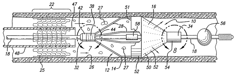
Another embodiment of the present invention is shown in FIGS. 9-11.
As can be seen in FIG. 9, the embolic protection device 100 includes a filter
assembly
102 having an expandable strut assembly 104 and a unique filter element 106.
The
particular strut assembly 104 utilized with this embolic protection device 100
is similar
to the structure of the expandable strut assembly 14 shown in the previous
embodiment. The filter element 106, which will be described in greater detail
below,
is utilized in its expanded position to collect any embolic debris for removal
from the
blood stream of the patient. '
The various elements making up this particular embodiment of the
embolic protection device 100 are shown in FIG. 10. In this particular
embodiment,
the strut assembly 104 does not necessarily have to be made from a self
expanding
material, as the strut assembly 14 disclosed in the previous embodiment.
Rather, it
could be made from stainless steel or other materials which require the
application of
external axial force on the proximal end 110 and distal end 112 of the strut
assembly
104 to move the struts 108 between the contracted and expanded positions. As
is
shown in FIGS. 10 and 11, the proximal end 110 of the assembly 104 includes a
short
tubular or sleeve-like segment 114 and a similar distal segment 116. The
struts 108
are moved from a contracted to a deployed position by imparting an inward
axial force
on the proximal end I 10 and distal end 1 I2 of the strut assembly 104. This
can be
accomplished by first attaching the distal end 112 of the assembly 104
directly to the
guidewire I 18. The proximal end I 10 of the strut assembly 104, can then, in
turn, be
attached to an outer tubular member 120 which, along with the guidewire 118,
has a
proximal end which extends outside of the patient. The proximal ends (not
shown) of
both the outer tubular member 120 and the guidewire 118 can be manipulated by
the
physician to either impart an inward axial force on the two ends 110 and 112
of the
CA 02395332 2002-06-20
WO 01/49215 PCT/US00/35556
-25-
strut assembly 104 to move the struts 108 to the deploy position or can be
moved to
impart an outward axial force on both ends 110 and 112 to collapse the struts
108 back
to their collapsed position.
The struts 108 of the strut assembly 104 can be made from a piece of
tubing (hypotube) in which select portions of the tubing are removed to form
the
particular size and shape of each strut. The strut assembly 104 could also be
made
from a self expanding material such as nickel-titanium (NiTi) if desired. The
struts
108 would then be biased into either the collapsed or expanded position with
the outer
tubular member 120 being used to move the proximal end 110 in order to expand
or
contract the strut assembly 104, depending upon, of course, the manner in
which the
expandable struts 108 are biased. Again, in the embodiment shown in FIG. 10,
the
struts 108 have a similar shape as the struts 28 shown in the embodiment of
FIGS.1-4.
This particular embodiment of an embolic protection device thus eliminates the
need
to utilize both a restraining sheath and recovery sheath which would be
otherwise
needed in order to deploy and contract the embolic protection device. This
particular
design, however, does not allow for the filter assembly 102 to rotate as
freely along the
guidewire 118 as does the previous embodiments, although there can be some
rotation. However, the outer tubular member I20 and guidewire 1 I 8 are
utilized in a
similar fashion by allowing interventional devices to be delivered over the
outer
tubular member in an over-the-wire fashion after the embolic protection device
110 is
in place within the patient's vasculature.
It should be appreciated that the strut assembly 104 could also be made
from a self expanding material which maintains the struts 108 biased in their
expanded
position. The outer tubular member 120 would still be utilized in order to
move the
expanded struts 108 back into their collapsed position. The proximal ends of
the outer
tubular member 120 and guidewire 118 can be attached to a simple locking
mechanism
600 (shown in FIGS. 39 and 40) which can be utilized to move the outer tubular
member relative to the guidewire for maintaining the strut assembly 104 in its
collapsed position until ready to be deployed within the patient's
vasculature. It
should further be appreciated that the particular embolic protection device
100 can also
CA 02395332 2002-06-20
WO 01/49215 PCT/US00/35556
-26-
be modified to eliminate the outer tubular member 120 and be a self expanding
assembly like the one shown in FIGS. 1-2. In such a case, the proximal end 110
of the
strut assembly 104 can be rotatably attached to the guidewire 118 with the
distal end
112 being slidably mounted on the guidewire to allow for longitudinal motion
and
rotational motion about the guidewire 118.
The filter element 106 utilized in conjunction with this particular
embodiment, or which can be utilized with any of the other embodiments
disclosed
herein, has a unique shape to provide a large reservoir to collect and
maintain any
embolic debris which may be trapped within the filter 106. Referring now to
FIGS.
9-I2, the various sections of the filter element 106 will be described in
greater detail.
It should be noted that the filter element I22 of FIG. 22 incorporates many of
the same
filter sections as the filter element 106 shown in FIGS. 10-12. Therefore,
corresponding sections of these filters will be described simultaneously in
order to
better understand the principles underlying these unique filter elements. Both
filter
elements include a proximal cone section 124 which expands to fit within the
diameter
of the artery. This particular proximal cone section 124 blocks or funnels
blood flow
and embolic debris into the main or central filter 126. In both of the filter
elements
shown in FIGS. 9 and 22, the proximal cone section 124 includes a plurality of
openings 128 which axe utilized in filtering the embolic debris. However, it
is possible
to eliminate the openings 128 on the proximal cone section 124 to allow it to
primarily
direct blood flow and embolic debris directly into the central filter 126.
This central
filter 126 is integral with the proximal cone section 124 and includes a
number of
openings 128 utilized to permit blood flow through this section of the filter
but to
retain any embolic debris which is larger than the size of the openings 128.
The
openings 128 can be laser cut or otherwise punched into this central filter
126. This
A central filter 126 has a substantially cylindrical shape and acts as a large
reservoir for
holding the embolic debris. Ideally, it is sized such that when it is
completely full of
embolic material, it does not collapse to a smaller profile. However, is
should be able
to be withdrawn into the guiding catheter (not shown) when in its fully
expanded
condition with embolic debris trapped therein. Thus, the maximum outer
expanded
CA 02395332 2002-06-20
WO 01/49215 PCT/US00/35556
-27-
diameter of this central filter 126 should be smaller than the inner diameter
of the
guiding or sheath utilized in deploying the embolic protection device 100 in
the
patient's vasculature. The central filter can be made from a stiffer polymeric
material
which will maintain the shape and outer diameter to prevent the filter from
collapsing
afteruse. The resulting stiffer central filter cannotbe squeezed during the
collapse and
removal of the filtering assembly from the artery which should prevent any
trapped
embolic debris from being squeezed out of the reservoir portion of the central
filter.
Both filters 106 and 122 include a distal tapered region 130 which tapers
down to the shaft of the guidewire 118. The taper of this particular region of
the filter
elements 106 and 122 facilitates the delivery of the embolic protection device
100 and
helps prevent the "snowplow" effect when being delivered through the patient's
vasculature. There is a small distal section 132 which also forms a part of
the filter
element and is utilized to attach the distal end of the filter directly onto
the guidewire.
This distal section 132 can be fastened utilizing well-known adhesives or
other
bonding techniques to permanently affix it to the guidewire 118 and prevent
any
embolic debris from escaping through the distal opening of this distal section
132.
The primarybenefit of utilizing a large central filter with a proximal cone
section is that there is a large filtering area provided by the central filter
126 which is
less likely to squeeze out trapped embolic debris when the embolic protection
device
100 is being removed from the patient's vasculature. As can be seen in FIG.
22, the
central filter 126 has a general cylindrical shape while the central filter
126 of FIG. 9
can be a generally cylindrically shaped but can also include side creases 134
which
produce a unique-looking design. The particular cross-sectional view of the
central
filter 126 of filter element 106 is shown in FIG. 16 and shows just one of a
number of
different shapes that can be used to create the central filter 126. In use,
the filter
element 122 of FIG. 22 would be attached to the strut assembly 104 and
guidewire 118
utilizing adhesives or other bonding techniques.
The filter element 106 of FIG. 9 also incorporates some unique features
which are not shown in the more basic filter design shown in FIG. 22. These
advantages include the unique cross-sectional shape of the central filter 126
shown in
CA 02395332 2002-06-20
WO 01/49215 PCT/US00/35556
-28-
FIG. 16, along with other features which help maintain the filter element 106
securely
attached to the struts 108 of the strut assembly 104. Referring again to FIGS.
10-12,
the filter element 106 includes a short outer rim 136 which is proximal to the
end of
the cone section 124 and has a large inlet opening 125 for receiving the blood
flow and
any embolic debris released into the bloodstream. This proximal outer rim 136
is ring-
shaped and can be utilized to help attach the filter onto the struts 1 OS of
the assembly
104. As can be seen in FIG. 10, this proximal outer ring is attached to the
middle
section 13 8 of each strut 108 and includes a tab 123 which can be wrapped
around and
attached to the strut 1 O8. This proximal outer ring 136 also helps maintain
the circular
inlet opening 125 which must be expanded and maintained within the artery of
the
patient. Attached to the front of the outer rim 13 6 are restraining straps
142 which are
likewise utilized to help hold the filter onto the struts 108 of the assembly
104. Each
restraining strap 142 includes tab-like projections 144 which can wrap around
each
individual strut and be affixed thereto utilizing a bonding agent such as
adhesive.
These elements allow the restraining straps 142 to hold the filter element 106
onto the
strut assembly 104. It should be appreciated that any number of different tab-
like
projections 144 can be utilized in conjunction with these restraining straps
142 to help
secure the filter onto the assembly 104. The proximal end of each restraining
strap 144
is attached to a sleeve 146 which also can be adhesively fixed to the tubular
segment
114 formed at the proximal end 110 of the strut assembly 104. These various
sections
of the filter 106 can be made as one composite unit and can be formed by
cutting a
pattern into a pre-formed filter blank. Thereafter, the openings 128 along the
length
of the filter element 106 can be placed accordingly.
The proximal cone section 126 of the filter element 106 shown in FTG.
9 includes a plurality of indented flaps 148 which are utilized to help close
the opening
of the central filter 126 when the proximal cone 124 is in its collapsed
position. Each
of these indented flaps 148, as shown in FIGS. 11, 17 and 18, are created such
that as
the proximal cone section 124 is being closed, the flaps j oin together and
cooperate to
form a barner which prevents embolic debris from being released through the
inlet
opening 127 of the central filter 126. In the particular embodiment shown in
FIG. 9,
CA 02395332 2002-06-20
WO 01/49215 PCT/US00/35556
-29-
four such indented flaps can be utilized (only two of which are shown in FIGS.
11, 17
and 18) in order to create the barrier necessary to close the opening to the
central filter
126. However, the number of indented flaps 148 and the size and shape of each
flap
148 can be varied accordingly in order to create a protective barrier which
helps
S prevent trapped embolic debris from escaping from the central filter 126 as
the device
100 is being collapsed for removal from the patient.
Referring now to the FIGS. 19, 20 and 21, a variation of the indented
flaps 148 is shown in the proximal cone section 124 of the filter element 106.
As can
be seen in these figures, there are a pair of flap portions 150 which are
located within
the proximal cone section 124 and are utilized as a mechanism for closing the
inlet
opening 127 of the filter element 106 when the filter assembly is collapsed.
These flap
portions 150 act much like the indented flaps 148 in that as the proximal cone
section
124 is being collapsed, these flap portions 150 extend across the inlet
opening 127 of
the filter element 106 to create a barrier which helps prevent trapped embolic
debris
from being released back into the bloodstream. These flap portions 150 can be
small
appropriately shaped pieces which extend across the inlet opening when the
filter is
expanded but do not interfere with the flow of blood going into the filter
element 106.
Blood simply travels around the flap portions 150, along with any embolic
debris, to
the center filter 126 where the embolic debris will be trapped in the debris
reservoir.
This feature provides a preventive measure to diminish the possible release of
trapped
embolic debris when the embolic protection device 100 is being collapsed and
removed from the patient's vasculature.
Referring now to FIGS. 14 and 15, an alternative form of the restraining
straps and tabs which are utilized to affix the filter element 106 is shown.
In these
particular figures, the restraining strap 152 extends along each strut 108 and
a tab like
projection 154 is utilized to affix the restraining strap to each individual
strut 108.
Additional lateral strapping members 156 which extend laterally from each
restraining
strap 152 can also be utilized to help prevent the filter element 106 from
moving off
the strut assembly 104 during usage. These various designs shows alternative
ways
of affixing the filter element 106 onto the strut assembly 104. It should be
appreciated
CA 02395332 2002-06-20
WO 01/49215 PCT/US00/35556
-3 0-
that still other forms of attaching the filter element 106 to the strut
assembly 104 can
be utilized without departing from the spirit and scope of the present
invention.
Another preferred embodiment of the present invention is shown in
FIGS. 23 and 24. In this pal-ticular embodiment, the embolic protection device
200
includes a filter assembly 202 having a strut assembly 204 and a filter
element 206.
The strut assembly 204 is similar to the strut assembly shown in FIGS. 1-4. It
includes self expanding struts 208 which axe expandable from a collapsed
position to
a fully expanded position. This strut assembly 204 includes a proximal end 210
and
a distal end 212. This strut assembly 204 can be made from a piece of tubing
in which
the struts are created by selectively removing portions of the tubing. In this
particular
embodiment, the tubing can be hypotubing made from a shape memory material
such
as nickel-titanium (NiTi). The resulting strut assembly 204 is normally biased
to
remain in the expanded position and require the applications of force on the
ends 210
and 212 to deploy the struts 208 back to their collapsed position.
The proximal end 210 includes a segment of tubing 214 and the distal
end 212 includes a similar segment of tubing 216 as well. The distal end 212
is
permanently attached to the guide wire 218 near the distal coil 220 of the
guide wire.
The distal end 212 can be bonded using adhesives or welded, brazed or soldered
to the
guidewire 218. Likewise, the proximal end 210 of the strut assembly 204 can be
bonded, welded, brazed or soldered to an elongated outer tubular member 222
which
has a proximal end which extends outside of the patient. The proximal ends of
the
elongated tubular member 222 and the guidewire 218 can be manipulated by the
physician to either open or close the filter assembly 202. A suitable locking
mechanism 600 for maintaining the strut assembly 204 in its collapsed or
closed
position is disclosed in FIGS. 43 and 44 and is described in greater detail
below.
The filter element 206 comprises of a cone shape portion 224 which is
attached to the center section 226 of each strut 208. A plurality of openings
228 are
laser cut or otherwise formed in the filter 206 which allows blood to flow
through the
filter but captures embolic debris which is larger than the size of the
openings. This
CA 02395332 2002-06-20
WO 01/49215 PCT/US00/35556
-31-
is another more example of a variation of the embolic protection device which
can be
made in accordance with the present invention.
Another embodiment of the present invention is shown as a embolic
protection device 300 in FIGS. 25-28. Like the other embodiments, this device
300
includes a filtering assembly 302 which has an expandable strut assembly 304
and a
filter element 306 attached to the strut assembly 304. Individual struts 308
are formed
on the strut assembly 304 for moving the filtering element 306 into an
expanded
position within the patient's vasculature. The strut assembly 304 is some what
similar
similar to the previous embodiments disclosed above in that an outer elongated
tubular
member 310 is utilized in conjunction with a guide wire 312 to collapse and
deploy the
strut assembly 3 04. Although not shown in FIGS. 25 and 26, the outer tubular
member
310 has a proximal end which extends with the proximal end of the guidewire
outside
of the patient to allow the physician to move the proximal ends to deploy or
collapse
the filtering assembly 302. The strut assembly 304 can be formed by
selectively
removing material from the outer tubular member 310 near its distal end to
create the
individual struts 308. The struts will open upon application of an inward
force on
ends of the individual struts 308. Alternatively, the strut assembly 304 can
be made
from a piece of hypotubing which can be affixed to the outer tubular member
310 as
is shown in some of the previous embodiments of the invention. The entire
outer
tubular member 310 with the strut assembly 304 is.free to slide along the
length of the
guidewire 312 which allows the filtering assembly 302 to be positioned within
the
patient's vasculature in an over-the-wire fashion.
As can be seen in FIGS. 25-28, a stop element 320 is located near the
distal coil 322 of the guidewire 312. This distal stop element 320 is utilized
in
conjunction with the outer tubular member 310 to produce the force necessary
to
expand the struts 308 into the expanded position. The embolic protection
device 300
can be utilized in the following matter. First, the physician maneuvers the
guide wire
312 into position past the lesion or area of treatment. Thereafter, the outer
tubular
member 310 with the strut assembly 304 is advanced over the guidewire 312 in
an
over-the-wire technique. The embolic protection device 300 remains in its
collapsed
CA 02395332 2002-06-20
WO 01/49215 PCT/US00/35556
-32-
position while being delivered over the guide wire 312 to the distal end 313
of the
guide wire, as is shown in FIG. 27. Thereafter, the physician allows the
distal sleeve
312 of the outer tubular member 3I0 to contact the stop element 320 located on
the
guide wire 312. By applying additional force at the proximal end of the
elongated
S tubular member 310, the physician will cause the struts 308 to expand
radially
outward for deployment within the artery. The resulting expansion of the
struts 308
thereby opens up the filter element 306 within the artery. The physician can
then
deliver interventional debris into the area of treatment and perform the
procedure on
the lesion. Any embolic debris which may be created during the interventional
procedure will be collected within the interior of the filter 306.
A simple locking mechanism 600 device located at the proximal end of
the outer tubular member and guidewire, as is shown in FIGS. 43 and 44, can be
utilized to move and maintain the stl-ut assembly 304 in the expanded
condition.
Thereafter, once the embolic protection device 300 is desired to be removed
from the
1 S vasculature, the physician merely retracts the proximal end of the outer
tubular member
310 to remove the force on the strut assembly 304 allowing the struts 308 to
move
back to the collapsed position. Thereafter, the embolic protection device 300
and
guidewire 312 can be removed from the patient's vasculature.
The filter element 306 takes on a some what different shape from the
previous filter element in that the main portion of the filter element 306 has
a shape of
a half of a dilatation balloon utilized in angioplasty procedures. Perfusion
openings
313 are located on the alter elements 306 for allowing blood perfusion while
capturing
embolic debris. The proximal end of the filter element 306 includes a
plurality of
restraining straps 314 which extend to a proximal sleeve 316 which is affixed
to the
2S outer tubular member 310 proximal of the struts 308. The distal end 318 of
the filter
element 306 is also attached to the distal sleeve 321 which is formed on the
outer
tubular member 310 when the struts 308 are formed.
FIGS. 29 and 30 show another embodiment of a embolic protection
device 400 made in accordance with the present invention. This particular
embodiment is somewhat similar to the previous embodiments in that an external
force
CA 02395332 2002-06-20
WO 01/49215 PCT/US00/35556
-33-
is generated on the ends of the struts of the strut assembly to facilitate the
outward
expansion and inward contraction of the struts. Referring specifically now to
FIG. 29,
the embolic protection device 400 includes a filter assembly 402 having a
strut
assembly 404 which has a filter element 406 attached thereto. The individual
struts
408 are formed on an outer tubular member 410 which has a distal end 412
attached
to the distal end 413 of an inner tubular member 414. Both the inner member
414 and
the outer member 410 have proximal ends which are located outside of the
patient's
vasculature. The struts 408 are radially expanded by moving the outer tubular
member
410 relative to the inner tubular member 414 to apply the necessary axial
force to cause
the struts to deploy outward. An opposite axial force is necessary to cause
the struts
408 to move back to the collapsed position when the device is to be removed
from the
patient's vasculature. In this embodiment, more than four struts 408 are used
to
expand the filter element 406 within the artery 420. Again, the number, size
and shape
of the struts 408 can be varied without departing from the spirit and scope of
the
present invention.
The filter element 406 also has the shape of one half of a dilatation
balloon utilized in angioplasty procedures and includes openings 416 which
allows
blood to flow through the filter but captures the desired size of the embolic
debris.
The proximal end of the filter element 406 which includes an inlet opening 417
is
attached to each of the center sections 418 of the struts 408. The distal end
420 of the
filter 406 is attached to the distal end 412 of the strut assembly 404.
The lumen 422 of the inner tubular member 414 can be utilized for a
number of purposes, such as blood perfusion past the deployed filter assembly
402
when placed in the artery. Therefore, should the openings 416 of the filter
element
406 become clogged with debris which prevents blood from flowing through the
filter,
oxygenated blood can be perfused to downstream vessels via the inner lumen of
the
inner tubular member 414. This lumen can also be utilized for delivering the
embolic
protection device 404 over a guidewire in an over-the-wire fashion.
FIG. 31 and 32 show a variation of the previous filter element which can
be utilized in conjunction with the present invention. The filter embolic
protection
CA 02395332 2002-06-20
WO 01/49215 PCT/US00/35556
-34-
device 400 is basically the same device shown in FIGS. 29 and 30 except that
the filter
element 430 has a different design. As can be seen in FIG. 31, the filter
element 430
includes a proximal cone shape portion 431 which extends in front of the inlet
opening
432 of the filter element 430. This type of filter 430 has advantages in that
it may be
easier to attach to the strut assembly 404. Additionally, the wall of the
artery is
insulated from the struts 408 by restraining straps 434. This device also has
the
benefits of being low profile and allows the use of any guide wire, as well as
allowing
for guidewire exchanges. This particular embodiment, like the previous
embodiments,
allows for the exchange of the interventional device in an over-the-wire
procedure.
Referring now to FIGS. 33-3 8, two different embodiments of the present
invention are shown which utilize a different mechanism for deploying the
struts of the
strut assembly. In FIG. 33, an embolic protection device 500 is shown as
including a
filter assembly 502 having an expandable strut assembly 504 and a filter
element 506.
As with the other embodiments, the strut assembly 504 includes a plurality of
radially
1S expandable struts 508 which are utilized to place the filter element 506
into an
expanded position within the patient's vasculature. The mechanism for
deploying the
radially expandable struts 508 utilizes a number of self expanding deployment
members 510 which are attached to each of the struts 508 making up the
expandable
strut assembly 504. The self expanding deployment members S 10 are made from
self
expanding materials, such as nickel-titanium alloy, which can be compressed to
a very
small profile and expanded to a rather large expanded position which moves the
struts
508 and filter 506 to the fully expanded position. As is seen in FIGS. 33 and
34, there
are a number of deployment members S 10 which are located along the length of
each
of the struts 508. There is a proximal set 512 of deployment members 510
located
2S along the proximal region of each strut 508. There is a center set 514 of
deployment
members 510 located at the center section of each stmt SO8. As can be seen in
FIG.
34, the coverage of the filter element 506 begins at this center set 514. A
third or distal
set S 16 of deployment members 510 is located on the struts in the region
where the
filter element S06 is placed to enhance the deployment of each strut.
CA 02395332 2002-06-20
WO 01/49215 PCT/US00/35556
-35-
As can be seen in FIG. 37, each deployment member 510 is basically a
collapsible piece of self expanding material which will expand to a final size
when
fully deployed. FIG. 38 shows an end view of the center set 514 and distal set
516 of
the deployment members as they are located along the sthuts 508. Each of the
sets of
deployment members 510 will fully expand to a quarter-circle segment which
cooperate to form a "ring" when the sets of the deployment members are fully
expanded. As a result of using this particular construction, the filter
element 506 will
fully deploy and maintain a circular-shaped opening 507 which will contact the
wall
of the artery when the embolic protection device 500 is deployed within the
patient's
vasculature.
In the first embodiment of this particular embolic protection device 500,
the distal end 518 of the expandable strut assembly 504 is permanently
attached to the
guidewire 520. The proximal end 522 of the strut assembly 504 is, in turn,
attached
to an elongated outer tubular member 524 which has a proximal end (not shown)
1 S which extends outside of the patient's vasculature along with the proximal
end of the
guidewire. The embolic protection device 500 can be moved into its collapsed
position as shown in FIG. 3 5 by simplyretracting the proximal end of the
outer tubular
member 524 to impart an outward force on the ends of the strut assembly 504.
The
force which will be imparted on the ends of the strut assembly 504 should be
sufficient
to collapse each deployment members 510 which will, in turn, cause each of the
struts
508 to move back to the collapsed position. As with the other embodiments,
once the
struts 508 are placed in its collapsed position, the filter element 506 will
likewise
collapse and will trap and encapsulate any embolic debris which may have been
trapped within the filter element 506.
Referring now to FIG. 36, an alternative embodiment of an embolic
protection device similar to the one shown in FIG. 33 is disclosed. This
particular
embolic protection device 530 utilized the same filter assembly 502 and strut
assembly
504 as shown in the previous embodiment. The differences between the strut
assembly 532 of the embolic protection device 530 includes the elimination of
the
proximal set 512 of deployment members 510 from this strut assembly 532.
CA 02395332 2002-06-20
WO 01/49215 PCT/US00/35556
-36-
Otherwise, the filter assembly 534 is virtually the same as the filter
assembly 502 of
the previous device 500. .
The distal end 518 of the strut assembly 534 is also permanently affixed
to the guidewire 520 in this particular embodiment. The proximal end of this
particular strut assembly 534 is free to move longitudinally along the length
of the
guidewire when being moved from a deployed to a contracted position and visa
versa.
The mechanism for deploying the filter assembly 532 is restraining sheath 536
which
places a force on the and deployment members 510 which prevent them from
expanding until the restraining sheath 536 is retracted. Once the embolic
protection
device 530 is properly in place within the patient's vasculature, the proximal
end (not
shown) of the restraining sheath 53 6 is retracted to allow the deployment
members 510
to open the struts 508 and filter element 506 to the fully expanded position
within the
artery. When the device is to be removed from the patient's vasculature, the
restraining sheath 536 is placed against the proximal region 535 of the struts
508 and
is retracted over the struts to force the deployment members 510 back into
their
collapsed position. Thereafter, any embolic debris which may be trapped within
the
filter element 506 is retained and safely removed from the patient's
vasculature. A
proximal set of deployment members 510 may not have to be used with this
particular
embodiment since there may be a need to reduce the amount of expansive force
applied to the stl-uts in this proximal region 535. However, it is still
possible to place
a first set of deployment members at this proximal region 53 5 provided that
the sheath
has sufficient strength to collapse the struts in this region.
The filter element 506 shown in FIGS. 33-38 is made from a mesh
material which allows blood to perfuse therethrough but captures embolic
material.
The mesh material can be made from any interwoven fabric which contains small
size
openings which will trap the desired size of emboli. Alternatively, the filter
506 can
be made from a polymeric material with perfusion openings found therein.
Referring now to FIGS. 39A, 39B and 40, an alternative strut assembly
550 which could be utilized in conjunction with any of the filtering
assemblies made
in accordance with the present invention is shown. The strut assembly 550
includes
CA 02395332 2002-06-20
WO 01/49215 PCT/US00/35556
-37-
struts 552 and a deployment member 554 which is used to expand the struts 552
into
the deployed expanded position. This deployment member 554 acts in the same
manner as the previously described deployment members in that the deployment
member 554 can be made from a self expanding material which will expand to a
final
size once fully deployed. The deployment member 554 also could be collapsed to
an
unexpanded position when an external force is placed on the assembly to
maintain the
deployment member 554 in its collapsed position. As can be seen in FIGS. 39A,
39B
and 40, the deployment member 554 has a serpentine pattern made of peaks 556
and
valleys 558 which are accordingly attached to the struts 552 of the assembly
550. In
these particular embodiment of the invention, the deployment member 554 has a
sinusoidal wave pattern which includes the peaks 556 and valleys 558 that are
attached
to the ends of the struts 552. This particular pattern allows the struts to be
offset or
staggered from one another to allow the assembly 550 to be collapsed to a
lower
profile which enhances the assembly's ability to reach tighter lesions and to
be
maneuvered into even distal anatomy. The staggered strut design also increases
the
assembly's flexibility which enhances the ability to move the assembly within
the
patient's anatomy. A filter element could be likewise placed over or within
the struts
552 to create a composite filter assembly. The deployment member 554 provides
complete vessel wall opposition, forcing a seal of the filter edge to the wall
of the
vessel. The deployment member 554 can have multiple geometries without
departing
from the spirit and scope of the present invention. This particular strut
assembly 550
also could be created from a lazed hypotube which incorporates the staggered
strut
design. The number of struts can be varied along with the particular lengths
of the
struts. Alternatively, the deployment member 5 54 could be made from a
separate piece
of material from the struts and could be attached using methods such as
soldering,
brazing or bonding, using suitable adhesives. As can be seen from FIGS. 39A
and
39B, the attachment of the struts 552 to the peaks 556 and valleys 558 of the
deployment 554 can be varied as shown. Both of these particular designs allow
the
strut assembly to be collapsed to a Iow profile.
CA 02395332 2002-06-20
WO 01/49215 PCT/US00/35556
-3 8-
Refernng now to FIGS. 41 and 42, an alternative filter element 570 with
an angulated filter edge 572 is shown which is used to help in the loading and
retrieval
of the embolic protection device into a restraining sheath. The filter element
570 is
similar to the filters previously described in that the filter element 570
includes a
central section 574 which has a plurality of openings 576 that are utilized in
filtering
the embolic debris. The filter element 570 includes an edge 572 which is
configured
similar to a crown, with pointed peaks 578 and valleys 580. This configuration
of the
filter edge 572 allows the alter to be incrementally introduced into the
restraining
sheath, thus preventing the material from entering the sheath all at once. As
can be
seen in FIGS. 41 and 42, the edge 572 has a somewhat sinusoidal configuration
which
would reduce the stress concentration in the valley regions 5 80 of the
filter. The peaks
578 of the filtering element 570 would be matched up with the struts of the
struts of
582 of the strut assembly 584. The number of peaks 578 could vary with the
number
of struts 582 on the strut assembly 584. In this particular embodiment, the
filtering
element 570 could be placed within the inside of the strut assembly 584, or,
alternatively, the filter could be placed on the outside of the assembly 584.
It should
be appreciated that other filter elements described herein also could either
replace on
the inside or outside of the strut assembly used in connection with a
particular filtering
assembly. As the strut assembly 584 is being loaded or retrieved, the peaks
578 of the
filter element 570 would enter the restraining sheath first. This prevents all
of the
filtering material from entering the sheath at once, causing a gradual and
incremental
loading of the filter element 570 into the sheath. Additionally, dimensions A
and B
shown in FIG. 42 show the difference in the valley depths in the sinusoidal
pattern of
the filter edge 572. This allows for a variety of configurations. One possible
configuration is A = B = 0. Additionally, B >_A>_ 0 so that the loading of the
filter into
the sheath will be in a smooth operation. This particular configuration
eliminates or
virtually eliminates all of the valley portions 580 from entering the sheath
at the same
time. The filter edge 572 may or may not have openings 576. The peaks 578 can
also
have varying heights. Dimensions C, D and E shown in FIG. 42 shows a
difference
in the peak heights on the sinusoidal pattern of the filter edge 572. This
particular
CA 02395332 2002-06-20
WO 01/49215 PCT/US00/35556
-39-
pattern also allows for a variety of configurations. One possible
configuration is C =
D = E = 0. Additionally, E>_D>_C>_0 to correspond, or alternatively, not to
correspond
with the depths of the
valleys 580.
Referring now to FIGS. 43 and 44, a simple locking mechanism 600 for
expanding and collapsing the filter assembly described herein are shown. These
particular mechanisms are useful whenever the embolic protection device
utilizes an
inner shaft member and outer tubular member for moving the strut assemblies
into the
expanded or collapsed position. Referring first to FIG. 43, the proximal end
602 of
the outer tubular member 604 is shown with a locking mechanism 600 which can
be
utilized to lock the embolic protection device in either an expanded or
unexpended
position. The locking mechanism 600 includes an elongated slot 606 which is
cut into
the wall of the outer tubular member 604 and includes a first locking position
608 and
a second locking position 610. The inner shaft member 612, which can be either
a
solid shaft such as a guidewire or a hollow tubular shaft, has a raised dimple
6I4
which moves within this elongated slot 606. This raised dimple 614 can be
moved
into either the first locking position 608 or second locking position 610 to
either
maintain the filter assembly in an expanded or unexpended position. It should
be
appreciated that only two locking positions are shown on this particular
embodiment,
however, it is possible to use a number of different locking positions if the
user desires
to have several expanded positions. If the filter assembly is self expanding,
then a
removable handle that pushes and pulls the inner and outer members could be
used.
The handle would push/pull the inner and outer members to hold the assembly
closed,
then be removed so that other interventional devices could be passed over the
inner
tubular member. Thereafter, the handle could be placed back onto the proximal
ends
of the inner and outer members to collapse and remove the filter assembly.
The proximal end 602 of the outer tubular member includes a small
section of knurling 616, as does the inner shaft member 612, which provides
the
physician with a surface to grip when holding and maneuvering the proximal
ends of
these devices. The locking mechanism 600 can also include a biasing spring 618
CA 02395332 2002-06-20
WO 01/49215 PCT/US00/35556
.Q_
located within the inner lumen 620 of the outer tubular member 604 for biasing
the
inner shaft member 612 with an outward force which maintain the raised dimple
614
near the first locking position 608. This biasing mechanism includes a
shoulder region
621 located at the proximal end of the outer tubular member and a collar 622
located
on the inner shaft member 612. The force of the spring 618 again helps to
maintain
the dimple 614 at or near the first locking position 608. Such a mechanism is
preferable when the device is designed to be maintained in an unexpanded
position
until it is ready to be deployed. It may be beneficial to keep the filter
assembly in its
unexpanded position until ready for use since it is possible to cause damage
to the
filter assembly if left in an expanded position. When the filter assembly is
desired to
be placed into the deployed or expanded position, the physician merely grasps
the
proximal end of the inner shaft member and pulls it back until the dimple 614
is placed
into the second locking position 610. When the strut assembly is made from
elements
which are self expanding, then there may not be a need to have a biasing
spring 618
since the struts on the strut assembly will act somewhat like a biasing spring
to
maintain the filter assembly in an expanded position.
The strut assemblies of the present invention can be made in many ways.
However, the preferred method of making the strut assembly is to cut a thin-
walled
tubular member, such as nickel-titanium hypotube, to remove portions of the
tubing
in the desired pattern for each strut, leaving relatively untouched the
portions of the
tubing which are to form each strut. It is preferred to cut the tubing in the
desired
pattern by means of a machine-controlled laser.
The tubing used to make the strut assembly may be made of suitable
biocompatible material such as stainless steel. The stainless steel tube may
be alloy-
type: 316L SS, Special Chemistry per ASTM F138-92 or ASTM F139-92 grade 2.
Special Chemistry of type 316L per ASTM F 138-92 or ASTM F 139-92 Stainless
Steel
for Surgical Implants in weight percent.
The strut size is usually very small, so the tubing from which it is made
must necessarily also have a small diameter. Typically, the tubing has an
outer
diameter on the order of about 0.020 - 0.040 inches in the unexpanded
condition. The
CA 02395332 2002-06-20
WO 01/49215 PCT/US00/35556
-41-
wall thickness of the tubing is about 0.076 mm (0.003 - 0.006 inches). For
strut
assemblies implanted in body lumens, such as PTA applications, the dimensions
of the
tubing maybe correspondingly larger. While it is preferred that the strut
assembly be
made from laser cut tubing, those skilled in the art will realize that the
strut assembly
can be laser cut from a flat sheet and then rolled up in a cylindrical
configuration with
the longitudinal edges welded to form a cylindrical member.
Generally, the hypotube is put in a rotatable collet fixture of a machine-
controlled apparatus for positioning the tubing relative to a laser. According
to
machine-encoded instructions, the tubing is then rotated and moved
longitudinally
relative to the laser which is also machine-controlled. The laser selectively
removes
the material from the tubing by ablation and a pattern is cut into the tube.
The tube is
therefore cut into the discrete pattern of the finished struts. The strut
assembly can
thus be laser cut much like a stmt is laser cut. Details on how the tubing
CaI1 be cut by
a laser are found in U.S. Patent Nos. 5,759,192 (Saunders) and 5,780,807
(Saunders),
which have been assigned to Advanced Cardiovascular Systems, Inc. and are
incorporated herein by reference in their entirely.
The process of cutting a pattern for the strut assembly into the tubing
generally is automated except for loading and unloading the length of tubing.
For
example, a pattern can be cut in tubing using a CNC-opposing collet fixture
for axial
rotation of the length of tubing, in conjunction with CNC XfY table to move
the length
of tubing axially relative to a machine-controlled laser as described. The
entire space
between collets can be patterned using the C02 or Nd:YAG laser set-up. The
program
for control of the apparatus is dependent on the particular configuration used
and the
pattern to be ablated in the coding.
A suitable composition of nickel-titanium which can be used to
manufacture the strut assembly of the present invention is approximately 55%
nickel
and 45% titanium (by weight) with trace amounts of other elements making up
about
0.5% of the composition. The austenite transformation temperature is between
about
-15 ° C and 0 ° C in order to achieve superelastecity. The
austenite temperature is
measured by the bend and free recovery tangent method. The upper plateau
strength
CA 02395332 2002-06-20
WO 01/49215 PCT/US00/35556
-42-
is about a minimum of 60,000 psi with an ultimate tensile strength of a
minimum of
about 155,000 psi. ~ The permanent set (after applying 8% strain and
unloading), is
approximately 0.5%. The breaking elongation is a minimum of 10%. It should be
appreciated that other compositions of nickel-titanium can be utilized, as can
other
self expanding alloys, to obtain the same features of a self expanding stmt
made in
accordance with the present invention.
The strut assembly of the present invention can be laser cut from a tube
of super- elastic (sometimes called pseudo-elastic) nickel-titanium (Nitinol)
whose
transformation temperature is below body temperature. After the strut pattern
is cut
into the hypotube, the tubing is expanded and heat treated to be stable at the
desired
final diameter. The heat treatment also controls the transformation
temperature of the
strut assembly such that it is super elastic at body temperature. The
transformation
temperature is at or below body temperature so that the stmt is superelastic
at body
temperature. The strut assembly is usually implanted into the target vessel
which is
smaller than the diameter if the strut assembly in the expanded position so
that the
struts apply a force to the vessel wall to maintain the filter element in the
expanded
position.
The piece of tubular hypotube which can be utilized in accordance with
the present invention to form the strut assemblies can be one continuous piece
which
forms both the outer tubular member and the strut assembly as well. In some of
the
embodiments disclosed herein, the strut assembly is shown as being made from a
short
segment of hypotube which is selectively cut to form the strut patterns.
Thereafter, the
proximal end of the strut assembly is bonded to, either by adhesives, welding,
brazing
or soldering to the distal end of the outer tubular member. However, these two
separate pieces can be formed from a piece of single tubing in a preferred
embodiment
of the invention.
The dampening element which is shown in one of the embodiments of
the present invention could also be used with any of the other embodiments
disclosed
herein. The dampening element could either be cut into the proximal end of the
strut
assemblies, as is shown in FIGS. 1 and 2, or an alternative dampening element
could
CA 02395332 2002-06-20
WO 01/49215 PCT/US00/35556
-43-
be attached to the strut assembly. For example, a separate spring made from a
different
material or similar material could be welded, brazed or soldered to the end of
the strut
assembly. Also, other dampening materials could be used besides a helical
spring in
order to achieve dampening. For example, segment of elastomeric material could
be
bonded to the strut assembly as well to act as a "shock absorber" for the
system.
The outer tubular member could be made from various materials such as
stainless steel, nickel-titanium alloy or materials which have memory. As
discussed
above, when using a separate outer member attached to the strut assembly, the
distal
end can be easily affixed to the strut assembly by known bonding methods. The
inner
diameter of the outer tubular member must of course be comparable to the outer
diameter of the inner shaft member to allow the outer tubular member to slide
in a
coaxial arrangement. The inner shaft member can also be made from stainless
steel,
nickel-titanium alloys or shape-memory materials. In one embodiment, the inner
shaft
member is shown as a tubular member which has an inner lumen which allows the
device to slide over a guidewire in an over-the-wire fashion. Other
embodiments show
the inner shaft member as a guidewire or guidewire-like shaft. Generally, when
the
inner shaft member is utilized as a guidewire, it should include an atraumatic
guidewire coil tip to prevent injury to the vessel as the guidewire is being
maneuvered
through the patient's vasculature. It should be appreciated that the coil tip
does not
have to be placed directly next to the filtering assembly in those embodiments
which
utilize a guidewire as the inner shaft member. The filtering assembly could be
placed
much more proximal to the coil tip to create a short, distal segment of
guidewire which
may be pre-bent by the physician to aid in steering through the patient's
vasculature.
Again, the tubing or hypotube which could be utilized to create the strut
assembly can be a nickel-titanium alloy, such as Nitinol, or other shape-
memory
materials. It is also possible to utilize stainless steel to form the strut
assembly as well.
The strut assembly could also be made from a self expanding material even in
embodiments in which the outer tubular member and inner shaft member are
utilized
to provide the axial forces necessary to expand or contract the device during
use.
CA 02395332 2002-06-20
WO 01/49215 PCT/US00/35556
-44-
Additionally, the strut assembly could be either biased to remain in its
collapsed
position or expanded position as may be desired.
The polymeric material which can be utilized to create the filtering
element include, but is not limited to, polyurethane and Gortex, a
commercially
available material. Other possible suitable materials include ePTFE. The
material can
be elastic or non-elastic. The wall thickness of the filtering element can be
about
0.001-0.005 inches. The wall thickness may vary depending on the particular
material
selected. The material can be made into a cone or similarly sized shape
utilizing
blow-mold technology. The perfusion openings can be any different shape or
size.
A laser, a heated rod or other process can be utilized to create to perfusion
openings
in the filter material. The holes, would of course be properly sized to catch
the
particular size of embolic debris of interest. Holes can be lazed in a spinal
pattern with
some similar pattern which will aid in the re-wrapping of the media during
closure of
the vice. Additionally, the filter material can have a "set" put in it much
like the "set"
used in dilatation balloons to make the filter element re-wrap more easily
when placed
in the collapsed position.
The materials which can be utilized for the restraining sheath and
recovery sheath can be made from similar polymeric material such as cross-
linked
HDPE. It can alternatively be made from a material such as polyolifin which
has
sufficient strength to hold the compressed strut assembly and has relatively
low
frictional characteristics to minimize any friction between the filtering
assembly and
the sheath. Friction can be further reduced by applying a coat of silicone
lubricant,
such as Microglide~, to the inside surface of the restraining sheath before
the sheaths
are placed over the filtering assembly.
In view of the foregoing, it is apparent that the system and device of the
present invention substantially enhance the safety of performing certain
interventional
procedures by significantly reducing the risks associated with embolic
material being
created and released into the patient's bloodstream. Further modifications and
improvements may additionally be made to the system and method disclosed
herein
CA 02395332 2002-06-20
WO 01/49215 PCT/US00/35556
-45-
without departing from the scope of the present invention. Accordingly, it is
not
intended that the invention be limited, except as by the appended claims.