Note: Descriptions are shown in the official language in which they were submitted.
CA 02395393 2002-06-25
WO 01/62191 PCT/USO1/05638
ANTERIOR IMPACTED BONE GRAFT AND DRIVER INSTRUMENTS
CROSS-REFERENCE TO RELATED APPLICATIONS
The present application claims the benefit of commonly owned U.S.
Provisional Patent Application No. 60/183,930, filed 22 February 2000, which
is
hereby incorporated by reference in its entirety.
BACKGROUND OF THE INVENTION
The present invention relates generally to instruments and implants for
intervertebral spacing. More specifically, the present invention provides
instruments and implants that may be utilized to provide multi-directional
insertion
techniques to establish and maintain intervertebral spacing. Still more
preferably,
the present invention provides implants made of bone adapted to be inserted
from
more than one direction while maintaining proper orientation in the disc
space.
The removal of damaged or diseased discs and restoration of disc space
~5 height to treat chronic back pain and other ailments, is well-known.
Spacers are
often utilized to maintain or reestablish disc space height after removal of
all or a
portion of the disc. Such spacing implants may include those promoting fusion
between adjacent vertebral bodies, inert implants, and artificial disc
implants.
Such implants are typically designed to be inserted from an anterior,
posterior or.
lateral approach. However, such implants are often designed for insertion only
from one of the particular approaches to the spine. This is particularly true
where
implants are intended to maintain non-parallel angulation between adjacent
vertebrae. Therefore, multiple implants each designed for insertion from one
of
the various approaches to the spine must be maintained in inventory to
accommodate the various surgical demands of each procedure. Maintaining
multiple implant designs may create inventory problems for both manufacturers
and their customers. Moreover, the complications of creating multiple implants
to
accomplish the same desired spacing is compounded when implants are made of a
scarce resources, such as allograft bone.
Therefore, there remains a need for instruments, techniques, and implants
that reduce implant inventory without sacrificing desired implant
configurations.
CA 02395393 2002-06-25
WO 01/62191 PCT/USO1/05638
2
SUMMARY OF THE INVENT10N
The present invention provides for instruments to implant a single implant
design from multiple approaches to the disc space. In a preferred aspect of
the
present invention, instruments are provided for inserting an implant from a
direct
anterior approach to the spine and from an oblique-anterior approach to the
spine.
In a further aspect of the present invention, an implant is provided that
includes features permitting insertion into the disc space from multiple
directions.
In a preferred aspect of the present invention, the implant may be configured
for
1o insertion from a direct anterior approach as well as an anterior-lateral
approach to
the spine. Still more preferably, the anterior-lateral approach to the spine
is from
an oblique angle with respect to the sagittal plane.
In still a further aspect of the present invention, a multi-faceted implant is
provided comprising an implant body having a first pair of substantially
parallel
side walls and a second pair of substantially parallel side walls. The second
pair of
substantially parallel side walls are disposed at an oblique angle with
respect to the
first pair of substantially parallel side walls. The angulation between-the
first and
second set of parallel side walls permits insertion of the implant into the
disc space
from multiple directions. Further in one preferred embodiment the distance
2o between the first pair of side walls is substantially identical to the
distance between
the second pair of side walls. One choice is to dispose the second pair of
side
walls at an angle of approximately 30 degrees with respect to the first pair
of side
walls. In a more preferred aspect of the present invention, the implant body
has
upper and lower bone engaging surfaces that are tapered to maintain angulation
between adjacent vertebrae. In still further preferred aspects of the
invention, one
of each of the first and second pair of side walls includes an insertion tool
bore.
In yet a further aspect of the present invention, a method of making an
implant of honey material is provided. The method comprises forming a first
pair
of substantially parallel side walls on the honey material. A second pair of
3o substantially parallel side walls is formed at an oblique angle with
respect to the
first pair of side walls. In one aspect the method further includes forming a
CA 02395393 2002-06-25
WO 01/62191 PCT/USO1/05638
3
plurality of driving surfaces on the donor bone. Still more preferably, the
upper
and lower bone engaging surfaces are disposed at an angle with respect to each
other.
In still a further aspect of the invention an implant inserter is provided.
Preferably, the implant inserter includes anti-rotation components to limit
rotation
of the implant about the longitudinal axis of the inserter and rotation about
the axis
of the implant itself. In one preferred embodiment, the anti-rotation
components
comprise a pair of angled side walls on the inserter adapted to engage a pair
of
corresponding surfaces on the implant. Instill a further preferred aspect, a
to threaded post engages a corresponding opening on the implant and the angled
surfaces are spaced from the opening to limit stress placed on the implant
adjacent
the opening.
These and other objects of the present invention will become apparent from
the following description of the preferred embodiments.
CA 02395393 2002-06-25
WO 01/62191 PCT/USO1/05638
4
BRIEF DESCRIPTION OF THE DRAWINGS
Fig. 1 is a perspective view of an implant according to the present
invention.
Fig. 2(a) is a side view of the implant of Fig. 1.
Fig. 2(b) is an enlarged~view of a portion of Fig. 2(a).
Fig. 3 is an end view of the implant of Fig. 1.
to Fig. 4 is a cross-sectional view taken along line 4-4 of Fig. 2(a).
Fig. 5 is a top view of an implant inserter according to the present
invention.
Fig. 6 is a side view of the implant inserter of Fig. 5.
Fig. 7 is a perspective view of a distal guide of the implant inserter of Fig.
5.
Fig. 8 is a perspective view of an implant and an implant inserter according
to the present invention.
Fig. 9 is a top view of the combination shown in Fig. 8.
Fig. 10 is a top view of a further embodiment of an implant inserter
2o according to the present invention.
Fig. 11 is a side view of the implant inserter of Fig. 10.
Fig. 12 is an end view of the distal guide of Fig. 10.
Fig. 13 is a perspective view of the distal guide of Fig. 12.
Fig. 14 is a cross-sectional view taken along line 14-14 of Fig. 12.
Fig. 15(a) is a top view of an implant and an implant inserter according to
the present invention.
Fig. 15(b) is an enlarged perspective view of a portion of Fig. 15(a).
Fig. 16 is a top view of a further embodiment of an implant according to
the present invention.
3o Fig. 17 is an end view of the implant of Fig. 16.
Fig. 18 is a cross-sectional view taken along line 18-18 of Fig. 17.
CA 02395393 2002-06-25
WO 01/62191 PCT/USO1/05638
Fig. 19(a) is a side view of the implant of Fig. 16.
Fig. 19(b) is a perspective view of the implant of Fig. 16.
Fig. 20(a) is a top view of a further embodiment of an implant inserter
according to the present invention.
5 Fig. 20(b) is a side view of the implant inserter of Fig. 20(a).
Fig. 21(a) is a perspective view of the distal guide of the implant inserter
of
Fig. 20(a).
Fig. 21(b) is an end view of the distal guide of Fig. 21(a).
Fig. 21(c) is a cross-sectional view of the distal guide of Fig. 21(b) taken
along line 21 (c)-21 (c).
Fig. 22(a) is a top view of an implant inserter and an implant according to
the present invention.
Fig. 22(b) is an enlarged perspective view of a portion of the drawing Fig.
22(a).
Fig. 23(a) is a perspective view of an implant inserter, implant, and guide
tube according to the present invention.
Fig. 23(b) is an enlarged perspective view of a portion of Fig. 23(a).
Fig. 24(a) is a perspective view of an implant positioned adjacent a
vertebral body according to the present invention.
2o Fig. 24(b) is a top view of the implant and vertebral body of Fig. 24(a).
Fig. 24(c) is a further perspective view of the implant and vertebral body of
Fig. 24(a).
Fig. 25(a) is a top view of an alternative embodiment of an implant inserter
according to the present invention.
Fig. 25(b) is a side view of the implant inserter of Fig. 25(a).
Fig. 26 is a perspective view of a distal guide of the implant inserter of
Fig.
25(a).
Fig. 27(a) is an end view of the distal guide of Fig. 26.
Fig. 27(b) is a side view of the distal guide of Fig. 26.
Fig. 27(c) is a rear end view of the distal guide of Fig. 26.
CA 02395393 2002-06-25
WO 01/62191 PCT/USO1/05638
6
Fig. 28 is a cross-sectional view of the distal guide taken along line 28-28
of Fig. 27(b).
Fig. 29(a) is a top view of an implant and an implant inserter according to
the present invention.
Fig. 29(b) is an enlarged perspective view of a portion of Fig. 29(a).
Fig. 30(a) is a perspective view of an implant, implant inserter, and guide
tube according to one aspect of the present invention.
Fig. 30(b) is an enlarged top view of a portion of Fig. 30(a).
Fig. 31(a) is a perspective view of an implant positioned adjacent a
1o vertebral body according to the present invention.
Fig. 31(b) is a top perspective view of the implant and vertebral body of
Fig. 31 (a).
Fig. 31(c) is a further perspective view of the implant and vertebral body of
Fig. 31 (a).
CA 02395393 2002-06-25
WO 01/62191 PCT/USO1/05638
DESCRIPTION OF THE PREFERRED EMBODIMENTS
For the purposes of promoting an understanding of the principles of the
invention, reference will now be made to the embodiments illustrated in the
drawings and specific language will be used to describe the same. It will
nevertheless be understood that no limitation of the scope of the invention is
thereby intended, such alterations and further modifications in the
illustrated
devices, and such further applications of the principles of the invention as
illustrated therein being contemplated as would normally occur to one skilled
in
the art to which the invention relates.
The present invention provides implants and instruments for multi-
directional implantation of an intervertebral spacer. Additional
instrumentation
and techniques for disc space preparation are disclosed in Provisional
Application
entitled "Instruments and techniques for Disc Space Preparation," filed
February
22, 2000. The disclosure of the referenced Provisional Application is
incorporated
herein by reference in its entirety. Referring now to Figs. 1-4, there is
shown an
implant according to a preferred embodiment of the present invention. Implant
10
includes an upper bone engaging surface 12, a lower bone engaging surface 14,
and a central opening 16 extending from upper surface 12 to lower surface 14.
While it is contemplated that implant 10 may be formed of any suitable bio-
compatible material (e.g. steel, titanium, composites, ceramics, zenograft,
composite bone material, etc.), in a preferred aspect of the invention,
implant 10 is
formed of allograft bone. Referring specifically to Fig. 4, outline 36
represents a
typical outline of an allograft ring suitable for use to form an implant
according to
the present invention. It will be understood that central opening 16 conforms
generally to the medullary canal, typically found in an allograft ring.
Implant 10 includes a pair of opposing side walls 24 and 26 formed in
substantial parallel alignment with longitudinal axis 64. A further pair of
oblique
angled side walls 20 and opposing side wall 28 are formed at an angle AS with
respect to side walls 26 and 24. In a preferred embodiment, angle AS is
CA 02395393 2002-06-25
WO 01/62191 PCT/USO1/05638
8
approximately 30 degrees. In a preferred aspect, from driving wall 18 extends
substantially perpendicular to longitudinal axis 64 and at an angle of A4 with
respect to angled surface 20. In a preferred embodiment, angle A4 is
substantially
60 degrees. Implant 10 includes a front face 18 and an opposing end face 30.
While not required, front face 18 and face 30 are planar surfaces in
substantially
parallel alignment. Further , front face 18 is substantially perpendicular to
end face
30. A first opening 40 is formed in implant 10 and is internally threaded to
received an externally threaded post. Internally threaded opening 40 extends
substantially along longitudinal axis 64 and in substantial alignment with
side
1o walls 24 and 26. A second bore 42 has an axis 66 extending substantially
parallel
to axis 64 and spaced at a distance D9 therefrom. Bore 42 is adapted to
receive a
substantially smooth pin. It will be understood that a pin extending in bore
42 will
limit the tendency of implant 10 to rotate as an externally threaded rod is
inserted
into threaded opening 40. In a preferred aspect, distance D9 is approximately
5mm.
Referring now to Fig.4, front face 18 and opposing end face 30 are
substantially parallel and spaced by distance D2. In a preferred aspect,
opposing
side walls 24 and 26 are substantially parallel and spaced by a distance of
D3.
Opposing angled walls 20 and 28 are substantially parallel and spaced by a
2o distance D6. In a preferred embodiment, distances D2, D3, and D6 are
approximately equal. Still more preferably, in at least one preferred
embodiment
adapted for implantation in the lumbar spine, distances D2, D3, and D6 are
approximately 26mm.
Referring still further to Fig. 4, an angled driving wall 22 is provided at an
approximately 30 degree angle with respect to front wall 18. Internally
threaded
bore 44 extends through angled wall 22 along axis 62. Axis 62 is substantially
parallel to side walls 20 and 28.
As shown most clearly in Fig. 4, the multi-faceted implant provides three
pairs of substantially parallel side walls. A reference point 60 is provided
on the
3o drawing as an indication of the starting point of the formation of the
various walls
of the implant. Side wall portions 32 and 34 are not machined, thereby
preserving
CA 02395393 2002-06-25
WO 01/62191 PCT/USO1/05638
at least a portion of the original configuration of the donor bone. It will be
understood that the amount of machining required to form an implant according
to
the present invention depends in large measure on the configuration of the
donor
bone available and the dimensions of the implant intended to be manufactured
from the available donor bone. As will be explained further herein, it is
advantageous in a preferred embodiment that the maximum outer dimensions of
the implant permit the implant to be inserted from a direct anterior approach
to the
spine, an oblique angle to the spine and, while not specifically shown in the
drawings, a lateral approach to the spine.
Dimensions of donor bone vary depending on the source of the bone, as
well as the specific location of the source of an allograft ring taken along a
bone,
such as the femur. In one aspect of the invention, intended for use in the
lumbar
spine, it is preferred that the implant have certain minimal dimensions for
the
safety and efficacy of the device. While such dimensions are disclosed herein,
it is
contemplated that dimensions may be altered for various implants in the
lumbar,
thorasic, and cervical spine without deviating from the present invention
provided
that the implant provides the desired strength and stability. Specifically,
minimum
dimensions are given from the surface of the outer side walls to central
channel 16.
As previously indicated, central channel 16 is preferably defined by the
naturally
occurnng medullary canal. However, it may be altered or increased by
additional
machining to form a channel having desired dimensions or shapes. Side wall 19
has a dimension D5. Side wall 25 has a dimension D7. Side wall 31 has a
dimension D4. Side wall 27 has a dimension D8. In a preferred aspect,
dimensions D5, D7, and D8 are limited to a minimum thickness of 4mm.
Dimension D4 may have an even smaller minimum thickness of approximately
3mm.
Refernng now to Fig. 2(a), implant 10 includes end wall 30 having a height
H2 and front wall 18 having a height H1. In a preferred aspect, height H1 is
substantially greater than height H2. Furthermore, opposing bone engaging
surfaces 12 and 14 substantially, uniformly taper from height Hl at end wall
30 to
height H2 at front wall 18. In a preferred embodiment, height H1 is
approximately
CA 02395393 2002-06-25
WO 01/62191 PCT/USO1/05638
l7mm. Further, the substantially uniform taper between the upper and lower
surfaces 12 and 14 creates an angle A1. In a particular application, angle A1
is
approximately 8 degrees.
In a preferred embodiment, upper surface 12 includes buttressed ridges 13
5 providing an anti-migration surface to engage adjacent vertebral bone upon
insertion and limit movement out of the disc space. In a similar fashion,
lower
bone engaging surface 14 includes a plurality of buttressed bone engaging
ridges
15. Bone engaging ridges 15 are shown in greater detail in Fig. 2(b). The bone
engaging ridges include a leading angled surface 50 and a trailing surface 54
1o disposed substantially perpendicular to the intervening flat surface 52
disposed
between ridges. Angled surface 50 is disposed at an angle A3, which in a
preferred
embodiment is substantially 30 degrees. Trailing surface 54 is disposed at an
angle
A2, which in a preferred embodiment is substantially 90 degrees. Individual
ridges
have a height of approximately H3, which in a preferred embodiment is
approximately .Smm. Further, individual ridges are spaced by a distance of
approximately l.Smm, as shown by dimension D1.
The present invention further includes an implant inserter, such as that
shown in Figs. 5 and 6. Implant inserter 80 includes an outer shaft 82 and an
inner
shaft 85 rotatably disposed therein. Inner shaft 85 includes a thumb wheel 84
2o connected to its proximal end and an externally threaded portion 90 on the
distal
end. Implant inserter 80 further includes a proximal guide 86, a distal guide
88,
and a stop 87. The proximal and distal guides are intended to guide and
maintain
alignment of the inserter within an outer guide sleeve (not shown) while stop
87
provides the function of limiting further movement of the implant inserter
into the
outer guide sleeve (see Fig. 23a), thereby limiting the advancement of the
implant
into the disc space. While the implant inserter is shown with features
suitable for
use with a guide sleeve, it is contemplated that the inserter may be used
without a
guide sleeve.
Distal guide 88 includes upper and lower tapered guiding surfaces 89 and
95, respectively. Guide 88 also includes substantially parallel opposed side
walls
91 and 93. Guide 88 has a width W1 extending between side walls 91 and 93.
CA 02395393 2002-06-25
WO 01/62191 PCT/USO1/05638
11
Further, with reference to Fig. 7, a substantially smooth pin 92 extends from
opening 96 while inner shaft 85 extends through opening 94 of guide 88. Guide
88
includes a substantially planar bearing wall 98 extending substantially
perpendicular to the longitudinal axis of the implant inserter.
Refernng now to Figs. 8 and 9, the implant inserter of Figs. 5 and 6 is
shown interconnected with the implant of Figs. 1-4. Implant inserter 80 is
interconnected with implant 10 by threaded engagement of externally threaded
portion 90 of inner shaft 85 with the internally threaded opening 40 of
implant 10.
Although inner shaft 85 is shown acting as a locking mechanism, it should be
1o understood that other types of generally known locking mechanisms can also
be
used to secure the implant. Further, pin 92 may be inserted into bore 42 to
limit
rotation of implant 10 while externally threaded portion 90 is threadedly
inserted
into internally threaded bore 40. Pin 92 also limits rotation of the implant
about its
own axis as force is applied to advance the implant into the disc space. Front
face
18 is in substantial abutting engagement with bearing wall 98 such that
implant 10
may be impacted into a disc space by forcing bearing wall 98 against front
face 18.
Furthermore, substantially parallel side walls 24 and 26 of the implant are in
substantial alignment with side walls 91 and 93 of the implant inserter. In a
preferred aspect, the width W1 of distal guide 88 is substantially equal to or
greater
2o than the width D3 of implant 10. The implant inserter Figs. 8 and 9 may be
referred to as a straight inserter as it is intended to function in a
preferred aspect of
the invention from a direct or straight anterior approach to the spine.
In still another aspect of the invention, an oblique inserter is shown in
Figs.
10 and 11. The oblique inserter is configured for engaging the implant of
Figs. 1-4
to permit insertion from an oblique angle to the spine. As a general
reference, this
approach may be carried out by approaching the disc space in substantial
alignment with the axial plane and at an oblique angle with respect to the
sagittal
plane. Oblique inserter 110 includes an outer shaft 112 and an inner shaft 115
movably disposed therein. Inner shaft 115 includes a proximal thumb wheel 114
3o and has a distal end 120 with an external thread pattern. Inserter 110
includes
proximal guide 116, distal guide 118, and stop 117. Distal guide 118 includes
CA 02395393 2002-06-25
WO 01/62191 PCT/USO1/05638
12
opposing tapered surfaces 132 and 134 tapering from opposing upper and lower
surfaces 136 and 138, respectively. Distal guide 118 has a maximum width W2
extending from opposing side surfaces 122 and 124. The features of implant 110
are substantially similar to the features of implant inserter 180 with the
exception
of the driving surfaces of distal guide 118.
Referring now to Figs. 12-14, distal guide 118 includes a central driving
surface 128 substantially perpendicular to longitudinal axis 131 and the
planes of
side walls 122 and 124. Distal guide 118 further includes a first oblique
driving
surface 126 disposed at an angle A6 with respect to surface 128. In a
preferred
aspect, angle A6 is approximately 30 degrees. Distal guide 118 further
includes a
second angled driving surface 130 disposed at an angle A7 with respect to
driving
surface 126. In a preferred embodiment, angle A7 is approximately 90 degrees.
Refernng now to Figs. 15(a) and 15(b), implant inserter 110 is shown here
connected with implant 10. Implant 10 is coupled to implant inserter 110 by
engagement of externally threaded portion 120 of the inner shaft with
internally
threaded opening 44. Driving surfaces 126, 128, and 130 of distal guide 118
substantially engage surfaces 26, 22, and 18, respectively, of implant 10. It
will be
understood that driving surfaces of distal guide 118 are configured to
substantially
mate with the external surfaces of implant 10 such that force transmitted on
the
implant inserter tending to urge the implant into the disc space is
substantially
transmitted to implant 10. Additionally, angled side walls 126 and 130 inhibit
rotation of implant 10. Further, in a preferred aspect, substantially parallel
side
walls 20 and 28 of implant 10 are in substantial parallel alignment with
opposing
parallel side walls 122 and 124 of distal guide 118. Width W2 of distal
portion
118 is substantially equal to or greater than the width D6 between opposing
side
walls 20 and 28 of implant 10.
Refernng now to Figs. 16-19(b), a further embodiment of an implant
according to the present invention is shown. Implant 200 includes an upper
bearing surface 228 and opposing lower bearing surface 230. Each of the upper
3o and lower bearing surfaces include anti-migration members. In a preferred
aspect
of the invention, the anti-migration members are comprised of buttressed
ridges
CA 02395393 2002-06-25
WO 01/62191 PCT/USO1/05638
13
extending substantially perpendicular to side walls 212 and 220. Still more
preferably, upper and lower bearing surfaces 228 and 230 extend at an angle
A25
with respect to one another forming a tapered implant. It is contemplated that
angle A25 may have a variety of angles, but in a preferred embodiment
specifically
adapted for establishing and maintaining lumbar lordosis, angle A25 is
approximately 8 degrees. Further, the implant has a maximum height of H20,
which in a preferred aspect is approximately 2lmm.
As with the implant according to the first embodiment shown in Fig. 1,
implant 200 includes two pair of opposing parallel side walls. Specifically,
side
l0 wall 212 opposes substantially parallel side wall 220. Similarly, angled
side walls
214 and opposing angled side wall 222 are in substantially parallel alignment.
Side wall 222 extends at an angle A23 with respect to side wall 220. Angled
side
wall 214 extends at an angle A21 with respect to side wall 212. In a preferred
aspect, angles A21 and A23 are substantially identical. Still more preferably,
angles A21 and A23 are approximately 30 degrees. Implant 200 further includes
end wall 216 and unmachined portion 215 extending between end wall 216 and
angled wall 214. A further unmachined portion maintaining substantially the
natural shape of donor bone 202 includes wall portion 218 extending between
end
wall 216 and side wall 220.
2o The driving walls of implant 200 have been modified in comparison to the
implant of Fig. 1. Specifically, implant 200 includes a short drive wall 206
extending generally perpendicular to longitudinal axis 223. An internally
threaded
opening 224 is formed extending substantially along and in alignment with
longitudinal axis 223. It is contemplated that driving wall 206 may be
substantially unmachined and may include arcuate portions such as those found
in
the naturally occurnng outer portion of donor bone 202. Referring to Fig. 16,
angled driving walls 210 and 208 extend away from reference line 227 at an
angle
of A20 and A24, respectively. In a preferred embodiment, angles A20 and A24
are
substantially identical. Still more preferably, angles A20 and A24 are
substantially
18 degrees. Angled driving wall 210 further includes a recess surface 229
extending into surface 210 at an angle of A22. Preferably, angle A22 is
CA 02395393 2002-06-25
WO 01/62191 PCT/USO1/05638
14
approximately 12 degrees, thereby making surface 229 substantially
perpendicular
to angled side walls 214 and 222. Referring more specifically to Fig. 18, an
internally threaded bore 226 is defined through the implant extending along
axis
231. Axis 231 extends in substantial parallel alignment with side walls 214
and
222. In a preferred aspect, implant 200 is asymmetrical about axis 231. More
specifically, in a preferred aspect of the invention axis 231 is approximately
l2mm
from angled side wall 214 and approximately 14.5mm from angled side wall 222.
Implant 200 further includes central opening 204, which as previously
described,
will typically be defined by the naturally occurnng medullary canal formed in
the
to donor bone graft.
Refernng now to Figs. 20(a)-21(c), a straight implant inserter according to
another aspect of the present invention is illustrated. Implant inserter 250
is
substantially identical to the implant inserter of Fig. 5 with the exception
of distal
guide 252. Distal guide 252 includes a first angled drive surface 256 and an
opposing angled drive surface 258 separated from the first drive surface by a
concave surface 260. Surfaces 256 and 258 each extend at an angel A26 with
respect to reference line 261 (Fig. 21(c)). Reference line 261 is
substantially
perpendicular to the surface of side walls 257 and 259. In a preferred aspect,
angle
A26 is substantially 18 degrees to matingly engage corresponding surfaces on
implant 200. Distal guide 252 further includes an internal bore 262 extending
through surface 260 adapted to receive the inner shaft. The inner shaft has an
externally threaded portion 254 extending beyond distal guide 252.
Referring now to drawing Figs. 22(a) and 22(b), implant inserter 250 is
shown selectively coupled to implant 200. Distal guide 252 abuttingly engages
implant 200. More specifically, angled drive surfaces 256 and 258 abuttingly
engage angled drive surfaces 210 and 208, respectively. It will be understood
that
angled surfaces act to inhibit rotation of implant 200. Angled surfaces 256
and 258
limit rotation of the implant about the longitudinal axis of the inserter as
the
threaded post is engaged to implant 200 and rotation of the implant about
itself as
3o force is applied to urge the implant into the disc space. Thus, the angled
drive
surfaces provide secure engagement with the implant without the need for
CA 02395393 2002-06-25
WO 01/62191 PCT/USO1/05638
additional openings that may weaken the implant walls. Concave surface 260 is
intended to be spaced from naturally occurring surface 206 such that machining
of
surface 206 is not required to provide the requisite clearance. Further, by
spacing
the driving walls from the wall having the threaded opening, force applied to
the
5 implant during insertion is concentrated away from the implant opening
thereby
having less tendency to cause fracture. This may be particularly beneficial
where
somewhat brittle materials, such as bone or ceramics, are used to form the
implant.
As shown in Figs. 22(a)-(b), with implant 200 securely engaged with driver
250,
opposing implant side walls 200 and 220 are in substantial alignment with
implant
10 driver side walls 257 and 259. It will be understood that by providing
angled
driving surfaces rather than a single planar drive surface, more of the
natural
architecture of the bone may be maintained, thereby increasing the strength of
the
implant. While angled drive surfaces are shown as substantially planar
surfaces it
will be understood that they may also be arcuate, concave, convex, or complex
15 surfaces.
Implant 200 may be inserted into a vertebral disc space properly prepared
for receipt from a direct anterior approach. As shown in Fig. 23(b), a
distraction
window 268 is disposed adjacent a vertebral body V1 with distraction
extensions
270 and 272 extending into the vertebral disc space (the opposing upper
vertebra is
not shown). Guide tube 262 is selectively coupled to distraction window 268.
Distraction window and guide tube define a substantially rectangular working
channel (not shown) substantially confirming to the dimensions of the distal
guide
252. Inserter 250 with selectively coupled implant 200 attached thereto may
then
be inserted through guide tube 266 and distraction window 268 and guided to
the
disc space. Implant inserter is slidably advanced in the guide tube 266 with
distal
guide maintaining alignment until stop 271 engages the distal end 273 of guide
tube 266. Implant 200 will thereby be positioned in the proper location in the
disc
space with the intended orientation. The thumb wheel of implant inserter 250
may
then be rotated to threadedly disengage the inserter from implant 200. Once
3o implant inserter 250 has been disengaged from implant 200. The inserter may
be
CA 02395393 2002-06-25
WO 01/62191 PCT/USO1/05638
16
removed from the guide tube and distraction window. Guide tube 266 and
distraction window 268 may then be removed from the disc space.
Referring now to Figs. 24(a)-24(c), implant 200 is shown disposed in a
prepared end plate of vertebral V 1. It will be understood that an opposed
vertebra
is disposed above the implant creating a disc space, but the upper opposed
vertebra
has been removed from the illustration for the purpose of clarity. Implant 200
is
shown disposed in channel C 1 defined in the end plate of vertebra V 1. One
method of forming channel C1 is disclosed in Provisional Application entitled
"Instruments and Techniques for Disc Space Preparation," filed on February 22,
2000, which is incorporated herein by reference. Channel C1 extends in a
direction extending from the anterior to the posterior portion of the vertebra
and is
configured for direct anterior insertion of an implant. End surface 216 is
shown in
substantial alignment with posterior portion 274 of channel C1. Thus, end
surface
216 is disposed substantially adjacent the posterior portion 275 of vertebra V
1.
Side walls 212 and 220 are disposed laterally with respect to vertebra V 1.
Thus,
implant 200 is disposed in the disc space between vertebra V 1 and the upper
opposed vertebra (not shown) such that the taper between opposed bone engaging
surfaces 228 and 230 is in proper alignment and orientation to maintain the
appropriate angular relationship between the opposing vertebral bodies.
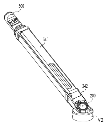
Referring now to Figs. 25(a)-28, there is shown an implant inserter 300
adapted for insertion of implant 200 from an anterior-oblique approach to the
spine. Inserter 300 includes features also found in implant inserter 250 with
the
exception that distal guide 302 has been configured to permit engagement with
an
implant for oblique insertion. Distal guide 302 includes a first angled drive
surface
310 disposed at an angle A33 with respect to side wall 306. In a preferred
embodiment, A33 is approximately 42 degrees. A second angled drive surface 314
is disposed at an angle A32 with respect to side wall 308. In the preferred
aspect,
A32 is approximately 30 degrees. A third angled surface 312 is disposed at an
angle A30 with respect to angled drive surface 310 and an angle A31 with
respect
3o to angled drive surface 314. In a preferred embodiment, angle A30 is
approximately 144 degrees and angle A31 is approximately 108 degrees.
CA 02395393 2002-06-25
WO 01/62191 PCT/USO1/05638
17
Additionally, an internal bore 316 is formed through distal guide 302. Bore
316 is
formed a distance D30 from side wall 308 and a distance D31 from side wall
306.
In a preferred aspect of the invention, D31 is greater than the distance D30
such
that bore 316 is offset with respect to the longitudinal axis of guide 302.
More
specifically, distance D30 is approximately l2mm and distance D31 is
approximately l5mm.
Refernng to Figs. 29(a) and 29(b), implant inserter 300 is shown selectively
coupled to implant 200. Angled driving surfaces 310 and 314 are in abutting
engagement with driving surfaces 212 and 208. It will be noted that angled
surface
l0 312 and 310 have sufficient length such that side wall 206 is not intended
to be in
substantial contact with the implant driver. Further, it is contemplated that
surface
312 may be spaced slightly from wall 210 to limit stress on the implant
adjacent
opening 226. Implant 200 is aligned with distal guide 302 such that opposing
side
walls 214 and 222 are in substantial alignment with side walls 308 and 306,
respectively, of distal guide 302. Moreover, angled driving surfaces 310 and
314
cooperate to limit implant rotation.
Refernng now to Figs. 30(a)- 31(c), a distraction window 342 is disposed
in a disc space created by vertebra V2 and an opposing upper vertebra (not
shown)
with distraction extensions 344 and 346 extending into the disc space.
Distraction
2o window 342 is positioned in the disc space from an anterior-oblique angle
approach to the spine. Specifically, reference line 348 represents a direct
anterior
approach to the spine, in substantial alignment with the sagittal plane. In
the
anterior-oblique approach, distraction window 342 is positioned into the disc
space
from an angled approach shown by angle A35. In a preferred embodiment, with
opposing angled side walls disposed at an approximately 30 degree angle, angle
A35 is approximately 30 degrees. A guide tube 340 is selectively coupled to
distraction window 342, thereby forming a. substantially rectangular working
channel into the disc space. Inserter 300 with interconnected implant 200 is
then
inserted through guide sleeve 340 until implant 200 is disposed in the disc
space in
3o preformed channel C2. The guide sleeve has dimensions substantially
corresponding to the implant dimensions, thereby limiting the amount of
tissue,
CA 02395393 2002-06-25
WO 01/62191 PCT/USO1/05638
18
vessels and other structures that must be removed or retracted for placement
of the
implant. The inner shaft is then rotated to release implant inserter from
implant
200. The implant inserter, guide tube, and distraction window may then be
removed. The orientation of implant 200 in comparison to vertebra V2 is
substantially identical to the orientation of implant 200 with respect to
vertebra V1
shown in Figs. 24(a)-24(c). End wall 216 is in substantial alignment with
posterior
portion 274 of channel C2. End wall 216 is disposed substantially adjacent
posterior portion 275 of vertebra V2. Further, opposed side walls 212 and 250
are
in substantial lateral alignment with the lateral portions of vertebra V2.
Thus, it
will be understood that implant 200 is positioned in the disc space with the
tapering surfaces 228 and 230 extending in the proper orientation to provide
maintenance of angulation between vertebra V2 and the opposing upper vertebra
(not shown).
While not shown by illustration, it will be understood that the implants
described herein may be inserted from a direct lateral approach to the spine.
The
same orientation in the disc space may be achieved regardless of the direction
of
insertion and the guiding instruments used.
Thus, the present invention provides an implant having multiple facets or
substantially parallel side walls allowing uniform orientation of the implant
in the
disc space although it is inserted by multiple, often guided, approaches to
the
spine. Specifically, the embodiments of the implants according to the present
invention permit insertion from a direct anterior, oblique-anterior and a
direct
lateral approach to the spine. While preferred embodiments of the invention
has
disclosed three pair of substantially parallel side walls disposed at a
various angles,
it is contemplated that more than three pair of substantially parallel side
walls
could be utilized to provide for implant insertion from a plurality of angles.
Further, while a particular angle of 30 degrees has been utilized for the
purposes of
illustration in a preferred embodiment, it will be understood that any oblique
angle
might be utilized to provide for insertion from multiple approaches from the
spine.
3o While the invention has been illustrated and described in detail in the
drawings and foregoing description, the same is to be considered as
illustrative and
CA 02395393 2002-06-25
WO 01/62191 PCT/USO1/05638
19
not restrictive in character, it being understood that only the preferred
embodiments have been shown and described and that all changes and
modifications that come within the spirit of the invention are desired to be
protected.