Note: Descriptions are shown in the official language in which they were submitted.
CA 02395672 2002-06-25
WO 01/49358 PCT/US00/35121
-1-
MEDICAL DEVICES THAT RESIST RESTENOSIS
BACKGROUND OF THE INVENTION
The invention relates to medical devices
associated with agents that inhibit restenosis.
Prostheses, i.e., prosthetic devices, are used
to repair or replace damaged or diseased organs, tissues
and other structures in humans and animals. Prostheses
must be generally biocompatible since they are typically
implanted for extended periods of time. Prostheses can
be constructed from natural materials such as tissue,
synthetic materials or a combination thereof.
Invasive procedures are commonly used to treat
various forms of cardiovascular disease. Routine
procedures include angioplasty, atherectomy, insertion
of stents and laser surgery. However, damage resulting
from this contact with a patient's blood vessels can
result in restenosis, the blockage of blood vessels. In
particular, balloon angioplasty and atherectomy are
associated with relatively high incidents of restenosis
in the three to six months following the procedure.
A common procedure for treating
arteriosclerosis is balloon angioplasty. To perform
balloon angioplasty, a catheter is guided through an
artery to a location with restricted flow. A balloon
catheter is then inflated to a pressure between, for
example, 3 and 6 atmospheres for about 60 seconds. The
inflation ~of the balloon cracks the plaque lining the
vessel walls stretching the arterial wall, so that the
lumen of the artery is expanded following the inflation
of the balloon. The increased lumen allows for
increased blood flow, but restenosis can result in
subsequent loss of blood, flow through the lumen.
CA 02395672 2002-06-25
WO 01/49358 PCT/US00/35121
-2-
Vascular stems involve structures that are
expanded within a blood vessel to maintain or expand the
lumen of the vessel. While intravascular stents can be
used to successfully achieve increased internal lumen
diameter, stent restenosis also can result in intimal
thickening that reduces the effectiveness of the stent.
Restenosis is characterized by platelet
aggregation and adhesion, and by smooth muscle cell
migration and proliferation, which individually or
together result in the narrowing of the vessel lumen.
The narrowing of the vessel restricts vasodilation and
causes an increase in blood pressure . The smooth muscle
cells along the vessel lining begin proliferating within
two to three days of disruption of the vessel and
continue for several days. In addition to vessel
narrowing, restenosis can lead to blood clotting, which
further threatens stroke, lung damage and heart damage
if the blood clots travels from the formation site.
Some approaches for the prevention or treatment of
restenosis include the delivery of radioactive compounds
or of nitric oxide, which is implicated in platelet
accumulation.
SUMMARY OF THE INVENTION
In a first aspect, the invention pertains to
a medical device suitable for implantation including a
biocompatible material and a plurality of exogenous
biological macromolecules bound to the biocompatible
material. The exogenous biological macromolecules store
a therapeutic agent which acts to inhibit restenosis.
In another aspect, the invention pertains to
a medical device suitable for implantation comprising a
biocompatible material and a plurality of exogenous
f
storage structures bound to the biocompatible material.
CA 02395672 2002-06-25
WO 01/49358 PCT/US00/35121
-3-
The exogenous storage structures store isotopically
enhanced radioactive metal ions.
In a further aspect, the invention pertains to
a method for inhibiting restenosis comprising binding an
exogenous storage structure to a biocompatible material
forming at least a portion of a vascular prosthesis.
The storage structure comprises a biological
macromolecule. The method further includes associating
the storage structure with a therapeutic agent which
acts to inhibit restenosis.
In addition, the invention pertains to a
medical device suitable for implantation comprising a
biocompatible material and a therapeutic agent
covalently bonded to the biocompatible material. The
therapeutic agent actsa to inhibit restenosis.
In another aspect, the invention pertains to
a medical device including an expandable structure and
therapeutic particles on the surface of the expandable
structure. The therapeutic particles include a
therapeutic agent that acts to inhibit restenosis.
Furthermore, the invention pertains to a
method of producing a medical device comprising applying
therapeutic particles to the exterior of an expandable
structure. The therapeutic particles include a
therapeutic agent that inhibits restenosis.
BRIEF DESCRIPTION OF THE DRAWINGS
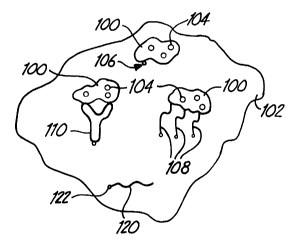
Fig. 1 is a schematic diagram that shows a
medical device having bound exogenous storage structures
associated with a therapeutic agent.
Fig. 2 is a side view of a vascular stmt.
Fig. 3 is an end view of the vascular stmt of
Fig. 2.
r
Fig. 4 is a perspective view of a vascular
graft .
CA 02395672 2002-06-25
WO 01/49358 PCT/US00/35121
-4-
Fig. 5 is a side view of the vascular graft of
Fig. 4 attached to a blood vessel.
Fig. 6 is a sectional side view of a blood
vessel with an angioplasty balloon positioned for use,
in which the cross section is taken through the center
along the length of the vessel.
Fig. 7 is a sectional side view of the blood
vessel of Fig. 6 with an expanded angioplasty balloon,
in which the cross section is taken through the center
of the vessel.
Fig. 8 is a sectional side view of the blood
vessel of Fig. 7 wherein the angioplasty balloon and
catheter have been removed following deflation of the
angioplasty balloon to leave behind therapeutic
particles, in which the cross section is taken through
the center of the vessel.
DETAILED DESCRIPTION OF THE ILLUSTRATIVE EMBODIMENTS
Therapeutic agents can be delivered locally to
vascular sites that have been subject to intervention,
where the agent is effective to inhibit restenosis,
i.e., to reduce the severity -and/or to reduce or
eliminate the incidence of restinosis. The intervention
sites generally are subject to some form of treatment
for vascular disease. "Vascular" sites. and structures
as used herein include cardiovascular sites and
structures and other blood contacting sites and
structures. Furthermore, the treatment of strictures of
the urinary tract are contemplated. Due to the medical
treatment, these locations are prone to the development
of restenosis. The therapeutic agents can be lethal or
inhibitory to proliferating cells and/or inhibitory to
deposition of plateletstor other clotting agents.
Local delivery of the therapeutic agent can be
directed to the specific potential restenosis sites
CA 02395672 2002-06-25
WO 01/49358 PCT/US00/35121
-5-
without introducing larger systemic quantities of the
therapeutic agent. In some embodiments, the local
delivery of the agent involves the use of storage
structures that carry and store the therapeutic agent in
association with a medical device. The storage
structures can be associated with a substrate that forms
part of a medical device. In some alternative
embodiments, a therapeutic agent is directly covalently
bonded to the biocompatible material without the use of
a binder matrix of an adhesive.
In alternative embodiments, the therapeutic
agents are delivered as particulates that are delivered
with the use of an expandable medical device. The
expansion of the device delivers the therapeutic agent.
In particular, an angioplasty balloon can be used to
deliver therapeutic particles by inflating the balloon
to open up the lumen of the blood vessel. The
particulates are located initially on the surface of the
balloon and are driven into the plaque and/or tissue of
the vessel wall when the balloon in deployed. In some
embodiments, the therapeutic agent is bound to
extracellular matrix of the vessel with ultraviolet
light.
A variety of medical articles can be used to
contact bodily fluids of a patient. Relevant
biocompatible medical articles generally incorporate a
biocompatible material which is intended to contact the
patient's biological fluids and/or tissues. Bodily
fluids include, for example, blood, plasma, serum,
interstitial fluids, saliva and urine. The patient can
be an animal, especially a mammal, and preferably is a
human.
t
Relevant medical articles include devices that
contact a person's bodily fluids for varying lengths of
CA 02395672 2002-06-25
WO 01/49358 PCT/US00/35121
-6-
time, for example, prostheses, catheters and surgical
instruments. Prostheses, i.e., prosthetic articles, are
used to repair or replace damaged or diseased organs,
tissues and other structures in humans and animals.
Prostheses generally must be biocompatible since they
are typically implanted for extended periods of time.
Preferred prostheses include prostheses used in the
vascular system at locations prone to restenosis.
The therapeutic agents generally can be any
agent effective for the inhibition of restenosis
including agents that are toxic or inhibitory to
proliferating smooth muscle cells or that inhibit
platelet accumulation or adhesion. Preferred
therapeutic agents include radioactive isotopes that
emit radiation at a suitable rate to inhibit
proliferating cells. Suitable radioactive ions include
anions, such as 32P04-1, l2sl-, 1311-, and cations of metals,
such as, s9Fe, 6'Ga, saCo, 6'Cu, 9°Y, 99Rh, lasRe, and l9aAu.
Nitric oxide is known to inhibit platelet aggregation,
as described in U.S. Patent 5,665,077 to Rosen et al.,
incorporated herein by reference. Preferred approaches
for delivering nitric oxide generally involve the
delivery of compounds that release nitric oxide.
Exogenous storage structures are
macromolecular structures that are not inherent or
native to the biocompatible material. In other words,
the exogenous storage structures are joined to the
biocompatible material to provide relevant storage
capability to the biocompatible material or to enhance
any low level inherent storage capability of the
biocompatible material with respect to the therapeutic
agent. Through the use-of exogenous storage structures,
r
a therapeutic agent can be delivered locally at a
region, such as a blood vessel, susceptible to
CA 02395672 2002-06-25
WO 01/49358 PCT/US00/35121
restenosis. For example, the therapeutic agent can be
associated with the particular medical device whose use
is correlated with the restenosis risk. Thus, an
effective amount of therapeutic agent can be delivered
locally without causing undesirable systemic treatment
and possible toxicity. With local delivery of the
therapeutic agent at the treatment site, only a small
amount of therapeutic agent is required to yield an
effective treatment.
Exogenous storage structures can provide
flexibility in directing and maintaining the therapeutic
agent at specific, particularly relevant portions of a
medical article. Further, use of an exogenous storage
structure permits control of the release rate of the
therapeutic agent, if release is desirable for
effectiveness of the therapeutic agent. The association
of a therapeutic agent with the exogenous storage
structures can be performed before or after attachment
of the exogenous storage structures to the biocompatible
material.
Generally, the exogenous storage structure is
bound to biocompatible material forming the medical
device or a portion thereof. This binding is shown
schematically in Fig. 1. In the schematic diagram of
Fig. 1, exogenous storage structures 100 are bound to
biocompatible material 102. Exogenous storage
structures 100 are associated with therapeutic agents
104.
Fig. 1 displays three possible storage
structure binding approaches. For example, exogenous
storage structures 100 can be bound to the biocompatible
material 102 with a covalent bond 106. Alternatively,
linkers 108 having a plurality of functional groups,
such as crosslinking agents, can be used that form
CA 02395672 2002-06-25
WO 01/49358 PCT/US00/35121
_g_
covalent bonds with both biocompatible material 102 and
exogenous storage structures 100: Alternatively, a
linker 110, such as an antibody, can bind with specific
bonding interactions with exogenous storage structures
100 and/or biocompatible material 102.
In alternative embodiments, the therapeutic
agent 120 is covalently bonded to biocompatible material
102, as shown in Fig. 1. A linker compound can be used
in the formation of covalent bond 122. Any linker
compound can be considered to be a portion of the
therapeutic compound.
In alternative embodiments, the therapeutic
agent is delivered from the surface of a medical device,
such as an angioplasty balloon. The medical device
generally expands, flexes or otherwise moves to apply
some force against a blood vessel wall or other region
susceptible to restenosis, e.g., a passageway of the
urinary tract. The force of the angioplasty balloon or
other device against the vessel wall or other region
after the balloon or device is deployed propels the
therapeutic agent into the plaque and/or tissue in the
targeted region. The therapeutic agent can then be
effective in inhibiting restenosis in the treated
structure. The medical device used to deliver the
therapeutic agent can be removed from the treated region
or it can remain in the treated region.
The approaches described herein for delivery
of therapeutic agents for the treatment of restenosis
are an effective and versatile approach for treatment of
restenosis. For example, local delivery of the
therapeutic agent results in fewer side effects than
systemic delivery, and-a reduced amount of therapeutic
r
agent is needed as compared to systemic delivery. The
delivery approaches described herein provide effective
CA 02395672 2002-06-25
WO 01/49358 PCT/US00/35121
_g_
control of the amount of therapeutic agent delivered at
a location susceptible to restenosis. A variety of
different therapeutic agents can be delivered alone or
in combination.
Medical Articles
Relevant medical articles include a
biocompatible material, at least as a component, that is
suitable for contacting a patient' s bodily fluids and/or
tissues. Biocompatible materials are non-toxic, non-
carcinogenic and do not induce hemolysis or a severe
immunological response. For embodiments based on-
exogenous storage structures, the medical articles
generally are designed for implantation into a patient
for extended periods of time. For embodiments based on
the delivery of particulate therapeutic agents, the ..
medical article can be designed for implantation, or the
medical article can be used percutaneously extending
from outside the body into the body for delivery of the
therapeutic agent.
Suitable medical articles or components of
such medical articles for implantation include, for
example, artificial organs such as artificial hearts,
anatomical reconstruction prostheses, coronary stems,
vascular grafts and conduits, vascular and structural
stem s, vascular shunts, biological conduits, stents,
valued grafts, permanently in-dwelling percutaneous
devices, intrauterine devices (IUDs), urinary stem s,
and combinations thereof.
Other biomedical devices that are designed to
dwell for extended periods of time within a patient are
also suitable for the inclusion of therapeutic agents
described herein. These~,devices include, for example,
Hickman catheters and other percutaneous articles that
are designed for use over a plurality of days.
CA 02395672 2002-06-25
WO 01/49358 PCT/US00/35121
-10-
Percutaneous devices include items that penetrate the
skin, thereby extending from outside the body into the
body.
Medical devices of particular interest are
susceptible to initiation of undesirable cell growth
and/or platelet aggregation. Implantable devices of
particular interest include, for example, implantable
vascular devices, implantable cardiovascular devices and
implantable urinary stents. Implantable vascular
devices include, for example, vascular stents, vascular
grafts and conduits and valued grafts. Implantable
cardiovascular devices of particular interest include,
for example, coronary stem s.
Especially relevant medical articles for use
with exogenous storage structures include, for example,
vascular stems, urinary stem s and vascular grafts,
especially small vascular grafts. Vascular stents are
used to form an internal scaffolding within the blood
vessel that maintains or increases the lumen of the
blood vessel. The stmt is generally introduced through
a catheter for deployment at the desired position within
the blood vessel.
Vascular stents generally are formed from
synthetic materials, such as metals or synthetic
polymers. The stmt can be formed from a resorbable
material, for example a resorbable polymer, such that
the stmt forms a temporary scaffolding to promote
healing while maintaining patency of the blood vessel.
A representative stmt design is shown in Figs . 2 and 3 .
Vascular stent 150 is formed from a biocompatible
material 152 with a form consistent with its expandable
nature. -
Y
Preferred vascular and urinary stem s have
radial and torsional flexibility, biocompatibility,
CA 02395672 2002-06-25
WO 01/49358 PCT/US00/35121
-11-
visibility by x-ray, and reliable expandability.
Expandability is desirable since vascular stems
generally are implanted through a catheter. Thus, the
vascular stmt preferably expands to fit securely
against the vessel walls once deployed. Expandable
stents can operate, for example, with a spring-like
design that expands from a small diameter to a
predetermined dimension when a constraint is removed, or
with a thermal memory metal that changes shape upon
heating. In addition, a balloon expandable stmt can.
undergo plastic deformation, expanding the material
beyond its elastic limit with pressure. The
biocompatible materials for forming these stents are
described further below.
Vascular grafts are used to replace portions
of damaged/diseased vascular tissue. The damaged/
diseased section of vascular tissue is removed, and the
vascular graft replaces the removed section. The
vascular graft is attached with suture or other
fasteners to the free ends of the vessel that remain
after a damaged/diseased portion of vessel is extracted.
The vascular graft can be constructed from tissue and/or
synthetic materials, as described further below.
A representative vascular graft 170 is
depicted in Fig. 4. Vascular graft 170 includes a
flexible tubular structure 172 and optional sewing cuffs
174, 176. Flexible tubular structure 172 can include
one or more biocompatible materials, such as tissue,
synthetic polymer or combinations thereof. Sewing cuffs
174, 176 are formed from fabric, tissue or the like.
Sewing cuffs 174, 176 assist with the implantation of
the prosthesis and may~provide reinforcement of the
prosthesis at the site of anastomoses, i.e., attachment
of the vessel to the graft. A cross section of vascular
CA 02395672 2002-06-25
WO 01/49358 PCT/US00/35121
-12-
graft 170 attached to natural vessel sections 180, 182
is depicted in Fig. 5. As shown in Fig. 5, suture 184
is used to secure vascular graft 170 to vessel sections
180, 182.
Restenosis is particularly problematic in
thinner arteries. Embodiments of the vascular grafts
and vascular stem s of particular interest have an
implanted internal diameter less than about 5 mm, less
than about 4 mm or even less than about 3 mm.
In alternative embodiments involving the
delivery of particulate therapeutic agents, particularly
relevant medical devices for the delivery of the agent
include expandable stems, angioplasty balloons, and
surgical instruments. Angioplasty balloons are brought
into a partially obstructed artery using a catheter.
When positioned at the point of obstruction, the balloon
is expanded under pressures generally in the range of 3-
6 atmospheres. Similarly, a vascular stmt is
positioned within a vessel using a catheter and expanded
against the vessel walls to increase the lumen. The
force of the expanding balloon or stent can propel the
particle of therapeutic agent into the tissue and/or
plaque. Surgical instruments, such as forceps, can be
coated with a particulate therapeutic agent for delivery
by applying pressure to the vessel wall with the coated
surface of the instrument. Photochemical coupling can
be used to secure the therapeutic agent to the vessel
walls if the delivery force is not sufficient to deliver
the therapeutic agent to a stable location within the
vessel wall.
Referring to Fig. 6, angioplasty balloon 200
has a deposit of therapeutic particles 202 on its outer
surface. Angioplasty balloon 200 is deployed within
blood vessel 204 at a point of partial blockage 206.
CA 02395672 2002-06-25
WO 01/49358 PCT/US00/35121
-13-
Angioplasty balloon 200 can be deployed through a
catheter 208.
Referring to Fig. 7, balloon 200 is expanded
by flowing fluid, either a gas or a liquid, into balloon
200 through channel 210. If the fluid is blood
compatible, such as sterile saline, balloon 200 can be
designed to have the fluid flow through the walls of
balloon 200. Balloon 200 generally is deployed for
about one minute. When balloon 200 is deployed, at
least some of therapeutic particles 202 are deposited
within the tissue and/or plaque in the vessel wall.
After the balloon is deflated, balloon 200 and catheter
208 are withdrawn from blood vessel 204. Therapeutic
particles 202 remain in blood vessel following the.
removal of balloon 200, as shown in Fig. 8.
Biocompatible Materials
The biocompatible medical devices can be made
from one or more biocompatible materials described
below. Biocompatible materials are suitable for contact
with a patient's bodily fluids and tissues. Appropriate
biocompatible materials include natural materials,
synthetic materials and combinations thereof.
Natural, i . a . , biological, material for use in
the invention includes relatively intact (cellular)
tissue as well as decellularized tissue. These tissues
may be obtained from, for example, natural blood
vessels, such as veins or arteries, pericardial tissues
such as pericardial patches, connective tissues, bypass
grafts, blood vessels, dura matter, fascia, submucosa,
umbilical tissues, and the like.
Natural tissues are derived from a particular
animal species, typically mammalian, such as human,
bovine, porcine, canine, seal, kangaroo or transgenic
mammals. Suitable natural tissues generally include
CA 02395672 2002-06-25
WO 01/49358 PCT/US00/35121
-14-
collagen-containing material. Natural tissue is
typically, but not necessarily, soft tissue.
Appropriate tissues also include tissue equivalents such
as tissue-engineered material involving a cell-
s repopulated matrix, which can be formed from a polymer
or from a decellularized natural tissue. Tissue
materials are particularly useful for the formation of
vascular grafts.
Tissues can be fixed by crosslinking. This
provides mechanical stabilization, for example, by
preventing enzymatic degradation of the tissue.
Glutaraldehyde is typically used for fixation, but
formaldehyde, other difunctional aldehydes, epoxides,
genipin or derivatives thereof can be used. Tissues can
be used in either crosslinked or uncrosslinked form,
depending on the type of tissue, the use and other
factors.
Relevant synthetic materials include, for
example, polymers, metals and ceramics. Appropriate
ceramics include, without limitation, hydroxyapatite,
alumina and pyrolytic carbon. Appropriate metals
include medals approved for medical use, such as
titanium, cobalt, stainless steel, nickel, iron alloys,
cobalt alloys. For use in vascular stents, preferred
metals include, for example, resilient metals, such as
Elgiloy°, a cobalt-chromium-nickel alloy, and MP35N, a
nickel-cobalt-chromium-molybdenum alloy, and Nitinol~,
a nickel-titanium alloy. Appropriate synthetic
materials include hydrogels and other synthetic
materials that cannot withstand severe dehydration.
Polymeric materials can be fabricated from
synthetic polymers as swell as purified biological
polymers. The polymeric materials can be woven into a
mesh to form a matrix or substrate. Alternatively, the
CA 02395672 2002-06-25
WO 01/49358 PCT/US00/35121
-15-
polymer materials can be molded or cast into appropriate
forms. Appropriate synthetic polymers include, without
limitation, polyamides (e. g., nylon), polyesters,
polystyrenes, polyacrylates, vinyl polymers (e. g.,
polyethylene,polytetrafluoroethylene,polypropylene and
poly vinyl chloride), polycarbonates, polyurethanes,
poly dimethyl siloxanes, cellulose acetates, polymethyl
methacrylates, ethylene vinyl acetates, polysulfones,
nitrocelluloses and similar copolymers.
Biological polymers can be naturally occurring
or produced in vitro by, for example, fermentation and.
the like. Purified biological polymers can be
appropriately formed into a substrate by techniques such
as weaving, knitting, casting, molding, extrusion,
cellular alignment and magnetic alignment. Suitable
biological polymers include, without limitation,
collagen, elastin, silk, keratin, gelatin, polyamino
acids, polysaccharides (e.g., cellulose and starch) and
copolymers thereof.
Other suitable polymers include natural or
synthetic resorbable polymers such as dextran.,
hydroethyl starch, gelatin, derivatives of gelatin,
polyvinylpyrrolidone, polyvinylalcohol, poly[N-(2-
hydroxylpropyl) methacrylamide], polyglycols,
polyesters, poly (orthoesters), polyester amides),
polyanhydrides. Resorbable polyesters include, for
example, poly (hydroxy acids) and copolymers thereof,
poly(E-caprolactone), poly (dimethyl glycolic acid), and
poly (hydroxy butyrate). Preferred resorbable polymers
include, for example, D, L-polylactic acid, L-polylactic
acid, poly(glycolic acid), and copolymers of L-lactic
acid, D-lactic acid and ~lycolic acid.
Biocompatible materials can form the entire
medical device or they can form portions of the medical
CA 02395672 2002-06-25
WO 01/49358 PCT/US00/35121
-16-
device. Biocompatible materials can include one or a
combination of the various natural materials and
synthetic materials described above.
Vascular grafts can be primarily tissue based
or polymer based. Polyesters, such as polyethylene
terephthalates, are particularly suitable for the
formation of vascular grafts. To prevent undesirable
levels of bleeding through knitted or woven vascular
grafts or the like, a small volume of the patient's
blood can be forced into and through the graft's
interstices prior to implantation. A clot results that
is ultimately replaced by ingrowth of fibroblast cells
and collagen. Alternatively, the graft can be
manufactured with albumin or collagen in the fabric
interstices.
Vascular stems can be formed from metals,
polymers and combinations thereof. Resorbable polymers
are particularly well suited for the formation of
temporary vascular stent embodiments. Exogenous storage
structures can be attached to the stmt with or without
a chemical linker based on the binding properties of the
material. The binding of a exogenous storage structure
to a biocompatible material is described in detail
below.
Suitable polymers for the formation of
angioplasty balloons include, for example, cellulose
acetate, polyvinyl chloride, polysulfone,
polyacrylonitrile, polyurethanes, polyolefins,
polyesters, fluoropolymers and other natural and
synthetic elastomers.
With respect to relevant embodiments, the
exogenous storage structures can be associated with an
entire biocompatible material or a portion of the
biocompatible material. Similarly, if the medical
CA 02395672 2002-06-25
WO 01/49358 PCT/US00/35121
-17-
article includes more than one biocompatible material,
storage structures can be associated with one or more of
the biocompatible materials. For example, an
appropriately treated natural blood vessel can be
combined with fabric sewing cuffs to form a vascular
graft, where the tissue and/or the fabric can be
associated with exogenous storage structures.
A medical article can include one or more
types of exogenous storage structures and/or one or more
therapeutic agents. If a plurality of types of
exogenous storage structures are used, the different
types of storage structures can be associated with the
same biocompatible materials) or portions thereof, or
with different biocompatible materials) or portions'
thereof. For example, one type of storage structure can
be associated with the tissue portion of a tissue
vascular graft while a second type of exogenous storage
structure is associated with the sewing cuff.
Similarly, a first type of exogenous storage structure
can be associated with the entire medical article, such
as both the tissue portion and the sewing cuff portion
of a vascular graft, while a second type of exogenous
storage structure is only associated with the sewing
cuff portion. Other variations can be used.
The exogenous storage structures can be
associated with the biocompatible material before or
after the various components of the medical device are
combined into the medical device. The selected
approaches for association of the exogenous storage
structure with the biocompatible material may influence
the order of construction of the medical device.
Therapeutic Agents
The approaches described herein are suitable
for the delivery of therapeutic agents to sites
CA 02395672 2002-06-25
WO 01/49358 PCT/US00/35121
-18-
susceptible to risk for the development of restenosis.
Suitable therapeutic agents are able to inhibit
restenosis, for example, by inhibiting platelet
deposition, the proliferation of smooth muscle cells and
excretion of extracellular matrix or a combination
thereof. Exogenous storage structures can be used for
the delivery of one or more therapeutic agents in
association with a medical device to inhibit 'the
development of restenosis. Preferred therapeutic agents
include, for example, ionic agents that can be stored
with preferred exogenous storage structures. In
alternative embodiments, therapeutic agents are
formulated into particulates for delivery from the
surface of a medical device. Some therapeutic compounds'
can be directly covalently bonded to the biocompatible
material.
Natural macromolecular storage structures are
capable of storing a variety of ionic therapeutic
agents. Suitable ionic therapeutic agents include, for
example, radioactive ions and ionic nitric oxide (NO)
precursors. Nitric oxide is known to inhibit platelet'
aggregation, as described in U.S. Patent 5,665,077 to
Rosen et al., incorporated herein by reference.
Radioactive ions act as antiproliferative
agents. Suitable radioactive ions include anions, such
as 32PO4-1, l2sl-, 1311-, and cations of metals, such as,
s9Fe ~ s~Ga ~ saCo ~ s~Cu ~ 9oY ~ s9Rh ~ iasRe ~ and isaAu .
Radioactive ions refer to metal atoms that have been
isotopically enhanced relative to any naturally
occurring radioactive ions. Radioactive ions of
interest generally have half-lives considerably less
than a year such that-t~ey are not found naturally,
although some of the ions can be produced as decay
products of long lifetime isotopes, such that very small
CA 02395672 2002-06-25
WO 01/49358 PCT/US00/35121
-19-
amounts can occur in nature. Isotopically enhanced
radioactive metal atoms/ions are preferably enhanced at
least about 10 times relative to any naturally occurring
values and more preferably at least 100 times any
naturally occurring values. Preferred radioactive
isotopes are beta emitters.
Preferred ways of delivering nitric oxide
include the delivery of compounds that release nitric
oxide. Ionic compounds that decompose to release nitric
oxide include, for example, organic and inorganic
compounds that include an -NONO- functional group. For
example, suitable organic compounds have the structure
XNONO-, where X can be a primary amine, such as
(CH3)zCHNH-, a secondary amine, such as (CH3CHz)zN-, or a
polyamine, such as, the zwitterionic species with X
being spermine , i . a . , H2N ( CHz ) 3NHz+ ( CHz ) 4N [NONO] - ( CHz ) 3NHz
.
A suitable inorganic species is Na0[NONO]Na,
nitropercide. The synthesis of 1-(2S-carboxypyrrolidin-
1-yl)-1-oxo-2-hydroxydiazene disodium salt,l-hydroxy-2-
oxo-3-carboxymethyl-3-methyl-1-triazene disodium salt,
1-hydroxy-2-oxo-3-carboxymethyl-3-methyl-1-triazine N-
methylamide sodium salt, the bis (nitric oxide) adduct of
L-prolyl-L-leucylglycinamide, and corresponding protein
adducts are described in U.S. Patent 5,632,981 to
Saavedra et al., entitled "Biopolymer-Bound Nitric
Oxide-Releasing Compositions, Pharmaceutical
Compositions Incorporating Same and Methods of Treating
Biological Disorders Using Same," incorporated herein by
reference. The protein adducts can be used directly as
exogenous storage structures in which the nitric oxide
releasing functional groups are the therapeutic agents.
The amine groups of spermine are suitable for
covalent bonding to a biocompatible material that has
functional groups that bond to the amines . For example,
CA 02395672 2002-06-25
WO 01/49358 PCT/US00/35121
-20-
aldehyde functional groups react with amines. Aldehyde
crosslinked tissue generally has free aldehyde groups
that can bond with the amines. Other nitric oxide
releasing compounds can be formulated with reactive
functional groups that can be covalently bonded to a
biocompatible material with corresponding reactive
functional groups.
In certain embodiments, the therapeutic agents
are incorporated into a particle that can be delivered
from the surface of a medical device into tissue and/or
plaque lining the blood vessel wall. The radioactive
ions and the ionic nitric oxide releasing compounds
described above, along with appropriate counter ions,
can be formed into particles or combined with a suitable
binder to form particles, as described below.
Similarly, exogenous storage structures which are not
attached to a medical device can be used as particles
for delivery from the surface of a medical device. In
addition, non-ionic therapeutic agents can be formed
into particles or combined with a suitable binder to
form the particles.
Suitable non-ionic therapeutic agents for
delivery as particles include, for example, neutral
compounds containing the radioactive isotopes listed
above and non-ionic nitric oxide-releasing compounds.
Suitable non-ionic nitric oxide releasing compounds
include, for example, a-substituted nitroso compounds
(R-NO, where R is a tertiary carbon group), such as 2-
methyl-2-nitrosopropane. These a-substituted nitroso
compounds are relatively stable at room temp and
decompose at body temperature. Use of a-substituted
nitroso compounds as a nitric oxide source is described
in U.S. Patent 5,665,077 to Rosen et al., entitled
"Nitric Oxide-Releasing Nitroso Compositions and Methods
CA 02395672 2002-06-25
WO 01/49358 PCT/US00/35121
-21-
and Intravascular Devices for Using Them to Prevent
Restenosis," incorporated herein by reference.
Alternatively, insoluble or slightly soluble compounds
incorporating radioactive atoms can be formed into
powders that are used as the source of particles. For
example, ferric phosphate (Fe04P) can incorporate
radioactive iron and/or phosphate atoms. Ferric
phosphate is practically insoluble in water, and the
non-radioactive form of ferric phosphate is used as a
food supplement.
Appropriate doses of the agent may depend on
several factors, such as size of the blood vessel,
physical condition of the patient, nature of the medical
device, and the properties of the therapeutic agent.
Generally, for nitric oxide-releasing agents, a suitable
amount of therapeutic agent releases from about 0.05 mg
to about 100 mg of NO. Spermine has an IC-50
concentration, i.e., a concentration at 50 percent
effectiveness, of 1.97 x 10-a molar. Thus, an effective
amount of spermine would be delivered to yield a local
concentration in this order of magnitude or higher.
Similarly, an effective concentration of nitro percide
is 2.5 x 10-e molar. A suitable treatment period would
be about 10 days, such that the cumulative NO quantities
would range from about 0.00086 moles to about 0.086
moles. Treatment periods can be extended to longer
times, for example, 6 weeks. Over a six week period,
the total amounts of NO would be from about 0.0036 moles
to about 0.36 moles. Suitable amounts of radioactive
compounds will further depend on the isotopic
enrichment, the lifetime of the isotope and the
penetrative ability of-the radiation. For metal ions
that are beta emitters with a lifetime over a few days,
a suitable amount of radioactive metal ions ranges from
CA 02395672 2002-06-25
WO 01/49358 PCT/US00/35121
-22-
about 0.001 milligrams(mg)/gram(g) biocompatible
material to about 100 mg/g and preferably from about
0.001 mg/g to about 30 mg/g. The amount of radioactive
ions delivered preferably are below toxic levels by a
factor of 10 or more, where toxic levels are levels at
which observable physical effects of the radiation
manifest themselves.
Exoctenous Storage Structures
Storage structures can be used for the
delivery of cationic or anionic therapeutic agents to a
blood vessel. Exogenous storage structures preferably
are microscopic, natural macromolecules such as
proteins, carbohydrates, nucleic acids and combinations
thereof. It is to be understood, however, that
aggregations of the preferred compositions need not be
microscopic. Suitable macromolecules generally have a
molecular weight greater than about 5,000 atomic mass
units (amu), preferably greater than about 10,000 amu
and more preferably greater than about 25,000 amu. The
term "protein" includes peptides and polypeptides alone
as well as peptides and polypeptides conjugated with'
carbohydrates, nucleic acids, lipids and/or other
compounds.
Exogenous storage structures are distinct from
any naturally occurring structures, such as ferritin
already present in the material. In other words,
exogenous storage structures are non-inherent or
extrinsic structures that are associated with the
biocompatible material, as depicted in Fig. 1.
Exogenous structures are in contrast with endogenous
structures that are inherent to the biocompatible
materials. Generally, the exogenous storage structures
are attached as discrete entities to the biocompatible
CA 02395672 2002-06-25
WO 01/49358 PCT/US00/35121
-23-
material, using the methods described below, rather than
applied as a coating.
Appropriate protein storage structures within
the scope of the present invention include metal binding
proteins such as ferritin, transferrin, hemoglobin,
globulins, albumin, glutathione, metallothiens,
myoglobin, ceruloplasmin and hemocyanin, as well as
modified proteins having attached bifunctional chelators
to generate metal binding capability. Ferritin is a
preferred metal binding protein because of its generally
large storage capacity.
Ferritin protein without bound metal is called
apoferritin. Apoferritin is a 24-subunit protein with.
a molecular weight of approximately 450,000 amu,
although the molecular weight varies depending on the
animal species from which the ferritin is isolated.
Isoferritins, related proteins with differing numbers of
subunits, are also within the scope of the present
invention and are included within the term "ferritin."
The ferritin core can store between about 2000
and about 4500 iron ions. For example, horse spleen
ferritin can bind about 4500 iron ions, while human
ferritin can bind about 2500 iron ions. The iron is
stored within the core as ferric oxide or ferric
hydroxyphosphate. Ferritin can also bind large
quantities of other metal cations, as well as anions
that are bound to generally maintain, overall electrical
neutrality. Binding of these non-iron ions is enhanced
by the simultaneous binding of a moderate quantity of
iron ions. Generally, storage structures, such as
ferritin, can be bound to biocompatible material with
the storage structures~preloaded in vitro with desired
cations or anions.
CA 02395672 2002-06-25
WO 01/49358 PCT/US00/35121
-24-
The selection of a particular storage
structure can be based on its storage capacity,
availability and ease of handling. For example,
ferritin or other metal binding proteins generally need
not be saturated with the metal ions or ions of interest
to be useful in the invention. The ferritin can be
charged with, for example, desired ions by incubating
purified ferritin with a relatively concentrated
solution of the ions. The binding of the ions to the
protein can be accelerated by heating and by pH
adjustment. After a sufficient period of incubation,
the free ions can be removed by passing the solution
over an ion exchange resin or through a size exclusion
membrane.
In addition, storage structures can be formed
from other proteins modified to create metal binding
capability. Preferred proteins for modification have
high molecular weight, such as immunoglobulins. The
modification can involve, for example, covalent bonding
of metal sequestering compounds to the protein.
More specifically, significant metal binding
capability can be created by binding a bifunctional
chelator, such as a polyaminocarboxylate or a
polyaminophosphonate, to the protein as the metal
sequestering compound. Preferred bifunctional chelators
include electrophilic and nucleophilic moieties such as
bromoacetamide, maleimide, imidoester, thiophthalimide,
N-hydroxysuccinimyl ester, pyridyl disulfide, phenyl
azide, o-acylisourea, diazonium hydrazine, carbonyl
hydrazine, amino hydrazine, acyl hydrazine, diazonium
semicarbazide, carbonyl semicarbazide, amino
semicarbazide, acyl semitcarbazide, thio semicarbazides
and cyclic polyaminocarboxylates and cyclic
polyaminophosphonates having 12 to 16 atom rings. The
CA 02395672 2002-06-25
WO 01/49358 PCT/US00/35121
-25-
specific chelator can be selected to produce a desired
release rate of the bound ions if release is a
consideration for function.
The bifunctional chelators generally can be
covalently bonded to the protein by conventional
methods. Typically, the covalent bonds will be formed
between selected amino acid residues of the protein and
a specific functional group in the chelator, in which
the distinct functional groups are distinct from the
metal chelating sites. The number of chelating agents
bound to a protein will depend on the structures of the
chelator and protein and on the reaction conditions.
It is preferable to have at least one
bifunctional chelator bound to each protein, and it i.s
more preferable to have multiple bifunctional chelators
bound to each protein. Metal ions can be bound to the
chelator before, at the time of, or after the covalent
binding of the chelator to the protein. The reaction
conditions may influence the selected order of the
processing steps.
In addition, organic anions R-NONO- with NONO-
functional groups can be attached directly onto protein
side chains or other natural macromolecules by way of
other reactive functional groups located in the R group.
In this way a variety of natural macromolecules can be
modified to deliver NO as a therapeutic agent.
Binding of Exogenous Storage Structures to Biocompatible
Material
Binding of the exogenous storage structures to
the biocompatible material can involve specific binding
interactions to target specific structures within the
material. Alternative~.y, the binding can involve
covalent bonding due, for example, to reaction with
general crosslinking agents or other specifically
CA 02395672 2002-06-25
WO 01/49358 PCT/US00/35121
-26-
designed linker molecules. For tissue substrates, the
binding of the exogenous storage structures preferably
takes place at or near a physiological pH, preferably
ranging from a pH of about 6 to a pH of about 8.5 and
more preferably from a pH of about 7.0 to a pH of about
a.o.
One procedure for non-specific binding makes
use of glutaraldehyde, which crosslinks proteins by way
of two aldehyde groups. This procedure is particularly
appropriate for binding protein exogenous storage
structures to tissue based biocompatible material.
Since glutaraldehyde is typically used for fixation. of
some biocompatible materials, e.g. tissues, the non-
specific crosslinking to bind the exogenous storage
structures to the tissue material can be performed
simultaneously with fixation of the tissue.
Alternatively, the non-specific binding of the exogenous
storage structures to the biocompatible material can be
performed as a separate step before or after the
completion of a fixation process, assuming a fixation
step is performed.
Similarly, the exogenous storage structure can
be bound directly or indirectly to the biocompatible
material using other covalent chemical bonding
reactions. For example, other polyfunctional linker
molecules, besides dialdehydes, can be used to join the
exogenous storage structure and the biocompatible
material. At least one functional group of the
polyfunctional linker molecule reacts with the exogenous
storage structure, and at least one functional group of
the polyfunctional linker molecule reacts with the
biocompatible material. For example, with a synthetic
polymer biocompatible material, the linker molecule can
react with a functional group in the polymer. In
CA 02395672 2002-06-25
WO 01/49358 PCT/US00/35121
-27-
particular, if the polymer is polyester, such as
polyethylene terephthalates, ester functional groups can
decompose or contain residual carboxyl groups. For
polyesters, the linker can include a carbodiimide, such
as in the compound 1-ethyl-3f3-dimethylaminopropyl~-
carbodiimide hydrochloride (EDC), functional group to
bond with the polymer and an aldehyde functional group
to bond with, for example, a protein storage structure.
Use of a photoactivated linker is described below. If
the storage structure includes appropriate functional
groups to bond with the biocompatible material directly,
a linker molecule may not be necessary.
Specific binding interactions can form a basis
for attachment of the exogenous storage structures. The
character of the specific binding interactions involve
a plurality of non-covalent interactions such as
hydrogen bonding, van der Waals interactions and
molecular rearrangements, which characterize, for
example, antibody-antigen, specific binding protein-
receptor and enzyme-substrate associations. Thus,
specific binding interactions involve molecular'
recognition characteristics wherein a plurality of
interactions over a region of both interacting molecules
are involved to bind or dock the two molecules together.
One approach for taking advantage of specific
binding interactions involves covalent binding of a
linker to the storage structure and association of the
linker with the prosthetic material by specific binding
interactions. A variety of commercially available
antibodies, receptors, substrates and other specific
binding reagents may be used as linkers. Such linkers
can function as binding molecules for cellular or
extracellular sites in tissue having specific binding
sites. Similar binding can be used for synthetic
CA 02395672 2002-06-25
WO 01/49358 PCT/US00/35121
-28-
materials if, for example, antibodies were raised
against the synthetic materials.
As an alternative to using commercially
available antibodies, cellular or extracellular
components from the biological material can be isolated
by conventional techniques. For example, nuclear
membranes or a specific portion of the nuclear membrane
corresponding to an antigen or groupings of antigens can
be isolated. The isolated materials then are used to
produce polyclonal or monoclonal antibodies by
conventional techniques. The resulting antibodies are
covalently bonded to the exogenous storage structure to
prepare it for binding to the biocompatible material.
A storage structure having an attached
antibody or any other comparable targeting molecule is
considered a "storage structure" for the purposes of the
present application. The binding of compounds to
antibodies is well established, especially where the
compound is a protein. Due to its high iron content,
ferritin is commonly linked to antibodies to serve as an
electron microscopy probe in the histology field. In a
preferred embodiment, glutaraldehyde is used to join the
respective proteins. In addition, as noted above, the
antibody itself can be modified with a therapeutic agent
to become, itself, an exogenous storage structure,
rather than serving as a linker portion of an exogenous
storage structure.
In an alternative embodiment, photochemical
coupling can be used for covalent coupling.
Photochemical coupling is based on the use of high
energy light, e.g., ultraviolet light, to form reactive
intermediates of certain functional groups. These
reactive intermediates can form carbon-carbon bonds
CA 02395672 2002-06-25
WO 01/49358 PCT/US00/35121
-29-
between two compositions. Aryl ketone functional groups
are particularly useful in this respect.
Photochemical coupling is particularly
appropriate for the attachment of exogenous storage
structures to synthetic polymeric materials,
uncrosslinked tissues or biological polymers. See, for
example, Dunkirk et al.,.J. Biomaterials Applications
6:131-156 (1991), incorporated herein by reference.
Photochemical techniques are useful also for the
attachment of exogenous storage structures to metal
surfaces and decellularized tissue substrates.
Photochemical coupling can be used for the direct
attachment of an exogenous storage structure to the
biocompatible material. Alternatively, photochemical
coupling can be used to attach a linker to the
biocompatible material either before or after the
attachment of the linker to the exogenous storage
structure.
In addition, photochemical coupling can be
used to attach a therapeutic agent directly to tissue or
plaque following delivery as particles into the vessel
wall or other region susceptible to restenosis. Light
is directed into the vessel following delivery of the
therapeutic agent.
A specific embodiment of photochemical
coupling involves the use of a linker with a functional
group that reacts with primary amines and a second
functional group that reacts with functional groups of
the biocompatible material only following activation by
ultraviolet light. Thus, if the exogenous storage
structure has primary amine functional groups, the
linker can attach to the exogenous storage structure.
The linker with bound exogenous storage structure is
contacted with the biocompatible material. Then,
CA 02395672 2002-06-25
WO 01/49358 PCT/US00/35121
-30-
ultraviolet light is used to activate the second
functional group of the linker and bind the exogenous
storage structure to the biocompatible material.
A suitable difunctional compound is N-5-azido
2-nitrobenzoyloxysuccinimide (ANB-NOS). The ANB-NOS
compound reacts with primary amines of~the exogenous
storage structure, such as ferritin, as shown in the
following reaction:
NOz O NOz
. O ~~ O
C
C-O-N T HN.z-R ----T ~ ~ C-O-N-R
v
C
~i
N3 O N3
where R is the ferritin protein or other amine
containing exogenous storage structure. Upon exposure
to ultraviolet light, a ring expansion takes place to
form a highly reactive species, as indicated in the
following:
NOz NOz
O UV light
C-O-N-R -~ ~ ~ C-O-N-R
N3 N
The ring expanded compound is highly reactive and will
react with crosslinked tissue or other hiocompatible
materials, as shown below for reaction with an amine:
N0,
NOz O
C-O-N-R
C-O-N-R f .N~z-~ ~ ?v
N~ NH
R~
CA 02395672 2002-06-25
WO 01/49358 PCT/US00/35121
-31-
where R~ is the tissue or other biocompatible material.
ANB-NOS can also be used with therapeutic
agents delivered directly into or onto the walls of a
blood vessel. The ANB-NOS compound is reacted with
amine groups of the therapeutic agent. The complex
formed as the reaction product of ANB-NOS with the
therapeutic agent is delivered into the walls of the
blood vessel or other region susceptible to restenosis.
Then, ultraviolet light is directed at the blood vessel
with the complex. Absorption of the ultraviolet light
results in the ring expansion and subsequent reaction
with the tissue or plaque by the highly reactive ring
expanded compound. In this way, the therapeutic agent
is bound to the vessel walls.
Covalent Bondincr of Therapeutic Accent
As noted above the therapeutic agent can be
directly covalently bonded to the biocompatible
material. This approach is similar to the use of an
exogenous storage structure except that no
macromolecular structure is used to store the
therapeutic agent. However, there is generally at least
a portion of the compound, e.g., spermine, that acts to
support the active therapeutic portion of the compound,
such as a radioactive atom or NONO group. The entire
portion of the exogenous compound bound to the
biocompatible material can be considered the therapeutic
agent.
As noted above, spermine can be used to link
a nitric oxide releasing group NONO to a biocompatible
material using amine groups. Similar organic and
inorganic compounds with NONO groups and other reactive
functional groups can' ~e bonded to a biocompatible
material to introduce nitric oxide releasing activity.
In addition, metal chelators can be produced with
CA 02395672 2002-06-25
WO 01/49358 PCT/US00/35121
-32-
reactive functional groups. Rather than reacting the
functional groups with a macromolecule to form an
exogenous storage structure, the metal chelators can be
covalently bonded directly with the biocompatible
material. Radioactive metal ions can be associated with
the chelator prior to or after bonding the chelator to
the biocompatible material.
Particulate Agents
In some embodiments, the therapeutic agent is
formulated into a particle for delivery to a blood
vessel wall or other region susceptible to restenosis.
The particle is applied to the outer surface of an
expandable medical device or an otherwise flexible or
movable medical device for delivery. The particles
should remain in a particulate form on the outer surface
of the medical device since delivery of identifiable
particles will occur more readily than other coatings.
When the medical device is expanded to contact the
targeted region, the force of the expansion propels the
particulates into the tissue and/or plaque of the
contacted walls. The particles can be formed directly
from the therapeutic agent or the therapeutic agent can
be formed into particles using a binder or the like .
The binder can be an exogenous storage structure or a
matrix into which the therapeutic agent is incorporated.
The discrete nature of the particles, even
with some aggregation typical of particles in a powder,
results in some penetration of the particles into the
walls contacted by the delivery device. Thus,
especially in the case of an angioplasty balloon, the
blood flow does not easily remove the therapeutic agent
after it is deposited.- When an angioplasty balloon is
used to deposit the therapeutic agent, at least a
CA 02395672 2002-06-25
WO 01/49358 PCT/US00/35121
-33-
portion of the particles of therapeutic agent remain
after the angioplasty balloon is removed.
Suitable particles include the exogenous
storage structures described above. To function as
particulates, the storage structures are deposited
without any binding to the biocompatible material.
Since there is no binding with the biocompatible
material, the storage structures can be applied to the
biocompatible material and, subsequently, deposited into
the blood vessel wall or other region susceptible to
restenosis.
Alternatively, the particles can be formed by
incorporating the therapeutic agents into a polymer
matrices. Suitable polymers include synthetic polymers
and purified natural polymers, such as those described
above. The polymers with the therapeutic agent can be
formed into particles of appropriate size to apply onto
the biocompatible material and for deposition into the
targeted region. The therapeutic agent can be
incorporated into the polymer matrix using an
appropriate technique based on the nature of the
therapeutic agent and the polymer. For example, a
polymer particle can be formed by spray drying a polymer
solution from a non-aqueous solvent where the solution
contains a dissolved composition with radioactive atoms.
Preferably, the radioactive composition is sparingly
soluble in aqueous solutions. For therapeutic agents
that are sufficiently heat insensitive, the particles
can be formed from a polymer melt.
Furthermore, some therapeutic agents
themselves can be formed into particles . In particular,
inorganic compounds that are relatively insoluble in
aqueous solutions, such as radioactive ferric phosphate,
can be formed into fine powders comprised of suitable
CA 02395672 2002-06-25
WO 01/49358 PCT/US00/35121
-34-
particles for delivery under the approaches described
herein. These inorganic compounds can include
appropriate radioactive atoms.
The particles can be deposited on the surface
of the medical device using a dispersion of the
particles. The dispersion can be applied as a spray, by
dipping the medical device into the dispersion or any
other reasonable application approach. The liquid used
to deposit the particles can be removed by drying the
particles and do not need to remain moist. Following
application of the particles, the medical device can be
stored appropriately to maintain the particles
appropriately on the surface of the medical device.
Combination of Treatments
A plurality of therapeutic agents and/or
delivery approaches can be used in association with a
single medical device. With respect to combinations of
therapeutic agents, a plurality of radioactive isotopes
can be delivered, where the different isotopes have:
different lifetimes and/or emit radiation with different
penetration distances. Similarly, radioactive isotopes.
can be used along with a nitric oxide-releasing agent,
such that both radiation and nitric oxide can be
delivered to inhibit restenosis.
Combinations of therapeutic agents can be
delivered in similar ways or with different delivery
approaches. For example, a plurality of therapeutic
agents can be combined within a single portion of
exogenous storage structures. In this way, association
of this single portion exogenous storage structure with
a biocompatible material would deliver the plurality of
therapeutic agents. SZ~ilarly, different therapeutic
agents can be associated each with different portion of
exogenous storage structures. Each portion of exogenous
CA 02395672 2002-06-25
WO 01/49358 PCT/US00/35121
-35-
storage structures can be equivalent to other portions
of exogenous storage structures or of different type
from other portions of exogenous storage structures used
with different therapeutic agents.
For example, a nitric oxide releasing compound
can be bound to a hemoglobin protein as an exogenous
storage structure, while radioactive metal cations are
associated with ferritin protein as an exogenous storage
structure. After the proteins are loaded with
therapeutic agent, the storage structures can be bound
to a biocompatible material simultaneously or
sequentially. In addition, one or more therapeutic
agents can be directly bonded to the biocompatible
material.
Similarly, for embodiments based on
application of particles on the surface of a medical
device, individual particles can incorporate a plurality
of therapeutic agents, or different .therapeutic agents
can be segregated within different quantities of
particles that are then applied to the surface of the
medical device simultaneously or sequentially..
Different therapeutic agents can be applied to the same
portion of a biocompatible material or to different
portions of a biocompatible material.
In this application, approaches have been
described for the delivery of therapeutic agents with
bound exogenous storage structures as well as with
particles that are applied on the surface of an
expandable medical device. These approaches can be
combined using the same therapeutic agent or with
different therapeutic agents. For example, a particular
therapeutic agent can be~bound in an exogenous storage
structure that is bound to a medical device and applied
as particles. Alternatively, a therapeutic agent can be
CA 02395672 2002-06-25
WO 01/49358 PCT/US00/35121
-36-
bound in an exogenous storage structure and within a
different matrix to form particles. The exogenous
storage structures are bound to the medical device while
the particles are applied to the surface of the medical
device. Similarly, a plurality of therapeutic agents
can be applied in various combinations and forms.
When both bound exogenous storage structures
and surface applied particles are used on an expandable
medical device, deployment of the device deposits the
particles within the vessel wall or other region
susceptible to restinosis and leaves the exogenous
storage structures in association with the medical
device. This combination approach can be particularly
advantageous with vascular stems. The exogenous
storage structures can be bound to all or a portion of
the stent, while the therapeutic particles are bound to
the outside of the stent. When the stmt is deployed,
such that it expands to support the blood vessel wall,
at least some of the particles on the outside of the
stmt are deposited in the vessel wall and the exogenous
storage structures remain associated with the stmt.
The therapeutic agents associated with the particles and
with the exogenous storage structures can both be
effective to inhibit restenosis. Direct bonding of the
therapeutic agent can be used in place of the exogenous
storage structures.
Storacre, Packaginct, Distribution and Use
If the assembly process is not harsh, the
medical device can be assembled following the
association of therapeutic agent with the biocompatible
material. Alternatively, the therapeutic agent can be
associated with the bioco~patible material following the
assembly of the medical device. If the medical device
includes multiple biocompatible materials that are
CA 02395672 2002-06-25
WO 01/49358 PCT/US00/35121
-37-
assembled into the medical device, the therapeutic
agents can be associated with one or more of the
biocompatible materials. For example, a vascular graft
can include a therapeutic agent associated with the
walls of the graft and/or associated with a sewing cuff
or the like.
The biocompatible material with associated
therapeutic agent can be stored appropriately prior to
and following formation into a medical device.
Appropriate storage conditions will depend significantly
on the nature of the biocompatible material and the
therapeutic agent/storage structure. For example,
tissue biocompatible materials generally should be kept
moist to prevent irreversible degradation of the
material. The tissue can be immersed in a liquid such
as an aqueous glutaraldehyde solution to keep the tissue
moist. Even if the biocompatible material is not
moisture requiring, some storage structures, such as
biological macromolecules, may require moisture to avoid
degrading. Materials with moisture requiring storage
structures can be stored by immersing the material or by
storage in a moist atmosphere.
Immersion of a biocompatible material is
appropriate with therapeutic agents stored in exogenous
storage structures or with therapeutic agents covalently
bonded to the biocompatible material, but may not be
appropriate with particles deposited on the exterior of
the medical article. However, moisture sensitive
composites of biocompatible material and therapeutic
agents can be stored in a moist environment, for
example, using a storage container that generates a
moist environment without immersing the device. Such a
storage container is described in U.S. Patent 5,960,956
to Langanki et al., entitled "Storage Container,"
CA 02395672 2002-06-25
WO 01/49358 PCT/US00/35121
-38-
incorporated herein by reference. If neither the
biocompatible material nor the therapeutic agent
including any exogenous storage structures are moisture
requiring, the biocompatible material with the
therapeutic agent can be stored in a dry, sterile
environment.
For distribution, the medical devices are
placed in sealed and sterile containers. The containers
can be dated such that the date reflects the maximum
advisable storage time, if components of the medical
device should not be stored indefinitely. The
containers are packaged along with instructions for the
proper use and/or implantation of the medical device and
along with other appropriate and/or required labeling.
The containers are distributed to health care
professionals for use in appropriate medical procedures,
such as implantation of a prosthesis, temporary
deployment of an angioplasty balloon and the like.
The embodiments described above are intended
to be illustrative and not limiting. Additional
embodiments are within the claims. Although the present
invention has been described with reference to preferred
embodiments, workers skilled in the art will recognize
that changes may be made in form and detail without
departing from the spirit and scope of the invention.