Note: Descriptions are shown in the official language in which they were submitted.
CA 02395924 2007-10-12
STEERABLE FIBEROPTIC EPIDIJRAL BALLOON
CATHETER AND SCOPE
F1ELU OF THE INVENTION
The present invention generally relates to medical devices and methods for
using the same. In particular, the present invention relates to a method for
treating
fibrotic lesions in the epidural space of the spinal column and to a device
for
facilitating such a method. Specifically, the present invention relates to
steerable
fiberoptic catheter/scope with a balloon tip that is to be used in and around
the
epidural space (and other body cavities) to dilate the epidural space,
decompress
adhesions, and remove scar tissue.
BACKGROUND OF THE INVENTION
Back pain, and particularly lower back pain, is a conunon disabling problem of
the body. In the back or posterior end of the body, the epidural space is
located in and
extending the length of the spine. The epidural space contains fat, connective
tissue,
blood vessels, lyinphatic vessels, nerve fibers, as well as other structures.
Figures 9a
and 9b illustrate, the crescent shaped cross-section of the epidural space 110
and its
position within the spinal column 118. The epidural space 110 is defined along
the
edge (or side) by dura mater 112 that surrounds spinal cord 118. The epidural
space is
further defined along a second edge (or side) by the periosteum of the bony
vertebrae
or by ligamentum-flavum 114 at the vertebral interspaces. Along the interior
surface
1
WO 01/49356 CA 02395924 2002-07-04 pCT/USO1/00405
of the periosteum or ligamentum-flavum 114 lies venus plexis 119, a complex
configuration of veins. Web-like fibrosis 120 may adhere to dura mater 112 and
the
periosteum and/or the ligamentum-flavum 114. These fibrosis may be formed in a
random manner or in layers that form lesions extending across epidural space
110 or
parallel thereto.
The various lesions, as well as cystical masses and nerve damage, which occur
in and around the epidural space can cause various back problems for the human
body. Fibrosis often comprise an epidural lesion, which may have a consistency
ranging from very soft to tougher, scar-tissue.
An epidural lesion may extend through the epidural space over the length of
two or three vertebrae and are believed to be a source of lower back pain and
possibly
sciatica in human beings. These lesions are believed to be caused by post
operative
scarring of nerves, particularly from laminectomy procedures. A ruptured disc
or a
leaking disc, caused by an annular tear, also are believed to be a cause.
Adhesions are
often attached to the nerve roots or sleeves themselves causing compression
and/or
tethering of these neural elements, causing intractable pain and disability.
This
condition is often related to post surgical changes related to inflammation or
bleeding
in the epidural spaces resulting in scar tissue formation with resultant
contraction over
time. Many other conditions can contribute to the above affliction in the
epidural
space, including leakage of material from the compromised inter-vertebral
disc,
infection, tumor, and a number of other medical conditions. The result of
these
afflictions is loss of the epidural space and/or inflammation in the same
space. These
2
CA 02395924 2007-10-12
lesions generally have their greatest negative effect when they exist in the
anterior
lateral epidural space.
Epidural lesions and other epidural afflictions have been treated by numerous
methods. One known method is surgical exploration. Unfortunately, surgical
exploration is difficult, time-consuming and often results in a painful post-
operative
recovery.
Epidural afflictions have also been investigated and treated through the
methods and devices disclosed in U.S. Patent No 5,232,442. Epidural lesions
also
have been treated by fluid lysis. In fluid lysis, an epidural catheter often
comprising a
flexible tubular shaft having an open distal end is introduced between the
vertebrae of
the spinal column and into the epidural space. The distal end of the epidural
catheter
is positioned adjacent the fibrosis comprising the lesion. A desired volume of
fluid is
then delivered through the catheter and directed against the fibrosis with
enough force
to break the web-like layers comprising the lesion. Unfortunately, fluid lysis
can be
ineffective because the fluid takes the path of least resistance upon leaving
the distal
end of the catheter and fails to impact the fibrosis with enough force to
destroy the
lesion. Consequently the lesion is not removed and the procedure must be
repeated.
Fluoroscopic observation techniques have also been used to investigate and
treat various sources of problems associated with back pain. See, for example,
U.S.
Patent No. 5,215,105. These fluoroscopic techniques help guide devices, but
fail to
give a detailed picture of structures within vessels or cavities, such as the
epidural
space, and therefore are
3
WO 01/49356 CA 02395924 2002-07-04 PCT/USO1/00405
limited in their ability to identify the source of back pain. For example,
fiber optic
scopes (or fiberscopes) have been used for various types of surgery. These
fiberscopes often are inserted into a vein or an artery for viewing blockage
or the like
within the vein or artery. The epidural space, however, has not fully been
explored
using visual techniques because the epidural space, as described above, does
not take
the form of a vein or artery. Instead, the epidural space collapses around an
instrument or device inserted therein.
Endoscopes have been used to investigate and treat internal areas or organs
within a body vessel or cavity, such as the epidural space. An elongated
insertable
part of the endoscope is inserted through a tube or sleeve that is itself
inserted into a
body vessel or cavity, or directly into the body vessel or cavity itself. See,
for
example, U.S. Patent No. 5,195,541, the disclosure of which is incorporated
herein by
reference. These endoscopes, however, are relatively large with respect to a
catheter
and, therefore, difficult and dangerous to operate.
Practitioners have also used contrast injections under fluoroscopy to
investigate and treat epidural afflictions. More recently, epidurography
and/or
epiduroscopy has improved diagnosis and treatment. Equipment and technology
have
only recently allowed epidurography to diagnose and treat these most difficult
and
incapacitating medical conditions. Because the epidural space is continuous
with the
dura and the neuro-foramina, it is the obvious starting cavity to diagnose and
treat
many of the epidural afflictions.
Therefore, there is still a need for a device for and a method of epidural
exploration and surgery that allows a physician or use to effectively enter
the epidural
4
CA 02395924 2007-10-12
space, visually observe a problem area, and therapeutically treat the problem
area in or
around the epidural space in a minimal amount of time and with minimal amount
of
damage.
SUMMARY OF THE INVENTION
The present invention provides a medical device for treating disorders within
an epidural space, including a body, a catheter portion connected to the body
at a
proximal end and extending from the body to a distal end and an inflatable
member
disposed on an exterior surface of the catheter portion and spaced away from
the distal
end, such that the distal end of the catheter extends beyond the inflatable
member so as
to penetrate patient tissue ahead of the inflatable member during use. A
steering
mechanism is integrated into the catheter portion. The catheter portion is
flexible and
the steering mechanism controls the angle of the distal end with respect to an
adjacent
segment of the catheter portion such that the distal end and the inflatable
member can
be steered without, or after being advanced beyond an end of, a guidewire. An
optical
system is connected to the body, and is configured to obtain an image of the
epidural
space around the distal end of the catheter and provide that image to an
operator of the
medical device. The medical device is specifically configured for treating
disorders
within an epidural space with minimal damage to a patient.
BRIEF DESCRIPTION OF THE DRAWINGS
Figures 1-4, 5a-5b, 6a-6b, 7a-7d, 8, and 9a-9b are views of medical devices
and methods of using the same according to the present invention. Figures 4,
5a-5b,
6a-6b, 7a-7d, 8, and 9a-9b presented in conjunction with this description are
views of
only particular-rather than complete-portions of the medical devices and
methods of
using the same.
DETAILED DESCRIPTION OF THE INVENTION
The following description provides specific details in order to provide a
thorough understanding of the present invention. The skilled artisan, however,
would
understand that the present invention can be practiced without employing these
specific details. Indeed, the present invention can be practiced by modifying
the
illustrated structural member and method and can be used in conjunction with
CA 02395924 2007-10-12
apparatus and techniques conventionally used in the industry. For example, the
devices and methods are described with reference to the epidural space and the
afflictions associated therewith. The devices and methods of the present
invention,
however, could easily be adapted for other body cavities and their associated
afflictions.
The devices of the present invention are able to perform at least three
functions simultaneously. First, the devices of the present invention are
steerable.
Second, the devices of the present invention are optical. Finally, the devices
of the
present invention are inflatable. By exhibiting these three functions
simultaneously,
the devices (and methods) of the present invention are more effective and
easier to
use than known devices.
The devices of the present invention are made steerable using any suitable
means known in the art. Suitable means include any mechanism that allows a
user of
the device to control the direction of the device. Examples of suitable
steerable
means include those described in U. S. Patent Nos. 5,857,996 and 5,399,164.
The devices of the present invention are made optical using any suitable
means known in the art. Suitable means include any mechanism that allows a
user of
the device to view the proximity of the body cavity near the device. Examples
of
suitable optical means include those described in U. S. Patent Nos. 5,857,996,
4,961,738, 5,399,164, and 5,215,105.
6
CA 02395924 2007-10-12
The devices of the present invention are made inflatable using any suitable
means known in the art. Suitable means include any mechanism that allows a
user of
the device to expand a portion of the device when desired. Examples of
suitable
inflatable means include balloons and the like, as well as those means
described in
U.S. Patent Nos. 4,961,738, and 4,519,403, 5,084,016, and 5,215,105.
Besides the above three functions, the devices can contain any other features
known in the art which aid the devices to serve additional functions. As well,
the
devices of the present invention can have any configuration that provides the
above
three features. A preferred configuration for the devices of the present
invention for
achieving the above three functions is illustrated in the Figures.
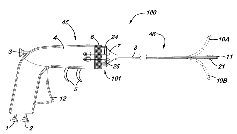
In the aspect of the present invention illustrated in the Figures, device 100
comprises three main portions: a disposable catheter portion, a reusable
handling
portion, and a connecting portion. The reusable handling portion allows an
operator to
hold--either by hand or by using a mechanical apparatus--device 100 and to
control
the device. Thus, any suitable mechanism which serves such functions can be
employed in the present invention. See, for example, U.S. Patent Nos.
5,857,996 and
5,399,164. Preferably, the handling portion 45 illustrated in Figure 1 is
employed in
the present invention. In one aspect of the invention, the handling portion
can be
made disposable instead of reusable.
The disposable catheter portion allows an operator to access, analyze, and
treat
the desired body cavity, such as the epidural space. Thus, any suitable
mechanism
7
CA 02395924 2007-10-12
which serves such a function can be employed in the present invention. See,
for
example, U.S. Patent Nos. 5,857,996, 5,215,105, 4,519,403, and 4,961,738.
Preferably, the catheter portion 46 illustrated in Figure 1 is employed in the
present
invention.
The handling portion and the disposable catheter portion are connected using a
connecting portion, e.g., suitable connection means known in the art. Suitable
connection means are those devices or apparatus which removably connect these
two
portions, yet (as described below) allow communication between these portions.
Preferably, the connection portion 101 illustrated in Figure 1 is employed in
the
present invention. The connecting portion can be part of or separate from
either the
handling portion or the catheter portion.
The material for the handling portion, catheter portion, and connection
portion
(unless specified otherwise) can be made of any suitable medical grade
material.
Examples of suitable medical grade materials include polymeric materials,
plastic
materials, rubber materials, elastomeric materials, silastic, silicone, and
PVC.
Preferably, polyurethane is used in the present invention as this material.
In the aspect of the present invention is illustrated in the Figures, body 45
(handling portion) is connected to catheter 46 (catheter portion) by locking
collar 6
and proximal collar 7 (collectively, the connecting portion). Body 45
comprises
housing 4 which contains at least one control means and at least one
communication
means. The control means allow a user to actuate and control the various
functions of
device 100, including the three functions described above. Thus, any suitable
control
means known in the art can be employed in the present invention. The
conveyance
8
CA 02395924 2002-07-04
WO 01/49356 PCT/US01/00405
means allows the handling portion to convey the instructions from the control
means
in the handling portion to the catheter portion. Thus, any suitable conveyance
means
known in the art can be employed in the present invention.
In one aspect of the invention, a single control means and a single conveyance
means can be employed for all the functions desired of device 100. In a
preferred
aspect of the invention, however, multiple control means and multiple
conveyance
means are used, with each control means and conveyance means controlling a
single
function. Thus, for example, a first control means and a first conveyance
means could
be used for the inflatable function, a second control means and a second
conveyance
means could be used for the visualization (fluoroscopic) function, etc...
For example, a first control means and a first conveyance means are employed
to aid in the inflatable function of device 100. In Figure 7a, trigger 12 is
hinged at 49
and is connected to a pump plunger 47 and a pump 34. Pump 34 can pump any
suitable fluid 9, e.g., a liquid such as saline solutions, water, contrast
agents,
pharmaceuticals, or anesthetics; a gas such as air; a gas containing a solid
such as a
suspension; a liquid containing a solid such as a slurry; or a gas containing
a liquid.
The fluid 9 is pumped from a reservoir (not shown) that is either internal or
external to
body 45. Pump 34 pumps the selected fluid 9 through tube 35 and into catheter
46.
Tube 35 is made of any suitable material which will handle the selected fluid,
such as
plastic. Tube 35 is provided with fitting 48 that will connect tube 35 to pump
34.
Tube 35 is connected at the other end to manifold 51. Since manifold 51 is
attached
to housing 4, manifold 51 serves to anchor tube 35 to housing 4. By actuating
trigger
12, a user is able to pump fluid 9 from the reservoir, through tube 35,
through
9
CA 02395924 2002-07-04
WO 01/49356 PCT/USO1/00405
manifold 51, and to the catheter portion. Fluid 9 will be used, as described
below, to
inflate the inflatable means of device 100.
In a similar manner, additional control means and conveyance means
can be provided for the additional functions desired of device 100 as shown in
Figure
7b. Tube 36 can also be provided for the optical function of device 100. Tube
36 is
provided in housing 4 and is connected at one end to manifold 51 and at the
other end
to viewing port 2, which in one aspect of the invention is a video connection
port.
Tube 37 can also be provided for the optical function of device 100. Tube 37
is
connected at one end to manifold 51 and, at the other end to light source port
1, which
in one aspect of the invention is a light injection port. These tubes, along
with their
associated ports, aid a user to use device 100 to view the body cavity under
inspection.
Other control and conveyance means can be used for the steering
function of device 100 as shown in Figure 7a. Two triggers 5 are connected to
spring
loaded chamber 39 that is connected to housing 4. Spring loaded chamber 39 is
connected to attachment rod 40 that has an attachment point 102 to allow
connection
of a first deflection wire 24 and a second deflection wire 25. The first and
second
deflection wires are not connected to manifold 51, but instead pass through
manifold
51 via ports 27. As described below, these elements of device 100 are used to
aid a
user in steering device 100.
As shown in Figure 7b, Tube 38 can be provided for additional functions, such
as introducing other instruments or injecting other fluids. Tube 38 is
connected at one
end to manifold 51 and, at the other end, to port 3, which in one aspect of
the
invention can serve to introduce fluids, gas, or micro surgical/therapy
instruments. If
WO 01/49356 CA 02395924 2002-07-04 pCT/US01/00405
desired, additional control and conveyance means can be provided for
additional
functions for device 100.
As illustrated in Figure 1, body 45 contains manifold 51, which secures the
conveyance means within handling portion. The control means of the handling
portion are already affixed thereto, so there is no need to secure them The
conveyance
means (tubes and wires), however, are attached to the control means at one end
and
therefore need to be secured to manifold 51 at their other end. Manifold 51 is
also
connected to tubes 35, 36, 37, 38 (and others, if desired) so as to allow
materials
associated with that respective tube to pass through manifold 51 and into the
catheter
portion. Any suitable connection which allows such a transfer can be employed
in the
present invention. One suitable connection is indexing ports 41, 42, 43, 50 as
illustrated in Figures 7c and 7d that are respectively associated with tubes
35, 36, 37,
and 38, and serve as an "end" to the tubes, and allow the materials to pass
through
manifold 51.
As shown in the Figures, connection portion between handling portion
and disposable catheter portion comprises several elements besides the
connection
means which aid in the operation of the device. Connection portion comprises
locking collar 6 and proximal collar 7. These two collars serve to removably
connect
handling portion and catheter portion using any suitable mechanism known in
the art.
For example, proximal collar 7 which has previously been attached to catheter
46-
can be screwed onto body 45 using locking collar 6 and threads 52, thereby
removably
connecting the handling portion with the catheter portion as shown in Figures
7a and
11
CA 02395924 2002-07-04
WO 01/49356 PCTIUSOI/00405
7b. In this process of connection, manifold 51 that is located in body 45
abuts to
manifold 30 that is located in proximal collar 7 as shown in Figure 6a.
As depicted in the Figures, proximal collar 7 contains manifold 30 which
serves a similar function as manifold 51. Manifold 30 also contains indexing
ports
31, 32, 33, and 53 that serve the same functions as the indexing ports 41, 42,
43, and
50, but are merely located in manifold 30 instead of manifold 51. When
attached to
locking collar 6 (which is connected to body 45), manifold 30 abuts manifold
51 with
the indexing ports of manifold 30 matched with the corresponding indexing
ports of
manifold 51.
As illustrated in the Figures, the combination of the two sets of indexing
ports
allows materials in the tubes located in the handling portion to pass into the
catheter
portion. The fluids and light from tubes 35, 36, 37, and 38 pass through the
indexing
ports in manifold 51, through the index ports of manifold 30, and to the
catheter
portion. Manifold 30 is connected to tubes 54, 55, 56, 57 as described below.
Thus,
the materials associated with that respective tube to pass through manifold
51, through
manifold 30, and into matching tubes in the catheter portion.
As shown in Figure 8, connecting portion contains means for aligning the
handling portion and the catheter portion. One suitable aligning means is
indexing lug
44, which makes sure catheter portion is aligned properly with the handling
portion.
Indexing lug 44 also makes sure that manifold 51 is aligned properly with
manifold
30. Indexing lug provides the alignment by indexing with slot 28 in a proximal
collar
7 as shown in Figures 5a and 6a.
12
CA 02395924 2002-07-04
WO 01/49356 PCT/US01/00405
If necessary, sealing means-such as gaskets-can be provided in device 100
where necessary. For example, as shown in Figures 5a and 6a, manifold 30 is
provided with gasket 29 that seals a injection index port 33 from leaking when
fluids
or materials are passed through it. In another example, gasket 154 can be
provided for
port 53 from leaking when fluids or materials are passed through it.
Additional
gaskets for the other indexing ports, and other parts of device 100, can be
added where
necessary.
As shown in Figure 5b and 6b, catheter portion comprises catheter shaft 8
which contains at least one conveyance means. The conveyance means allows the
catheter portion to convey the materials from the handling portion through
manifold
30 to the respective location of the catheter portion where these materials
perform the
desired operation. Thus, any suitable conveyance means known in the art can be
employed in the present invention. In one aspect of the invention, tubes 54,
55, 56, 57
are employed as the conveyance means in the catheter portion (similar to tubes
35, 36,
37, 38 used as the conveyance means in the handling portion and depicted in
Figures
7a and 7b).
As illustrated in Figures 6a and 6b, manifold 30 is connected to tubes 54, 55,
56, and 57 with any suitable connection means known in the art. Any suitable
connection which allows such a transfer can be employed in the present
invention.
One suitable connection means are indexing ports 31, 32, 33, and 53 as
illustrated in
Figure 6a that are respectively associated with tubes 54, 55, 56, and 57, and
serve as
an "end" to the tubes and allow the materials to pass through manifold 30.
13
WO 01/49356 CA 02395924 2002-07-04 PCT/US01/00405
Like the tubes in the handling portion, the tubes in the catheter portion aid
device 100 in carrying out the specified functions. For example, as depicted
in Figure
2, tube 55 is associated with a video port 17 in catheter shaft 8. Tube 56 is
associated
with a light injection port 15 in catheter shaft 8. Tube 54 is associated with
balloon
injection port 13 in catheter shaft 8. Tube 57 is associated with
injection/instrument
port 16 in catheter shaft 8.
Catheter shaft 8 is connected to proximal collar 7 using any suitable means
known in the art that will allow these two components to remain in a fixed
orientation
or alignment. By fixing the alignment between these two components, two holes
27
can be placed a proximal collar 7 that will allow first deflection wire 24 and
second
deflection wire 25 to exit from proximal collar and be connected to attachment
rod 40
in body 45 when the handling portion is connected to the catheter portion.
As depicted in Figure 3, catheter shaft 8 extends about a central axis to
distal
end 11. Shaft 8 may be formed from a material-such as a semi-soft polymer like
a
polyester elastomer-which provides good columnar strength and collapse
resistivity
while allowing some flexibility. The end 11 of a catheter shaft 8 is a soft
clear tip
made of any suitable material. Suitable materials include those which will not
damage or otherwise adversely impact sensitive and delicate internal
structures.
Suitable materials include any of the medical grade materials described above.
The
catheter shaft 8 can be made to fit any desired length or diameter.
Optionally, the tip
of catheter shaft can be tapered (as known in the art) to facilitate
penetration of tissues
during insertion into the body cavity.
14
CA 02395924 2002-07-04
WO 01/49356 PCT/US01/00405
As illustrated in Figure 4, first deflection wire 24 and second deflection
wire
25 run the length of flexible catheter shaft 8 through ports 14 and 18. These
deflection wires are anchored in the soft tip of catheter shaft 8 at positions
22 and 23.
These deflection wires can be made of any suitable material, such as stainless
steel,
and configured so they are strong, yet flexible. When these wires are
contracted, they
"pull" flexible shaft into positions 10a and l Ob, as shown in Figure 1,
because they are
anchored to the tip. The tip can be "pulled" by any either wire at an angle
(relative to
the axis of shaft 8) ranging from 0 degrees to about 180 degrees. Thus, the
tip can
encompass a full 360 degree range of motion.
Catheter shaft 8 also contains balloon injection port 13, as illustrated in
Figure
3. This port runs the length of the shaft and terminates at the flexible
balloon 21 near
the end of catheter shaft 8. This port also encloses and contains tube 54. As
fluid 9 is
pumped from the handling portion, it enters through port 13 and travels the
length of
tube 54 and enters flexible balloon 21. Balloon 21 inflates as additional
amounts of
fluid 9 are pumped into it.
Balloon 21 is attached to catheter shaft 8 at the ends of the balloon at
locations
103 and 104. Thus, when fluid 9 enters the cavity created by balloon, the ends
remain
attached to the shaft while the middle inflates. The ends of balloon 21 can be
attached
to the catheter shaft by any suitable mechanisms or method known in the art,
such as
by thread winding and a bonding agent. In one aspect of the invention, the
ends of the
balloon are attached by RF welding. Balloon 21 can be made of any suitable
material
known in the art, such a s compliant material like latex or silicone rubber.
WO 01/49356 CA 02395924 2002-07-04 PCT/USO1/00405
Balloon 21 is preferably a low pressure, high-volume balloon. The inflated
outer diameter of the balloon is dependent upon the space in which the balloon
is
inflated. Once the balloon reaches a maximum radial dilation, it expands
longitudinally within the epidural space. In this manner, the expansion of the
balloon
is adequate to rupture the fibrosis of the epidural lesion while preventing
damage
within the nerves within the cavity such as the epidural space or damage to
the dura
mater and the spinal column itself. The maximum pressure at which the balloon
may
be inflated is about 250 mm Hg. Balloon inflation time preferably should not
exceed
seconds, while balloon deflation time should not exceed 30 seconds. The volume
10 of balloon 21 when inflated is preferably less than 1 cc.
Tubes 56, 57, and 55 run the length of catheter shaft respectively through
ports
15, 16, and 17. Ports 15, 16, and 17 are open at their respective ends. The
light 19
(for illuminating) enters through tube 56, travels along port 15, and then
exits into the
body cavity. The light 20 (for viewing) is then reflected from the body
cavity, travels
along port 17, and back through tube 55. Instruments or other fluid injections
are
inserted through tube 57, along port 16, and exit into the body cavity.
Device 100 operates in the following manner. The device is steered by using
triggers 5. The force applied to a trigger by a user will pull the desired
attachment rod
which will, in turn, pull on the appropriate wire. The action of pulling the
wire will
cause the distal end 11 of the catheter shaft 8 to deviate from the distal end
at the
desired angle from the shaft axis. Releasing the trigger will then return the
distal end
11 to its position along the shaft axis.
16
CA 02395924 2002-07-04
WO 01/49356 PCT/US01/00405
In one aspect of the invention, additional wires (with the accompanying
elements such as ports in the manifolds, triggers, etc...) can be added for
additional
directions and dimensions of tip deviation. In another aspect of the
invention, the
mechanical means for tip deviation (the wires and associated elements) can be
replaced with non-mechanical (i.e., magnetic or electromagnetic) means for tip
deviation.
A user can view the body cavity using device 100 in the following manner.
Although device 100 is described using fiberoptics, any fluoroscopic means
could be
employed. A light source is attached to port 1 to send light along tube 37.
The light
passes through manifold 51 via port 43, through manifold 30 via port 33, into
tube 56
along port 15, and then exits into the body cavity. The light 20 (for viewing)
is then
reflected from the body cavity, travels along port 17, and back through tube
55,
through manifold 30 via port 32, through manifold 51 via port 42, through tube
36,
and through video connection port 2 where the image is displayed in any
desired
display medium.
A user inflates the inflatable means (balloon) of device 100 in the following
manner. Actuating trigger 12 will cause pump 34 to pump fluid 9 from the
reservoir
and inject the fluid along tube 35, through manifold 51 via port 41, through
manifold
30 via port 31, through tube 54 in port 13, and into balloon 21. The more
fluid 9 that
is injected, the more the balloon is inflated. Once the balloon is inflated to
the desired
level, the trigger 12 is slowly released. The negative pressure caused by the
release
will cause fluid 9 to reverse direction along this path and return to the
reservoir.
17
CA 02395924 2002-07-04
WO 01/49356 PCT/USO1/00405
Instruments or other injections are inserted through port 3, along tube 38,
through manifold 51 via port 50 through manifold 30 via port 53, through tube
57 in
port 16, and into the body cavity. These instruments can be used for various
surgical
procedures as known in the art. Other liquids, such as steroic liquids for
treatment or
radioactive liquids for fluoroscopic analysis, can also be injected in a
similar manner.
Generally, by using device 100 a user can control and manipulate the catheter
46 while simultaneously viewing the body cavity under inspection. Further, a
user can
positionally locate, isolate, and view problem, as well as visually record and
visually
document the problem area. Since catheter 46 is flexible and maneuverable
within
the epidural space, the method also provides less radical interspinal surgical
operations because problem areas can more effectively be observed and accessed
with
the optical and steerable combined functions. The device of the present
invention can
be used for any type of surgery known in the art, including laser, ultrasound,
and
electrocautery surgeries.
Specifically, the devices of the present invention are used to treat
afflictions
within any cavity of the body. In one aspect of the invention, the devices of
the
present invention are employed in methods for treating fibrotic lesions in the
epidural
space of the spinal column. One such method involves inserting a device of the
present invention into the epidural space using the steerable and fiberoptic
functions
of the device. Once located in the epidural space, the fiberoptic and
steerable
mechanisms of the device are used to quickly and efficiently explore and
analyze the
epidural space. Once an affliction is located in the epidural space-such as a
fibrotic
lesion, adhesion, or scar tissue-the inflatable mechanism is used to dilate
the
18
WO 01/49356 CA 02395924 2002-07-04 pCT/USO1/00405
epidural space, decompress adhesions, and remove scar tissue. When a balloon
is
used as the inflatable mechanism, the balloon is positioned across the
fibrotic lesion,
and the balloon is inflated radially and/or longitudinally to sever or disrupt
the fibrosis
comprising the epidural lesion.
To perform a treatment, as known in the art, a needle is first used to access
the
sacral foramen. The ligamentum-flavum, 24 is then pierced and the needle tip
is
inserted in the sacral hiatus or other spinal levels. A guide wire is inserted
and
advanced through the needle and into the epidural space. The needle is
extracted from
the epidural space 110 and discarded. A dialating or introducer sheath can
then be
placed over the guide wire.
The catheter 46 is then inserted through the introducer sheath over the guide
wire and into the opening to the epidural space 110. The guide wire functions
to
guide catheter 46 into the sacral hiatus. Because the catheter 46 is a
steerable catheter,
the body 45 and flexible distal end 11 ease the advancement and positioning of
the
catheter within and around the epidural space. If desired, the position of the
steerable
catheter within the epidural space may also be fluoroscopically observed as
known in
the art. Once in the epidural space, device 100 is advanced into the distended
portion
of the epidural space. The optical function of device 100 illuminates the
distended
portion of the epidural space to thereby visualize and display the epidural
space and a
problem area therein. The problem area is then analyzed. The catheter can be
manipulated to place the distal end 11 into an optimal position, e.g., one
where
balloon 21 could be inflated but without hindering positioning of instruments
or
devices used in surgical procedures.
19
CA 02395924 2002-07-04
WO 01/49356 PCT/USO1/00405
Then, the requisite treatment is performed using the balloon or other
instruments/injections to disrupt a fibrotic lesion, performing a diskectomy,
or other
types of procedures. In one aspect of the invention, fluid 9 is used to
inflate balloon
21. As balloon 21 inflates, it expands radially outwardly, concentrically
about shaft 8,
rupturing and dislodging the fibrosis as it expands. As inflation of the
balloon 21
continues, the outer diameter of the balloon expands toward the walls of the
epidural
space compressing the fibrosis therebetween, exerting a force against the
walls of the
epidural space. As inflation continues further, the pressurized fluid in the
balloon 21
interior finds the path of least resistance, causing the balloon to expand
longitudinally
within the epidural space and parallel to the axis of shaft 8. As balloon 21
expands
longitudinally, the force against the walls of the epidural space is
maintained. In this
manner, the longitudinal expansion of balloon 21 further increases the surface
area of
the balloon which contacts, ruptures and compresses the fibrosis of the
lesion. As
known in the art, the size and shape of the epidural space will affect the
extent of
radial and longitudinal expansion of balloon 21. After balloon 21 has been
inflated
and the fibrosis treated, negative pressure is applied to the interior of the
balloon as
described above, causing deflation of the balloon. The catheter 46 then may be
repositioned to treat an adjacent portion of the same lesion or to treat
another lesion at
a different site. After the dilatation(s) and any other desired medical
procedures have
been completed, the catheter is withdrawn from the epidural space.
The method of the present invention thereby provides improved visualization
of the epidural space and more effective treatment of problems areas therein.
The
method allows the user to effectively observe and document the problem area
and then
CA 02395924 2002-07-04
WO 01/49356 PCT/USO1/00405
determine the most effective treatment for the patient. Since the steerable
catheter is
preferably quite flexible and maneuverable within the epidural space, the
method also
provides less radical interspinal surgical operations because problem areas
can more
effectively be observed and accessed with the optical and steerable
combination.
Having described the preferred embodiments of the present invention, it is
understood that the invention defined by the appended claims is not to be
limited by
particular details set forth in the above description, as many apparent
variations thereof are possible without departing from the spirit or scope
thereof.
21