Note: Descriptions are shown in the official language in which they were submitted.
WO 01/54761 CA 02397697 2002-07-29 PCT/IB01/00267
-1-
ENDOVASCULAR M:=DICAL DEVICE WITH PLURALITY OF WIRES
Description
Technical Field
The present invention relates to the field of medical devices and
more particularly to vascular devices such as catheters and delivery systems
for implantable devices.
Background of the Invention
Catheters for medical diagnostic or therapeutic use are well
known. A catheter has a distal end and a proximal end, with a body
extending therebetween and a lumen extending therethrough from end to
end. A wide variety of catheters exists for percutaneous insertion by the
Seldinger technique into the vascular system to accomplish diagnostic or
therapeutic objectives. The vessels of the peripheral vasculature have a
relatively large diameter and low tortuosity, the coronary vasculature is
somewhat smaller and more tortuous, and the vasculature in the soft tissue
of the brain and liver is of small lumen and is very tortuous.
In order to be able to access the various parts of the vasculature,
the catheter needs to be flexible and to maintain its column strength when
it follows a tortuous path. The contradictory requirements for flexibility and
column strength are particularly pronounced in catheters for intracranial
catheterizations used in a variety of diagnostic and interventional
neurological techniques including delivery of contrast fluids, drugs or a
vasoocclusive agent, treatment of tumors, aneurysms, AVS (arteriovenous
shunts) and so forth.
When a central member is to be moved within a catheter or
sheath to perform an activity at or beyond the distal end of the catheter,
after the catheter has been positioned, the central member is to be pushed
through the catheter lumen. The more tortuous the path and the smaller
the catheter the more difficult it is to advance the central member through
the catheter lumen. This difficulty is in particular pronounced in coaxial
systems for intracranial use. Where the central member is a delivery device
WO 01/54761 CA 02397697 2002-07-29 PCT/IB01/00267
-2-
for an embolization coil and must be rotated to disconnect from the coil upon
release at the treatment site, the central member must be capable of
transmitting torque to its distal end for assured coil disconnection; one such
prior art coil delivery system is disclosed in US Patent No. 5,122,136; but
it is a common problem that such prior art coil delivery members have
relatively high rigidity which is problematic in small or tortuous vessels
with
aneurysms. Where the device is a pusher to push a device such as a stent
from the distal end of the catheter, the pusher must have substantial column
strength as well as great flexibility.
Where a catheter is to be used for delivery of an endovascular
prosthesis to a treatment site, such as a stent, a stent graft, a valve
member, or a filter, where the prosthesis is compressed to pass through the
catheter and then selfexpand upon release therefrom within a body lumen,
the prosthesis must be constrained while within the catheter and imposes
significant forces against the surrounding catheter body.
It is an objective of the present invention to provide a medical
device that includes a distal area that is very flexible and yet easily
pushable
and capable of transferring torque in an assured, controllable manner.
It is another objective to provide a catheter system that makes it
easier to advance the central member through the catheter also in cases
where the catheter exhibits sharp turns.
It is further an objective to provide a catheter that resists the
substantial radially outward forces of a compressed endovascular prosthesis
contained within the distal end thereof, and yet be very flexible and capable
of transferring torque.
It is yet another objective to provide a central member for
movement within a catheter lumen that is very flexible, has substantial
column strength and/or is capable of transferring torque.
Summary of the Invention
The foregoing and other problems are solved and a technical
advance is achieved in an illustrative medical device for passage along the
WO 01/54761 CA 02397697 2002-07-29 PCT/IB01/00267
-3-
vasculature of a patient, having a body portion comprising primarily a
plurality of coils or turns of a plurality of wound filaments or wires. The
medical device may be a catheter or may be one or more components of a
delivery system for endovascular devices, such as a central member within
a catheter, for example, a pusher or delivery device for an embolization coil.
Two to twelve filaments such as wires, and preferably from four to eight
wires, are preferably helically wound adjacent to each other as a group or
row with a pitch corresponding generally to the aggregate width of the
adjacent wires in the row.
The wound wires transfer torque and also force components
directed in the axial direction of the medical device to the distal end
thereof,
and this construction is found to give a very high resistance to kinking of
the
medical device. When a catheter according to the present invention is
heavily bent, the cross-section of the catheter maintains a circular shape.
This provides a distinct advantage over prior art catheters which are
deformed into an oval shape in cross-section when bent leading to kinking.
The catheter surprisingly maintains its capabilities for transferring torque
and
push when it follows a tortuous path involving two or more loops, probably
because of the excellent kinking resistance. These qualities facilitate
placement of the catheter at the desired position in the vascular system, and
by making the catheter system so that the inner surface of the catheter is
mainly undeformable by a central member moving axially therewithin, it is
virtually impossible for the central member to get stuck in the catheter wall,
even in situations where the catheter is heavily curved. This is in contrast
to prior art coaxial systems where the catheter is made of a soft material
such as a resin, the inner surface of which is readily deformable in a local
area, causing the formation of a small bead in front of the tip of the central
member bearing against the wall of the curved catheter. It is an advantage
of the catheter according to the present invention that the wall is primarily
made of wires that provide a hard and relatively slippery inner surface
WO 01/54761 CA 02397697 2002-07-29 PCT/IB01/00267
-4-
resulting in low resistance to advancing the central member through the
lumen of the catheter.
The inventive catheter maintains three valuable characteristics of
very high flexibility, pushability and torqueability even when set in a very
tortuous pattern involving two or more tight loops, and the catheter can thus
be of use in very small and distant vessels such as deep brain sites accessed
by intracranial catheterization. Preferably, a thin sealing coating of
elastic,
low-friction material, or adhesive material may be provided over the
outwardly directed surfaces of the coiled wires or along the inner surfaces
that define a lumen, or at least in recesses between abutting wires or in
interstices between nonabutting turns between the groups of wires, thus
sealing the interstices between the wires so that the catheter wall is
leakproof especially where the device is a catheter or sheath.
Further, wires may have the same diameter in the group and
extend the entire length of the device, or the device may have portions with
wires of different diameters, lessening toward the distal end and thereby
decreasing gradually in outer diameter; the device may also have a noncoiled
part in the proximal region such as a supplementary cannula or tubing.
In the present context, the term "catheter" is to be understood in
the sense that it can be an ordinary catheter, but also a sheath, which is a
short catheter, and in the latter case the central member can be a catheter,
e.g., a catheter according to the present invention. The sheath can have a
check-flow valve or a fitting at the proximal end in order to stop bleeding
out
of the puncture site. In one aspect, the catheter may be utilized without a
guidewire. When intended for use in a soft tissue region, it is preferred that
the distal end of the catheter is provided with a buffer member, such as a
soft obturator, that distributes the force from the catheter tip over a large
area so that damage to the vascular wall is avoided. The term "central
member" can be a member that simply blocks the distal opening of the
catheter during inflation of a balloon for percutaneous transluminal coronary
angioplasty; it may also be an embolization means such as a sack containing
WO 01/54761 CA 02397697 2002-07-29 PCT/IB01/00267
-5-
several occlusion coils, c;r a stent for expansion on a balloon, a sensor body
for measuring pressure or temperature or the composition of blood, a
physical shunt member, a retrieval wire or a forceps used to retrieve another
member from a vascular site; or it can be a central member of some other
kind.
In another aspect, the number of wires may vary along the length
of the catheter, such as reducing the number of wires in the row during the
winding operation in the distal direction, enabling a larger pitch angle and
increasing the flexibility of the catheter proximate to the distal end.
In a second embodiment, the medical device may be a delivery
system for a prosthesis such as a stent, a stent graft, a valve member, or
a filter, wherein the prosthesis is compressible to be placed within a
receptacle at the distal end of the delivery catheter and is then radially
expandable upon delivery to a treatment site after being urged from
receptacle. The delivery system has a catheter shaft with a receptacle that
may be simply a distal end portion of the catheter shaft, but the receptacle
may also be a separate tubular member that extends from the distal end of
the catheter shaft, or optionally partially within the distal end. The
receptacle, whether integral with the catheter shaft or a separate member,
is primarily defined by a group of wires wound about a lumen, thus having
the same advantageous properties of high flexibility and kink resistance as
the catheter shaft; optionally and preferably, when the receptacle is a
separate member, the catheter shaft may also be of the inventive type
hereinbefore set forth. The receptacle may have a larger lumen dimension
than the lumen of the catheter shaft, such as by having a smaller wall
thickness through use of smaller diameter wire or grinding away an
innermost portion of the coiled wires of the distal tip when integral with the
catheter shaft, since the wall thickness required for resisting the outward
pressure from the radially compacted prosthesis is smaller than the wire
thickness required to transmit axial thrust over a long shaft distance, such
WO 01/54761 CA 02397697 2002-07-29 PCT/IB01/00267
-6-
as 80 cm or more, enabling the outer diameter to remain the same as that
of the catheter shaft portion.
In a third embodiment, a prosthesis receptacle is a separate
member and is fixed to the helically wound multiple filament row of wires
of the catheter shaft, in axial extension thereof. This allows the prosthesis
receptacle to be designed and manufactured independently of the shaft
portion. The mounting in direct extension of the wire or wires of the
catheter shaft makes the prosthesis receptacle follow torsional actions on
the shaft portion. Although the prosthesis receptacle can be designed in any
manner capable of resisting the outward pressure applied to the inside of the
receptacle by the compressed prosthesis, it is preferred that the prosthesis
receptacle be a tubular segment of multiple filament construction, such as
a braided wire construction providing the prosthesis receptacle with a high
flexibility. More preferably, the receptacle is a construction of a second
helically wound group or row of multiple wires; this makes it possible to
obtain a very diminutive outer diameter as only a single layer of wires is
required.
In yet another embodiment, the medical device may be a pusher
for use in a delivery system of the type described above, where the pusher
is primarily comprised of multiple wires that are helically coiled, resulting
in
a hollow construction with torqueability and pushability similar to the shaft
portion of the delivery device and with slightly greater flexibility due to
the
smaller outer diameter of the row of wires.
In still another embodiment, the medical device may be used in an
introducer for an embolization device, where the delivery member comprises
primarily a plurality of wires to provide the advantageous torqueability of
the
present invention. The distal end of the delivery member thus is able to be
rotated from rotation of the proximal end thereof, and thus being
disconnectable through unscrewing from the embolization device, a
technique that causes only negligible influence on the vasculature while
enabling precise maintenance of the embolization device in its desired
WO 01/54761 CA 02397697 2002-07-29 PCT/IB01/00267
-7-
position during detachment even in very tortuous paths to treatment sites
such as intracranial locations.
Brief Description of the Drawings
Embodiments of the present invention will now be described by
way of example with reference to the accompanying drawings, in which:
FIG. 1 is a side view of a catheter according to the present
invention;
FIGS. 2 and 3 are enlarged partial views in longitudinal section of
embodiments of the catheter in FIG. 1;
FIG. 4 is a partial view in longitudinal section of an embodiment
where the number of wires in a row varies along the length of the catheter;
FIG. 5 is an enlarged partial and sectional view of the transition
between two catheter segments having wires of different diameter;
FIG. 6 is an enlarged view of an embodiment having a catheter tip
with a buffer member;
FIG. 7 depicts a winding operation on a multiple-wire row;
FIG. 8 depicts a catheter segment having decreasing outer
diameters;
FIG. 9 is an illustration of the catheter of FIG. 1 in position in the
vascular system;
FIG. 10 is an illustration of a device of the present invention used
in a delivery system having a central member that serves as a pusher;
FIGS. 11 and 12 are enlarged views of central members of FIG.
10 being advanced out of the distal end of the catheter;
FIG. 13 is an illustration of a delivery system of the present
invention, for delivery of a prosthesis such as a stent;
FIGS. 14 to 18 are enlarged partial views in longitudinal section
of various embodiments of the delivery system of FIG. 13;
FIG. 19 depicts a partial view of a delivery member of an
embolization device introducer according to the present invention;
WO 01/54761 CA 02397697 2002-07-29 PCT/IB01/00267
-8-
FIG. 20 is a sketch of the introducer of FIG. 19 ready for
disengaging an embolization device;
FIG. 21 is an enlarged illustration of the distal end of the delivery
member of FIG. 20 with an embolization device during placement in a
catheter;
FIGS. 22 and 23 are partial view of the delivery members of other
embodiments of embolization device introducers;
FIG. 24 is an enlarged view of a coil connection means of FIG. 19;
FIGS. 25 and 26 are views of different embodiments of
embolization device introducers providing increased flexibility in the distal
end area of the delivery member; and
FIG. 27 illustrates delivering an embolization device by the
embolization device introducer of FIG. 19.
Detailed Description
In the following description of the depicted embodiments, the
same reference numerals are used for features of the same type. FIGS. 1
to 12 illustrate luminal medical devices such as catheters and sheaths, FIGS.
13 to 18 illustrate prosthesis receptacles and delivery systems therefore;
and FIGS. 19 to 27 illustrate embolization device delivery systems.
A vascular medical device according to the present invention and
illustrated in FIG. 1 is generally denoted 1, and it has a distal end 2, a
body
portion 3 extending from the distal end to a proximal end 4. The body
portion is made of a first helically wound multiple-filament sequence, group
or row of wires 5 and it has a central longitudinally extending lumen 6. The
medical device may be a catheter, and a catheter is normally open ended at
both the proximal and the distal end; but for special uses such as a single
lumen balloon dilatation catheter, the distal end can be provided with means
for barring the distal end opening (see FIG. 6).
For example, a catheter according to the present invention can be
a balloon dilatation catheter used for percutaneous transluminal coronary
WO 01/54761 CA 02397697 2002-07-29 PCT/IB01/00267
-9-
angioplasty, an angiography catheter, a drug delivery catheter, a guiding
catheter, an infusion catheter, and so forth.
The wires 5ised in the helically wound multifilament group or
row are of a linear elastic material, such as stainless steel, titanium or
tantalum, or it is made of a superelastic alloy, such as nitinol. Preferably,
the wires have an ultimate tensile strength in the range of 1800 to 2700
N/mm2 but lower or higher values are also possible. The body portion 3 of
the catheter is made by placing a group of from two to twelve wires of
desired wire diameter in a row next or closely adjacent to each other,
whereafter the group of wires is wound according to the desired pitch angle
in a common movement into the body portion. Because a row of wires is
wound, an individual wire is restricted in movement by the other wires and
is plastically deformed into a permanent helical shape which is kept without
any further restraints other than the remaining wires in the row. The
winding can be done on the inside end of a tubular support member where
the row of wires is inserted at said end by rotating and simultaneously
pushing the wires against the inside of the support. The wound wire then
exits at the other end of the support. This produces a wire body with a very
precise outer diameter.
Alternatively, the winding operation can take place about a
mandrel 7. FIG. 7 depicts a winding of a row A of four identical wires 5.
After the winding the mandrel with the coiled wires can be subjected to heat
treatment in order to remove residual stresses from the wires. As an
example the heat treatment can last for about two hours in an oven at a
temperature of about 500 C. Generally, the temperature can be in the range
of 400 to 600 C and the holding time at the temperature can last for many
hours, such as up to 20 hours or more. After the heat treatment the
mandrel is removed from the wires. The wires in the resulting helically
wound multiple-wire group maintain their mutual position even when heavily
torqued, bent or pushed, presumably because each single wire is supported
by the contiguous wires in the row. The winding operation can be effected
WO 01/54761 CA 02397697 2002-07-29 PCT/1B01/00267
-10-
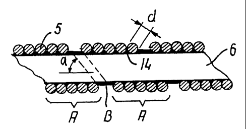
so that the windings are touching each other, but preferably it is performed
so that an interstice B is present between the turns (FIG. 2). The interstice
facilitates bending of the body portion in tight turns along the vasculature
such as is shown in FIG. 9.
The size of the pitch angle a (FIG. 2) depends on the diameter
of the wires, the diameter of the body portion 3 and the number of wires in
the row. The most preferred pitch angle a for the catheter is in the range
of 40 to 68 or 50 to 70 . However, the combination of torque-transferral,
pushability and transverse flexibility is normally well-balanced for pitch
angles in the range of 50 to 68 . The diameter d of the wire is typically in
the range of 0.03 to 0.75 mm, and preferably in the range of 0.15 to 0.45
mm. The present invention includes providing a medical device having
different segments wherein the row of wires is set to different pitch angles,
or wherein different rows of wires have different pitch angles.
In order to make the tip portion of the catheter more visible on a
screen it is desirable to use some kind of radiopaque material, such as
platinum or gold. It can be of annular shape and be located at a
predetermined distance from the distal end 2, or the terminal end of the
distal tip of the catheter can be provided with a marker means for making
it radiopaque, such as a gold layer or a gold thread.
The catheter can be made with a uniform diameter throughout its
length. In case the catheter has a diminishing diameter towards the distal
end, a prefabricated catheter of uniform diameter can be ground to the
desired dimensions.
As an alternative or supplement to grinding, the catheter can be
composed of several segments in which the wires have mutually different
diameters and cross-sectional areas. In a proximal segment 8 the wires can
have a larger diameter than the wires in a distal segment 9. The segments
can be joined together in axial extension by laser welding 10 as depicted in
FIG. 5, by soldering, by bracing or in another manner such as mutual
geometrically locking of the wires in the segments or by mechanical locking,
WO 01/54761 CA 02397697 2002-07-29 PCT/IB01/00267
-11-
such as press-fitting one segment into the lumen of the other segment, or
binding the segments in axial extension with threads or suture.
When the catheter body is of multi-segment construction, the
inner lumens of the segments are preferably of even size which brings the
advantage that an advancing guidewire can not snag or grip onto a step in
the inner wall of the body portion.
In the embodiment illustrated in FIG. 4, the number of wires in
said helically wound group or row of wires varies along the length of the
catheter. During the winding operation the number of wires in the row is
reduced one by one at the points in time where the individual segment
having a certain number of wires has obtained the desired length. The
segment marked "VI" has six wires in the row, and the segments marked
"V", "IV" and "III" have five, four and three wires, respectively, in the row.
Each time a wire is left out of the row, the pitch gets shorter and the pitch
angle grows resulting in an even more flexible consecutive segment. The
advantage of this embodiment is that the wires extending into the distal end
segment are continuous from the distal end to the proximal end of the
catheter, thus avoiding any need for joining the various segments. It is
possible to secure the wire ends of the discontinuous wires onto the other
wires, such as by welding, soldering or the like.
A grinding procedure can also be used to produce one or more
tapered segments 11 in the body portion 3 (FIG. 8). The taper can extend
along a substantial length of the body portion. In the tapered segment the
outer diameter of the catheter diminishes toward the distal end. Due to the
taper, the catheter obtains a gradually increasing transverse flexibility and
a higher softness, but column strength and torque are nevertheless
surprisingly transferred to the distal end.
When the catheter is to be advanced without a guidewire, the
distal end 2 can be provided with a soft buffer 12 , as shown in FIG. 6,
having a rounded distal end which acts gently on the vascular wall when the
catheter is pushed forwardly. A thread 13 can be securely embedded into
WO 01/54761 CA 02397697 2002-07-29 PCT/IB01/00267
-12-
the soft pliable material of buffer 12 and be ensnared around one of the
distal wires, so that the thread will keep the buffer connected to the
catheter body portion when the buffer is pushed out and cleared from the
lumen of the catheter.
Referring now to FIG. 3, the wound wires 5 are provided with a
sealing coating 14 on the inside, or on the outside or on both, surfaces of
the catheter body. The coating is relatively thin and is preferably made of
an elastic material which can be hydrophilic. The coating extends along the
entire length of the catheter and is typically applied after winding and heat
treatment of the catheter body have been completed. As an example, the
coating can be of PTFE applied onto the outside surface of the body portion
in the same manner as such a coating is traditionally applied onto the
exterior of a guidewire. When the coating is to be applied on the external
and the internal surfaces of the body portion the catheter length can be
dipped briefly into a bath of liquid coating material, which is then allowed
to
solidify following removal from the bath.
In case it is desirable to use a hydrophilic coating, the coating can
comprise a hydrophilic polymer selected from the group comprising
polyacrylate, copolymers comprising acrylic acid, polymethacrylate,
polyacrylamide, poly(vinyl alcohol), poly(ethylene oxide), poly(ethylene
imine), carboxymethylcellulose, methylcellulose, poly(acrylamide sulphonic
acid), polyacrlonitril, poly(vinyl pyrrolidone), agar, dextran, dextrin,
carrageenan, xanthan, and guar. The hydrophilic polymers can comprise
ionizable groups such as acid groups, e.g., carboxylic, sulphonic or nitric
groups. The hydrophilic polymers may be cross-linked through a suitable
cross-binding compound. A cross-binder generally comprises two or more
functional groups which provide for the connection of the hydrophilic
polymer chains. The actually-used cross-binder depends on the polymer
system: if the polymer system is polymerized as a free radical
polymerization, a preferred cross-binder comprises 2 or 3 unsaturated double
bonds.
WO 01/54761 CA 02397697 2002-07-29 PCT/IB01/00267
-13-
By making the inventive device primarily of a group or row of two
or more wires, which row is helically wound with a pitch roughly
corresponding to the aggregate width of the adjacent wires in the row, the
wound wires transfer torque and also force components directed in the axial
direction of the catheter to the distal end thereof, and this construction is
found to give a very high resistance to kinking of the device. When the
device is heavily bent the cross-section of the device maintains a circular
shape, and the forces transmitted through the helically wound wires have
less tendency to be concentrated in the area of the bend. This is a distinct
advantage over prior art devices of the type that define a lumen (e.g.,
catheters and sheaths), which are deformed into oval shape when bent, and
thus they are much more prone to kinking. The device surprisingly maintains
its capabilities for transferring torque and push when it follows a tortuous
path involving two or more loops, probably because of the excellent kinking
resistance; and in curved areas the torque and push is mainly transmitted
within the device resulting in a favorably low influence on the vascular
walls.
Due to the very high flexibility, pushability and torqueability and
the ability of the construction of the inventive device to maintain each of
these three characteristics even when set in a very tortuous pattern
involving two or more tight loops, the device can be of use in very small and
distant vessels such as deep brain sites accessed by intracranial
catheterization.
If required, the flexibility of the distal portion of a luminal device
during advancement along a tortuous path, can be further increased by
avoiding the use of a guidewire. The body portion of a catheter, for
example, can be maneuvered to the desired prosthesis deployment site like
a guidewire because it is made of the multiple wire coils so in terms of
maneuverability there is no need for using the catheter in conjunction with
a guidewire. However, a guidewire can be used to diminish the action of the
catheter tip on the vascular wall because the tip will follow the guidewire
when such is advanced in front of the catheter prior to pushing the catheter
WO 01/54761 CA 02397697 2002-07-29 PCT/IB01/00267
-14-
forward. It is an advantage of the catheter according to the present
invention that the wall is primarily made of wires that provide a hard and
relatively low-friction or slippery inner surface resulting in low resistance
to
advancing a member through the lumen of the catheter.
When the catheter is used without a guidewire in a soft tissue
region it is preferred that the distal end of the catheter is provided with a
buffer member, such as a soft obturator. The buffer member distributes the
force from the catheter tip over a large area so that damage to the vascular
wall is avoided.
In one embodiment the group or row of wires is made up of from
2 to 12 helically wound wires, preferably of from 4 to 8 helically wound
wires. By using several wires their aggregate width can be adapted to
correspond to the desired pitch for the given diameter of the device. A row
of more than 12 wires would have a tendency to buckle when the wires are
helically wound in the common winding operation. For wires of round cross-
sectional shape a number of from 4 to 8 wires in the row is preferred, but
for flat wires or wires of oval shape two or three wires in a row can be more
suitable.
In order to promote uniform and well-defined characteristics of the
inventive device along its length the wires in the row can be located closely
next to each other so that the mutually contact each other almost
continuously and support each other. In this manner a possible deflection
of a single wire strand is reduced to a minimum by the others wires in the
row. As the wires in the row are wound into a helical course in a common
movement there can be an interstice between the turns of the row of wires.
The inside surface of an inventive catheter is also more even, which
promotes advancing of a central member axially therewithin. The
capabilities of torque and push are presumably a result of a kind of mutual
interlocking of the individual wire strands in the group or row of wires. If
one wire in the row has a tendency to kink or bend heavily under influence
of the load applied to the delivery member, the other wires in the row keep
WO 01/54761 CA 02397697 2002-07-29 PCT/1BO1/00267
-15-
said wire in place because they are all extending in a common helical course,
which interlocks the wires.
Where the inventive device is a delivery member for an
embolization coil, after advancement of the introducer to the desired
deployment site, a rotational movement at the distal end of the delivery
member is immediately transmitted into an almost identical rotational
movement of the connection means at the distal end (viz., about 1:1 torque
transferral). Such an introducer is particularly useful in association with
the
connection means being designed for detachment by unscrewing from the
embolization device, because the rotation of the delivery member during
unscrewing will cause only negligible influence on the vasculature, and the
embolization device can thus easily be kept exactly at the desired position
during detachment, and furthermore there is obtained a very precise control
of the detachment when, for example, three turns at the proximal end
immediately results in an identical three turn rotation at the distal end of
the
delivery member.
In an embodiment the wires in said row have a pitch angle in the
range of 26 to 76 , preferably a pitch angle in the range of 40 to 65 .
Although it is possible to use other pitch angles, angles chosen in these
ranges provide a balanced solution to the requirements for the desired high
flexibility, high column strength and fine torqueability. The inner range of
40 to 65 is in particular useful for advancing a catheter to very distant,
small sized vessels, such as in blood vessels in the brain, whereas the
subrange of 35 to 40 is applicable when very high flexibility is a dominant
requirement, and the subrange of 70 to 76 is applicable when very high
pushability is a dominant requirement. It is of course possible to choose
different pitch angles in different segments of the device.
At the time of performing the winding operation of the body
portion, the individual wires in the row wound in the helical pattern have
preferably a mainly circular cross-section. This facilitates the winding
operation because twisting of a wire does not result in disorder in the row.
WO 01/54761 CA 02397697 2002-07-29 pCT/1BO1/00267
-16-
The sealing coating is preferably elastic. The wires are to a large
extent mutually locked in position because several wires are wound in a
common movement and thus one wire in the row is kept in place by the
other wires in the row, but nevertheless some mutual movement can occur
between the wires and in particular between the distal wire in one turn and
the proximal wire in the consecutive turn. The sealing coating seals the
interstices between the wires so that the catheter wall is leakproof. The
elasticity of the sealing coating allows the wires to effect small mutual
movements so that the excellent flexibility of the helically wound row of
wires is maintained, and the elasticity also allows the catheter wall to stay
Ieakproof when the wires move. The elasticity is a particular advantage
when the device is pulled back as the pulling action can tend to elongate the
body portion.
It is possible to provide the sealing coating only on the inner
surface of the body portion which will result in a device of a very small wall
thickness relative to its diameter. If a slightly enlarged diameter is
acceptable, the coating can also or as an alternative be placed on the
outside of the body portion. The increase in diameter will be relatively
modest as the sealing coating can be made thin. The sealing coating
provided on the outside of the body portion can, for example, result in no
more than a 5 to 15% increase of the outer diameter of the catheter body.
In an embodiment the sealing coating is a low-friction coating,
such as polytetrafluoroethylene (PTFE) coating. A low-friction coating
applied on the external side of the device wall acts to reduce the forces
required to push forward the device inside a larger guiding catheter or a
sheath, and a low-friction coating applied on the internal side of the
catheter
wall acts to reduce the forces required to push forward a guidewire or
another member such as a pusher member advanced through the device.
In yet another embodiment the sealing coating is a hydrophilic
coating. Such a coating can traditionally be applied to the exterior of a
device for reducing the tendency of the device to stick to the vascular wall,
WO 01/54761 CA 02397697 2002-07-29 PCT/IB01/00267
-17-
but according to the preEent invent on in addition to the lubricating effect
of
the coating it also effects the sealing of the body portion. The sealing
coating is preferably th:n and constitutes only a minor part of the wall
thickness of the body portion. The thickness of the coating at the middle of
the wire can be less than 0.1 mm, and preferably it is less than 0.02 mm.
It is possible to promote the flexibility of the device by machining
the wires in said row to a lesser outer diameter, e.g., by grinding, at a
region
of the device. The region can extend along the whole length of the body
portion, so that it is given a very precise outer dimension by the machining.
In another embodiment the region is a distal region machined to a tapering
shape with decreasing outer diameter in the distal direction causing the
device to have an increasing flexibility towards the distal end which
promotes the introduction into very diminutive vessels. The reduced cross-
sectional area of the wires produced by the machining greatly increases the
bending flexibility of the device without sacrificing its ability to transfer
torque.
Where the device of the present invention is utilized for delivery
of a prosthesis such as a stent, it is preferred that at least in a 30 cm long
distal area the delivery system have a maximum outer diameter of 3.0 mm,
and suitably less than 2.0 mm. As use of a traditional separate sheet for
keeping the prosthesis compressed can be wholly dispensed with because
the prosthesis receptacle is in itself capable of keeping the prosthesis in
the
fully compressed state, the outer diameter of the receptacle and the shaft
portion is identical to the maximum outer diameter of the delivery system
portion introduced into the vascular system. A maximum diameter of 3 mm
in the part of the device advanced through the vascular system allows for
straightforward percutaneous introduction by the Seldinger technique and
easy navigation through the curves in the larger vessels.
It is preferred that for most other forms of the invention, the
device at least in a 30 cm long distal area, have a maximum outer diameter
of less than 2.0 mm. A maximum diameter of less than 1.00 mm allows
WO 01/54761 CA 02397697 2002-07-29 PCT/IBO1/00267
-18-
introduction into quite fine and diminutive vessels such as into the external
and internal carotid arteries. It is further possible to restrict the maximum
outer diameter to at the most 0.75 mm which makes it possible to easily
advance the inventive catheter into, for example, the liver or other soft
tissue areas, and by keeping the maximum outer diameter below 0.30 mm
in a distal end area having a length of at least 10 cm even the most distant
vascular regions are accessible and this embodiment of the catheter is
excellent as a neuro-microcatheter.
When the inventive medical device is to be an embolization device
introducer, it is preferred that at least the distal area have a maximum outer
diameter of 1.0 mm. A maximum diameter of 1.0 mm in the part of the
embolization device introducer advanced through the vascular system allows
for a straightforward percutaneous introduction by the Seldinger technique
and easy navigation through the curves in the larger vessels. Coils having
the relatively large diameters in the range of 0.7 to 1.0 mm are suitable for
embolization in larger vessels, and in particular at locations where the blood
flow rate is high, e.g., due to a malformation or trauma. A maximum
diameter of 1.00 mm allows introduction into quite fine and diminutive
vessels such as into the external and internal carotid arteries.
In a further embodiment the number of wires in said helically
wound group or row of wires varies along the length of the device. This can
be attained by reducing, during the winding operation, the number of wires
in the row. The lower number of wires in the row can be utilized to wind
the wires with a larger pitch angle which increases the flexibility of the
device. It is preferred that the number of wires diminishes in the distal
direction so that the softness of the device increases without any change of
material and without bonding together several separate device segments.
When the device has to traverse large lumen vascular paths in
order to reach the more difficult small size vascular vessels, the helically
wound row of wires can be stiffened in a proximal segment of said body
portion by a supplementary tubular member, such as a cannula tubing.
WO 01/54761 CA 02397697 2002-07-29 PCT/IB01/00267
-19-
In the following, some examples of catheters are described that
are made according to the invention.
EXAMPLE 1:
A catheter was made of a helically wound row of four wires of
0.35 mm wire diameter. The body portion of wound wires had initially an
outside diameter of 1.67 mm and an inner lumen of 0.97 mm. A coating of
PTFE of a minimum thickness of 0. 1 mm was applied onto the inside of the
catheter. The catheter was set in a complex curved shape involving three
consecutive loops of a loop diameter of 24 mm axially separated by two
loops of a loop diameter of 18 mm and a number of further turns
representative of a complex vascular structure. Then the body portion of
the catheter was manipulated and it proved to be easily pushed forward and
retracted as well as easily torqued. Then a guidewire was pushed forwardly
in relation to the body portion, and it proved to be easily pushed out past
the
distal end of the catheter without causing noticeable flexion or movement
of the catheter.
EXAMPLE 2:
A catheter was made of a helically wound row of five wires of
0.30 mm wire diameter. The winding of a first segment of the body portion
was made with an outside diameter of 1.20 mm and an inner lumen of 0.6
mm. Another segment was made up of a second helically wound row of
four wires of 0.15 mm wire diameter. This segment had a length of 20 cm
and an outside diameter of 1.20 mm and an inside diameter of 0.9 mm. The
segments were joined by laser welding. The catheter body was provided
with a flexible coating on its outside. The catheter was advanced through
a complex curved vascular system involving several consecutive retrograde
turns in vessels having a lumen of only 2 mm and less. Then the catheter
was torqued and moved both forwardly and backwardly without any
problems.
WO 01/54761 CA 02397697 2002-07-29 PCT/IB01/00267
-20-
EXAMPLE 3:
A catheter was made of a first helically wound row of eight wires
of 0.075 mm wire diameter. The winding was made with an outside
diameter of 0.25 mm and an inner lumen of 0.1 mm. The body portion had
a length of 160 cm and was coated with a hydrophilic material of
polyacrylamide on its outside surface. When tested the catheter shows no
problems. After placing the catheter in a very complex pattern involving
several sharp turns (see an example in FIG. 9), a guidewire could be
advanced with only very low friction, and after removal of the guidewire, a
fluid could be injected through the catheter without leakage through the
coating.
When the catheter is to be introduced into the vascular system
there is firstly established a percutaneous puncture site, e.g., by the
Seldinger technique, or an existing puncture site is used. Then the body or
shaft portion of the catheter is inserted through the cannula, sheath or
hemostatic valve at the puncture site and the catheter is advanced and
navigated through the vascular system to the treatment site or the
prosthesis deployment site. Due to the very high flexibilility, pushability
and
torqueability of the catheter it can be advanced to the site without use of a
guidewire, or a sheath to negotiate the sharp curves in the path. When large
lumen vessels are to be traversed in order to enter the vasculature near the
target site, it can be an advantage to stiffen the proximal portion of the
catheter by inserting it through a cannula 14 (FIG. 3), a tubing or another
kind of a more rigid structure.
The catheter according to the invention can be used as a
traditional catheter, and it can also be used as a sheath which has normally
a shorter length than a traditional catheter.
Individual features of the various embodiments can be combined
into further embodiments according to the present invention. It is possible
to effect the sealing coating as a multilayer coating, e.g., comprising a
primer-coating and a top-coat where the primer-coating is chosen to provide
WO 01/54761 CA 02397697 2002-07-29 PCT/IB01/00267
-21-
a strong bonding to the vjires, and the top-coat provides the sealing action
and can be a hydrophilic slippery coating providing a low friction surface.
A catheter system is illustrated in FIG. 10 to include a central
member 100 such as a pusher, and a catheter 1 having a distal end 2 and
a body portion 3 extending from the distal end to a proximal end 4, the
catheter being similar to catheter 1 of FIG. 1. The central member may be
used to block the distal opening during inflation of a balloon of a balloon
dilatation catheter for percutaneous transluminal coronary angioplasty. The
catheter system can also be for placing the central member in the vascular
system. To give some examples, the center member can include (or can be)
an embolization means in the form of a sack 102 containing several
occlusion coils, as shown in FIG. 11. It also can be a stent for expansion on
a balloon, or it can be a sensor body for measuring pressure or temperature
or the composition of blood, or it can be a physical shunt member. It also
can be or include a retrieval wire or a forceps 104, as shown in FIG. 12 used
to retrieve another member from a vascular site, or it can be a central
member of some other kind.
Following are three examples of catheter systems made according
to the invention.
EXAMPLE 4:
A catheter was made in accordance with the catheter of Example
1 and deployed in the complex vascular structure described therein. Then
a bag 102 with four occlusion coils was pushed forward by the pusher 100
(FIG. 10) until it discharged through the opening at the distal end 2, as
shown in FIG. 11. There was no noticeable sticking of the bag 102 against
the inside surface of the catheter.
EXAMPLE 5:
A catheter was made in accordance with the catheter of Example
2 and provided with a PTFE coating on its outside surface. The catheter
WO 01/54761 CA 02397697 2002-07-29 PCT/IB01/00267
-22-
was advanced through a complex curved vascular system involving several
consecutive, retrograde turns in vessels having a lumen of only 2 mm and
less. Then a pair of forceps 104 was advanced through the catheter as
shown in FIG. 12, and activated to grab the desired item, such as a kidney
stone, and retracted through the catheter lumen.
EXAMPLE 6:
A catheter was made having the wire structure and dimensions
of the catheter in Example 3. The body portion was uncoated, and when
tested the catheter showed no problems. After placing the catheter in a
very complex pattern involving several sharp turns (see an example in FIG.
9) a guidewire could be advanced with only very low friction, and after
removal of the guidewire, central members in the form of fluid injected
embolization coils were delivered through the catheter.
Shown in FIGS. 13 to 18 is a delivery system according to the
present invention, for use in the delivery of a prosthesis to a treatment site
in the vasculature. The prosthesis may be of the radially compressible, self-
expandable type such as a stent, a stent graft, a valve member or a filter,
and may be formed of shape memory alloy. When the delivery system has
been maneuvered to the desired location, the prosthesis is discharged by
application of a pushing force against the proximal end of the prosthesis
relative to the delivery system by means of a pusher member; alternatively,
the prosthesis may be discharged by being held by a trigger wire against
proximal movement as the surrounding catheter or sheath is pulled
proximally.
Delivery system 200 in FIG. 13 includes a delivery device 202
having a distal end 204 and a shaft portion 206 extending between a
prosthesis receptacle 208 at the distal end and a proximal mounting member
210 fixedly mounted to the shaft portion. The shaft portion is made of a
first helically wound multiple filament row of wires 212 and it has a central
longitudinally extending lumen 214.
WO 01/54761 CA 02397697 2002-07-29 PCT/IB01/00267
-23-
The delivery system 200 further comprises a pusher member 216
which can be inserted through the lumen 214. A handle or pin vise 218 is
mounted on the pusher member for pushing it forwardly in the distal
direction when a prosthesis 220 located in receptacle 208 is to be released
from the introducer device by being pushed out of receptacle 208. Pin vise
218 and mounting member 210 can be parts of a unitary control device to
be manually actuated when the prosthesis has been introduced and
positioned at the desired vascular site.
At the distal end of the pusher member 216 an engagement
means 222 can act on the prosthesis 220. The engagement means can be
for example a plate of a dimension fitting into receptacle 208 and abutting
the proximal end of the prosthesis so that the plate pushes the prosthesis
out of the receptacle when the pusher member is pushed forwardly. The
engagement means can also be designed as an elongate member that
extends coaxially inside the radially compressed prosthesis and engages the
prosthesis at several locations along the length thereof so that the
prostheses is partly pulled, partly pushed out of the receptacle. These
engagement points or areas can be effected by radial projections, hooks,
ridges, or another kind of engagement means such as a high friction
material. This can be an advantage if the prosthesis has an extensive
length, and in particular if it has a construction having a tendency to buckle
when pushed upon.
By the term "prosthesis receptacle" is meant any structure or
region near or at the distal end of a delivery device where a radially
compressible tubular prosthesis is carried during maneuvering of the delivery
device and prosthesis within a body lumen. The prosthesis receptacle 208
can be made of a length of tubular material that is flexible in itself or is
made
flexible by incisions or due to its construction, such as a construction of
wound or braided wires. If the prosthesis is rather short in length or is for
deployment in a large sized vessel of a rather straight shape, such as in the
WO 01/54761 CA 02397697 2002-07-29 pCT/IB01/00267
-24-
aorta, the receptacle need not be flexible and can be made out of a stiff
tubular member.
The length of the prosthesis receptacle 208 is at least of the same
size as the length of the loaded prosthesis 220. However, other lengths are
also possible. As depicted in FIG. 18, the receptacle 208can have a length
that is considerably longer than the loaded prosthesis 220, so that the
prosthesis can be loaded into a position at the proximal end of the receptacle
leaving empty a distal length of the receptacle. This free distal length will
not be stiffened by the presence of the loaded prosthesis and will
consequently be very soft and flexible. The length can for example by
chosen so that the free distal length is in the range of from 5 to 150 mm,
preferably in the range of 10 to 50 mm.
In a preferred embodiment, the prosthesis receptacle 208 is made
of a second helically wound multiple filament row of wires 224. As depicted
in FIG. 14, the second row of wires 224 can be made independently of the
first row of wires 212 and in different dimensions or different materials than
the first row of wires, and the receptacle 208 is then fixed in axial
extension
of the first row of wires, e.g., by laser welding, soldering bracing, or
mechanical locking such as press-fitting into the lumen of the shaft portion,
or binding with threads or suture. An alternative embodiment is depicted in
FIGS. 15 and 16 where prosthesis receptacle 208 is made integral with
shaft portion 206 by using a distal segment 226 of said first row of wires
212 as the receptacle.
In the embodiment of FIG. 15, the inner lumens in the shaft
portion 206 and in receptacle 208 are of even size which brings the
advantages of being able to load prostheses of various lengths in one and
the same delivery system and of being able to lead from the proximal end of
the delivery device a pusher member having a solid engagement means 222
of a diameter that is only slightly less than the diameter of the inner lumen
214.
WO 01/54761 CA 02397697 2002-07-29 PCT/IB01/00267
-25-
In the embodiment of FI 3. 16, the pusher member 216 is inserted
from the distal end of the shaft portion prior to leading the prosthesis 220
into receptacle 208. This ailows the engagement means 222 to be of a
larger diameter than the Iumen 214 of shaft portion 206.
In the embodiment of FIG. 18, the radially compacted prosthesis
220 projects radially inwards beyond the step in inner lumen diameter at the
transition between receptacle 208 and shaft portion 206. Consequently, it
is possible to use a pusher member 216 having an engagement member 222
of less diameter than lumen 214 and yet push the prosthesis out of
receptacle 208 by its pressing against the proximal end of the prosthesis.
The shaft of the pusher member 216 can be of a small diameter
solid wire or rid as depicted in FIG. 15 or it can be made of a third
helically
wound multiple filament row of wires 228 as depicted in FIG. 14. The
receptacle 208 in the embodiment of FIG. 16 is made by machining the
inside of the wound wires 226 to a larger lumen. This can for example by
done by spark erosion or grinding. In the latter case, the distal end portion
of the wound wires are placed in a retaining ring (not shown) that is
longitudinally displaceable with respect to a coaxially mounted grinding
wheel.
A grinding procedure can also be used to produce a tapered
section 230 in shaft portion 206 (seen in FIG. 17). The taper can extend
along a substantial length of the shaft portion. In the tapered section the
outer diameter of the delivery device 202 diminishes to diameter D2 . Due
to the taper the delivery device obtains a gradually increasing transverse
flexibility and a higher softness, but torque is nevertheless surprisingly
transferred fully to the receptacle 208. As an alternative or supplement to
grinding, the shaft portion 206 can be composed of several portions in
which the wires of each portion have mutually different diameters and cross-
sectional areas.
Preferably, the distal tip of the delivery system is provided with
marker means 230 for making it radiopaque, e.g., by a gold or platinum
WO 01/54761 CA 02397697 2002-07-29 PCT/IB01/00267
-26-
plating, or by soldering, brazing or laser welding a radiopaque member onto
the distal tip (FIG. 17). The marker 230 promotes precise positioning of the
prosthesis at a treatment site in the vasculature.
For some applications it is desirable to deploy a prosthesis that
has been provided with an active substance, such as a cell growth inhibitor.
The active substance can have such a short shelf life that it needs to be
applied to the prosthesis immediately prior to deploying the prosthesis. This
can be done by dipping the distal end of the delivery device, viz., the
prosthesis in the receptacle, into a fluid of active substance.
Following are some examples of delivery systems made according
to the invention:
EXAMPLE 7:
A delivery device was made of a first helically wound row of four
wires of .35 mm wire diameter. The shaft of wound wires had initially an
outside diameter of 1.67 mm and an inner lumen of 0.97 mm. The
receptacle was made up of a second helically wound row of four wires of
0.20 mm wire diameter. The receptacle had a length of 37 mm and initially
an outside diameter of 1.70 mm and an inside diameter of 1.3 mm. A
radially compressed stent was arranged inside the receptacle. The loaded
stent had a length of 35 mm and was recessed a little in relation to the
distal end of the receptacle. The pusher member was made of a third
helically wound row of four wires of 0.28 mm wire diameter and a shaft
outer diameter of 0.91 mm. A plunger element or an engagement member
was located on the distal end of the shaft. The shaft and the receptacle of
the delivery device was ground to a common outer diameter of 1.5 mm (4.5
French). In its fully self-expanded state the stent had an outer diameter of
8 mm. The delivery device was set in a complex curved shape involving
three consecutive loops of a loop diameter of 20 mm axially separated by
two loops of a loop diameter of 15 mm and a number of further turns
representative of a complex vascular structure. Then the shaft of the
WO 01/54761 CA 02397697 2002-07-29 pCT/IB01/00267
-27-
delivery device was manipulated and it proved to be easily pushed forwardly
and retracted as well as easily torqued. Then the pusher member was
pushed forwardly in relation to the shaft portion, and the stent was easily
pushed out of the receptacle without causing noticeable flexion or
movement of the delivery device.
EXAMPLE 8:
A delivery device was made of a first helically wound row of five
wires of 0.30 mm wire diameter. The winding of the shaft was made with
an outside diameter of 1.20 mm and an inner lumen of 0.6 mm. The
receptacle was made up of a second helically wound row of four wires of
0.15 mm wire diameter. The receptacle had a length of 60 mm and an
outside diameter of 1.20 mm and an inside diameter of 0.9 mm. A radially
compressed prosthesis was arranged inside the receptacle. The loaded
prosthesis had a length of 20 mm and was positioned at the proximal end
of the receptacle with a 40 mm very soft free distal receptacle end. The
pusher member was made of a single 0.35 mm diameter wire rod that
carried an engagement member at its distal end. In its fully self-expanded
state the prosthesis had an outer diameter of 3 mm. The delivery device
was advanced through a complex curved vascular system involving several
consecutive, retrograde turns in vessels having a lumen of only 2 mm or
less. Then the pusher member was pushed forwardly in relation to the shaft
portion, and the stent was easily pushed out of the receptacle in a well-
controlled manner.
EXAMPLE 9:
A combined receptacle and distal shaft segment of a delivery
device was made of a first helically wound row of eight wires of 0.075 mm
wire diameter. The winding was made with an outside diameter of 0.25 mm
and an inner lumen of 0.1 mm. The combined receptacle and distal shaft
segment had a length of 12 cm. A prosthesis was compressed radially to
WO 01/54761 CA 02397697 2002-07-29 pCT/IB01/00267
-28-
an outer diameter of 0.07 mm and was pushed into the receptacle. The
loaded prosthesis had a length of 10 mm and was positioned in the
receptacle with its proximal end 25 mm from the distal receptacle end. The
pusher member was made of a single 0.08 mm diameter solid wire rod. The
pusher member was used to push the stent out of the receptacle.
Shown in FIGS. 19 to 27 is a delivery system for an embolization
coil, made according to the present invention. A delivery system 300 has
a length in the range of 50 to 250 cm and a diameter in the range of 0.08
to 2.0 mm, depending on the relevant field of application. The delivery
system utilizes a delivery member 302 within an introducer 304, and in a
distal section 306 the delivery member has a connection means 308 for an
embolization device 310.
The delivery system may utilize any of a number of kinds of
connection means 308, among which are: an electrolytically erodable
means, a heat erodable means, a latch, a coupling, a threading coil, a thread,
a deflatable balloon, and a hydraulically or pneumatically activated gripper
means. As shown in FIG. 19, the delivery member 302 preferably has in its
distal section 306 a central core 312 with a blade-shaped portion, and the
connection means 308 is a threading coil 314 which is fixed to the central
core at least at the edges of the blade-shaped portion. The blade-shaped
portion carrying the threading coil is much more flexible and easy to bend in
the thickness direction of the blade than in the direction of the width where
the blade dimension is the largest. The blade-shaped portion is a distal end
portion 316 of the central core 312 and if it is subjected to a torque, the
central core twists. When the delivery wire is advanced and has to pass
through a curvature, the blade-shaped portion touches the inner wall of the
lumen and is subjected to a torque until the blade-shaped portion has turned
itself with the direction of width transverse to the curvature. The result is
that the bending occurs in the thickness direction which is most flexible.
The fixation of the threading coil 314 at the edges provides control of the
WO 01/54761 CA 02397697 2002-07-29 PCT/IB01/00267
-29-
positioning of the threa&; so that the unthreading of the embolization device
310 is very smooth-runring.
The connecticn means 308 can be made of radiopaque material
in order to be discerned on an image screen by the radiologist or
neuroradiologist that introduces the detachable embolization device 310 into
the vascular system of a patient, but in order to be seen clearly the
radiopaque area ought to have relatively large dimensions. This can be
obtained by positioning a radiopaque marker at a predefined first distance,
such as about 3 to 3.5 cm, proximal to the distal termination of the
connection means 308. In this embodiment, the connection means in itself
need not be radiopaque, because the marker is clearly seen and the
radiologist is aware that the embolization device is positioned said first
distance ahead of the marker.
In the following description of several embodiments, the same
numerals are used to denote features of the same kind. In one embodiment
the connection means 308 comprises a central core 312 of stainless steel,
nitinol, or another suitable material and a threading coil 314. The central
core 312 has at its distal end section 316 a blade-shaped portion with a
blade thickness and a blade width, which is more than twice as large as the
blade thickness. The threading coil 312 is fixed onto the blade-shaped
portion, e.g., by soldering, welding, brazing or gluing at joints 346 (as seen
in FIG. 23). The threading coil wire can be of stainless steel and can have
a wire diameter in the range of 0.02 to 0.12 mm, typically a diameter of
about 0.06 to 0.075 mm. The wire is set with a pitch corresponding to or
being larger than twice the thickness of the wire so that a mating threading
in the proximal end of the detachable embolization device 310 can be
threaded into and out of threading coil 314, as shown in FIG. 24. The outer
diameter of the threading coil can, for superselective use, be in the range of
0.08 to 1.0 mm, and typically from 0.20 to 0.45 mm.
Other embodiments of the connection means 308 include a
connecting area, which is eroded away by applying current or head when
WO 01/54761 CA 02397697 2002-07-29 PCT/IB01/00267
-30-
the embolization device 310 is positioned at the desired site, or a latch or a
coupling providing a geometrical locking, such as a bayonet coupling, two
mating parts held together by a thread that can be pulled out for detachment
of the embolization device, or a deflatable balloon positioned inside a
tubular
proximal end area of the embolization device 310. Other embodiments of
threads can also be used, such as spaced ball-like enlargements on the
central member, a helix-shaped groove cut into a cylindrical or conical distal
end part on delivery member 302. These kinds of connection means are
well known in the art, e.g., from EP-A-0 720 838; US Patent No.
5,217,484; WO 94/06503; WO 94/06502; WO 94/00104; and EP-A-0 717
969. In FIG. 23 such a connection means 308 is shown in a general
manner, and an activation member 318 is shown to extend inside the
delivery member 302. The activation member can be, for example, the
above mentioned thread to be pulled out, an optical fiber, an electrical wire,
and so forth.
The embolization device 310 can be a Gianturco stainless steel
coil of traditional design, or coils with a regular helical shape or irregular
coil
shape as described in US Patent No. 4,994,069; US Patent No. 5,122,136;
WO 93/06883; WO 94/11051; WO 94/07560; WO 94/10936; WO
95/25480; DE-295 18 932-U 1; WO 96/18343; EP 0 623 012 or the
embolization device can be a random matrix shape as described in US Patent
No. 4,994,069 and WO 94/09705. The embolization device can also be of
a regular linear shape as described in WO 98/09570, which is hereby
incorporated into the present description by reference. The embolization
device can also be called an occlusion device.
Referring now to FIGS. 20 and 21, placement of the embolization
device 310 in an aneurysm 320 will be described. A catheter is introduced
percutaneously through a fitting 322 by the Seldinger technique and
advanced transluminally in a well known manner along a suitable path until
the distal end of the catheter is located in the neck of the aneurysm 320.
Then an introducer 304 with the embolization device 310 mounted on the
WO 01/54761 CA 02397697 2002-07-29 pCTRB01/00267
-31-
delivery member 302 is inserted into the catheter and pushed forwardly until
the embolization device is pushed out of the catheter and is in the desired
deployment position in aneurysm. So positioned, the delivery member
extends along a complexly curved path. Then the embolization device is
released from connection means 308. This can be done, for example, by
activating member 318 or by rotating the proximal end of the delivery
member with the aid of a pin vise 324 which is fixed onto a proximal section
326 of delivery member 302.
Referring now to FIGS. 22 to 27, wires 330 are wound by a
winding operation in a manner such as that described with respect to FIG.
2. The winding operation can be effected so that the windings are touching
each other, but preferably it is performed so that a slight interstice B is
present between the turns (FIG. 23). The interstices facilitate bending of the
body portion in tight turns of the vasculature (FIG. 20). The size of the
pitch
angle depends on the diameter of the wires, the diameter of the delivery
member 302 and the number of wires in the sequence, group or row. The
most preferred pitch angle for the delivery member is in the range of 40 to
65 . However, the combination of torque-transferral, pushability and
transverse flexibility is normally well-balanced for pitch angles in the range
of 50 to 68 . The diameter of the wire is typically in the range of 0.03 to
0.75 mm, and preferably in the range of 0.15 to 0.45 mm.
In order to make the tip portion of the delivery member more
visible on a screen, it is desirable to use some kind of radiopaque marker
332 or radiopaque material, such as platinum or gold. It can be of annular
shape and be located at a predetermined distance c from the distal end
334, as shown in FIG. 22. The marker can be of platinum wire inserted into
delivery member 302 in distal extension of wires 330, or it can be a
separate member such as a platinum or gold ring. A catheter 336 used
when advancing the introducer 304 can also have a radiopaque marker 338
located at such a distance from the distal end 340 of the catheter that the
WO 01/54761 CA 02397697 2002-07-29 pCT/IB01/00267
-32-
embolization device 310 is in position for release when the marker 332 has
been advanced to be positioned at marker 338.
In the embodiment illustrated in FIG. 23 the number of wires 330
in portions of the length of the delivery member 302 varies along the length.
During the winding operation the number of wires in the group is reduced
one by one at the points where individual portions having a constant number
of wires have obtained their desired lengths. The segments marked V, IV
and III have five, four and three wires, respectively, in the group. Each time
a wire is left out of the group, the pitch gets shorter and the pitch angle
grows resulting in an even more flexible consecutive segment. The
advantage of this embodiment is that the wires extending into the distal end
segment are continuous from the distal end to the proximal end of the
delivery member, thus avoiding any need for joining the various portions. It
is possible to secure the thread ends of the discontinuous wires onto the
other wires, such as by welding, soldering and so forth.
The delivery member can be made with uniform diameter
throughout its length. Incase the delivery member is to have diminishing
diameter toward the distal end, a prefabricated delivery member of uniform
diameter and be ground to the desired dimensions. As an alternative or
supplement to grinding, the delivery member can be composed of several
segments in which the wires have mutually different diameters and cross-
sectional areas, as described with respect to FIG. 5.
As illustrated in FIGS. 25 and 26, a grinding procedure can also
be used to produce one or more tapered segments 340,342 in delivery
member 302. The taper can extend along a substantial length of the
delivery member to produce a gradually increasing flexibility. In the tapered
segments, the outer diameter of the delivery member 302 diminishes toward
the distal end 334. Due to the taper or tapers, the delivery member obtains
a gradually increasing transverse flexibility and a higher softness, but
column
strength and torque are nevertheless surprisingly transferred to the distal
end.
WO 01/54761 CA 02397697 2002-07-29 PCT/IB01/00267
-33-
In the embodirnent of FIG. 22, the wound wires 330 are provided
with a low-friction coatirig 344 on the radially outwardly facing surface of
delivery member 302. The coating is relatively thin and is preferably made
of an elastic material which can by hydrophilic. The coating extends along
part of or along the entire length of the delivery member and is typically
applied after winding and heat treatment of the delivery member have been
completed. As an example, the coating can be of PTFE applied onto the
outside of the body portion in a traditional manner.
The helically wound row of wires in the delivery member makes
it possible to manufacture the connection means as an integral part of the
delivery member. This can be done by removing one or several of the wires
in the distal end portion of the delivery member. The wires are very
diminutive so that they can be cut, for example, by a laser beam or manually
with a tool under a microscope. If required, a thread cutter tool or a thread
shaping tool can be used to set the remaining wire or wires with the desired
pitch corresponding to the pitch on the mating coupling member on the
embolization device. The resulting unitary delivery member has in its distal
end only the wires which extend toward the proximal end.
Following are several examples of delivery members made
according to the present invention:
EXAMPLE 10:
A delivery member was made of a helically wound row of four
wires of 0.30 mm wire diameter. The delivery member had initially an
outside diameter of 0.90 mm. The delivery member was set in a complex
curved shape involving three consecutive loops of a loop diameter of 24 mm
axially separated by two loops of a loop diameter of 18 mm and a number
of further turns representative of a complex vascular structure. Then the
proximal section of the delivery member was manipulated and it proved to
be easily pushed forward and retracted as well as easily torqued.
WO 01/54761 CA 02397697 2002-07-29 pCT/IBOl/00267
-34-
EXAMPLE 11:
A delivery member was made of a helically wound row of five
wires of 0.25 wire diameter. The winding of a first segment of the delivery
member was made with an outside diameter of 0.80 mm. Another segment
was made up of a second helically wound row of four wires of 0.1 5 mm
wire diameter. This segment had a length of 20 cm and an outside diameter
of 0.45 mm. The segments were joined by laser welding. The delivery
member was provided with a coating on its outside surface. The delivery
member was advanced through a complex curved vascular system involving
several consecutive, retrograde turns in vessels having a lumen of only 2
mm and less. Then the delivery member was torqued and moved both
forwardly and backwardly without any problems.
EXAMPLE 12:
A delivery member was made of a first helically wound row of
eight wires of 0.075 mm wire diameter. The winding was made with an
outside diameter of 0.25 mm. The delivery member had a length of 160
cm. When tested, the delivery member showed no problems. After placing
the delivery member in a very complex pattern involving several sharp turns,
the distal end could be rotated in a 1:1 relationship with a rotation of the
proximal end of the delivery member.