Note: Descriptions are shown in the official language in which they were submitted.
CA 02398719 2002-07-29
WO 01/54614 PCT/USO1/01504
-1-
SELF-E~ANDING STENT WITH EN~IANCED
DELIVERY PRECISION AND STENT DELIVERY SYSTEM
BACKGROUND OF THE INVENTION
This application is a continuation-in-part of application Serial No.
09/313,780 filed on May 17, 1999, which is assigned to the same Assignee as
the
present application.
The present invention relates to expandable endoprosthesis devices,
generally called stems, which are adapted to be implanted into a patient's
body
lumen, such as a blood vessel, to maintain the patency thereof, along with
systems
for delivering and deploying such stems. Stems are particularly useful in the
treatment and repair of blood vessels after a stenosis has been compressed by
percutaneous transluminal coronary angioplasty (PTCA), percutaneous
transluminal
angioplasty (PTA), or removed .by atherectomy or other means, to help improve
the results of the procedure and reduce the possibility of restenosis.
Steuts axe generally cylindrically shaped devices which function to
hold open and sometimes expand a segment of a blood vessel or other arterial
lumen, such as a coronary artery. Stems are usually delivered in a compressed
condition to the target site and then deployed at that location into an
expanded
condition to support the vessel and help maintain it in an open position. They
are
particularly suitable for use to support and hold back a dissected arterial
lining
which can occlude the fluid passageway there through.
A variety of devices are known in the art for use as stems and have
included coiled wires in a variety of patterns that are expanded after being
placed
intraluminally on a balloon catheter; helically wound coiled springs
manufactured
from an expandable heat sensitive metal; and self expanding stems inserted
into a
compressed state for deployment into a body lumen. One of the difficulties
encountered in using prior art stems involve maintaining the radial rigidity
needed
to hold open a body lumen while at the same time maintaining the longitudinal
CA 02398719 2002-07-29
WO 01/54614 PCT/USO1/01504
-2-
flexibility of the stmt to facilitate its delivery and accommodate the often
tortuous
path of the body lumen. '
Prior art stems typically fall into two general categories of
construction. The first type of stmt is expandable upon application of a
controlled
force, often through the inflation of the balloon portion of a dilatation
catheter
which, upon inflation of the balloon or other expansion means, expands the
compressed stmt to a larger diameter to be left in place within the artery at
the
target site. The second type of stmt is a self expanding stmt formed from, for
example, shape memory metals or super-elastic nickel-titanum (NiTi) alloys,
which
will automatically expand from a compressed state when the stmt is advanced
out
of the distal end of the delivery catheter into the blood vessel. Such stems
manufactured from expandable heat sensitive materials allow for phase
transformations of the matehial to occur, resulting in the expansion and
contraction
of the stmt. Other stems include those made with a branded configuration that
does not go through plastic deformation.
Details of prior art expandable stents can be found in U.S. Patent No.
3,868,956 (Alfidi et al.); U.S. Patent No. 4,512,1338 (Balko et al.); U.S.
Patent No.
4,553,545 (Maass, et al.); U.S. Patent No. 4,733,665 (Palmaz); U.S. Patent No.
4,762,128 (Rosenbluth); U.S. Patent No. 4,800,882 (Gianturco); U.S. Patent No.
5,514,154 (Lau, et al.); U.S. Patent No. 5,421,955 (Lau et al.); U.S. Patent
No.
5,603,721 (Lau et al.); U.S. Patent No. 4,655,772 (Wallsten); U.S. Patent No.
4,739,762 (Palmaz); and U.S. Patent No. 5,569,295 (Lam), which are hereby
incorporated by reference.
Further details of prior art self expanding stems can be found in U.S.
Patent No. 4,580,568 (Gianturco); and U.S. Patent No. 4,830,003 (Wolff, et
al.),
which are hereby incorporated by reference.
Some prior art stmt delivery systems for implanting self expanding
stems include an inner lumen upon which the compressed or collapsed stmt is
mounted and an outer restraining sheath which is initially placed over the
compressed stmt prior to deployment. When the stmt is to be deployed in the
CA 02398719 2002-07-29
WO 01/54614 PCT/USO1/01504
-3-
body vessel, the outer sheath is moved in relation to the inner lumen to
"uncover"
the compressed stel~t, allowing the stmt to move to its expanded condition.
Some
delivery systems utilize a "push-pull" type technique in which the outer
sheath is
retracted while the inner lumen is pushed forward. Still other systems use an
actuating wire which is attached to the outer sheath. When the actuating wire
is
pulled to retract the outer sheath and deploy the stmt, the inner lumen must
remain
stationary, preventing the stmt from moving axially within the body vessel.
However, problems have been associated with prior art delivery
systems. For example, systems which rely on a "push-pull design" can
experience
movement of the collapsed stmt within the body vessel when the inner lumen is
pushed forward which can lead to inaccurate positioning and, in some
instances,
possible perforation of the vessel wall by a protruding end of the stmt.
Systems
which utilize an actuating wire design will tend to move to follow the radius
of
curvature when placed in curved anatomy of the patient. As the wire is
actuated,
tension in the delivery system can cause the system to straighten. As the
system
straightens, the position of the stent changes because the length of the
catheter no
longer conforms to the curvature of the anatomy. This change of the geometry
of
the system within the anatomy can also lead to inaccurate stmt positioning.
Another difficulty which can be encountered with some existing self
expanding stems is the fact that the length of the stmt can shorten
dramatically
during deployment, making it difficult to precisely position the stmt within
the
artery. Since proper positioning of the stem is critical to the performance of
the
stmt, it is imperative that the physician know the exact length and diameter
that the
stmt will expand to upon deployment. A self expanding stmt which shortens in
length upon radial expansion of the device can cause problems to the physician
attempting to accurately position the stent within the target site.
Additionally, s~me
existing self expanding stems can store energy axially as the outer
restraining
sheath is retracted. Frictional force generated as the outer sheath is
retracted over
the self expanding stmt can cause the stmt to act somewhat like a spring,
storing
energy as the frictional force acts on the stmt. The stored energy is released
as the
CA 02398719 2002-07-29
WO 01/54614 PCT/USO1/01504
-4-
stmt expands beyond the end of the sheath, and this release of energy can
cause the
stmt to move or "jump" from the desired position, resulting in inaccurate
placement. The amount of energy stored is dependent on the flexibility of the
stmt
and the friction between the stmt and the outer sheath.
The above-described stmt delivery systems also can be somewhat
difficult to operate with just one hand, unless a mechanical advantage system
(such
as a gear mechanism) is utilized. Often, deployment with one hand is desirable
since it allows the physician to use his/her other hand to support a guiding
catheter
which is also utilized during the procedure, allowing the physician to prevent
the
guiding catheter from moving during deployment of the stmt. Neither of the
above-described prior art stmt delivery systems prevents any axial movement of
the catheters of the system during stmt deployment. Even a slight axial
movement
of the catheter assembly during deployment can cause inaccurate placement of
the
stmt in the body lumen.
What has been needed and heretofore unavailable is a self expanding
stmt which has a high degree of flexibility so that it can be advanced through
tortuous passageways of the anatomy and can be expanded up to its maximum
diameter with minimal, or no longitudinal contraction, and yet have sufficient
mechanical strength to hold the body lumen open. The self expanding stmt
should
also store little or no energy during sheath retraction to prevent "jumping"
of the
stmt from occurring to allow for more accurate positioning within the body
lumen.
Also, there is a need for a stmt delivery system which facilitates minimal
movement during stmt deployment, provides accurate stmt placement, and
provides single handed operation by the physician. The present inventions
disclosed herein satisfy all of these needs.
SUMMARY OF THE INVENTION
The present invention is directed to a self expanding stent having a
configuration which permits the stmt to be expanded radially to larger
diameters
while preventing longitudinal shortening of the stmt during expansion. As a
result,
CA 02398719 2002-07-29
WO 01/54614 PCT/USO1/01504
-5-
the present invention provides a stmt which maintains a constant length from
its
fully compressed cdnditioi~ all the way through to its fully expanded
condition. A
self expanding stmt made in accordance with the present invention provides for
more accurate placement during the delivery of the stmt to the target site in
the
body lumen. The stmt remains relatively flexible along its longitudinal axis
in
order to facilitate delivery through tortuous body lumens, but is strong
enough
radially in its expanded condition to maintain the patency of the body lumen,
such
as an artery or other vessel, when implanted therein.
The stmt of the present invention also minimizes the potential for
storing energy as the outer restraining sheath of the stmt delivery catheter
is
retracted over the compressed stmt. The structure of the stmt made in
accordance
with the present invention stores little or no energy during deployment,
reducing
the likelihood that the stmt will "jump" off of the delivery catheter during
deployment. As a result, a smooth and controlled deployment can be achieved
when utilizing the stmt of the present invention. This stmt design results in
a low
profile device which maintains good flexibility to reach even distal lesions.
The stmt of the present invention includes a plurality of adjacent
cylindrical elements (also referred to as "rings") which are independently
expandable in the radial direction and arranged along a common longitudinal
axis.
The cylindrical elements are formed in an irregular serpentine wave pattern
transverse to the longitudinal axis and continuing in a plurality of
alternating peaks
and valleys. Each cylindrical element is connected to an adj acent cylindrical
element by at least one interconnecting member which is aligned longitudinally
with another interconnecting member to create a continuous spine which runs
the
length of the stmt to prevent any significant stmt shortening during
expansion.
The continuous spine also helps prevent unwanted storage of energy in the stmt
as
the outer restraining sheath of the delivery catheter is retracted to deploy
the stmt.
In one embodiment of the present invention, each cylindrical element
is connected to an adjacent cylindrical element by three interconnecting
members
which are circumferentially positioned 120 degrees apart. In this embodiment,
the
CA 02398719 2002-07-29
WO 01/54614 PCT/USO1/01504
-6-
interconnecting members are aligned to form three continuous spines along the
length of the stent, again to prevent any significant shortening of the stmt
during
radial expansion and to prevent unwanted storage of energy as the outer
restraining
sheath is retracted for deployment.
One preferred structure for the expandable cylindrical elements
which form the stmt of the present invention generally has a circumferential
serpentine pattern along a plurality of alternating peaks and valleys. Each
cylindrical element contains three (3) (W) and three (3) (U) shaped patterns
which
form the valleys of the stmt. Each (W) and (U) shaped valley is connected by
an
(inverted U) shaped pattern which forms the peaks of the cylindrical element.
As
the stmt expands, the (W), and (U) and inverted (inverted U) patterns open
circumferentially, with the interconnecting members maintaining the spacing
between each cylindrical element. To minimize the gaps between the struts when
the stmt is expanding, each serpentine cylindrical element is designed to
extend
into the space between the (W), the (U) and the (inverted U) of an adjacent
cylindrical element. The interconnecting members ensure minimal longitudinal
contraction during radial expansion of the stmt in the body vessel. Preferably
the
serpentine patterns have varying degrees of curvature in the regions of the
peaks
and valleys and are adapted so that radial expansion of the cylindrical
elements are
generally uniform around their circumferences during expansion of the stmt
from
the contracted condition to the expanded condition.
The resulting stmt structure is a series of radially expandable
cylindrical elements that are spaced longitudinally close enough so that small
dissections in the wall of a body lumen may be pressed back into position
against
the luminal wall, yet does not compromise the longitudinal flexibility of the
stmt
both when being negotiated through the body lumens in the unexpended state and
when expanded into position. The serpentine patterns allow for even expansion
around the circumference by accounting for the relative differences in stress
created
by the radial expansion of the cylindrical elements. Each of the individual
cylindrical elements may rotate slightly relative to their adjacent
cylindrical
CA 02398719 2002-07-29
WO 01/54614 PCT/USO1/01504
elements without significant deformation, cumulatively providing a stent which
is
flexible along its length arid longitudinal axis, but which is still very
stable in the
radial direction in order to resist collapse after expansion.
The stem of the present invention can be laser cut from a tube of
super elastic nickel-titanium (Nitinol) whose transformation temperature is
below
body temperature. All of the stmt diameters are cut with the same stmt
pattern, and
the stmt is expanded and heat treated to be stable at the desired final
diameter. The
heat treatment also controls the transformation temperature of the Nitinol
such that
the stmt is super elastic at or below body temperature. The stmt is electro-
polished
to obtain a smooth finish with a thin layer of titanium oxide placed on the
surface.
The stmt is usually implanted into the target vessel which is smaller than the
stmt
diameter so that the stmt applies a force to the vessel wall to keep it open.
After the stent is expanded, some of the peaks and/or valleys may, but
not necessarily, tip outwardly and embed in the vessel wall. Thus, after
expansion,
the stmt might not have a smooth outer wall surface. Rather, they might have
small
projections which embed in the vessel wall and aid in retaining the stmt in
place in
the vessel.
The elongated interconnecting members which interconnect adj acent
cylindrical elements should have a transverse cross-section similar to the
transverse
dimensions of the undulating components of the expandable cylindrical
elements.
The interconnecting members may be formed in a unitary structure with the
expandable cylindrical elements formed from the same intermediate product. The
stmt could also be made from a sheet of material with the pattern of the
cylindrical
elements and interconnecting elements cut by a laser. The sheet could then be
formed into a cylinder by welding a longitudinal seam using laser welding or
other
known techniques.
Preferably, the number and location of the interconnecting members
can be varied in order to develop the desired longitudinal flexibility
provided by the
rings in the stmt structure both in the compressed condition as well as in the
expanded condition. These properties are important to minimize alteration of
the
CA 02398719 2002-07-29
WO 01/54614 PCT/USO1/01504
_g_
natural physiology of the body lumen into which the stmt is implanted and to
maintain the compliance o'f the body lumen which is internally supported by
the
stmt. Generally, the greater the longitudinal flexibility of the stems, the
easier and
the more safely they can be delivered to the implantation site, especially
where the
implantation site is on a curved section of a body lumen, such as a coronary
artery
or a peripheral blood vessel, and especially saphenous veins and larger
vessels.
The number of spines formed by the collinear arrangement of interconnecting
elements can vary from one to as many as can be reasonably placed on the stmt,
however, for minimal energy storage with maximum flexibility, two to four
spines
are preferred.
The stmt of the present invention is particularly useful for
implantation in body lumens .which are located along the outer portions of the
body
where external forces could possibly be applied to the stmt. For example, the
stmt
of the present invention is particularly advantageous for implantation in the
carotid
arteries which are susceptible to external forces. Since the Nitinol stmt is
crush
resistant, it will spring back to its original expanded condition even after
an
external force is applied to it. As a result, there is less likelihood that
the stmt
would be deformed or crushed by an external force. Additionally, due to the
springy and softer composition of the stmt, there is less likelihood that the
struts of
the stmt would cut into the underlying plaque build-up upon application of a
force
which may otherwise create small pieces of plaque that would enter the
bloodstream.
Another embodiment of the present invention enables the stmt to
expand to a larger maximum size and collapse to a smaller size. The stmt also
has
increased axial rigidity through the use of additional interconnecting members
at
the outermost cylindrical elements which not only increases the end strength
of the
stmt, but may also provide additional radiopacity to the device. In this
embodiment, the (W), (U) and (inverted U) portions of each cylindrical element
are
modified somewhat so that each arc angle "wraps" more than 1 ~0 degrees,
providing a smaller collapsed capacity, yet has the same size strut width.
This
CA 02398719 2002-07-29
WO 01/54614 PCT/USO1/01504
-9-
particular embodiment of the present invention also utilizes a number of
spines
formed by a collinear arrangement of interconnecting elements. The number of
spines can vary from one to as many as can be reasonably placed on the stmt,
however, for minimum energy storage with maximum flexibility, two to four
spines
are preferred.
The present invention also is directed to a stmt delivery system which
can be used to provide accurate deployment of a self expanding stmt into a
target
site in a patient's body lumen. The stmt delivery system in accordance with
the
present invention incorporates unique features which facilitate minimal
movement
during stmt deployment, accurate stmt placement, and single-handed system
operation. The scent delivery system can be used to deploy the novel self
expanding stmt disclosed herein, or any self expanding stmt.
One embodiment of a stmt delivery system made in accordance with
the present invention includes an elongated catheter body having a proximal
and
distal end. The elongated catheter body is made up of an inner tubular member
which extends within an outer tubular member in a coaxial arrangement. The
outer
tubular member has a restraining sheath at its distal end which holds the
stmt,
which is mounted on tile inner tubular member, in its compressed delivery
position
until ready for deployment. The outer tubular member and restraining sheath
are
retractable to release the compressed stmt to its expanded condition. The
proximal
ends of the inner and outer tubular members are connected to a housing
assembly
which provides a manual mechanism for retracting the restraining sheath and
immobilizing the inner tubular member, preventing it from moving relative to
the
restraining sheath during stmt deployment. The proximal end of the outer
tubular
member is attached to a pull- back handle located on the housing assembly
which is
moved by the physician in order to retract the restraining sheath and deploy
the
compressed stmt. A luer fitting attached to the proximal end of the inner
tubular
member is rigidly fixed to the housing base to prevent the inner tubular
member
from moving when the outer tubular member is retracted.
CA 02398719 2002-07-29
WO 01/54614 PCT/USO1/01504
-10-
The inner tubular member has a guide wire lumen which extends
from the distal end of the Timer tubular member to the proximal end to allow a
guide wire to be used to advance the elongated catheter body to the target
area in
the body lumen in an "over the wire" techlzique. In this regard, the catheter
stmt
assembly can be introduced within the patient's vasculature in a conventional
Seldinger technique through a guiding catheter. The distal end of the inner
tubular
member includes a soft, low profile tip assembly with a radiopaque marker. An
additional radiopaque marker is placed proximal to the collapsed stmt.
In one embodiment of the present invention, the inner tubular member
is made with three (3) coaxial layers of materials. The inner most layer is
the guide
wire lumen (described above) which runs the entire length of the catheter
body. A
second layer of the inner tubular member is composed of a proximal portion
made
from stainless steel hypotube and a distal reinforcing portion which can be
made
from a rilaterial with high compressive strength such as polyetheretherketone
(PEED). The outermost part of the inner tubular member is a thin layer shrink
tubing.
In another embodiment, the tip assembly of the inner tubular member
includes a tubular element made from a piece of stainless steel hypotube to
which a
wound coil is welded. The coil and the distal end of the tubular element are
encased in molded urethane. The distal end of the urethane body is loaded with
radiopaque
tungsten making the tip assembly radiopaque. The proximal end of the tubular
segment can include circumferential slots which are cut into the proximal end
to
provide a channel which allows air and fluid to escape when the catheter
assembly
is flushed to evacuate air from the system.
The housing assembly of the stent delivery system is designed so that
the operator retracts only the outer restraining sheath while the inner
tubular
member remain stationary. Due to the unique design of the housing assembly,
the
physician pushes down on the housing assembly during deployment and not
forward. This prevents the inner tubular member assembly fiom moving forward
CA 02398719 2002-07-29
WO 01/54614 PCT/USO1/01504
-11-
toward the patient. The housing assembly includes a uniquely curved base which
has a contour which' conforms to the patient's leg. The design of the housing
allows the system to be operated by just one hand, freeing the physician's
other
hand for other purposes, such as stabilizing the guiding catheter during stmt
deployment.
The scent delivery system of the present invention also includes a
unique flushing system which is used to evacuate air from the system. The
flushing
system consists of small openings extending through the inner tubular member
near the end of the proximal portion of the inner member. The openings are
drilled
through the guide wire lumen to effectively open up a passageway from the
guide
wire lumen to the annular space formed between the inner tubular member and
the
outer tubular member. A syringe is attached to the luer fitting at the housing
assembly and sterile fluid is pumped into the guide wire lumen in order to
flush air
from the system. A mandrel placed in the guide wire lumen at the tip assembly
blocks the flow of the sterile fluid through the distal tip. The sterile fluid
is thus
forced to flow out of the small openings into the annular space formed between
the
inner tubular member and outer tubular member. The fluid flows past the
collapsed
scent where the fluid will eventually escape either through the small
circumferential slots cut into the tubular element of the tip assembly or from
the
sheath directly. Once fluid is observed dripping from the end of the
restraining
sheath, the mandrel can be removed since air has been evacuated from the
system.
Since the gap sizes are so small between the various components, capillary
force
prevents air from infiltrating the delivery system once the evacuation has
been
completed.
These and other advantages of the present invention become apparent
from the following detailed description and the accompanying exemplary
drawings.
CA 02398719 2002-07-29
WO 01/54614 PCT/USO1/01504
-12-
BRIEF DESCRIPTION OF THE DRAWINGS
FIG. 1 is an elevational view, partially in section, depicting the self
expanding stmt embodying features of the present invention which is mounted on
a
stmt delivery system made in accordance with the present invention and
disposed
within a vessel.
FIG. 2 is an elevational view, partially in section, similarly to that
shown in FIG. 1, wherein the stmt is expanded within the vessel.
FIG. 3 is a plan view showing the housing assembly of the stmt
delivery system shown in FIG. 1 in its locked position.
FIG. 4 is a plan view of the housing assembly of the stmt delivery
system shown in Fig. 1 in its unlocked position.
FIG. 5 is a cross-sectional view of the housing assembly taken along
lines 5-5.
FIG. 6 is a cross-sectional view of the housing assembly taping along
lines 6-6.
FIG. 7 is an elevational view of the inner tubular member of the
catheter portion of the stmt delivery system made in accordance with the
present
invention.
FIG. 8 is an elevational view showing the housing assembly of the
present invention being manually operated.
CA 02398719 2002-07-29
WO 01/54614 PCT/USO1/01504
-13-
FIG. 9 is a plan view of a preferred embodiment of a flattened section
of a stmt of the present invention, which illustrates the serpentine pattern
with the
interconnecting members arranged collinearly to form a continuous spine along
the
stmt.
FIG. 10 is an enlarged partial view of the stmt of FIG. 9 depicting the
serpentine pattern along the peaks and valleys which form the cylindrical
element
of the stmt of the present invention.
FIG. 11 is a cross-sectional view of the inner tubular member taken
along lines 11-11.
FIG. 12 is a cross-sectional view of the catheter body shown in FIG. 1
taken along lines 12-12.
FIG. 13 is a perspective of an alternative embodiment of the stmt
embodying features of the present invention in an unexpanded position.
FIG. 14 is a plan view of a flattened section of a stmt of the present
invention which illustrates the undulating pattern on the stmt shown in FIG.
13.
FIG. 15 is an enlarged view of one cylindrical element of the stmt of
FIGS. 13 and 14 depicting the serpentine pattern with the peaks and valleys
made
in accordance with the present invention.
FIG. 16 is an enlarged view of one of the peak portions (inverted U)
of a cylindrical element of the present invention which shows the slight
inward
angulation of the legs of this element.
CA 02398719 2002-07-29
WO 01/54614 PCT/USO1/01504
-14-
FIG. 17 is an enlarged view of one of the double curved portions (W)
of a cylindrical element of'the present invention which shows the slight
inward
angulation of the outer legs of this element.
FIG. 18 is an elevational view, partially in section, depicting the self
expanding stmt embodying features of the present invention as it is expanded
within a body vessel along with an alternative embodiment of the tip assembly
of
the stmt delivery system which is made in accordance with the present
invention.
DETAILED DESCRIPTION OF THE PREFERRED EMBODIMENTS
The present invention is directed to a self expanding stmt with
enhanced delivery precision and a stmt delivery system for accurately placing
self
expanding stems into a target site in a body lumen. While the present
invention is
described in detail as applied to the coronary arteries of a patient, those
skilled in
the art will appreciate it that it can also be used in other body lumens as
well,
peripheral arteries such as the carotid artery, and veins.
FIGS. 1-4 illustrate a self expanding stmt 10 incorporating features
of the present invention. The scent 10 is mounted onto a stmt delivery system
11
which is also made in accordance with the present invention. The stem 10
generally comprises a plurality of radially expandable cylindrical elements 12
disposed generally coaxially and connected by interconnecting members 13
disposed between adjacent cylindrical elements 12. Additional details
regarding
the particular structure and shape of the various elements making up the stmt
10 are
provided below.
The stmt delivery system 11 has an elongated catheter body I4 for
delivering and deploying the compressed stmt 10 (as shown in FIG. 1) within an
artery 15 or other vessel. The artery 15, as shown in FIGS. 1 and 2, has an
area of
treatment 16 which has just undergone an angioplasty procedure, or similar
procedure, in which atherosclerotic plaque of a stenosis has been compressed
CA 02398719 2002-07-29
WO 01/54614 PCT/USO1/01504
-15-
against the inside wall 17 of the artery 15 to increase the diameter of the
occluded
area of artery 15. The expanded stmt I O (shown in FIG. 2) is implanted within
the
artery 15 to help hold open the artery in this area and to help prevent
restenosis.
The stmt delivery system 11 includes a housing assembly 18 attached
to the proximal end 19 of the delivery catheter 14 which is used to manually
deploy
the compressed stmt 10 mounted on the distal end 20 of the delivery catheter
14
into the diseased artery 15. The delivery catheter 14 includes an inner
tubular
member 21 which extends within an outer tubular member 22 in a coaxial
arrangement. The inner tubular member 21 has a luer fitting 23 attached at its
proximal end 24 which is rigidly attached to the base 25 of the housing
assembly 18
to prevent the inner member 21 from moving relative to the outer member 22
during stent deployment. The outer member 22 has a proximal end 26 which is
attached to a pull-back handle 27 which is designed to move axially (along the
longitudinal axis of the delivery catheter 14) within the base 25. At the
distal end
of the outer tubular member 22 is a flexible restraining sheath 29 which is
welded
or otherwise attached to the elongated shaft 28 of the outer W bular member
22.
This restraining sheath 29 is designed to hold the stmt 10 in its compressed
or
collapsed state and is retracted by moving the pull back handle 27 (in the
direction
of the avows 30 shown in FIG. 4) which moves the restraining sheath in a
likewise
fashion while maintaining the inner tubular member 21 stationary during stmt
deployment.
FIG. 8 shows how the pull-baclc handle 27 of the housing assembly
18 can be grasped by a single hand 31 of the physician to deploy the collapsed
stmt
I 0. The housing assembly 18 includes a pair of thumb grooves 32 which are
located at the proximal end 33 of the base 25 and are adapted to receive the
thumb
of the physician when the stmt is to be deployed. The pull-baclc handle 27
includes
a pair of recesses 34 adapted for the fingers of the physician. The physician
simply
pulls back on the pull-back handle 27 to deploy the stmt 10 once in proper
position. Since the thumb grooves 32 are perpendicular to the axis of the
restraining sheath 29, the physician can usually only push downward on the
base 25
CA 02398719 2002-07-29
WO 01/54614 PCT/USO1/01504
-16-
of the housing assembly 18 and not forward. This helps prevent the housing
assembly 18 from moving forward, towards the patient. By directing the force
of
the physician's hand down on the base and away from the patient via the pull-
back
handle 27, rather than forward, the distal end of the delivery catheter 14
should be
prevented from moving within the artery to insure an accurate placement of the
stmt 10 in the body lumen. Since the stmt delivery system 11 can be used with
just one hand, the physician's other hand is free to perform other tasks, such
as
stabilizing the guiding catheter used during the procedure. Stabilizing the
guide
catheter enhances deployment accuracy. Details concerning additional features
of
the housing assembly 18 are provided below.
In one embodiment of the present invention, the inner tubular member
21 is a composite structure formed from three coaxial layers of materials,
each
material having a specific function. The innermost layer is a guide wire lumen
3 5
which runs the entire length of the delivery catheter 14. This guide wire
lumen 35
can be made from a material such as a high density polyethylene (HDPE) or
similar
material which provides a low friction interface between the delivery catheter
and
the guide wire (not shown) which is also used in the procedure to advance the
catheter body 14 to the target site using over-the-wire techniques that are
well
known in the art. For example, the guide wire lumen 35 can be made from tubing
which is compatible with a .014 inch guide wire for an over-the-wire
configuration.
The application of tensile force to the shaft of the outer tubular
member 22 and restraining sheath 29 during stmt deployment creates an equal
and
opposite compressive force on the inner tubular member 21. For the restraining
sheath 29 to retract (via the movement of the pull-back handle 27) without
causing
the rest of the delivery catheter 14 to buckle, the inner tubular member 21
must
possess sufficient column strength to prevent buckling or deformation.
Otherwise,
buckling or deformation to the inner tubular 21 can cause the distal end 20 of
the
delivery catheter 14 to move within the artery, causing inaccurate deployment
of the
stmt. Therefore, the second layer of the ilmzer tubular member may be
comprised of
tubular elements which possess sufficient rigidity to prevent unwanted
buckling or
CA 02398719 2002-07-29
WO 01/54614 PCT/USO1/01504
-17-
deformation, yet are flexible enough to track along the torturous anatomy to
the
target site. ' '
In a preferred embodiment of the present invention, the second layer
of the inner tubular member 21 includes a proximal portion 36 made from a
stainless steel hypotube, or similar material, and a distal portion 37
comprising of
more flexible material such as polyethereketone (PEEK) or similar material or
a
wound coil which possess excellent compressive strength yet is reasonably
flexible.
The proximal portion 36 is made from stainless steel hypotube which provides
maximum strength, but is fairly rigid. However, this is not a concern since
this
proximal portion 36 of the inner tubular member 21 remains relatively straight
within the guiding catheter during the procedure. The distal portion 37, which
is
approximately 15 centimeters in length, must exit the guiding catheter and
track
through the torturous anatomy to reach the target site. Therefore, this
portion must
possess sufficient compressive strength yet be fairly flexible.
The outermost layer of the inner tubular member 21 may be made
from a layer of shrink tubing 38 having low frictional characteristics. A
suitable
material would be linear low density polyethylene (LLDPE). The outer layer of
shrink tubing 38 is utilized to reduce the amount of friction created when the
outer
tubular member 22 is retracted over the length of the inner tubular member 21.
The
outer surface of the inner tubular member 21 can also be coated with a
silicone
lubricant such as Microglide manufactured by Advanced Cardiovascular Systems,
Inc., Santa Clara, California, to further reduce the amount of fi-ictional
buildup
between the outer tubular member 22 and inner tubular member 21.
A luer fitting 23 attached to the proximal portion 36 of the inner
tubular member 21 is rigidly mounted to the base 25 of the housing assembly 18
to
permanently secure the inner member to the housing assembly. The luer fitting
23
can be attached to the inner tubular member 21 by trimming the guide wire
lumen
3 5 at the proximal end and then gluing the fitting 23 and proximal portion 3
6
together with a suitable adhesive. It should be appreciated that the mounting
of the
CA 02398719 2002-07-29
WO 01/54614 PCT/USO1/01504
-18-
inner tubular member to the housing assembly 18 can be achieved in any number
of
ways without departing from the spirit and scope of the present invention.
The distal end 39 of the inner tubular member 21 includes a stmt
holder 40 upon which the compressed stmt 10 is mounted. A tip assembly 41
having a tapered configuration is located at the distal end of the delivery
catheter 14
to help cross an occluded area in the diseased artery. A tantalum marker 42 is
attached to the proximal end of the stmt holder 40 by adhesive or other means.
The
tantalum marker 42 is radiopaque and is used to locate the proximal end of the
stmt
10. In addition, the marker 42 is larger than the inner diameter of the
compressed
stmt 10 to provide an abutting surface for the scent I O to push against when
the
restraining sheath 29 is being retracted. The stmt holder 40 can be made from
a
piece of tubing which correctly sizes the mismatch between the inner diameter
of
the collapsed stmt 10 and the rest of the inner tubular member 21. For
example,
the stmt holder can be made from a composite material having a mix of 75%
LLDPE which makes it soft and flexible with 25% HDPE to improve process
ability. The stmt holder 40 has a tapered distal tip 43 to facilitate
attachment to the
tip assembly 41. The stmt holder 40 can be glued directly onto the guide wire
lumen 3 S and is encased under the layer of shrink tubing 3 8 which forms the
outermost layer of the inner tubular member 21.
The tip assembly 41 is made from a tubular element 44 made from a
small segment of stainless steel hypotube which has a tapered wound coil 45
welded to the distal end of the tubular element 44. The coil 45 and the distal
portion of the mounting segment are incased in molded urethane to form the tip
component 46. A radiopaque tungsten element 47 is placed at the distal end of
the
tip component 46. The guide wire lumen 35 extends through the tip component to
the distal tip 48. An opening (not shown) at the distal end of the assembly
tip 41
permits the guide wire to advance therethrough to allow the delivery catheter
14 to
track along the wire into the diseased artery.
The tubular element 44 has a number of circumferential slots 49 cut
into the proximal end of the element 44. The slots 49 provide a channel which
CA 02398719 2002-07-29
WO 01/54614 PCT/USO1/01504
-19-
allows fluid to escape when the device is being flushed to evacuate air from
the
delivery system. The proximal end 50 of the tubular element 44 abuts the
distal end
of the stmt holder 48 and is partially covered by the restraining sheath 29.
At least
a small segment of the slots 49 should be unsheathed to allow the flushing
fluid and
air to escape from the system during the air evacuation step. The tip
component 46
includes a shoulder 51 which is raised from the outer surface of the tubular
element
44 so that the distal end 52 of the restraining sheath 29 remain flush with
the tip
component 46. This particular configuration prevents the distal end 52 of the
restraining sheath 29 from being exposed while the delivery catheter is being
maneuvered through the curves of the anatomy.
The elongated shaft 28 of the outer tubular member 22 can be made
from a material such as cross-linked HDPE. The restraining sheath 29 can be
made from a material such as polyolefin which is welded or otherwise attached
to
the shaft 28 of the outer tubular member. A material such as polyolefin is
used
since it has sufficient strength to hold the compressed stmt and has
relatively low
frictional characteristics to minimize any friction between the stem 10 and
the
sheath 29. Friction can be further reduced by applying a coat of silicone
lubricant,
such as Microglide, to the inside surface of the restraining sheath 29 before
the
stmt 10 is loaded onto the stmt holder 40.
Refernng now to FIGS. 3-6, the housing assembly 18 is shown
including a lock mechanism 53 which is designed to maintain the pull-back
handle
27 in its forward position until the stmt is ready to be deployed. The base
includes
a cover 54 which extends from the distal end of the base 25 to its proximal
end.
This cover 54 includes an opening 55 for receiving the lock mechanism 53. The
lock mechanism 53 is operated by simply grasping the control knob 56 and
rotating
it to either the locked or unlocked position. FIGS. 3 and 5 show the lock
mechanism 53 in the locked position. In the locked position, a shoulder,
portion 57
of the lock mechanism 53 comes in contact with a raised projection 58 formed
on
the pull-back handle 27. The shoulder portion 57 includes a slotted opening 59
through which the raised projection 58 slides when the pull-back handle 27 is
CA 02398719 2002-07-29
WO 01/54614 PCT/USO1/01504
-20-
retracted to deploy the stmt. The shoulder portion 57 abuts the raised
projection 58
preventing it from moving passed it since the slotted opening 59 is oriented
90° out
of phase with the raised protection 58. Referring now to FIGS. 4 and 6, which
show the lock mechanism in the unlocked position, the slotted opening 59 on
the
shoulder 57 is now aligned with the raised projection 58 to allow it to pass
therethrough. In the open position shown in FIGS. 4 and 6, the lock mechanism
53
allows the pull-back handle 27 to be pulled back in the direction of the arrow
30,
which retracts the restraining sheath to deploy the compressed stmt.
The base 25 of the housing assembly 18 includes a slotted channel 60
which is adapted to receive the central section 61 of the pull-back handle 27.
The
central portion 61 includes an opening 62 through which the proximal portion
36 of
the inner tubular member 21 extends to a location where the luer fitting 23 is
rigidly
mounted in a recess (not shown) or similar mounting element on the base 25.
The
proximal end 26 of the outer tubular member 22 is affixed to the front plate
63 of
the pull-back handle 27 so that as the pull-back handle is retracted, the
outer
member 22 and restraining sheath 29 are likewise retracted, accordingly while
the
inner member 21 remains stationary.
As can be seen in FIGS. 5 and 6, the base 25 has an unique contour
which increases the surface area of the base and is contoured to fit the
patient's leg.
Thus, during the procedure, the physician can place the housing assembly 18
directly onto the leg of the patient where it should remain stationary as the
sheath is
being retracted. The unique design of the housing assembly permits the
physician
to use just one hand to retract the pull-back handle 27 to deploy the
compressed
stmt into its expanded condition without the worry of possibly moving the
delivery
catheter during the deployment process.
The stmt delivery system of the present invention also includes a
unique flushing system which is used to evacuate air from the system. It is
important to evacuate air from the system when the stmt delivery system is
being
used to place a stmt in the carotid artery since it is undesirable to have
even a small
air bubble enter the arteries in the brain. In other instances, it may be
desirable to
CA 02398719 2002-07-29
WO 01/54614 PCT/USO1/01504
-21-
have a fluid preplaced into the system to prevent the possible accumulation of
blood between the retractable sheath and the inner tubular member since
stagnated
blood has the tendency to coagulate and cause thrombosis. For these reasons,
it
may be beneficial to pre-flush the system before placing the delivery catheter
in the
patient.
Referring now to FIGS 7, 11 and 12, the flushing system consists of
openings 64 extending through the inner tubular member 21 in the area of where
the proximal portion meets the distal portion of the inner member (FIG. 7).
The
openings 64 are drilled through to the guide wire lumen 35 to effectively open
up a
passageway from the guide wire lumen 35 to the annular space formed between
the
inner tubular member 21 and the outer tubular member 22. A syringe is attached
to
the luer fitting 23 of the housing assembly 18 and sterile fluid is pumped
into the
guide wire lumen 3 5 in order to flush air from the system. A mandrel (not
shown)
placed in the guide wire lumen 35 at the tip assembly 41 blocks the flow of
the
sterile fluid through the distal tip. The sterile fluid is thus forced to flow
out of the
small openings 64 into the annular space formed between the inner tubular
member
and outer tubular member. The fluid eventually flows past the collapsed stmt
(FIG
12) where the fluid and ally air in the system will escape through the small
circumferential slots 49 cut into the tubular element 44 of the tip assembly
41.
Once fluid is observed dripping from the distal end 52 of the restraining
sheath 29,
the mandrel is removed since air has been evacuated from the system. Since the
gap sizes are so small between the various components, capillary force
prevents air
from infiltrating the delivery system once the evacuation has been completed.
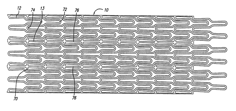
Referring now to FIGS. 9 and 10, a preferred embodiment of the stmt
10 of the present invention is shown. As can be seen in FIG. 10, the
cylindrical
element 12 of stmt 10 illustrates the serpentine pattern having a plurality of
peaks
and valleys which aid in the even distribution of expansion forces. In this
embodiment, the interconnecting members 13 serve to connect adjacent valleys
of
each adjacent cylindrical element 12 as described above. The various peaks and
valleys generally have U, W and (inverted U) shapes, in a repeating pattern to
form
CA 02398719 2002-07-29
WO 01/54614 PCT/USO1/01504
-22-
the cylindrical element 12. During expansion, doubled curved portions (W) 70
located in the region of the' valley where interconnecting members 13 are
connected, have the most mass and are the stiffest structure during
deformation,
while peak portions (inverted U) 72 are the least stiff, and valley portions
(U) 74
have an intermediate stiffness. In the embodiment shown in FIGS. 9 and 10,
there
are three repeating patterns of peaks and valleys in each cylindrical element
I2,
which allows the stmt to be collapsed to a very small profile. Each peak
portion
(inverted U) 72 has a shoulder portion 75 which has a different radius of
curvature
than the radius of curvature for the valley portions (U 74) and peak portion
(inverted U) 72. This shoulder region 75 provides a transition region between
the
peak portion (inverted U) 72 and the valley portions (U) 74 and double curved
portion (W) 70 to allow adjacent cylindrical elements to overlap and thereby
better
support the artery walls with smaller gaps between stmt struts. In this
manner, the
shoulder portion 75 provides more dense coverage of the serpentine pattern of
the
cylindrical element to create a fairly uniform strut pattern which fully
supports the
walls of the diseased artery. For this reason, there are no or few areas of
the stmt
wall which do not have struts for supporting the walls of the artery.
Each interconnecting member 13 is aligned collinearly with each
other to form a substantially continuous spine 76 which extends along the
length of
the stmt 10. This continuous spine 76 prevents the stmt from shortening
longitudinally when the cylindrical elements 12 are expanded radially. The
spine
76 also helps prevent the stmt from storing energy as the restraining sheath
29 is
retracted over the stmt during deployment. As a result, the stmt 10 will not
"jump"
off the stmt holder 40 as the stmt rings 12 are released by the restraining
sheath 29.
Therefore, more accurate deployment of the stmt can be achieved. The number
and
location of the interconnecting members I3 can be varied in order to develop
the
desired longitudinal flexibility in the stmt structure both in the compressed
condition as well as the expanded condition. The interconnecting members do
not
provide flexibility per se, but their location and frequency can enhance the
flexibility derived from the cylindrical elements. Generally, the greater the
CA 02398719 2002-07-29
WO 01/54614 PCT/USO1/01504
-23-
longitudinal flexibility of the stmt, the easier and more safely it can be
delivered to
the target site, especially where the implantation site is on a curved section
of the
body lumen, such as a coronary artery or a peripheral blood vessel. The number
of
spines 76 formed by the collinear arrangement of interconnecting elements 13
can
vary from one to as many as can be reasonably placed on a stmt, however, for a
minimal energy storage with a maximum flexibility, two to four spines are
recommended.
As shown in FIG. 2, stmt 10 serves to hold open artery 15 after the
catheter body 14 is withdrawn from the artery and help reduce the likelihood
of
restenosis. Due to formation of stent 10 from an elongated tubular member, the
undulating component of the cylindrical elements 12 of stmt 10 is relatively
flat in
transverse cross-section, so that when the stent is expanded, the cylindrical
elements 12 are pressed into the wall of the artery 15 and do not result in an
interference with the blood flow through the artery 15. Cylindrical elements
12
which are pressed into the wall of artery 15 will eventually be covered with
endothelial cell growth which further minimizes blood flow turbulence. The
serpentine pattern of cylindrical sections 12 provide good packing
characteristics to
prevent stmt movement within the artery. Moreover, the closely spaced
cylindrical
elements 12 at regular intervals provide uniform support for the wall of
artery 15.
While FIGS. 1 and 2 depict a vessel having an area of compressed plaque, the
stmt
10 can be used for purposes such as repairing a detached lining in the artery,
or to
assist in attaching a vascular grasp (not shown) when repairing an aol-tic
abdominal
aneurysm.
Another embodiment of a stmt 80 made in accordance with the
present invention is disclosed in FIGS. 13-17. FIG. 13 is an enlarged
perspective
view of a stmt 80 having a number of interconnecting elements 13 between
adjacent radially expandable cylindrical elements 12. As can be seen in both
FIGS. 14 and 15, the cylindrical element 12 of scent 80 has a serpentine
pattern
having a plurality of peaks and valleys which aid in the even distribution of
expansion forces. In this embodiment, as with the previously described
CA 02398719 2002-07-29
WO 01/54614 PCT/USO1/01504
-24-
embodiment, the various peaks and valleys generally have U, W and inverted U-
shapes in a repeat pattern to form the cylindrical element 12. During
expansion,
double-curved portions (W) 82, located in the region of the valley where
interconnecting members 13 are connected, have the most mass and are the
stiffest
structure during deployment. Valley portions (U) 84 have an intermediate
stiffness.
The peak portions (inverted U) 86 are the least stiff. A shoulder portion 88
which
has a different radius of curvature than the radius of curvature for the
valley
portions and peak portion is located between each peak portion (inverted U) 86
and the respective valley portion (U) 84 or double-curved portion ( W) 82.
This
shoulder region 88, like the shoulder region 75 shown in the previous
embodiment
of the stmt 10, provides a transition region between the peak portions and the
valley portions to allow adjacent cylindrical elements to overlap and
therefore
better support the arterial walls with smaller gaps between stmt struts.
The double-curved portions (W) 82 is somewhat similar to the
double-curved pol-tion (W) 70 described in the previous embodiment, except
that
the double-curved portion (W) 82 has a slight inward angulation. Likewise, the
peak portion (inverted U) 86 and valley portion (U) 84 also have a more inward
angulation than their counterparts shown in FIGS. 9 and 10. This inward
angulation of the (U), (W) and (inverted U) portions allow each of the
cylindrical
elements 12 to collapse closer to each other, resulting in a smaller collapsed
diameter.
FIGS. 16 and I7 show how the (U) and (W) portions have a inward
angulation, where the arc angle of the radius "wraps" more than 180 degrees.
FIGS. 16 and 17 shows a line A which denotes the longitudinally axis of the
stmt
80. Line B denotes the angle at which the legs of the U's and W's invert
inward to
allow a cylindrical element to be aligned closer to an adj acent cylindrical
element.
Hence, the stmt can be collapsed to a smaller diameter without having to
resort to a
narrower strut width. Additionally, the cell height (indicated by arrow 90 in
FIG. 15) can be longer to enable a larger expansion size without exceeding the
material's strain limits. This allows the embodiment of the stmt 80 shown in
CA 02398719 2002-07-29
WO 01/54614 PCT/USO1/01504
-25-
FIGS. 13 - 15 to expand to a laxger maximum size (up to about l Omm) while
allowing the same device to be collapsed to a smaller size, down to about
0.069
inches, rather than about 0.073 inches.
The stmt 80 further includes end rings 92 and 94 which include
double-curved portions (W) 82 at the outermost edge to increase the strength
of the
stmt 80 at its ends. Additional interconnecting members 13 are utilized in
this
particular design to further increase the axial strength of the ends of the
stmt 80.
As can be seen in FIG. 14, each of the double-curved portions (W) 82 of the
end
rings 92 and 94 have an interconnecting member 13 which attaches to the
respective adjacent cylindrical element 12. This allows for greater strength
at the
ends of the stmt where it may be needed to prevent the stmt from deploying
into a
"cigar" shape once implanted in the patient's vasculature. The end rings 92
and 94
also increase the amount of material at the ends of the stmt which may
increase
radiopacity as well. As result, the physician may have an easier time in
visualizing
the location of the stmt 80 both on the delivery catheter and once the stmt is
implanted within the patient's vasculature.
Stems of various lengths can be made by adding or subtracting
cylindrical elements between the end rings 92 and 94 of the stmt 80. For
example,
the stmt 80 can be made in sizes of 20mm, 30mm and 40mm, but could also be
made as small as 9lnm to as large as about 200mm, depending upon the
application
to which the stmt will be utilized. The stmt 80 also includes a number of
continuous spines 76 which extend along the length of the stmt 80 to help
prevent
the stmt from shortening longitudinally when the cylindrical elements 12 are
expanded radially. As with the previously described embodiments shown in
FIGS. 9 and 10, the spines 76 also help prevent stems from storing energy as
the
restraining sheath is retracted over the stent during deployment. As a result,
the
stmt 80 should not "jump" off the stmt holder as the cylindrical elements are
released by the restraining sheath. Therefore, more accurate deployment of the
stmt can be achieved. The number of spines 76 formed by the collinear
arrangements of interconnecting members 13 can vary from one to as many as can
CA 02398719 2002-07-29
WO 01/54614 PCT/USO1/01504
-26-
be reasonably placed on the stmt, however, for a minimum energy storage with
maximum flexibility; again, two to four spines are recommended.
Referring now to FIG. 18, an alternative tip assembly 100 for the
delivery system 11 is shown. It should be noted that where like elements have
already been described in conjunction with the stmt delivery system 11
disclosed in
FIGS. 1-8, similar reference numbers are used herein. The modifications of tip
assembly 100 include the elimination of the tubular element 44 which provided
a
conduit to the tip for evacuating air from the delivery system. Additionally,
the tip
assembly eliminates need for a round coil 45 and the radiopaque tungsten
element 47 which was placed at the distal end of the tip component 46. As a
result,
the tip assembly 100 may be less costly to manufacture and easier to place on
the
stmt holder 40.
Referring again to FIG. 18, a tip component 102 is shown bonded
directly to the stmt holder 40. This tip component 102 can be made from
material
such as poly-ether-block-amide sold under the name of PEBAX, or other similar
polymeric material or alloy suitable for use. A highly radiopaque material,
such as
BaS04, could be compounded with the PEBAX, or any other polymeric material
used to form the tip component 102, to increase the radiopacity of the tip
component 102. A marker band 104, which is located on the stmt holder 40
distal
to the expandable stmt 80, provides the physician with an additional marker
for
determining the location of the stmt when placing it within the patient's
vasculature. The stmt holder 40 is shown extending approximately halfway into
the lumen 106 of the tip component 102 to provide a sufficient area for
bonding
purposes. Adhesives are used to bond the tip component to the portion of the
stmt
holder 40 which extends into the lumen 106. The guide wire lumen 35 also
extends
into the lumen 106 of the tip component 102 to allow the guide wire (not
shown)
to extend through the distal opening 108 of the tip component 102. The tip
component 102 includes a recessed area 99 over which the restraining sheath
extends once the stmt is mounted on the stmt holder 40. A rounded proximal
edge 101 formed on the tip component 102 provides a smooth edge and reduces
the
CA 02398719 2002-07-29
WO 01/54614 PCT/USO1/01504
-27-
possibilities of the tip component 102 catching on a deployed stmt when the
delivery system is being removed from the patient.
In an alternative method of attaching the tip component 102 to the
stmt holder 40, the end of the stmt holder 40 does not have to extend into the
lumen 106 of the tip component 102, but rather, can terminate proximal to the
opening 110 of the component 102. The guide wire lumen 3 5 would thus extend
into the lumen 106 of the tip component 102. Adhesive could be applied to the
guide wire lumen 3 5 directly to adhesively bond the tip component 102
thereto. It
should be appreciated that still other methods of attaching the tip component
102 to
the stmt holder can be employed without departing from the spirit and scope of
the
present invention.
Additional minor modifications to the catheter system could be made
such as placing the marker band 42 over the layer of shrink tubing 3 8 as is
shown in
FIG. 18, rather than have the layer of shrink tubing 38 encapsulate the marker
42 as
is shown in FIGS. 1 and 2. The proximal end of the inner tubular member 21 can
be further stiffened by placing an inner hypotube inside the existing guide
wire
lumen 35. In this manner, the shaft of the inner tubular member 21 can be
reinforced to resist buckling. Additionally, the luer fitting 23 on the
housing
assembly 18 can be increased in size to allow for guide wire exchanges.
The elimination of the tubular member 44 utilized in the flushing
system in this particular embodiment does not sacrifice the ability of the tip
assembly 100 to evacuate trapped air from the catheter system. A similar
method of
evacuating air fiom the system, as previously described and shown in FIGS. 7,
11
and 12, can be used. For example, a syringe can be attached to the luer
fitting 23 of
the housing assembly 18 to pump sterile fluid into the guide wire lumen 35 to
flush
air from the system. Again, a mandrel or stylet (now shown) is placed in the
guide
wire lumen at the distal opening 108 to block the flow of sterile fluid. Any
trapped
air will be forced to escape through the distal end of the restraining sheath
29 or
through the distal opening 108 on the tip component i 02. Once fluid is
observed
dripping from the distal end of the retraining sheath and the distal opening
108, the
CA 02398719 2002-07-29
WO 01/54614 PCT/USO1/01504
-28-
mandrel can be removed since air has been evacuated from the system. Since the
gap sizes axe so small between the various components, capillary force
prevents air
from infiltrating the delivery system once evacuation has been completed.
The stmt of the present invention can be made in many ways.
However, the preferred method of making the stmt is to use the well-known
process of laser cutting to cut a thin-walled tubular member, to remove
portions of
the tubing in the desired pattern for the stmt, leaving relatively untouched
the
portions of the metallic tubing which are to form the stmt. It is preferred to
cut the
tubing in the desired pattern by means of a machine-controlled laser.
Generally, the tubing is put in a rotatable collet fixture of a machine-
controlled apparatus for positioning the tubing relative to a laser. According
to
machine-encoded instructions, the tubing is then rotated and moved
longitudinally
relative to the laser which is also machine-controlled. The laser selectively
removes
the material from the tubing by ablation and a pattern is cut into the tube.
The tube
is therefore cut into the discrete pattern of the finished stmt. Further
details on how
the tubing can be cut by a laser are found in U.S. Patent Nos. 5,759,192
(Saunders)
and 5,780,807 (Saunders), which have been assigned to Advanced Cardiovascular
Systems, Inc. and are incorporated herein by reference in their entirety.
The process of cutting a pattern for the stmt into the tubing generally
is automated except for loading and unloading the length of tubing. For
example, a
pattern can be cut in tubing using a CNC-opposing collet fixture for axial
rotation
of the length of tubing, in conjunction with CNC X/Y table to move the length
of
tubing axially relative to a machine-controlled laser as described. The entire
space
between collets can be patterned using the CO2, Nd or YAG laser set-up of the
foregoing example. The program for control of the apparatus is dependent on
the
particular configuration used and the pattern to be ablated in the coding.
A suitable composition of Nitinol used in the manufacture of the stmt
of the present invention is approximately 55% nickel and 45% titanium (by
weight)
with trace amounts of other elements making up about 0.5% of the composition.
The austenite transformation temperature is between about -15 °C and
0°C in order
CA 02398719 2002-07-29
WO 01/54614 PCT/USO1/01504
-29-
to achieve superlastecity. The austenite temperature is measured by the bend
and
free recovery tangent method. The upper plateau strength is about a minimum of
60,000 psi with an ultimate tensile strength of a minimum of about 155,000
psi.
The permanent set (after applying 8% strain and unloading), is approximately
0.5%.
The breaking elongation is a minimum of 10%. It should be appreciated that
other
compositions of Nitinol can be utilized, as can other self expanding alloys,
to
obtain the same features of a self expanding stmt made in accordance with the
present invention.
The stmt of the present invention can be laser cut from a tube of
super- elastic (sometimes called pseudo-elastic) nickel titanium (Nitinol)
whose
transformation temperature is below body temperature. The stmt diameters can
cut
with the same stmt pattern, and the stmt is expanded and heat treated to be
stable at
the desired final diameter. The heat treatment also controls the
transformation
temperature of the Nitinol such that the stem is super elastic at body
temperature.
The transformation temperature is at or below body temperature so that the
stmt is
superelastic at body temperature. The stmt is electro polished to obtain a
smooth
finish with a thin layer of titanium oxide placed on the surface. The stem is
usually
implanted into the target vessel which is smaller than the stmt diameter so
that the
stmt applies a force to the vessel wall to keep it open.
The stmt tubing may be made of suitable biocompatible material
besides super-elastic nickel-titanium (NiTi) alloys. In this case the stmt
would be
formed full size but deformed (e.g. compressed) into the restraining sheath of
the
delivery catheter to facilitate intraluminal delivery to a desired
intraluminal site. In
the compressed state, a portion of the stmt material is in the martensite
phase, and
upon release of the restraining sheath when the stmt reaches the desired
intraluminal location, the stmt expands due to the transformation back to the
more
stable austenite phase. Further details of how NiTi super-elastic alloys
operate can
be found in U.S. Patent Nos. 4,665,906 (Jervis) and 5,067,957 (Jervis).
The stmt diameters are very small, so the tubing from which it is
made must necessarily also have a small diameter. For PTCA applications,
CA 02398719 2002-07-29
WO 01/54614 PCT/USO1/01504
-3 0-
typically the stmt has an outer diameter on the order of about 1.65 mm (0.065
inches) in the unexpended condition, the same outer diameter of the hypotube
from
which it is made, and can be expanded to an outer diameter of 5.08 mm (0.2
inches)
or more. The wall thickness of the tubing is about 0.076 mm (0.003 inches).
For
stems implanted in other body lumens, such as PTA applications, the dimensions
of
the tubing are correspondingly larger. This stmt is also designed for carotid
applications, so the outer diameter of the tubing would typically be about
0.095
inches with a wall thickness of about 0.007 inches. The diameters of a carotid
stmt typically would be about 5-l Omm. While it is preferred that the stems be
made from laser cut tubing, those skilled in the art will realize that the
stmt can be
laser cut from a flat sheet and then rolled up in a cylindrical configuration
with the
longitudinal edges welded to form a cylindrical member.
While the invention has been illustrated and described herein in terms
of its use as intravascular stems, it will be apparent to those skilled in the
ant that
the stems can be used in other instances in all conduits in the body, such as,
but not
limited to, the urethra and esophagus. Since the stmt of the present invention
has
the novel feature of self expanding to a large diameter while retaining its
structural
integrity, it is particularly well suited for implantation in almost any
vessel where
such devices are used. This feature, coupled with limited longitudinal
contraction
of the stmt when it is radially expanded, provide a highly desirable support
member
for all vessels in the body. Other modifications and improvements may be made
without departing from the scope of the invention.