Note: Descriptions are shown in the official language in which they were submitted.
CA 02399570 2002-08-02
WO 01/72373 PCT/US01/09474
APPARATUS AND METHODS FOR INTRABODY THERMAL TREATMENT
TECHNICAL FIELD
The present application relates to medical devices and
procedures, and to ultrasonic energy emitters adapted for use in
such devices and procedures.
BACKGROUND ART
Contraction or "beating" of the heart is controlled by electrical
impulses generated at nodes within the heart and transmitted
along conductive pathways extending within the wall of the heart.
Certain diseases of the heart known as cardiac arrhythmias
involve abnormal generation or conduction of the electrical
impulses. One such arrhythmia is atrial fibrillation or "AF".
Certain cardiac arrhythmias can be treated by deliberately
damaging the tissue of the cardiac wall along a path crossing a
route of abnormal conduction. This causes formation of a scar
extending along the path where disruption occurred. The scar
blocks conduction of the electrical impulses. Such a scar can be
created by conventional surgery, but this entails all of the
risks and expense associated with cardiac surgery. Another
approach, described in Swartz et al., United States Patent
5,575,766, is to introduce a catheter bearing a localized energy
emitter such as an electrode for application of radio frequency
("RF") energy at its distal tip into a heart chamber, such as the
right or left atrium of the heart in the case of atrial
fibrillation. The physician then moves the catheter so that the
tip, and the localized emitter traces the desired path. In AF,
the desired path typically is a closed loop encircling the
openings or ostia of the pulmonary veins. RF energy applied
through the electrode heats the tissue to a degree sufficient to
cause death of the normal tissue and its replacement by scar
tissue. Heating to this degree is referred to herein as
"ablation". The elevated temperature required for ablation
CA 02399570 2002-08-02
WO 01/72373 PCT/US01/09474
varies with the time of exposure to the elevated temperature, but
heating to about 60-80 C is typically used. Tracing a precise
path along the interior of a chamber in the heart of a living
subject with the tip of a catheter involves inherent practical
difficulties. Although curved guide wires ca.n be placed within
the catheter so that the catheter tip will tend to follow the
guide wire as the physician moves it, the process is still
difficult.
Swanson et al., U.S. Patent 5,582,609 describes an elongated
catheter having numerous RF electrodes disposed along its length
in a distal region adjacent the tip. This distal region can be
formed into a curved, looplike configuration and manipulated so
that the electrodes lie along the desired path, whereupon RF
energy is applied so as to ablate cardiac tissue. In a variant
of this approach, the electrodes are mounted on a structure which
opens to form a ring-like configuration. Even with these
structures, however, it is difficult to assure the desired
placement of the RF electrodes. Lesh, U.S. Patent 5,971,983
describes an elongated catheter which is equipped with similar RF
electrodes distributed over its distal region, and uses guide
wires to position the distal region in place against the wall of
the heart. Although this patent mentions a "ultrasonic element
such as an ultrasound crystal element" along with numerous other
devices as theoretically applicable to cardiac tissue ablation,
it offers no structure for an elongated ultrasonic ablating
device.
As described in various publications including Swartz, U.S.
Patent 5,938,660 and Lesh, International Publication WO 99/02096,
the abnormal conduction routes in AF typically extend from the
wall of the heart along the pulmonary veins. Therefore, AF can
be treated by ablating tissue in a ring around each pulmonary
vein at the juncture between the pulmonary vein and the heart.
p
CA 02399570 2002-08-02
WO 01/72373 PCT/US01/09474
As described in the '096 publication, such ablation can be
performed by threading a catheter having a thermal ablation
element at its distal tip into the heart so that the tip is
lodged within the appropriate pulmonary vein. The catheter may
bear a balloon which is inflated within the vein and which holds
the catheter in place. The ablating element is then actuated so
as to apply heat in a region surrounding the ablating element.
In certain embodiments taught in the '096 publication, the
ablating element includes a radio frequency ("RF") emitting
element which is carried on the surface of the balloon. Ablation
of the pulmonary vein using RF energy can create a rough,
disrupted surface on the interior of the vein. This or other
factors can lead to thrombosis or clot formation.
Other embodiments described in the '096 publication disclose the
use of ultrasonic transducers. The preferred ultrasonic
transducer illustrated in the '096 publication is a rigid ceramic
piezoelectric element disposed on a catheter surrounded by a
balloon. When the balloon is inflated, the piezoelectric element
remains remote from the wall of the pulmonary vein. The
piezoelectric element can be actuated to apply sonic energy
through a fluid contained in the balloon, thereby heating the
ring of vein wall tissue surrounding the balloon. As a further
alternative, the '096 publication shows an ultrasonic emitter in
the form of a hollow concave disk. The '096 publication suggests
that such an emitter can be physically rotated around the axis of
a catheter so as to ablate a ring-like zone. These transducers
have numerous drawbacks even for use in ablation of a vein wall
and are not adapted for ablation of the wall of the cardiac
chamber.
Ultrasonic heating such as high intensity focused ultrasound
(HIFU) is utilized for many therapeutic applications. As
disclosed in commonly assigned International Application
PCT/US98/1062, published as International Publication WO/98/52465
3
CA 02399570 2004-12-02
HIFU heating typically is conducted using an ultrasonic emitter
having an array of transducers. The transducers are actuated
with a drive signal so as to emit ultrasonic waves. The
relative phasing of the waves is controlled by the physical
configuration of the array and the phasing of the drive signal.
These factors are selected so that the ultrasonic waves tend
to reinforce one another constructively at a focal location.
Tissue at the focal location is heated to a greater extent than
tissue at other locations. As described, for example in
assigned U.S. Patent 6,461,314 and in the corresponding
International Publication WO 00/45706, commonly assigned United
States Patent 6,492,762 and in the corresponding International
Publication WO 00/45706, HIFU may be applied by transducer
arrays such as arrays of polymeric piezoelectric transducers.
These arrays can be mounted on a probe such as a catheter which
can be introduced into the body as, for example, within the
vascular system or into a cavernous internal organ. U.S.
Patent 6,461,314 discloses certain transducer arrays which can
be deformed so as to vary the placement of the focal location.
DISCLOSURE OF THE INVENTION
One aspect of the invention provides apparatus for applying
thermal treatment to tissue of an internal organ of a living
subject. Apparatus according to this aspect of the invention
desirably includes one or more catheters and an elongated
energy emitter carried on one of the one or more catheters.
The elongated energy emitter desirably is adapted to assume a
desired shape when disposed within the interior of the organ.
The apparatus desirably also includes an expansible positioning
structure such as a balloon or other expansible element carried
4
CA 02399570 2002-08-02
WO 01/72373 PCT/US01/09474
on one of the one or more catheters. When the energy emitter is
in the desired curved shape, the energy emitter extends over the
expansible positioning structure so that the expansible
positioning structure can bias the elongated energy emitter
against an interior wall of the organ. Thus, when the
positioning element and energy emitter are in an operative
condition, the energy emitter extends along an elongated path on
the interior wall of the organ. The path has a shape
corresponding to the desired shape of the energy emitter. The
energy emitter desirably is operative to emit energy at a
plurality of locations along its length so as to heat tissue
surrounding the interior of the organ at a plurality of locations
along the path.
Most preferably, the energy emitter is formed separately from the
positioning element, so that the energy emitter can assume its
desired shape before it is biased against the wall of the organ.
The energy emitter may be adapted to assume a curved shape such
as a substantially closed loop, so that the path along the
interior wall of the organ will be generally in the form of a
loop. The energy emitter desirably is adapted to emit energy
substantially simultaneously at a plurality of locations along
its length to thereby heat tissue at a plurality of locations
along the path substantially simultaneously. In a particularly
preferred arrangement, the energy emitter is an elongated
ultrasonic transducer array.
The one or more catheters desirably include a treatment catheter
carrying the energy emitter, the emitter extending lengthwise
along the treatment catheter adjacent the distal end thereof. In
a particularly preferred arrangement, the energy emitter is an
elongated ultrasonic transducer array which is flexible in
directions transverse to the lengthwise direction of the catheter
to facilitate threading of the catheter into the body. The distal
end of the treatment catheter, and hence the energy emitter, may
5
CA 02399570 2002-08-02
WO 01/72373 PCT/US01/09474
be brought to the desired shape by structures within the
catheter, or by additional elements such as curved guide wires or
sheaths. The one or more catheters most preferably include a
holding structure such as a stabilizer catheter separate from the
treatment catheter, the holding structure carrying the expansible
positioning element. The apparatus may include an anchor linked
to the stabilizer catheter, the anchor being adapted to engage an
anatomical structure in or adjacent said organ. The expansible
positioning structure may be movable relative to the anchor while
the anchor is engaged with said anatomical structure. For
example, where the apparatus is used for treatment of atrial
fibrillation or other cardiac arrhythmias, the treatment catheter
bearing the energy emitter and the stabilizer catheter may be
threaded into a chamber of the heart and the treatment catheter
may be brought to the desired shape such as a generally loop-like
configuration. The expansible positioning structure may be
expanded and the anchor may be engaged in a pulmonary vein or
other blood vessel, with the treatment catheter disposed between
the positioning structure and the wall of the heart chamber. The
positioning structure is urged toward the wall of the heart, so
as to engage the energy emitter with the wall of the heart, as by
moving the stabilizer catheter relative to the anchor. While the
energy emitter is engaged with the wall of the heart, it is
activated to apply energy and ablate tissue in the heart wall,
thereby forming a lesion along a loop-like path. Desirably, the
entire lesion can be formed without repositioning or
reconfiguring the energy emitter.
A further aspect of the invention provides methods of applying
thermal treatment to tissue of an internal organ of a living
mammal. A method according to this aspect of the invention
desirably includes the steps of inserting an elongated energy
emitter into the interior of the internal organ and bringing the
energy emitter to a desired shape in a desired position relative
6
CA 02399570 2002-08-02
WO 01/72373 PCT/US01/09474
to the organ, inserting an expansible positioning element into
the interior of the organ, and expanding the positioning
structure so that the energy emitter is disposed between the
positioning structure and the wall of the organ and the
positioning structure biases the energy emitter against the
interior wall of the organ. In this condition, the energy
emitter extends along an elongated path on such interior wall
having a shape corresponding to the desired shape of the energy
emitter. While the energy emitter extends along this path, the
energy emitter is actuated to emit energy at a plurality of
locations along its length so as to heat tissue at a plurality of
locations along the path. In a particularly preferred method,
the entire lesion is formed in one actuation, or a few
actuations, of the energy emitter, without repositioning or
reconfiguring the emitter. Most preferably, the energy emitter
is brought at least approximately to the desired shape and at
least approximately to the desired position before the
positioning structure is fully expanded and before the energy
emitter is biased against the wall of the internal organ by the
positioning element.
In a particularly preferred method, the energy emitter includes
an array of one or more ultrasonic transducer elements, the array
extending in a lengthwise direction, the one or more transducer
elements emitting ultrasonic energy at plural locations along the
length of the array. For example, the array may extend
lengthwise along a treatment catheter as discussed above in
connection with the apparatus. The use of ultrasonic energy
allows formation of lesions in the wall with minimal damage to
the lining of the wall. In ablation of heart tissue, this
minimizes the possibility of thrombus formation. Most desirably,
the method includes the step of focusing ultrasonic energy
emitted by the one or more transducer elements onto a elongated
focal region extending generally parallel to said path. The term
7
CA 02399570 2002-08-02
WO 01/72373 PCT/US01/09474
"focusing" as used in this disclosure with reference to sonic or
ultrasonic energy, refers to providing such energy from
spatially-separated regions of a transducer or transducer array
such that the ultrasonic waves from plural spatially-separated
regions of the transducer or transducer array converge with one
another in passing from the transducer or array to a focal region
and are in phase within one another within the focal region so
that they mutually reinforce one another so as to provide a sonic
power density in the focal region higher than the sonic power
density at the transducer surface. Most typically, the focal
region is disposed on or in the wall of the organ and has an area
(measured in a plane normal to the direction of propulsion of the
ultrasonic waves) smaller than the area of the transducer. The
method may further include the step of varying the focus of the
ultrasonic energy so as to move the focal region towards or away
from the transducer or array and thereby position said focal
region deeper or shallower within the wall of said organ while
the array remains substantially in position along the path. The
ability to focus the ultrasonic energy allows rapid heating of
the tissue, and facilitates heating tissue in the focal region to
the extent necessary to= ablate it, while minimizing damage to
adjacent tissues.
As discussed above in connection with the apparatus, the energy
emitter desirably is flexible in directions transverse to its
length. The step of inserting the energy emitter may include the
step of advancing the array lengthwise through a tubular
anatomical structure and then deforming the array in directions
transverse to its lengthwise direction to the desired shape.
A further aspect of the invention provides a medical device
including an elongated catheter body with proximal and distal
directions in its direction of elongation and an elongated
ultrasonic transducer array extending in the proximal and distal
directions, the catheter body and the transducer array being
8
CA 02399570 2002-08-02
WO 01/72373 PCT/US01/09474
flexible in all directions transverse to said proximal and distal
directions.
Yet another aspect of the invention provides an elongated
ultrasonic transducer array having lengthwise directions, the
array including a sheetlike element having a first fold extending
in the lengthwise directions of the array and defining a first
pair of adjacent regions on opposite sides of the fold. These
desirably are non-parallel with one another and non-coplanar with
one another. For example, the first pair of adjacent regions may
define a structure which is generally V-shaped when seen in
cross-section with the viewing direction in the lengthwise
direction of the array.
Most preferably, at least one of the regions in the first pair is
an active region. The array includes a plurality of ultrasonic
transducer elements disposed on or formed integrally with the
sheetlike element in the active region or regions of such
element. The sheetlike element has notches in each of the
aforesaid regions, the notches extending along axes transverse to
the first fold at locations spaced apart from one another in the
lengthwise direction. The notches subdivide each of the regions
into panes, the notches in each region of the first pair of
adjacent regions being offset in the lengthwise direction of the
array from the notches in the other region of such first pair of
adjacent regions. Each pane of each region of said first pair has
a hinge zone aligned with the axis of a notch in the other region
of the first pair. The sheetlike element is flexible at least in
the hinge zones. As further discussed below, this arrangement
allows the array to bend in directions transverse to said
lengthwise direction of the array, and typically allows bending
in all directions transverse to the lengthwise direction. The
sheetlike element desirably has one or more electrical conductors
thereon, the conductors extending lengthwise along the sheetlike
9
CA 02399570 2002-08-02
WO 01/72373 PCT/US01/09474
element in a zigzag pattern so that the conductors pass through
the hinge regions of the panes and around the notches.
Yet another aspect of the invention provides a medical device
including an elongated catheter body with proximal and distal
directions in its direction of elongation, and an array as
discussed above, the lengthwise directions of the array and the
first fold extending in the proximal and distal directions of
said body. The active region or regions desirably are disposed
on or constitute an outwardly-facing surface of said body and
extend in lateral directions transverse to said lengthwise
directions.
Yet another aspect of the invention provides a medical ultrasonic
applicator including a first elongated catheter body having an
exterior surface and having proximal and distal directions; a
distributed array of one or more ultrasonic transducer elements
disposed on or constituting a portion of said exterior surface of
said first body, the array extending in the proximal and distal
directions; and an elongated lens overlying the array of
transducer elements and extending in said proximal and distal
directions, said lens being adapted to focus ultrasonic emissions
from the transducer elements into a elongated focal region
outside of said body but generally parallel thereto. Most
preferably, the body, lens and array are flexible in directions
transverse to the proximal and distal directions. The lens may
include a hollow enclosure extending in the proximal and distal
directions, the enclosure being filled with a lens fluid when the
device is in an operative condition.
These and other objects, features and advantages of the present
invention will be more readily apparent from the detailed
description of the preferred embodiments set forth below, taken
in conjunction with the accompanying drawings.
BRIEF DESCRIPTION OF THE DRAWINGS
CA 02399570 2002-08-02
WO 01/72373 PCT/US01/09474
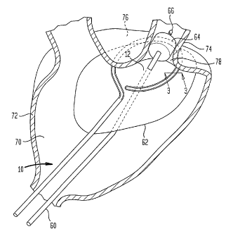
Figure 1 is a diagrammatic, cutaway perspective view depicting
apparatus according to one embodiment of the invention during
use.
Figure 2 is a fragmentary, diagrammatic perspective view of a
treatment catheter used in the apparatus of Fig. 1.
Figure 3 is a fragmentary sectional view taken along line 3-3 in
Fig. 1.
Figure 4 is a fragmentary, cutaway perspective view of the
treatment catheter used in the apparatus of Figs. 1-3.
Figure 5 is a perspective view of an element of the transducer
array used in the apparatus of Figs. 1-4.
Figure 6 is an elevational view of the transducer array depicted
in Fig. 5.
Figure 7 is a plan view of the transducer array depicted in Figs.
5 and 6.
Figure 8 is an exploded view depicting a portion of the
transducer array shown in Figs. 5-7.
Figure 9 is a fragmentary, cutaway perspective view depicting a
portion of the transducer array shown in Figs. 5-8.
Figure 10 is a fragmentary, diagrammatic perspective view
depicting a transducer array according to a further embodiment of
the invention.
11
CA 02399570 2002-08-02
WO 01/72373 PCT/US01/09474
Figure 11 is a diagrammatic sectional view depicting a treatment
catheter according to a further embodiment of the invention
incorporating the transducer array of Fig. 10.
Figure 12 is a sectional view taken along line 12-12 in Fig. 11.
Figure 13 is a sectional view depicting a treatment catheter
according to a further embodiment of the invention.
Figure 14 is a diagrammatic perspective view depicting elements
of apparatus according to yet another embodiment of the
invention.
Figure 15 is a view similar to Fig. 14 but depicting the elements
in a different operating condition.
Figure 16 is a diagrammatic perspective view of the elements
shown in Figs. 14 and 15, in conjunction with additional elements
of the apparatus.
Figure 17 is a sectional view taken along.line 17-17 in Fig. 16.
Figure 18 is a diagrammatic perspective view of the elements
shown in Figs. 14-17, in conjunction with additional elements.
Figure 19 is a diagrammatic perspective view of a stabilizer
catheter used with the apparatus of Figs. 14-18.
Figure 20 is a diagrammatic perspective view of the apparatus of
Figs. 14-19 in an assembled condition during one phase of
operation.
12
CA 02399570 2002-08-02
WO 01/72373 PCT/US01/09474
Figure 21 is a diagrammatic perspective view of a treatment
catheter in accordance with a further embodiment of the invention.
MODES FOR CARRYING OUT THE INVENTION
One aspect of the present invention provides apparatus for
applying thermal treatment such as ablation to tissue in the wall
of a cavernous internal organ of a living subject such as a
chamber of the heart. Apparatus according to one embodiment of
the invention includes a treatment catheter 10 having an
elongated body with a distal region 12 adapted to form a desired
curved shape when in an operative condition, deployed within a
chamber of the heart such as the atrium A schematically shown in
Fig. 1. The particular curved shape is generally in the form of
a closed or nearly closed loop, as best seen in Fig. 2. The
catheter, including distal region 12, should be flexible in
directions transverse to the proximal and distal directions,
i.e., directions transverse to the lengthwise axis of the
catheter, at least during introduction and removal of the
catheter.
Numerous techniques and structures known in the art for deforming
a catheter to a desired curved shape while the catheter is
disposed in an internal chamber of the body can be used in the
treatment catheter. Merely by way of example, a guide wire which
inherently tends to assume such shape, such as a resilient guide
wire 14 (Fig. 3) can be provided in the interior bore 16 of the
catheter before or after it is deployed. A shape memory alloy
guide wire such as a Nitinol (Trademark) wire which is straight
at room temperature but which tends to assume the desired
curvature at body temperature can be used. The treatment
catheter body itself may include these elements, and may be
constrained to a relatively straight form during introduction and
removal by threading the catheter through a bore of an introducer
catheter (not shown). Alternatively or additionally, the
13
CA 02399570 2002-08-02
WO 01/72373 PCT/US01/09474
treatment catheter may include controllable elements such as
steering wires extending through the interior bore for
controllably bending the catheter body to the desired shape. In
a further alternative, the catheter body may have "dead bend" or
non-resilient properties such that once bent to a particular
shape, the catheter body retains such shape until it is bent
again by external forces. Such a dead-bend treatment catheter
can be bent to the preselected shape by the physician after
inserting the distal end of the treatment catheter into the heart
but before applying energy to ablate tissue as described below.
Treatment catheter 10 has an elongated, flexible ultrasonic
transducer array 20 extending lengthwise along the distal region
12 of the catheter body. A signal cable 21 connected to the
transducer array extends to the proximal end of the catheter body
for connection to an external source of drive signals (not
shown). As further discussed below, the elongated transducer
array incorporates transducer elements incorporating an
electromechanical transduction material, most preferably a
polymeric electromechanical transduction material. As used in
this disclosure, the term "electromechanical transduction
material" means a material which changes dimensions in response
to an applied electrical signal. Polymeric electromechanical
transduction materials include polymeric piezoelectric materials
as, for example polyvinylidene difluoride ("PVDF") and copolymers
of PVDF with trifluoroethylene ("PVDF-TrFE"), as well as
electrostrictive polymers such as certain silicone polymers. The
term "transducer element" as used herein refers to a structure or
a region of a structure which is capable of converting a signal
in one form to a signal in another form as, for example, a mass
or portion of electromechanical transduction material and
electrodes juxtaposed with the transduction material. The
"transducer array" is used herein as referring to a structure
which includes one or more transducer elements. Where a
14
CA 02399570 2002-08-02
WO 01/72373 PCT/US01/09474
transducer array includes plural transducer elements, these may
be connected together, so that the same signal is applied to all
of the elements and the plural elements act in much the same way
as a single larger element. Alternatively, different elements of
a transducer array may be connected to different signal sources
as, for example, to sources of signals having preselected phase
relationships.
Several conflicting factors complicate the design of an elongated
transducer array for ablation along an elongated path within a
cavernous organ such as a heart chamber or within the vascular
system. These factors include the following:
Diameter: The transducer array must be constructed to fit on a
catheters of small diameter. For intracardiac use, the treatment
catheter carrying the array should be in the range from 3 French
to 12 French catheter size, i.e., about 1 to about 4mm in
diameter.
Flexibility: The necessary flexibility of the particular system
will be a function of the target area and the procedure to be
employed. For ablating the cardiac wall in a loop surrounding the
ostium of a single pulmonary vein in the procedure for treatment
of atrial fibrillation depicted in Fig. 1, the minimum radius of
curvature that the treatment catheter, and thus the transducer,
will have to make is approximately 15-20mm. Moreover, the
transducer array should be flexible in all directions transverse
to its direction of elongation to facilitate threading of the
treatment catheter through the vascular system and to facilitate
intimate engagement of the transducer array and treatment
catheter with the cardiac wall.
Power: The required power needed to perform treatment will depend
on the specific application. As with most ultrasonic devices,
methods to increase output power should be employed, as the
higher the power density (emission power per unit surface area),
the smaller the device can be and the shorter the treatments
CA 02399570 2002-08-02
WO 01/72373 PCT/US01/09474
become. A transducer array for use in an ablation procedure
preferably emits about 5 W/cm2 or more. If the ultrasonic waves
from the device are focused into a region smaller than the
emitting surface, somewhat lower power density can be employed.
Shielding: The emitter should limit electromagnetic emissions, to
avoid interference with other devices used in the hospital
environment. For ultrasonic emitters, this typically means the
hot electrical leads to the emitter should be shielded to the
outside world by grounded layers of conductive material, and that
the drive cable should be coaxial, with a grounded outer sheath.
Thermal Control: Ultrasonic emitters, and particularly emitters
incorporating a polymeric electromechanical transduction material
generate significant heat through dielectric and mechanical
losses. The performance (power and frequency) of the device is
somewhat a function of the operating temperature. A method of
removing heat from the structure should be provided to ensure
proper operation of the device (at its tuned frequency and
appropriate power levels) as well as to prevent unintended
thermal damage to the surrounding tissue as a result of the
tissue heating by conduction from hot transducer surface, as
opposed to deposition of acoustic power.
Bio-Compatibility: While the primary foreseeable applications of
the transducer array will have the transducer positioned inside
of an outer sheath or cover, it is still desirable to limit the
incorporation of materials which are not approved for patient
contact so as to minimize any concerns regarding accidental
contact in the event the outer sheath fails during use, and to
ease the regulatory process. This factor is more significant if
no outer sheath is employed.
Machinability: To make an inexpensive disposable catheter, the
transducer array should be designed in such a way so as to take
advantage of mass production techniques which can be employed to
limit construction costs while maximizing ease of fabrication.
16
CA 02399570 2002-08-02
WO 01/72373 PCT/US01/09474
Also, the transducer array should include electrical conductors
connected to the ultrasonic transducers so as to limit the number
of external connections which must be made within the limited
space available inside the treatment catheter. This implies that
continuous electrical conductors should extend lengthwise along
the transducer array. As disclosed in the aforementioned
commonly assigned applications, ultrasonic transducer arrays, and
particularly ultrasonic transducer arrays incorporating polymeric
transduction materials, can be fabricated economically in flat
form, using techniques similar to those used in fabrication of
printed circuits. It would be desirable to use such techniques
in fabrication of an elongated, flexible transducer array.
However, printed circuits which incorporate thin, flexible
sheetlike materials such as a polyimide dielectric and thin
metallic conductors are flexible in only some directions. Such a
sheet can bend readily around an axis in the plane of the sheet,
but will not bend readily around an axis perpendicular to the
plane of the sheet. Thus, a strip of such a sheet will bend
readily in a first direction transverse to its length, but will
not bend readily in a second direction transverse to the first
direction and transverse to the lengthwise direction.
The transducer array 20 addresses these factors as further
discussed below. The transducer array 20 and its disposition
relative to the catheter body are best seen in Fig. 4. Arrows
adjacent certain views indicate the directions referred to in the
text below. The transducer array has an active region 22
overlying a surface region 24 of the catheter body facing
outwardly, away from the interior of the catheter and away from
bore 16. In Fig. 4, portions of the active region are omitted
for clarity of illustration, to show surface region 24. The
active region 22 includes substantially planar transducer
elements 26, schematically indicated in broken lines in Fig. 4.
The treatment catheter may include a thin outer covering 23,
17
CA 02399570 2002-08-02
WO 01/72373 PCT/US01/09474
partially cut away in Fig. 4, closely overlying surface region 24
and active region 22 of the transducer array.
The transducer array also includes a radially-extensive
additional region 28, also referred to as a back plane region.
Region 28 extends inwardly, into the interior of the catheter
body and into bore 16. As used in this disclosure, the term
"laterally extensive" used with reference to a structural element
means that the element extends generally in the lateral
direction, and the term "radially extensive" means that the
element extends generally in the radial direction, but these
terms do not imply that the element extends exactly laterally or
exactly radially. Thus, although the particular embodiment
illustrated in Figs. 3-9 has planar regions 24 and 28
perpendicular to one another, these regions need not be exactly
planar or exactly perpendicular to one another. Also, the
radially-extensive additional or back plane region 28 need not
lie exactly on a radial plane of the catheter body.
As best seen in Fig. 5, the transducer array 20 is formed as a
sheetlike laminate structure generally in the form of a segmented
"L" shaped beam. The laminate structure preferably is
manufactured flat and folded about a first fold 30 extending in
the lengthwise direction of the array. Fold 30 thus subdivides
the sheet into the active region 22 and additional region 28,
which are non-coplanar and non-parallel to one another. Active
region 22 has an outer boundary 32 at an edge of the laminate
structure remote from fold 30. Notches 34 extend into active
region 22 from its outer boundary 32 along axes 36, generally in
the lateral direction transverse to fold 30 and thus transverse
to the lengthwise direction of the array. Each notch 34 extends
across fold 30 so that a small portion of each notch in the
active region extends into the radially-extensive region 28.
Each notch 34 is generally triangular, so that the notch is wider
at the outer boundary 32 than at fold 30. Notches 34 subdivide
18
CA 02399570 2002-08-02
WO 01/72373 PCT/US01/09474
the active region into a series of panes 38a, 38b, 38c, and so
on.
The radially-extensive additional or back plane region 28 has a
similar outer boundary 40 and notches 42 subdividing region 28
into a series of panes 44, seen in broken lines in Fig. 5. Here
again, the notches 42 extend transverse to the fold 30 from the
outer boundary 40 and slightly across the fold, so that the
notches in region 28 extend slightly into active region 22. The
notches 42 in region 28 are offset in the lengthwise direction
from the notches 34 in region 22, so that each notch 42 in region
28 is aligned with a zone 46 of each panel 38 of region 22. The
zone of each panel 38 which is aligned with a notch 46 is
referred to as the hinge zone. The hinge zone 46 of panel 38b is
indicated schematically by a line in Fig. 5. A single notch 42,
with the aligned panel 38 and hinge zone 46 are shown on a larger
scale in Fig. 9, in the flat state of the laminate, prior to
folding at fold 30. The hinge zone 46 of each panel 38 extends
from fold 30 to the outer boundary 32 of region 22. Preferably,
each hinge zone 46 lies near the center of the panel in the
lengthwise direction. In the same manner, each notch 34 in
active region 22 is aligned with a hinge zone of a pane 44 of the
additional region 28, one such hinge zone being indicated
schematically by a line 48 in Fig. 5.
The array is flexible in hinge zones 46 and 48. The notches and
hinge zones permit bending of the transducer array in all
directions transverse to the lengthwise direction of the array.
Thus, a bend in the YZ plane (in the plane of the radial (Y) and
lengthwise (Z) directions) flexes one or more hinge zones 46 in
one or more panes 38 of active region 22. Such flexure only
requires each hinge zone 46 to bend about a lateral or X-
direction line, in the plane of the laminate within the active
region, also referred to as "plate mode" bending. As this
bending of the radiating plane occurs, the angle between the
19
CA 02399570 2002-08-02
WO 01/72373 PCT/US01/09474
sides of the notches 42 in additional or back plane region 28
changes. In the same manner, flexure in the XY plane (in the
plane of the lateral (X) and lengthwise (Z) directions) flexes
one or more hinge zones 48 in the panes 44 of additional or back
plane region 28, in a similar plate mode bending action about a
line in the plane of additional region 28. Compound bending,
with components in both XY and YZ planes is accommodated by a
combination of these actions.
Although the structure is free to bend in all directions
transverse to the lengthwise direction, the laminate is
continuous; it is not separated into isolated pieces by the
notches 34 and 42. The laminate therefore can accommodate
continuous electrical conductors (further discussed below)
extending in the lengthwise direction; these conductors extend
around the notches, so that each conductor runs in part on a pane
38 of the active region, then runs on a pane 44 of the additional
or back plane region 28 past a notch 34 in the active region, and
then runs on a pane 38 of the active region past a notch 42 in
the back plane region, and so on.
As shown in Fig. 4, the hinge zone 46 of each panel 38 lies
between zones of the panel constituting the active transducer
elements 24. Panes 38 need not be, and preferably are not,
flexible in the regions constituting the transducer elements. The
two zones of each pane 38 constituting the transducer elements 24
are kept rigid and the bend is confined to the hinge zone 46 by a
patterned metallic ground/acoustic reflecting layer, further
discussed below. Likewise, a localized hinge is built into each
panel 44 of the back plane region 28 by controlling the location
of the metallic traces on panel 44.
The laminate is formed as a multi-layer flex circuit. One
example of a multilayer construction which provides the features
discussed above is shown in Figs. 8 and 9. A lower dielectric
layer K2 is formed from a polymeric dielectric such as 1 mil (25
CA 02399570 2002-08-02
WO 01/72373 PCT/US01/09474
m) thick Kapton (trademark) polyimide. A conductive lower
shield ground G2, which may be a thin sputtered layer of copper
or other metal, lies on the bottom surface of layer K2. In the
assembled catheter, this layer is connected to the coaxial shield
of the signal cable within the lumen of the catheter. A lower
hot lead H1 runs on the upper surface of dielectric layer K2, so
that layer K2 separates the lower G2 shield ground from lower hot
lead H1. As best seen in Fig. 9, layer K2 may have a depression
in its upper surface to accommodate hot 'lead H1. An main or
upper dielectric layer K1, also formed from a 1 mil polyimide,
overlies H1 and K2. Layer K1 has vias K1' extending through it.
A ground layer G1 overlies dielectric K1. Layer G1 is formed
from a metal such as copper. The thickness of the copper layer is
selected to optimize its acoustic reflecting properties. This
layer forms both a ground electrode and an acoustic reflecting
layer for the transducer elements. Layer G1 has holes G1'
aligned with vias K1'. Layer G1 has narrow regions Gla in the
areas which will form the hinge zones 46 of the active region,
and has narrow regions G1b in the areas which will form the hinge
zones of the back plane region 28.
Lower active polymer layer P1, formed from a polymeric
piezoelectric material such as PVDF-TrFE , with a frequency
selected to optimize its response at the desired emission
frequency, overlies layer G1 in the active region 22, and has
vias P1' aligned with vias K1' and holes G1'. A conductive hot
electrode layer H2 overlies layer P1 and a further active polymer
layer P2 overlies layer H2. Layer H2 may be formed as a
sputtered coating on the bottom surface of layer P2. Layer H2
has a narrow neck H2' at the hinge region 46, and two wide
regions H2" on opposite sides of the neck, in those regions which
will constitute active ultrasonic emitting transducer elements. A
top ground layer G3, such as a sputtered conductive coating,
21
CA 02399570 2002-08-02
WO 01/72373 PCT/US01/09474
overlies layer P2. Conductive tabs 12, which may be formed from a
material such as silver epoxy connect ground layers GI and G3.
The lower hot lead Hl extends into the additional or back plane
region 28 (Fig. 9) of the laminate, and extends past notches 34
between panes of the active region 22. Within the panes of the
active region, the lower hot lead H1 is interrupted at the hinge
region 46. Each portion of Hl extends upwardly through a via K1'
and through the corresponding hole G1'. Silver ink pads I1 may
be provided at the vias to ensure contact between HI and H2.
However, H1 does not make contact with G1. H1 and H2 thus
constitute a continuous "hot" or signal conductor extending
lengthwise along the transducer array, but alternately running on
the active region 22 and on additional or back plane region 28.
Ground layer G1 provides a similar ground conductor.
As best seen in Fig. 9, the neck region G1b and hot lead H1 are
offset from one another in the direction towards and away from
fold 30 within each hinge zone 48 of the back plane region 28,
which enhances flexibility at the hinge zone. The neck regions
H2' and Gla (Fig. 8) are similarly offset from one another in the
direction towards and away from fold 30 within each hinge zone 46
of active region 22.
The apparatus further includes a holding structure including a
stabilizer catheter 60 (Fig. 1). The holding structure further
includes an expansible positioning element in the form of a
balloon 62 disposed adjacent the distal end of the stabilizer
catheter. A lumen (not shown) communicating with the interior of
balloon extends to the proximal end of the stabilizer catheter.
The stabilizer catheter optionally has an expansible anchor in
the form of a further balloon 64 disposed between positioning
element 62 and the distal tip 66 of the stabilizer catheter, and
a further lumen (not shown) is provided in the stabilizer
catheter for inflation and deflation of the anchor balloon.
22
CA 02399570 2002-08-02
WO 01/72373 PCT/US01/09474
In a method according to one embodiment of the invention, the
distal region 12 of treatment catheter 10 is advanced through the
vascular system and into a chamber of the heart, such as an
atrium 70 of the subject's heart. The distal portion of
stabilizer catheter 60 is also threaded into the atrium and into
a pulmonary vein 74 so that the expansible positioning element or
balloon 62 lies within the atrium and so that the tip 66 and
anchor balloon 64 lie within the pulmonary vein. The threading
operation may be performed by conventional techniques, using
conventional expedients such as guide wires and introducer
catheters. The two catheters may be threaded simultaneously or
sequentially. The distal region 12 of the treatment catheter,
and hence transducer 20, are brought to the desired curved shape
and positioned against the interior surface of the wall 72 of the
atrium so that the distal region of the catheter and the
transducer array 20 extend along the desired path 76 on the
interior surface of wall 72, with the active region 22 of the
transducer facing the wall surface. For treatment of atrial
fibrillation, this path may encircle the ostium (opening) 78 of a
pulmonary vein. The proper shape and positioning of the
treatment catheter and transducer relative to the heart may be
confirmed by imaging such as fluoroscopy, X-ray , CAT, MRI or
other conventional imaging techniques, or by means of position
sensors (not shown) in the treatment catheter. Such position
sensors may include magnetic or radio frequency transmitters or
receivers disposed along the distal region. Using known
techniques, the location of each sensor can be determined in a
sensing frame of reference, and this position can be correlated
to the frame of reference of a preexisting image.
After the treatment catheter has been brought to the desired
shape and position, positioning element or balloon 62 is expanded
within the atrium so that the balloon urges the treatment
catheter 10 and transducer array 20 into engagement with the wall
23
CA 02399570 2002-08-02
WO 01/72373 PCT/US01/09474
72 of the atrium. Before or during this step, anchor 64 may be
expanded to hold the stabilizer catheter in place.
Alternatively, if anchor 64 is omitted or is not inflated, the
physician can hold the proximal end of the stabilizer catheter
against movement in the proximal direction. Other techniques,
such as an anchor in the vascular system proximal to balloon 62
or outside of the patient's body may be used to hold the
stabilizer catheter in place. The positioning element or balloon
holds the treatment catheter and transducer array in place with a
substantially uniform pressure over the entire path 76.
While the treatment catheter is engaged in this manner, the
transducer array is actuated by applying an electrical signal
through cable 21 of the treatment catheter at an appropriate
ultrasonic frequency such as 1-5 MHz or higher. The signal
voltage is applied to hot layer H2 (Fig. 8) causing piezoelectric
layers P2 and P1 within each transducer element 26 to expand and
contract in the direction normal to the plane of the active
region 22, so that the transducer elements emit ultrasonic waves.
These waves are absorbed by the tissue of wall 74 overlying the
active region, so that the tissue within a treated region 80,
extending through wall 74 on path 76 is ablated to form a scar or
conduction block. The catheters are then removed.
A transducer array according to a further embodiment of the
invention (Fig. 10) includes three folds 130, 131 and 133
extending lengthwise along the array and generally parallel to
one another. First fold 130 lies between a first active region
122 and a first additional region 128. These regions constitute
a first pair of adjacent regions, and may be configured in
essentially the same way as regions 22 and 28 discussed above.
For example, the notches 142 dividing the first additional region
128 are offset in the lengthwise direction of the array from the
notches 134 in the first active region. Second fold 131 lies
between the first additional region 128 and the second additional
24
CA 02399570 2002-08-02
WO 01/72373 PCT/US01/09474
region 129, whereas the third fold 133 lies between the second
additional region 129 and a second active region 123. Regions
123 and 129 form a second pair of adjacent but non-coplanar
regions. This pair can also be similar to regions 22 and 28
discussed above. Here again, each of the regions is subdivided
into panes by notches, and notches 143 of the second additional
region 129 are offset in the lengthwise direction from the
notches 135 in the second active region 123. In this embodiment,
the second fold 131 constitutes the outer boundaries of regions
128 and 129, i.e., the boundaries of those regions remote from
first fold 130 and third fold 133, respectively. The two
additional regions 128 and ,129 may lie in planes which are
parallel or nearly parallel to one another. The notches 142 and
143 in the additional regions are aligned with one another in the
lengthwise direction of the array. Likewise, the notches 134 and
135 in the active regions are aligned with one another. This
arrangement also provides flexibility in all directions
transverse to the lengthwise direction.
The treatment catheter shown in cross-section in Figs. 11 and 12
includes a transducer array 120 as, for example, a transducer
array as discussed above with reference to Fig. 10. Active
regions 122 and 123 of the transducer array are disposed on an
outwardly-facing surface portion 124 of the catheter body. The
treatment catheter further includes a lens 190 overlying the
transducers. Lens 190 extends lengthwise along the treatment
catheter. The treatment catheter, lens and transducer array are
flexible in directions transverse to the lengthwise or proximal
to distal direction of the catheter. In this embodiment, the
lens is formed by a hollow enclosure 192 defining a lumen 194
which may be filled with a fluid referred to herein as the lens
fluid such as a dense, fluorinated fluid of the type sold under
the trademark Fluorinert. The lens serves to refract the
ultrasonic waves from the transducers so that they constructively
CA 02399570 2002-08-02
WO 01/72373 PCT/US01/09474
reinforce one another in an elongated focal region F outside of
the catheter body but extending generally parallel to it.
The lens fluid should have an acoustic velocity different from
the acoustic velocity in water, so that the ultrasonic waves will
be refracted at the interface between the lens and the
surrounding tissue of the body. However, the acoustic impedance
of the lens fluid should be close to that of water, to minimize
reflection at the interface. The focused waves provide rapid
heating within the focal region. By varying the pressure of the
lens fluid, the shape of the lens can be varied so as to vary the
refractive properties of the lens and move the focal region
towards or away from the catheter. The focal region can be moved
while the treatment catheter and transducer array remain in place
along the desired path. The lens fluid may also act as an
imaging marker to render the treatment catheter more visible in
an imaging procedure. For example, where X-ray procedures such
as fluoroscopy or CAT imaging are used, the lens fluid may be
radioopaque. Where magnetic resonance imaging is used, the lens
fluid may include a substance with magnetic resonance properties
distinct from those of the surrounding tissue to enhance
visibility of the treatment catheter in a magnetic resonance
image. Fluids having such distinct magnetic resonance properties
may include substances such as paramagnetic ions, as, for
example, transition metal cations (e . g. , Gd+3, U+4, U+3) In a
further alternative, the lumen used to hold the lens fluid may be
filled with a fluid which acts as a marker during imaging and the
same lumen may be filled with another fluid more suitable for use
as a lens during operation of the transducer array. In a further
variant, the refractive properties of the lens can be varied so
as to move the focal region by replacing the lens fluid with a
different lens fluid. Where the fluid in the lens lumen is to be
varied during the course of the procedure, the lens lumen 194
optionally may communicate with another lumen 196 on the interior
26
CA 02399570 2002-08-02
WO 01/72373 PCT/US01/09474
of the catheter body at an opening 197 disposed distally of the
transducer array 120 so that fluid can be passed into the lens
lumen from a source 191 connected at the proximal end of the
catheter, pass through the lens lumen 194 and pass into lumen
196, where it is conducted to the proximal end of the catheter
and out to a drain 193 or back to source 191. Such an
arrangement can be used to assure bubble-free filling of the lens
lumen. Moreover, circulation can be maintained during operation,
so that the circulating fluid helps to conduct heat from the
transducer array. Flow can be provided either continuously or
intermittently. The reverse flow (through lumen 196 to opening
197 and back out through lens lumen 194) can be used.
In a further variant, the lens lumen can be pre-filled with a
bubble-free fluid before use, desirably during manufacture. The
treatment catheter may be maintained in a substantially gas-
impermeable wrapper which may have its interior at vacuum to
maintain the lens fluid bubble-free after manufacture but before
use. The gas-impermeable wrapping may also serve as a sterility-
preserving package.
Array 120 is electrically connected as two separate subarrays
121a forming the proximal portion of the array and 121b forming
the distal portion of the array. The subarrays are connected to
separate signal leads 127a and 127b, respectively, in the signal
cable so that the transducer elements in each subarray can be
excited independently. This allows operation of the array to
treat tissue overlying different portions of the treatment
catheter at different times and/or at different intensities. The
ground connections of the subarrays may be common.
The treatment catheter 210 seen in cross-section in Fig. 13
includes a transducer array 220 which, like the arrays discussed
above, has a lengthwise fold 230 (seen in end view) subdividing
the sheetlike element forming the array into two non-coplanar
regions 222 and 228. In this array, however, both regions are
27
CA 02399570 2002-08-02
WO 01/72373 PCT/US01/09474
active, and both include transducer elements 226 and 227. The
regions 222 and 228 desirably include notches (not shown)
,similar to the notches discussed above with reference to Figs 1-
9, subdividing each region into panes. Here again, the notches
in each region desirably are offset from the notches in the
adjacent region, so that a notch in one region is aligned with a
hinge zone of a pane in the adjacent region to provide multi-
directional flexibility. The array as seen in cross-section is
generally V-shaped. Thus, region 222 and planar transducer
elements 226 on that region slope radially outwardly, away from
the center of the catheter body (in the +Y direction, toward the
top of the drawing in Fig. 12) in a first or +X lateral direction
(to the right in Fig. 13). Region 228 and transducers 227 slope
laterally outwardly (in the +Y direction) in the opposite or X
lateral direction (to the left in Fig. 13). Thus, the
transducers are aimed along converging directions, towards an
elongated focal region F outside of the catheter body but
parallel thereto. The treatment catheter body defines a lumen
296. By varying the pressure of the fluid in lumen 296 to deform
the catheter wall 224, the angle between regions 222 and 228 at
fold 230 may be varied, as shown in broken lines, so as to the
vary the position of the focal region.
The approaches shown in Figs. 11 and 12 may be combined. Thus,
by varying the pressure of a fluid in the lumen 196 of the
treatment catheter relative to the pressure of the lens fluid in
lumen 194, the wall of the treatment catheter can be deformed so
as to tilt active regions 122 and 123 relative to one another.
In such an arrangement, the port 197 shown in Fig. 12 would be
omitted or equipped with a valve (not shown) to permit
maintenance of different pressures in the two lumens 194 and 196
of the catheter.
In the discussion above, the sheetlike structure forming the
transducer array is referred to as having one or more folds. The
28
CA 02399570 2002-08-02
WO 01/72373 PCT/US01/09474
term "fold" as used herein should be understood broadly as
including a crease or juncture between regions of a sheetlike
element extending in different planes or tangent to different
planes. Thus, although structures incorporating folds are most
preferably formed by making the structure in planar form and then
deforming it to form the fold, this is not essential. For
example, the folded structures discussed above can be formed by
fabricating a backing element with a fold, such as by extruding a
polymeric structure with an L-shaped or V-shaped cross-section,
and forming transducer elements in place on the preexisting
folded structure.
Also, although the elements constituting the transducer array
have been described separately from the structure of the catheter
carrying the array, this is not essential. Thus, the structures
constituting the transducer array can also form portions of the
catheter walls. The polymeric electromechanical transduction
material can form part of the catheter wall, or can be applied as
a coating thereon. Where "poling" or exposure to high electric
fields under controlled conditions is required to impart
piezoelectric properties to a polymer, this procedure can be
performed with the polymer in place on, or as part of, the
catheter. Electrodes and/or backing elements in the transducer
structure can be fabricated by depositing metals or other
suitable materials on the catheter wall itself.
Apparatus according to a further embodiment of the invention,
shown in Figs. 14-20 includes a treatment catheter 310 (Fig. 18)
similar to those discussed above, having a distal region 312
bearing an elongated transducer array. The apparatus further
includes a stabilizer catheter 360 (Fig. 14) having an internal
guide wire 361, which may be permanently installed within the
stabilizer catheter or which may be removable. The stabilizer
catheter has an expansible anchor in the form of a balloon 364
disposed adjacent its distal end.
29
CA 02399570 2002-08-02
WO 01/72373 PCT/US01/09474
The apparatus further includes a delivery system catheter 302
having a head 303 at its distal end and a main portion 304
extending from the proximal side of the head to the proximal end
of the delivery system catheter. Head 303 is generally
cylindrical, whereas main portion 304 has the shape of a cylinder
with a sector removed (Fig. 17) so as to define a face 305
recessed radially relative to the head 303. The delivery system
catheter has a stabilizer lumen 306 aligned with the recess in
main portion 304 and extending through the head 303. The
delivery system catheter 302 also has a treatment catheter lumen
307 and pusher catheter lumen 308 extending through the head' 303
and through the main portion 304 to the proximal end 309 of the
main portion.
A pusher catheter 331 has an elongated body and an expansible
positioning balloon 362 mounted adjacent the distal end of such
body. The pusher catheter has an internal lumen (not shown) for
inflation and deflation of balloon 362.
In use, the stabilizer catheter 360 is advanced through the
vascular system with anchor 364 in the collapsed condition
illustrated in Fig. 14, until the anchor is disposed within a
pulmonary vein. The anchor balloon 364 is expanded as depicted
in Fig. 15 to anchor the stabilizer catheter in place. The
proximal end 367 of the stabilizer catheter remains accessible,
desirably outside of the body of the patient.
With the stabilizer catheter and anchor balloon in place, the
proximal end 367 of the stabilizer catheter is threaded through
the stabilizer catheter lumen 303 of delivery system catheter
302. An appropriate guide or threading aid (not shown) may be
used to facilitate this procedure. Alternatively, the proximal
end of the stabilizer catheter may be threaded into the lumen 303
of the delivery system catheter before the stabilizer catheter is
advanced into the subject. The delivery system catheter is then
advanced along the stabilizer catheter until head 303 is disposed
CA 02399570 2002-08-02
WO 01/72373 PCT/US01/09474
in or near the chamber of the heart to be treated. The
stabilizer catheter guides the delivery system catheter during
its advancement. The stabilizer catheter 360 lies within the
recess defined by the main portion 304 of the delivery system
catheter, alongside face 305 (Fig. 17).
After the delivery system catheter is in place, the treatment
catheter is advanced through the treatment catheter lumen 307 of
the delivery system catheter and the distal region 312 of the
treatment catheter is brought to the desired shape (Fig. 18) and
positioned within the heart chamber in the correct location. In
this condition, the proximal end 311 of the treatment catheter
remains accessible at the proximal end 309 of the delivery system
catheter 304. Even if the distal region 12 of the treatment
catheter is resilient and hence tends to deform to the desired
shape during the threading process, the delivery system catheter
confines the distal region to a substantially straight condition
during threading and facilitates the threading process. The
delivery system catheter desirably has a smooth, low-friction
surface on the interior of lumen 307. The interior of the lumen,
the exterior of the treatment catheter or both may be lubricated
to further facilitate threading.
Pusher catheter 331 is threaded through the pusher catheter lumen
308 of the delivery system catheter until the expansible
positioning element 362 passes out of the distal end of the
delivery system catheter and into the heart chamber. The
proximal end 333 of the pusher catheter remains accessible at the
proximal end 309 of the delivery system catheter. The delivery
system catheter guides the pusher catheter and facilitates the
threading operation. Preferably, the pusher catheter is threaded
after the treatment catheter is in place and in the desired
shape.
The expansible positioning element 362 of the pusher catheter is
then expanded by inflating it to the condition illustrated in
31
CA 02399570 2002-08-02
WO 01/72373 PCT/US01/09474
Fig. 20. In this condition, the pusher catheter 331, stabilizer
catheter 360 and delivery system catheter 304 form a composite
holding structure, with positioning element 362 is movable
relative to the anchor 364. The positioning element 362 is thus
movable relative to the distal region 312 of the treatment
catheter. The distal region, and the elongated transducer array
320 carried thereon, can be biased against the interior of the
heart chamber by urging the proximal end 333 of the pusher
catheter in the distal direction, thereby engaging positioning
element or balloon 362 with the distal region 312 of the
treatment catheter. Balloon 362 will bear against all portions
of the treatment catheter distal region with substantially
uniform pressure, and assure good engagement of the treatment
catheter with the chamber wall.
After treatment has been applied with a treatment catheter in one
configuration, the expansible positioning structure can be
partially or fully collapsed, while leaving the delivery system
catheter in place. The distal region of the treatment catheter,
and hence the transducer array can be brought to a different
configuration and the positioning structure can be expanded
again, so that the treatment may be repeated along a different
path on the interior surface of the organ. Alternatively, the
treatment catheter can be withdrawn and replaced by a different
treatment catheter to provide a different configuration of the
transducer array while the positioning structure is collapsed,
and the treatment can be repeated using the new treatment
catheter.
In a variant of this structure, the expansible positioning
structure or balloon 362 is carried on the delivery system
catheter 302, at head 303, and inflated using a lumen within the
delivery system catheter itself. With this alternative
structure, the positioning element can be moved relative to the
distal region of the treatment catheter by sliding the delivery
32
CA 02399570 2002-08-02
WO 01/72373 PCT/US01/09474
system catheter along the stabilizer catheter. Thus, the
delivery system catheter acts as a pusher catheter. In a further
alternative, the catheter carrying the positioning structure can
remain fixed relative to the treatment catheter, and the degree
of engagement between the positioning structure and the treatment
catheter can be controlled by controlling the degree of expansion
of the positioning structure, such as the degree of inflation of
a balloon constituting the positioning structure.
The procedures and apparatus set forth above can be used to treat
linear paths along the wall of a bodily organ instead of, or in
addition to, looplike paths. For example, if the distal region
of the treatment catheter bearing the transducer array is brought
to a straight shape lying along the wall of the heart before the
treatment catheter and array are biased into engagement with the
wall of the heart, tissue along a linear path can be ablated or
otherwise treated. Such a procedure can be used to form a maze
of ablated tissue surrounding a region of the cardiac wall, and
can also be used in conjunction with ablation of looplike regions
to form a composite maze. For example, individual ablated loops
each encircling home the ostium of one or more pulmonary veins
can be joined by linear ablated paths.
In further variants, the ultrasonic array and treatment catheter
can be brought to a looplike shape which encircles the ostia of
plural pulmonary veins, and the transducer array can be actuated
to ablate tissue in the heart wall along a path surrounding all
of these ostia. Such a structure may include a larger
positioning balloon or other positioning element. Plural anchors
arranged for engagement with plural pulmonary veins can be used
with one positioning element. Conversely, the anchor can be
omitted.
The elongated ultrasonic transducer and the structures and
methods discussed above can be used to treat tissue surrounding
other cavernous or tubular internal organs such tissue in the
33
CA 02399570 2002-08-02
WO 01/72373 PCT/US01/09474
wall or adjacent structures of a blood vessel, a part of the
respiratory tract, a part of the digestive tract or a part of the
urinary tract as, for example, to ablate a portion of the
prostate gland surrounding the urethra or to ablate a sphincter
surrounding the urethra or rectum.
A flexible, elongated' ultrasonic transducer according to a
further embodiment of the invention (Fig. 21) includes an
elongated flexible tape 421 wound in a helix around the exterior
of a region of a catheter body 420. The tape desirably is a
laminate including a relatively high-modulus backing layer such
as a metallic layer, one or more layers of a polymeric
electromechanical transduction material such as a piezoelectric
material, together with two or more metallic electrode layers.
The backing layer may serve as one of the electrode layers. The
electrode layers, including the backing layer, may be continuous,
so that the entire array includes only one continuous transducer
element. Alternatively, one or more of the layers may be
interrupted so as to provide a plurality of individual transducer
elements. Where the layers are continuous, the transducer array
will emit uniformly in all radial directions. Individual
transducer elements can be positioned on the tape so that they
form a strip of transducer elements along one side of the
catheter when the tape is wound into the helix. The catheter,
with the helical tape, can be flexed in all directions transverse
to the direction of elongation of the catheter.
An elongated, flexible ultrasonic transducer array as shown in
Fig. 21 can be used as part of the apparatus discussed above with
respect to Figs. 1-20. Alternatively, the catheter bearing the
transducer element can be provided with a balloon 424 or other
suitable anchoring device for use, for example, within the
urinary bladder. Such a catheter can be threaded into the
urethra and anchored therein by the balloon, and can be used to
ablate prostate tissue. Also, the transducer elements discussed
34
CA 02399570 2002-08-02
WO 01/72373 PCT/US01/09474
above with reference to Figs. 1-13 can be provided along the
length of a catheter as shown in Fig. 21.
The term "catheter" as used herein should be understood in the
broad sense as encompassing devices suitable for introduction
into the body of a living subject, and hence as including other
elongated probes which can be introduced into the body, as, for
example, the devices commonly referred to as endoscopes,
nasogastric tubes, endotracheal tubes, and the like.
Numerous variations of the features discussed above can be
employed. For example, the transducer arrays discussed above can
incorporate ceramic piezoelectric materials rather than polymeric
materials. For example, ceramic piezoelectric elements can be
mounted on a flexible printed circuit similar to those discussed
above. Although those regions occupied by the ceramic elements
will be substantially rigid, the remainder of the printed circuit
can remain flexible. Thus, flexible regions can be provided
between adjacent ceramic elements. In the embodiments discussed
above with reference to Figs. 5-9 and with reference to Fig. 10,
the ceramic elements can be disposed in the panes, leaving the
hinge regions between panes flexible. For example, the treatment
catheter need not be separate from the stabilizer catheter. For
example, the energy emitter can be disposed on a region of a
catheter distal to a positioning balloon. After the distal
region carrying the emitter is brought to the desired shape, the
balloon is inflated. Inflation of the balloon moves a wall of
the balloon distally relative to the catheter, so that this wall
engages the shaped distal region of the catheter and forces it
into engagement with the wall of the heart. Also, expansible
positioning elements other than balloons can be employed as, for
example, mechanically expansible structures can be used. The
anchor element need not be a balloon; a mechanically expansible
element similar to a vascular stent can be employed instead.
Such an element provides a benefit in that it does not block
CA 02399570 2002-08-02
WO 01/72373 PCT/US01/09474
blood flow through the blood vessel. The entire transducer array
need not be activated simultaneously; where the transducer array
includes separate signal inputs for various groups of elements,
the groups can be actuated separately. The flexible ultrasonic
transducers and treatment catheters can be applied in other
techniques. Conversely, the technique of shaping and positioning
a treatment catheter before engaging the positioning element can
be applied to treatment catheters having operative elements other
than ultrasonic transducers.
Although the invention herein has been described with reference
to particular embodiments, it is to be understood that these
embodiments are merely illustrative of the principles and
applications of the present invention. It is therefore to be
understood that numerous modifications may be made to the
illustrative embodiments and that other arrangements may be
devised without departing from the spirit and scope of the
present invention as defined by the appended claims.
36