Note: Descriptions are shown in the official language in which they were submitted.
CA 02399621 2002-08-08
WO 01/58349 PCT/USO1/04343
PACIFIER PULSE OXIMETER SENSOR
I. FIELD OF THE INVENTION
This invention is directed to an apparatus and a method for measuring blood
oxygenation from locations within the oral cavity of a subject, for example, a
small child
or a small/newborn animal. More particularly, the invention relates to using
pulse
oximeter sensors to perform reflective pulse oximetry within the oral cavity
of a subject.
II. BACKGROUND OF THE INVENTION
With a few exceptions, tradition and technology have favored transillumination
pulse oximetry in the operating theater. The principle of operation of the
pulse oximeter
is fairly simple but is arguably the most important development in anesthesia
monitoring in the twentieth century. Two wavelengths of light (usually 660 nm
and 940
nm) are used to spectrophotometrically determine the ratio of oxidized to
reduced
hemoglobin noninvasively as well as to determine the pulsatility of blood
plethysmographically.
However, reflectance oximetry rather than transillumination oximetry was the
earliest investigative form of the technique for taking oximeter readings.
Transillumination pulse oximetry, without question, is the most effective form
when
oximetry is obtained through skin. However, when skin is not interposed as a
barrier to
capillary bed access, reflectance pulse oximetry easily can be achieved with
very
accurate results. Indeed, it is used commonly and effectively among
intrapartum and
neonatal patients whose capillary beds are easily accessed through their skin.
The
technique has also been applied to adult and pediatric burn patients by
placing the
reflectance sensor in wounds or over hyperemic sites such as healed partial
thickness
burns. The effect is achieved by the backscattering of incident bispectral
light that
traverses and, on reflection from nonabsorptive collagenous tissues,
retraverses
formed elements in the blood back to the oximeter elements. Rather than
superseding
1
CA 02399621 2002-08-08
WO 01/58349 PCT/US01/04343
transillumination pulse oximetry, this technique broadens the scope of
possible
monitoring sites, adding to the clinician's armamentarium.
Presently, the most common application of this in a medical setting is via
transillumination through the capillary bed of a peripheral digit. However,
young
patients such as babies are apt to remove or reject foreign objects such as
finger
oximeters or inserted tubes upon realizing their placement when recovering
from
anesthesia or awaking from sleep. Sick children, in particular, are more
likely to be
restless and easily agitated and thus will resist any attempts to have medical
readings
taken like temperature or oximeter readings. Additionally, it is not unusual
for
multitrauma and thermally injured patients to either have severe peripheral
vasoconstriction or to have severely damaged (or missing due to amputation)
peripheral vascular beds.
There are other often overlooked capillary beds readily accessible in most
adult
burn patients and young children that are as amenable to reflectance oximetry
similar
to the forehead of the premature infant. The buccal surface, posterior soft
palate, hard
palate, lingual surfaces, and gums of a burned patient and/or child are seldom
compromised no matter how severe the burn, and the capillary beds are very
close to
the surface in those areas. Transillumination pulse oximetry of the tongue and
cheek
has been documented as a viable method of monitoring, but not everyone has the
equipment available to place a transilluminating pulse oximeter on the tongue
or
cheek.
Recent studies indicate that oral pulse oximetry is a superior modality when
compared to peripheral transillumination pulse oximetry. A variety of studies
have
shown that oral pulse oximeters are more reliably and rapidly responsive than
peripheral pulse oximeters. However, these studies use oral transillumination
pulse
oximetry, held in place via complex devices or pieces of improvised malleable
metal.
2
CA 02399621 2002-08-08
WO 01/58349 PCT/US01/04343
Oral secretions, equipment failure, and placement difficulty often render
these
techniques ineffective.
Prior pulse oximeter sensors inserted through the mouth are usable only when
the patient is under general anesthesia. These pulse oximeter sensors are
inserted to
reach the larynx area, for example, U.S. Patent No. 5,282,464 to Brain et al.
Another
known method uses transillumination pulse oximetry of the posterior tongue,
but this
method possibly may not be used with a patient, who is awake, for example.
U.S.
Patent No. 5,205,281 to Buchanan. Also, the posterior tongue is not the most
accessible body part to take oximetry measurements.
Notwithstanding the usefulness of the above-described devices, and the above-
identified recognized viability of transilluminating pulse oximetry, a need
exists for a
more convenient method for obtaining oximeter readings from a subject.
III. SUMMARY OF THE INVENTION
The invention while addressing the problems of the prior art obtains
advantages
that were not achievable with the prior art apparatuses and methods.
An object of this invention is to provide an effective method for taking pulse
oximetry measurements from oral capillary beds.
Another object of the invention is the use of reflectance pulse oximetry via
the
oral cavity for a variety of surgical, anesthetic, or critical care procedures
or situations
to include patients that are awake, undergoing general anesthesia, or
recovering from
general anesthesia.
Another object of the invention is to allow for lingual placement of a pulse
oximeter sensor for reflectance readings to provide efficient and clinically
accurate
pulse oximetry measurements.
3
CA 02399621 2002-08-08
WO 01/58349 PCT/USO1/04343
Another object of the invention is to allow for buccal placement of a pulse
oximeter sensor for reflectance readings to provide efficient and clinically
accurate
pulse oximetry measurements.
Yet another object of the invention is to monitor oxygen levels in newborns
and
young children who are difficult to monitor because of their natural
restlessness and
young age.
Still another object of the invention is to monitor oxygen levels in severely
burned ICU patients who are difficult to monitor.
An advantage of the invention is an improvement in the quality of care
resulting
from using a straightforward method with easily used devices to take internal
oximetry
measurements and readings.
Another advantage of the invention is that EMS crews and personnel will be
able to use this invention easily in the field during, for example, emergency
situations.
Another advantage of the invention is improved pulse oximetry readings.
Another advantage of the invention is reflectance pulse oximetry requires less
power to function and thus less heat is produced than transilluminance pulse
oximetry.
The decrease in produced heat lowers the risk the subject will be burned.
Yet another advantage of the invention is that ambient light will not degrade
the
oximeter readings because the invention is within the mouth of a subject.
The apparatus and the method accomplish the above objectives and achieve
the advantages. The apparatus and the method are easily adapted to a wide
variety of
situations.
Furthermore, intraoral buccal, palatal, or lingual placement of a pulse
oximeter
probe in a configuration relying upon reflected light will provide pulse
oximetry
measurements comparable to those obtained by peripheral pulse oximetry. Test
protocols suggest that buccal and palatal reflectance pulse oximetry provides
a simple,
4
CA 02399621 2002-08-08
WO 01/58349 PCT/USO1/04343
accurate means of monitoring arterial oxygen saturation in the severely burned
patient
where oximetric monitoring presents a challenge.
Furthermore, the apparatus to perform this method is extremely useful in cases
where it is difficult at best or not even possible to attach prior art pulse
oximeter
sensors with clips or straps to the patient. The types of patients that this
apparatus
would be useful with are critically ill or injured patients including
newborns, babies,
young children, young animals, and burn or trauma patients without alternative
sites
and maxillofacial injuries.
Given the following enabling description of the drawings, the apparatus and
the
method should become evident to a person of ordinary skill in the art.
IV. DESCRIPTION OF THE DRAWINGS
The use of cross-hatching within these drawings should not be interpreted as a
limitation on the potential materials used for construction. Like reference
numerals in
the figures represent and refer to the same element or function.
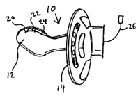
Figure 1 depicts a side view of the preferred embodiment of the invention.
Figure 2 illustrates a side cross-section of a nipple of the preferred
embodiment
of the invention.
Figures 3-10 depict side cross-sections of a nipple to illustrate various
alternative placements and arrangements of pulse oximeter elements according
to the
invention.
Figures 11-16 illustrate top views of various alternative placements and
arrangements of the pulse oximeter elements according to the invention.
Figures 17 and 18 depict the invention in use in a subject.
Figures 19 and 20 illustrate cross-sections of examples of attaching the
nipple
to the shield for the preferred embodiment of the invention.
CA 02399621 2002-08-08
WO 01/58349 PCT/USO1/04343
Figures 21 (a)-(b) depict an example of a shield structure for use in the
preferred
embodiment of the invention.
Figure 22 illustrates a top view of an alternative embodiment of the
invention.
Figure 23 depicts a top cross-section of another alternative embodiment of the
invention.
Figure 24 illustrates a rear view of the alternative embodiment of the
invention
illustrated in Figure 23.
Figure 25 depicts a block diagram for an alternative embodiment of the
invention illustrated in Figures 23 and 24.
Figure 26 illustrates a top view of another alternative embodiment of the
invention.
Figure 27 depicts a flowchart illustrating the steps for performing the
preferred
embodiment.
V. DETAILED DESCRIPTION OF THE INVENTION
Figures 1-18 illustrate a preferred embodiment and alternative component
arrangements of the pacifier oximeter sensor assembly. The assembly preferably
includes a pacifier 10, pulse oximeter sensor elements 20, 22, and wiring 24.
The pacifier 10 preferably includes a nipple (or baglet) 12 and a shield (or
guard) 14. The nipple 12 may be a variety of shapes in addition to those shown
in
Figures 1-18 that will allow the subject to apply a suction force to the
nipple 12.
Exemplary shapes for the nipple 12 include orthodontic, bottle nipple,
spherical, and
thumb shaped. The nipple 12 preferably is a flexible material typically used
to make
pacifiers and baby bottle nipples such as polypropylene, polyvinyl chloride,
silicones,
epoxies, polyester, thermoplastics, rubber, or similar flexible material.
Preferably, the
material used to make the nipple 12 will be at least partially translucent to
allow light to
pass through in the area of the pulse oximeter sensor elements 20, 22.
Preferably, the
6
CA 02399621 2002-08-08
WO 01/58349 PCT/US01/04343
nipple 12 will have an inner cavity 124 formed as a void in the nipple
material 122.
However, the nipple 12 may be solid or filled with a flexible material to
increase the
protection of the pulse oximeter sensor elements 20, 22 and wiring 24.
The pulse oximeter sensor elements 20, 22 preferably are within the material
122 making up the nipple 12 to reduce the impact of the material 122 on the
transmission of light through the material 122. However, the pulse oximeter
sensor
elements 20, 22 may be nested within the nipple material 122 as shown, for
example,
in Figure 4 or the pulse oximeter sensor elements 20, 22 may abut the nipple
material
122 on the inner cavity surface as shown, for example, in Figure 3. The pulse
oximeter sensor elements 20, 22 preferably will be placed in a position to
transmit light
and receive backscattered light from a capillary bed within the oral cavity of
the subject
as illustrated, for example, in Figures 17 and 18. The preferred locations are
along the
top of the nipple 12 (Figures 2-4), at the tip of the nipple 12 (Figure 5),
and along the
bottom of the nipple 12 (Figure 6). Also, the pulse oximeter elements 20, 22
may be
located in and/or along the nipple shank 126 as illustrated, for example, in
Figures 7-
10.
Preferably, the pulse oximeter sensor elements include a light source 20 and a
light detector 22. The placement and location of the light source 20 and the
light
detector 22 depicted in Figures 1-18 may be switched with respect to each
other.
Furthermore, the light source 20 and the light detector 22 may be in a variety
of
exemplary spatial locations relative to each other as shown, for example, in
Figures
11-16. Although Figures 11-16 illustrated the pulse oximeter sensor elements
20, 22
on the top of the nipple 12, these elements may have similar spatial locations
on other
portions of the nipple 12 such as the tip, bottom, and along the shank 126.
The light source 20 preferably emits at least two frequencies of light in the
red
region, for example with a wavelength of 660 nm, and in the infrared region,
for
7
CA 02399621 2002-08-08
WO 01/58349 PCT/USO1/04343
example with a wavelength of 940 nm, preferably in response to a signal from a
spectrophotometer, other similar oximeter monitoring devices or multiparameter
patient
monitoring systems that provide oximetry readings. The light source 20
preferably is
one or more of the following: two light emitters such as light emitting diodes
(LED), a
bispectral emitter, a dual spectral emitter, a photoemitter, or a
semiconductor die.
However, any light source that facilitates reflectance pulse oximetry may be
employed.
Typically, the two emitter arrangement will include a red LED around or at 660
nm and
a near-infrared LED emitting in the range of 890 to 950 nm and more
particularly at
about 940 nm. The light source 20 may emit light having a bandwidth, for
example, in
the range of 20 to 50 nm.
Preferably, the light detector 22 detects light emitted by the light source
20.
Signals representing the detected light are transmitted by the light detector
22 to a
spectrophotometer, an oximeter monitoring device or a multiparameter patient
monitoring system that provides oximetry readings by discriminating between
the
relative intensity of these emissions and provides an index as to the degree
of oxygen
saturation of hemoglobin in blood. Preferably, the light detector 22 may be
one of the
following: a photoelectric receiver, a photodetector, or a semiconductor die.
The wiring 24 preferably includes conductive lines and contact electrodes. The
wiring 24 preferably is embedded within the nipple material 122, or passes
through the
nipple cavity 124, or some combination of these two. An external cord 26
preferably is
insulated and connects to the wiring 24 at a proximal end of the pacifier 126
so that the
external cord 26 is outside of the oral cavity of the subject. The external
cord 26
preferably includes a standard plug design to interface with a pulse oximetry
spectrophotometer, a pulse monitor such as a plethysmograph, or other external
device. Alternatively, the external cord 26 may be a jack to connect to a
reusable
8
CA 02399621 2002-08-08
WO 01/58349 PCT/USO1/04343
cable such as the cable sold with the Nellcor~ OxiCliq~ systems (Mallinckrodt,
Inc., St.
Louis, Missouri, U.S.A.).
The nipple 12 preferably is attached or mounted to the shield 14. An example
of one type of mounting is integrally forming the nipple 12 with the shield
14, for
example by mechanically coupling the nipple 12 to the shield 14. Another
mounting
arrangement, as illustrated in Figure 19, is to have the nipple 12 include a
shank 126'
with two integral spaced collars 1262, 1264 to form a channel to receive the
shield 14.
Preferably, the shield 14 is at or near the proximal end of the shank 126'.
Preferably to
prevent the shield 14 from being pulled off the shank 126', a handle 16 is
looped
through the shank 126' as illustrated in Figure 19.
Another example of attaching the nipple 12 to the shield 14 is illustrated in
Figure 20. The shield 14 includes an opening for the nipple shank 126 to pass
through
preferably such that a rim or section of rolled up material 1266 is located on
the
proximal side of the shield 14. A plug 18 is inserted into the shield opening
142 to hold
the nipple shank 126 in place with respect to the shield 14. More preferably,
the plug
18 will include a securing mechanism that is compressed as it travels through
the
shield opening 142 and then expands on the distal side of the shield 14 to
secure the
plug 18 in place and hold the nipple 12 securely to the shield 14.
The shield 14 preferably is curved or bowed to form fit to the average baby's
face. The shield 14 may be any shape that prevents it from being pulled into
the
subject's mouth from the suction force placed upon the nipple 12 by the
subject. More
preferably, the shield 14 will be shaped or include a reference indicator such
that the
top of the pacifier 10 can be readily determined by looking at the shield 14.
In an
alternative embodiment, the shield 14 preferably includes a plurality of holes
(or relief
openings) 142 to allow for spit to be discharged without interference from the
pacifier
as illustrated, for example, in Figures 21 (a), 23, and 24. Figure 21 (b)
illustrates a
9
CA 02399621 2002-08-08
WO 01/58349 PCT/USO1/04343
relief opening 142' that allows insertion of a catheter such as an
endotracheal tube. A
further alternative is for the shield to include a mesh pattern over at least
a portion of it.
Another alternative embodiment adds a ring (or annular or other shaped handle)
16 on
the opposite side of the shield 14 from the nipple 12 as illustrated in
Figures 21 (a) and
22 that may attach to either the shield 14 or the nipple 12. Preferably, the
ring is
hinged, collapsible, and/or flexible.
An alternative embodiment of the invention is the placement of the oximeter
signal processing device within a housing 30 extending from the shield 14 on
the side
opposite the nipple 12 as illustrated, for example, in Figures 23 and 24. The
oximeter
signal processing device preferably is a miniature spectrophotometer. The
oximeter
signal processing device preferably will include a display 32, a power supply
(such as
a battery) 34, and a processor 36 to perform calculations and to drive the
display 32,
and an on-off button (or switch/mechanism) 38 as illustrated in Figure 25. The
display
32 preferably will show the blood oxygenation level of the subject as
illustrated in
Figure 24. More preferably, the display 32 is a digital display. The processor
36
preferably will connect to the wiring 24 running from the pulse oximeter
sensor
elements 20, 22, calculate the blood oxygenation level, and drive both the
display 32
and the light source 20. The processor 36 preferably is a circuit that
includes either an
analog circuit or an integrated circuit, which is either hardwired or
programmed.
Preferably, the display 32, the power supply 34, the processor 36 will reside
on a
printed circuit board that includes appropriate circuitry and provides a
connection to
wiring 24.
Another alternative embodiment of the invention is that the light source 20
and
the light detector 22 may be in wireless communication with the external
device
instead of connected with the external cord 26 as illustrated in Figure 26 as
a rod (or
antenna or transmitter) 40. Alternatively, the antenna 40 may take the shape
as a
CA 02399621 2002-08-08
WO 01/58349 PCT/USO1/04343
handle 16 similar to the one illustrated, for example, in Figures 21 and 22
without the
external cord 26. Preferably, the wireless communication will occur through an
antenna 40 extending away from the pacifier 10. The transmitter may be
incorporated
within the antenna 40 or some other housing incorporated into the shield 14.
Preferably, the antenna 40 will be sufficiently sturdy to withstand tugging
and being
played with during use by the subject. This alternative embodiment also
preferably
includes a power source such as a battery to power all of the electrical
components.
The power source preferably is located within the shield, a housing, or as
part of the
antenna 40.
A further alternative embodiment of the invention is to provide a bite block
on
the distal side of the shield 14 between the shield 14 and the nipple 12. The
bite block
may be an extension of the shield material or a hardened nipple shank 126. The
flexible nipple 12 preferably is attached to the bite block. Preferably, the
bite block will
provide a passageway through which the wiring 24 may pass through. The shield
14
and nipple 12 preferably would be shaped such that multiple catheters would
have
space to enter the oral cavity, for example, for suction and supplying oxygen.
This
alternative embodiment preferably would be for use during surgery of a variety
of
subjects other than infants and young children.
The device may be a retrofit of current pacifiers by inserting the pulse
oximeter
sensor elements from a disposable pulse oximeter like the Nellcor~ Oxisensor~
II
oximeters (Mallinckrodt, Inc., St. Louis, Missouri, U.S.A.) by stripping away
the
packaging and adhesive strip. The ring attached to must pacifiers would be
removed
leaving access to the interior cavity of the nipple into which the pulse
oximeter sensor
elements would be inserted such that they faced in the same general direction.
The
ring then would be reattached.
11
CA 02399621 2002-08-08
WO 01/58349 PCT/USO1/04343
In accordance with the present invention, there is a method to take oximeter
readings from different sites within a subject, which may be either human or
animal, for
the purposes of determining the amount of oxygen within the blood of the
subject. The
oximeter readings are accomplished using reflectance oximetry from capillary
beds
that are readily accessible within the subject. The capillary beds include,
for example,
the hard palate, the soft palate, the superior lingual surface, the inferior
lingual surface,
the gingivae, the mouth floor, the buccal surface, and any other surface
within the oral
cavity. Each of these capillary beds is accessible through the oral cavity,
which
extends from the lips to the oral portion of the pharynx, i.e., pars oralis.
Figure 27 illustrates a flowchart showing the steps for taking oximeter
readings
pursuant to the present invention. In the first step 110, which may actually
occur at a
later point but no later then the initiation of taking pulse oximeter
readings, the pulse
oximeter sensor elements are connected to an oximeter device such as a
spectrophotometer. In step 120, the pacifier 10 is inserted into the subject
through the
mouth. The placement of the pacifier 10 with a pulse oximeter sensor is
illustrated, for
example, in Figures 17 and 18. In step 130, reflectance pulse oximeter
readings are
taken from the relevant capillary bed. While taking the pulse oximeter
readings, the
pulse oximeter sensor elements preferably remain in contact with the relevant
capillary
bed to continue the flow of accurate oximeter readings.
The method according to the invention may be used in a variety of surgical,
anesthetic, critical care procedures or situations that include patients that
are awake,
sedated or recovering from general anesthesia.
The method of taking pulse oximeter readings from different surfaces within a
patient has been submitted to actual testing in the below-described population
and
according to the following protocols.
12
CA 02399621 2002-08-08
WO 01/58349 PCT/USO1/04343
Reflectance Oximetry from the Buccal Surface
The first protocol involved taking readings from the buccal surface. Nine
patients were monitored via buccal reflectance pulse oximetry over 20
consecutive
surgical procedures, which procedures consisted of burn excision and grafting.
Patients ranged in age from 23 to 56 years (Mean = 264.8, Standard Deviation
(SD) _
11.2) and ranged from 17 to 75 percent total body surface area (%TBSA) burned
(Mean = 274.3%, SD = 28.9). Each patient received from one to eight operations
(Mean = 4.01 ). Five of these nine patients arrived at the operating room
intubated for
all of the operations in this study. Four patients were induced and intubated
in a
standard fashion for all surgical procedures.
A Nellcor~ Oxisensor~ II D-25 was placed intraoraly between the lower teeth
and the left or right buccal surface of the cheek and lip, with the bispectral
emitter and
detector facing the buccal surface. This pulse oximeter sensor orientation was
used
for the duration of each case. In addition, a similar disposable oximetric
probe was
placed on a peripheral digit in the commonly accepted transillumination
configuration.
At five minute intervals throughout the case, values for both oximetric probes
were
coded on the anesthesia record.
The differences between the peripheral and buccal Sp02 (oxygen saturation of
hemoglobin) values were insignificant by t-tests for correlated means.
Concordance
rates as percent agreements were calculated for all cases. Average percent
agreement was 84% ranging from 25% to 100%. Three of the 20 samples had
percent
agreements less than 91%. In each of these cases, the peripheral pulse
oximeter
sensor appears to have failed, in two cases secondary to sepsis, and in
another
secondary to peripheral vasoconstriction in the face of a norepinepherine
infusion.
Buccal Sp02 readings in all three cases continued to be 97% or greater.
13
CA 02399621 2002-08-08
WO 01/58349 PCT/USO1/04343
This data suggests that buccal reflectance oximetry is a simple, accurate
means
of monitoring arterial oxygen saturation in the severely burned patient where
oximetric
monitoring presents a challenge. Given that central oximetry has been shown in
numerous studies to be more rapidly responsive to oxygen saturation
variability than
peripheral oximetry, as well as more directly reflective of central oxygen
saturation,
there are few drawbacks and considerable benefit from this method. Indeed, in
the
three examples in this study where percent agreements were low, the peripheral
oximetric probes were returning apparently erratic and/or generally low values
while
buccal oximetric readings remained at 97% or higher. All three of these
patients had
peripheral vascular compromise secondary to sepsis and/or a vasoconstricting
agent
(norepinepherine infusion).
It may appear from the study results, at first blush, that a full range of
Sp02
values was not tested and that the continuously high Sp02 readings are
spurious to the
technique. On the contrary, in order to obtain a Sp02 value greater or less
than 85% a
very specific set of relationships must be present relative to the bispectral
emitter and
light sensing oximetric elements. Thus, spuriously high values in particular
do not
consistently occur. High Sp02 values require the presence of saturated
hemoglobin.
Posterior Pharyngeal Reflectance Oximetry
The second protocol involved comparing posterior pharyngeal reflectance pulse
oximetry to conventional peripheral transillumination pulse oximetry in
difficult to
monitor burn patients. Eight patients' records were reviewed over fourteen
consecutive surgical procedures, all consisting of excision and grafting.
Patients
ranged in age from 9 to 43 years and ranged from 14.5% to 77.5% TBSA burned
(Mean = 30.4, SD = 22.1 ). The number of operations per patient ranged from
one to
fou r.
14
CA 02399621 2002-08-08
WO 01/58349 PCT/USO1/04343
A Nellcor~ Oxisensor~ II pulse oximeter probe was placed in the distal lumen
of
an appropriately sized oropharyngeal airway with sensor and emitter facing the
posterior pharynx. A similar probe was placed on a peripheral digit as a
transilluminating pulse oximeter. Sp02 values were noted at five-minute
intervals.
Concordance statistics as well as a t-test for correlated means were
calculated
between the simultaneously obtained Sp02 values.
The mean differences between pharyngeal reflectance and peripheral digital
transillumination Sp02 values were insignificant for all cases. Concordance
statistics
were as follows: 0.75 (n = 1 ) and 1.0 (n = 12).
Given the near perfect concordance statistics in this study, this data
suggests
that posterior pharyngeal reflectance oximetry is a simple, highly accurate
means of
monitoring arterial oxygen saturation in the severely burned patient where
oximetric
monitoring presents a challenge.
Lingual Surface Reflectance Oximetry
The third protocol involved taking readings from the lingual surface. Data was
reviewed for eight difficult to monitor patients who were monitored via
lingual
reflectance pulse oximetry over twenty-five consecutive surgical procedures,
all
consisting of burn excision and grafting. Patients ranged in age from 26 to 57
years
(Mean = 36.0, SD = 10.3). Patients ranged from 20% to 92% TBSA burned (Mean =
66.75%, SD = 26.42). Number of operations per patient ranged from one to five
(Mean
= 3.13, SD = 1.55). Six of these eight patients arrived at the operating room
intubated
for all of the operations in this study. Two patients were induced and
intubated in a
standard fashion.
In each case, a Nellcor~ Oxisensor~ II D-25 was centered flat on the superior
lingual surface with the detector and the bispectral emitter facing the
lingual surface.
This pulse oximeter configuration was used for the duration of each case. When
CA 02399621 2002-08-08
WO 01/58349 PCT/USO1/04343
clinically indicated, an arterial blood gas (ABG) sample was drawn and the
Sp02 noted
for clinical monitoring and prior to transfusion in every case. All had
multiple ABG's
drawn and all patients were transfused. The ABG Sa02 (oxygen saturation of
arterial
blood) was noted in each case.
Descriptive statistics and a concordance rate as well as a t-test for
correlated
means were calculated between the simultaneously obtained Sp02 and Sa02
values.
The difference between the Sp02 and Sa02 values was insignificant by t-test
for
correlated means (t = 1.25, df = 24, NS). Upon inspection, the means were very
close
and the standard deviations were very small, as were the SEM's, all suggesting
very
little difference or variability between these two measures of oxygen
saturation. A
concordance rate of 92% was calculated (+ 1.5%) showing a high degree of
relationship between lingual and ABG Sa02.
This data suggests that lingual reflectance oximetry is a simple, accurate
means
of monitoring arterial oxygen saturation in the severely burned patient where
oximetric
monitoring presents a challenge. An existing disposable pulse oximeter sensor
was
utilized in this study saving the cost of specially designed equipment. Given
that
central oximetry has been shown to be more rapidly responsive to oxygen
saturation
variability than peripheral oximetry, there are few drawbacks and considerable
benefit
from this method.
VI. INDUSTRIAL APPLICABILITY
The invention is particularly useful for monitoring the blood oxygen content
of a
subject, more particularity a child or infant. The invention is also useful
when other
sites are not available on the patient such as a patient with severe burns
covering most
of their body or a restless child who is prone to remove attached oximeters to
fingers
and other body parts. The invention may be used by hospital personnel,
emergency
16
CA 02399621 2002-08-08
WO 01/58349 PCT/USO1/04343
medical crews, in-home medical personnel, laboratory and veterinary personnel
and
battlefield medical personnel.
Those skilled in the art will appreciate that various adaptations and
modifications of the above-described devices and steps can be configured
without
departing from the scope and spirit of the their use in the method. Therefore,
it is to be
understood that, within the scope of the appended claims, the method may be
practiced and arranged other than as specifically described herein.
Furthermore, the
above-described embodiments may be used in a variety of combinations.
17