Note: Descriptions are shown in the official language in which they were submitted.
CA 02400319 2006-12-08
1
COATING THAT PROMOTES ENDOTHELIAL CELL ADHERENCE
FIELD OF THE INVENTION
The present invention relates to the field of medical devices implanted in
vessels
within the body. More particularly, the present invention relates to stents or
synthetic
grafts implanted in blood vessels that incorporate a matrix which promotes
adherence of
endothelial cells to the sent or synthetic graft.
BACKGROUND OF THE INVENTION
It is well known that vascular disease and atherosclerosis in particular is
one of the
leading causes of death and disability in the world. Atherosclerosis involves
the deposition
of fatty plaques on the lumenal surface of arteries. The deposition of fatty
plaques on the
lumenal surface of the artery causes narrowing of the cross-sectional area of
the artery.
Ultimately, this deposition blocks blood flow distal to the lesion causing
ischemic damage
to the tissues supplied by the artery.
Coronary arteries supply the heart with blood. Coronary artery atherosclerosis
disease (CAD) is the most common, serious, chronic, life-threatening illness
in the United
States, affecting more than 11 million persons. The social and economic costs
of coronary
atherosclerosis vastly exceed that of most other diseases. Narrowing of the
coronary artery
lumen causes destruction of heart muscle resulting first in angina, followed
by myocardial
infarction and finally death. There are over 1.5 million myocardial
infarctions in the
United States each year. Six hundred thousand (or 40%) of those patients
suffer an acute
myocardial infarction and more than three hundred thousand of those patients
die before
reaching the hospital. (Harrison's Principles of Internal Medicine, 14" '
Edition, 1998).
CAD can be treated using percutaneous translumenal coronary balloon
angioplasty
(PTCA). More than 400,000 PTCA procedures are performed each year in the
United
States. In PTCA, a balloon catheter is inserted into a peripheral artery and
threaded
through the arterial system into the blocked coronary artery. The balloon is
then inflated,
the artery stretched, and the obstructing fatty plaque flattened, thereby
increasing the cross-
sectional flow of blood through the affected artery. The therapy, however,
does not usually
result in a permanent opening of the affected coronary artery. As many as 50%
of the
patients who are treated by PTCA require a
CA 02400319 2002-08-22
WO 01/68158 PCT/US01/08244
2
repeat procedure within six months to correct a re-narrowing of the coronary
artery. Medically,
this re-narrowing of the artery after treatment by PTCA is called restenosis.
Acutely, restenosis
involves recoil and shrinkage of the vessel. Subsequently, recoil and
shrinkage of the vessel are
followed by proliferation of medial smooth muscle cells in response to injury
of the artery from
PTCA. In part, proliferation of smooth muscle cells is mediated by release of
various
inflammatory factors from the injured area including thromboxane A2, platelet
derived growth
factor (PDGF) and fibroblast growth factor (FGF). A number of different
techniques have been
used to overcome the problem of restenosis, including treatment of patients
with various
pharmacological agents or mechanically holding the artery open with a stent.
(Harrison's
Principles of Internal Medicine,14'' Edition, 1998).
Of the various procedures used to overcome restenosis, stents have proven to
be
the most effective. Stents are metal scaffolds that are positioned in the
diseased vessel segment to
create a normal vessel lumen. Placement of the stent in the affected arterial
segment prevents
recoil and subsequent closing of the artery. Stents can also prevent local
dissection of the artery
along the medial layer of the artery. By maintaining a larger lumen than that
created using PTCA
alone, stents reduce restenosis by as much as 30%. Despite their success,
stents have not
eliminated restenosis entirely_ (Suryapranata et al. 1998. Randomized
comparison of coronary
stenting with balloon angioplasty in selected patients with acute myocardial
infarction.
Circulation 97:2502-2502).
Narrowing of the arteries can occur in vessels other than the coronary
arteries,
including the aortoiliac, infrainguinal, distal profunda femoris, distal
popliteal, tibial, subclavian
and mesenteric arteries. The prevalence of peripheral artery atherosclerosis
disease (PAD)
depends on the particular anatomic site affected as well as the criteria used
for diagnosis of the
occlusion. Traditionally, physicians have used the test of intermittent
claudication to determine
whether PAD is present. However, this measure may vastly underestimate the
actual incidence of
the disease in the population. Rates of PAD appear to vary with age, with an
increasing incidence
of PAD in older individuals. Data from the National Hospital Discharge Survey
estimate that
every year, 55,000 men and 44,000 women had a first-listed diagnosis of
chronic PAD and 60,000
men and 50,000 women had a first-listed diagnosis of acute PAD. Ninety-one
percent of the
acute PAD cases involved the lower extremity. The prevalence of comorbid CAD
in patients with
PAD can exceed 50%. In addition, there is an increased prevalence of
cerebrovascular disease
among patients with PAD.
PAD can be treated using percutaneous translumenal balloon angioplasty (PTA).
The use of stents in conjunction with PTA decreases the incidence of
restenosis. However, the
CA 02400319 2002-08-22
WO 01/68158 PCT/US01/08244
3
post-operative results obtained with medical devices such as stents do not
match the results
obtained using standard operative revascularization procedures, i.e., those
using a venous or
prosthetic bypass material. (Principles of SurgerX, Schwartz et al. eds.,
Chapter 20, Arterial
Disease, 7th Edition, McGraw-Hill Health Professions Division, New York 1999).
Preferably, PAD is treated using bypass procedures where the blocked section
of
the artery is bypassed using a graft. (Principles of Surgery, Schwartz et al.
eds., Chapter 20,
Arterial Disease, 7th Edition, McGraw-Hill Health Professions Division, New
York 1999). The
graft can consist of an autologous venous segment such as the saphenous vein
or a synthetic graft
such as one made of polyester, polytetrafluoroethylene (PTFE), or expanded
polytetrafluoroethylene (ePTFE). The post-operative patency rates depend on a
number of
different factors, including the lumenal dimensions of the bypass graft, the
type of synthetic
material used for the graft and the site of outflow. Restenosis and
thrombosis, however, remain
significant problems even with the use of bypass grafts. For example, the
patency of infrainguinal
bypass procedures at 3 years using an ePTFE bypass graft is 54% for a femoral-
popliteal bypass
and only 12% for a femoral-tibial bypass.
Consequently, there is a significant need to improve the performance of both
stents
and synthetic bypass grafts in order to further reduce the morbidity and
mortality of CAD and
PAD.
With stents, the approach has been to coat the stents with various anti-
thrombotic
or anti-restenotic agents in order to reduce thrombosis and restenosis. For
example, impregnating
stents with radioactive material appears to inhibit restenosis by inhibiting
migration and
proliferation of myofibroblasts. (U. S. Patent Nos. 5,059,166, 5,199,939 and
5,302,168).
Irradiation of the treated vessel can pose safety problems for the physician
and the patient. In
addition, irradiation does not permit uniform treatment of the affected
vessel.
Alternatively, stents have also been coated with chemical agents such as
heparin or
phosphorylcholine, both of which appear to decrease thrombosis and restenosis.
Although
heparin and phosphorylcholine appear to markedly reduce restenosis in animal
models in the short
term, treatment with these agents appears to have no long-term effect on
preventing restenosis.
Additionally, heparin can induce thrombocytopenia, leading to severe
thromboembolic
complications such as stroke. Nonetheless, it is not feasible to load stents
with sufficient
therapeutically effective quantities of either heparin or phosphorylcholine to
make treatment of
restenosis in this manner practical.
Synthetic grafts have been treated in a variety of ways to reduce
postoperative
restenosis and thrombosis. (Bos et al. 1998. Small-Diameter Vascular Graft
Prostheses:Current
CA 02400319 2002-08-22
WO 01/68158 PCT/US01/08244
4
Status Archives Physio. Biochem. 106:100-115). For example, composites of
polyurethane such
as meshed polycarbonate urethane have been reported to reduce restenosis as
compared with
ePTFE grafts. The surface of the graft has also been modified using
radiofrequency glow
discharge to add polyterephalate to the ePTFE graft. Synthetic grafts have
also been impregnated
with biomolecules such as collagen. However, none of these approaches has
significantly reduced
the incidence of thrombosis or restenosis over an extended period of time.
Because endothelial cells possess certain intrinsic characteristics such as
cell
regulatory molecules that decrease the incidence of thrombosis or restenosis,
stimulating the
development of an endothelial cell monolayer on the surface of stents or
synthetic grafts may
prevent both restenosis and thrombosis. (Belle et al. 1997. Stent
Endothelialization. Circulation
95:438-448; Bos et al. 1998. Small-Diameter Vascular Graft Prostheses:Current
Status Archives
Physio. Biochem. 106:100-115).
Endothelial cells have been deposited on the surface of stents by local
delivery of
vascular endothelial growth factor (VEGF), an endothelial cell mitogen, after
implantation of the
stent. (Belle et al. 1997. Stent Endothelialization. Circulation 95:438-448.).
Because the
application of VEGF can have systemic as well as local effects, this form of
treatment may be
unreliable.
Synthetic grafts have also been seeded with endothelial cells, but the
clinical results
with endothelial seeding have been generally poor, i.e., low post-operative
patency rates (Lio et
al. 1998. New concepts and Materials in Microvascular Grafting: Prosthetic
Graft Endothelial
Cell Seeding and Gene Therapy. Microsurgery 18:263-256).
Accordingly, there is a need for development of new methods and compositions
for coating medical devices, including stents and synthetic grafts, with
endothelial cells. This type
of coating will not only prevent restenosis, but also thromboembolic
complications resulting from
stent implantation. Methods and compositions that provide such improvement
will elinunate the
drawbacks of previous technology and have a significant positive impact on the
morbidity and
mortality associated with CAD and PAD. It is the object of this invention to
prepare stents and
synthetic grafts coated in such a manner as to stimulate adherence of
endothelial cells to a medical
device such as a stent or synthetic graft.
SUMMARY OF THE INVENTION
The invention provides methods and compositions for coating medical devices
with a matrix that promotes adherence of endothelial cells to a medical
device. The matrix
CA 02400319 2002-08-22
WO 01/68158 PCT/US01/08244
incorporates antibodies that stimulate adherence of endothelial cells to the
surface of the medical
device.
As used herein, "medical device" refers to a device that is introduced
temporarily
or permanently into a mammal for the prophylaxis or therapy of a medical
condition. These
5 devices include any that are introduced subcutaneously, percutaneously or
surgically to rest within
an organ, tissue or lumen. Medical devices may include stents, covered stents
such as those
covered with polytetrafluoroethylene (PTFE), or expanded
polytetrafluoroethylene (ePTFE),
synthetic grafts, artificial heart valves, artificial hearts and fixtures to
connect the prosthetic organ
to the vascular circulation, venous valves, abdominal aortic aneurysm (AAA)
grafts, inferior venal
caval filters, permanent drug infusion catheters, embolic coils, embolic
materials used in vascular
embolization (e.g., PVA foams), and vascular sutures.
Coating of the medical device with the compositions and methods of this
invention
may stimulate the development of an endothelial cell layer on the surface of
the medical device,
thereby preventing restenosis as well as other thromboembolic complications
that result from
implantation of the medical device.
Synthetic grafts and stents can be used for treating CAD or PAD. A stent or
synthetic graft may be coated with a matrix incorporating antibodies that
stimulate adherence of
circulating progenitor endothelial cells to the medical device. The antibodies
may comprise
monoclonal antibodies reactive with endothelial cell surface antigens such as
CD34, an antigen
expressed on the surface of progenitor endothelial cells. Fab fragments of the
monoclonal
antibody may be used. In another embodiment, monoclonal antibodies directed
against other
endothelial surface antigens such as KDR or Tie-2, may also be used. In one
embodiment, a
single type of antibody that reacts with one antigen may be used.
Alternatively, a plurality of
different antibodies directed against different endothelial cell surface
antigens may be mixed
together and added to the matrix.
The matrix coating the medical device may be composed of synthetic material,
such as polyurethane, poly-L-lactic acid, cellulose ester or polyethylene
glycol. In another
embodiment, the matrix is composed of naturally occurring materials, such as
collagen, fibrin,
elastin or amorphous carbon. The matrix may comprise several layers with a
first layer being
composed of synthetic or naturally occurring materials and a second layer
composed of
antibodies. The layers may be ordered sequentially, with the first layer
directly in contact with the
stent or synthetic graft surface and the second layer having one surface in
contact with the first
layer and the opposite surface in contact with the vessel lumen.
CA 02400319 2006-01-16
6
In a third embodiment, the matrix may comprise fullerenes, where the
fullerenes range from
about C60 to about C 100. The fullerenes may also be arranged as nanotubes,
that incorporate
molecules or proteins. The fullerene matrix may also be mixed with PTFE or
ePTFE, or antibodies.
Alternatively, the PTFE or ePTFE may be layered first on the medical device
followed by a second
layer of fullerenes.
The matrix may be noncovalently or covalently attached to the medical device.
Antibodies
may be covalently attached to the matrix using hetero- or homobifunctional
cross-linking reagents.
Methods of treatment of atherosclerosis are also provided. The artery may be
the either a
coronary artery or a peripheral artery such as the femoral artery.
According to the present invention, then, there is provided a medical device
comprising a
coating rendering the medical device compatible for in vivo attachment and
proliferation of cells on
the surface thereof, wherein the coating comprises a therapeutically effective
amount of at least one
type of antibody, or fragment thereof, which reacts with an endothelial cell
surface antigen, one or
more layers of a matrix.
According to a further aspect of the present invention, there is provided a
composition for
rendering a medical device compatible for in vivo attachment and proliferation
of cells on the
surface thereof, wherein the coating composition comprises a matrix and a
therapeutically effective
amount of at least one type of antibody, or fragment thereof, that reacts with
an endothelial cell
surface antigen.
According to yet a further aspect of the present invention, there is provided
a method for
rendering a medical device compatible for in vivo attachment and proliferation
of cells on the
surface thereof, comprising the steps: (a) coating the medical device with at
least one layer of a
matrix; and (b) adding to the matrix layer a therapeutically effective amount
of at least one type of
antibody, or fragment thereof, which reacts with an endothelial cell surface
antigen.
CA 02400319 2002-08-22
WO 01/68158 PCT/US01/08244
7
Brief Description of the Figures
Figure 1 shows an antibody tethered covalently to the matrix by a cross-
linking molecule.
Figure 2 shows a diagram of the C600 molecule anchoring the matrix.
DETAILED DESCRIPTION OF THE INVENTION
Overview
The present invention provides methods and compositions that involve coating a
medical device such as a stent or synthetic graft with a matrix which is then
used to coat the
medical device. In one embodiment, the matrix incorporates a therapeutically
effective amount of
at least one type of antibody that promotes adherence of endothelial cells to
the medical device.
Following adherence, the endothelial cells differentiate and proliferate on
the surface of the
matrix. The presence of endothelial cells on the medical device reduces the
occurrence of
restenosis and thrombosis after medical device implantation into a vessel.
As used herein, the term "antibody" refers to one type of monoclonal or
polyclonal
antibody, where the monoclonal or polyclonal antibody binds to one antigen or
a functional
equivalent of that antigen. The term antibody encompasses any fragment of an
antibody such as
Fab, F(ab')2 or Fc fragments. (An antibody encompasses a plurality of
individual antibody
molecules equal to 6.022 x 10²³ molecules per mole of antibody).
As used herein, a "therapeutically effective amount of the antibody" means the
amount of an antibody that promotes adherence of endothelial cells to the
medical device. The
amount of an antibody needed to practice the claimed invention varies with the
nature of the
antibody used. For example, the amount of an antibody used will depend on the
binding constant
between the antibody and the antigen against which it reacts. It is well known
to those of
ordinary skill in the art how to determine therapeutically effective amounts
of an antibody to use
with a particular antigen.
As used herein, "medical device" refers to a device that is introduced
temporarily
or permanently into a mammal for the prophylaxis or therapy of a medical
condition. These
devices include any that are introduced subcutaneously, percutaneously or
surgically to rest within
an organ, tissue or lumen. Medical devices may include, stents, covered stents
such as those
covered with PTFE, or ePTFE, synthetic grafts, artificial heart valves,
artificial hearts and fixtures
to connect the prosthetic organ to the vascular circulation, venous valves,
abdominal aortic
aneurysm (AAA) grafts, inferior venal caval filters, permanent drug infusion
catheters, embolic
coils, embolic materials used in vascular embolization (e.g., PVA foams), and
vascular sutures.
CA 02400319 2006-01-16
8
As used herein,"restenosis" refers to the accumulation of a layer of smooth
muscle cells and
matrix protein in the intima of an arterial wall. Vessels may become
obstructed because of
restenosis. After PTCA or PTA, smooth muscle cells from the media and
adventitia, which are not
normally present in the intima, proliferate and migrate to the intima and
secrete proteins, forming
an accumulation of smooth muscle cells and matrix protein within the intima.
This accumulation
causes a narrowing of the lumen of the artery, reducing blood flow distal to
the narrowing. As used
herein, "inhibition of restenosis" refers to the inhibition of migration and
proliferation of smooth
muscle cells accompanied by prevention of protein secretion so as to prevent
restenosis and the
complications arising therefrom.
The subjects that can be treated using the methods and compositions of this
invention may
be a mammal, or more specifically, a human, dog, cat, pig, rodent or monkey.
The methods of the present invention may be practiced in vivo or in vitro.
The term "endothelial cell" refers to endothelial cells at any developmental
stage, from
progenitor to mature. Fully differentiated endothelial cells may be isolated
from an artery or vein
such as a human umbilical vein, while progenitor endothelial cells are
isolated from peripheral
blood or bone marrow. The endothelial cells are bound to the medical devices
by incubation of the
endothelial cells with a medical device coated with the matrix that
incorporates an antibody or other
agent that adheres to endothelial cells.
The methods of this invention may be practiced on any artery or vein. Included
within the
scope of this invention is atherosclerosis of any artery including coronary,
infrainguinal, aortoiliac,
subclavian, mesenteric and renal arteries. Other types of vessel obstructions,
such as those resulting
from a dissecting aneurysm are also encompassed by the invention.
The medical device may be coated with endothelial cells after insertion into a
vessel.
Alternatively, the medical device is coated with the endothelial cells before
insertion of the medical
device. In either case, the presence of endothelial cells on the lumenal
surface of the medical device
inhibits or prevents restenosis and thrombosis.
Endothelial Cells
Human umbilical vein endothelial cells (HUVEC) are obtained from umbilical
cords
according to the methods of Jaffe, et al., J. Clin. Invest., 52:2745-2757,
1973. Briefly, cells are
stripped from the blood vessel walls by treatment with collagenase and
cultured in gelatin-coated
tissue culture flasks in M199 medium containing 10%
CA 02400319 2006-01-16
9
low endotoxin fetal calf serum, 90 ug/ml preservative-free porcine heparin, 20
ug/mI endothelial
cell growth supplement (ECGS), glutamine and antibodies.
Progenitor endothelial cells are isolated from human peripheral blood
according to the
methods of Asahara et al. (Isolation of putative progenitor endothelial cells
for angiogenesis.
Science 275:964-967, 1997). Magnetic beads coated with antibody to CD34 are
incubated with
human peripheral blood. After incubation, bound cells are eluted and can be
cultured in M- 199
containing 20% fetal bovine serum and bovine brain extract. (Clonetics, San
Diego, CA). Cells are
characterized by fluorescent antibodies to CD45, CD34, CD3 1, Flk-1, Tie-2,
and E-selectin.
Endothelial cells are transfected with any mammalian expression vectors that
contains any
cloned genes encoding proteins such as platelet derived growth factor (PDGF),
fibroblast growth
factor (FGF), or nitric oxide synthase (NOS) using conventional methods. (See,
for example,
mammalian expression vectors and transfection kits commercially available from
Stratagene, San
Diego, CA). For example, purified porcine progenitor endothelial cells are
transfected with vascular
endothelial growth factor (VEGF) using an adenoviral expression vector
expressing the VEGF
cDNA according to the methods of Rosengart et al. (Six-month assessment of a
phase I trial of
angiogenic gene therapy for the treatment of coronary artery disease using
direct intramyocardial
administration of an adenovirus vector expressing the VEGF121 cDNA. Ann. Surg.
230
(4):466-470 (1999)).
Antibodies
Monoclonal antibodies useful in the method of the invention may be produced
according to
the standard techniques of Kohler and Milstein (Continuous cultures of fused
cells secreting
antibody of predefined specificity. Nature 265:495-497, 1975). Endothelial
cells can be used as the
immunogen to produce monoclonal antibodies directed against endothelial cell
surface antigens.
Monoclonal antibodies directed against endothelial cells are prepared by
injecting
HUVEC or purified progenitor endothelial cells into a mouse or rat. After a
sufficient time, the
mouse is sacrificed and spleen cells are obtained. The spleen cells are
immortalized by fusing them
with myeloma cells or with lymphoma cells, generally in the presence of a non-
ionic detergent, for
example, polyethylene glycol. The resulting cells, which include the fused
hybridomas, are allowed
to grow in a selective medium, such as HAT-medium, and the surviving cells are
grown in such
medium using limiting dilution conditions. The cells are grown in a suitable
container, e. g.,
microtiter wells, and the supernatant is screened for monoclonal antibodies
having the desired
CA 02400319 2002-08-22
WO 01/68158 PCT/US01/08244
specificity, i.e., reactivity with endothelial cell antigens.
Various techniques exist for enhancing yields of monoclonal antibodies such as
injection of the hybridoma cells into the peritoneal cavity of a mammalian
host which accepts the
cells and then harvesting the ascites fluid. Where an insufficient amount of
monoclonal antibody
5 collects in the ascites fluid, the antibody is harvested from the blood of
the host. Various
conventional ways exist for isolation and purification of monoclonal
antibodies so as to free the
monoclonal antibodies from other proteins and other contaminants.
Also included within the scope of the invention are useful binding fragments
of
anti-endothelial cell monoclonal antibodies such as the Fab, F(ab')2, or Fc
fragments of these
10 monoclonal antibodies. The antibody fragments are obtained by conventional
techniques. For
example, useful binding fragments may be prepared by peptidase digestion of
the antibody using
papain or pepsin.
Antibodies of the invention are directed to an antibody of the IgG class from
a murine source;
however, this is not meant to be a limitation. The above antibody and those
antibodies having
functional equivalency with the above antibody, whether from a murine source,
mammalian
source including human, or other sources, or combinations thereof are included
within the scope
of this invention, as well as other classes such as IgM, IgA, IgE, and the
like, including isotypes
within such classes. In the case of antibodies, the term "functional
equivalency" means that two
different antibodies each bind to the same antigenic site on an antigen, in
other words, the
antibodies compete for binding to the same antigen. The antigen may be on the
same or different
molecule.
In one embodiment, monoclonal antibodies reacting with the endothelial cell
surface antigen CD34 are used. Anti-CD34 monoclonal antibodies attached to a
solid support
have been shown to capture progenitor endothelial cells from human peripheral
blood. After
capture, these progenitor cells are capable of differentiating into
endothelial cells. (Asahara et al.
1997. Isolation of putative progenitor endothelial cells for angiogenesis.
Science 275:964-967.)
Hybridomas producing monoclonal antibodies directed against CD34 can be
obtained from the
American Type Tissue Collection. (Rockville, NID). In another embodiment,
monoclonal
antibodies reactive with endothelial cell surface antigens Flk-1 or Tie-2 are
used.
Polyclonal antibodies reactive against endothelial cells isolated from the
same
species as the one receiving the medical device implant may also be used.
Stent
CA 02400319 2006-01-16
11
The term "stent" herein means any medical device which when inserted into the
lumen of a
vessel expands the cross-sectional lumen of a vessel. The term "stent"
includes covered stents such
as those covered with PTFE or ePTFE. In one embodiment, this includes stents
delivered
percutaneously to treat coronary artery occlusions or to seal dissections or
aneurysms of the splenic,
carotid, iliac and popliteal vessels. In another embodiment, the stent is
delivered into a venous
vessel. The stent can be composed of polymeric or metallic structural elements
onto which the
matrix is applied or the stent can be a composite of the matrix intermixed
with a polymer. For
example, a deformable metal wire stent can be used, such as that disclosed in
U. S. Pat. No.
4,886,062 to Wiktor. A self-expanding stent of resilient polymeric material
such as that disclosed in
published international patent application W091/12779 "lntraluminal Drug
Eluting Prosthesis", can
also be used. Stents may also be manufactured using stainless steel, polymers,
nickel-titanium,
tantalum, gold, platinum-iridium, or ElgiloyTM and MP35NTM and other ferrous
materials. Stents are
delivered through the body lumen on a catheter to the treatment site where the
stent is released from
the catheter, allowing the stent to expand into direct contact with the
lumenal wall of the vessel. It
will be apparent to those skilled in the art that other self-expanding stent
designs (such as resilient
metal stent designs) could be used with the antibodies and matrices of this
invention.
Synthetic Graft
The term "synthetic graft" means any artificial prosthesis having
biocompatible
characteristics. In one embodiment this includes synthetic grafts made of
DacronTM (polyethylene
terephthalate, PET) or TeflonTM (ePTFE). In another embodiment, synthetic
grafts are composed of
polyurethane. In yet a third embodiment, a synthetic graft is composed of an
inner layer of meshed
polycarbonate urethane and an outer layer of meshed DacronTM. It will be
apparent to those skilled
in the art that any biocompatible synthetic graft can be used with the
antibodies and matrices of this
invention. (Bos et al. 1998. Small-Diameter Vascular Prostheses : Current
Status. Archives Physio
Biochem. 106:100-115). Synthetic grafts can be used for end-to-end anastomosis
of vessels or for
bypass of a diseased vessel segment.
Matrix
(A) Synthetic Materials - The matrix that is used to coat the stent or
synthetic graft may be selected
from synthetic materials such as polyurethane, segmented polyurethane-urea and
heparin complex,
poly-L-lactic acid, cellulose ester or polyethylene glycol.
CA 02400319 2006-01-16
12
(B) Naturally Occurring Material - The matrix may be selected from naturally
occurring substances
such as collagen, laminin, heparin, fibrin, cellulose or carbon. A primary
requirement for the matrix
is that it be sufficiently elastic and flexible to remain unruptured on the
exposed surfaces of the
stent or synthetic graft.
(C) Fullerenes - The matrix may also comprise a fullerene (the term
"fullerene" encompasses a
plurality of fullerene molecules). Fullerenes are carbon-cage molecules. The
number of carbon (C)
molecules in a fullerene species varies from about C60 to about C 100.
Fullerenes are produced by
high temperature reactions of elemental carbon or of carbon-containing species
by processes well
known to those skilled in the art; for example, by laser vaporization of
carbon, heating carbon in an
electric arc or burning of hydrocarbons in sooting flames. (U.S. Patent No.
5,292,813, to Patel et al.;
U.S. Patent No. 5,558,903 to Bhushan et al.). In each case, a carbonaceous
deposit or soot is
produced. From this soot, various fullerenes are obtained by extraction with
appropriate solvents,
such as toluene. The fullerenes are separated by known methods, in particular
by high performance
liquid chromatography (HPLC). Fullerenes may be synthesized or obtained
commercially from
Dynamic Enterprises, Ltd., Berkshire, England or Southern Chemical Group, LLC,
Tucker,
Georgia.
Fullerenes may be deposited on surfaces in a variety of different ways,
including,
sublimation, laser vaporization, sputtering, ion beam, spray coating, dip
coating, roll-on or brush
coating as disclosed in U.S. Patent No.5,558,903.
An important feature of fullerenes is their ability to form "activated
carbon." The fullerene
electronic structure is a system of overlapping pi-orbitals, such that a
multitude of bonding
electrons are cooperatively presented around the surface of the molecule.
(Chemical and
EngineeringNews, Apr. 8, 1991, page 59). As forms of activated carbon,
fullerenes exhibit
substantial van der Waals forces for weak interactions. The adsorptive nature
of the fullerene
surface may lend itself to additional modifications for the purpose of
directing specific cell
membrane interactions. For example, specific molecules that possess chemical
properties that
selectively bind to cell membranes of particular cell types or to particular
components of cell
membranes, e. g., lectins or antibodies, can be adsorbed to the fullerene
surface. The fullerene
surface may also be chemically modified to present specifically reactive
groups to the cell
membrane, e. g., oxidants or reductants. Attachment of different molecules to
the fullerene surface
may be manipulated to create surfaces that selectively bind various cell
types, e.g., epithelial cells,
fibroblasts, primary explants, or T-cell subpopulations. U. S. Patent No.
5,310,669 to Richmond et
al.; Stephen R.
CA 02400319 2006-01-16
13
Wilson, Biological Aspects of Fullerenes, Fullerenes : Chemistry, Physics and
Technology, Kadish
et al. eds., John Wiley & Sons, NY 2000.
Fullerenes may also form nanotubes that incorporate other atoms or molecules.
(Liu et al.
Science 280:1253-1256 (1998)). The synthesis and preparation of carbon
nanotubes is well known
in the art. (U. S. Patent No. 5,753,088 to Olk et al., and U. S. Patent No.
5,641,466 to Ebbsen et al.).
Molecules such as proteins may also be incorporated inside carbon nanotubes.
For example,
nanotubes may be filled with the enzymes, e.g., ZnZCdZ metallothionein,
cytochromes C and C3,
and beta-lactamase after cutting the ends of the nanotube. (Davis et al.
Inoraanica Chim. Acta
272:261 (1998); Cook et al. Full Sci. Tech. 5(4):695 (1997)).
Three dimensional fullerene structures may also be used. U. S. Patent No.
5,338,571 to
Mirkin et al., discloses three-dimensional, multilayer fullerene structures
that are formed on a
substrate surface by (i) chemically modifying fullerenes to provide a bond-
forming species; (ii)
chemically treating a surface of the substrate to provide a bond-forming
species effective to
covalently bond with the bond-forming species of the fullerenes in solution;
and, (iii) contacting a
solution of modified fullerenes with the treated substrate surface to form a
fullerene layer
covalently bonded to the treated substrate surface.
(D) Application of the Matrix to the Medical Device - The matrix should adhere
tightly to the
surface of the stent or synthetic graft. Preferably, this is accomplished by
applying the matrix in
successive thin layers. Each layer of matrix may incorporate the antibodies.
Alternatively,
antibodies may be applied only to the layer in direct contact with the vessel
lumen. Different types
of matrices may be applied successively in succeeding layers. The antibodies
may be covalently or
noncovalently coated on the matrix after application of the matrix to the
stent.
In order to coat a medical device such as a stent, the stent is dipped or
sprayed with a liquid
solution of the matrix of moderate viscosity. After each layer is applied, the
stent is dried before
application of the next layer. In one embodiment, a thin, paint-like matrix
coating does not exceed
an overall thickness of 100 microns.
For example, a suitable matrix coating solution is prepared by dissolving 480
milligrams
(mg) of a drug carrier, such as poly-D, L-lactid acid (available as R203 of
Boehringer Inc.,
Ingelheim, Germany) in 3 milliliters (ml) of chloroform under aseptic
conditions. In principle,
CA 02400319 2006-01-16
14
however, any biodegradable (or non-biodegradable) matrix that is blood-and
tissue-compatible
(biocompatible) and can be dissolved, dispersed or emulsified may be used as
the matrix if, after
application, it undergoes relatively rapid drying to a self-adhesive lacquer-
or paint-like coating on
the medical device.
For example, coating a stent with fibrin is well known to one of ordinary
skill in the art. In
U.S. Patent No. 4,548,736 issued to Muller et al., fibrin is clotted by
contacting fibrinogen with
thrombin. Preferably, the fibrin in the fibrin-containing stent of the present
invention has Factor
XIII and calcium present during clotting, as described in U.S. Patent No.
3,523,807 issued to
Gerendas, or as described in published European Patent Application 0366564, in
order to improve
the mechanical properties and biostability of the implanted device.
Preferably, the fibrinogen and
thrombin used to make fibrin in the present invention are from the same animal
or human species as
that in which the stent will be implanted in order to avoid any inter-species
immune reactions ,e.g.,
human anti-cow. The fibrin product can be in the form of a fine, fibrin film
produced by casting the
combined fibrinogen and thrombin in a film and then removing moisture from the
film osmotically
through a semipermeable membrane. In the European Patent Application 0366564,
a substrate
(preferably having high porosity or high affinity for either thrombin or
fibrinogen) is contacted with
a fibrinogen solution and with a thrombin solution. The result is a fibrin
layer formed by
polymerization of fibrinogen on the surface of the medical device. Multiple
layers of fibrin applied
by this method could provide a fibrin layer of any desired thickness.
Alternatively, the fibrin can
first be clotted and then ground into a powder which is mixed with water and
stamped into a desired
shape in a heated mold (U.S. Patent No. 3,523,807). Increased stability can
also be achieved in the
shaped fibrin by contacting the fibrin with a fixing agent such as
glutaraldehyde or formaldehyde.
These andother methods known by those skilled in the art for making and
forming fibrin may be
used in the present invention.
If a synthetic graft is coated with collagen, the methods for preparing
collagen and forming
it on synthetic graft devices are well known as set forth in U.S. Patent No.
5,851,230 to
Weadock et al. This patent describes methods for coating a synthetic graft
with collagen. Methods
for adhering collagen to a porous graft substrate typically include applying a
collagen dispersion to
the substrate, allowing it to dry and repeating the process. Collagen
dispersions are typically made
by blending insoluble collagen (approximately 1- 2% by weight) in a dispersion
at acidic pH (a pH
in a range of 2 to 4). The dispersion is typically injected via syringe into
the lumen of a graft and
massaged manually to cover the entire inner surface area with the collagen
slurry. Excess collagen
slurry is removed through one of the open
CA 02400319 2006-01-16
ends of the graft. Coating and drying steps are repeated several times to
provide sufficient
treatment.
In yet another embodiment, the stent or synthetic graft is coated with
amorphous carbon. In
U.S. Patent No. 5,198,263, a method for producing a high-rate, low-temperature
deposition of
5 amorphous carbon films in the presence of a fluorinated or other halide gas
is described. Deposition
according to the methods of this invention can be performed at less than 100
C, including ambient
room temperature, with a radio frequency, plasma-assisted, chemical-vapor
deposition process. The
amorphous carbon film produced using the methods of this invention adheres
well to many types of
substrates, including for example glasses, metals, semiconductors, and
plastics.
10 Attachment of a fullerene moiety to reactive amino group sites of an amine-
containing
polymer to form the fullerene-graft, amine-containing polymers may be
performed as described in
U.S. Patent No. 5,292,813. Chemical modification in this manner allows for
direct incorporation of
the fullerenes into the stent. In another embodiment, the fullerenes may be
deposited on the surface
of the stent or synthetic grafts as described above. (see, WO 99/32184 to
Leone et al.). Fullerenes
15 may also be attached through an aldehyde bond (Yamago et al., Chemical
Derivatization of
Organofullerenes through Oxidation, Reduction and C-O and C-C Bond Forming
Reactions. J. OW.
Chem., 58 4796-4798 (1998)). C600 may also be attached directly through an
epoxide group on the
fullerene to a stent. The attachment is through a covalent linkage to the
oxygen. This compound and
the protocols for coupling are commercially available from BuckyUSA.
(BuckyUSA, Houston,
Texas).
(E) Addition of Antibodies to the Matrix - Antibodies that promote adherence
of progenitor
endothelial cells can be incorporated into the matrix, either covalently or
noncovalently. Antibodies
may be incorporated into each layer of matrix by mixing the antibodies with
the matrix coating
solution. Alternatively, the antibodies may be covalently or noncovalently
coated on to last layer of
matrix that is applied to the medical device.
In one embodiment, the antibodies are added to a solution containing the
matrix. For
example, Fab fragments on anti-CD34 monoclonal antibody are incubated with a
solution
containing human fibrinogen at a concentration of between 500 and 800 mg/dl.
It will be
appreciated that the concentration of anti-CD34 Fab fragment will vary and
that one of ordinary
skill in the art could determine the optimal concentration without undue
experimentation. The stent
is added to the Fab/fibrin mixture and the fibrin activated by addition of
concentrated thrombin (at a
concentration of at least 1000 U/ml). The resulting polymerized fibrin mixture
CA 02400319 2006-01-16
16
containing the Fab fragments incorporated directly into the matrix is pressed
into a thin film (less
than 0.12 cm) on the surface of the stent or synthetic graft. Virtually any
type of antibody or
antibody fragment can be incorporated in this manner into a matrix solution
prior to coating of a
stent or synthetic graft.
In another embodiment, antibodies are covalently coupled to the matrix. In one
embodiment, the antibodies are tethered covalently to the matrix through the
use of hetero- or
homobifunctional linker molecules. As used herein the term "tethered" refers
to a covalent coupling
of the antibody to the matrix by a linker molecule. The use of linker
molecules in connection with
the present invention typically involves covalently coupling the linker
molecules to the matrix after
it is adhered to the stent. After covalent coupling to the matrix, the linker
molecules provide the
matrix with a number of functionally active groups that can be used to
covalently couple one or
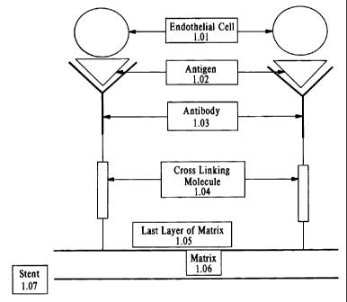
more types of antibody. Figure 1 provides an illustration of coupling via a
cross-linking molecule.
An endothelial cell, 1.01, binds to an antibody, 1.03, by a cell surface
antigen, 1.02. The antibody is
tethered to the matrix, 1.05-1.06, by a cross-linking molecule, 1.04. The
matrix, 1.05-1.06, adheres
to the stent, 1.07. The linker molecules may be coupled to the matrix directly
(i.e., through the
carboxyl groups), or through well-known coupling chemistries, such as,
esterification, amidation,
and acylation. The linker molecule may be a di- or tri-amine functional
compound that is coupled to
the matrix through the direct formation of amide bonds, and provides amine-
functional groups that
are available for reaction with the antibodies. For example, the linker
molecule could be a
polyamine functional polymer such as polyethyleneimine (PEI), polyallylamine
(PALLA) or
polyethyleneglycol (PEG). A variety of PEG derivatives, e.g., mPEG-
succinimidyl propionate or
mPEG-N-hydroxysuccinimide, together with protocols for covalent coupling, are
commercially
available from Shearwater Corporation, Binmingham, Alabama. (See also, Weiner
et al., Influence
of a poly-ethyleneglycol spacer on antigen capture by immobilized antibodies.
J. Biochem.
Bionhys. Methods 45:211-219 (2000)). It will be appreciated that the selection
of the particular
coupling agent may depend on the type of antibody used and that such selection
may be made
without undue experimentation. Mixtures of these polymers can also be used.
These molecules
contain a plurality of pendant amine-functional groups that can be used to
surface-immobilize one
or more antibodies.
Antibodies may be attached to C600 fullerene layers that have been deposited
directly on
the surface of the stent. Cross linking agents may be covalently attached to
the fullerenes. The
antibodies are then attached to the cross-linking agent, which in turn is
attached to the stent. Figure
2 provides an illustration of coupling by C600. The endothelial cell, 2.01, is
CA 02400319 2006-01-16
17
bound via a cell surface antigen, 2.02, to an antibody, 2.03, which in turn is
bound, covalently or
non-covalently to the matrix, 2.04. The matrix, 2.04, is coupled covalently
via C600, 2.05, to the
stent, 2.06.
EXPERIMENTAL EXAMPLES
This invention is illustrated in the experimental details section which
follows. These
sections set forth below the understanding of the invention, but are not
intended to, and should not
be construed to, limit in any way the invention as set forth in the claims
which follow thereafter.
EXAMPLE 1
ADHERENCE OF HUMAN ENDOTHELIAL CELLS TO CD34 Fab-COATED STENTS
Materials and Methods
1. Cells
HUVEC will be prepared from human umbilical cords by the method of Jaffe
(Jaffe, E. A.
In "Biology of Endothelial Cells", E. A.Jaffe, ed., Martinus-Nijhoff, The
Hague (1984)) and
cultured in Medium 199 supplemented with 20% fetal calf serum (FCS), L-
glutamine, antibiotics,
130 uglml heparin and 1.2 mg/ml endothelial cel) growth supplement (Sigma-
Aldrich, St. Louis,
MO).
Progenitor endothelial cells will be isolated from human peripheral blood by
the method of
Asahara et al. (Isolation of Putative progenitor endothelial cells for
angiogenesis. Science
275:964-967). Monoclonal anti-CD34 antibodies will be coupled to magnetic
beads and incubated
with the leukocyte fraction of human whole blood. After incubation, bound
cells will be eluted and
cultured in M-199 containing 20% fetal bovine serum and bovine brain extract.
(Clonetics, San
Diego, CA). Cells will be characterized by fluorescent antibodies to CD45,
CD34, CD31, Flk-l,
Tie-2, and E-selectin.
CA 02400319 2002-08-22
WO 01/68158 PCT/US01/08244
18
2. Coating of Stents
A. R stents produced by Orbus International B.V. (Leusden, The Netherlands)
will be incubated with human fibrinogen (Sigma, St. Louis, MO) 500-800 mg/ml
together with
Fab fragments of anti-CD34 monoclonal antibody and the fibrinogen will be
polymerized by the
addition of 1000 units/ml of thrombin. After incubation of the stent with the
polymerized fibrin
mixture containing the anti-CD34 monoclonal Fab fragments, the fibrin will be
compressed into a
thin film (less than 0.012 cm) against the R Stent. The R-Stent having the
thin, fibrin film
containing the Fab fragments will be washed three times with phosphate-
buffered saline (PBS)
containing 0.5% bovine serum albumin (BSA) at room temperature.
B. Alternatively, R stents will be coated with mPEG-succinimidyl propionate
(Shearwater Corporation, Birmingham, Alabama). The succinimidyl group will be
reacted with
the anti-CD34 monoclonal Fab fragments (Fab-PEG coated R stents) according to
the
manufacturer's instructions to form a stable amide linkage between the PEG
derivative and the
Fab fragment.
3. Endothelial Cell binding assay
The fibrin-anti-CD34 Fab coated R-stents or Fab-PEG coated R stents will be
incubated with isolated HUVEC or isolated progenitor endothelial cells at
cellular concentrations
of between 100,000 and 1,000,000 cells/ml in M199 containing 0.5% BSA at 37 C
in a 5% COz
humidified atmosphere. Prior to incubation with the stent, the HUVEC or
progenitor endothelial
cells will be labeled with [3H]-thymidine for 24 hours. After incubation of
the labeled endothelial
cells with the stents coated with fibrin and Fab anti-CD34 for between 4 and
72 hours, the stents
will be removed from the solution and washed 5 times with M199 containing 0.5%
BSA. Bound
endothelial cells will be removed by trypsinization and binding of labeled
endothelial cells to the
stents will be assessed by scintillation counting of [3H]-thymidine. As
negative control, stents
coated with fibrin alone or uncoated stents will be incubated with [3H]-
thymidine-labeled
endothelial cells. Results will be evaluated statistically using a t-test to
determine differential
binding. Stents coated with fibrin which incorporate monoclonal anti-CD34 Fab
fragments will
show significantly increased binding of endothelial cells as compared with
uncoated stents.
EXAMPLE 2
PROLIFERATION OF HUMAN ENDOTHELIAL CELLS
CA 02400319 2002-08-22
WO 01/68158 PCT/US01/08244
19
ON CD34 FAB-COATED STENTS
Endothelial Cell Proliferation Assay
R stents coated with fibrin that incorporates anti-CD34 Fab fragments will be
incubated with HUVEC or progenitor endothelial cells for between 4 and 72
hours in M199
containing 0.5% BSA. After incubation of the stents with the HUVEC or
progenitor endothelial
cells, the stents will be washed 5 times with M199 containing 0.5% BSA and
then incubated with
[3H]-thymidine. [3H]-thymidine incorporation will be assessed in washed and
harvested HUVEC
or progenitor endothelial cells (cells will be harvested with trypsin).
Proliferation of HUVEC or
progenitor endothelial cells on fibrin-coated stents will be compared with
endothelial cell
proliferation in standard microtiter dishes. Proliferation of HUVEC or
progenitor endothelial
cells on fibrin-coated stents will be equal to or greater than proliferation
of endothelial cells in
microtiter dishes
EXAMPLE 3
PRODUCTION OF MONOCLONAL ANTIBODIES REACTIVE WITH HUVEC AND
PROGENITOR ENDOTHELIAL CELLS
BALB/c mice will be immunized, intraperitoneally 3-4 times at 2-4 weekly
intervals, with 1.5 x 10⁶ HUVEC in PBS or 1.5 x 10⁶ progenitor endothelial
cells and challenged
3 days prior to spleen-cell removal with 1.5 x 10⁶ HUVEC or 1.5 x 10⁶
progenitor endothelial
cells. A spleen-cell suspension will be prepared, fused with the myeloma NS
1/1 AG4.1 and
hybridomas grown up and cloned. To improve hybridoma growth and cloning
efficiencies, 10%
endothelial-cell conditioned medium (HUVEC) will be included in culture media.
Initially,
hybridoma culture supernatants will be tested for reactivity with HUVEC or
progenitor
endothelial cells by immunofluorescence flow cytometry (FACS). Briefly, H[JVEC
(1.5 x 10⁴) or
progenitor endothelial cells (1.5 x 10⁴) will be incubated (30 min, 4 C) with
undiluted hybridoma
supematant, washed and incubated with fluorescein-isothiocyanate (FITC)-sheep
F(ab')2 anti-
mouse Ig(l00 ug/ml). Following final washing, the endothelial cells will be
examined for
monoclonal antibody binding by FACS analysis. Positive hybridoma supernatants
will be screened
on the human melanoma cell line MM-170 to eliminate non-endothelial specific
mAbs.
Endothelial specificity will be further confirmed by screening of monoclonal
antibodies on a panel
of human tumor cell lines as well as human lymphocytes, monocytes,
neutrophils, red cells and
platelets.
CA 02400319 2002-08-22
WO 01/68158 PCT/US01/08244
EXAMPLE 4
PORCINE BALLOON INJURY STUDIES
Implantation of antibody-covered stents will be performed in juvenile
Yorkshire
5 pigs weighing between 25 and 30 kg. Animal care will comply with the "Guide
for the Care and
Use of Laboratory Animals" (NIH publication No. 80-23, revised 1985). After an
overnight fast,
animals will be sedated with ketamine hydrochloride (20mg/kg). Following the
induction of
anesthesia with thiopental (12 mg/kg) the animals will be intubated and
connected to a ventilator
that will administer a mixture of oxygen and nitrous oxide (1:2 [vol/vol]).
Anesthesia will be
10 maintained with 0.5-2.5 vol% isoflurane. Antibiotic prophylaxis will be
provided by an
intramuscular injection of 1,000 mg of a mixture of procaine penicillin-G and
benzathine
penicillin-G (streptomycin).
Under sterile conditions, an arteriotomy of the left carotid artery will be
performed and a 9F-
introducer sheath will be placed in the left carotid artery. All animals will
be given 7,500 IU of
15 heparin sodium and 100 mg of acetylsalicylic acid intravenously.
Additiona12,500 IU boluses of
heparin will be administered periodically throughout the procedure in order to
maintain an
activated clotting time above 300 seconds. An 8F guiding catheter will be
introduced through the
carotid sheath and passed to the origin of the iliac artery. Angiography will
be performed after
the administration of lmg of isosorbide dinitrate and images will be analyzed
using a quantitative
20 coronary angiography system. A 3F-embolectomy catheter will be inserted
into the common
femoral artery, and passed distal to the segment selected for stent
implantation. The
embolectomy balloon will be inflated to a size 0.5 mm larger than the arterial
segment and
withdrawn twice to denude the vessel. Immediately after denudation, a fibrin-
coated stent
incorporating an Fab fragment of a monoclonal antibody will be inserted
through the guiding
catheter and deployed in the denuded segment of the femoral artery. Animals
will be sacrificed
both at 3 days and at eight weeks after stent implantation. The animal will
first be sedated and
anesthetized as described above. The stented femoral segments will be
explanted and then placed
in 4% paraformaldehyde in 0.1 M phosphate buffer pH 7.2 at 4 C for 48 h. A
rectangular section
of the vessel wall will be removed for further processing for electron
microscopic evaluation of
surface coverage of endothelial cells. This portion of the stented vessel will
be placed in 0.15
cacodylate buffer and further fixed with 2.5% glutaraldehyde in 0.15 M
cacodylate. The tissue
will then be post-fixed with 0.1 M cacodylate buffer containing 1% Os04 and 50
mM ferricyanide
(K3[Fe(CN)6]), and further processed. (Reduction in thrombotic events with
heparin-coated
CA 02400319 2006-01-16
21
Palmaz-SchatzTM stents in normal porcine coronary arteries, Circulation 93:423-
430).
The remaining sections of the stented arterial segments will be impregnated
with three
changes of methy methacrylate as described by van Beusekom et al. (Cardiovasc
Pathol 5:69-76
(1996)). Embedded arterial segments with the stent in place will be cut into
sections 3 to 5 n thick
on a motor-driven rotary microtome (HM-350, Microm GmbH, Munich, Germany)
using stainless
steel disposable knives. On chrome aluminum coated slides, sections will be
stretched on a hot plate
at 40 C using a mixture of 60% 2-butoxyethanol and 10% ethanol in water.
Sections will be
covered by a plastic film, excess butoxyethanol-ethanol mixture removed and
the slides will be left
overnight to dry in a 40 C oven. Sections will then be deplasticized in a
solution of equal volumes
of xylene-chloroform for 30 to 60 minutes. Standard staining procedures for
light microscopy will
then be performed on the prepared sections. Statistics: Data will be presented
as the mean
standard error of the mean (SD) of the independent experiments. Statistical
significance will be
determined by one way analysis of variance (ANOVA) and Fisher's PLSD test
(StatViewTM 4.01;
Brain Power, Inc., Calabasas, Calif). For data of treated and untreated
segments of femoral arteries,
a paired t test (StatViewTM 4.01) will be used. A p value of <0.05 will be
considered a statistically
significant difference between the means. Animals treated with a stent
incorporating an anti-porcine
endothelial cell monoclonal Fab fragment will show increased endothelial cell
coverage and
significantly reduced restenosis as compared with controls having an uncoated
stent implanted.
EXAMPLE 5
TRANSFECTION OF PORCINE PROGENITOR ENDOTHELIAL CELLS
Porcine progenitor endothelial cells will be isolated from pig peripheral
blood by the
method of Asahara et al. (Isolation of Putative progenitor endothelial cells
for angiogenesis.
Science 275:964-967). Monoclonal anti-CD34 antibodies will be coupled to
magnetic beads and
incubated with the leukocyte fraction of pig whole blood. After incubation,
bound cells will be
eluted and cultured in M-199 containing 20% fetal bovine serum and bovine
brain extract.
(Clonetics, San Diego, CA). Cells will be characterized by fluorescent
antibodies to CD45, CD34,
CD31, Flk-1, Tie-2, and E-selectin.
For example, purified porcine progenitor endothelial cells will be transfected
with vascular
endothelial growth factor (VEGF) using an adenovirus vector expressing the
VEGF cDNA
according to the methods of Rosengart et al. (Six-month assessment of a phase
I trial of angiogenic
gene therapy for the treatment of coronary artery disease using direct
intramyocardial
CA 02400319 2006-01-16
22
administration of an adenovirus vector expressing the VEGF 121 cDNA. Ann.
Sura. 230 (4):466-
470 (1999)).
The transfected purified porcine progenitor cells expressing VEGF will be
infused into the
porcine femoral artery model after balloon injury and stent implantation as
described in
Example 4 using a double-balloon chamber infusion catheter (Cordis Corp) which
isolates the
stented portion of the femoral artery. Restenosis will be compared in balloon
angioplasty stent-
treated pigs infused with VEGF-transfected porcine progenitor cells as
compared with pigs infused
with un-transfected porcine progenitor endothelial cells. Expression of VEGF
in the re-infused
porcine progenitor endothelial cells will result in a decreased incidence and
severity of restenosis in
the anti-CD34 coated stents.
EXAMPLE 6
PREPARATION OF AMINOSILANE PEO TETHERED ANTIBODIES
Stent Preparation - stents will be made from 316L stainless steel and will be
cleaned and
passivated by first washing in an anionic detergent in an ultrasonic cleaner
and then soaked in hot
nitric acid with agitation, followed by a final deionized water rinse.
Derivatized stents will be prepared as follows - stents will be dipped into a
2% mixture of
N- (2-aminoethyl-3-aminopropyl) trimethoxysilane in 95% ethanol for three
minutes, removed, air
dried at room temperature and then cured for 10 minutes at 110 C.
Polyethylene glycol (PEG) Spacer Coupling - Derivatized stents will be placed
in 100 ml of
0.1 M MES buffer containing 10 mM Dicarboxymethyl-PEG and 500 mg of EDC added
and
incubated at 25 C with constant stirring for two hours.
Tethered Antibody - Antibodies to endothelial cells will be immobilized to the
PEG
functionalized stents in a one-step carbodiimide coupling reaction by
immersing the stents into 150
ml of 0.1 M MES buffer (pH 4.5) into which 1.0 mg of murine anti-CD34 IgG,
antibody is dissolved
and incubated at 25 C for two hours. Stents will be removed from the solution
and rinsed five times
with 50 ml of phosphate buffered saline (pH 7.2) with 0.02% Tween 20.
Reagents include: N-(2-aminoethyl-3-aminopropyl) trimethoxysi lane (Degussa-
Huls); MES
buffer-morpholine ethane sulfonic acid buffer (Sigma, St. Louis, MO); EDC -I-
ethyl-3-
(3-dimethylaminopropyl) carbodiimide (Sigma, St. Louis, MO); Dicarboxymethyl-
PEG-
Dicarboxymethyl-poly(ethylene glycol) [MW 3400] (Shearwater, Huntsville, AL).
Having described several different embodiments of the invention, it is not
intended that the
invention is limited to these embodiments it is intended that modifications
and variations
CA 02400319 2002-08-22
WO 01/68158 PCT/US01/08244
23
may be made by one skilled in the art without departing from the spirit and
scope of the invention
as defined in the claims.