Note: Descriptions are shown in the official language in which they were submitted.
74?5 71276
CA 02400601 2002-08-29
-1-
REAL-TIME GENE QUANTIFICATION WITH INTERNAL STANDARDS
BACKGROUND
The polymerise chain reaction (PCR) is a technique of synthesizing
large quantities of a preselected DNA segment. The technique is fundamental to
molecular biology and is the first practical molecular technique for the
clinical
laboratory. PCR is achieved by separating the DNA into its two complementary
strands, binding a primer to each single strand at the end of the given DNA
segment
where synthesis will start, and adding a DNA polymerise to synthesize the
complementary strand on each single strand having a primer bound thereto. The
process is repeated until a sufficient number of copies of the selected DNA
segment
have been synthesized.
During a typical PCR reaction, double stranded DNA is separated into
its single strands by raising the temperature of the DNA containing sample to
a
denaturing temperature where the two DNA strands separate (i.e., the "melting
temperature of the DNA") and then the sample is cooled to a lower temperature
that
allows the specific primers to attach (anneal), and replication to occur
(extend). In
illustrated embodiments, a thermostable polymerise is utilized i ~ the
polymerise
chain reaction, such as Taq DNA Polymerise and derivatives thereof, including
the
Stoffel fragment of Taq DNA polymerise and KlenTaql polymerise (a
5'-exonuclease deficient variant of Taq polymerise -- see U.S. Patent No.
5,436,149).
The years 1991 to 1998 have seen a 10 fold increase in the number of
papers using quantitative PCR methods. One of the major reasons for this
increased
use of quantitative PCR derives from the fact that PCR has a sensitivity five
orders of
magnitude better than the best blotting procedures. This sensitivity makes PCR
as a
quantitative tool highly desirable. However, the use of a system undergoing
exponential amplification is not ideally suited to quantification. Small
differences
between sample sizes can become huge difference in results when they are
amplified
through forty doublings.
Kinetic PCR
A typical PCR reaction profile can be thought of has having three
segments: an early lag phase, an exponential growth phase, and a plateau. The
lag
7475 71276
CA 02400601 2002-08-29
-2-
phase is mainly a reflection of the sensitivity of the instrument and the
background
signal of the probe system used to detect the PCR product. The exponential
growth
phase begins when sufficient product has accumulated to be detected by the
instrument. During this "log" phase the amplification course is described by
the
equation Tn = To(E)n, where Tn is the amount of target sequence at cycle n, To
is the
initial amount of target, and E is the efficiency of amplification. Finally,
in the
plateau phase, the amplification efficiency drops off extremely rapidly.
Product
competes more and more effectively with primers for annealing and the amount
of
enzyme becomes limiting. The exponential equation no longer holds in the
plateau
phase.
Most of the quantitative information is found in the exponential cycles,
but the exponential cycles typically comprise only 4 or 5 cycles out of 40.
With
traditional PCR methods, finding these informative cycles requires that the
reaction
be split into multiple reaction tubes that are assayed for PCR product after
varying
numbers of cycles. This requires either assaying many tubes, or a fairly good
idea of
the answer before the experiment is begun. Once the position of the
exponential
phase is determined, the experimental phase can be compared to known standards
and
the copy number can be calculated. .
Competitive Quantitative PCR
Competitive quantitative PCR methods were developed to attempt to
overcome difficulties associated with finding the exponential phase of the
reaction
and to obtain greater precision. A competitor sequence is constructed that is
amplified using the same primers as are used to amplify the target sequence.
Competitor and target are differentiated, usually by length or internal
sequence, and
the relative amount of competitor and target are measured after amplification.
If the
target and the competitor are amplified with equal efficiency, then their
ratio at the
end of the reaction will be the same as the ratio had been at the beginning.
This holds
true even into the plateau phase as long as both decline in efficiency at the
same rate.
Thus, finding the exponential region is no longer a problem. Providing
standards in
the same tubes with the unknown targets allows for additional control not
possible
with kinetic methods. For example, adding the competitor before mRNA
purification
would control for variations in sample preparation and reverse transcription.
7475 71276
CA 02400601 2002-08-29
-3-
The use of currently available competitive PCR techniques continues
to suffer from several deficiencies. Firstly, the competitor sequence must be
constructed to be as similar as possible to the target sequence with regard to
the
efficiency of amplification, yet the two sequences must be distinguishable
from one
S another. If the competitor is too close in sequence to the target,
heteroduplexes form
during the PCR that skew the ratio of the product to the template.
In addition, competitor must be added to the unknown sample at a
concentration approximating that of the target. If one product reaches plateau
before
the other rises above background, no quantitative information can be obtained
from
that sample. Usually an unknown sample is split and mixed with multiple
concentrations of competitor.
Other concerns have been raised regarding competitive quantification
methods. A common criticism is that despite all efforts, the target and the
competitor
together in a sample may be amplified at different efficiencies, even if
target and
competitor are amplified at the same efficiencies when amplified separately
(the
obvious control). When the target and competitor are combined in one vessel
and the
reagents are limiting, the efficiencies of the two amplification reactions may
change at
different rates. Length differences between target and competitor are of most
concern
here as the longer product may compete more effectively with the primers and
may be
more affected by reagent limitations. Both of these concerns could be
addressed by
making the target and competitor sufficiently alike, if it were not for the
problem of
forming heteroduplexes during the PCR reaction.
Real-Time Quantitative PCR
Developments in instrumentation have now made real-time monitoring
of PCR reactions possible and thus have made the problem of finding the log
phase of
the reaction trivial.
Thermocycling may be carned out using standard techniques known to
those skilled in the art, including the use of rapid cycling PCR. Rapid
cycling
techniques are made possible by the use of high surface area-to-volume sample
containers such as capillary tubes. The use of high surface area-to-volume
sample
containers allows for a rapid temperature response and temperature homogeneity
7475 71276
CA 02400601 2002-08-29
-4-
throughout the biological sample. Improved temperature homogeneity also
increases
the precision of any analytical technique used to monitor PCR during
amplification.
In accordance with an illustrated embodiment of the present invention,
amplification of a nucleic acid sequence is conducted by thermal cycling the
nucleic
acid sequence in the presence of a thermostable DNA polymerise using the
device
and techniques described in U.S. Patent No. 5,455,175. In accordance with the
present invention, PCR amplification of one or more targeted regions of a DNA
sample is conducted while the reaction is monitored by fluorescence.
The first use of fluorescence monitoring at each cycle for quantitative
PCR was developed by Higuchi et al., "Simultaneous Amplification and Detection
of
Specific DNA Sequences," Bio. Technology, 10:413-417, 1992, and used ethidium
bromide as the fluorescent entity. Fluorescence was acquired once per cycle
for a
relative measure of product concentration. The cycle where observable
fluorescence
first appeared above the background fluorescence (the threshold) correlated
with the
starting copy number, thus allowing the construction of a standard curve. A
probe-
based fluorescence detection system dependent on the 5'-exonuclease activity
of the
polymerise soon followed. This improved the real-time kinetic method by adding
sequence specific detection.
Alternatively, PCR amplification of one or more targeted regions of a
DNA sample can be conducted in the presence of fluorescently labeled
hybridization
probes, wherein the probes are synthesized to hybridize to a specific locus
present in a
target amplified region of the DNA. In an illustrated embodiment, the
hybridization
probe system comprises two oligonucleotide probes that hybridize to adjacent
regions
of a DNA sequence wherein each oligonucleotide probe is labeled with a
respective
member of a fluorescent energy transfer pair. In this embodiment, the presence
of the
target nucleic acid sequence in a biological sample is detected by measuring
fluorescent energy transfer between the two labeled oligonucleotides.
These instrumentation and fluorescent monitoring techniques have
made kinetic PCR significantly easier than traditional competitive PCR. More
particularly, real-time PCR has greatly improved the ease, accuracy, and
precision of
quantitative PCR by allowing observation of the PCR product concentration at
every
cycle. In illustrated embodiments of the present invention, PCR reactions are
conducted using the LightCycler~ (Roche Diagnostics), a real-time PCR
instrument
7475 71276
CA 02400601 2002-08-29
-5-
that combines a rapid thermal cycler with a fluorimeter. Through the use of
this
device, the PCR product is detected with fluorescence, and no additional
sample
processing, membrane arrays, gels, capillaries, or analytical tools are
necessary.
Other PCR instrumentation, as known in the art, may be used in the practice of
the
present invention.
SUMMARY OF THE INVENTION
The present invention is directed to a nucleic acid quantification kit
and method for determining the initial concentration or mass fraction of a
target
nucleic acid present in a sample. More particularly, the present invention
relates to
the use of real-time competitive quantitative polymerise chain reaction (PCR)
and
fluorescently labeled oligonucleotide probes to monitor the PCR reaction in
real time
to determine the copy number of a target nucleic acid sequence in a sample.
The
method of determining the copy number of a target nucleic acid present in a
biological sample comprises the steps of combining in a single reaction vessel
at least
a portion of the biological sample, a thermostable polymerise, a known amount
of a
competitor nucleic acid sequence, a pair of oligonucleotide PCR primers, one
or more
oligonucleotide probes, initiating the PCR reaction, and conducting real time
monitoring of the reaction and/or melting curve analysis.
In an illustrated embodiment, the competitor nucleic acid sequence is
prepared to have the identical sequence as the target nucleic acid sequence
with the
exception of a unique section located at an internal position on the
competitive
nucleic acid sequence. The unique section has the same overall nucleotide
composition as the corresponding region of the target nucleic acid sequence
but
having a substantially different sequence from the corresponding region of the
target
nucleic acid sequence. The term substantially different is used herein to mean
that a
probe complementary to the unique region of the competitor will not cross-
hybridize
to the corresponding region of the target nucleic acid sequence above
background
levels under the reaction conditions used to conduct the PCR reaction. In one
embodiment, the unique region has a randomized sequence relative to the
corresponding region of the target nucleic acid sequence.
In another embodiment, the unique section of the competitor nucleic
acid sequence differs from the target nucleic acid sequence by only one base
pair,
7475 71276
CA 02400601 2002-08-29
-6-
similar to a point mutation. In still another embodiment, the unique section
of the
competitor nucleic acid sequence may be quite a bit different from the
corresponding
region of the target, varying in length and/or composition, but the competitor
and
target nucleic acid sequences are amplified with essentially the same
efficiency. Such
S amplification efficiencies can be determined based on CG content and routine
experimentation.
The anchor probe is configured to hybridize adjacent to the unique
region of the competitor nucleic acid sequence and adjacent to the region of
the target
nucleic acid sequence corresponding to the unique region of the competitor
nucleic
acid sequence. The competitor probe is configured to hybridize to the unique
region
of the competitor nucleic acid sequence, and the target probe is configured to
hybridize to the region of the target nucleic acid sequence corresponding to
the unique
region of the competitor nucleic acid sequence. Accordingly, when the anchor,
target
and competitor probes hybridize to their respective complementary target
nucleic acid
sequences and competitor nucleic acid sequences, the donor fluorophore and the
first
acceptor fluorophore as well as the donor fluorophore and the second acceptor
fluorophore are placed in a resonance energy transfer relationship. Therefore,
the
measurement of fluorescence from the acceptor fluorophore can be used to
determine
the relative concentrations of the target nucleic acid sequence and the
competitor
nucleic acid sequence. In illustrated embodiments, the first fluorophore and
the
second fluorophore both accept energy transfer from the fluorophore donor, but
the
two acceptor fluorophores emit fluorescent energy at different wavelengths.
Thus, the
concentrations of the target nucleic acid sequence and the competitor nucleic
acid
sequence can be measured at the same time.
In still another embodiment, a single-labeled oligonucleotide is used
and the desired information is obtained through melting curve analysis.
Another aspect of this invention is a method of quantifying the initial
target nucleic acid sequence concentration based on the cycle shift between
competitor and target. Provided that the efficiency of amplification is
essentially
equal for target and competitor, logCo = logE(~n) + logTo, where Co is the
initial
amount of competitor, E is the average efficiency, 0n is the cycle shift
between target
and competitor, and To is the initial amount of target. Because this equation
has the
form y=mx+b, a plot of the initial competitor concentration versus the cycle
shift
7475 71276
CA 02400601 2002-08-29
_7_
between competitor and target will yield a line with the slope equal to the
log of the
efficiency and a y-intercept equal to the log of the initial target
concentration. Since
the competitor may be provided in a variety of known initial concentrations,
the initial
concentration of the target may be determined with relative ease.
One particularly useful application for DNA quantification may be in
determining the genomic equivalents of particular viruses in any given
clinical
sample. Several viruses exhibit their pathological effects at various stages
of their
replication cycle, and the amount of virus in host cells can serve as an
indicator of
infection progression and prognosis.
In yet another aspect of this invention is method of determining mass
fractions of first and second target nucleic acids present in a test sample,
said method
comprising the steps of contacting the target nucleic acids with a fluorescent
nucleic
acid indicator, the indicator being configured to provide a signal related to
the
quantity of indicator hybridized to the target nucleic acid, the indicator
further
configured to discriminate the target nucleic acids based on melting
temperature,
illuminating the test sample, monitoring fluorescent change to generate a
melting
curve, and using a thermodynamically based signal processing algorithm to
determine
the mass fraction of the target nucleic acids. The internal standard may
consist of an
artificial competitor or an endogenous allele that is different from the
target nucleic
acid sequence by one or more bases. If a known amount of the internal standard
is
added to the sample, then the initial copy number of the target nucleic acid
sequence
can be calculated from the mass fraction or ratio against the known amount of
internal
standard. Particularly useful applications for this type of quantification may
be in
determining allele frequencies in pooled population samples, monitoring
differential
allele expression in various cell and tissue types, monitoring gene
amplification, or
deletion, using imbalance of copy number against the copy number of a
pseudogene,
and assessing the ratio between different cell types in a mixed tissue sample,
such as
in margin dissected tissue samples from cancer patients.
Additional features of the present invention will become apparent to
those skilled in the art upon consideration of the following detailed
description of
illustrated embodiments exemplifying the best mode of carrying out the
invention as
presently perceived.
7475 71276
CA 02400601 2002-08-29
_g_
BRIEF DESCRIPTION OF THE DRAWINGS
Fig. 1 is a diagrammatic representation of the mechanical and optical
design of the LightCycler~;
Figs. 2a-f are diagrammatic representations of the various fluorescent-
based methods of detecting amplification products. Figs. 2a-b represent
detection of
amplified products by double strand specific dyes. Figs. 2c-d represent the
Taq Man
strategy wherein synthesis of the amplified product results in donor emission.
Figs.
2e-f represent the hybridization probe method wherein two separately labeled
probes
hybridize to adjacent regions of a nucleic acid sequence resulting in
fluorescent
resonance energy transfer;
Figs. 3a-b represent typical external standard curves using
hybridization data. Fig. 3a is a plot of the log fluorescence ratio vs. cycle
number.
Fig. 3b is a plot of the log copy number vs. the second derivative maximum;
Figs. 4a-b represent a typical standard curve generated by plotting
fluorescence vs. temperature (Fig. 4a) and the derivative of that curve
plotted against
temperature (Fig. 4b), with homozygous mutant (~ ~ ~ ~), homozygous wild type
(-
-), heterozygous mutant (-), and no DNA (- -);
Figs. Sa-c represent melting analysis of several nucleic acids. Fig. 5a
shows melting peaks generated from a melting curve. The area under each curve
is
calculated using non-linear regression to fit the melting peak data to a
Gaussian curve.
Fig. 5b shows various amplification curves on a log fluorescence vs. cycle
number
plot. Fig. 5c shows the data from Fig. 5b converted into a logCo= log E (0n) +
logTo
curve (solid line show crossing points from the data of Fig. 5b and dashed
line is
linear regression);
Fig. 6 represents the nucleotide sequences of the competitive DNA
fragment for HPV 16 and the targeting, competitive and anchor probes;
Fig. 7 represents the nucleotide sequences of the HER-2/neu (target),
its competitor, the reporter and anchor probes; the predicted melting
temperatures Tm
of the reporter probe hybridized to either the target or competitor are shown.
Fig. 8 is a diagrammatical representation of the strategy used to create
the competitive DNA fragment for HPV 16;
Fig. 9 is a diagrammatical representation of the hybridization probes
used to detect the internal quantification standards and the HPV 16 artificial
template;
7475 71276
CA 02400601 2002-08-29
-9-
Fig. 10 is a graphic representation of the detection efficiency of the
Internal Quantification Standard ( ~ )and Artificial HPV 16 template (~). The
data
are presented as the average of at least three separate data points, with
standard
deviations for each;
Figs. 11 a-b illustrate a typical internal control reaction demonstrating
fluorescent data from an internally controlled hybridization probe reaction.
Internal
quantification standards at concentrations of 1x10⁹ (1); 5x10⁸ (2); 1x10⁸ (3);
5x10
(4); 1x10 (5); 5x10⁶ (6); 1x10⁶ (7); 5x10⁵ (8); 1x10⁵ (9) are plotted in Fig.
11a. HPV
16 at 1x10⁶ in each of the samples is shown in Fig. 1 1b;
Figs. 12a-c are graphic representations of the detected fluorescence vs.
cycle number for the Internal quantification standard (open triangles) and HPV
16
(closed squares). In each case HPV 16 is at an initial template concentration
of 1x10⁴.
The internal quantification standard is at initial template concentrations of
1x10⁵ (Fig.
12a), 1x10⁴ (Fig. 12b), and 1x10³ (Fig. 12c);
Fig. 13 is a graphic representation of the log of initial competitor copy
number versus difference in crossing threshold (delta C.T.). A graph of
internal
quantification standard reaction with distinct concentrations of HPV 16
artificial
template. HPV 16 initial template concentrations are: 1 x 1 Oz (open circles),
1 x 10³
(open triangles), 1x10⁴ (open squares), 1x10⁵ (closed circles), 1x10⁶ (closed
triangles). Error bars are determined from the standard deviation from four
independent reaction data points. The 95% confidence interval at each ratio of
competitor to target is indicated on the x-axis.
Fig. 14 represents the correlation between melting peak area and
product concentration for mutant and wild-type HER-2/neu targets detected by
hybridization probes using melting curve analysis software. Artificial
oligonucleotide
templates were mixed with probes at various concentrations and melting peak-
area
was determined using LightCycler melting curve analysis software.
Fig. 15 represents quantification of mutant (M) and wild-type (WT)
HER-2/neu targets by melting curve analysis following PCR amplification.
Mutant
and wild-type templates, both individually and mixed at various ratios (input
ratio),
were amplified for 40 cycles of PCR and melting curves were generated from the
PCR products. Melting curves were analyzed by the TMBSP algorithm to determine
the ratios of mutant and wild-type PCR products (output ratio).
74,75 71276
CA 02400601 2002-08-29
-10-
Figs. 16a-d are plots of melting analysis of a wild type (WT) sample
(-), a mutant (Mut) sample (- ~ ~ ~), and a mixture (Mix) of wild and mutant
alleles at
50:50 ratio (----), detected by the Sensor probe only (Figs. 16a, and b), or
with the
FRET pair probes (Figs. 16c, and d); Fig 16a and c show melting data
(fluorescence
vs temperature) and Figs. 16b and d show the melting peak data (negative first
derivative -dF/dT).
Figs. 17a-d are plots of melting analysis of a wild type (WT) sample
(-), a mutant (Mut) sample (~ ~ ~ ~), and a mixture (Mix) of wild and mutant
alleles at
95:5 ratio (----), detected by the Sensor probe only (Figs. 17a, and b), or
with the
FRET pair probes (Figs. 17c, and d); Fig 17a and c show melting data
(fluorescence
vs temperature) and Figs. 17b and d show the melting peak data (negative first
derivative -dF/dT).
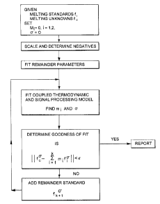
Fig. 18 is a flow chart of the Thermodynamic Modeling based Signal
Processing algorithm.
Fig. 19 is a plot of Input (the actual fraction of the wild-type allele in
samples) vs the difference between Input and Output (the fractions estimated
by the
analysis software). Results from the Thermodynamic Modeling based Signal
Processing algorithm (open circle), and the melting peak area ratio software
(closed
circle) are shown.
Fig. 20 is a graphic representation of a model melting curve which has
three phases: the non-linear "annealed phase," the melting transition
(depicted as the
"melting phase"), and the linear "melted phase." The basis function
approximation
algorithm is based on this model to approximate the melting curve.
DETAILED DESCRIPTION OF THE INVENTION
The present invention allows the quantification of analytes, including
analytes that are too low in concentration to be quantitated using standard
techniques.
The method uses a competitive PCR reaction with real time monitoring during
amplification or melting curve analysis, and the presence of an internal
standard as a
means of calculating the initial concentration of the target sequence. To
date, all real-
time PCR quantification applications have been limited to quantification
relative to an
external standard curve. While this technique is very useful, it lacks control
for tube-
to-tube differences in PCR efficiency. This limitation of quantification with
external
7475 71276
CA 02400601 2002-08-29
-11-
standards has been addressed by competitive quantitative PCR methods. In these
techniques a competitor, with the same primer sites as the target but
differing in
internal sequence, is spiked at a known concentration into an unknown sample.
However, no real-time version of this method is available.
The present disclosure is directed to the use of real-time methods to
differentiate target from competitor and thus allow for gene quantification by
reference to an internal standard. The methods provide investigators with the
advantages of a homogenous, real-time PCR system while giving the added
control
that internal standards provide.
In accordance with one embodiment, a method is described for
conducting real-time competitive quantitative PCR using a competitor with a
unique
hybridization probe binding site. The competitor will be distinguished from
the target
by using differently colored hybridization probes for the target and the
competitor.
In another embodiment, a method is described for conducting real-time
competitive quantitative PCR using a competitor differing from the target by
only a
single base. The target and the competitor will be distinguished from one
another by
the differential melting of fluorescently labeled hybridization probes.
Fig. 1 provides a schematic representation of an embodiment 400 the
LightCycler~, a thermal cycler that may be used in accordance with the
described
methods. As shown in Fig. l, air is taken in through an aperture 470 and
generally
follows the flow path indicated by the lines 472. The temperature of the air,
and thus
the temperature of the sample container 450, is controlled with heating
cartridge 474,
which is positioned within a central duct 476, and fan 498, which is provided
to move
the air in the indicated path 472. The fan is driven via a shaft 496 and a
motor 494.
The fan 498 forces air into the aperture 470 and out via exhaust ports 478. In
the
illustrated embodiment, twenty-four sample containers 450 (two of which are
represented in Fig. 1 ) are symmetrically arranged around the heating
cartridge 474
and the central duct 476. The sample containers 450 are received by sleeves
452 in a
circular carousel 480. The carousel 480 is positioned by a stepper motor 488
provided with a drive gear 484 that is connected to the motor 488 via a shaft
486.
Fluorescence from each sample container is obtained by photo array 460, which
includes an excitation radiation source 468 and photodetectors 464 and 466.
More
details of the LightCycler~ may be found in U.S. Patent Application No.
08/869,275.
7475 71276
CA 02400601 2002-08-29
-12-
It is understood that this described embodiment is merely exemplary and that
other
thermal cyclers may be used within the scope of the invention.
By way of illustration, amplifying an analyte by PCR comprises the
steps of placing a biological sample comprising the target nucleic acid
sequence in a
capillary vessel, raising the temperature of the biological sample from a
first
temperature ("annealing temperature") to a second temperature ("denaturation
temperature") wherein the second temperature is higher than the first
temperature,
illustratively at least 15°C higher than the first temperature, holding
the biological
sample at the second temperature for a predetermined amount of time, lowering
the
temperature of the biological sample from the second temperature to at least
as low as
the first temperature and holding the biological sample at a temperature at
least as low
as the first temperature for a pre-determined length of time. The temperature
of the
biological sample is then raised back to the second temperature, and the
biological
sample is thermocycled a predetermined number of times.
In one embodiment, the method of amplifying a DNA sequence
comprises a two temperature profile wherein the samples are cycled through a
denaturation temperature and an annealing temperature for a predetermined
number
of repetitions. Other PCR profiles may be used within the scope of this
invention.
For example, the PCR reaction can also be conducted using a three temperature
profile wherein the samples are cycled through a denaturation temperature, an
annealing temperature, and an elongation temperature for a predetermined
number of
repetitions.
In illustrated embodiments, the PCR reaction is conducted in the
presence of fluorescent entity to allow real-time monitoring of the reaction.
Several
detection formats based on target dependent fluorescent signaling have been
disclosed
which enable continuous monitoring of the generation of amplification
products. See,
for example, Wittwer et al., "Continuous Fluorescence Monitoring of Rapid
Cycle
DNA Amplification," BioTechniques, Vol. 22, No. 1, 130-138, 1997). These
detection formats include but are not limited to:
7475 71276
CA 02400601 2002-08-29
-13-
1. Use of fluorescent double-stranded DNA recognizing compounds (see Figs.
2a-b)
Since the amount of double stranded amplification product usually
exceeds the amount of nucleic acid originally present in the sample to be
analyzed,
double-stranded DNA specific dyes may be used, which upon excitation with an
appropriate wavelength show enhanced fluorescence only if they are bound to
double-
stranded DNA (Fig. 2b). Preferably, only dyes such as SYBRTM Green I, which do
not affect the efficiency of the PCR reaction are used.
2. Taq Man principle (see Figs. 2c-d)
In order to detect the amplification product, a single-stranded
hybridization probe is used. The hybridization probe is labeled with a
fluorescent
entity, the fluorescence emission of which is quenched by a second label on
the same
probe that acts as a quenching compound. During the annealing step of the PCR
reaction, the probe hybridizes to its target sequence (Fig. 2c), and,
subsequently,
during the extension of the primer, a DNA polymerise having a S'-3'-
exonuclease
activity digests the hybridization probe into smaller pieces, separating the
fluorescent
entity from the quencher compound (Fig. 2d). After appropriate excitation,
fluorescence emission can be monitored as an indicator of accumulating
amplification
product.
3. Molecular beacons
Similar to the Taq Man Probes, a molecular beacon oligonucleotide is
labeled with a fluorescent compound and a quencher compound, which due to the
secondary structure of the molecule are in close vicinity to each other. Upon
binding
to the target DNA, the intramolecular hydrogen bonding is broken, and the
fluorescent compound located at one end of the probe is separated from the
quencher
compound, which is located at the opposite end of the probe. See, for example,
U.S.
Patent No. 5,118,801.
4. Increased FRET upon hybridization (see Figs. 2e-f)
For this detection format, two oligonucleotide hybridization probes,
each labeled with a fluorescent moiety, are used which are capable of
hybridizing to
7475 71276
CA 02400601 2002-08-29
-14-
adjacent but non-overlapping regions of one strand of the amplification
product.
Preferably, one oligonucleotide is labeled at the 5' end and the second
oligonucleotide
is labeled at the 3' end. When hybridized to the target DNA, the two
fluorescent
labels are brought into close contact, such that fluorescence resonance energy
transfer
(FRET) between the two fluorescent moieties can take place (Fig. 2f). As a
consequence, the hybridization can be monitored through excitation of the
donor
moiety and subsequent measurement of fluorescence emission of the second
acceptor
moiety.
In a similar embodiment, only one fluorescently labeled probe is used,
which together with one appropriately labeled primer may also serve as a
specific
FRET pair. See Bernard et al., "Integrated Amplification and Detection of the
C677T
Point Mutation in the Methylenetetrahydrofolate Reductase Gene by Fluorescence
Resonance Energy Transfer and Probe Melting Curves," Anal. Biochem. 255, p.
101-
107 (1998).
Usually, the hybridization probes as disclosed have sequences
completely identical with or exactly complementary to the sequence of the
analyte.
However, it is also within the scope of the invention for probes to contain
one or
several mismatches, as long as the probes are capable of hybridizing to the
analyte
under appropriate hybridization conditions. In any case, it has been proven to
be
particularly advantageous if the sequence identity or complementarity is 100%
over a
range of at least 10 contiguous residues. It has also been proven to be
advantageous if
the length of the probe does not exceed 100 nucleotides, preferably not more
than 40
nucleotides.
Fluorescence resonance energy transfer occurs between two
fluorophores when they are in physical proximity to one another and the
emission
spectrum of one fluorophore overlaps the excitation spectrum of the other. The
rate
of resonance energy transfer is
(8.785E~5) (f1) (k2) (n 4) (qD) (R 6) (JvA), where:
t= excited state lifetime of the donor in the absence of the acceptor;
k2= an orientation factor between the donor and acceptor;
n= refractive index of the visible light in the intervening medium;
qD= quantum efficiency of the donor in the absence of the acceptor;
74,75 71276
CA 02400601 2002-08-29
-15-
and
R= distance between the donor and acceptor measured in Angstroms;
Jpp= the integral of (FD) (eA) (W4) with respect to W at all overlapping
wavelengths with:
Fa = peak normalized fluorescence spectrum of the donor;
eA = molar absorption coefficient of the acceptor (M-lcrri'); and
W4 = wavelength (nm).
For any given donor and acceptor, a distance where 50% resonance
energy transfer occurs can be calculated and is abbreviated R.o. Because the
rate of
resonance energy transfer depends on the 6th power of the distance between
donor
and acceptor, resonance energy transfer changes rapidly as R varies from Ro.
At 2 Ro,
very little resonance energy transfer occurs, and at 0.5 Ro, the efficiency of
transfer is
nearly complete, unless other forms of de-excitation predominate.
The fluorescently labeled oligonucleotides are designed to hybridize to
the same strand of a DNA sequence such that the donor and acceptor
fluorophores are
separated by a distance ranging from about 0 to about 25 nucleotides, more
preferably
about 0-5 nucleotides, and most preferably about 0-2 nucleotides. A
particularly
preferred spacing between the donor and acceptor fluorophores is about 1
nucleotide.
When one of the labeled oligonucleotides also functions as a PCR
primer ("probe-primer"), then the two fluorescent entities are on opposite
strands of a
DNA sequence. In this embodiment, the donor and acceptor fluorophores are
preferably within about 0-15 nucleotides and more preferably within about 4-6
nucleotides.
Unless both of the fluorescently labeled oligonucleotides are
hybridized to their complementary sequence on the targeted DNA, the distance
between the donor fluorophore and the acceptor fluorophore generally is too
great for
resonance energy transfer to occur. Thus, in the absence of hybridization, the
acceptor fluorophore and the donor fluorophore are not in resonance energy
transfer
relationship and excitation of the donor fluorophore will not produce a
detectable
increased fluorescence by the acceptor fluorophore.
Acceptable fluorophore pairs for use as fluorescent resonance energy
transfer pairs are well known to those skilled in the art and include, but are
not limited
7475 71276
CA 02400601 2002-08-29
-16-
to, fluorescein/rhodamine, phycoerythrin/Cy7, fluorescein/CyS,
fluorescein/Cy5.5,
fluorescein/LCRed 640 or fluorescein/LCRed 705. LCRed 640 and LCRed 705 have
been previously described in European Publication EP 0 567 622.
The thermal stability of a DNA duplex relies on duplex length, GC
S content, and Watson-Crick base pairing. Changes from Watson-Crick base
pairing
destabilize a duplex by varying degrees depending on the length of the
mismatched
duplex, the particular mismatch, the position of the mismatch, and neighboring
base
pairs. Accordingly, the percent identity of the hybridization probes to their
target
complementary sequence directly affects the temperature at which the
hybridization
probe will separate (melt) from the complementary strand. The greater the
difference
between the probe and the target complementary sequence, the lower the
temperature
needed to separate the hybridizing strands.
5. Single-Labeled Oligonucleotides
Single-labeled oligonucleotides are oligonucleotides having a singular
fluorescent label. The single-labeled oligonucleotides may be used
independently of
any other fluorescent entities, and fluorescent change occurs due to the
sequence of
the bases located on the complementary strand. See U.S. Patent Application No.
09J927,842, filed August 10, 2001. Depending on various factors, such as the
fluorescent entity used and the sequence of the complementary strand,
hybridization
may result in either a decrease or increase in fluorescence.
Probe Systems for the LightCycler~
A sequence specific probe system for the LightCycler has been
developed for use in the present invention wherein two fluorophores of a FRET
pair
are brought close together by hybridization during PCR so that resonance
energy
transfer occurs (see Figs. 2e-f). Two adjacent hybridization probes are
designed to
hybridize between the PCR primers, one labeled at the 3' end with a donor
fluorophore, the other labeled at the 5' end with an acceptor fluorophores. As
product
accumulates during PCR, the probes hybridize next to each other during the
annealing
segment of each cycle. Fluorescence energy transfer to the acceptor dye
increases
with hybridization and is plotted as a ratio of acceptor to donor
fluorescence. For
quantification, the fluorescence preferably is monitored once each cycle near
the end
7475 71276
CA 02400601 2002-08-29
-17-
of a two-temperature annealing extension segment. A version of the LightCycler
has
been optimized for use with one donor dye, fluorescein, and two different
acceptor
dyes, LightCycler Red 640 (LCRed 640) and LightCycler Red 705 (LCRed 705).
While FRET oligonucleotide pairs are commonly used with the LightCycler and
are
used various examples herein, it is understood that other sequence specific
probes
may be used within the scope of this invention.
Real-Time Kinetic PCR on the LightCycler~
The LightCycler~ can be used with either double stranded DNA
binding dyes such as SYBRTM Green I or hybridization probes to monitor the PCR
reaction. Fig. 3a and 3b show typical external standard curves using
hybridization
probes. The donor probe was labeled with fluorescein and the acceptor with
LCRed
640. The data are plotted as the ratio of acceptor to donor fluorescence. The
initial
concentration of standard ranged from 105 to 101 copies of target per 10 ~1
reaction.
Mutation Detection using the LightCycler
Monitoring once each cycle provides useful information for
quantification. Additional information is available if fluorescence is
monitored
continuously during temperature transitions. The hybridization state of the
probes can
be determined by measuring the change in fluorescence as the temperature is
varied.
Hybridization probe melting occurs at a characteristic temperature that can be
exploited for product identification and mutation detection.
Quantification by Kinetic PCR
The temperature dependence of the fluorescence from hybridization of
the probes may be demonstrated with fluorescence vs. temperature plots (Fig.
4a).
The illustrated plots were generated by monitoring a single sample every
0.1°C during
a slow (0.2°C/second) temperature ramp from 45°C to 75°C.
The product is denatured
and then rapidly cooled ( 10°C/second) to 45°C. At low
temperature the probes
hybridize to single-stranded product and the fluorescence ratio (for example
LCRed
640/ fluorescein) increases. During heating, the probes dissociate in the 55
to 65°C
range, returning the fluorescence ratio to background levels. The derivative
of this
74,75 71276
CA 02400601 2002-08-29
-18-
curve is calculated with respect to temperature and plotted against
temperature (Fig.
4b). This produces a melting peak centered around the Tm of the probe.
Discrimination based on hybridization temperatures is a powerful tool for
mutation
detection.
A Method Combining Mutation Detection with Quantification
The use of an internal standard in competitive quantitative PCR assays
involves careful selection of the competitor used as the internal standard.
The
competitor and the target in competitive quantitative PCR assays must fulfill
contradictory criteria. The two nucleic acid sequences must amplify with the
same
efficiency, generally requiring them to be as similar as possible. But they
must also
be differentiable and not prone to heteroduplex formation, requiring them to
be
dissimilar.
The ultimate in similarity between target and template is a single base
pair change. It is extremely unlikely that a single base change would have a
significant effect on efficiency of amplification. In accordance with one
embodiment
of this invention, the LightCycler is used to differentiate between a target
and a
competitor differing by only a single base pair, as in a single base pair
mutation.
Under proper conditions, hybridization probes detect only one of the DNA
strands, so
heteroduplex formation during amplification does not affect the results.
In the course of the development of the LightCycler°, software has
been developed for analysis of real-time fluorescence data. Fig. 5a is a
representative
melting curve. The software calculates the area under each curve using non-
linear
regression to fit the melting peak data to a Gaussian curve. This module
serves as the
basis of the software for quantification using the Tm method. The relative
peak areas
of target and competitor are used to calculate the relative amounts of the two
products.
Fig. 5b shows various amplification curves on a log fluorescence vs.
cycle number plot. For each curve, the point in the amplification curve where
the
second derivative is at a maximum is identified, that is, the point of maximal
increase
in the rate of increase. This fractional cycle number is used to describe the
position of
the amplification curve. Unlike traditional "threshold" methods that define
the curve
position relative to background noise, this approach allows the automatic
7475 71276
CA 02400601 2002-08-29
-19-
determination of the positions of the amplification curves based on the shape
of the
curve. See U.S. Patent No. 6,387,621. This module serves as the basis of the
software for the mufti-color method. The relative amounts of target and
competitor
are determined by looking at the fractional cycle difference in the positions
of the two
amplification curves, as shown in Fig. 5c.
A Method Combining Kinetic PCR with Internal Standards
In an alternative embodiment, the competitor/internal standard is
distinguished from the target nucleic acid by differential probe hybridization
during
the PCR reaction. Thus, the reaction is monitored and hybridization is
detected as it
occurs: a "real-time probe capture." This makes it possible to determine the
amount
of the target and competitor kinetically, not merely from an endpoint
measurement.
In an illustrated embodiment, a kinetic internal standard quantification
method is used where the target and competitor differ only at the probe
binding site.
The competitor probe and the target probes are labeled with differently
colored
fluorophores (LCRed 640 and LCRed 705). Both of these probes are paired with a
longer fluorescein "anchor probe." Both target and competitor are monitored
simultaneously, once-each-cycle. Illustratively, the optical design of the
system used
in this embodiment is three color and based on paraxial epifluorescent
illumination of
the capillary tip. Total internal reflection along the capillary axis
increases signal
strength by about 10-fold. The excitation source is a "super bright" blue
light
emitting diode. Fluorescence signals are acquired from photodiodes after
bandpass
filtering in the three channels at 520 nm, 640 nm and 705 nm.
Like the Tm method, heteroduplex formation is not a concern, as only
one of the DNA strands is detected by the hybridization probes. Work with
external
standards has shown that the position of amplification curves is more
reproducible
than the final fluorescence levels. Accordingly, since data are collected
every cycle in
this kinetic method, the more reliable data from earlier cycles are used.
Advantageously, the present method does not depend on a single measurement to
define the product ratios. Instead, the relative positions of the entire
amplification
curves are used to determine the ratio of the two products.
If reactions containing the same target and competitor concentrations
give amplification curves that are in the same position, then the shift in the
curve
7475 71276
CA 02400601 2002-08-29
-20-
position between target and competitor can be used to calculate the ratio of
target and
competitor. This method provides precise estimates of target and competitor
amounts.
Delta C.T. Equation Determination
The above approach has not previously been used with quantification
with internal standards. Thus, a convenient, preferably linear mathematical
relationship between the target and the competitor's curve positions and their
relative
concentrations is needed. If target and competitor have the same efficiency,
then at
the second derivative maximum for the target:
Tnt - To ~E)nt
where T"t is the amount of target at the second derivative maximum, To is the
initial
1 S amount of target, E is the average efficiency, of the reaction, and nt is
the fractional
cycle number of the second derivative maximum. Similarly at the second
derivative
maximum for the competitor:
nc
Cnc - Co~E
where Cn~ is the amount of competitor at the second derivative maximum, Co is
the
initial amount of competitor, E is the average efficiency of the reaction, and
nc is the
fractional cycle number of the second derivative maximum.
The second derivative method is sensitive to the shape of the
amplification curve, not the absolute fluorescence level. The position of the
amplification curve should not be significantly affected by differences in
signaling
efficiency between LCRed 640 and LCRed740. The point where the second
derivative is at a maximum does not reflect a certain signal level but rather
the
accumulation of a certain amount of product. At their respective second
derivative
maxima, the concentrations of target and competitor should be equal.
Therefore:
Cnt -Tnc
7475 71276
CA 02400601 2002-08-29
-21-
And so it follows that:
Co(E)nc = To(E)nt
Rearranging:
C~Z.o - (E)nt/(E)nc
Taking the log of both sides:
log(Co/To) = log[(E)°'/(E)°~~
logCo - logTa = ntlogE - nclogE
logCo - logTo = logE(nt-nc)
nt-nc is the cycle shift between target and competitor which we can call Vin,
substituting:
logCo - logTa = logE(On)
And rearranging:
logCo = logE(On) + logTo
This delta C.T. equation has the form y = mx + b, so a plot of the
initial competitor concentration versus the cycle shift between competitor and
target
will give a line with the slope equal to the efficiency and a y-intercept
equal to the log
of the initial target concentration.
EXAMPLE 1
The following experiment is conducted to confirm that equal
concentrations of initial target and competitor template give equal second
derivative
maxima.
7475 71276
CA 02400601 2002-08-29
-22-
Equal concentrations of purified target and competitor PCR are mixed
together at concentrations ranging from 10 to 106 copies per reaction in 10
fold steps
and amplified for 35 cycles. The positions of the second derivative maximum
for all
of the target and competitor pairs are compared and it is expected that the
second
S derivative maxima are the same for equal concentrations. This experiment is
repeated
five times and statistical tests are conducted to determine if a zero
difference in
crossing point is within the 95% confidence interval of 0n. If the difference
is not
zero, but the difference is consistent, a "00n" can be used, that is, the
difference in
curve position less any systematic difference between the two channels.
EXAMPLE 2
The following experiment is conducted to confine that the dynamic
range of the assay is at least an order of magnitude on either side of the
target
concentration.
If either the target or the competitor is present in great excess, the more
concentrated product will reach a plateau before the less concentrated product
rises
above the detection limit of the instrument. The LightCycler has a detection
range
of approximately two orders of magnitude. This detection range defines the
upper
limit of the dynamic range. A minimum dynamic range of at least a one order of
magnitude difference is desirable.
The maximum difference in target/competitor ratio that still allows
both products to be detected is tested. Target at 104 copies per reaction is
mixed with
competitor ranging from 102 to 106 copies per reaction in one third log steps.
A
dynamic range of between one and two orders of magnitude is expected. The
target
copy number is calculated using the kinetic method and is compared to the
actual
target concentration. This experiment is repeated five times and the precision
of the
calculated target number is determined.
Once the maximum target to competitor difference has been
established with 104 copies of target, the maximum difference in
target/competitor
ratio across a range of target concentrations is determined. Target from 10l
to 106
copies per reaction is mixed with competitor differing by 2-fold, 5-fold, 10-
fold, 20-
fold up to the maximum difference in target/competitor ratio defined by the
experiments above. The target copy number is calculated using the kinetic
method
7475 71276
CA 02400601 2002-08-29
-23-
and is compared to the actual target concentration. This experiment is
repeated five
times and the precision of the calculated target number is determined.
EXAMPLE 3
The following experiment is conducted to determine the effect of
target copy number on the accuracy and precision of the assay.
Results of the PCR experiments are analyzed for precision and
accuracy. For each starting copy number of target from 10' to 106, a 95%
confidence
interval is calculated. The inter-assay and intra-assay precision is also
calculated by
measuring the coefficient of variation (%CV) within and across experiments for
each
starring copy number of target from 10' to 106. At 10' or 102 copies, it is
expected
that the %CVs will be around 100%. At the higher copy numbers the %CVs are
expected to be around 25%. A 25% CV would allow easy discrimination of two-
fold
differences.
Software
The curve positions are calculated using the second derivative
maximum method. This method, which depends on curve shape and not absolute
signal, is believed to be more resistant to differences in signaling
efficiency between
the channels. The cycle shift is plotted against the initial competitor
concentration
and a line is fit to the data. If the single point method gives reasonable
answers
(%CV < 50), then the software supports this calculation as well.
EXAMPLE 4
A method for real-time competitive quantitative PCR in the
LightCycler~ using a competitor which differs from the target by only a single
base is
described in the following experiment. The target and the competitor are
distinguished by the differential melting of fluorescently labeled
hybridization probes.
Experimental Design
The target for quantification in this example is the human HER-2/neu
gene. The HER-2/neu gene is amplified in 25% of breast tumors and the degree
of
amplification (usually 2-50 fold) correlates with survival time. Fig. 7 shows
a design
7475 71276
CA 02400601 2002-08-29
-24-
of probes for HER-2/neu. With this design, the competitor has a CA mismatch
with
the hybridization probe. A CA mismatch in the center of a probe results in a
Tm shift
of 5-10°C, sufficient to allow for separation of the matched and
mismatched melting
peaks. The primers that flank these probes (not shown) were designed using the
Primer DesignerTM software (Scientific and Educational Software).
Construction of the Competitor
Wild type HER-2/neu PCR product generated from human genomic
DNA is used as the target. The competitor is generated by amplification of HER-
2/neu from genomic DNA with a mutagenic PCR primer containing a G to A change,
as shown in Fig. 7. The PCR products are gel purified, diluted, and then
reamplified
with the amplification primers. These products are gel purified and used as
target and
competitor. The introduction of the mutation is confirmed by sequencing.
Target and competitor concentrations are determined by Molecular
Probes PicoGreen dsDNA quantification assay or by the limiting dilution method
as
discussed above.
Probe Synthesis and Purification
The probes are shown in Fig. 7. The anchor probe is 3' fluorescein
labeled. The acceptor probe is labeled on the 5' end with LightCycler Red 640
and is
blocked on the 3' end by a phosphate. Probes are synthesized and purified as
discussed above.
Quantification with Internal Standards
First, a determination is made that the signals from target and
competitor (that is, the melting peak areas) are proportional to the amount of
target
present. This is first done with purified PCR products. Wild type and
competitor
HER-2/neu are mixed in equal concentrations from 101° to 1012 copies
per tube. The
melting peaks are obtained by rapidly dropping the temperature below the
annealing
temperature of the probes and then slowly heating (0.2 °C/second) to a
temperature
15°C above the melting temperature of the probes. Fluorescence is
acquired every
0.1°C during the ramp. The ratio of the areas under the best-fit
Gaussians is
7475 71276
CA 02400601 2002-08-29
-25-
compared to the known initial target/competitor ratio of 1Ø Statistical
tests produce
a ratio of 1.0 that falls within the 95% confidence intervals.
Preferably, not only do equal amounts of purified PCR product
produce equal signal; the proportions should stay constant throughout
amplification.
Accordingly, purified target and competitor PCR products are mixed together at
equal
concentrations from 10' to 106 copies per reaction in 10-fold steps, amplified
for 35
cycles, and then studied by performing a melting curve analysis. This
experiment is
repeated five times. The ratio of the areas under the best fit Gaussians is
compared to
the known initial target/competitor ratio of 1Ø Statistical tests are
conducted to
determine whether a ratio of 1.0 falls within the 95% confidence intervals,
and results
show that the amplification efficiencies of the target and competitor
molecules are
indistinguishable.
The final amount of PCR product produced, and thus available for
melting curve analysis, is dependent upon many variables, but will not exceed
the
amount of primer used. Hybridization probe reactions typically use between 0.1
~M
and 0.5 ~ M primers, so the highest concentration of product that can
theoretically be
produced by PCR would be between 0.1 and 0.5 ~ M. Preliminary experiments
indicated that accurate measurement of product amounts by melting-peak areas
needed probe concentrations in excess of the total amount of PCR product
produced
after amplification. This posed problems for the standard LightCycler~ optics,
since
fluorescein probe concentrations higher than ~0.2 ~ M will exceed the
detection level
in the F1 channel. To over come this problem the F1 optics of a LightCycler~
was
modified to block ~90% of the fluorescent signal transmitted to the F1
detector. In
this manner higher concentrations of probe could be used so that the probe
concentrations are always in excess of product. Reconstructed melting
experiments
using artificial templates of known concentration were designed to measure
peak
areas with this modified instrument using excess probe. Fig. 14 shows that
there is a
linear correlation between melting peak areas and product concentrations
between 0.1
and 0.4 ~ M using 1.0 0 M of each probe. These results indicate that end-point
PCR
product (using primer concentration of 0.5 qM or less) will consistently
produce
melting peak areas within this linear range and yield quantitative
information.
7475 71276
CA 02400601 2002-08-29
-26-
Dynamic range of quantification by melting peak analysis
A linear relationship between melting peak area and amount of PCR
product could be established for a ten-fold difference in the relative amounts
of the
two molecules in reconstructed melting experiments using the conventional
LightCycler melting analysis software. To broaden the dynamic range of this
technique, a novel method of melting curve analysis was developed based on a
Thermodynamic Modeling based Signal Processing (TMBSP, see Example 6) of the
melting curve data: the components of a heterogeneous melting curve are
quantitatively described in terms of their volume fractions with respect to
homogeneous melting curves for each component.
Figure 15 shows the results of mixing wild-type (WT) and mutant (M)
template molecules at input ratios ranging from 20:1 to 1:100, followed by 45
cycles
of PCR amplification and melting curve analysis to identify the relative
amounts of
wild-type and mutant product after amplification (output ratios). These
results show
that TMBSP analysis of melting curves can distinguish 1 molecule in 100
following
45 cycles of PCR amplification.
Precision of the assay
Table 1 summarizes the accuracy of quantification by melting-peak
analysis. Ratios of as much as 1 in 50 are discernable with reasonable
accuracy and at
a 100-fold difference the minor species can still be routinely detected, but
with poorer
accuracy.
7475 71276
CA 02400601 2002-08-29
-27-
Table
1.
Ratios
of
Mutant
and
Wild-type
alleles
calculated
from
melting-curve
analysis
Amount
of
input
wild-type
compared
to
mutant
equal 2X 5X lOX 20X 50X 100X
Mutant M/WT M/WT M/WT M/WT M/WT M/WT M/WT
Copy Ratio Ratio Ratio Ratio Ratio Ratio Ratio
number
st dev st dev st dev st dev st dev st dev st dev
106 1.040
0.041
105 1.030 0.544 0.230 0.108 0.052
0.069 0.044 0.008 0.007 0.008
104 1.010 0.517 0.227 0.117 0.062 0.027 0.012
0.055 0.009 0.006 0.010 0.005 0.011 0.007
103 0.943 0.503 0.216 0.104 0.051 0.034 0.018
0.068 0.034 0.015 0.005 0.003 0.005 0.003
10z 0967 0.493 0.207 0.116 0.058 0.022 0.011
0.173 0.036 0.030 0.018 0.006 0.004 0.006
Because of the exponential nature of PCR, small differences in
reaction efficiencies will have ever greater effects with increasing cycle
number.
However, the fact that quantitative information can be obtained after 45
cycles of
amplification indicates that reaction efficiencies of mutant and wild-type
molecules in
practice do not differ significantly enough to affect product quantification.
Software
Current analysis software used to assess the data takes melting curve
data, differentiates with respect to temperature to give melting peaks, and
then
calculates the best fit of 1 to 3 Gaussian curves to the melting peak data.
The only
user input is the number of Gaussians to fit. The current software can be
further
modified to make it possible to analyze melting data for quantification.
The parameters in a Gaussian curve equation are the area of the peak,
the position of the center of the peak (mean) and the width of the peak
(standard
745 71276
CA 02400601 2002-08-29
-28-
deviation). The preferred currently available software allows all three values
to float.
For quantification with internal standards, the number of curves is
illustratively two,
and the means are known to be within the reproducibility of the machine. Only
the
area and standard deviation of the curve need to float completely free. The
non-linear
regression software can be modified to allow the user to enter the expected
melting
temperatures of target and competitor and the concentration of the competitor
in each
sample.
The relative melting peak areas are used to calculate HER-2/neu target
copy number. Users enter the competitor copy number for each sample. The
software takes the data from multiple samples and plots the log of the final
target/competitor ratio versus the log of the competitor concentration. This
plot
should give a line a slope of -1 with a y-intercept equal to the log of the
initial target
concentration.
EXAMPLE 5
The following experiment is conducted to determine quantification of
HPV 16 using internal quantification standards with real-time fluorescence PCR
on
the LightCycler~.
DNA/oligonucleotides
Human Papilloma virus DNA is subcloned into pBR322. The
following probes and primers are used for cloning, amplification, and
detection.
16HI13: 5'-GGGGATCCACTTCAGTATTGC-3' (SEQ ID NO.1);
16RI9: 5'-GGGAATTCCATGGCTGATCCTGCAGGTAC-3' (SEQ ID N0.2);
16ICS: 5'-GATCCTGCAGGTACCGATCGGATAGTGAGCGAGAGATAGGTA
GGGATGGTTTTATGTAG-3' (SEQ ID N0.3);
ICSp913/640: S'-LC640-CTACCTATCTCTCGCTCACTATCCATC-P-3'
(SEQ ID N0.4);
16p913: 5'-LC705-ATTACATCCCGTACCCTCTTCCCCATT-p-3'
(SEQ ID NO.S);
900f16: 5'-CCATGGCTGATCCTGCAGGTAC-3' (SEQ ID N0.6);
1300r16:S'-CCACTTCAGTATTGCCATACCC-3' (SEQ ID N0.7);
16an913: 5'-CTCGTCATCTGATATAGCATCCCCTGTTTTTTTTTCCACTA
7475 71276
CA 02400601 2002-08-29
-29-
CAGCCTCTACATAAAACC-FITC-3' (SEQ ID N0.8)
Fluorescent Dyes
5' LCRed 640 labeled oligonucleotide (Roche Molecular Systems) is
conjugated to the oligonucleotide post-synthesis. 5' LCRed 705 labeled
oligonucleotide (Roche Molecular Systems) is conjugated to the oligonucleotide
during the synthesis reaction, as a phosporamidite. 3' Fluorescein labeled
oligonucleotide (Operon, Inc.), is purified by HPLC.
Reactions
An artificial system for the detection of initial template DNA copy
number has been created from HPV 16 genomic DNA that had been previously
cloned into a bacterial plasmid. The HPV 16 artificial template was produced
by
introducing an EcoRI restriction endonuclease site in the forward primer, and
a
BamHI restriction endonuclease site in the reverse primer. The PCR product was
amplified from the HPV 16 plasmid to produce a sequence that could be readily
cloned into a pUCl9 plasmid.
Similarly, the internal quantification standard was created from the
HPV 16 containing plasmid DNA using a combination of nested PCR primers. The
design for creating this artificial sequence can be seen in Fig. 8. In
summary, plasmid
DNA containing HPV 16 genomic DNA top was amplified with PCR primers 900F16
and 1300816. 16ICS is a long primer with an internal HPV 16 sequence that has
been randomized. The DNA was then amplified with this primer to create the
Internal
Quantification Standard (IQS) sequence. This randomized region serves as the
internal quantification standard probe-binding site. Primers 168I9 and 16HI13
have
been designed to introduce EcoRI and BamHI restriction endonuclease sites for
directional subcloning of the final artificial sequence into a pUC 19 plasmid.
To
ensure similar template backgrounds, HPV 16 was also amplified with the
primers
168I9 and 16HI13, to facilitate the directional subcloning of this amplicon
into a
pUCl9 plasmid.
7475 71276
CA 02400601 2002-08-29
-30-
Producing the Artificial IQS and HPV 16 plasmids
HPV 16 plasmid DNA at 107 copies per O1 were aliquoted into 96-
well microliter plates. Solutions containing the following final
concentrations were
prepared: 0.1 C7M 16HI13 primer, and 0.1 ~M of either 16RI9 primer or 16IQS
primer, SOmM Tris pH 8.3 (25°C), 4.0 OM MgCl2, 0.25 mg/ml Bovine serum
albumin, 2000M each dNTP, and KlenTaq DNA polymerase 0.2 Units/~1, 1:30,000
dilution of SYBR~ Green I (Molecular Probes). Thermal cycling conditions for
the
amplification of the artificial HPV 16 and IQS, templates included 1 cycle of
sample
denaturation at 97°C for 30 seconds. The amplification protocol
included 30 cycles of
denaturation at 90°C for one second, annealing at 55°C for 2
seconds, extension at
78°C for 18 seconds with a fluorescence acquisition following
extension. The ramp
rates for each transition was set to the maximum of 20°C/second, except
for the
transition between the annealing and extension step at 10°C/second.
Reactions were
run on a 0.8% SeaKem Agarose gel (lxTris, borate, EDTA, ethidium bromide) gel
at
80mA for 1.5 hours. The reaction products were visualized by UV light, and
product
bands were excised from the gel. The products were purified from the gels by
Amicon Gel NebulizersTM (Part No. 42600, Beverly, MA) according to the
manufacturer's directions. Following purification, partial IQS template was
subjected
to a second round of amplification to complete the artificial IQS sequence.
The
reaction is the same as above, save for the template DNA, which was the
partial IQS;
and the primers 16RI9, and 16HI13. The final complete IQS sequence was band
isolated from a 0.8% agarose gel, and purified as described above.
Purified artificial HPV 16 template, IQS template, and pUC 19 DNA
were restriction endonuclease digested with Eco RI and Bam HI (Boehringer
Manneheim Biochemicals) according to the manufacturers directions. Following
digestion, DNA was band isolated on 0.8% Agarose gels and purified as
described
above. Purified template DNAs were ligated into the digested pUC 19 DNA with
T4
DNA ligase (Boehringer Manneheim Biochemicals) at 14°C overnight.
Ligated
DNAs were transformed into competent E.coli DHSa cells, and plated onto Luria
Broth Agar plates containing ampicillin at 125~,g1ml. Cells were incubated
overnight
at 37°C. Single colonies were isolated and grown in Luria Broth
containing
ampicillin at 125~g/ml for 16 hours. Plasmids were isolated by Promega Wizard
Minipreps. Final preparations were boiled for 5 minutes, and DNA concentration
was
7475 71276
CA 02400601 2002-08-29
-31-
determined by spectrophotometer readings at A26o and AZBO. Inserts were
confirmed
by amplification with the 900f16 and 1300r16 primers and FRET probe
specificity
pBECIQS or pBEC 16.
The artificial IQS and HPV 16 templates served as the templates in all
subsequent reactions. The design of the detection of the IQS product and the
HPV 16
product is based around the objective of minimizing the differences between
the
target and the competitor DNAs. Both IQS and HPV 16 were amplified with a
single
primer set, 900f16 and 1300r16. A single fluorescein labeled "anchor" probe
was
used to position the FRET inducing fluorophore adjacent to the detection
probes, as
seen in Fig. 6. The detection probes are designed specifically to hybridized
to either
the IQS product, IQSp913, or to the HPV 16 product,16p913. IQSp913 is an LCRed
640 labeled probe that can be detected in Channel 2 of the LightCycler~.
16p913 is an
LCRed 705 labeled probe that can be detected in Channel 3 of the LightCycler~.
This
internal standard design allows for simultaneous amplification of both the
competitor
and target DNA in a single reaction cuvette, as well as providing a color-
based
method for distinguishing the two products.
Fig. 9 illustrates detection of internal quantification standards (IQS)
and the HPV 16 artificial template. A single primer pair was designed to
amplify the
BPV 16 artificial template (900f16/1300r16). This same primer pair also
amplifies
the internal quantification standard sequence. A 58-mer fluorescein labeled
oligonucleotide (16an913), that exactly matches both the artificial HPV 16 and
IQS
sequences, serves as the FRET anchor. Two additional probes were designed, one
to
specifically detect the HPV 16 sequence (16p913) and the other for detecting
the IQS
sequence (ICSp913).
Amplifications for quantification analysis
Serial dilutions of plasmid pBECIQS and plasmid pBECl6 were made.
DNA templates were aliquoted and mixed into 96-well microtiter plates. Master
mix
solutions for the quantification contained the following final concentrations:
0.4p,M
900f16 primer, 0.1 pM 1300r16 primer, 0.3~M of 16an913 fluorescein probe, 0.1
pM
of each 16p913 LCRed 705 and IQSp913 LCRed 640 probes, SOmM Tris pH 8.3
(25°C), 3.25p.M MgCl2, 0.25 mg/ml Bovine serum albumin, 200p,M each
dNTP, and
Taq DNA polymerase 0.2 Unitslpl. Thermal cycling conditions for the
amplification
7475 71276
CA 02400601 2002-08-29
-32-
of the internal quantification standard and the artificial HPV 16 DNA
templates
included 1 cycle of sample denaturation at 97°C for 30 seconds. The
amplification
protocol included SO cycles of denaturation at 92°C for 1 second,
annealing at 47°C,
fluorescence acquisition following a hold at S°C for 6 seconds and
extension at 78°C
for 12 seconds. The ramp rates for each transition was set to the maximum of
20°Clsecond, except for the transition between the annealing and
fluorescence
acquisition step that was at 0.4°C/second.
Results
As indicated above in the derivation of the delta C.T. equation, the
detection efficiency of both the target and internal quantification standard
DNAs
should be equal. To determine whether, in fact, this is met by this system,
the
crossing threshold for either HPV 16 or the IQS was determined in reactions
where
both probes were present and only one DNA template was available for
detection. As
seen in Fig. 10, the crossing thresholds for both the target and competitor
DNA are
similar. Fig. 10 shows the crossing threshold of the amplification curves
following
color compensation, baseline subtraction, setting of the noise-band, and
finally
detecting the cycle threshold at which the amplification curves can be
detected.
Although the amplification or detection efficiency of these reactions is not
linear over
the range of concentrations tested, the crossing thresholds are consistent for
both the
internal quantification standard and the target DNA.
Fig. 10 shows the detection efficiency of Internal Quantification
Standard and Artificial HPV 16 template. The data are presented as the average
of at
least three separate data points, with standard deviations for each.
A typical internal control reaction is depicted in Figs. l la and l 1b.
Serial dilutions of the IQS template were prepared (1x10⁹, SxlOg, 1x10⁸,
SxIO~,....1x103). Each sample contained 1x10⁶ initial copies of HPV 16 target
DNA,
and was spiked with one of the serial dilutions of the competitor IQS DNA. The
internal standard is detected in channel two (Fig. 11 a), the decreasing
concentrations
of IQS show a typical crossing threshold cycle shift as the initial number of
template
copies decreases. In Fig. l 1b, HPV 16 DNA is shown as detected in channel 3.
As
expected with a single concentration of initial template DNA, 1x10⁶, the cycle
threshold of detection is consistently at cycle 28. The data in Figs. 11 a and
1 1b have
7475 71276
CA 02400601 2002-08-29
-33-
been compensated for color overlap from channel 2 into channel 3, using a
color
compensation file.
The cycle shift that occurs during the amplification when differing
initial copies of target DNA and competitor DNA are present is regularly
observed in
reactions where one DNA template is maintained at a single starting copy
number and
the other template is varied. In Figs. 12a-c, an example of a typical cycle
shift is
presented. Three reactions are represented. Each reaction comprises the
templates
and the probes for IQS and HPV 16 amplification and detection. Internal
quantification standard (triangles) and HPV 16 (squares) are multiplexed. In
each
case, HPV 16 is at an initial template concentration of 1x10⁴. The internal
quantification standard is at initial template concentrations of 1x10⁵ (Fig.
12a), 1x10⁴
(Fig. 12b), and 1x10³ (Fig. 12c). Accordingly, the initial copies of IQS DNA
range
from ten fold greater than the HPV 16 (Fig. 12a), to ten fold less than the
HPV 16
DNA (Fig. 12c). As can be seen in Fig. 12b, where the initial copy number of
both
the target and the competitor DNAs are the same, the crossing thresholds are
identical. However, when the competitor is either ten fold greater (Fig. 12a)
or ten
fold less than (Fig. 12c) the cycle threshold for the internal quantification
standard is
earlier or later, respectively, than the cycle threshold for the HPV 16 DNA.
The cycle shift for copy number differences between the competitor
DNA (IQS) and the target DNA (HPV 16) was plotted as the change in cycle
threshold, between the IQS in channel 2 and the HPV 16 in channel 3, versus
the log
of the initial copy number of the IQS in the reaction. Fig. 13 represents the
data from
two separate experiments for each target, HPV 16, DNA concentration each
performed in triplicate. HPV 16 initial template concentrations are: 1 x l OZ,
1 x 10³,
1x10⁴, 1x10⁵, and 1x10⁶. Error bars are determined from the standard deviation
from
four independent reaction data points. The standard deviations are hence a
combination of intra- as well inter-experimental variation. The majority of
the cycle
threshold error arises from inter-experimental variation. The lines plotted
represent
the trendlines for the averaged delta C.T. data points for each IQS and HPV 16
concentration. A trendline for the averaged delta C.T. data points for each
IQS
concentration and the 1x10⁴ HPV 16 concentration is shown in Fig. 13. The
trendlines are calculated from a least squares analysis of the best linear fit
to the
points. Table 2 presents the equations for the linear fit to the trendlines.
7475 71276
CA 02400601 2002-08-29
-34-
The analysis of delta C.T. data from internal quantification standard
curves is shown in Table 2. The trendline equations used to calculate target
concentrations are shown with the log [To] indicated in bold. Amplification
efficiencies and the actual and calculated concentrations of HPV 16 target DNA
are
S also indicated.
Table 2.
Linear best-fit trend
line to the data
in Figure 8.
Trendline Equation EfficiencyCalculatedActual % Error*
Copy
logCo = logE(@n) Copy # #
+ logTo
y = 0.2802x + 5.93471.91 0.86x10⁶ 106 13.96
R2 = 0.989
y = 0.2872x + 4.92351.94 0.84x10⁵ 105 16.15
R2 = 0.9911
y = 0.2826x + 3.95271.92 0.90x10 104 10.32
R2 = 0.988
y = 0.2944x + 2.96211.97 0.92x10' 103 8.36
R2 = 0.9885
y = 0.289x + 1.8822 1.95 0.76x10² 102 23.76
R2 = 0.9922
* The absolute difference
between the observed
copy # and calculated
copy # is represented
as
the % Error for each
particular initial
target copy #.
The log of the slopes of each of these lines was calculated to produce
the average reaction efficiency, and the log of the y-intercept was used to
calculate the
observed target DNA concentration. The observed target concentrations were in
each
of the samples no greater than 24 percent varied from the estimated
concentration
based on limiting dilution determination of the DNA concentration and
subsequently
the initial template copy number in each reaction.
The use of a common set of primers to amplify similar templates and
two hybridization probe systems to detect the products of those templates
apparently
7475 71276
CA 02400601 2002-08-29
-35-
results in samples that have similar crossing thresholds. The application of
this two
color detection system to internal quantification standards has been
facilitated by the
derivation of an equation that uses only a simple manipulation of the crossing
threshold data to produce internal quantification with a minimum dynamic range
of
10 fold on either side of the target DNA concentration.
While the above examples have incorporated FRET oligonucleotide
probe systems, it is understood that other probe systems may be used within
the scope
of this invention. For example, single-labeled oligonucleotide probes may be
used,
eliminating the need for the anchor probe. The following example uses both the
FRET oligonucleotide probe system (Sensor and Acceptor) and the single-labeled
probe system (Sensor probe only).
EXAMPLE 6
This example demonstrates that ratios between different nucleic acid
targets in a mixture can be quantified using a Thermodynamic Modeling based
Signal
Processing algorithm. In an exemplary bi-allelic system, allele fractions as
low as 1
part in 100 can be determined accurately by aid of this algorithm. This method
can be
used, for example, to determine allelic patterns of gene expression, allele
frequencies
in pooled populations, and ratios between different cell types in a mixed
tissue
sample.
Model Bi-allelic System
A single nucleotide polymorphism (SNP) locus in the human p53 gene
(GenBank Accession # U94788) is used as target. Detection and analysis of the
SNP
locus is possible by a 3'-fluorescein-labeled Sensor Probe
(5'GTTCCTGCATGGGCGGCATGAAC-F (SEQ ID N0.9)). When matched
perfectly to the wild-type target sequence, this probe has a Tm of
70°C. When
hybridized to the mutant allele, probe Tm is shifted to about 62°C due
to the GA
mismatch at position 12 from the 5' end of the probe.
This Sensor Probe can be used alone to detect the SNP locus through
the fluorescence quenching mechanism in which the signal of the probe
decreases
upon hybridization by the effect of a G residue on the target strand (See
Crockett and
Wittwer Anal Biochem. 2001, 290(11:89-97). Signal change is observed in the F1
7475 71276
CA 02400601 2002-08-29
-36-
channel of the LightCycler apparatus. Illustratively, this probe can also be
paired
with an Acceptor Probe that is labeled with a fluorescence resonance energy
acceptor
dye, LCRed640, at its 5' end (5'640-GGAGGCCCATCCTCACCATCATCACAC
TGGAAG (SEQ ID NO.10), Tm=75°C). Signal change from this FRET pair
probe
system is observed in the F2 channel.
Target Preparation
Targets with wild-type and mutant alleles are generated by PCR using
Forward primer 5' GCGCACTGGCCTCATCTT (SEQ ID NO.11 ) (Tm=62.9°C),
and
Reverse primer 5' GGTCAGCGGCAAGCAGA (SEQ >D N0.12) (Tm=62.6°C).
The
amplified samples are purified, quantified spectrophotometrically, and mixed
at
various known molar ratios.
Melting Curve Analysis
The reaction mixture consists of DNA (2000 copies/1 Op,L), KlenTaq
enzyme (0.8 U/lOpL), TaqStart antibody (0.088 pg/lOpL), 0.2mM dNTP, 1X PCR
buffer including 3mM Mg++ (Idaho Technology Inc., UT), and 0.2p,M of the
Anchor
probe and/or the Sensor probe. Unlike Example 4, it is not necessary to use a
high
amount of probe as the heterozygote sample in this system provides a melting
peak
area ratio of about 1. Thermal cycling conditions are 94°C (reached by
a transition
rate of 20°C /s, held for 0 seconds); 56°C (transition rate of
20°C /s, held for 5
seconds); 74°C (transition rate of 2°C /s, held for 7 seconds).
After forty PCR cycles,
melting curve analysis is conducted by denaturing the sample at 94°C,
annealing at
40°C, and melting the double-stranded DNA using a ramp rate of
0.2°C/s.
Fluorescence is monitored continuously during melting. The resulting melting
curve
data (shown as example in Figs. 16a and 16c for allele ratio of 50:50, and
Figs. 17a,
17c for allele ratio of 95:5) are directly analyzed with the Thermodynamic
Modeling
based Signal Processing (TMBSP) software, and allele ratios estimated. Two
external standards (100% of wild type allele, and 100% mutant allele) are
provided
for the TMBSP analysis method. For estimations of allele fractions using the
peak
area ratio method, first the melting curve data are converted into melting
peak data by
taking the negative first derivative (shown as example in Figs. 16b, 16d for
allele ratio
of 50:50, and Figs. 17b, 17d for allele ratio of 95:5). The data are then
analyzed as
7475 71276
CA 02400601 2002-08-29
-3 7-
described in Example 4, using software such as the LCDA Software (Roche
Molecular Biochemicals).
Thermodynamic Modeling Based Signal Processing (TMBSP) Algorithm
This algorithm couples digital signal processing with a thermodynamic
observation to calculate the mass fractions of materials in a mixture. Digital
signal
processing is performed using Fast Fourier Transformations and by associating
small
amplitude Fourier modes with noise in the signal. The thermodynamic modeling
is
based on the Gibbs free energy of a mixture, and assumes that there are no
chemical
interactions between melted materials. Additionally, the algorithm includes
the
ability to analyze the melting signal of a mixed sample in the absence of
standards.
The method provides the melting temperature and the fraction of the unknown
components.
Prior technologies differ from the illustrative method in four ways.
First, most technologies perform the signal processing using Fourier-based
deconvolution to identify individual components of a signal that is composed
of many
different species. These methods assume the input signal is a convolution of
the
individual signals with a predetermined convolution kernal. Examples of this
type of
method are found in U.S. Patent Nos. 5,273,632, 5,748,491, and 5,346,306.
Illustrative methods of this disclosure determine the deconvolution kernal as
a
component of determining the mass fractions of the materials.
Second, prior technologies determine the desired quantities one at a
time. Once a component of the signal is determined, these methods subtract the
result
from the signal and determine the next component. An example of this type of
technology is found in U.S. Patent No. 5,985,120. The methods of this
disclosure
determine the mass fractions with an "all at once" approach.
Third, prior technologies using digital signal processing have been
involved in the analysis of images of PCR amplified samples on an
electrophoretic gel
or similar devices, as in U.S. Patents Nos. 5,906,919; 5,912,165; 6,066,459;
and
6,054,268, or with mass spectrometry as in U.S. Patents Nos. 6,054,268 or
6,268,131.
The methods of the present invention do not require post-amplification
manipulation
of the PCR samples.
7475 71276
CA 02400601 2002-08-29
-38-
Fourth, prior technologies using digital signal processing in PCR based
applications, such as U.S. Patent No. 6,221,600, do not use thermodynamic
modeling.
The present invention couples the process of determining a set of
desired material parameters and simultaneously determining an optimal signal
processing scheme.
Digital signal processing with the Discrete Fourier Transform (DFT)
has been widely used since the discovery of the Fast Fourier Transform (FFT).
The
basic idea is to represent the signal as a linear combination of sinusoidal
signals (or
basis functions), and to keep only those sinusoidal basis functions that
contain reliable
information about the signal.
A DFT uses a finite number of terms from a Fourier series to
approximate a periodic function. The Fourier series can represent periodic
function
with a reasonable amount of smoothness. Suppose f(T) is a fluorescence melting
signal, then the Fourier series of f(T) is
f (z) _ ~ g(k)e zmkr
k=-~o
where the temperature is rescaled by the change of variables
T-min
z=
Tmax - Tm;"
The variables g(k) axe the discrete Fourier coefficients of f(T). Each
coefficient g(k)
is calculated by computing the integral
1 ~ zmkr
g(k)=-~f(z)e dz
~o
and the FFT is an efficient method of computing a set of these integrals.
In practice, only a finite number of these terms can be computed, and
some terms are meaningless because they represent noise in the signal. The DFT
7475 71276
CA 02400601 2002-08-29
-39-
provides a simple method of eliminating the noise from a signal by setting
those
discrete Fourier coefficients associated with the noise equal to zero. One is
left to
decide which coefficients correspond to noise and which correspond to signal.
A mathematical truism is useful to accomplish this. The sum of the
moduli of the discrete Fourier coefficients is equal to the norm of the
function f(T), or
Ilf(z>IIZ= ~lg(k)IZ
k=-~
Assuming the noise in the signal is small, a common and safe method
of eliminating noise is to use this property and keep enough terms of the DFT
so the
norms of the actual signal and processed signal are close to one another.
Specifically,
one sets g(k) = 0 if I g(k) I < 6 where a is a small tuning parameter. If K(6)
is the set
of discrete Fourier coefficients that have not been set to zero, then the
processed
signal is
fa(z)= ~,g(ji)ezmk
keK(a)
and it has the property that the processed signal is close to the actual
signal since
II f (z) - ~ g(k)e2mk I
keK(~)
is small by construction. Additionally, the periodic basis functions that only
contribute a small amount to the actual signal are ignored. Usually these
basis
functions oscillate rapidly and are identified with noise. This procedure has
the added
benefit that it approximates the actual signal with a small set of data. The
only data
that need to be stored are the wave numbers in the set K(6) and the
corresponding
discrete Fourier coefficients.
Thermodynamic Modeling
The fluorescence signal of a PCR product decreases or increases when
the product denatures. This process is a phase transition that can be
understood using
thermodynamics. The thermodynamics of phase transitions of a mixed material
are
based on the thermodynamics of a phase transition of the base substance.
747,5 71276
CA 02400601 2002-08-29
-40-
Consider a mixture of many substances, labeled 1, 2, . . ., N. If G,~(T) is
the Gibbs free energies of substance i as a function of temperature, then the
Gibbs
free energy of a mixture of these substances is given by
Gmix = ~ miGi (T ) + ~ OGmix,i;
i i>;
where m; is the mass fractions of the substance i. The energy from mixing is
OGm;X,
and it accounts for changes in entropy introduced by mixing species i and j.
In
aqueous solutions, this term is usually small.
Fluorescence melting signals do not measure the Gibbs free energy of
a material. However, since the signals are monotonically changing as a
function of
temperature, temperature itself can be thought of as a function of
fluorescence, i.e.,
T(f) where f is the fluorescent melting signal.
This is a useful observation since Gi(T) is typically a monotone
function of temperature, particularly at temperatures near a phase transition.
Since
temperature is a function of f, composing G;(T) with T(f) implies that G; is a
function
of fluorescence. Finally because G is a monotone function one can think of
fluorescence as a function of G;.
These observations suggest that one can model the fluorescence of a
PCR mixture as
J mix ~ miJ i (T ) + ~ ~~mix,ij
i i>j
where f,~ is the fluorescence melting signals of species i, and where fm;X is
the
fluorescence melting signal of the mixture of species i with j.
Given the fluorescence melting signals, f, fm;X, and ignoring the
fluorescence mixing terms, a good approximation for the mass fractions of the
substances can be found by minimizing the following objective function over
all
choices of m; between zero and one.
.fmi.T - ~ mi.fOT ) dT
~ i
Basis Function Approximation
?475 71276
CA 02400601 2002-08-29
-41-
The fluorescence melting signal to be analyzed may or may not have
standard melting signal such as f; described above. When the standard signals
are
missing, they must be approximated. The illustrative approximation scheme is
based
on the observation that the fluorescence melting signal of the products is
essentially
linear at temperatures greater than the melting transition (i.e., at the
"melted phase"),
and non-linear at temperatures less than the melting transition (i.e., at the
"annealed
phase," see Fig. 20).
To approximate the standard signals, the fluorescence melting signals
of all PCR mixtures are scaled relative to one reasonable signal, and an
approximation
of the remainder data is calculated. The mathematical model used is:
.fr~T)-P,~T)~l_~~m;M;~T)~+P2~T)~JmJMJ~T)
and the terms in the model are defined as the following:
~ T- temperature
~ f"(T) - approximated fluorescence of the melting curve
~ P~(T) - nonlinear function representing the fluorescence in the annealed
phase
~ Pz(T) - linear polynomial representing the fluorescence in the melted phase
~ M~(T) - fraction of species j that has melted; M(T)=0 implies species j is
annealed and T~ is the melting temperature
~ m~ - constant mass fraction of species j in the sample vessel
Finally all the summations in the model include N terms, with one term for
each melt.
The model was constructed using a combination of observed behavior
and elementary thermodynamics. The background terms Pl(T) are solely based on
experience with the data, while the linear combinations of the fractional
melting
equations are based on the free energy of a mixture of materials. The free
energy is
equal to the sum of the free energies of the components of the mixture to
first order.
Changes in the free energy of the materials are the driving force for a phase
transition
in the probe-melting experiments, therefore, it is expected that the
fluorescence of the
samples will be nearly linear combinations of the fluorescence of the
individual
species. In this context, the terms M~(T) represent the probability that a
type j probe is
attached to its target. The melting terms MJ(T) depend on two parameters: the
melting
7475 71276
CA 02400601 2002-08-29
-42-
temperature T at which the curve is steepest in the melting transition, and
the width of
the melting transition wj.
Algorithm Coupling
In the first step of the full algorithm, scaling is performed, and the
samples with no melting signal ("negatives") are found. Then parameters that
specify
the remainder functions are found. Finally an iterative process adds one
remainder at
a time, and minimizes the objective function
min E(m;,~,T,~) _ ~T~ f a - ~ mi f,~ (T) - f'(T) dT
mr,a o i
to simultaneously find the smoothing parameter a, the mass fractions of the
known
standards m1 , and the melting temperature and mass fraction parameters of the
remainder functions, T~ and m~. The iterative process is terminated when the
mass
fractions sum are bigger than 1-swhere E is a tolerance, and when the
approximated
melting signal of the mixed material is sufficiently close t0 fm;x. The
previously
computed results serve as inputs to the optimization software that minimizes
the
objective function. The tolerance limit Bused in the algorithms is
proportional to the
relative size of the noise in the signal. Other methods of selecting the
tolerance could
be used.
A flow chart of the full algorithm process is shown in Fig 18. The top
box is the entry into the algorithm, and users specify the standards and
unknowns.
The second box determines the scale factors of all the signals and determines
whether
any of the signals are negatives. The third box signifies where the parameters
of the
remainder function are determined. If combinations of the standards adequately
model the unknown melting curve, then the remainder parameters will be zero.
In the
absence of known standards f, then the approximated curves f ~ will be used
exclusively.
The bottom three boxes form the iterative algorithm to find all the
components of the unknown. First the minimization problem defined in this
section is
solved for the current set of standards and remainders. Then the model is
compared
against the unknown, and if the fit is within a tolerance limit, the algorithm
stops and
7475 71276
CA 02400601 2002-08-29
-43-
reports its results. If the fit is not within the tolerance limit, then the
algorithm
determines a new standard and repeats the solution of the minimization
problem.
Results
S The wild-type allele fraction is estimated (as "Output") using (1) the
Thermodynamic Modeling Based Signal Processing (TMBSP) method, and (2) the
melting peak area ratio analysis method by use of the LCDA software. In Tables
3
and 4, those outputs are compared against the actual allele fraction in the
sample
("Input"). Output of TMBSP analysis is obtained for all allele fractions
regardless of
the probe system used. The values agree well with the Input values. Output of
melting peak area ratio analysis is obtained only for the FRET pair probes in
which
allele fractions are greater than 10% and lower than 90% (Table 3). The LCDA
software used for this analysis was unable to provide the melting peak area
ratio for
the Sensor probe-only system due to the opposite direction of signal change in
the
1 S melting curve data (Figures 16b, 17b). The LCDA software is also not able
to detect
allele fractions of 10% and lower, or 90% and greater.
TABLE 3. Data obtained Sensor robe onl
usin
Input fractionOutput of meltingOutput of
of
wild-type allelepeak area ratioTMBSP analysis
(%) analysis (%) (%)
2 2
5 S
10 11
20
50 49
80 78
90 85
95 92
98 94
7475 71276
CA 02400601 2002-08-29
-44-
TABLE 4. Data
obtained using
FRET pair
(Sensor and
Acce tor)
robes
Input fractionOutput of meltingOutput of TMBSP analysis
of
ild-type allelepeak area ratio(%)
(%)
anal sis (%)
2 -- 3
-- 7
7 14
25 16 24
33 29 34
40 33 39
50 47 52
60 56 61
66 67 70
80 78 80
90 -- 87
95 -- 92
98 -- 95
The difference between Output and Input values are plotted against the
Input value (Fig 19). A difference of zero indicates complete concordance
between
Output and Input values. The difference between Input, and Output using the
TMBSP
5 method has a mean of - 0.1 S (SD=2.4), and the confidence interval at 95%
includes
zero, indicating the high accuracy of estimations generated by the TMBSP
algorithm.
The mean difference between Input, and Output using the melting peak area
ratio
analysis is -4.2 (SD=2.9). The 95% confidence interval does not include zero,
indicating a bias with the melting peak area ratio method.
EXAMPLE 7
This example demonstrates that the gene dosage in a mixture can be
quantified using the Thermodynamic Modeling based Signal Processing (TMBSP)
algorithm.
In an exemplary system, a gene locus of interest is studied for deletion
or duplication using the addition of a known amount of non-amplifiable
competitor
DNA prior to PCR. After PCR, dosage ratio between the gene locus and the
competitor in a wild type sample is compared against the ratio in unknown
samples
by aid of the algorithm. For instance, deletions and duplications at the exon
level are
7475 71276
CA 02400601 2002-08-29
-45-
known to exist in tumor suppressor genes, and while they are considered
important in
a variety of tumors, including breast cancer, bladder cancer, and hereditary
non-
polyposis colorectal cancers, detailed studies of these large deletions and
duplications
have been difficult due to limitations of conventional analytical methods. In
this
exemplary system, analysis of such large deletions or duplications is
facilitated.
PCR primers are selected to amplify a segment of the gene locus of
interest. Typically, the segment is 100 to 200 by in length, although the
segment can
be longer or shorter. A sequence specific probe system, for example a set of
hybridization probes, or alternatively, a single-labeled probe, that is
complementary to
a portion of the amplified segment is provided. Illustratively, this segment
is void of
known single nucleotide polymorphisms. In addition, a single-stranded
competitor
polynucleotide, also generally complementary to the probes) but mismatched at
one
or more bases, is provided. This competitor strand is illustratively shorter
than the
amplified segment, typically 50 to 60 bases, and lacks the region required for
primer
hybridization, so the competitor does not amplify during PCR. Also, the 3' end
of the
competitor is phosphorylated to suppress self priming. For a typical reporter
probe
that is 17 to 19 bases long, a single base mismatch and/or an additional one
base
deletion on the competitor strand creates a 10 to 12 °C shift in
melting temperature
(Tm). With such a change to the competitor sequence, the competitor and the
target
of interest may be differentiated by Tm. Alternatively, the probe can be
designed to
match the competitor fully (with a mismatch to the target).
Illustratively, 1 ~,M of competitor is added to l Ong of human genomic
DNA in a 10 ~,L PCR reaction mixture that contains 0.1 to 0.2pM of primers and
other reagents for amplification. The probes) are also provided at a
concentration of
0.2~M.
A$er PCR, melting curve analysis is performed on samples that all
contain the same known amount of competitor. Typically, it is not necessary to
start
the melting curve analysis at the exponential phase of PCR Amplified material
produced after 40 to 45 cycles of PCR, and thus in the plateau phase, will
provide
adequate data. Two tiers of standards are used: the first tier comprises 1) a
wild type
sample without the competitor and 2) the competitor by itself; the second tier
comprises the wild type sample mixed with the competitor. From melting curves
of
the first tier standards, the TMBSP algorithm computes the ratio of gene locus
to the
7475 71276
CA 02400601 2002-08-29
-46-
competitor in the mixed wild type standard. Unknown samples are then similarly
analyzed and normalized to the ratio in the wild type. Samples that are wild
type have
a normalized ratio of 1Ø Samples in which the locus of interest is deleted
in one
chromosome (but not in the other) have a normalized ratio of 0.5. Samples with
a
one-fold duplication of the locus in one chromosome have a normalized ratio of
1.5.
EXAMPLE 8
The exemplary system in Example 7 is further modified to
accommodate situations in which the amount of sample DNA prior to PCR is not
controlled. The use of a housekeeping gene to normalize the amount of DNA
across
samples is well known. A second set of probes for the housekeeping gene,
preferably
labeled with a dye of different fluorescent color than the first set of
probes, and a
second competitor for the housekeeping gene in exactly the same amount as the
first
competitor, are added to the sample. Alternatively, a chimerical competitor
carrying
the sequences of the first and second competitors is used to ensure equal
dosage of the
two competitor sequences. The ratio of the housekeeping gene versus the
competitor
is calculated by use of the algorithm in all samples, and then used to
normalize the
dosage of the gene locus.
EXAMPLE 9
The system described in Example 7 is further simplified by the
additional use of the basis function approximation algorithm (detailed in
Example 6).
In this case, only one standard melting curve is required. The approximation
algorithm takes the melting curve of the wild type sample that is mixed with
its
competitor, and separates the curve into two standard curves (one for the gene
alone
and the other for the competitor alone). The ratio of gene locus to competitor
in the
wild type sample is assigned 1Ø The algorithm-generated standard curves are
then
used to calculate the ratios in unknown samples using the TMBSP algorithm. The
final answers are the same as those generated in Example 7.
EXAMPLE 10
This example demonstrates that the mass fraction (or molar ratio) of
two or more nucleic acids in a biological sample can be quantified using the
7475 71276
CA 02400601 2002-08-29
-47-
Thermodynamic Modeling based Signal Processing (TMBSP) algorithm without
limitation to use of the same PCR primer set for amplification, or the same
probe sets
for the different nucleic acids.
Segments of the human HER-2/neu gene and the housekeeping gene
beta-actin are amplified using separate PCR primers for each gene, and melting
analysis is performed also using separate probes. The probes are fluorescently
labeled
to allow detection of both genes by one detection channel on the LightCycler
instrument. The probes also have different melting temperatures (Tm) so that
the two
genes can be distinguished. Example 4 describes the HER-2/neu probes in which
the
LCRed640 dye is used on the reporter probe that has a Tm of 64°C. U.S.
Patent No.
6,174,670, describes beta-actin probes in which the reporter probe is labeled
with Cy5
and has a Tm of about 74°C (U.S. Patent No. 6,174,670 SEQ ID N0:3 and
SEQ ID
N0:4). The melting curve data from the wild type standard is first analyzed by
the
basis function approximation algorithm to convert the data into two separate
melting
curves. Then the ratio of Her-2/neu to beta-actin in other samples is
calculated by the
TMBSP algorithm, using the ratio in the wild type standard as 1Ø It is also
contemplated that using similar approaches, mass fractions (or molar ratio) of
more
than two nucleic acid species can be quantified in a biological sample.
Although the invention has been described in detail with reference to
preferred embodiments, variations and modifications exist within the scope and
spirit
of the invention as described and defined in the following claims.
CA 02400601 2002-08-29
48
SEQUENCE LISTING
(1) GENERAL INFORMATION:
(i) APPLICANT: UNIVERSITY OF UTAH RESEARCH FOUNDATION -AND- IDAHO
TECHNOLOGY
(ii) TITLE OF INVENTION: REAL-TIME GENE QUANTIFICATION WITH INTERNAL
STANDARDS
(iii) NUMBER OF SEQUENCES: 12
(iv) CORRESPONDENCE ADDRESS:
(A) ADDRESSEE: SMART & BIGGAR
(B) STREET: P.O. BOX 2999, STATION D
(C) CITY: OTTAWA
(D) STATE: ONT
(E) COUNTRY: CANADA
(F) ZIP: K1P 5Y6
(v) COMPUTER READABLE FORM:
(A) MEDIUM TYPE: Floppy disk
(B) COMPUTER: IBM PC compatible
(C) OPERATING SYSTEM: PC-DOS/MS-DOS
(D) SOFTWARE: ASCII (text)
(vi) CURRENT APPLICATION DATA:
(A) APPLICATION NUMBER: CA
(B) FILING DATE:
(C) CLASSIFICATION:
(vii) PRIOR APPLICATION DATA:
(A) APPLICATION NUMBER:
(B) FILING DATE:
(viii) ATTORNEY/AGENT INFORMATION:
(A) NAME: SMART & BIGGAR
(B) REGISTRATION NUMBER:
(C) REFERENCE/DOCKET NUMBER: 64005-994
(ix) TELECOMMUNICATION INFORMATION:
(A) TELEPHONE: (613)-232-2486
(B) TELEFAX: (613)-232-8440
(2) INFORMATION FOR SEQ ID NO.: 1:
(i) SEQUENCE CHARACTERISTICS
(A) LENGTH: 21
(B) TYPE: nucleic acid
(C) STRANDEDNESS:
(D) TOPOLOGY:
(ii) MOLECULE TYPE: DNA
(vi) ORIGINAL SOURCE:
(A) ORGANISM: Artificial Sequence
(ix) FEATURE
(C) OTHER INFORMATION: Synthesized sequence for cloning.
(xi) SEQUENCE DESCRIPTION: SEQ ID NO.: 1:
GGGGATCCAC TTCAGTATTG C 21
(2) INFORMATION FOR SEQ ID NO.: 2:
(i) SEQUENCE CHARACTERISTICS
(A) LENGTH: 29
(B) TYPE: nucleic acid
(C) STRANDEDNESS:
(D) TOPOLOGY:
(ii) MOLECULE TYPE: DNA
(vi) ORIGINAL SOURCE:
CA 02400601 2002-08-29
49
(A) ORGANISM: Artificial Sequence
(ix) FEATURE
(C) OTHER INFORMATION: Synthesized sequence for cloning.
(xi) SEQUENCE DESCRIPTION: SEQ ID NO.: 2:
GGGAATTCCA TGGCTGATCC TGCAGGTAC 29
(2) INFORMATION FOR SEQ ID NO.: 3:
(i) SEQUENCE CHARACTERISTICS
(A) LENGTH: 59
(B) TYPE: nucleic acid
(C) STRANDEDNESS:
(D) TOPOLOGY:
(ii) MOLECULE TYPE: DNA
(vi) ORIGINAL SOURCE:
(A) ORGANISM: Artificial Sequence
(ix) FEATURE
(C) OTHER INFORMATION: Synthesized sequence for cloning.
(xi) SEQUENCE DESCRIPTION: SEQ ID NO.: 3:
GATCCTGCAG GTACCGATCG GATAGTGAGC GAGAGATAGG TAGGGATGGT TTTATGTAG 59
(2) INFORMATION FOR SEQ ID NO.: 4:
(i) SEQUENCE CHARACTERISTICS
(A) LENGTH: 27
(B) TYPE: nucleic acid
(C) STRANDEDNESS:
(D) TOPOLOGY:
(ii) MOLECULE TYPE: DNA
(vi) ORIGINAL SOURCE:
(A) ORGANISM: Artificial Sequence
(1x) FEATURE
(C) OTHER INFORMATION: Synthesized sequence for detection of internal
quantification standard.
(ix) FEATURE
(A) NAME/KEY: misc_feature
(B) LOCATION: (1) . (1)
(C) OTHER INFORMATION: 5'-LC640 Fluorescent label
(xi) SEQUENCE DESCRIPTION: SEQ ID NO.: 4:
CTACCTATCT CTCGCTCACT ATCCATC 27
(2) INFORMATION FOR SEQ ID NO.: 5:
(i) SEQUENCE CHARACTERISTICS
(A) LENGTH: 27
(B) TYPE: nucleic acid
(C) STRANDEDNESS:
(D) TOPOLOGY:
(ii) MOLECULE TYPE: DNA
(vi) ORIGINAL SOURCE:
(A) ORGANISM: Artificial Sequence
(ix) FEATURE
(C) OTHER INFORMATION: Synthesized sequence for detection of artificial
HPV 16 sequence.
(ix) FEATURE
(A) NAME/KEY: misc_feature
(B) LOCATION: (1). (1)
(C) OTHER INFORMATION: 5'-LC705 Fluorescent label
(xi) SEQUENCE DESCRIPTION: SEQ TD NO.: 5:
ATTACATCCC GTACCCTCTT CCCCATT 27
CA 02400601 2002-08-29
(2) INFORMATION FOR SEQ ID NO.: 6:
(i) SEQUENCE CHARACTERISTICS
(A) LENGTH: 22
(B) TYPE: nucleic acid
(C) STRANDEDNESS:
(D) TOPOLOGY:
(ii) MOLECULE TYPE: DNA
(vi) ORIGINAL SOURCE:
(A) ORGANISM: Artificial Sequence
10 (ix) FEATURE
(C) OTHER INFORMATION: Synthesized sequence for cloning.
(xi) SEQUENCE DESCRIPTION: SEQ ID NO.: 6:
CCATGGCTGA TCCTGCAGGT AC 22
(2) INFORMATION FOR SEQ ID NO.: 7:
(i) SEQUENCE CHARACTERISTICS
(A) LENGTH: 22
(B) TYPE: nucleic acid
20 (C) STRANDEDNESS:
(D) TOPOLOGY:
(ii) MOLECULE TYPE: DNA
(vi) ORIGINAL SOURCE:
(A) ORGANISM: Artificial Sequence
(ix) FEATURE
(C) OTHER INFORMATION: Synthesized sequence for cloning.
(xi) SEQUENCE DESCRIPTION: SEQ ID NO.: 7:
CCACTTCAGT ATTGCCATAC CC 22
(2) INFORMATION FOR SEQ ID NO.: 8:
(i) SEQUENCE CHARACTERISTICS
(A) LENGTH: 59
(B) TYPE: nucleic acid
(C) STRANDEDNESS:
(D) TOPOLOGY:
(ii) MOLECULE TYPE: DNA
(vi) ORIGINAL SOURCE:
(A) ORGANISM: Artificial Sequence
(ix) FEATURE
(C) OTHER INFORMATION: Synthesized sequence for detection of internal
quantification
standard and artificial HPV sequence.
(ix) FEATURE
(A) NAME/KEY: misc_feature
(B) LOCATION: (59) .(59)
(C) OTHER INFORMATION: 3'-fluoresein Fluorescent label
(xi) SEQUENCE DESCRIPTION: SEQ ID NO.: 8:
CTCGTCATCT GATATAGCAT CCCCTGTTTT TTTTTCCACT ACAGCCTCTA CATAAAACC 59
(2) INFORMATION FOR SEQ ID NO.: 9:
(i) SEQUENCE CHARACTERISTICS
(A) LENGTH: 23
(B) TYPE: nucleic acid
(C) STRANDEDNESS:
(D) TOPOLOGY:
(ii) MOLECULE TYPE: DNA
(vi) ORIGINAL SOURCE:
(A) ORGANISM: Homo Sapiens
CA 02400601 2002-08-29
51
(ix) FEATURE
(A) NAME/KEY: misc_feature
(B) LOCATION: (23) .(23)
(C) OTHER INFORMATION: 3'-fluoresein Fluorescent label
(xi) SEQUENCE DESCRIPTION: SEQ ID NO.: 9:
GTTCCTGCAT GGGCGGCATG AAC 23
(2) INFORMATION FOR SEQ ID NO.: 10:
(i) SEQUENCE CHARACTERISTICS
(A) LENGTH: 33
(B) TYPE: nucleic acid
(C) STRANDEDNESS:
(D) TOPOLOGY:
(ii) MOLECULE TYPE: DNA
(vi) ORIGINAL SOURCE:
(A) ORGANISM: Homo Sapiens
(ix) FEATURE
(A) NAME/KEY: misc_feature
(B) LOCATION: (1). (1)
(C) OTHER INFORMATION: 5'-LC640 Fluorescent label
(xi) SEQUENCE DESCRIPTION: SEQ ID NO.: 10:
GGAGGCCCAT CCTCACCATC ATCACACTGG AAG 33
(2) INFORMATION FOR SEQ ID NO.: 11:
(i) SEQUENCE CHARACTERISTICS
(A) LENGTH: 18
(B) TYPE: nucleic acid
(C) STRANDEDNESS:
(D) TOPOLOGY:
(ii) MOLECULE TYPE: DNA
(vi) ORIGINAL SOURCE:
(A) ORGANISM: Homo Sapiens
(xi) SEQUENCE DESCRIPTION: SEQ ID NO.: 11:
GCGCACTGGC CTCATCTT 18
(2) INFORMATION FOR SEQ ID NO.: 12:
(i) SEQUENCE CHARACTERISTICS
(A) LENGTH: 17
(B) TYPE: nucleic acid
(C) STRANDEDNESS:
(D) TOPOLOGY:
(ii) MOLECULE TYPE: DNA
(vi) ORIGINAL SOURCE:
(A) ORGANISM: Homo Sapiens
(xi) SEQUENCE DESCRIPTION: SEQ ID NO.: 12:
GGTCAGCGGC AAGCAGA 17