Note: Descriptions are shown in the official language in which they were submitted.
CA 02400789 2002-08-16
WO 01/62377 PCT/USO1/05695
MICROARRAY FABRICATION TECHNIQUES AND
APPARATUS
This invention claims the benefit of priority to U.S. Provisional Application
Nos.:
60/I83,737, filed on February 22, 2000; 60/188;872, filed on March 13, 2000;
60/216,265,
filed on July 6, 2000; 60/220,085, filed on July 21, 2000; 60/244,711, filed
on October 30,
2000. AlI of the above applications are incorporated by reference herein in
their entireties
as if fully set forth below.
FIELD OF THE INVENTION
The invention relates to mechanisms and methods used to form a microarray of
multiple probes used to detect the presence of a target biological material or
a target
chemical.
BACKGROUND
A microarray is an array of spots of biological or chemical samples ("probes")
immobilized at predefined positions on a substrate. Each spot contains a
number of
molecules of a single biological or chemical material. To interrogate the
array, the
microarray is flooded with a fluid containing one or more biological or
chemical samples
(the "target"), elements of which typically interact with one or more
complementary probes
on the microarray. In DNA microarrays in particular, the probes are
oligonucleotide or
cDNA strains, and the target is a fluorescent or radioactive-labeled DNA
sample. The
molecular strands in the target hybridize with complementary strands in the
probe
microarray. The hybridized microarray is inspected by a microarray reader,
which detects
the presence of the radioactive labels or which stimulates the fluorescent
labels to emit light
through excitation with a laser or other energy sources. The reader detects
the position and
strength of the label emission in the microarray. Since the probes are placed
in
predetermined and thus known positions in the microarray, the presence and
quantity of
target sequences in the fluid are identified by the position at which
fluorescence or
radiation is detected and the strength of the fluorescence or radiation.
Microarray technology provides an extremely useful tool to conduct biological
or
chemical experiments in a massive parallel fashion because of the large number
of different
CA 02400789 2002-08-16
WO 01/62377 PCT/USO1/05695
probes that one can fabricate onto the microarray. It is particularly powerful
in screening,
profiling and identifying DNA samples.
Microarrays today come as two-dimensional probe matrices fabricated on solid
glass or nylon substrates. Because the target samples are generally hard to
produce or very
expensive, it is highly desirable to perform assays on as many features as
possible on a
single microarray. This calls for a significant increase in probe density and
quantity on a
single substrate. In general, microarrays with probe pitch smaller than SOO~m
(i.e. density
larger than 400 probes per sqr. centimeter) is referred as high density
microarrays,
otherwise, they are "low density" microarrays.
There are two microarray fabrication techniques on the market,
photolithographic
and robotic spotting techniques. The photolithographic technique [IJS Patents
5445934,
5744305] adapts the same fabrication process for electronic integrated
circuits ~to synthesize
probes i~z situ base by base. This.technique requires a large capital outlay
for equipment
running up to hundreds of millions of dollars. The initial setup of new
microarray designs is
also very expensive due to the high cost of producing photo masks. This
technique is
therefore only viable in mass production of standard microarrays at a very
high volume.
Even at high volumes, the complexity in synthesis still limits the production
throughput
resulting in a high microarray cost. This complexity also limits the length of
the
synthesized DNA strain to the level of a short oligonucleotide (~25 bases),
which reduces
the specificity and sensitivity of hybridization in some applications.
The established robotic spotting technique [LJS Patent 5807522] uses a
specially
designed mechanical robot, which produces a probe spot on the microarray by
dipping a
pin head into a fluid containing an off line synthesized DNA and then spotting
it onto the
slide at a pre-determined position. Washing and drying of the pins are
required prior to the
spotting of a different probe in the microarray. In current designs of such
robotic systems,
the spotting pin, andlor the stage carrying the microarray substrates move
along the XYZ
axes in coordination to deposit samples at controlled positions of the
substrates. Because a
microarray contains a very large number of different probes, this technique,
although
highly flexible, is inherently very slow. Even though the speed can be
enhanced by
employing multiple pin-heads and spotting multiple slides before washing,
production
throughput remains very low. This technique is therefore not suitable for high
volume mass
production of microarrays.
2
CA 02400789 2002-08-16
WO 01/62377 PCT/USO1/05695
In addition to the established quill-pin spotting technologies, there are a
number of
microarray fabrication techniques that are being developed. These include the
inkjet
technology and capillary spotting.
Inkjet technology is being deployed to deposit either cDNA/oligonucleotides,
or
individual nucleotides at defined positions on a substrate to produce an
oligonucleotide
microarray through i~ situ synthesis. Consequently, an oligonucleotide is
produced in situ
one base at a time by delivering monomer-containing solutions onto selected
locations,
reacting the monomer, rinsing the substrate to remove excess monomers, and
drying the
substrate to prepare it for the next spot of monomer reactant.
An emerging spotting technique uses capillaries instead of pins to spot DNA
probes
onto the support. Four references discuss capillary-based spotting techniques
for array
fabrication:
~ WO 98/29736, "Multiplexed molecular analysis apparatus and method",
by Genometrix Inc.
~ WO 00/01859, "Gene pen devices for array printing", by Orchid
Biocomputer Inc.
~ WO 00/13796, "Capillary printing system", by Incyte Pharmaceuticals
Inc.
WO 99/55461, "Redrawn capillary imaging reservoir", by Corning Inc..
In summary, due to the high cost of production, microarrays fabricated with
existing
technologies are far too expensive as a single use lab supply.
SUMMARY OF THE INVENTION
The invention provides a probe printing system having a print head composed of
one or more bundles of randomly bundled or discretely bundled capillaries as
described
herein. A bundle of capillaries has a'portion where at least the proximal ends
of the
capillaries are immobilized in a planar matrix and a facet is formed for
printing. The
immobilized portion is preferably sufficiently rigid that it may be used to
print a probe
microarray upon a substrate with minimal or no deformation (deformation may
result in
portions of the microarray not being printed to the substrate). The
immobilized portion is
therefore sufficiently rigid to ensure good contact with the substrate across
the portion of
the facet in contact with the substrate. The distal ends of the capillaries
may be free or may
be attached to reservoirs. The capillaries include, but are not limited to,
fiber optic or other
3
CA 02400789 2002-08-16
WO 01/62377 PCT/USO1/05695
light-conducting capillaries, through which light as well as fluid can be
conveyed; and
other flexible or rigid capillaries.
A capillary bundle in one embodiment of the invention has a plurality of
individual
capillaries having proximal and distal ends. The outer diameter of a capillary
is typically
Iess than about 300 micron, preferably the outer diameter is less than about
100 micron.
Each of the capillaries of the bundle has a channel extending from the
proximal end to the
distal end of the capillary, and each of the capillaries has a channel-facing
wall. The
channel diameter is preferably less than 100 micron.
A bundle of individual capillaries is distinguished from a unitary structure
in which
tubular preforms are fused to one another to form a large array of preforms
and then
stretched to form a unitary array of channels.
The proximal ends of capillaries of a bundle may be secured to one another in
a
solid mass such that the proximal ends of the capillaries are substantially
coplanar at a facet
of the solid mass. Proximal ends are substantially coplanar when liquid
flowing through the
capillaries form spots on a flat surface of the substrate when the facet of
the solid mass is
either pressed against the surface or is in sufficient proximity to the
surface that droplets
from the capillaries are deposited on the surface. Generally, proximal ends
are substantially
coplanar when all ends terminate within about 100 microns of one another.
Preferably,
proximal ends terminate within about 50 microns of one another. More
preferably,
proximal ends terminate within about 20 microns of one another. Even more
preferably,
proximal ends terminate within 5 microns of one another.
A capillary bundle may contain any number of capillaries. Preferably, the
bundle
contains at least about 1000, 5000, 10,000, .50,000, 100,000, or 500,000
capillaries. A
capillary bundle also preferably contains at least about 83, 416, 500, 833,
1000, 4166,
5,000, 8333, 41,666, 10,000, 20,000, or 40,000 capillaries per cm2 that print
non-
overlapping spots on a substrate.
Capillaries of the bundle may individually have a well formed at their distal
ends.
Such wells may be formed by etching the proximal end of a silica capillary
that has a
region near the channel of the capillary that is doped compared to the region
nearer the
outer wall. The facet of the solid mass may be coated with an electrically-
conductive
material to facilitate establishing a potential difference that moves probe
molecules. Each
of the capillaries may have a substantially uniform inner diameter from their
distal ends to
their proximal ends, and each of the capillaries preferably has substantially
the same
4
CA 02400789 2002-08-16
WO 01/62377 PCT/USO1/05695
diameter. This assures a uniform flow rate of fluid through the capillaries,
so that spot sizes
are approximately equal and so that individual spots do not join together and
mix.
Preferably, the diameter along a capillary has no more than about 10%, more
preferably no
more than about 3% variation, and preferably the diameters of all of the
capillaries are
within about 10%, more preferably about 3% of the mean diameter of the
capillaries.
The invention also provides methods of making capillary bundles, methods of
correlating the myriad number of individual capillaries of a print head to the
reservoirs to
which they are attached, and methods of printing microarrays using any of the
printing
systems, capillaries, and print heads further described herein.
A capillary bundle may be formed by a number of different methods. In one
method, individual capillaries are gathered together in no particular order
and secured to
one another to form a random bundle. In such a random bundle, the distal ends
of the
capillaries are grouped in a first arrangement, the proximal ends of the
capillaries are
grouped in a second arrangement, and the first arrangement is not identical to
the second
arrangement. Often, it is not possible to know which distal end corresponds to
which
proximal end in such a random bundle until the proximal ends and the distal
ends are
registered to one another.
The proximal and distal ends of the capillaries may be registered to one
another
using any of a number of methods. If the capillaries are light-conducting
capillaries, light
may be launched into a distal end of each capillary and the position of light
exiting the
proximal end of the capillary is noted and recorded. Other methods include
registering the
position using a temperature change induced by an air or another fluid flowing
through the
capillary or by visually observing e.g. an ink that passes through the
capillary.
In another method, individual capillaries are secured to one another to form
an
ordered bundle. In an ordered bundle, the correlation between distal ends and
proximal
ends is known at the time the ordered bundle is made. No registration of
distal and
proximal ends is necessary. In one method of making an ordered bundle,
individual
capillaries are inserted into a guide plate or a set of guide plates, and the
capillaries at or
near the proximal and/or distal ends or over most or all of the capillaries'
lengths are
bonded together in a solid mass using, e.g., epoxy. The ends or capillaries
may optionally
be fused to form the solid mass. The guide plate or plates may be removed,
since a
sufficient portion of the capillaries are bonded or fused together in a solid
mass at the point
5
CA 02400789 2002-08-16
WO 01/62377 PCT/USO1/05695
that the guide plates are removed. Removal of the guide plate forms a facet of
the solid
mass.
A print head of the invention has a capillary bundle as described herein
attached or
secured to a frame that is adapted to hold the capillary bundle in a print
system. A print
head may alternatively have a frame that holds a plurality of capillary
bundles.
A print system has a print head and a plurality of reservoirs (such as those
contained
in a microtiter plate) in fluid communication with distal ends of the
capillary bundle of the
print head. A print system may have a voltage source connected to an
electrically- .
conductive material on a facet of the print head and to an electrically
conductive material
contacting the probe-containing liquid near the proximal ends of the
capillaries. A voltage
regulator may be used to regulate the voltage and thus the rate of deposition
of probe
molecules.
Another print system of the invention may have a print head, a plurality of
reservoirs, and a magnetic field generator that is positioned sufficiently
closely to the print
head to move a magnetic probe-containing fluid (such as a fluid containing
magnetic beads
or paramagnetic beads having probes attached to their surfaces) through the
capillaries of
the bundle.
A print system may have a flexible mount on which the substrate, the print
head, or
both are mounted. A flexible mount permits the substrate and/or print head to
move and
align themselves to one another to provide for improved print quality.
The print head of a print system may be configured so that it moves in only
one
direction (toward and away from the substrate on which probes are to be
printed, or. in the
z-direction of an x-y-z coordinate system), with the substrates moving beneath
the print
head. Alternatively, the print head may be configured to move in all
directions or to be
stationary, with substrates being moved to the print head.
The reservoirs of a print system of the invention preferably reside in fixed
positions, .
whereas the print head of the print system is free to move. Consequently, the
capillaries of
the capillary bundle of the print system have sufficient flexibility to allow
capillary
movement without requiring the reservoirs to also move. In addition, the
reservoirs of a
print system of the invention preferably reside in a regulated pressure
chamber, wherein
change of pressure moves solution in or out of the capillaries.
The invention provides a probe microarray comprising an arrangement of non-
identical probes on a substrate in a honeycomb pattern, wherein, at the same
center-to-
6
CA 02400789 2002-08-16
WO 01/62377 PCT/USO1/05695
center pitch, the density of probes is higher than that in a chessboard
pattern. By
"honeycomb" is meant a pattern of regular triangles and regular hexagons
wherein each
spot is at the center of a regular hexagon formed by six neighboring spots of
equal distance
to the center. The substrate may be porous or nonporous.
The invention further provides a probe microarray comprising a random
arrangement of non-identical probes on a substrate. A random arrangement of
non-identical
probes is one in which probes on a substrate may appear to be organized
locally into
columns and rows or in a honeycomb pattern, but the probes do not have column
and row
order or honeycomb pattern across the entire microarray as is found in an
array that is
fabricated on a substrate using photolithographic techniques or robotic
spotting techniques.
Further, the individual probes of a first probe microarray having a random
arrangement of
non-identical probes printed using a first random bundle of capillaries will
have positions
on the substrate that differ from the positions of the same individual probes
of a second
probe microarray printed using a second random bundle of capillaries. The
spatial positions
of the individual probes are determined by the order and spatial relationship
of the
individual capillaries of the random bundle, and the order and spatial
relationship of the
individual capillaries in the bundle are random. A probe microarray printed
using a random
bundle is one example of a probe microarray made by placing non-identical
probes on a
substrate in a random pattern.
The probes are printed on print surface of the substrate, and the number of
probes
per unit area of the print surface is the print density. The print surface is
that area of the
substrate on which the individual probes are printed, plus the surface area
between the
individual probes. If there axe two or more groupings of a substantial number
of probes on
surface of the substrate separated by surface axes in which few or no probes
are printed, the
print surface includes the surface area between probes of a group but not the
surface: area of
the substrate between groupings. Preferably, the print density is high so that
a large number
of probes can fit on a substrate. Preferably, the print density is at least
about 200, 500,
1,000, 5,000, 10,000, 20,000, or 40,000 probes per cm2.
The probes of the probe microarray may be oligonucleotides (the term
"oligonucleotides" as used herein also includes polynucleotides, especially
polynucleotides
having more than about 40 bases), or the probes may be proteins, cells, or
chemical
compounds. A microarray may contain any number of probes, and preferably the
number
of probes in the microarray is at least about 1,000, 5,000, 10,000, 50,000,
100,000, or
7
CA 02400789 2002-08-16
WO 01/62377 PCT/USO1/05695
500,000. A probe microarray may be formed by attaching any of the probes
discussed
above individually to beads, which beads are affixed to the substrate:
covalently; non-
covalently through e.g. ionic, polax, or Van der Waals forces or
conformational interaction
of binding moieties attached to the beads and substrate (such as biotin-avidin
or biotin-
streptavidin); magnetically; or any other method for attaching beads to a
substrate.
One method of the invention forms a probe microarray on a substrate. This
method
comprises the acts of providing a print head having a bundle of individual
capillaries;
passing non-identical probe-containing liquids through a number of the
capillaries
simultaneously; and printing the non-identical probe-containing liquids onto
the substrate
to form the probe microarray. The probe-containing liquids may contain the
probes in a
suitable liquid carrier, or the probe-containing liquids may contain probes
attached to e.g.
beads such as magnetic beads that are deposited onto the substrate using a
magnetic field to
move the beads through the capillaries. .
The individual capillaries of the bundle may be light-conducting capillaries.
For
instance, a light-conducting capillary is formed of a transparent material and
has a properly
designed refractive index profile across its cross section so that the
capillary transports light
from the distal end to the proximal end of the capillary. The capillary can
therefore conduct
light and fluid individually or simultaneously.
In one embodiment of the invention, a light-conducting capillary has a first
portion
having a first refractive index and a second portion having a second
refractive index whose
value is greater than the first refractive index wherein said second portion
is inside the first
portion. The light-conducting capillary further has a proximal end, a distal
end, an axis, an
inner wall defining a channel through the capillary, and an outer wall. The
inner wall
extends coaxially with the axis of the capillary, and the outer wall also
extends coaxially
with the axis of the capillary. The first portion and the second portion are
configured such
that a light beam launched into the proximal end is transmitted along the
capillary and exits
the capillary at the distal end. The channel of the capillary has a cross-
sectional area that is
sufficiently large that a fluid entering the channel at the proximal end of
the capillary
discharges at the distal end of the capillary. In one instance, a light-
conducting capillary is
formed by selecting a liquid carrier which has a refractive index that is
sufficiently high
compared to the refractive index of the capillary that the liquid acts as a
light-conductive
core and the capillary acts as cladding. Preferably, a light-conducting
capillary is an optical
fiber capillary, in which the capillary itself is configured to be light-
conducting by
8
CA 02400789 2002-08-16
WO 01/62377 PCT/USO1/05695
providing a region of high refractive index along the length of the capillary
that is bounded
by regions of lower refractive index. The optical fiber capillary may be
formed of doped
silica, for instance. The cross-sectional area and outer diameter of the
capillary is such that
at least about 1000, 10,000, 100,000, or 500,000 non-overlapping spots of
liquid may be
deposited in an area of 12 cm2 on a substrate by bundling capillaries
together. A bundle of
light-conducting capillaries may be formed, and the bundle may be utilized as
part of a
print-head or printing system as described herein.
A capillary as used in a print head of the invention typically has a large
ratio of
length to outer diameter. The length of a capillary can be at least about 20
cm, and
preferably at least about 100 cm. A capillary as used in the invention
typically has an outer
diameter less than 200 micron and preferably less than 100 micron.
Consequently, the ratio
of length to outer diameter ranges can be the ratio of any of these values,
and typically the
ratio of length to outer diameter is greater than 500, 4000, 10,000, or
30,000.
Thus, this invention features a unique carrier that simultaneously conduct
light and
transport minute quantity of material. The light can be used to carry
information and/or
energy. Individual carriers may be used as medical devices (e.g., for
observing and treating
diseased tissues or organs) or industrial devices (e.g., for inspecting and
treating cracks or
leaks). A plurality of a carrier can be bundled together to provide massive
parallel
capability in handling multiple samples and multiple information channels.
Light may be conducted through light-conducting capillaries of a print head
before
depositing probes or during probe deposition to e.g. prepare a light-sensitive
area to receive
the probes. Light may be conducted through the light-conducting capillaries of
a print head
during probe deposition to measure the distance between the capillary facet
and the
substrate and to detect in real time whether the probe fluid contacts the
substrate surface.
Light may be conducted through the capillaries after depositing probes as a
quality control
measure to determine if probes have been deposited, especially where some of
the
molecules of each probe incorporate a tag that fluoresces when illuminated
with light of the
appropriate wavelength. Preferably, the facet of the print head used to print
the random
probe microarray has at least about 83, 416, 833, 4166, 8333, or 41,666
capillaries per
square centimeter. An electric potential may optionally be applied across the
capillaries to
move the probes in the probe-containing liquids through the capillaries. A
probe microarray
of the invention can be formed using any of the methods specified above.
9
CA 02400789 2002-08-16
WO 01/62377 PCT/USO1/05695
A probe microarray of this invention may also comprise a substrate that is
coated
with a layer of light sensitive material, and a plurality of probes (i.e.
spots of probe
molecules) on a surface of the substrate. A light sensitive material may be
hydrophobic but
turn hydrophilic upon exposure to light of the appropriate wavelength. Probes
can be more
easily positioned on a portion of the substrate that is hydrophilic if the
liquid in which
probe molecules are carried is polar (e.g. water).
The invention also provides a method of using the probe microarrays discussed
herein. The method includes contacting a probe microarray with a liquid which
contains
target components for a sufficient period of time to allow target components
in the liquid to
associate with complementary probes of the probe microarray, if any, to form
target-probe
complexes, and determining the positions of the target-probe complexes in the
microarray.
The positions may be correlated with a probe identity or with a target
identity using, e.g., a
software file or dedicated memory such as read-only memory that contains data
on the
probe and/or target identities as a function of probe position on the
substrate.
In addition, the invention provides systems and methods of printing
microarrays,
even when the substrate and print head are not perfectly aligned and would
otherwise not
print a complete microarray of probes that the print head is capable of
printing.
The invention further provides quality control instruments and methods for
inspecting microarrays after their formation.
In one method of detecting the unintentional absence of probes from a probe
microarray or the unintentional overlapping of adjacent probes, or mis-sizing
of probe spots
on the array, the method comprises positioning a microarray beneath a light
detector and
shining light on a probe-containing surface of the microarray at an angle to
the microarray.
The angle is sufficient to reflect light from the probe-containing surface in
a first area of the
surface that contains no probes. The angle is also sufficient to scatter light
to the detector in
a second area of the surface that contains probes. A light pattern array
formed by scattering
the light to the detector is detected, and the light pattern array is compared
to an expected
pattern array to determine if the light pattern array matches the expected
pattern array.
In another method of detecting the unintentional absence of probes from a
probe
microarray or the unintentional overlapping of adjacent probes, or the mis-
sizing of probe
spots on the array, the method comprises positioning a microarray beneath a
light detector
and shining light on a surface of the microarray at an angle sufficient to
cause total internal
reflection of the light within the microarray. A light pattern array is formed
by detecting the
CA 02400789 2002-08-16
WO 01/62377 PCT/USO1/05695
light refracting from within the microarray at a probe-containing area of the
microarray and
comparing the light pattern array to an expected pattern array to determine if
the light
pattern array matches the expected pattern array.
The invention also provides quality control instruments. One instrument
detects the
unintentional absence of probes from a probe microarray or the unintentional
overlapping
of adjacent probes, or the mis-sizing of probe spots on the array. This
quality control
instrument has a light detector and a light source configured to shine light
onto a probe-
containing surface of the microarray at a first angle to the microarray. The
light contacting
a first set of areas of the probe-containing surface that contain no probes
reflects away from
the light detector. The light contacting a second set of probe-containing
areas of the probe-
containing surface is scattered sufficiently that the detector detects the
presence of the light
at the second set of areas. A microprocessor receives data signals from the
light detector,
which data signals correspond to a light pattern array formed by the light
scattered from
said probe-containing areas of the microarray. The microprocessor is
configured to
compare the data signals corresponding to the light pattern array to data
corresponding to
an expected pattern array to determine if the light pattern array matches the
expected
pattern array.
Another quality control instrument of the invention also detects the
unintentional
absence of probes from a probe microarray or the unintentional overlapping of
adjacent
probes, or the mis-sizing of probe spots on the array. This quality control
instrument has a
light detector and a light source configured to shine light onto a surface of
a microarray
placed beneath the light detector. The light shines at an angle sufficient to
cause total
internal reflection of the light.within the microarray. A microprocessor
receives data
signals from the light detector, which data signals correspond to a light
pattern array .
formed by the light refracting from within the microarray at probe-containing
areas of the
microarray. The microprocessor is configured to compare the data signals
corresponding to
the light pattern array to data corresponding to an expected pattern array to
determine if the
light pattern array matches the expected pattern array.
A preferred arrayer based on the invention is simple and low cost and capable
of
producing one high-density (down to 10 ~,m probe pitch), large scale (500,000
or more
probes per slide) microarray in a single stamping action. The production
throughput for a
single arrayer can be as high as 5, 10 ar 20 slides per second. Such a
throughput gives it
advantage in production of high volume and standard microarray products. In
addition, it
11
CA 02400789 2002-08-16
WO 01/62377 PCT/USO1/05695
has great flexibility for custom microarrays as the entire or part of the
capillaries in the
stamp can be quickly washed clean and reused for different probe samples.
The invention thus provides a number of systems, components, means, and
methods
for producing probe microarrays as are more fully described below. This
Summary section
of the disclosure provides a summary of some salient points of the invention,
but this
section is not to be interpreted as limiting the scope of the invention to
only those features
and embodiments discussed in this section. Instead, the invention involves all
components,
systems, and methods discussed in this and the following sections in addition
to those
defined by the appended claims.
BRIEF DESCRIPTION OF THE DRAWINGS
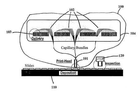
Figure 1 is a schematic diagram of one embodiment of a microarray fabrication
system.
Figure 2 illustrates a print-head containing the immobilized portion of twenty-
one
capillary bundles.
Figure 3 illustrates a random capillary bundle linked to a frame that has
suction
portions that dip into the wells of a standard microtiter plate.
Figure 4 illustrates equipment for and a method of using light to register the
proximal and distal ends of capillaries in a random bundle.
Figure 5 illustrates one method of identifying the position of a proximal end
of a
capillary in the immobilized portion of the bundle.
Figure 6 illustrates steps in fabricating a capillary bundle using a guide
plate that is
removed to form the finished bundle.
Figure 7 illustrates two methods of controlling flow rate of probe-containing
solution through capillaries, i.e. the use of pressurized gas, and the use of
voltage. . .
Figure 8 illustrates probe deposition by mechanical tapping.
Figure 9 illustrates a spring-mounted substrate holder that provides improved
alignment between substrate and print head.
Figure l0 illustrates probe deposition by electrostatic printing.
Figure 11 illustrates equipment for and a method for inspecting a microarray
using
light-scattering.
Figure 12 illustrates equipment for and a method for inspecting a microarray
using
total internal reflection of light within the substrate.
12
CA 02400789 2002-08-16
WO 01/62377 PCT/USO1/05695
Figure 13 illustrates two ordered spot patterns that can be formed when
individual
fibers are used to form a print head using a guide plate.
Figure 14 illustrates how a fluid transfer device composed of multiple
capillary
bundles can be configured to draw liquid from multiple microtiter plates
having wells of
large capacity and place that liquid in small reservoirs contained in a single
microtiter plate.
Figure 15 illustrates a honeycomb pattern of probes that can be formed by a
print
head made using a guide plate having holes in a honeycomb pattern.
Figure 16 illustrates a random pattern of probes which can form when printed
using
a random-bundle print head.
Figure 17 depicts a print system suitable for depositing probes immobilized on
a
magnetic support onto a substrate.
Figure 18 illustrates a print system having a collar or support that contacts
the
substrate or structure around the substrate and is sufficiently long to
prevent the print head
from contacting the substrate. At the same time, it is not so high that it
prevents droplets
from the capillaries from contacting the substrate surface.
Figure 19 illustrates an alternative printing arrangement where the facet of
the print
head is flat but there is a riser at the edge of, or around, the substrate
that is. sufficiently
high to prevent the print head from directly contacting the substrate. At the
same time, it is
not so high that it prevents droplets from the capillaries from contacting the
substrate
surface.
DETAILED DESCRIPTION OF THE INVENTION
In the description below, a DNA microarray is used as one embodiment of the
invention. The techniques described herein can also be used to produce
microarrays of a
wide range of biological and chemical probe materials which include but are
not limited to
deoxyribonucleic acids (DNA), ribonucleic acids (RNA), synthetic
oligonucleotides,
antibodies, cells, tissue, proteins, peptides, lectins, modified
polysaccharides, synthetic
composite macromolecules, functionalized nanostructures, synthetic polymers,
modified/blocked nucleotides/nucleosides, modified/blocked amino acids,
fluorophores,
chromophores, ligands, chelates, haptens, drug compounds, and chemical
compounds that
have associated substance which binds, associates, or interacts with the probe
material. The
samples being deposited on the microarray substrate using the technology
disclosed herein
can take or be carried by any physical form that can be transported through a
capillary.
13
CA 02400789 2002-08-16
WO 01/62377 PCT/USO1/05695
These include but are not limited to aqueous or non-aqueous fluid, gel, paste,
bead, powder
and particles suspended in aqueous or non-aqueous liquid.
The substrate may be formed of any material on which the probes can be
deposited.
The substrate itself may be capable of immobilizing the particular probes
used, or the
substrate may be capable of modification (for example, by coating) so that it
is capable of
such immobilization. The substrate may be porous or nonporous materials.
Preferred
materials for the substrate of the present invention include silica, glass,
metals, plastics, and
polymers.
For immobilizing polynucleotides and polypeptides, glass is a preferred
material
because polynucleotides and polypeptides can be covalently attached to a
treated glass
surface and glass gives out minimal fluorescent noise signal. The glass may be
layered on
another material, or it may be core or base material of the apparatus, or
both. Another
example of a substrate includes a plastic or polymer tape as a base substrate,
with a coating
of silica for probe embodiment. In this embodiment, a further layer of
metallic material
may be added, either on the opposite side of the tape from the silica layer,
or sandwiched
between the silica layer and the polymer or plastic.
A microarray fabrication system based on this invention is illustrated
schematically
in Figure 1. The heart of the system 100 is a print-head 101 comprising a
large number of
flexible capillaries 102. Each capillary in the print-head is fluidly linked
to a reservoir 103
containing a specific DNA sample. The reservoirs may take the form of fluid
wells in
standard microtiter plates 104. Probes are delivered to the print-head via the
capillaries and
the entire set of probes can be deposited on to the substrate 110 in a single
printing action.
There is an inspection system 120 to inspect the quality of the fabricated
microarrays online
or off line.
In the invented system shown in Figure 1, multiple microarray substrates are
carried
on a translation stage, which moves in a single axis in a stepping fashion to
align a blank
substrate under the print head. The translation stage can be a rotation stage
or a conveyor
belt based system equipped with substrate loading and unloading stations. In
this way,
blank substrates can be fed to a print position beneath the print head in a
continuous
fashion. The print head can deposit an entire set of probes by moving only a
very short
distance (< lmm) in one axis (up and down in the z axis). Or the print head
may not have to
move at all if electric or magnetic induced deposition methods are used, which
are
14
CA 02400789 2002-08-16
WO 01/62377 PCT/USO1/05695
described below. As a result, microarray manufacturing can be carried out in a
continuous
fashion at a very high throughput.
In robotic pin deposition methods and other deposition methods in which probes
may be placed on a substrate, the print head moves in the x and y axes as well
as the z axis.
The pins travel a long distance, in the order of a meter, and thus such
conventional
deposition methods require a substantial period of time to fabricate an array
on a substrate.
A print system of the invention can thus be configured to travel a short
distance and require
little time to print a microarray.
The probe reservoirs in the system can be positioned above the print head and
substrates, as shown in Figure 1. The print head deposits the probe down to
the upper
surface of the substrate. The advantage of such an arrangement is that, after
priming, the
fluid flow inside the capillaries can be driven by the gravity, which is very
stable and
uniform among capillaries and can be precisely controlled by adjusting
the.height of the
reservoirs. An alternative arrangement is to place the reservoirs below the
print head. The
print head moves up to deposit probes on substrates, which are held "face-
down" on the
stage. In this configuration, the capillaries are short and relatively
straight. The probe-
containing fluid can be moved to the substrate by pressurizing the reservoirs,
for instance.
The basic elements of the technology of this invention include methods and
apparatus for print-head, fluid delivery, probe deposition and inspection. The
details of
these technological elements are discussed in the following sections.
1. Print-Head
The print-head receives probe fluids from their individual reservoirs and
deposits
them in small volumes onto the microarray substrate at each printing action. A
print-head is
a solidified piece of e.g. polymer such as a thermo-setting or other polymer
(for example,
an epoxy polymer) that surrounds the proximal ends of the capillaries, and its
facet or face
that contacts the substrate is fabricated to conform to the surface contour of
the microarray
substrate in order to facilitate uniformed probe deposition.
The print-head is solid or has su~cient flexibility to conform to the
substrate
surface on which a micro-array is to be printed. The print-head 200 may
contain a single
capillary bundle or, as shown in Figure 2, multiple capillary bundles 201,
202, 203, 204,...,
221. In the multiple bundle configuration, it is preferred that the outline
shape of each
bundle is rectangular or square so that the capillary bundles can easily be
assembled to
form a structured matrix in a rectangular print-head 200 (although other
shapes are
CA 02400789 2002-08-16
WO 01/62377 PCT/USO1/05695
possible). In this way, 1) the print head can be configured to print on most
or all of the
surface area of a standard microscope slide; 2) the position and orientation
of each bundle
in the system is known; and 3) it is easier to identify each capillary in a
bundle.
Alternatively, the outline shape of each bundle could be round or in other
shapes.
Capillaries used in the system can be made of silica or other suitable
materials such
as glass, ceramics, polymer or metal. The capillaries conduct the probes of
interest from the
distal ends of the capillaries to the proximal ends of the capillaries, and
thus capillaries that
are bundled to form a print head are manufactured from a material that does
not remove a
substantial number of probe molecules from their carrier liquid and attach the
molecules to
the walls or to another material positioned within the capillaries.
The capillary bundle is assembled from a large number of individual, ready-
made
capillaries. Capillaries are bundled together, solidified into a single mass
or block at their
proximal ends using an adhesive or by fusing the capillary walls at the
proximal ends of the
capillaries together, and eventually assembled into the print-head while the
distal ends of
capillaries are left loose or attached to reservoirs or a plate that dips into
a set of reservoirs.
The proximal ends of the capillaries may be solidified together using a cement
or
epoxy that forms a rigid block, or the proximal ends may be solidified
together using a
polymer that is somewhat flexible, so that the surface conforms to the
substrate when
pressed against it to provide better printing in the event that the printing
face or facet of the
block is not perfectly parallel to the surface of the substrate to be printed.
The printing face
may optionally be polished to provide a very flat surface, so that the
proximal ends of the
capillaries terminate within 100 micron of each other, for instance. That is,
if the printing
face is held above and parallel to a plane and separated by a nominal distance
z, the
difference between the shortest distance that a proximal end in the facet
terminates fromahe
plane and the greatest distance that a proximal end in the facet terminates
from the plane is
no more than about e.g. 100 micron. Preferably, the difference in termination
distances is
no more than about 50 micron, more preferably no more than about 20 micron,
and more
preferably no more than about 5 micron. The trimmed block has sufficient
rigidity to assure
its facet remains parallel to the substrate during printing.
In one embodiment of the invention, the solid mass contains no more than about
10
cm of the lengths of the capillaries (and thus the printhead in this
embodiment is no more
than about 10 cm thick), and the loose or free ends of the capillaries are
from about 1 to
about 3 meters in length. Consequently, the ratio of length of loose capillary
to thickness of
16
CA 02400789 2002-08-16
WO 01/62377 PCT/USO1/05695
solid mass is preferably at least about 10 and more preferably at least about
30. The solid
mass may be about 2 cm thick or thinner, and in this instance the ratio of
length of loose
capillary to thickness of solid mass is preferably at least about 50 and more
preferably at
least about 150. The solid mass needs only to be sufficiently thick that the
print head, alone
S or in combination with a frame that forms part of the print system, is
sufficiently rigid that
the solid mass does not deform appreciably under printing conditions, so that
a microarray
is formed when probes are printed onto a substrate. The loose ends of the
capillaries are
sufficiently long to be in fluid communication with the reservoirs or with
outlet pipes
connected to the reservoirs. Preferably, the loose ends are also sufficiently
long that the
loose portions of the capillaries accommodate any up-and-down movement of the
print
head with little stress to the capillaries, so that the capillaries do not
crack or break during
use.
In another embodiment of the invention as illustrated in Figure 18, the print
head
1800 is equipped with a supporting bracket or collar 1802 that prevents the
facet 1804 of
the print head from contacting the substrate 1806 held on substrate support
1808. The facet,
especially any functional coating on the surface (such as a coating of an
electrically-
conductive material), may be damaged after repeated contact with the
substrate.
Consequently, the supporting bracket helps to prolong the life of the
printhead..The vertical
distance d illustrated in Figure 18 between the edge of the collar 1810 and
the facet 1804 is
selected so that the printhead does not contact the substrate but is still
sufficiently close to
deposit droplets 1812 of probe-containing fluids 1814 onto the substrate. The
collar need
not be a solid piece of cylindrically-shaped material as illustrated. The
collar may consist of
a frame that attaches to the print head and has feet or shafts that protrude
to prevent the
facet~from contacting the substrate, for instance.
Alternatively, as shown in Figure 19, the facet 1902 of print head 1900 can be
flat
and a riser 1904 may be placed on the outer region of the substrate 1906 to
prevent the
printhead from contacting the substrate while still depositing droplets 1908
of probe-
containing fluids. Further, this same effect can be achieved by positioning a
collar of
suitable dimensions around the substrate. The collar can be rigid, or
alternatively the collar
may contain a cushioning portion formed from a polymer or felt, for instance,
upon which
edges of the facet press when the facet is moved toward the substrate. The
cushioning
portion is positioned so that the facet does not contact the substrate, even
though the
cushioning material is compressed and the print head is printing the
microarray on the
17
CA 02400789 2002-08-16
WO 01/62377 PCT/USO1/05695
substrate. The cushioning portion provides a "softer" portion upon which the
facet lands,
helping to prevent the facet from being damaged.
Each capillary can be fluidly linked to a probe reservoir, which may be the
well in a
standard microtiter plate. The linkage can be made permanent by gluing the
capillary to a
hole at the bottom of a microplate well. Alternatively, as shown in Figure 3,
the capillaries
301 can be permanently fixed to a frame 302, which holds the positions of
capillary tips
303 in a grid, which has the same spatial pattern and pitch as a standard
microplate 304.
Then the frame can be locked on to a standard microplate to establish the
fluid linkage for
each capillary. In this way, the microplate after fabrication can be taken off
the arrayer for
long-term storage. It is also possible to wash the capillaries after the
fabrication of a
particular microarray, then install a new set of microplates to make a
different microarray.
Following is a description of two different methods for making the assembled
capillary bundle. These are the "tight-pack" and "guide-plate" methods,
respectively.
1.1 Tight-Pack Method
In the tight-pack method, a large number of hair-thin, flexible capillaries
are tightly
packed in random order into a bundle at the proximal ends of the capillaries,
in which the
outer surface of a capillary is in direct contact with that of adjacent
capillaries. In a tight
packing of random capillaries, the capillaries take up positions in reference
to each other.
The local spatial pattern may be regular, e.g. the centers of every three
adjacent spots may
form an equilateral triangle, and six spots surrounding any spot may form a
hexagon.
However, minute misalignment in the random bundle of capillaries soon
accumulates and
results in distortion of the global alignment of the spots as illustrated in
Figure 2 and Figure
16. As the number of spots increases, the distortion is amplified. The global
spatial pattern
becomes random.
However, although such a bundle may be used to print a probe microarray at
high
density, the microaxray is useless for printing because the association
between a capillary
facet in the bundle and the fluid reservoir that it linked to, thus the probe
identity, is lost.
Capillaries randomly packed to form a capillary bundle can be made suitable
for
microarray printing by re-establishing the one-to-one association of each
capillary between
the proximal and distal end of the bundle after the bundle has been made.
There are a number of ways to re-establish the capillary association in a
tightly
packed bundle. These are:
18
CA 02400789 2002-08-16
WO 01/62377 PCT/USO1/05695
1 ) Use a type of capillary that is not only capable of transporting fluid
through
the capillary, but is also capable of transmitting light like an optical
fiber. Capillary-
reservoir association can then be re-established by launching light into each
capillary
from the reservoir end and observing the position of the exiting light at the
bundle end,
using an imaging device as shown in Figure 4. This imaging device can be
either a
CCD based digital microscope or a scanning microscope. Light guiding
capillaries can
be produced by creating an inner region in the capillary, in which the optical
refractive
index is higher than the outer region around it. Such a region will be able to
trap the
light inside it and guide the light all the way through the capillary.
This light trapping region inside capillary can be created in many different
ways. A first method is to coat the outer surface of a silica capillary with a
polymer
with lower refractive index. A second method is to fill a silica capillary
using a
transparent fluid with a higher refractive index than that of the capillary to
create a
temporary fluid core capable of transmitting light through the capillary. A
third, also
preferred, method is to draw the capillary out of a preform. Such a preform
can be made
by following the modified chemical vapor deposition (MCVD) procedure widely
used
in the optical fiber industry for optical fiber perform fabrication, then
drawing the
preform without collapsing the central cavity at the final step.
Alternatively, this
preform can also be made by drilling a hole of suitable size through the axis
of a
multimode optical fiber preform or depositing a layer of fluoride doped silica
outside a
suitable pure silica tube. Since fluoride doping lowers the refractive index
of pure
silica, it forms a cladding to help trapping light inside the pure silica
region around the
central cavity.
2) Blow air into the capillaries one by one from the distal end and use a
micro-
flow detection device at the bundle proximal end to locate the outlet of the
air flow. The
position coordinate of the capillary facet is registered among other
capillaries in the
bundle. A micro sized hot wire or temperature probe can be used for the flow
detection
because the air current caused by air exiting the capillary alters the thermal
balance at
the probe.
3) Fill capillaries with ink from the distal end and observe where the ink
exits
the proximal end at the bundle facet using an imaging microscope and register
its
position. Capillaries can be filled one at a time or several at a time using
ink of different
colors.
19
CA 02400789 2002-08-16
WO 01/62377 PCT/USO1/05695
4) Use metal capillaries insulated from one another by e.g. a dielectric such
as
a silica coating, or form dielectric capillaries with a metal layer and
dielectric coating
over the metal layer. The capillary-reservoir association can be established
by placing a
voltage on the distal or proximal end of the capillary and sensing the voltage
on the
proximal or distal end of the capillary, respectively, and determining the
position of the
capillary relative to the other capillaries.
The invention also provides two ways to automatically register the identity of
a
specific capillary in a bundle formed using any of the four methods described
above.
Capillary position may be registered by way of an absolute coordinate system,
or capillary
position may be registered to an image of the facet face.
1 ) Absolute coordinates Referring to Figure 5, an XY coordinate system, 501
for example, can be established with reference to the edges of the bundle, and
the
identity of each capillary 502a, 502b, etc. can be registered by the system
through its
unique coordinates in the coordinate system. In this instance, the coordinates
represent
a mathematical vector that can be drawn from the origin of the coordinate
system to the
capillaries. The coordinates can be recorded in a database or otherwise saved
in digital
or analog form, and the coordinates can be associated with information on the
position
of corresponding reservoirs or distal ends to correlate or register the
proximal end of
each capillary with its associated reservoir or distal end. This approach is
relatively
easy to implement if the outline shape of the bundle is square or rectangular
and the
capillaries are packed tightly, so that, as shown in Figure 5, the capillaries
form a
honeycomb pattern or other regular pattern. This method also tolerates at
least a
moderate degree of positional randomness in the bundle.
2) Image matching When the capillaries are completely random and there is no
obvious spatial pattern in the bundle, an image matching method can be
employed to
register capillary identities. In this method, as illustrated in Figure 4, a
computer
records data representing an image 401 of the bundle facet to a file. Each
capillary in
the image (e.g. 402a, 402b,...) is correlated to its probe reservoir using
e.g. one of the
methods above, thereby building a database or forming other data correlating
reservoirs
with their corresponding capillaries. The spot pattern of the printed
microarray will be a
precise hard copy of the capillary facets in the bundle. Therefore DNA placed
in a
reservoir is printed in a known position, and this information can be
correlated with the
facet image to determine where probes are in the microarray. After
hybridization, the
CA 02400789 2002-08-16
WO 01/62377 PCT/USO1/05695
microarray is scanned by a microarray scanner, which generates a pair of
digital images
at different fluorescent wavelengths as described in U.S. Patent No.
5,800,992, which is
incorporated by reference in its entirety herein. The scanned image can then
be
compared to the facet image stored in the computer to establish the DNA
identity of
each spot in the microarray. To make the image comparison easier, selected
small
number of wells in the plate can be filled with a special paint or ink or a
fluid tagged
with a distinctive dye. These distinctive spots 403a, 403b, on the scanned
image of the
probe microarray can then be used as reference points to match spots 404a,
404b on the
scanned image 405 with the ID tagged image file of the capillary bundle, pre-
stored in
the computer.
A single bundle consisting of 100,000 or more capillaries can be fabricated
and ID
tagged in this way. However, it may be~more beneficial to limit the number of
capillaries in
a random bundle to a smaller number, e.g., 1536. Then, multiple such random
bundles can
be assembled into an orderly bundle matrix as shown in Figure 2 to form a
print-head. This
allows the utilization of standard microtiter plates with 1536 or fewer wells
widely in use.
Secondly, this arrangement provides greater printing flexibility. Multiple
probes can be
organized into different groups with one bundle per group, then mixed-and-
matched to
produce different microarrays for different applications. Finally, this
arrangement gives the
user the option and flexibility to scan only one group of probes on the
microaxray, wherever
~ necessary to save time.
Considering a particular embodiment of the above described arrangement:
Assuming capillaries with an outer diameter of 100~m are used and each bundle
is
linked to a 1536-well or four 384-well microtiter plates, the capillary bundle
would have a
4mm x 4mm cross section. 75 such bundles can be easily assembled into a 5x15
orderly
bundle matrix, which could produce a microarray consisting 115,200 probes in
one stamp
and covering a 2cm x 6cm area on a microscope slide.
A modified form of the "tight-pack" method may also be used to form an
assembled
capillary bundle. Instead of tightly packing the capillaries, the capillaries
may be packed
more loosely. The local order as well as long-range order of the capillaries
becomes
random, resulting in a random array of probes in the microarray when printed.
1.2 Guide-Plate Method
The guide-plate method for capillary bundle fabrication is illustrated in
Figure 6. A
guide-plate 601 as seen from above in Figure 6a has an orderly matrix of small
holes 602a,
21
CA 02400789 2002-08-16
WO 01/62377 PCT/USO1/05695
602b,...etc. fabricated through precision drilling. Alternatively, the guide
plate can be
made of glass and produced by slicing fused capillary array tubing drawn from
a larger
glass preform as described in U.S. Pat. No. 4,010,019 and 5,276,327. The plate
can be
made of any suitable material such as metal, glass or plastic and can also be
relatively thin
andlor deformable and/or fragile. The hole diameter should be slightly larger
than the outer
diameter of the capillaries to be used. Capillaries 603a, 603b,... are
carefully plugged into
the holes to form a loose bundle 604, as illustrated in Figure 6b. The bundle
604 is
solidified at the section near the guide-plate as shown in Figure 6c using
epoxy 605, cement
or other suitable solidification techniques. Finally, the solidified portion
is cut at a position
very close to the guide-plate, to remove the guide plate, as shown in Figure
6d.
Because the holes are positioned in an orderly matrix at the guide-plate and
the
bundle is cut very close to the guide-plate, the spatial position of each
capillary in the
fabricated bundle will be in an orderly matrix the same as the holes in the
guide-plate. Also,
because the bundle is in one solid piece, it can be polished to achieve a high
degree of
flatness and at the same time, is mechanically robust for printing. In
addition, since the
capillaries are in an orderly matrix, the position of the capillary in matrix
is known, and
therefore the position of the capillary establishes the position of a probe in
a microarray
printed on a substrate. No ID tagging procedure is required.
A guide plate may be configured in any shape desired. It may be, e.g., a
block, a
sphere, a plate, or any other shape so long as the shape has holes or pores
into which the
capillaries may be inserted.
Instead of using a plate, a grid of wires or strings or strands (preferably
interwoven)
can be formed, and the individual capillaries can be inserted within spaces in
the loose grid
to form the capillary bundle. The grid can be tightened to pull the
capillaries close to one
another, and the proximal end, distal end, and/or intermediate portions can be
adhered
together using e.g. an adhesive to form a solid mass. Any strands of the grid
that form part
of the solid mass may be trimmed flush with the solid mass, and other free
strands may be
removed to provide the fiber bundle.
2. Fluid Delivery
The functions of the fluid delivery sub-system in the arrayer are to
~ Transport probe fluid from the reservoir to the print-head through its
respective capillary;
22
CA 02400789 2002-08-16
WO 01/62377 PCT/USO1/05695
~ Ensure the flow rate to be constant in each capillary and uniform across
the print-head.
2.1 Fluid Transport
This invention offers several methods to drive the probe fluid from its
reservoir into
the capillary and towards the print-head. They can be used alone or in any
combination of 2
or more in the fluid delivery sub-system. These methods include:
~ Air pressure A differential air (or other gas such as nitrogen) pressure
can be established and maintained between the proximal and distal ends of the
capillary bundles, which will translate into hydraulic pressure to drive the
probe
fluids.
~ Gravi Once the capillaries are filled with the probe fluids, a constant
flow can be maintained and controlled by adjusting the vertical positions of
the
fluid reservoirs, e.g. the microtiter plates, with respect to the position of
the print-
head.
~ Electric-field Because DNA fluids are negatively chaxged, a voltage
applied between the reservoir and the print-head can be used to control the
flow of
the fluid through electrostatic and electroosmotic force (EOF) [1 ].
~ Vacuum The proximal ends of the capillaries may be placed under
relative vacuum. The print head and substrate holder may be placed within a
vacuum chamber, and the capillaries may extend through a wall of the vacuum
chamber and to the reservoirs. The print head in this instance preferably
extends to
the wall of the chamber so that thin capillaries are not exposed directly to
vacuum if
no liquid flows through them. '
2.2 Flow Rate Control '
In order to ensure that the spot sizes on the substrate are constant from
microarray
to microarray and uniform across each microarray, the flow rate has to be
controlled to be
constant in each capillary arid uniform across the print-head.
It takes routine techniques to hold the fluid flow in a single capillary to a
constant
rate. All fluid driving methods described in Section 2.2.1 can be used to
control the flow
rate. Air pressure and gravity are relatively blunt mechanisms for flow rate
control. When
air pressure or elevation differences disappear, the flow does not stop
instantly due to back-
23
CA 02400789 2002-08-16
WO 01/62377 PCT/USO1/05695
pressure built up in the capillary. In comparison, electric fields are more
precise in
controlling flow rate.
It takes more measures to ensure the uniformity of flow rates in every
capillary of
the print-head because the flow rate in a capillary is dependent upon many
factors besides
the driving force, which include cavity size and surface characteristics of
the capillary as
well as fluid viscosity. Also, clogging and bubble entrapment in capillaries
will prevent
probe flow and cause unwanted vacancies on the fabricated microarray.
This invention provides the following measures to ensure the flow rate
uniformity:
~ Use of silica based capillaries Silica capillaries are renowned for precise
dimensions. Both inner and outer diameters can be controlled to vary less than
2%
in a same draw and less than 5% between different draws. (A "draw" is the
pulling
or stretching of larger, more easily fabricated preforms at a sufficiently
high
temperature that the tubular preforms thin to form capillaries. This
technology is
common in optical fiber manufacturing.) Capillaries from the same draw can be
used to enhance uniformity of channel diameter in the capillaries. Because the
drawing is carried out at melting point of the silica, the surface is
extremely smooth.
In addition, the silica surface in the capillary is naturally negatively
chaxged, which
makes it "phobic" to DNA samples, resulting in minimum friction between DNA
probes and capillaries, ensuring smooth delivery of sample fluids to the print-
head.
Coating cavity walls with other hydrophobic films such as a fluorocarbon
polymer
such as polytetrafluoroethylene may further enhance the durability and
uniformity
of the capillaries.
~ Buffering the probe fluids Different probes may have different viscosity.
The viscosity of different probe fluids can be made more uniform by adding a
suitable amount of inert buffering material, e.g., sugar, to increase the
viscosity of
probe fluids of low viscosity.
~ Clogging and bubble prevention All probe fluids can be purified and
handled in a clean room environment to prevent capillary clogging. Fluids can
also
be preprocessed with ultrasound and vacuum suction to eliminate bubble
entrapment.
~ Control flow rate in each capillary with individual electric fields The
flow rate variation across the print-head can be kept within a small range
(e.g. 20%)
24
CA 02400789 2002-08-16
WO 01/62377 PCT/USO1/05695
under a uniform driving force such as air pressure or gravity. This is
sufficient when
fabricating most microarrays. For applications that require more accurate flow
rate
control, the electric field method can be used to control the flow rate in
each
capillary individually. In one specific embodiment of the flow control sub-
system,
as shown in Figure 7, gravity and/or pressurized air 701 is used as the
primary fluid
driving force and an electric field of the original capillary is used as an
additional,
fine adjustment mechanism. The end-facet 702 of the print-head 703 at the
proximal
end of the capillaries 704 a, 704b,... and each capillary tip 705a, 705b,...
at the
distal end of capillaries are coated with metal. All capillaries are held at a
common
ground at the print-head and different voltages Vi, Vj are applied to the
different
capillary tips at the distal end. This produces appropriate electric fields to
fine-tune
the flow rate in the capillary. Because the electric field is only a fine-
tuning device,
a relatively small voltage is sufficient. Voltage can be adjusted based on
feedback
from inspection devices, as discussed below, or by monitoring the size of
droplets
deposited using e.g. an optical or scanning microscope.
3. Probe Deposition
The probe deposition sub-system in the arrayer ensures that a constant and
uniform
volume of probe fluids are deposited onto the substrate and there axe minimal
or no missing
or overlapped spots on the microarray.
3.1 Mechanical Tapping
As illustrated in Figure 8, probes can be deposited on to the microarray
substrate by
mechanically tapping the print-head 805 on the substrate. As shown in Figure
8a, the
constant flow of probe solutions 801 in the capillary 802 produces a micro
sphere 803 of
fluid at the facet 804 of each capillary. When the print-head 805 is tapped on
the substrate
806, the droplet bonds to the substrate due to surface tension as shown in
Fig. 8b. This
surface tension overcomes the binding force in the fluid. The droplet thus
breaks away
from the fluid column at its weakest point, i.e. exiting point of the
capillary cavity, when
the print-head withdraws as shown in Fig. 8c. A probe spot 807 is deposited on
the
substrate.
Two potential problems associated with microarrays produced with this type of
printing method are missing and overlapping probes in the microarray. This
invention
provides the following measures, which can be used alone or in combination, to
prevent
missing spots on the fabricated microarray:
CA 02400789 2002-08-16
WO 01/62377 PCT/USO1/05695
1) The distance between the print-head facet and substrate during printing
is selected to be no more than the minimum diameter of the probe-containing
droplets formed at the tips of the capillaries. Because the radius of the
droplets is
typically in the order of 1030 micron, the distance between the print-head
facet
and substrate is typically in the order of 5 to 20 micron. The surface of the
print-
head facet is polished to a high degree of flatness when, for example, a
microscope
slide or other flat substrate is used as the microarray substrate.
2) One of the contacting parts, i.e. print-head or the substrate, is rigidly
supported while the other is fixed on a soft or spring-loaded platform, as
shown in
Figure 9. If these two surfaces are slightly unparallel, the one on the soft
support
will yield to the one on the rigid mounting to ensure perfect contact (Figure
9). The
platform may be spring loaded, mounted on joints or gimbals, or may be a
polymeric or sponge-like block on which the substrate rests, fox example.
Probe cross-talk occurs when excess amount of probe fluid is deposited on the
substrate and there is a lack of means to confine the deposited fluid within a
certain area on
the substrate. The flow rate control described in Section 2.2 helps to prevent
fluid overflow.
In addition, capillary force may be created between the print-head facet and
the substrate
when the print-head is brought very close to the substrate and a fluid link is
established
between the two surfaces. This capillary force may act to pull extra fluid out
of the cavity.
This invention further provides the following measures, which can be used
alone or in
combination, to prevent overlapping spots on the fabricated microarray:
1) Making both the print-head and substrate surfaces hydrophobic.
2) A micro well 808 can be fabricated at the tip of each capillary (as shown
in Figure 8), which can accommodate the fluid volume of the droplet to be
placed
on the substrate. The micro wells can be produced one-by-one using a diamond
tipped precision drill or in parallel using photolithographic methods. When
the
capillary has a central region doped with Germanium (originally designed for
light
transmission as described in Section 2.1.1), these micro wells can be
fabricated in
parallel by dipping the print-head into an etching fluid such as fluoride acid
solution
(e.g., HF). A very small amount of Ge doping can dramatically accelerates the
etching rate of the silica in the vicinity of the Ge.
3) A spacer can be installed between the print head facet and the surface of
the substrate as shown in Fig. 18. During tapping, the spacer face contacts
the
26
CA 02400789 2002-08-16
WO 01/62377 PCT/USO1/05695
substrate while the print head facet is suspended closely above the substrate,
allowing fluid spheres to contact the substrate to deposit droplets of probe-
containing liquid on the substrate.
4) Increase the viscosity of the probe materials to be printed by increasing
the sample density in its solution or by adding sufficient amount of inert
buffering
materials. Print probes in bead, gel or paste forms can eliminate overlapping
problem.
5) Reduce the time in which the print-head is in fluid contact with the
substrate.
6) Use capillaries with a smaller inner diameter, which will reduce the
effect of the capillary pulling force generated in the fluid layer between the
print-
heat and the substrate during contact printing.
7) Deposit probes on hot substrates in a dry environment, which accelerates
the evaporation of fluids in the probe and reduces overflow.
8) Deposit probes on a substrate that has a surface temperature below the
freezing point of the probe fluid.
3.2 Electrostatic printing
As shown in Figure 10, a conductive layer 1001, such as metal, can be coated
on the
facet 1002 of the print-head 1003 and the microarray substrate is placed on a
conductor
. 1004 or conductor coated support 1005. Alternatively, a special microarray
substrate with
conductive layer can be used. When a voltage V is applied between the stamp
head and the
substrate or its support with positive polarity at the substrate end, the DNA
samples in the
capillary will be attracted towards the substrate because of their negative
charges. If a short
pulse of sufficiently high voltage is applied when the stamp head facet is
close to the
substrate, spots 1006a, 1006b,... of the various probe fluids are torn from
the fluid columns
in the capillaries and are propelled to the substrate. One advantage of this
method is that the
stamp head does not have to touch the substrate surface, thus eliminating many
potential
problems associated with missing or overlapping spots on the fabricated
microarray. In
addition, the stamp head does not have to move, and no microwell is needed at
the capillary
tip.
' 3.3 Printing Beads
Probes may also be immobilized on beads and a colloidal suspension formed, and
the suspension can be deposited through the capillaries and onto the substrate
to deposit the
27
CA 02400789 2002-08-16
WO 01/62377 PCT/USO1/05695
beads onto the substrate. In this event, the beads may be functionalized as
described below
so that the beads attach to the surface of the substrate. The beads typically
have a diameter
less than 20 microns, preferably between about 0.1 and 20 micron, and
preferably less than
100 nm.
The beads may be transparent, so that the light used to stimulate the
fluorescent
moieties refracts and reflects a number of times, thereby providing more light
to the probes
on the illuminated beads to stimulate fluorescence. This leads to a stronger
fluorescence
signal. Beads are also capable of carrying many more probe molecules on their
surfaces
than the flat surface of the substrate. Consequently, signal strength also
increases because
I O of the large number of target molecules that associate or hybridize to the
probe molecules
on the surface of the bead.
The beads may optionally be magnetic or paramagnetic. Magnetic beads are
currently commercially available. In this instance, a magnetic field can be
established to
help drive the magnetic beads from the print head 1706 to the surface of the
substrate
I S where they are to be deposited. A suitable magnetic field may be
established between the
reservoir or the capillaries and the substrate, for instance, by installing a
high bandwidth
electromagnetic coil 1702 under the microarray substrate 1704 as shown in
Figure 17.
3.4 Electromagnetic printing
Probe molecules may be attached to ferrofluids (magnetic liquids) to form
ferrofluid
20 particles and deposited on the substrate. Ferrofluids are colloidal
magnetic particles having
a size of about 3 to about 50 nm (approximately the size of an antibody) and
composed of ,
surfactant coated single or multiple crystals of e.g. magnetite (Fe304)
dissolved in water or
kerosene or other compatible solvent. Alternatively, probe molecules are
dissolved or
suspended in ferrofluids and not attached to magnetic or paramagnetic
particles.
25 Ferrofluid particles typically have a size ranging from 3 to 100 nm and can
be
synthesized by a variety of methods which result in 'flocs' composed of
polymer (typically
dextran or protein) and magnetite and/or other iron oxide crystals. Ligands
such as biotin,
avidin, streptavidin, or other ligands for attaching,the ferrofluids to the
substrate may also
be coupled to the ferrofluid particles.
30 The coupling chemistries for attaching antibodies and other relevant
molecules such
as oligonucleotides to ferrofluids to form ferrofluid particles are known, and
molecules
coupled to ferrofluids are available from Immunicon Corp., Huntingdon Valley,
PA.
28
CA 02400789 2002-08-16
WO 01/62377 PCT/USO1/05695
The magnetic properties of ferrofluids derive from the magnetic properties of
magnetite and the colloidal nature of the material. Magnetite crystals are
typically about 3 -
nm in size and therefore exhibit superparamagnetism (i.e. they only exhibit
magnetic
properties when in a magnetic field; when the field is reduced or eliminated,
the magnetism
5 disappears).
Ferrofluid particles are easily transported to the substrate surface by
applying a
magnetic field. The particles can diffuse through solution via Brownian motion
and deposit
rapidly on the substrate surface. Removing the magnetic field essentially
stops the
ferrofluid particles from depositing onto the substrate.
10 3.5 Vacuum printing
The proximal ends of the capillaries may be placed under relative vacuum in
order
to draw probe-containing fluid through the capillaries. The print head and
substrate holder
may be placed within a vacuum chamber, and the capillaries may extend through
a wall of
the vacuum chamber and to the reservoirs which are at atmospheric pressure.
The print
head in this instance preferably extends to the wall of the chamber so that
thin capillaries
are not exposed directly to vacuum if no liquid flows through them. The
pressure in the
vacuum chamber can be reduced to below atmospheric pressure to draw fluid from
the
reservoirs and print on the substrate, and the pressure in the vacuum chamber
can be
increased to about or slightly above atmospheric pressure to prevent further
fluid from
depositing on the substrate when the print head is lifted away from the
substrate.
3.6 Probes
The probes may be DNA, RNA, proteins, cells, or other constituents as
discussed
previously. The probes may be attached to the substrate or to beads
covalently. Thus, one
may use a variety of approaches to bind an oligonucleotide to the solid
substrate. By using
chemically reactive solid substrates, one may provide for a chemically
reactive group to be
present on the nucleic acid, which will react with the chemically active solid
substrate
surface. One may form silicon esters for covalent bonding of the nucleic acid
to the surface.
Instead of silicon functionalities, one may use organic addition polymers,
e.g. styrene,
acrylates and methacrylates, vinyl ethers and esters, and the like, where
functionalities are
present which can react with a functionality present on the nucleic acid.
.Amino groups,
activated halides, carboxyl groups, mercaptan groups, epoxides, and the like,
may also be
provided in accordance with conventional ways. The linkages may be amides,
amidines,
29
CA 02400789 2002-08-16
WO 01/62377 PCT/USO1/05695
amines, esters, ethers, thioethers, dithioethers, and the like. Methods for
forming these
covalent linkages may be found in U.S. Pat. No. 5,565,324 and references cited
therein.
Alternatively, the probes may be attached to the substrate or to beads non-
covalently by e.g. functionalizing the surface of the substrate and the probe
to provide
binding moieties on each. Generally, this will be accomplished by providing
each of the
probe and the support with one of a pair of corresponding affinity binding
partners, such
that the probe and the support may be bound together selectively, and if
desired, reversibly.
Typical non-covalent coupling agents include biotin/streptavidin,
Staphylococcus aureus
protein A/IgG antibody F~ fragment, and streptavidin/protein A chimeras. See,
e.g., T.
Sano and C. R. Cantor, Bio/Technology 9:1378-81, 1991. Most conveniently, the
affinity
binding partner will comprise biotin and avidin or streptavidin, the biotin
being bound to
the probe and the avidin or streptavidin to the support. In such an
embodiment, the surface
of the substrate may be functionalized with avidin or streptavidin, and the
probe molecules
may be functionalized with biotin by methods well-known in the art. See, e.g.,
U.S. Pat.
No. 5,948,624 and "Applications of Avidin-Biotin Technology: Literature
Survey," by
Wilchek, M., and Bayer, E.A., Methods in Enzymology, vol. 184, pp. 14-45, 529-
537, 588-
600 (1990) which are incorporated herein by reference in their entirety. Both
biotin-labeled
oligonucleotide probes and streptavidin-coated particles are commercially
available (Dynal
AS). Alternatively, the probe and the support may be bound together non-
selectively and
reversibly. One of the most commonly used techniques for immobilizing DNA onto
glass
microscope slide is to coat the slides with polylysine as discussed by, e.g.,
Schena M,
Shalon D, Davis RW, Brown PO, Quantitative monitoring of gene expression
patterns with
a.complementary DNA microarray, Science 270(5235):467-70 (Oct. 20, 1995). Most
commercially produced slides have positively charged amino-silane surface
chemistry.
These slides are prepared by reacting activated glass slides with different
silanes, leading to
the covalent addition of positively charged primary amine groups free to
attract negatively
charged sugar phosphate backbone of cDNA: Newly developed immobilization
methods
included end point, covalent attachment of amine- or thiol-modified
oligonucleotides and
PCR products to the amine- or thiol-reactivating groups on glass surfaces as
discussed in,
e.g., Beier M, Hoheisel JD, versatile derivatisation of solid support media
for covalent
bonding on DNA-microchips, Nucleic Acids Res. 27(9):1970-7 (May 1, 1999) and
Rogers
YH, Jiang-Baucom P, Huang ZJ, Bogdanov V, Anderson S, Boyce-Jacino MT,
Immobilization of oligonucleotides onto a glass support via disulfide bonds: A
method for
CA 02400789 2002-08-16
WO 01/62377 PCT/USO1/05695
preparation ofDNA microarrays, Anal Biochem. 266(1):23-30 (Jan 1, 1999). In
addition,
nitrocellulose solution containing DNA has been used to form DNA microarrays,
as
discussed in, e.g., Pinkel D, Segraves R, Sudar D, Clark S, Poole I, Kowbel D,
Collins C,
Kuo WL, Chen C, Zhai Y, Dairkee SH, Ljung BM, Gray JW, Albertson DG, High
resolution analysis of DNA copy Number variation using comparative gehomic
hybridization to microarrays, Nat Genet. 20(2):207-11 (Oct. 1998). All above
method can
be used to link DNA to glass surface under current configuration.
Beads may also be attached to the surface of the substrate either covalently
or non-
covalently as described above. Beads may also be attached to the surface by
functionalizing
the end of probe molecules, so that some of the probes bind the beads to the
substrate
surface.
Oligonucleotide probes of the invention are affixed, immobilized, provided,
and/or
applied to the surface of the solid support using any available means to fix,
immobilize,
provide and/or apply oligonucleotides at a particular location on the solid
support. The
various species may be placed at specific sites using ink jet printing (LT.S.
Pat. No.
4,877,745), photolithography (See, U.S. Pat. Nos: 5,919,523, 5,837,832,
5,831,070,
5,770,722 and 5,593,839), silk printing, offset printing, stamping, mechanical
application
with micropipets using an x-y stage or other rastering technique, or any other
method
which provides for the desired degree of accuracy and spatial separation in
placing the
bound component.
Combinatorial array approaches, such as described by Southern et al. (U.S.
Pat.
Nos. 5,770,367, 5,700,637, and 5,436,327), Pirrung et al. (U.S. Pat. No.
5,143,854), Fodor
et al. (U.S. Pat. Nos. 5,744,305 and 5,800,992), and Winkler et al. (U.S. Pat.
No.
5,384,261), have been used with success in cases in which polymers of short
sequences are
required. In these "GeneChips," oligonucleotide probes (20-25-mers) or peptide
nucleic
acids (PNAs) are produced either in situ during microarray fabrication, or
offline using
traditional methods and spotted on the microarrays. U.S. Pat. Nos. 5,445,934
and 5,744,305
to Fodor et al. describe the manufacture of substrates containing multiple
sequences at
density of 400 different probes per square centimeter or higher. These chip
are synthesized
using solid-phase chemistry and photolithographic technology. The
combinatorial
approaches generate significant biological and chemical diversity but are
unable to
construct microarrays of large macromolecules and can also be expensive and
difficult to
implement.
31
CA 02400789 2002-08-16
WO 01/62377 PCT/USO1/05695
Ink jet dispenser devices are used to deposit small drops of liquid on a solid
substrate. The fabrication of biological and chemical arrays by such
technology has been
shown by Brennan (LT.S. Pat. No. 5,474,796), Tisone (U.S. Pat. No. 5,741,554),
and Hayes
et al. (LJ.S. Pat. No. 5,658,802). These non-contact technologies are unable
to array large
numbers of samples easily and to control the quality of the resultant
microarrays.
A third category of arraying devices work by direct surface contact printing
as
described by Augenlicht (U.S. Pat. No. 4,981,783), Drmanac et al. (U.S. Pat.
No.
5,525,464), Roach et al. (IJ.S. Pat. No. 5,770,151), Brown et al. (LJ.S. Pat.
No. 5,807,522)
and Shalon et al. (U.S. Pat. No. 6,110,426). In this format, the probes are
long
complementary DNAs (cDNAs) 500-5000 bases long, synthesized by traditional
methods
before immobilization. Deficiencies of such technologies as quill-based
spotters include
imprecise sample uptake and delivery as well as lack of durability.
Martinsky et al. (U.S. Pat. No. 6,101,946) describe the use of an electronic
discharge machine (EDM) which can be attached to a motion control system for
precise
and automated movement in three dimensions. The oligonucleotide primers may
also be
applied to a solid support as described in Brown and Shalom U.S. Pat. No.
5,807,522
(1998). Additionally, the primers may be applied to a solid support using a
robotic system,
such as one manufactured by Genetic Microsystems (Woburn, MA), GeneMachines
(San
Carlos, CA) or Cartesian Technologies (Irvine, CA).
4. Array Inspection
The array inspection sub-system monitors the quality of fabricated
microarrays.
This can be carried out off line or online and in real-time. Arrays with
missing and
overlapped spots are automatically detected, registered and eventually
rejected as defect
products. The device may also be used to monitor the spot sizes in real time
and feed the
information back to the fluid delivery sub-system to control the flow rate in
the capillaries.
If the spot sizes are uniformly too large or small in the print-head, the
system has the option
to adjust the printing rate accordingly to compensate for the spot size change
by e.g.
adjusting the voltage applied to the individual capillaries.
This invention offers two different optical designs for the inspection sub-
system.
The first design, shown in Figure 1 l, is based on the detection of light
scattered by
the spots on the microarray. A fabricated microarray 1101 is illuminated with
light project
at a large angle a. A digital camera 1103 observes the substrate surface from
above. Due to
32
CA 02400789 2002-08-16
WO 01/62377 PCT/USO1/05695
their small fluid volume, probes 1104a, b,... deposited on the substrate will
dry almost
instantly and a high salt content in the probe fluid solution deposits. The
salt is present in a
sufficient amount to scatter light that shines upon it. At areas on the
substrate where there
are no spots, there is no salt to scatter the light and therefore the light is
reflected at the
same large angle to the side. The camera registers a dark background in these
areas. At
areas where there is a spot, the salt scatters the light towards the camera,
and the camera
registers bright spots where probes are deposited.
The second design, shown in Figure 12, is based on the principle of total
internal
reflection and is suitable for the inspection of spots where there is nothing
in spots that
scatters sufficient light to register it. A collimated light beam 1201 is
projected to the
bottom surface 1202 of the slide 1203 on which the probe microarray is
deposited. The
angle of incidence to the bottom surface 1202 is slightly larger than the
critical angle of
total internal reflection at the substrate-to-air interface. A digital imaging
camera 1204 is
used to observe the illuminated region above the substrate surface. In an area
1205 of the
surface where there is no probe, total internal reflection occurs and little
light can be
detected by the camera pixel aimed at this location. However, the presence of
the probe
1206 destroys the condition of total internal reflection at the substrate-air
interface. Part of
the light beam will be refracted into the space above the substrate surface
and captured by
the imager. This method can significantly increase the contrast of most
transparent objects.
5. Spatial Pattern of the Spots on the Microarray Substrate
The "chessboard" spatial pattern as shown in Figure 13a is the most common
microarray format on the market. This pattern arises because of the prevalent
manufacturing method of making these microarrays. Photolithography is used to
build
oligomeric sequences in situ, on the substrate, and the x-y positioning stage
of the
fabrication equipment is configured to provide an orderly matrix in a
chessboard pattern.
An inkjet print system is expected to also produce a chessboard pattern of
spots.
Because a probe microarray of this invention is produced in a printing
process, the
spatial pattern of the probes on the substrate is identical to the pattern of
the capillary facets
in the print-head. As described above, the printhead can be fabricated by two
different
methods, i.e. the guide plate and the random tight bundle. These two methods
provide great
flexibility in the probe pattern of the microarray.
When the guide plate method is used to fabricate a print head, the spatial
pattern of
the capillaries is determined by that of the holes in the guide plate. The
capillaries and
33
CA 02400789 2002-08-16
WO 01/62377 PCT/USO1/05695
therefore the probe pattern can be a highly organized matrix in either a
chessboard pattern
as shown in Figure 13(a) or a honeycomb pattern as shown in Figures I3(b) and
15. In a
honeycomb matrix, the centers of every three adjacent spots form an
equilateral triangle
1501, and six spots surrounding any spot form a hexagon 1502. In addition,
spots align in
straight lines globally across the entire microarray, as illustrated by lines
1503 and 1504.
Consequently, the microarray of probes is formed of rows of probe spots, where
the probes
of every other row (e.g. row n, n+2, n+4, etc. where n=1 or n=2) are also
aligned in
columns, but an adjacent row is shifted so that a probe of one row lies
between two probes
of the next row (i.e. the majority of probes of row n are centered between the
probes of row
n+1 ).
When the random tight bundle method is used to make a print head, the spot
pattern
appears well organized locally, i.e. it is still generally true that centers
of three adjacent
spots form an equilateral triangle 1601, and six spots surrounding a spot form
a hexagon
1602 as illustrated in Figure 16. However, globally, the spatial pattern
becomes random.
Spots across the microarray no longer form a true array but become shifted
compared to
one another, as illustrated by lines 1603 and 1604 in Figure 16. This is
because that in a
tight pack, capillaries take up positions in reference to each other. This
preserves the order
of local spatial pattern. However, minute misalignments among capillaries soon
accumulate
distorting the global alignment of the spots. With the increase in the number
of spots, such
distortions can grow worse and worse. Eventually, there is no global alignment
for spots
across the array, or there are localized regions of order with discontinuous
regions between
the ordered regions. The global spatial pattern becomes more random than is
the case with
an ordered microarray.
When the capillaries are not tightly packed, even the local spatial pattern
illustrated
in Figure 16 may not be preserved. The probe positions throughout the
microarray can be
completely random.
It is highly unlikely that a first random bundle of capillaries, made either
by tightly
packing the capillaries or by loosely packing the capillaries, will be
identical to a second
random bundle of capillaries formed of identical capillaries. The consequence
of.this is
twofold. First, the print face of the first random bundle is not identical to
the print face of .
the second random bundle. Consequently, a microarray pattern formed by the
probes
printed using the first random bundle will not be identical to the microarray
pattern formed
by the probes printed using the second random bundle.
34
CA 02400789 2002-08-16
WO 01/62377 PCT/USO1/05695
Second, the position of a particular probe in the first microarray is likely
to differ
significantly from the position of that probe in the second microarray. The
positions of both
the distal and proximal ends of a single capillary in a first random bundle
comprised of
thousands of capillaries is unlikely to be the same in a second bundle formed
of identical
capillaries. As a consequence, a microarray containing identical probes but
printed using
the first print head is likely to have the probes in an entirely different
arrangement of
probes from a microarray printed with the second print head. As discussed
previously, the
registry of probes to reservoirs performed by, e.g., launching light into the
reservoirs to
correlate proximal and distal ends of the capillaries, is used to determine
the positions of
probes in the microarray.
A microarray printed using a random bundle may have software associated with
it
that provides data which correlates the identity of the target or probe
molecules with a
particular location on the substrate or within the microarray, as discussed
above. The
software may be provided as a database providing this correlation and may be
on. a portable
medium such as a CDROM or may be downloaded to a user's equipment via a
telephone
line, cable modem, satellite link, or other form of data communication. The
software is
loaded into a computer or into dedicated equipment associated with a scanner,
so that the
hybridization pattern read by the scanner can be translated into information
on the target
molecules or probe molecules that have hybridized (or otherwise associated) on
the
substrate.
6. Other Potential Benefits of Light Guiding Capillaries
Light guiding capillaries have other important utilities in the microarray
fabrication.
For example, the microarray substrate can be coated with a layer of light
sensitive material,
which is hydrophobic in the dark and becomes hydrophilic after exposure to
light.
Examples of this material include O-carboxymethylated calyx resorcinaren, or
other
compounds containing photochromic azobenzenes. A light pulse can be sent down
the
capillary at the very moment that the print head deposits the probe microarray
onto the
substrate. It will make the'region immediately under the micro-fluid well at
each capillary
tip hydrophilic while leaving the rest of the substrate surface hydrophobic.
In this way, not
only the probe will be confined to a well-defined area, taxget sample fluid
will also
concentrate in the probe region during the hybridization stage, which helps to
improve
hybridization efficiency and reduce the required amount of target fluid. One
may also
choose the appropriate substrate coating material and light wavelength, so
that the
CA 02400789 2002-08-16
WO 01/62377 PCT/USO1/05695
substrate-probe cross-linking can occur instantly when the probe is laid down
in fluid
phase. The substrate includes, in one aspect, a glass support, a coating of a
polycationic
polymer, such as polylysine or polyarginine on the surface of the support, and
a microarray
of distinct polynucleotides electrostatically bound non-covalently to said
coating, where
each distinct biopolymer is disposed at a separate, defined position in a
surface microarray
of polynucleotides.
Since optic fiber capillaries can transmit near UV light, photoimmobilization
can
also be used to covalently link biopolymers such as DNA, protein or other
substances to a
substrate support such as a glass surface. Photophores such as benzophenone
derivatives
I O can be anchored to the silica surface using an established method such as
that disclosed by
Ayadim M and Soumillion JP, Photosehsitizers covalently anchored to the silica
surface:
modulation of the excited state efficiency through electron transfer
fi°om the liking arm or
,from the surface, Tetrahedron Letters, 1995, Vol. 36, pp. 4615-4618. When
soluble DNA
or proteins are printed onto the glass slide, a near UV light-irradiation can
be launched
through the optic fiber capillaries to initiate the covalent attachment of DNA
or protein to
the glass surface. Alternatively, photophores can be conjugated with DNA or
proteins first,
then photoimmobilized to the glass surfaces upon photo-irradiation as
discussed in, e.g.,
Dorman G and Prestwich GD, Using photolabile ligands in drug discovery and
development, Trends Biotechnol. 8(2):64-77 (Feb. 2000).
Light guiding capillaries can be used to incorporate photon cleavable linkers
in the
probe samples and alter the molecular structures of certain probe or to
prevent the fragment
from entanglement when they are being laid. For example, streptavidin- or
avidin- to biotin
interaction can be cleaved by a laser. A photolabile cross-linker such as 3-
amino-(2-
nitrophenyl)propionic acid (Brown et al: Molecular Diversity 4-12 (1995) and
Rothschild et
~ al. Nucleic Acids Res. 24:351-66 (1996)) can be employed to provide a means
for cleaving
a nucleic acid from the solid support, if desired. For further examples of
cross-linking
reagents, see, e.g., S. S. along, "Chemistry of Protein Conjugation and Cross-
Linking,"
CRC Press (1991), and G. T. Hermanson, "Bioconjugate Techniques," Academic
Press
(1995) and U.S. Pat. No. 5,900,481.
Light guiding capillaries can also be used to activate chemical reactions
within the
probe by illuminating the probe microarrays at certain conditions. G Protein
Coupled
Receptors (GPCRs) suitable for use in the present invention are those in which
agonist
binding induces G protein-coupled receptor kinase (GRK) phosphorylation and
subsequent
36
CA 02400789 2002-08-16
WO 01/62377 PCT/USO1/05695
translocation of arrestin from the cytosol of the cell to the cell membrane,
as in light-
activated GPCRs, such as rhodopsin.
In addition, light guiding capillaries can be used to conduct spatially
addressable
combinatorial synthesis of oligonucleotide or peptide libraries under the
current invention.
One feature of this parallel synthesis technique is the combination of
photolabile protecting
groups and lithography. It allows a pattern-directed photolytic cleavage in
each cycle,
followed by a coupling reaction with a new amino acid or a new nucleotide,
protected,
again with photolabile groups. The sequence diversity is generated by the
different patterns
in each cycle. In traditional photolithographic method, such pattern is
generated through the
use of photomasks. Under the current invention, photolytic cleavage can be
induced at
desired spots by shining light through selected, individual capillaries from
the distal end.
This method avoids the most expensive and time-consuming steps of making
photomasks
in the traditional method utilizing photolithography.
Furthermore, light guiding capillaries and/or a microarray of the current
invention
can be used as a high throughput screening device for drug discovery. The
device can be .
used to conduct massive parallel solid-phase combinatorial synthesis of
chemical
compounds. Such chemical compound libraries can be used to screen for drug
leads or for
lead optimization. Alternatively, a massive number of pre-synthesized chemical
compounds can be arrayed and screened using capillaries, bundles, print
systems, and
methods of the current invention. For example, a library of chemical compounds
can be
arrayed and photo-immobilized. The compounds can be screened for their ability
to bind a
target or to modulate the activity of a target. The target could be a protein
or DNA or any
substance known to be involved in any disease process.
Under the current invention, one can determine the targets of compound
libraries.
Photolabelling groups can be covalently attached to compound libraries and
photoaffmity
labeling could be carried out to identify interacting targets. The interacting
targets could be
proteins or DNA or other substances.
7. ADDITIONAL APPLICATIONS OF THE DISCLOSED INVENTION
Microtiter plates are the most widely used device for the storage, transport
and
handling of chemical or biological samples or used as reaction vessel to
perform multiple
chemical or biological reactions in parallel. In addition to the application
of microarray
fabrication described above, a capillary bundle of the invention can be
adapted to transport
37
CA 02400789 2002-08-16
WO 01/62377 PCT/USO1/05695
biological and chemical samples from one or multiple microtiter plates to
other locations in
a laboratory test system. In particular, it is ideally suited to transfer
samples between a
standard microtiter plate to other mufti-well or mufti-channel devices or
between standard
microtiter plates with the same or different formats (for example from 96-well
plate to 364-
well plate and vise versa). In this application, multiple flexible capillaries
1401a, 1401b,...
are attached to two frames 1403 and 1404, respectively, one at each end, as
shown in
Figure 14. The frame at one end holds the capillaries in the same spatial
pattern and pitch
as the wells in the microtiter plate that is the source of the sample while
the frame at the
other end holds the opposite ends of the capillaries in the same spatial
pattern and pitch as.
I O the wells in the destination plate. A frame for higher density plate
(1404) can be linked to
multiple frames (1403 and 1405) for lower density plates in this way. For
example, a
capillary frame for 364-well microtiter plates fixes 364 capillary terminals
into.a 16x24
matrix. It can be linked to four frames for 96-well microtiter plates, each of
which forms a
8x12 capillary matrix. To implement sample transfer, frames that hold
capillaries are
locked onto the source and destination plates respectively so that capillary
terminals are
plugged into its respective wells. Source plate or plates together with the
capillary matrix
are put into a pressure chamber. A positive pressure will drive the samples
from the source
plate to the destination plate. Alternatively, the destination plate or plates
can be placed in
the pressure chamber and a negative pressure is applied to achieve the sample
transfer.
A light-conducting capillary or capillary bundle can be used for any
application in
which it is desirable to transport light and fluid simultaneously. For
example, information .
can be encoded on a substrate at the same time that a microarray is printed by
inducing a
change in a photo-sensitive chemical layer on the substrate during microarray
printing or
before or after a microarray is printed. A light-conducting capillary or
capillary bundle can
also be used to deliver both a photodynamic therapy drug and its activating
light to a
treatment site on or in a patient's body. Further, fluid and information
encoded in light can
be simultaneously transmitted through a light-conducting capillary or
capillary bundle. In
the telecommunications field, optical fibers carry light signals of various
wavelengths
(channels). Light of each wavelength is individually modulated to encode
information in
the form of light pulses. A light-conducting capillary allows the simultaneous
transmission
of a fluid (gaseous or liquid) and one or more data channels, either
individually or
multiplexed.
38
CA 02400789 2002-08-16
WO 01/62377 PCT/USO1/05695
A light-conducting capillary can be constructed by forming a waveguide
structure
through the capillary which is made of a material that is transparent to the
light and has a
refractive index higher than a cladding material surrounding the waveguide.
Such a
structure may be formed in many ways. One way is to make a capillary of silica
and coat
the outer surface of the capillary with a polymer of lower refractive index
along the length
of the capillary. Another way is to form the capillary of a material having a
single
refractive index that is selected so that light transmitted into the distal
end of the capillary is
conducted through the capillary to exit at the proximal end of the capillary.
In this instance,
air may form the cladding. A third way of forming a light-conducting capillary
is to form
the capillary of a material (a polymer, for example) having a refractive index
lower than
that of the fluid to be transmitted through the capillary. The fluid then acts
as the core, and
the capillary acts as the cladding. Consequently, light transmitted into the
fluid at the distal
end of the capillary reflects off the channel wall of the capillary and exits
the fluid at the
proximal end of the capillary. A fourth way to form a light-conducting
capillary has been
described previously, which is to deposit a layer of Ge or A1 doped silica
along the cavity
wall of a silica preform (the Ge or A1 doped silica having a higher refractive
index than the
material from which the preform is made), and stretching the preform to form
the light-
conducting capillary. A fifth way is to deposit a layer of Fluoride or Boron
doped silica
outside a pure silica tube preform (F or B doping lowers the refractive index
of the silica),
and then extrude or draw the preform to .form the light conducting capillary.
39