Note: Descriptions are shown in the official language in which they were submitted.
CA 02401720 2002-08-29
WO 01/64279 PCT/US01/06302
MEDICAL INTRODUCER APPARATUS
Description
Technical Field
The invention relates to medical devices, and more particularly to
introducer sheaths and the like.
Background of the Invention
Introducer sheaths are used as conduits for the placement of intravascular
medical devices into venous or arterial systems following percutaneous access
using
the Seldinger technique. The introducer sheath is placed into a major blood
vessel
and the introduced device is then advanced from the distal end of the sheath
and
maneuvered to the target site by the physician, usually under fluoroscopy. In
the
case of placement of devices such as pacemaker and defibrillator leads which
have
large proximal connectors, splittable sheaths are used so that the sheath can
be
removed from the patient without disturbing the lead which must be left in
place.
While current introducer sheaths for placing pacemaker leads and other
intravascular devices are adequate for most applications, new pacing
technologies
and strategies, such as Intracoronary Cardioverter Defibrillation (ICD) and
biventricular pacing, have been developed that require placing leads into the
coronary
sinus or into the coronary vessels themselves. Accessing these anatomical
sites is
difficult to impossible with current introducer devices whose function is
generally
limited to establishing a conduit through a relatively large vessel to site
that is
relative easy to access. One problem is that pacemaker leads and other such
devices are not particularly designed to have good pushability and
torqueability. This
especially true for leads inserted into or via the coronary sinus since they
are
generally thinner and even more flexible than their standard counterparts.
While the
reduced pushability and torqueability does not normally pose a concern
regarding
placement of right atrial and ventricular leads, it can be a problem when
placing a
lead to stimulate the left side of the heart. For example, one method is to
access
the peripheral or central vessel using a standard splittable sheath, as is
currently
CA 02401720 2002-08-29
WO 01/64279 PCT/US01/06302
-2-
done, then trying to push and maneuver the lead further, to enter the ostium
of the
coronary sinus. This approach has proven to be very time-consuming and quite
difficult to accomplish, especially if the cardiac vessels are to be accessed.
In the
case of standard straight splittable sheaths made of polytetrafluoroethylene
(PTFE)
such as a PEEL-AWAYTM Introducer Sheath (Cook Incorporated, Bloomington, IN),
merely lengthening the sheath creates difficulties in that long PTFE sheaths
are prone
to kinking when being negotiated through a tortuous path, while the pre-scored
sheaths made from other materials lack the pushability and torqueability to be
guided
through such a long, tortuous path. While adding a curve to the PTFE
introducer will
help in negotiating an initial tortuous bend, such as found in the subclavian
and
innominate veins, when a second, distal tortuous turn is required to access
the
target site, such as in the right atrium, the introducer sheath is not
designed to make
that bend. Additionally, to access a smaller target vessel such as the
coronary
sinus, a small introducer sheath is required that would lack the pushability
and
torqueability to be successfully maneuvered to that site without being prone
to
kinking. A second method has been to use a preformed guiding catheter to
access
the coronary sinus and associated vessels, then introducing the lead into the
guiding
catheter for placement. The primary disadvantage with this approach is that it
is
very difficult to remove the guiding catheter, which is not splittable, over
the lead
without dislodging it from the target site due to the amount of friction
between the
devices.
What is needed is an introducer system that can provide quicker and
easier placement of a pacing lead or other device through a complex tortuous
path
to a remote anatomical location, especially where the target location requires
a
small-diameter introducer. Desirable properties of such a system would include
splittability, resistance to kinking, minimal blood loss, and the ability to
track over
a wire guide to a precise location within a narrow vessel.
Summary of the Invention
The foregoing problems are also solved and a technical advance is
achieved in an introducer apparatus that includes co-extending splittable
introducer
CA 02401720 2002-08-29
WO 01/64279 PCT/US01/06302
-3-
sheaths, each having a different configurations. The use of co-extending
introducers, whether coaxially arranged or coupled in another manner, permits
advantageous use of the different properties or configurations of each in
accessing
a particular target site that may otherwise be difficult to reach. Typically,
the
introducer apparatus includes a first outer introducer sheath having a first
shape and
stiffness, which is used to reach a first target site. The smaller, inner
introducer
sheath uses the first sheath as a pathway and utilizes its increased
flexibility and/or
a second shape to advantageously reach a second, more distal target site that
would
otherwise be difficult to access using the outer introducer.
In one embodiment of an introducer apparatus used to place a pacemaker
or defibrillator lead through the coronary sinus to stimulate the left side of
the heart,
the introducer apparatus includes an outer splittable introducer sheath and at
least
a second splittable introducer sheath that is coaxially inserted therein. The
inner
introducer sheath, which is usually introduced following initial placement of
the outer
introducer sheath, is designed to extend beyond the distal end of the outer
introducer sheath into the coronary sinus to reach a coronary vessel for
placement
of a left-side lead. Preferably, the introducer sheaths comprise molecularly
oriented
(non-isotropic) polytetrafluoroethylene (PTFE) such as that used in the PEEL-
AWAYTM
Introducer Sheath, although pre-scored or other types of splittable introducer
sheaths
may be used for certain clinical applications.
In the embodiment used to place left-side pacing or defibrillator leads, the
distal tip of the first introducer sheath is designed to be placed at the
ostium to, or
just within the coronary sinus. To facilitate this, the first introducer
sheath includes
at least one preformed bend that approximates the vasculature through which
the
sheath is navigated, thereby reducing the likelihood of kinking the sheath
during its
introduction. The first introducer sheath is designed to be introduced into a
larger
vessel, usually over a wire guide in combination with a steerage member, such
as
an internal dilator, and advanced to a first target site, such as the coronary
sinus.
The first dilator is then removed from the outer introducer sheath and the
second
introducer sheath is advanced over the wire guide through the outer sheath and
CA 02401720 2002-08-29
WO 01/64279 PCT/US01/06302
-4-
maneuvered to a second, more distal target site where the lead or other device
is to
be placed. A second dilator or obturator can be used in combination with the
inner
introducer sheath as it is advanced into the smaller vessel. The second
introducer
is partially constrained and protected by the larger first introducer sheath
during its
initial path to the first target site. At that point, it is advanced from the
distal tip of
the outer introducer sheath until it reaches the second target site.
Optionally, the
inner introducer sheath itself may be shaped to generally correspond to that
of the
outer introducer sheath and provide greater protection against kinking, or it
can be
designed to assume the shape of the outer introducer sheath when placed
therein.
Additionally, a curve may be added to the distal portion of the inner
introducer
sheath to facilitate access of the desired site, which often involves making a
relatively acute lateral bend, such as the case with the coronary sinus ostium
and
ostium cardiac veins.
In another aspect of the invention, a preformed obturator may be used
with either or both introducer sheaths to help steer, position or rotate the
mated
sheath through the vasculature. For example, in an application used to place
pacing
or defibrillator leads into the coronary sinus and coronary veins, an
obturator can be
placed into the inner sheath as it tracks over the wire guide to help provide
the
torque and steerability needed to make the tight turn from the coronary sinus
into
a coronary vein. To allow for maximum maneuverability, the obturator is given
a
shape that is compatible with the shape of the introducer sheath to allow for
maximum maneuverability. The obturator includes a small central lumen so that
both
it and the introducer sheath can be fed over a wire guide already in place at
the
target site. After the introducer sheath and obturator are advanced to the
target
site, the obturator is removed. Another method of positioning the introducer
apparatus includes use of a steerable or deflecting tip catheter or wire guide
within
the passageway of the sheath. The steerable device is usually removed from the
outer introducer sheath for placement of the inner introducer sheath through
which
the lead or other device is navigated to the ultimate target site. As an
alternative
to adding one or more preformed curves to the introducer sheaths themselves
and/or
CA 02401720 2002-08-29
WO 01/64279 PCT/US01/06302
-5-
the steerage members used in their placement, the steerable device may be used
as
the sole means for providing a curved shape to outer and/or inner introducer
sheaths.
Still another aspect of the invention includes adding radiopaque markings
to the distal end of inner and/or outer introducer sheaths, dilators, or
obturators to
augment visualization under fluoroscopy. Radiopacity can achieved by
incorporating
radiopaque powders, such as barium sulfate or tantalum powder, into the
polymer
comprising the sheath material, or a separate radiopaque marker, e.g., a metal
band,
or an annular ring of radiopaque paint or other type of indicia can be affixed
to, or
printed onto the introducer sheath.
Yet still another embodiment of the invention includes adding an inflatable
balloon to the distal portion of the inner or outer introducer sheath which
provides
a seal against backflow during injection of contrast media. During certain
placement
of the devices within the coronary vasculature or other vessels, it is often
desirable
to be able to inject contrast media to improve visualization under
fluoroscopy. In
some situations, especially in the cardiac veins, the backflow of blood
prevents the
injected media from traveling to the desired site. The balloon is made to be
carried
away either intact, by being attached to only one half of the splittable shaft
or by
comprising two separate balloons that are attached to the respective halves of
the
splittable sheath, or the balloon is designed to split into two or more
portions by
including a predetermined separation line, such as a seam, that splits the
balloon
open when the shaft is split.
In still yet another embodiment, either the first or second introducer sheath
can include a retention means to help prevent dislodgement from the target
site.
This can include one or more inflatable balloons or other atraumatic elements,
such
a series of bidirectional projections that prevent egress of the device.
Brief Description of the Drawings
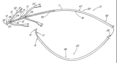
FIG. 1 depicts a side view of an embodiment of the present invention;
FIG. 2 depicts an obturator used with the embodiment of FIG. 1;
FIGs. 3-3a depict the device of FIG. 1 being used in the coronary sinus;
CA 02401720 2002-08-29
WO 01/64279 PCT/US01/06302
-6-
FIG. 4-4a depict use of the device of FIG. 1 in the coronary sinus with an
obturator;
FIG. 5 depicted a side view of a second embodiment of the present
invention that includes a balloon used inside a vessel;
FIG. 6 depicts a cross-sectional view taken along line 6-6 of FIG. 5;
FIGs. 7-8 depict cross-sectional views of third and fourth embodiments of
the present invention having a plurality of lumens;
FIG. 9 depicts a sectioned side view of the present invention that includes
an internal hemostatic valve;
FIG. 10 depicts a side view of the present invention that includes an
external hemostatic valve;
FIG. 11 depicts an end view of a membrane of the embodiment of FIG.
10;
FIG. 12 depicts a pictorial view of a fifth embodiment of the present
invention;
FIG. 13 depicts a side view of a second dilator embodiment of the present
invention;
FIG. 14 depicts a partially sectioned view of the present invention being
used with a steerable/deflectable positioning device;
FIGs. 15-16 depict cross-sectional views of separate balloon embodiments
used with the present invention;
FIG. 17 depicts a pictorial view of a splittable balloon used with the
present invention; and
FIGs. 18-19 depict pictorial views of separate embodiments that include
a retention means.
Detailed Description
FIG. 1 depicts the illustrative embodiment of an introducer apparatus 10
of the present invention which comprises a first introducer sheath 11, such as
an
outer introducer sheath 11, and a second introducer sheath 12, such as a
coaxial
inner introducer sheath 12. The first and second introducer sheaths 1 1,12 are
CA 02401720 2002-08-29
WO 01/64279 PCT/US01/06302
-7-
designed to be splittable longitudinally so that the separated sheath portions
can be
removed from within the body of a patient while the device introduced
therethrough,
such as a pacemaker or defibrillator lead, can remain in place without being
dislodged during their removal. The first and second introducer sheaths 1 1,12
are
designed to co-extend into the bodily passage at some point during the
procedure.
As used herein, co-extending means that the two introducer sheaths can be
introduced simultaneously or one sheath can introduced prior to the other,
e.g., the
outer introducer being initially placed to facilitate subsequent placement of
the
second introducer. In most applications, it is preferred that the first and
second
1 0 introducer sheaths 1 1,12 co-extend coaxially with the smaller (and
usually less stiff)
introducer being introduced inside a passageway of the first introducer. The
passageway can be internal, such as the main passageway 26; however, it may be
external, such as a series of loops or other guides attached to the first
introducer
sheath 11 that allow the second introducer sheath 12 to be introduced
alongside the
first introducer sheath in a non-coaxial arrangement. Additionally, the first
and
second introducer sheaths can be so configured to include a longitudinal
coupling
mechanism, such a track system whereby one introducer has a channel or
receiving
means to receive a corresponding feature on the other introducer, thereby
allowing
the two sheaths to be slidably coupled together at some point during a
procedure.
In another embodiment, the first and second introducer sheaths can be fixedly
interconnected. For example, the inner introducer sheath 12 can be designed to
evert from the outer introducer sheath 11 whereby it is connected about its
proximal
end 14 to the distal end 16 of the outer sheath 11 by a sleeve of a flexible
fabric or
polymer material such as expanded polytetrafluoroethylene (ePTFE).
In the illustrative embodiment of FIG. 1, the first introducer sheath 11
serves as an outer sheath for receiving the second introducer sheath 12, which
is
appropriately sized for introduction through the outer sheath passageway 26.
In the
illustrative embodiment, the outer introducer sheath 11 is sized to be
initially
introduced through the lumen of a vessel or duct to a first target site. In
the
embodiment of FIG. 1, which is particularly configured for navigating the
subclavian
CA 02401720 2002-08-29
WO 01/64279 PCT/US01/06302
-8-
vein and into the heart to place a pacemaker or defibrillator lead into the
coronary
sinus vein to reach and stimulate the left side of the heart, the preferred
sheath
diameter would range from 8 to 12 Fr, with a most preferred diameter of about
10
Fr. After the outer introducer sheath 11 has been placed at or within the
first target
site, the smaller introducer sheath 12 is advanced through the outer
introducer
sheath 11 to access a second target site which usually comprises a duct or
vessel
with a smaller diameter than the first target site and which could not be
safely
accessed by the larger outer introducer sheath 11. In this embodiment, the
inner
introducer sheath 12 normally ranges in diameter from about 5 to 8 Fr, with a
most
preferred diameter of about 7 Fr (when used with a 10 Fr outer introducer
sheath
11).
Introducer sheath 1 1,12 embodiments of the present invention, such as
FIGs 1-3a, that are designed for accessing remote sites within the body that
usually
comprise smaller, distally located vessels, must be made significantly longer
than
standard 12-15 cm introducer sheaths such as those used in the placement of
standard pacing or defibrillator leads. Depending on the application, the
introducer
sheaths 11,12 may range in length from 20 to 90 cm, with most applications
utilizing sheaths in the 25-65 cm range, the upper limit being more of a
practical one
due to the desire to limit the portion extending from the patient. For
example, in the
illustrative embodiment of FIG. 1, configured for placement of a cardiac
device, such
as a biventricular pacemaker lead or defibrillator lead, into the coronary
sinus of an
adult patient, the outer introducer sheath 11 measures approximately 45-55 cm
in
length and the inner introducer sheath 12 is approximately 55-65 cm in length,
with
the most preferred lengths for adult patients being approximately 50 and 60
cm,
respectively. Younger patients or small adults might require sheaths sized
anywhere
from 30 to 60% smaller than these ranges, e.g., outer and inner sheaths 11,12
being 35-45 and 45-55 cm, respectively.
Because the longer introducer apparatus is usually required to be navigated
along a more tortuous path than a standard splittable introducer, it is
desirable, but
not essential, to add at least one preformed bend 20 to the outer introducer
sheath
CA 02401720 2002-08-29
WO 01/64279 PCT/US01/06302
-9-
11 that at least somewhat corresponds in shape to the intended anatomical
pathway. This helps in the navigation of the sheath to the target site and
reduces
the likelihood of the sheath becoming kinked while negotiating a bend. It is
not
necessary that the preformed bends or bends exactly match the radii and shapes
of
the bends of the particular target vessels; however, the bend(s) should be
formed
in such a manner that it significantly reduces the bending stress on the
sheath when
negotiating the bend of the vessel or duct and/or orients the distal end 16 of
the
introducer into a favorable position to access the desired target site. For
example,
the embodiment of FIG. 1, used to access the coronary sinus, has both a
proximal
bend 47 having a radius falling within the range of 2.5 to 3.5" and a distal
bend 48
having a radius generally falling with the range of 1.5 to 2.75". Together,
the
proximal and distal bends 47,48 generally form a serpentine configuration 92.
The
distal bend 48 facilitates navigation through the curvature of the subclavian
34 and
innominate veins 35, shown in FIG. 3. As the first (outer) sheath 11 is
maneuvered
through the superior vena cava 36 into the right atrium 37, it is rotated such
that the
distal curve 48 is oriented toward the target site, the ostium 38 of the
coronary
sinus, while the portion of the sheath having the proximal curve 47 can permit
easier
navigation of the introducer sheath through the subclavian-innominate vein
bend.
Typically, the distal bend has a tighter radius in order to provide
posterolateral
access to the coronary sinus ostium. When a different embodiment of the
present
invention is used, for example to access the renal vasculature, urinary
system,
bronchial tree, cranial arteries, etc., the preformed curve(s) 20 would be
configured
to address the particular anatomical requirements. The inner introducer sheath
12
can either have a generally straight shaft 19 or include preformed bends that
approximate those found in the outer introducer sheath 11. As the smaller
diameter
and therefore, more flexible inner introducer sheath 12 is advanced through
the outer
introducer sheath 11, it tends to assume the shape of the outer introducer,
especially if it also has been configured to include its own preformed bends
that are
located correspondingly. For certain embodiments, such as that of FIG. 1, it
may be
advantageous for the inner introducer sheath 12 to include a distal curved
portion
CA 02401720 2002-08-29
WO 01/64279 PCT/US01/06302
-10-
17 to facilitate access of a particular vessel or duct. It should be noted
that
although the present invention is particularly useful for reaching a remote
location
within the body, thus requiring introducers of usually long length, a co-
extending
splittable introducer sheath of a more conventional length (i.e., less than 20
cm)
should be considered within the scope of the invention as well.
The inner and outer introducer sheaths 1 1,12 are made splittable by use
of any well-known means or material that permits each sheath to be separated
longitudinally along a relatively predictable path, such as a pre-determined
split line
46 by manual force generally applied at the proximal end 13,14 of the shaft
18,19.
The sheath 11,12 is usually, but not necessarily separated into two or more
portions, thereby opening a fissure along the length of the shaft 18,19 that
permits
its removal from around the lead or other indwelling device situated therein,
such
that the indwelling device can remain within the patient as the introducer
sheath is
removed. The predetermined split line 46 is a pathway along the length of the
sheath through which the tear or split progresses due to properties of, and/or
features incorporated into the sheath material. It is naturally preferred that
the
means to split the sheath be able to withstand being subjected to a curve to
the
degree required by the particular application without kinking or premature
separation.
In the illustrative embodiment a splittable polymer is used such as
molecularly
oriented, non-isotropic PTFE that is used to make the PEEL-AWAY Introducer
Sheath (Cook Incorporated, Bloomington, IN) which is fully described in U.S.
Patent
Nos. 4,306,562 to Osborne and 4,581,025 to Timmermans. In an alternative
embodiment, sometimes known in the art as a 'crack and 'peel' introducer,, the
sheath can be made splittable by adding at least one preweakened feature 59,
such
as a score line extending longitudinally along the sheath as depicted in FIG.
12. The
longitudinal preweakened feature 59 could include anywhere from one or more
orthogonal predetermined split lines 46, as shown, to a helical type
arrangement that
may comprise only a single predetermined split line 46.
As depicted in FIGs. 3-3a and 4a, the introducer apparatus 10 is normally
introduced over a wire guide. In the illustrative embodiment, a small diameter
wire
CA 02401720 2002-08-29
WO 01/64279 PCT/US01/06302
- 11 -
guide 45 with good torqueability in combination with an atraumatic tip is
preferred,
such as the COOK ROADRUNNERTM FIRMTM Wire Guide or COOK TORQ-FLEXO Wire
Guide (Cook Incorporated, Bloomington, IN). Generally, the tip 69 of the wire
guide
45, which may be angled, is guided to at least the first target site 67 (i.e.,
about
where the distal tip 16 of the outer introducer sheath 11 is to be placed),
and
possibly to the second target site 68 to which the distal tip 15 of the inner
introducer sheath 12 is to be placed. In the illustrative example, the wire
guide 45
is first placed into the ostium 38 leading to the coronary sinus 39 which
represents
the first target site 67. Then, as in the case of biventricular pacing, the
wire guide
45 is subsequently guided through the coronary sinus 39 and down a cardiac
vein
branching from the coronary sinus 39 (the second target site 68), for example,
the
posterior vein of the left ventricle 40 as shown in FIG. 4a, or another vein
such as
the middle cardiac vein 41 shown in FIG. 4.
While not always necessary, it is often advantageous to include a steerage
member, such as a dilator, obturator, deflectable tip device, etc., for
assisting with
the introduction and placement of the introducer sheaths 1 1,12. As used
herein, a
'steerage member' is defined as a device or apparatus that is used in
conjunction
with an introducer sheath 1 1,12 during advancement through a bodily passage
to
assist in some manner with the placement of the sheath at a target site.
Normally,
a steerage member is a placed inside the passageway 25,26 of the sheath to
provide
the desired torqueability, maneuverability, or shape for improved navigation
or
reduced risk of kinkage. In the case of a dilator, the tapered tip can be
useful when
guiding the sheath into a narrowed lumen or opening. In the illustrative
embodiment
of FIGs. 1-2, a dilator can be advantageously used with the outer introducer
sheath
11 for reaching the coronary sinus. With the wire guide 45 in place, a first
dilator
27, comprising a shaft 28 and proximal hub 29 and depicted in FIG. 2, can be
used
inside the outer introducer sheath 11 of FIG. 1 to facilitate its introduction
to the
target site, which in this embodiment, requires maneuvering through the right
atrium
37 and into the ostium 38 of the coronary sinus 39. The shaft 28 of the first
dilator
27, which can be made of PTFE or an other suitable polymer, includes a distal
taper
CA 02401720 2002-08-29
WO 01/64279 PCT/US01/06302
-12-
30 and narrow tip 31, with a passageway 32 sufficiently large to accommodate
an
appropriate wire guide 45. The purpose of the first dilator 27 is to provide a
relatively atraumatic means to guide the tip of the first introducer sheath 11
through
the vasculature and to access a relatively small opening such as the coronary
sinus
ostium 38. Without the dilator 27, increased precision would be required to
advance
the distal tip 16 of the outer introducer sheath 11 into the ostium 38
opening. As
with the mated introducer sheaths 1 1,12, the dilator 27 may be given a
preformed
shape 93 that corresponds to that of the other devices with which it is used.
Alternately, the preformed shape 93 of the dilator can provide a curved
configuration
to otherwise straight introducer sheaths 1 1,12, especially if having one or
more
preformed curves is primarily important during introduction and is not
particularly
advantageous once the sheath has been placed within the patient. It should be
noted that upon insertion therewithin, it is possible for the preformed inner
member,
such as a dilator 27, obturator, or inner introducer sheath 12, to either
elastically or
1 5 plastically deform the outer member, such an introducer sheath 1 1,12,
depending
on the physical properties of the inner and outer members. The sheaths 1 1,12
can
also be made such that the operator can manipulate the shape after they are
removed from the package to configure the them to a desired shape.
Once the outer introducer sheath 11 is in place, the dilator 27 is removed
and the inner introducer sheath 12 is inserted therethrough. As with the outer
introducer sheath 11, a second dilator 44, shown in FIGs. 3-3a, can be used to
guide
the inner introducer sheath further into the coronary sinus 39 to a more
distal target
site 68, such as the posterior vein of the left ventricle 40 as depicted in
FIG. 4a.
Once the inner introducer sheath 12 is advanced to the second target site 68
within
the vasculature, the second dilator 44 is removed and the pacing lead or other
device is advanced through the inner introducer sheaths 12 to the second
target site
68 or a more distal location. Once the lead or device has been properly
placed, the
outer introducer sheath 1 1(of FIG. 1) is then removed by splitting it into
two
portions from around the indwelling lead. This is accomplished by grasping the
handles 22 attached to the ears 21 extending from the sheath material. The
shaft
CA 02401720 2002-08-29
WO 01/64279 PCT/US01/06302
-13-
18 is torn into two separate portions along the predetermined split line 46
starting
from the cut point 24 in the material. As fabricated, the material forms a
folded cuff
23 at the proximal end 13 of the outer introducer sheath 11 such that the
material
is initially torn in the proximal direction, then starting at the proximal end
13, is split
along the predetermined split line 46 toward the distal 16 until the shaft 18
is
completely split apart. Ultimately, the inner introducer sheath 12 will be
removed
in a manner similar to that of the outer introducer sheath with the shaft 19
also
being torn along the predetermined split line 46 from the proximal end 14 to
the
distal end 15 until the shaft 19 separates and is removed from the patient.
FIG. 13 depicts another method of using the introducer apparatus with a
wire guide. In this embodiment, the first dilator 27 comprises a monorail
dilator
configuration 70 that includes a side opening 71 such that the wire guide 45
can
feed into the central passageway 72 of dilator 70, rather than the introducer
sheath
11 itself tracking over the wire guide 45 or the dilator and introducer sheath
both
tracking over the wire guide extending through the passageway 32 of the
dilator
shaft 28 of dilator 27. The monorail dilator can be used with either of the
inner or
outer introducer sheaths 1 1,12, such as those depicted in FIG. 1.
To add stiffness to the inner introducer sheath 12 for increasing
torqueability and pushability (as defined by common engineering testing
standards),
an obturator 42 may be used as shown in FIG. 4a. As with the introducer
sheaths
11,12 and dilators 27,44, the obturator can include a passageway to allow for
tracking over a wire guide. Preferably, the obturator is 42 designed to have
the
maximum amount of material and wall thickness with the smallest possible wire
guide lumen to yield the maximum stiffness for providing good maneuverability.
The
obturator, which can be made of PTFE or another suitable polymer for
fabricating
sheaths, can include at least one preformed curve to facilitate steering,
positioning,
and rotation of the inner introducer sheath 12. Additionally, an obturator 42
can be
used to assist with the positioning of the outer introducer sheath. Another
method
of positioning the introducer sheaths 1 1,12 into the target site, shown in
FIG. 14,
includes use of a well-known steerable or deflecting tip device 74, such as a
CA 02401720 2002-08-29
WO 01/64279 PCT/US01/06302
-14-
catheter (e.g., an Electrophysiology (EP) Catheter) or a wire guide, in place
of or in
combination with a dilator or pre-formed obturator. By introducing or
incorporating
the steerable/deflectable device into an outer or inner introducer sheath 1
1,12
permits the tip of the sheath to be deflected into the optimum position for
advancing
the sheath to the target area or providing an improved position such that the
inner
introducer sheath 12 can be then advanced to the target site. The
steerable/deflectable device 74 may include a passageway 86 for a wire guide,
and
be integral with either of the sheaths 1 1,12 or represent a separate
component of
the introducer apparatus 10. A separate steerable/deflectable steerage device
74
can being used to reach the vicinity of the ostium or target vessel such that
the
introducer sheath or sheaths 1 1,12 can then be advanced thereover to the
desired
target site. In the illustrative embodiment, the steerable/deflectable device
74
further includes a second passageway 87 that houses a deflection control means
75,
such as a flexible rod, wire, suture, etc., that is attached about the distal
end 88 of
the steerable/deflectable device 74 and extends proximally to a control handle
(not
shown) that affects the degree of deflection of the introducer sheath tip 16.
The
illustrative embodiment of FIG. 14 represents one example of how to make a
steerable/deflectable device 74 among many alternative methods that are known
in
the medical arts. The choice of the deflection control means 75, how or
whether
it is attached, and the specific configuration of the steerable/deflectable
device 74,
depends largely on intended use and physician preference. Again, it should be
noted
that the steerable/deflectable device 74 can be used with an introducer sheath
1 1,12 having one or more preformed bends 20, or it can be used to provide a
curved
configuration to an otherwise straight introducer sheath 11,12 when the
steerable/deflectable device 74 is deployed therewithin.
FIG. 5 depicts an embodiment of inner introducer sheath 11 that includes
an expandable member 49, such as an inflatable balloon 49 or other well-known
occlusion mechanism, mounted to the distal portion 17 of the shaft 19. The
balloon
49 communicates with a well-known inflation means, such as a syringe, via an
inflation port 61 and a separate inflation lumen 52, as depicted in FIG. 6.
The
CA 02401720 2002-08-29
WO 01/64279 PCT/US01/06302
-15-
inflatable balloon 49 can be made from a number of well-known compliant
materials,
such as latex or silicone, or a well-known noncompliant material, such as
polyethylene teraphthalate (PET) or a polyamide fabric, depending on the
medical
application. In the illustrative embodiment, the balloon 49 is made of PET. By
sizing
the balloon 49 to the target vessel, it helps prevent against overinflation
that could
lead to rupture of the vessel 50. The balloon 49 of this embodiment is used to
temporarily occlude the vessel 50 while contrast media 51 is injected into the
vein
to improve fluoroscopic guidance of the device to the target site. Without
occlusion
of the vessel 50 to prevent retrograde flow, the contrast media 51 may be
carried
back with the blood flow and thus not travel downstream to a sufficient degree
to
permit adequate imaging of the portion of the vessel containing the target
site. The
balloon 49 can be mounted on either the outer or inner introducer sheath 1
1,12,
depending on how the particular embodiment is used in the body. To allow the
balloon 49 to be carried away with the splittable introducer sheath portions
82,83,
the balloon 49 can configured such that the primary attachment points 77 are
on a
first half 82 of the introducer shaft 18, as depicted in FIG. 15, with the
balloon
extending circumferentially around the shaft from the respective attachment
points
77. The lateral edges 88 of the balloon 49 wrap around the shaft 18 and meet
over
the second half 83 where they can be affixed thereto using a bonding means 78
that
will readily yield to shearing forces that result from the shaft being split
into the two
portions 82,83. Because the balloon 49 is affixed to the second half 83 by a
weaker
bonding means 78 than that joining the balloon 49 to the first half 82, the
entire
balloon 49 is carried away intact with the first halve 82 during separation of
the
shaft 18. As shown in FIG. 16, another method that allows the balloon to
separate
is to have two adjacent balloons 80,81, each attached to opposite halves 82,83
of
the shaft 18, separated by the predetermined split lines 46, and together,
inflate to
function as a single composite balloon 49 capable of occluding the vessel. The
first
balloon 80 is attached to the first half 82 of the introducer shaft 18 where
is
communicates with a first inflation lumen 52. The first balloon 80 is
configured into
a hemispherical shape that generally wraps around and covers the surface of
the first
CA 02401720 2002-08-29
WO 01/64279 PCT/US01/06302
-16-
half 82. The second balloon 81 is attached to the second half 83 of the shaft
18
where it communicates with a second inflation lumen 53. It generally covers
the
surface of the second half 83 and abuts the first balloon 80 along the
predetermined
split lines 46. In a variation of this embodiment, the first balloon 80 can be
larger
than the second balloon 81 with its lateral edges 88, but not the attachment
points
77, extending over the predetermined split lines 46 to abut with the smaller
second
balloon 81. It may not be necessary for the balloon portion 81,82 to
completely
surround the circumference of the shaft 18 to accomplish the goal of infusing
contrast agent that remains for a sufficient period to allow diagnostic
imaging.
1 0 Yet another method of allowing an introducer sheath 1 1,12 with a balloon
49 to split, is shown in FIG. 17, wherein the balloon 49 includes a
longitudinal
weakened area 84 on the balloon 49 itself that allows the balloon to split
into two
portions as the shaft 18, to which it is attached, is separated. In the
illustrative
embodiment, the longitudinal weakened area 84 comprises a seam of overlapping
balloon material, although the edges of the seam could be designed to abut
each
other. The edges of the seam 84 can be sealed, e.g., with heat, or secured
together
with a separate strip of material, such as a plastic tape, or an adhesive such
as
silicone such that the seam 84 can be readily pulled apart when force is
applied to
the degree required to split the shaft 18. To help facilitate the split, a cut
point 85
can be positioned at the posterior edge of the material along the preweakened
area
84 to provide a start to the intended split. The longitudinal weakened area 84
can
also comprise a longitudinally extending zone in which the material has been
mechanically weakened (e.g., via abrasion) or molecularly altered, e.g., a
chemical
or radiation treatment, such that the balloon will generally rupture along the
preweakened area 84 when lateral force associated with the splitting of the
shaft
18 is applied. Alternatively, a balloon material may be selected, such as a
thin-wall
latex or silicone, that permits rupture and separation of the balloon 49 when
the
shaft 18 is split apart, without requiring the addition of a longitudinal
weakened area
84.
CA 02401720 2002-08-29
WO 01/64279 PCT/US01/06302
-17-
In addition to improving imaging by using the introducer apparatus 10 to
inject contrast media, the apparatus itself can be made radiopaque by one of
several
well-known methods that include incorporation of radiopaque materials, such as
barium sulfate, tantalum powder, etc. into the sheath polymer, the addition of
markers, such as radiopaque metal bands, applying radiopaque indicia to the
surface,
etc. Both the outer and inner introducer sheaths 1 1,12 can be made radiopaque
by
at least one of these methods.
FIGs. 6-8 depict various embodiments of multiple lumen introducer
sheaths. The embodiment of FIG. 6 depicts an inner introducer sheath 12 that
includes a first, primary passageway 25 and a second passageway 52
incorporated
into the sheath wall 62 that can be used as an inflation lumen or if made
larger,
could accommodate an ancillary device 54 such as a wire guide. FIG. 7 depicts
a
dual lumen outer introducer sheath 11 that includes a first passageway 26 and
a
smaller second passageway 52. The introducer apparatus 10 of this embodiment
can be used coaxially with an inner introducer sheath (not shown) in the first
passageway, or the outer introducer sheath 11 can be used alone with the lead
or
other device being placed through the first passageway. Typically, the second
passageway is used for an ancillary device 54 as shown, which could include a
wire
guide or a well-known control means to help make the sheath steerable or
deflectable. This could also include use of a steerable electrophysiology
catheter or
a control mechanism that operates in a similar manner, wherein the distal
portion of
the sheath can be manipulated via a well-known type of handle used for tip
deflection that is connected to the proximal end of the sheath. The
predetermined
split lines 46 can be positioned about the shaft 18 such that they permit both
passageways 26,52 to be peeled open and the sheath portions removed to allow a
lead, wire guide, or other device to remain in place. The intraluminal wall 63
separating the first and second passageways can be made sufficiently thin to
rupture
when the shaft is being separated, or it can be given a weakened feature 64
that is
added to the intraluminal wall during or after the extrusion process to
facilitate
rupture. If the outer introducer sheath 11 is only to be removed from over a
single
CA 02401720 2002-08-29
WO 01/64279 PCT/US01/06302
-18-
device, it may not be necessary to have the predetermined split line 46
intersect the
second passageway 52 which can remain intact while still allowing the first
introducer sheath 11 to be split and removed from the patient.
FIG. 8 depicts a three-lumen outer introducer sheath 11 that includes first
and second passageways 26,52 that are split open longitudinally when the
sheath
separates. The third passageway 53, typically used for injection of contrast
media
or for an ancillary device, such as a wire guide, can be left intact as it is
not
necessary to expose the third passageway 53 if any device contained therein is
removed prior to the separation of the shaft 18. Alternatively, the shaft 18
can be
made to split along three predetermined split lines 46 if all three
passageways
26,52,53 must be opened and exposed to remove devices that are left in place,
or
the intraluminal wall can be so designed to accomplish the same result with
only two
predetermined split lines 46.
FIGs. 9-10 depict embodiments of the present invention that include a
valve 55, such as a splittable hemostatic valve, to prevent loss of blood
during an
intravascular procedure, especially procedures of long duration such as
coronary
sinus or cardiac defibrillator lead placement. In the embodiment of FIG. 9,
the
hemostatic valve 55, preferably made of silicone, is insert molded into the
passageway 25 of an inner introducer sheath 12 near the distal portion 17 of
the
shaft 19 with the silicone material flowing into apertures 64 made in the
shaft wall
62 to help secure the hemostatic valve 55 and prevent longitudinal migration
and
allow the valve to be pulled apart with the shaft 19. To separate the
hemostatic
valve 55 when the sheath is separated, the valve body is given at least one
line of
fissure 60 extending therealong that can include a scored line or a thinned
region
such that the hemostatic valve halves rupture along the line of fissure 60
when the
shaft 19 halves to which they are attached, are being split apart. Besides
being
insert molded, the hemostatic valve 55 can be made as a separate component and
affixed within the shaft 19 using a well-known method such as gluing. The
hemostatic valve 55 can vary in its configuration and may comprise a simple 0-
ring.
CA 02401720 2002-08-29
WO 01/64279 PCT/US01/06302
-19-
The illustrative hemostatic valve 55 includes two primary seals in the
integral valve
body: a membrane 56 and an 0-ring 57.
The membrane depicted in FIG. 11 includes a series of slits 58 that define
a number of valve leaflets 65 designed to help seal about an elongated device
introduced therethrough. To facilitate separation of the hemostatic valve 55
along
with the introducer sheath 1 1,12, it is usually desirable to affix or join
the two
together, preferably aligning the predetermined split line 46 of the
introducer sheath
11,12 with the lines of fissure 60 of the hemostatic valve. This can be
accomplished by gluing the hemostatic valve therein or allowing the silicone
or
polymer used to form the hemostatic valve to flow through apertures 64 (FIG.
9)
made in the sheath wall 62 and cure to form a positive fixation that can
withstand
the forces required to separate the introducer apparatus 10 into two portions.
FIG.
10 depicts an embodiment wherein a hemostatic valve 55 is included on the
proximal end 14 of the inner introducer sheath 12.
The hemostatic valve 55 can be integrally attached to the introducer
sheath 21 or made to be detachable as shown in FIG. 10 wherein the valve
traverses
the proximal end 13 of the outer introducer sheath 11. While an integral
hemostatic
valve 55 may be designed to split along with the introducer sheath shaft 19,
in the
detachable embodiment, the hemostatic valve is split apart separately prior to
splitting the introducer sheath 11. This is accomplished by grasping the
integral
valve handles 66 and pulling them apart until the valve separates along the
lines of
fissure 60. The hemostatic valve 55 can be included on either the outer or
inner
introducer sheaths 1 1,12. To provide a seal between sheaths when used
together,
a hemostatic valve 55 consisting of an 0-ring or similar structure can be
either
affixed within the passageway 26 of the outer introducer sheath 11 or to the
exterior surface of the shaft 19 of the inner introducer sheath 12.
FIGs. 18-19 depict embodiments that include one or more retention
members 90 located about the distal end 16 of the introducer sheath 11 that
advantageously prevents or reduces unintended movement of the introduce sheath
11 during the procedure. This especially can be a problem when the friction
caused
CA 02401720 2002-08-29
WO 01/64279 PCT/US01/06302
-20-
by the withdrawal of the inner sheath or another indwelling device causes the
introducer sheath 11 to dislodge from the intended target site. FIG. 18
depicts an
introducer sheath 11 having a pair of expandable members 49 comprising a first
balloon portion 80 affixed to a first half 82 of the shaft 18, and a second
balloon
portion 81 affixed to the second half 83 of the shaft 18, with the
predetermined split
lines extending therebetween. Unlike the occlusion balloon 49 depicted in 15-
17,
it is not necessarily desirable for the first and section balloon portions
81,82 to
contact one another and surround the circumference of the shaft to provide a
seal
against fluid flow. Each balloon portion, which in this embodiment is a
complete
separate balloon 49, is inflated via dedicated second and third passageways
52,53
(inflation lumens) within the introducer sheath, with each communicating
proximally
with a common or separate inflation means, such as a syringe. Alternatively, a
single expandable member may be sufficient, in certain applications, to
prevent or
inhibit migration of the introducer sheath. Additionally, more than two
balloons 49
can be positioned about the shaft, including a longitudinal alignment at
various points
along the axis of the shaft, rather than the depicted circumferential
arrangement.
The expandable member 49 can simultaneously function as both a retention
member
90 and an occlusion balloon 49 for injection of contrast media as depicted in
FIGs.
5,15-17.
FIG. 19 shows a second main embodiment of an introducer sheath 11
having a plurality of retention members 90 that comprise a plurality of
bidirectional
retention elements 91 located about the distal end 16 of the sheath. These
bidirectional retention elements 91 can include a variety of configurations,
but are
preferably constructed of a material that is not traumatic to the tissues of
the bodily
passage. In the illustrative embodiment, the bidirectional elements comprise a
series
of annual projections that allow the introducer sheath 11 to be easily
advanced, but
provide limited resistance when the sheath 1 1 is urged in the opposite
direction. The
desired degree of the resistance to egress can be modified according to the
anatomical and clinical requirements. Certainly, the bidirectional elements
can be
modified in size, number, and placement along the shaft 18 of the introducer
sheath
CA 02401720 2002-08-29
WO 01/64279 PCT/US01/06302
-21-
1 1. For example, numerous, much smaller projections can be added to the outer
surface of the sheath, or even formed in the outer surface of the shaft 18
material,
to increase the coefficient of friction in one direction without significantly
adding to
the outer diameter of the sheath. While the outer introducer sheath 11 is
often more
prone to dislodgement during a procedure, it also may be desirable that the
inner
introducer sheath 12 can be modified to include one or more retention members
90
to reduce the possibility of its migration.
It should be understood that the present invention is not limited to a pair
of introducer sheaths. It is within the scope of the invention to include one
or more
additional introducer sheaths inside one or more of the first and second
introducer
sheaths 1 1,12. For example, the outer introducer sheath 11 could be sized to
accommodate two inner introducer sheaths 12 placed adjacent to one another to
access two different target sites (e.g., left and right renal vein), or there
could be
three or more concentric introducer sheaths with the smallest introducer
sheath
accessing perhaps a third target site that either is more distal than the
second target
site, or requires a different curvature of the distal portion 17 than the
second
introducer 12 in order to be accessed.
It is thus seen that the present invention has utility in a variety of medical
procedures, and variations and modifications of the introducer assembly of the
present invention additional to the embodiments described herein are within
the spirit
of the invention and the scope of the claims.
30