Note: Descriptions are shown in the official language in which they were submitted.
WO 01/66013 CA 02402429 2002-09-05PCT/US01/07189
DEVICE FOR CUSHIONING OF COMPRESSION SURFACES
BACKGROUND OF THE INVENTION
The present invention relates generally to medical apparatus and methods and
more
particularly to devices and methods for cushioning or padding the surface of
compression plates
applied to body parts for purposes of obtaining x-ray films, for example,
mammography, or other
scans of compressed tissue.
Currently, in the case of mammography, a patient's breast is placed under
compression by
opposing plates attached to a mammography machine. Once under compression an x-
ray is
taken to determine the presence or absence of suspect lesions in the breast
tissue (e.g.
calcifications, tumors). Approximately 25 million screening mammograms are
performed
yearly, which is estimated to be only a 50% compliance rate among potential
patients, meaning
that number would double if all potential patients complied with the
recommended screening
regime. One of the more common complaints from mammography patients is
discomfort during
compression of the breast. Most patients can only tolerate up to 10-11
compression units. The
current legal limit for clinical mammography is 16-18 units. A device which
would reduce
discomfort could likely improve compliance for screening.
An important reason for compressing the breast during mammography is to
provide a
thinner cross section of tissue for the x-rays to pass through. When the
breast is compressed, it
provides optimal imaging of the tissue abnormalities with the lowest possible
dose of x-ray
radiation to the patient. Furthermore, during a mammogram, it is important for
the x-ray plate to
be free from radiopaque material, so that the diagnostic film, once processed,
can give the
physician the best possible picture of the tissue and any abnormalities.
1
WO 01/66013 CA 02402429 2002-09-05PCT/US01/07189
Although patients may tolerate the pain during compression, there is a need
for improved
devices and techniques to provide better screening outcomes by enabling the
use of higher
compression force, and by providing increased patient comfort during
mammograms thereby
positively impacting patient compliance with mamrnographic screening and
ultimately impacting
early detection of cancer and improving patient survival.
Such improved devices must be radiolucent and made of a relatively homogeneous
material to avoid striations or other variations on the resulting x-ray image,
have a low profile to
allow for correct positioning of the breast in the mammography machine, be
easily cleaned or
disposable for sanitary reasons, and provide structural support and tactile
comfort to the patient
(both soft to touch and providing a less harsh or "cold" surface). In
addition, such improved
devices will permit the use of higher compression forces to be applied to the
breast during
mammograms without the patient reaching her tolerance level for discomfort,
resulting in a
thinner tissue section, better image quality, and reduced x-ray dose to the
patient.
It is an objective of the present invention to provide greater patient comfort
thereby
increasing screening compliance (e.g. patient willingness to have more regular
mammograms by
reducing discomfort of the procedure). Greater patient comfort also reduces
the risk of patient
movement (voluntary or involuntary). Motion artifact, caused by patient
movement or slippage
of the tissue, can result in loss of clarity of the mamrnographic image. It is
a further objective of
the present invention to allow for the use of an increased compressive force,
for example, up to
16-18 compression units or more thereby providing for a thinner cross-section
of breast tissue
during the mammogram resulting in an enhanced ability to detect abnormalities
in the
mammographic image. These objectives are met by the design and use of the
present invention.
2
WO 01/66013 CA 02402429 2002-09-05PCT/US01/07189
DESCRIPTION OF THE BACKGROUND ART
Various patents have issued illustrating inventions in the field of
mammography and
comfort during x-ray imaging. For example, in the field of mammography, US
Patent Nos.
3,963,933, 4,691,333, 4,943,986, 5,189,686, 5,553,111 and 5,398,272 describe
various fixtures
useful for breast compression. Further, patents have issued describing devices
for increasing
comfort during general x-ray procedures, such as US Patent No. 5,226,070
(radiolucent x-ray
mat), US Patent No. 5,081,657 (buckey warmer for mammography machine), US
Patent
5,541,972 (disposable padding device for use during mammography) and US Patent
No.
5,185,776 (padded cover for x-ray cassette).
SUMMARY OF THE INVENTION
According to the present invention, improved methods and apparatus are
provided for
cushioning or providing other patient comfort surfaces on devices used for
compressing the
patient's tissue, such as radiography machines, fluoroscopy units, mammography
units and the
like. In particular a pad element is provided for releasable attachment to at
least one surface of a
compression device to be used under x-ray, or other imaging modality.
In a preferred embodiment of the present invention a pad assembly is provided
consisting
of a pad element, an adhesive layer and a release paper layer allowing for
temporary attachment
to the applied surface (either the mammography paddle, x-ray plate or directly
to the patient's
skin).
An alternative embodiment of the present invention includes a reusable
cushioned paddle
configured of a self-skinned foam to allow for easy cleaning between patients.
This embodiment
3
CA 02402429 2011-10-19
50927-1
may be replaceable after many uses or formed integrally wherein the padded
surface
and the compression paddle are assembled as one unit.
The present invention may also incorporate a dispensing unit for access
to single pads for single use.
The invention also relates to a compression device for a mammography
unit, comprising: a radiolucent foam pad element radiolucent to x-ray and
having at
least one surface; and an adhesive layer on the at least one surface for
releasably
attaching the at least one surface to a compression surface of the mammography
unit.
The invention further relates to a method for using a mammography
unit, comprising: installing a radiolucent foam pad element on a patient
contact
surface of a compression plate in an x-ray field of the mammography unit
before a
mammography procedure, the radiolucent pad element being releasably secured to
the patient contact surface by an adhesive layer on the radiolucent pad
element, the
radiolucent pad element producing no significant visual artifact on the images
obtained using the mammography unit; and removing the radiolucent pad element
from the patient contact surface after the mammography procedure.
The invention also relates to a compression device for a mammography
unit, comprising: a compression plate having a compression surface in an x-ray
field
of the mammography unit and a front face surface; and a radiolucent cushioning
device releasably attached to the compression surface and the front face
surface, the
cushioning device being radiolucent to x-ray.
The invention also relates to a device for attachment to a compression
surface of a mammography unit, said device comprising: a cushioning device
having
a first and second surface, the cushioning device being radiolucent to x-ray;
an
adhesive layer on the first surface for releasably attaching the first surface
of the
cushioning device to the compression surface.
4
CA 02402429 2011-10-19
50927-1
BRIEF DESCRIPTION OF THE DRAWINGS
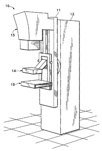
FIG. 1 illustrates a typical mammography unit having a base and a
pivoting x-ray unit attached thereto, the x-ray unit including a compression
paddle
and an x-ray plate.
FIGS. 2A-2B illustrate detailed construction of the x-ray plate and the
compression paddle respectively.
FIGS. 3A-3C illustrate various configurations of compression paddles
utilized during mammography in a standard mammography machine; the shape and
size depending both on the patient's anatomy and the type of x-ray view
desired by
the physician.
FIGS. 4A-4B illustrate various attachments that can be placed on the x-
ray plate to enhance the image, including devices for spot compression and
magnification.
FIG. 5 illustrates a compression paddle and x-ray plate configured for
use in a stereotactic biopsy procedure.
FIGS. 6A-6C illustrate the pad of the present invention having a
padding layer, an adhesive layer and a release paper layer.
FIGS. 7A-7B illustrate the installation of the pad of the present invention
on an x-ray plate.
4a
WO 01/66013 CA 02402429 2002-09-05 PCT/US01/07189
FIG. 8 illustrates the installation of the pad of the present invention on a
compression
paddle.
FIG. 9 illustrates various pad configurations and geometries according to the
present
invention depending on the type of compression paddle or x-ray unit used in a
given procedure.
FIG. 10A illustrates an alternative embodiment of the present invention,
showing the use
of a self-skinned foam fastened to a compression paddle intended for use on
multiple patients.
FIGS. 10B-10C further illustrate an alternative embodiment of the present
invention
wherein the pad and compression paddle are integral as one unit.
FIG. 10D illustrates an alternative embodiment of the present invention,
wherein the pad
and x-ray plate are integral.
FIG. 11 illustrates a further feature of the present invention, namely a
dispensing unit for
storing and dispensing the disposable pads of the present invention to promote
ease of use and
efficiency.
DETAILED DESCRIPTION OF THE PREFERRED EMBODIMENTS
A typical or standard mammography unit used to image the breast while under
compression is shown in Fig. 1. This unit 10 includes a base 12 and a rotating
x-ray source 11,
comprising an x-ray source 13, a movable compression paddle 14 and an x-ray
plate 15 that
holds the film cassette (not shown) .as well as serving as a compression
surface against which the
compression paddle 14 can compress tissue, e.g., a breast to be imaged. As
depicted in Fig. 2A,
typically the x-ray plate 15, in certain configurations known as a "bucky," is
stationary and
5
WO 01/66013 CA 02402429 2002-09-05PCT/US01/07189
includes an opening 16 into which an x-ray cassette 17 is placed prior to
imaging. The x-ray
plate has two patient contact surfaces, a front face 18, and a functional
surface 19. The x-ray
plate 15, may optionally include radiopaque markers 19A at the perimeter of
the functional
surface 19 to allow various marking schemes to be utilized during a procedure.
Fig. 2B illustrates a more detailed configuration of compression paddle 14,
including a
front patient contact surface 20 and a functional patient contact surface 21.
Paddle 14 is
typically constructed of a clear radiolucent plastic material and is designed
to be removably
attached by an interchange assembly 22, to the movable working arm of the
mammography
machine (not shown). These paddles are configured in various geometries as
depicted in Figs.
3A-3C to accommodate various patient anatomies and specific needs of
mammographers, such
as coned compression paddles (3A), spot compression paddles (3B) and the
axillary paddle
shown as Fig. 3C, all configured to attach to the mammography unit through
standard
interchange assembly 22, as shown earlier.
Similarly, the x-ray cassette holder may be adapted by various ancillary
modules such as
the spot compression fitting 41 shown in Fig. 4A, and a magnification fitting
42 shown in Fig.
4B. The entire compression system (compression paddle and x-ray plate) can
further be
modified to accommodate a,stereotactic biopsy procedure as illustrated in Fig.
5, In this
configuration, compression paddle 14 is modified to include a window 51,
allowing the clinician
access to the breast, while still under compression, for purposes of placing a
device to identify a
specific location in the breast, or to perform a biopsy of tissue.
A preferred embodiment of a pad assembly constructed in accordance with the
present
invention is illustrated in Fig. 6A. The pad assembly 60 comprises a padding
element 61, an
6
WO 01/66013 CA 02402429 2002-09-05PCT/US01/07189
adhesive layer 62, and a release paper 63 to be removed from contact with the
adhesive layer just
prior to installation on the surface to be padded. Pad element 61 may be
constructed of various
materials having the following characteristics: produce no significant visual
artifact on the
mammogram (i.e. is radiolucent), be deformable under the forces applied during
compression to
provide comfort. Furthermore, the material should provide conformance to the
tissue and the
compression surface so as to reduce the propensity for the material to create
air pockets or folds
that may be of sufficient size to be visible on the x-ray image. Additionally,
it may be desirable
for the material to be absorptive to external fluids such as sweat.
Such materials may be an elastomer or gel, open (reticulated) or closed (non-
reticulated)
cell foam consisting of polyolefin, or, preferably, a polyurethane open cell
foam because of its
radiolucent characteristics and soft tactile feel. Of particular advantage is
polyurethane foam
having a density of 5-6 lbs./cu ft, with approximately 90 pores per inch. The
padding material 61
may be a thickness of .050" to .500", preferably in the range of .200" and
.250". If an adhesive
layer is used, adhesive layer 62 may be one of a variety of currently
available pressure sensitive
adhesives such as acrylic or synthetic rubber based adhesives, to allow
sufficient tackiness for
secure attachment to the compression surface, while also allowing for easy
removal (e.g. leaving
no detectable residue of adhesive on the applied surface) and disposal.
Alternatively, a
non-adhesive gel may be used to secure the pad or another layer of material
having a greater
coefficient of friction against the applied surface. It is also anticipated by
the scope of the
present invention that the pad element may itself be textured such that it is
sufficiently "tacky" to
enable its use without an adhesive layer, i.e., by means of friction between
the element and the
tissue and the unit compression surface.
7
WO 01/66013 CA 02402429 2002-09-05 PCT/US01/07189
The pad element of Fig. 6A can be configured with adhesive on the entire
surface of the
pad, or at certain regions such as just along the border (see Fig. 6C). Fig.
6B depicts a "peel
away" packet configuration to house the pad assembly; optionally, the peel
away packet can
serve as a stiffening element to aid installation of the pad by keeping it in
a planar configuration
to minimize the possibility of misapplying the pad (leading to inadvertent air
pockets or folds in
the material, etc.) and to aid in positioning the pad prior to adhering it to
the applied surface.
Fig. 7A illustrates, in stepwise fashion, the installation of the pad assembly
60 of the
present invention onto the film holder 15. The first step comprises opening
the packing material
housing the pad assembly 60 (Si), and thereafter removing any release paper 63
therefrom (S2).
The pad assemblies can be packaged individually or in bulk. Installation on
the patient contact
surfaces of the x-ray plate 15 are shown in steps S3 and S4, S3 showing the
placement of the pad
element 61 on functional surface 19, and optionally extending to front face
surface 18. The
installed pad is depicted in Fig. 7A. Fig. 7B shows an alternative embodiment
of pad element 61
installed onto an x-ray plate where the pad is configured to stretch over side
face 23 of the x-ray .
plate, to provide added comfort for the patient. More particularly, the pad of
this embodiment
includes an adhesive layer which is preferably of a three part construction: a
high tack, high peel
permanent adhesive securing the adhesive layer to the pad element, a low tack,
removable
= adhesive for securing the adhesive layer to the patient contact surface of
the x-ray plate, and a
stretchable carrier material between the two adhesives, such as polyethylene.
A pad according to
the present invention can also be formed of a similar construction using,
e.g., polyester as a
carrier material. Finally, the pad element may be removed and disposed of and
the sequence
repeated for the next patient. It may be desirable to score or otherwise
provide a fold in the pad
8
WO 01/66013 CA 02402429 2002-09-05 PCT/US01/07189
element at a fixed point from the edge of the pad to accommodate folding the
pad onto the front
and/or side face of the applied surface.
A similar sequence of steps (Si to S4) is illustrated in Fig. 8A showing the
installation of
pad assembly 60 of the present invention onto compression paddle 14. The pad
assembly (PA)
of Fig. 8B is configured such that portions of the pad extend to cover side
surface 24 of the
compression paddle, providing added comfort for the patient.
It should be noted that the pad of the present invention may be installed on
the x-ray plate
15 and the compression paddle 14, or one and not the other, and further
optionally on the front
face and/or side face of either surface depending on the amount of additional
cushioning desired.
In experimentation with the present invention, increased comfort was noted in
all of the various
configurations as compared to unpadded compression surfaces.
An alternative technique for use of the pad is to attach it to the breast of
the patient
instead of on the mammography machine itself. In this technique (not shown)
the release paper
is removed and the adhesive side of the pad is placed directly on the breast
in an area of tissue to
be compressed prior to placing the breast into the mammography machine.
Typical geometries of the present invention are illustrated in Fig. 9,
including pad
elements for x-ray plate 15 (G1), pad elements with windows for stereotactic
use (G2), spot
compression paddles (G3), coned compression paddles (G4), and axillary paddles
(G5).
It is noted that while these configurations reflect the geometries of
various.commercially
available compression paddles and x-ray cassette holders, the present
invention may be
manufactured in a wide array of sizes and shapes. The present invention
includes pad
9
WO 01/66013 CA 02402429 2002-09-05PCT/US01/07189
assemblies, where the pad elements are modular (e.g. using more than one pad
to cover a desired
surface), or cut to fit the desired surface (oversized with an overlay pattern
to guide the operator
in cutting the pad to fit).
An alternative embodiment of the present invention is illustrated in Figs. 10A-
10C. Fig.
10A shows a modular configuration of the present invention wherein the pad
assembly is
constructed from a self-skinned foam (PA), i.e., foam having an impermeable
membrane
covering, such as a vinyl, deployed over a frame (not shown) and fastened to a
compression
paddle by suction cups, magnets rivets or adhesive (AD) on the non-functional
surface of the
compression paddle or x-ray plate. The self-skinned configuration of the pad
assembly allows
for washing or disinfecting and can therefore be applied for multiple
patients.
Fig. 10B illustrates a pad assembly (PA) attached to the paddle on the
nonfunctional
surface by snaps or rivets 110. Fig. 10C further illustrates an alternative
embodiment of the
present invention wherein the pad 120 and compression paddle 121 are a single
integral unit.
Fig. 10D illustrates an alternative embodiment of the present invention
wherein pad 122 and x-
ray plate 123 are a single integral unit.
Fig. 11 illustrates a dispensing unit according to the present invention for
housing and
dispensing the inventive pad assemblies. Dispensing unit 100 includes a
housing 101 allowing
multiple pad assemblies 103 to be stacked for compact storage, and an access
slot 102 for
allowing the user to access one pad assembly at a time. The pad assemblies as
shown are
individually packaged. Alternatively, pad assemblies may be packaged in bulk
(e.g., 25 pads per
bag/4 bags per case) for ease of storage at the user location.
10
WO 01/66013 CA 02402429 2002-09-05PCT/US01/07189
While the above is a complete description of the preferred embodiments of the
invention,
various alternatives, modifications, and equivalents may be used. Therefore,
the above
description should not be taken as limiting the scope of the present
invention.
11