Note: Descriptions are shown in the official language in which they were submitted.
CA 02402575 2008-01-31
METHOD AND APPARATUS FOR DETECTING THE PRESENCE OF
MICROBES AND DETERMINING THEIR PHYSIOLOGICAL STATUS
This invention relates to a method and apparatus for sensing the presence of
microbes (bacteria, fungi, protozoa, rickettsiae and/or other microorganisms)
and spores
on non-living surfaces, in air and in liquids.
BACKGROUND OF THE INVENTION
It is of course elementary that all microbial cells produce energy for their
cellular
activity through respiration. As cellular respiration occurs in living cells,
pyridine
nucleotides are reduced, flavins are oxidized, and other coenzymes and
metabolites are
produced. Alternatively, spores are found to be abundant with a calcium
dipicolinic acid
complex (a fluorescent compound otherwise rare in nature). The oxidation state
of
pyridine nucleotides, flavins and other cofactors, and/or the presence of
calcium
dipicolinate, can be simultaneously elucidated by concurrent excitation of
each
component with the appropriate electromagnetic radiation followed by detection
of the
characteristic radiation emitted by these individual fluorophores.
Simultaneous excitation
of a sample with multiple energies characteristic of the excitation for
fluorescent cellular
and endospore components with the subsequent collection and detection of
emitted and
reflected/scattered light energies (both associated with and independent of
the
fluorophores, respectively) is fundamental for the detection of microbes in a
sample or on
a non-living surface by the method described herein.
The detection of respiring cells in real world samples is made more reliable
by the
aforementioned method for two reasons. First, the simultaneous excitation of
microbes
by multiple excitation energies and ensuing coincident detection of numerous
fluorescence signals reduces the chance of interference, as the probability of
an
interference source duplicating the characteristics of numerous fluorophores
is extremely
CA 02402575 2008-01-31
small. Second, the relative quantities of the intrinsic metabolites, and thus
of the
resulting fluorescent signals, have been found to fall within defined
physiological ranges.
Analysis of the signals is achieved with a method capable of two things: (1)
separating
the detected fluorescent signals originating from any microbes present from
interferences
or background signals and/or scattered excitation signals, and (2) a
requirement that the
intensities of the signals from microbial metabolites, microbial components
and spore
components fall within physiological ranges. Thus, the basis for the detection
of
microbes in a sample is comprised of the following steps: first, excitation of
a sample
simultaneously with multiple excitation energies characteristic of cellular
metabolites,
microbial components, and spore components; second, the subsequent collection
of the
numerous individual fluorescence signals (associated with the maxima and
minima of the
emissions of these excited metabolites); and finally, analysis of the
collected signals with
a method capable of removing background fluorescence and comparing the
relative
signal magnitudes of metabolites to known physiological ranges.
Long-established technologies and methods used for microbial detection rely
upon detection of products resulting from metabolic reactions, immunological
capture or
the amplification of expected nucleotide sequences. Since this invention
employs
detection of multiple intrinsic fluorophores from microbes, coupled with an
analysis of
the relative amount of signals due to these fluorophores, it can not only
determine the
presence of microbes, but is also capable of differentiating between viable
cells, non-
viable cells and spores. This method and apparatus uses no reagents, requires
no physical
contact with the sample, and delivers `real-time' results.
2
CA 02402575 2008-01-31
There are other microbial detection methods that utilize fluorescence. Many of
the flow cytometry methodologies rely on the fluorescence of dye molecules
conjugated
to immunological proteins targeted to the analyte of interest. An example of
this can be
found in U. S. Patent 4,745,285 (to Recktenwald, et al.). Other fluorescence
methods use
added fluorescent metabolic dyes or dye conjugates (as in U. S. Patent
4,900,934 to
Peeters, et al.).
Some of the fluorescence-based microbe detection methodologies utilize
intrinsic
cellular fluorophores. One method (U. S. Patent 5,424,959 to Reyes, et al.)
simply
compares the fluorescence spectra of the sample with a library of spectra. The
method
described in U. S. Patent 5,474,910 to Alfano, compares the fluorescence of a
sample
surface to that of a clean surface. A popular intrinsic fluorophore used in
microbial
detection methods is the reduced pyridine nucleotide NADH. In U. S. Patent
5,701,012
to Ho, NADH fluorescence is detected in a forced airstream containing the
sample and
compared to a blank. Alternatively, the ratio of NADH fluorescence to either
the
scattered excitation signal or other fluorescence emissions is used in U. S.
patents to
Powers (5,760,406 and 5,968,766).
In U.S. Patent Nos. 5,760,406 and 5,968,766, which issued 02 June 1998 and 19
October 1999, respectively, there is
disclosed a method and apparatus for the detection of microbes on non-living
surfaces
and samples. The sample to be examined is excited with electromagnetic
radiation (1)
having a wavelength greater than 350 nm causing the excitation of pyridine
nucleotides
present in microbial cells, and (2) having a wavelength below 340 rim as a
measure of
other characteristics of the environment. The ratios of the microbial pyridine
nucleotide
3
CA 02402575 2008-01-31
fluorescence emission (resultant from the excitation at the different
wavelengths) to the
reflected excitation signals are calculated and compared, as the basis for
both the
detection and quantitation of microbes present on the sample. This invention
is able to
locate and quantitate microbes on non-living surfaces, including meats.
Whereas the aforementioned patents to Powers depend upon ratio fluorescence
for
the detection of a single metabolite, the present invention utilizes
excitation of one or
more fluorophores coupled with an algorithm that subtracts the detected
signals due to
the scattered/reflected excitation energies. This difference in design and
methodology
makes the current invention better able to detect and quantitate microbes on
non-living
surfaces, in liquids and in air relative to other fluorescence methods. The
current
invention is superior in its detection of microbes as the detection of
multiple intrinsic
fluorophores reduces the probability of false positive results due to
background
interferences. The detection of microbes with the foregoing method and
apparatus will
have uses in biowarfare agent detection, cell sorting, medical diagnostics,
sterilization
verification, water quality testing, food production and preparation safety,
and emergency
response teams tasked with the detection, decontamination and protection of
public
infrastructure facilities.
With recent announcements of bacterial contamination in foodstuffs (meats,
poultry, seafood, juices, fruits and vegetables), there has been a need to
provide a method
and apparatus that can be used to detect such microbial contamination in
foods, on foods
and on food preparation surfaces. This method and apparatus, as an object of
the
invention, should be operated inexpensively and rapidly in, for example, meat
and
poultry production facilities.
4
CA 02402575 2008-01-31
It is yet another object of the invention to provide a method and apparatus
for use
in the detection of microbial contamination on foods in which the fluorescence
of
pyridine nucleotides, flavins and other cofactors and spore components are
excited by
electromagnetic radiation to distinguish the metabolic reactions and spore
components of
microbes from the tissue of foodstuffs, allowing microbial contamination on
foods to be
determined without contact with said food.
It is accordingly an object of the invention to provide a method and apparatus
that
can be used in the detection of microbial contamination on non-living
surfaces, in liquids
and air. As a specific object of the invention, the method and apparatus can
be used to
find microbes and microbial contamination inexpensively and rapidly in, for
example,
health-care facilities, research laboratories, water treatment and testing
stations, public
buildings and on the battlefield.
It is yet another object of the invention to provide a method and apparatus
for use
in the detection of microbial contamination on non-living surfaces and in
liquid and air
samples in which the fluorescence of pyridine nucleotides, flavins and other
cofactors
and spore components are excited by electromagnetic radiation to distinguish
the
metabolic reactions of microbes and/or presence of spores from the background
of the
media or scattering, allowing microbial contamination in samples to be
determined
without contact with said sample.
It is yet another object of the invention to provide a method and apparatus
for use
in the differentiation between viable cells, non-viable cells, spores and non-
contaminated
samples in which the fluorescence of pyridine nucleotides, flavins and other
cofactors
and spore components are excited by electromagnetic radiation with the
differences in the
CA 02402575 2008-01-31
relative quantities of the intrinsic fluorophores in each used to distinguish
the presence of
microbes from the background of the media or scattering.
SUMMARY OF THE INVENTION
The concepts of the present invention reside in a method and apparatus for the
detection of microbes in which samples are exposed to electromagnetic
radiation of
numerous specific energies capable of exciting fluorescence from various
metabolites,
cofactors and cellular and spore components. Thus, the microbial cells and
spores to be
sampled (and more specifically the excited metabolites, cofactors and/or other
cellular,
viral and/or spore components) contained therein emit fluorescence that can be
measured.
The collected fluorescence signals (associated with the minima and/or maxima
of the
signals emitted from the cellular/viral/spore components) are analyzed with a
method
capable of (1) removing any background or reflected/scattered excitation
signal, and (2)
comparing the relative fluorescent signals of metabolites, cofactors and spore
components to known physiological ranges.
Thus, the method and apparatus of the present invention provides an
inexpensive
and rapid way in which to scan samples to detect and quantitate the presence
of microbial
contamination without contact with the sample. Being able to evaluate
microbial
contamination in a sample without contact reduces the risk of introducing
contamination.
In accordance with this form of the invention, it is frequently desirable to
utilize
light source(s) emitting electromagnetic radiation above 200 nm. In accordance
with the
present form of the invention, the light emitted by the light source is
specific to or filtered
to pass therethrough electromagnetic radiation of energies specific to excite
pyridine
nucleotides, flavins, porphoryns, cofactors and/or calcium dipicolinate.
6
CA 02402575 2008-01-31
In accordance with another embodiment of the invention, it is possible, and
sometimes desirable, to direct electromagnetic radiation of ultraviolet
energies
(wavelengths between 200 and 300 nm) at the sample. The ultraviolet light
excites
aromatic amino acids and nucleic acids, some of whose emission is self-
absorbed by the
sample sequentially exciting calcium dipicolinate and pyridine nucleotides,
some of
whose emission is self-absorbed by the sample in turn exciting cofactors
(e.g., flavins),
part of whose emission is used to excite porphyrins and other flavins. The
fluorescent
emissions of the sample are collected and analyzed as described previously.
The use of
ultraviolet light results in a relatively shallow sampling penetration depth
of a sample.
In accordance with another embodiment of the invention, it is possible, and
sometimes desirable, to direct electromagnetic radiation of energies capable
of exciting
specific metabolites, cofactors and cellular/spore components and also
energies that do
not interact with the microbes. Thus, in accordance with this embodiment of
the
invention, the resulting fluorescent signal emanating from the sample (both
from the
microbial components and those simply reflected/scattered from the sample) can
be
measured and the presence of microbes determined by comparing the ratios of
the
emitted signals from the microbes compared to those reflected/scattered from
the sample.
In accordance with the practice of the invention, a sensor is used to detect
not
only the fluorescence generated by the intrinsic fluorophores but also to
detect the
reflected or scattered electromagnetic radiation. This serves to normalize the
signal and
compensate for variations in the signal that might otherwise be caused by the
use of
varying distances between a probe and the sample being scanned and variations
between
different samples or surfaces.
7
CA 02402575 2008-01-31
It has also been found that by rapidly changing the electromagnetic radiation
directed to the sample at frequencies different than 60 Hertz, the effects of
ambient light
(and particularly fluorescent light) can be substantially minimized. The
modulation of
the excitation energy also permits the sensor to be moved to direct the
electromagnetic
radiation to various parts of a sample without substantially affecting the
accuracy of the
measurement of the microbial content.
Most commercially available microbe detectors rely on the growth of microbial
cultures to obtain sufficiently large samples (outgrowth) for the subsequent
application of
differential metabolic tests for species (genus) identification. However,
techniques
requiring bacterial outgrowth may fail to detect viable but nonculturable
cells.
Conversely, the growth media employed may favor the growth of bacteria with
specific
phenotypes.
Other approaches to microbial detection depend upon the immunological capture
of either the microbes themselves or their components. The most popular
immunoassay
method, enzyme-linked immunosorbent assay (ELISA), has a best detection limit
of
several hundred cells. (This is well below the ID50 of extremely infectious
bacteria such
as Shigella flexneri.) These techniques likewise involve significant problems
because the
antibodies employed are very sensitive to variations in pH, ionic strength and
temperature. Antibodies directed to microbial components not only are
relatively
expensive to develop and produce, but are also susceptible to degradation by a
host of
proteolytic enzymes in `dirty' samples. In addition, the density of antibody
molecules
supported on surfaces (e.g., microwell plates or magnetic beads) is not as
high as is
frequently necessary.
8
CA 02402575 2008-01-31
More sensitive but less rapid typing schemes utilize the polymerase chain
reaction
(PCR) for amplification of bacterial DNA or RNA, followed by nucleic acid
sequencing
to detect the presence of a particular bacterial species. Such general
amplification and
sequencing techniques require technical expertise and are not easily adaptable
outside of
specialized laboratory conditions. PCR-based techniques utilize the inference
of
microbial presence, since these techniques provide only a positive analysis
for an intact
target nucleic acid sequences, not necessarily microbes. Moreover, the
detection of
specific microorganisms in environmental samples is made difficult by the
presence of
materials that interfere with the effectual amplification of target DNA in
`dirty' or real-
world samples.
Mass spectral analysis of volatile cell components (e.g., fatty acids) after
sample
lysis or pyrolysis has been used for the detection of bacteria and viruses.
Unfortunately,
identification of the analyte is unreliable as the compositions of a microbe's
volatile
components change depending upon different environmental growth conditions.
Immunological, mass spectral and PCR-based methods are all unable to ascertain
if microbes in the sample were viable. Immunological and PCR-based methods
both use
relatively expensive reagents that require special handling. The microbial
detection
method and apparatus described herein is able to determine the viability of
detected
bacteria, fungi, protozoa, and rickettsiae. The method and apparatus requires
no reagents,
no contact with the sample, inexpensive to perform and delivers `real-time'
results.
These, and other objects, features and advantages of the present invention
will become
apparent upon review of the following detailed descriptions of the disclosed
embodiments and the appended claims.
9
CA 02402575 2008-01-31
BRIEF DESCRIPTION OF THE FIGURES
Figure 1 is a block diagram of the most basic features of the invention.
Figure 2 shows the emission spectra of a solution of bacteria (Bacillus
thuringiensis) in
low fluorescing media when excited with radiation of 345 nm. The solid line
shows the
emission spectra of the bacteria and the dashed line indicates the
contribution of the
Rayleigh scattering to this spectra.
Figure 3 shows the emission spectra of viable cell, non-viable cell and spore
(Bacillus
thuringiensis) solutions due to the various intrinsic fluorophores excited at
different
wavelengths (after background subtraction).
Figure 4 shows the distribution differences of the fluorescence signals
(normalized to the
emission at 440 nm after background subtraction) between viable cells, non-
viable cells
and spores of Bacillus thuringiensis in solution.
Figure 5 shows the distribution of the 440 rim to 480 nm fluorescence emission
ratios
after background subtraction for 22 species of bacteria.
Figure 6 shows the response of the reduced pyridine nucleotide (RPN)
fluorescence
emission of one embodiment of the invention to viable and non-viable
Salmonella typhi
cells on a sterile surface.
Figure 7 shows the response of the RPN fluorescence emission of Escherichia
coli on the
surface of turkey.
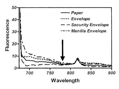
Figure 8 shows the spectroscopic window in the fluorescence emission spectra
of paper
and various samples of envelopes.
CA 02402575 2008-01-31
Figure 9 shows the response of the background-corrected 780 nm fluorescence
channel
of one embodiment of the invention to several milligrams of Bacillus
thuringiensis spores
in a sealed envelope.
DETAILED DESCRIPTION OF THE INVENTION
The basic elements for the apparatus described by this invention are shown as
a
block diagram in Figure 1. The apparatus consists of a light source,
excitation filters,
focusing optics, collection optics, emission filters and detectors.
Electromagnetic
radiation is directed from the light source towards the sample, passing
through the
excitation filters and focusing optics if necessary, to excite the intrinsic
fluorophores in
the sample. The scattered and reflected excitation radiation, along with the
emitted
fluorescence radiation, are collected with the collection optics and directed
towards the
detectors. Emission filters ensure that only the energies of interest are
measured.
Various embodiments of the invention, including different configurations and
utilizing diverse components, are possible. The fundamental components for
this
microbial detection method permit: the excitation of multiple intrinsic
microbial
fluorophores, collection and detection of emitted and reflected/scattered
light energies,
and analysis of the detected signals with a method that is able to correct for
background
interferences and compare the relative signal strengths to known physiological
parameters. The configuration and components employed in any apparatus using
this
method should be matched with the application requirements and expected
interferences.
It is possible, and sometimes desirable, to utilize a light source that
provides a
broad band illumination. The kind of light source employed is influenced by
its ability to
produce electromagnetic radiation of the wavelengths required to excite the
intrinsic
11
CA 02402575 2008-01-31
microbial components of interest. Additionally, it is sometimes desirable to
use a pulsed
light source allowing measurement of the environmental background during the
off cycle.
The light sources that can be used include lamps with various bulbs (e.g.,
mercury,
tungsten, deuterium, xenon), light emitting diodes (LEDs), and diode lasers
specific for
the required excitation energies. The kind of light source used depends upon
the intensity
of excitation radiation needed and detection limit required.
The excitation and emission filters used in the various embodiments of the
invention include interference filters, impregnated glass, series of cutoff
filters, gelatin
filters, monochrometers, gratings and the like. The light cutoff
characteristics of the
emission filters used depend on how much of the scattered and reflected
excitation
radiation signal can be tolerated by the analysis method or what detection
limit is
required. If light sources having only the energies of interest are employed,
the excitation
filters may not be necessary; if the light source is collimated (such as a
laser) then the
focusing optic may not be required. (The purpose of the focusing optic is to
direct the
excitation radiation to the sampling area or volume.) It is important to note
that with
multi-photon excitation it is possible to use light sources with energies less
than the
excitation energies of the fluorophores of interest.
The purpose of the collection optics is to deliver the light emitted from the
excited
microbial fluorophores and that scattered and reflected from the sample to the
detectors.
If interference filters are utilized to discriminate these emission energies,
then the
collected light needs to be collimated for these filters to work optimally.
Fiberoptic
cables can also be used to both deliver the excitation radiation to the sample
and to
collect the emitted radiation and direct it towards the detectors. It is
possible, and
12
CA 02402575 2008-01-31
sometimes desirable, to utilize polished metal reflective, sapphire, fused
silica, quartz,
MgF2, and/or CaF2 optical components as many optical components exhibit
fluorescence
in the ultraviolet and visible range.
The detectors are used to convert the emitted electromagnetic radiation into
an
electrical signal that can be measured. Numerous detectors, with different
sensitivities,
can be utilized in the embodiments of the invention: photomultiplier tubes
(PMTS),
avalanche photodiodes (APDs), pin diodes, CCDs, and the like. The detector
chosen
would depend upon the energy of the radiation to be detected, the strength of
the
emission signal, and the required detection limit of the apparatus.
The collected emission energies, having been converted to amplified electrical
signals, are analyzed with a method capable of removing any background
fluorescence
and scattered excitation contributions. The choice of excitation and emission
energies
used in a specific embodiment depends upon the target microbes and their
expected
physiological status. Table I lists the excitation and emission ranges of some
of the more
abundant intrinsic fluorescent compounds found in various microbes (and
proteinaceous
toxins) and indicates their likely presence in each. (Proteinaceous microbial
toxins can
be detected using this method and apparatus in a manner similar to that used
for the
detection of viruses.)
13
CA 02402575 2008-01-31
Table I. Excitation and Emission Ranges for Microbial Fluorophores.
Excitation Emission Viable Non-
Range Range Fluorophore Cells viable Spores Viruses Toxins
(111 11) Cells
260 - 285 340 - 360 Nucleic Acids X X X X
265 - 280 340 - 360 T to han X X X X X
265 - 280 340 - 360 Tyrosine X X X X X
270 - 280 380 - 400 ATP X
270 - 290 460 - 480 Ca-Di is X
310 - 330 400 - 430 Ca-Di is X
320 - 330 430 - 450 RPN X
340 - 365 430 - 450 RPN X
340 - 360 470 - 490 RPN X
430 - 450 520 - 535 Flavins X
470 - 485 560 - 580 Flavins X
560-585 615-680 Po h ns X X
560 - 580 620 - 700 Flavins X X
610 - 650 750 - 800 Unknown X X
650 - 670 730 - 780 Unknown X X
(In Table I, ATP is adenosine triphosphate and RPN refers to the reduced
pyridine
nucleotides.)
Figure 2 shows the emission spectra of a bacterial solution (Bacillus
thuringiensis) in a minimally fluorescing media when excited with light at
345nm. The
solid line shows the observed emission spectra of the bacteria and the dashed
line
indicates the contribution of the Rayleigh scattering to this spectra.
Subtraction of the
Rayleigh background from the observed spectra results in the true emission
spectra due to
the metabolites excited by 345 nm light (Figure 3 D). The magnitude of the
background
from Rayleigh scattering at wavelength A. can be described by the equation: I
= A/).4 + C.
(In this equation, I is the intensity of the incident light; A is determined
by the
experimental conditions; the value for the constant C is typically determined
by the
characteristics of the instrument used to collect the data.) The combined
emission
14
CA 02402575 2008-01-31
spectrum of the bacterial solution when excited with 325 nm, 345 nm and 570 nm
shows
minima near 515 nm and 850 nm. The measured fluorescence intensities at 515 nm
and
850 nm are used to calculate the unknown values of A and C from the
aforementioned
equation, ultimately allowing for the subtraction of the background signal
from the
detected signal. Ralyleigh scattering background subtraction is particularly
suited for
liquid and air samples; other sample media exhibit different backgrounds and
can be
treated with the appropriate methods (e.g., Me scattering, etc.).
Figure 3 shows the background-subtracted emission spectra of viable bacteri a,
non-viable bacteria and spore solutions (Bacillus thuringiensis) due to the
various
intrinsic fluorophores excited at 280 nm (A), 315 nm (B), 325 nm (C), 345 nm
(D), 570
nm (E) and 660 nm (F). Figure 4 shows the obvious differences of the
fluorescence
signals (normalized to the emission at 440 nm after background subtraction)
between said
viable cell (A), non-viable cell (B) and the spore (C) solutions. The analysis
method uses
these differences between the viable cells, non-viable cells and spore
solutions to
distinguish between these in samples. The magnitudes of the detected and
background-
subtracted signals are used to quantitate the number of microbes in the
sample.
In the one embodiment of the invention, the use of excitation filters at 325
nm,
345 nm and 570 nm would allow for the detection of and discrimination between
live
cells, dead cells and spores. These excitation filters would allow the
excitation of
reduced pyridine nucleotides, various flavins, calcium dipicolinate,
hemoproteins and
other components. The selection of filters for the emission detection of the
excited
fluorophores would include those at 405 nm, 440 nm, 480 nm and 650 nm; these
filters
correspond to maxima in the emission spectra of the excited flurophores.
Additionally,
CA 02402575 2008-01-31
other emission filters (545 nm and 850 nm) allow for the determination of the
magnitude
of the reflected/scattered background. To achieve a low detection limit, the
following
configuration was constructed. A pulsed xenon lamp was used as the light
source with
interference excitation filters. A focusing optic is added to collimate the
light before the
interference filters. The focusing and collection optical pieces were
constructed from
polished reflective optics to eliminate any background fluorescence. The
parabolic
collection optics, which collected ca. 90% of the emitted signal, were fitted
with
interference emission filters, collimating optics and PMTs. The instrument
functions,
data collection, integration and analysis were controlled by a
microcontroller.
In this embodiment of the invention, the detection method required the
relative
ratios of the detected and background-corrected signals to lie within certain
physiological
ranges. Analysis of greater than 500 samples from more than twenty different
species of
bacteria and spores showed that the numerous ratios could be used to ensure a
statistically
significant identification. Figure 5 shows the distribution for just one of
these ratios (440
nm/480 nm ratio after background subtraction) of 22 species of bacteria, thus
defining the
physiological range required for the detection method. The method could also
discriminate bacteria-containing solutions from sterile media and other
biochemical
buffers. Using a variety of methods and the following ratios (650/405,
405/440, 480/440,
650/440, 405/480 and 650/480), e.g., Neyman-Pearson test, fuzzy logic and a
trained
neural network (utilizing a multilayer perceptron), these gave a 99%, 95.6%
and 100%
probability of detection, respectively for the presence of bacteria. (False
alarm
probabilities of the 500 data points taken for these detection algorithms were
as follows:
Neyman-Pearson (0.01 %), fuzzy logic (0 %), and neural net (0 %).)
16
CA 02402575 2008-01-31
Figure 6 shows data from one emission (RPN) of the instrument for viable and
non-viable Salmonella typhi cells on the surface of a glass slide. The
difference between
viable and non-viable cells in the signal from this fluorescence is clear.
Figure 7 shows
the response of the RPN emission to Escherichia coli on the surface of turkey.
A
detection limit well below that observed for other microbial detection methods
is
observed in real-time, without the need for reagents or touching the meat
surface.
In another embodiment of the invention, LEDs centered around 570 and 660 nm
are used to excite the component(s) found in spores and dead cells shown in
Figures 3D
and 3F. Figure 8 shows the emission spectra of paper and various samples of
envelopes
when excited with light at 660 nm; the arrow in this figure shows the location
of the
emission expected from spores and non-viable cells at this excitation. As the
paper and
envelopes contain this spectroscopic window it is possible to detect bacterial
endospores
behind paper and inside envelopes. It is possible, and sometimes desirable, to
include
excitation at 570 nm as the resulting emission from non-viable cell
component(s)
between 610 and 680 nm excites the spore components that fluoresce in the
aforementioned spectroscopic window. Figure 9 shows the differences between
the 780
nm background-corrected fluorescence signals of an envelope and a sample of
freeze-
dried Bacillus thuringiensis spores sealed inside of the same envelope. With
this
embodiment of the invention it is possible to quickly detect spores in
envelopes without
the need for reagents, sample processing, contact with the sample or opening
the
envelope.
The embodiments of the present invention described above are intended to be
merely exemplary, with other configurations, variations and modifications
utilizing the
17
CA 02402575 2008-01-31
fore mentioned basic ideas available to those skilled in the art without
departing from the
spirit of the invention. The scope of this method and apparatus to detect
microbes
includes utilization of simultaneous excitation of multiple intrinsic
microbial
fluorophores with subsequent analysis of the detected emissions with methods
that
concurrently account for background signals and require said signals to lie
within
physiological ranges. All variations, modifications and configurations are
intended to be
within the scope of the present invention as defined in the appended claims.
18