Note: Descriptions are shown in the official language in which they were submitted.
CA 02402991 2002-09-12
-1-
Specification
Drugs for the Diag~osis of Tissue Reproductive Activity or
the Treatment of Proliferative Diseases
Technical Field
The present invention relates to use of radiolabeled nucleoside derivatives
for
diagnosis of tissue proliferation activity and treatment of proliferative
diseases.
$ack¾round Art
If proliferation activity of tumor cells can be determined non-invasively by
image diagnosis, it will be help for evaluation of growth rate and malignancy
of the
tumor. Detection of the most rapidly growing regions of a tumor by image
diagnosis
will be useful in preparing plans for radiation fields in radiotherapy and
identifying
suitable portions for biopsy. Such methods will permit an early and accurate
evaluation of therapeutic effects, which is difficult to identify by CT- or
MRI-based
anatomical evaluation or PET-based measurement of glucose-metabolic changes.
Particularly, they will be useful for an early assessment of therapeutic
effects of
anticancer agents that may cause strong side effects.
In order to solve these clinically important problems, use of 5-iodo-
deoxyuridine labeled with a radioactive iodine and thymidine labeled with
carbon-11
which is a positron-emitter, have been studied (Tjuvajev JG et al., J. Nucl.
Med. 35,
pp.1407-1417 (1994); Blasberg RG et al, Cancer Res. 60, pp.624-635 (2000);
Martiat
Ph et al., J. Nucl. Med. 29, pp.1633-1637 (1998); Eary JF et al., Cancer Res.
59, pp.
615-621 (1999); U.S. Patent No. 5,094,835; U.S. Patent No. 5,308,605). It is
considered that these radiolabeled compounds are taken into cells as
precursors for
DNA synthesis required for cell division of rapidly-growing tumors, and then
phosphorylated by thymidine kinase, followed by incorporation into DNA, to
reflect
CA 02402991 2002-09-12
,
-2-
proliferation activity of the tumor. These radiolabeled compounds, however,
are
decomposed rapidly in vivo, making it difficult to perform non-invasive
evaluation of
the proliferation activity of the tumor. The method using carbon- i l-labeled
thymidine,
in particular, requires very complicated mathematical model analysis, and
cannot
become popular as a diagnostic technique of nuclear medicine imaging.
The rapid metabolic decomposition of these radiolabeled compounds in vivo is
considered to be due to cleavage of C-N glycosidic bonds by thymidine
phosphorylase
and instability of the labels in vivo. If the C-N glycosidic bonds are
cleaved, the
compound loses its affinity to tumors, thereby decreasing in accumulation of
radioactivity in tumors, while the radioactive metabolites increase background
radioactivity, thereby making imaging of the tumors difficult.
To solve these problems, radiolabeled compounds with metabolic stability have
been synthesized by introducing fluorine atoms, which are high in
electronegativity, to
the 2' or 3' position in certain nucleosides, and have been studied for
imaging of tumors.
Thus, 3'-deoxy-3'-fluorothymidine that contains fluorine 18, a positron
emitter, at the 3'
position shows a high stability in vivo and an accumulation in tumor tissue
(Shields AF
et al., Nature Med. 4, pp.1334-1336 (1998)). Though this radiolabeled compound
is
stable in vivo, it is a radio-labeled compound with a short-life positron
emitter, and
therefore a cyclotron is required in the hospital, limiting the usage of the
compound.
For this radiolabeled compound, the major process responsible for its
accumulation in
cells is the phosphorylation caused by thymidine kinase that is an index of
DNA
synthesis, and thus it does not serve as an agent that essentially reflects
DNA synthesis.
A derivative of 5-iododeoxyuridine, in which fluorine is introduced to the 3'
position in the same manner as above to increase its stability in vivo, has
recently been
reported. Though stable in vivo, however, this radiolabeled compound was high
in
CA 02402991 2002-09-12
-3-
retention in blood and failed to show a significant accumulation in a tumor
compared to
5-iododeoxyuridine (Choi SR et al., J. Nucl. Med. 41, p. 233 (2000)).
2'-fluoro-5-iodoarabinouridine, in which fluorine is introduced to the 2'
position, shows a high stability in vivo, and has been used for identification
of
introduction and expression in vivo of a vector for gene therapy, utilizing a
phosphorylation reaction specific to thymidine kinase of human herpesvirus. It
has
also been applied to image diagnosis for virus infection, based on the high
specificity to
the viral thymidine kinase (Tjuvajev JG et al., Cancer Res. 56, pp.4087-95
(1996);
Tjuvajev JG et al., Cancer Res. 58, pp.4333-4441 (1998); Wiebe LI et al.,
Nucleosides
Nucleotides 18, 1065-1076 (1999); Gambhir SS et al., Nucl. Med. Biol. 26,
pp.481-490
(1999); Haubner R et al., Eur. J. Nucl. Med. 27, pp.283-291 (2000); Tjuvajev
JG et al.
Cancer Res. 59, 5186-193 (1999); Bengel FM et al., Circulation 102, pp.948-950
(2000)).
In view of the above situation, the present invention aims to provide a
radiolabeled compounds that are practically useful in clinical fields, stable
in vivo, and
able to retain in cells after being phosphorylated by thymidine kinase of
mammals, or
reflect the DNA synthesis activity after being incorporated in DNA,
particularly those
compounds which are labeled with a single-photon emitter to achieve a wide
spectrum
of use, and also aims to provide methods for diagnosis of tissue proliferation
activity
and for treatment of proliferative disease, utilizing agents that contain said
radiolabeled
compounds.
Disclosure of the Invention
To achieve the above-mentioned objectives, the present inventors have
synthesized a variety of radiolabeled compounds and have intensively studied
to see if
they are useful for image evaluation of tissue proliferation activity. As a
result, the
CA 02402991 2002-09-12
-4-
inventors have found that radiolabeled compounds as represented by the
following
formula can serve for diagnosis of tissue proliferation activity or treatment
of
proliferative disease, and have completed the present invention.
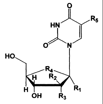
Specifically, the present invention provides an agent for diagnosis of tissue
proliferation activity or for treatment of proliferative disease, which
comprises, as an
active ingredient, a radiolabeled compound as represented by the following
formula or a
pharmaceutically acceptable salt thereof:
O
R5
HN
f
O N
HO
R4
H R2
H Ri
OH R3
wherein R, denotes hydrogen, or a linear- or branched-chain alkyl group having
1-8
carbon atoms; R2 denotes hydrogen, hydroxyl, or a halogen substituent; R3
denotes
hydrogen or fluorine substituent, R4 denotes oxygen, sulfur or a methylene
substituent,
and RS denotes a radioactive halogen substituent, excluding'the case where R,,
R2 and
R3 are hydrogen, R4 being oxygen, and Rg being radioactive fluorine, bromine,
iodine,
or astatine; the case where R, and R3 are hydrogen, RZ being fluorine, R4
being oxygen,
and R5 being radioactive bromine or iodine; and the case where R, and RZ are
hydrogen,
R3 being fluorine, R4 being oxygen, and RS being radioactive bromine or
iodine.
The radiolabeled compounds of the present invention are stable in vivo, and
can
retain in cells after being phosphorylated by mammalian thymidine kinase or
reflect the
CA 02402991 2002-09-12
-5-
DNA synthesis activity after being incorporated in DNA. Therefore, they
realize
effective diagnosis of tissue proliferation activity and treatment of
proliferative disease,
and are particularly useful as diagnostic radioactive imaging agents for
diagnosis of
tissue proliferation activity or as radioactive therapeutic agents for
treatment of
proliferative disease in accordance with internal radiotherapy, local
radiotherapy or the
like.
Thus, according to another aspect of the present invention, there are provided
methods for diagnosis of tissue proliferation activity, which comprise
administering an
effective amount of a radiolabeled compound as represented by the above
formula or a
pharmaceutically acceptable salt thereof to a mammal, followed by imaging in
vivo
distribution thereof, and methods for treatment of proliferative disease,
which comprises
administering an effective amount of said radiolabeled compound or salt to a
mammal.
Herein, mammal includes human beings.
In the present invention, the radiolabeled compounds as represented by the
above formula include salts thereof, or may be in a form of a hydrate or
solvate of these.
Such salts include phannaceutically acceptable salts, for example, one formed
with a
mirieral acid such as hydrochloric acid and sulfuric acid or with an organic
acid such as
acetic acid. As such a hydrate or solvate, mention may be made of the present
radiolabeled compounds or salts thereof to which water molecules or solvent
molecules
are attached. Furthermore, the compounds of the present invention include
their
various isomers such as tautomers.
In the above formula, the linear- or branched-chain alkyl group having 1-8
carbon atoms as represented by R, includes, for example, methyl group, ethyl
group,
propyl group, t-butyl group, and n-hexyl group, of which methyl group is
preferable.
The halogen-substituent as represented by R2 preferably includes fluorine,
chlorine, and
CA 02402991 2002-09-12
-6-
bromine. R4 is preferably oxygen or sulfur, of which sulfur is particularly
preferable.
The radioactive halogen-substituent as represented by R5 in the above formula
includes F-18, C1-36, Br-75, Br-76, Br-77, Br-82, I-123,1-124, I-125,143 1,
and At-211,
of which F-18, Br-76, I-123, and I-124 are preferable for diagnostic purposes
while Br-
77, 1-125, I-131, and At-211 are preferable for therapeutic purposes.
Preferred compounds as represented by the above formula include those
wherein R, is hydrogen or methyl, R2 is hydrogen or a halogen-substituent, R3
is
hydrogen, and R4 is oxygen or sulfur, particularly preferably those wherein
R,, R2 and
R3 are hydrogen, R4 is sulfur, R. is a radioactive halogen-substituent
selected from F- 18,
I-123, I-125, and I-131.
Certain 4'-thio nucleic acid derivatives as represented by the above formula
(where RS is a non-radioactive halogen-substituent) have been reported to be
resistant to
bacterial thymidine phosphorylase as a result of studies on antiviral agents
(Dyson MR
et al., J. Med. Chem. 34, pp.2782-2786 (1991); Rahim SG et al., J. Med. Chem.
39,
pp.789-795 (1996)). It has also been known that certain 5-iodine- and 5-methyl-
4'-
sulfur substitution products inhibit phosphorylation of thymidine by human
thymidine
kinase (Strosselli S et al., Biochem J. 334, pp.15-22 (1998)). The chemical
structures
of these compounds with sulfur at the 4' position and their use as an
antiviral agent are
already known (Intemational Publication W09101326, International Publication
W09104982, Japanese Patent Laid-Open No. HEI 10-087687), but neither the
corresponding radiolabeled compounds nor their use as a radioactive diagnostic
imaging
agent or radioactive therapeutic agent have been known.
The chemical structures of certain compounds with a substituent at the 1'
position as represented by the above formula (where RS is a non-radioactive
substituent)
and production methods thereof have already been known (Japanese Patent Laid-
Open
CA 02402991 2002-09-12
-7-
No. HEI 07-109289). However, neither the corresponding radiolabeled compounds
nor their use as a radioactive diagnostic imaging agent or radioactive
therapeutic agent
have been known.
The compounds as represented by the above formula can be used for various
diagnoses of tissue proliferation activity and treatment for proliferative
diseases by
virtue of their in vivo stability and their capability for retention in cells
or capability for
being incorporated in DNA.
Such diagnoses of tissue proliferation activity include, for example,
diagnosis
of hyperplasia, regeneration, transplantation or viral infection accompanied
by abnormal
prolifera.tion .
The diagnosis of hyperplasia accompanied by abnormal proliferation includes,
for example, diagnosis of hyperplastic inflammation, benign tuinors, or
malignant
tumors. The diagnosis of the hyperplastic inflammation includes, for example,
diagnoses concerning activity of chronic rheumatoid arthritis and
determination of
therapeutic effects. The diagnosis of the benign tumors includes, for example,
diagnoses concerning localization, activity and determination of therapeutic
effects.
The diagnosis of the malignant tumors includes, for example, diagnoses
concerning
localization, progress, malignancy and determination of therapeutic effects,
of primary
and metastatic malignant tumors. Benign tumors include, for example, prostatic
hyperplasia, endometrium hyperplasia (cystic hyperplasia, adenomyosis uteri,
hysteromyoma), ovarian tumor (cystadenoma), mammary gland (mastopathy, mammary
gland fibroadenoma), pituitary adenoma, craniopharyngioma, thyroid adenoma,
adrenocortical adenoma and pheochromocytoma. Malignant tumors include, for
example, malignant lymphoma (Hodgkin's disease, non-Hodgkin lymphoma),
pharyngeal cancer, lung cancer, esophagus cancer, gastric cancer, colon
cancer, hepatic
CA 02402991 2002-09-12
-g-
cancer, pancreatic cancer, nephric tumor (nephric cancer, nephroblastoma),
bladder
tumor, prostatic cancer, testicular tumor, uterine cancer, ovarian cancer,
breast cancer,
thyroid cancer, neuroblastoma, brain tumor (primary brain tumor, metastatic
brain
tumor), rhabdomyosarcoma, bone tumor (osteosarcoma, metastatic bone tumor),
Kaposi's sarcoma, and malignant melanoma.
The diagnosis of regeneration accompanied by abnormal proliferation is
exemplified by diagnosis of function of physiological regeneration of blood
and
diagnosis of pathological regeneration resulting from pathological loss of
blood cells,
such as evaluation of physiological hematopoietic functions of bone marrow
during
treatment with anti-cancer drugs and diagnosis of pathological functions of
the bone
marrow in patients suffering from hypoplastic anemia.
The diagnosis of transplantation accompanied by abnormal proliferation is
exemplified by diagnosis of blood cancer patients undergoing bone marrow
transplantation or very high-dose chemotherapy using an anticancer agent, such
as
diagnosis of take or proliferation of transplanted bone marrow cells in bone
marrow
transplantation.
The diagnosis of viral infection accompanied by abnormal proliferation
includes, for example, diagnosis of virus-infected portions and proliferation
thereof in
infectious diseases caused by Type I or Type II herpes simplex virus,
varicella-zoster
herpes virus, cytomegalovirus, Epstein-Barr virus, or human immunodeficiency
virus,
particularly infectious diseases of central nervous system (e.g., viral-
infectious cerebritis,
meningitis, etc.) caused by Type I or Type II herpes simplex virus or human
immunodeficiency virus.
The treatment for proliferative diseases is exemplified by treatment of
malignant tumors or viral infection accompanied by abnormal proliferation.
Such
~..~~.., a...._..... -.a.,.a._..,.~:~.. ~....G _ .W..,.~,...~_.W,_.,._:,.,~
.., CA 02402991 2002-09-12
-9-
malignant tumors include, for example, malignant lymphoma (Hodgkin's disease,
non-
Hodgkin lymphoma), pharyngeal cancer, lung cancer, liver cancer, bladder
tumor, rectal
cancer, prostatic cancer, uterine cancer, ovarian cancer, breast cancer, brain
tumor
(primary brain tumor, metastatic brain tumor), and malignant melanoma. Such a
viral
infection includes infectious diseases of central nervous system caused by
Type I or
Type II herpes simplex virus or human immunodeficiency virus, particularly
viral
encephalitis or meningitis.
Methods for labeling the compounds represented by the above formula at the
"5" position with a radioactive halogen may be known methods, such as methods
using
isotope exchange reaction, and a method using a 5-chloromercuri compound in
which
mercury is introduced into the "5" position of the compound or a 5-hydrogen
compound
in which there is no substitution at the "5" position of the compound. The
method
using the 5-chloromercuri compound is already known as an iodo-labeling method
for
producing 5-iodo-2'-deoxyuridine (United States Patent No. 4,851,520;
Baranowska-
Kortylewicz J et al., Appl. Radiat. Isot. 39, p.335 (1988)). This method is,
however,
disadvantageous for producing pharmaceuticals labeled with a short half-time
radioactive nuclide due to side reactions (formation of "5-chloro" compounds,
demercurization reaction), a long reaction time (6 hours), and formation of
inorganic
mercury compounds. The method using a 5-hydrogen compound is already known as
a method for producing 5-iodo-2'-deoxyuridine from 2'-deoxyuridine (Knaus EE
et al.,
Appl. Radiat. Isot. 37, p.901 (1986); Fin RD et al., J. Label. Comds.
Radiopharm. 40,
p.103 (1997)). This method, however, requires heating at 65-115 C, and
therefore, it
is not suitable for use with compounds that are easily decomposed under
heating
conditions and cannot be said to be an ideal labeling method, considering the
properties
of radioactive halogen atoms which preferably should not involve heating
operations
CA 02402991 2002-09-12
-10-
during the labeling reaction. Further, the radiolabeling method using isotope
exchange
reaction is also unsuitable for producing pharmaceuticals that must be
maintained at a
certain level of quality, because the method is not able to produce carrier-
free labeled
compounds and is difficult to control variation of specific activity among
different
labeling runs.
Another useful method for labeling the compounds represented by the above
formula at the "5" position with a radioactive halogen is to allow a compound
(5-
trialkyltin compound), in which the pyrimidine base is substituted by a
trialkylstannyl
group at the "5" position as represented by Formula 11 in Fig. 1, Formula 21
in Fig.2,
Formula 28 in Fig.3, Fonnula 40 in Fig.4, Formula 50 in Fig.5 or Formula 58 in
Fig.6,
to react with 0.1N sodium hydroxide solution of a radioactive halogen in an
appropriate
solvent such as chloroform, so that the trialkylstannyl group at the "5"
position is
converted into a radioactive halogen-substituent. This labeling method, which
uses a
5-trialkyltin compound, is preferable as it does not suffer such problems as
with the
above three labeling methods. Specifically, this method requires only a
relatively short
reaction time, and it does not produce "5-chloro" compounds or need heating as
the
reaction readily proceeds at room temperature. The resulting labeled compounds
are
free of carriers, and if a lower specific activity is desired, a labeled
compound with a
fixed specific activity can be readily prepared by adding a carrier. This
method is also
featured in that purification after the reaction is easy to operate.
Specifically, 5-
trialkyltin compounds are largely different from the corresponding radioactive
halogen-
labeled compounds in terms of overall molecular polarity as the electrical
properties at
the "5" position differ between them. Owing to the difference in the molecular
polarity, labeled compounds and unreacted precursors can be separated easily
by using a
commercial reverse-phase silica gel cartridge after the labeling reaction.
This permits
CA 02402991 2002-09-12
-11-
elimination of the need of troublesome high performance liquid chromatographic
purification.
Thus, according to another aspect of the present invention, there is provided
a
method for producing a radiolabeled compound as represented by the following
formula:
0
Rs
HN
O N
HO
Ra
H R2
H Ri
OH R3
wherein R, denotes hydrogen or a linear- or branched-chain alkyl groups having
1-8
carbon atoms, R2 denotes hydrogen, hydroxyl or a halogen substituent, R3
denotes
hydrogen or fluorine substituent, R., denotes oxygen, sulfur or a methylene
substituent,
and R. denotes a radioactive halogen substitient;
comprising reacting a nucleosicle derivative as represented by the following
formula:
CA 02402991 2002-09-12
-12-
O
R5
HN
I
O N
HO
R4
H R2
H Ri
OH R3
wherein R, denotes hydrogen or a linear- or branched-chain alkyl groups having
1-8
carbon atoms, R2 denotes hydrogen, hydroxyl or a halogen substituent, R3
denotes
hydrogen or fluorine substituent, R4 denotes oxygen, sulfur or a methylene
substituent,
and RS denotes a trialkylstannyl group,
with an alkaline solution of a radioactive halogen in a solvent, whereby the
trialkylstannyl group of RS is converted into the radioactive halogen
substituent.
The 5-trialkyltin compounds as represented by Formula 11 in Fig.l, Formula
21 in Fig.2, Formula 28 in Fig.3, Formula 40 in Fig.4, Formula 50 in Fig.5,
and
Formula 58 in Fig.6 are novel compounds which are useful intermediates for
producing
the radiolabeled compounds of the present invention.
Thus, according to another aspect of the present invention, there is provided
a
compound as represented by the following formula:
,,....,.....:.w.~._~. ._. . ._ r
CA 02402991 2002-09-12
-13-
O
R5
HN
f
O N
HO
Ra
H R2
H R,
OH R3
wherein R, denotes hydrogen or a linear- or branched-chain alkyl groups having
1-8
carbon atoms, ; R2 denotes hydrogen, hydroxyl - or a halogen substituent, R3
denotes
hydrogen or fluorine substituent, R4 denotes oxygen, sulfur or a methylene
substituent,
and R5 denotes a trialkylstan.nyl group.
In the above formula, the linear- or branched-chain alkyl groups having 1-8
carbon atoms as represented by R, include, for example, methyl group, ethyl
group,
propyl group, t-butyl group, and n-hexyl group, of which methyl group is
preferred.
The halogen-substituent as represnted by RZ preferably includes fluorine,
chlorine and
bromine. R4 is preferably oxygen or sulfur. The trialkylstannyl group as
represented
by R5 includes trimethylstannyl group, triethylstannyl group and
tributylstannyl group.
Preferred compounds as represented by the above formula include those
wherein R, is hydrogen or methyl, R2 is hydrogen or a halogen-substituent, R3
is
hydrogen, and R4 is oxygen or sulfur.
As seen from Figs.1-6, 5-trialkyltin compounds can generally be synthesized
by providing their corresponding halogen-containing compound (as represented
by
Formula 10 in Fig.1, Formula 20 in Fig.2, Formula 27 in Fig.3, Formula 39 in
Fig.4,
Formula 49 in Fig.5, or Formula 57 in Fig.6) as starting materials, reacting
the
CA 02402991 2002-09-12
-14-
compound with bis(trialkyltin) and bis(triphenylphosphine)palladium chloride
in
anhydrous 1,4-dioxane under heat at reflux in an argon atmosphere, followed by
purification.
Compound 10 (ITDU) in Fig.1 can be synthesized by a known method
(Formulae 1-8: Dyson, MR et al., Carbo. Res. 216, p.237 (1991), and Formulae 8-
10:
Oivanen, M et al., J. Chem. Soc., Perkin Trans. 2, p.2343 (1998)).
Specifically, 2-
deoxy-D-erythro-pentose (Compound 1) is reacted with a' 1% hydrochloric acid-
methanol solution to produce Compound 2, which is then reacted with sodium
hydride,
tetrabutylammonium iodide, and benzyl bromide to produce Compound 3, in which
hydroxyl groups are protected. The compound is reacted with a-toluenethiol and
concentrated hydrochloric acid to produce Compound 4, which is then reacted
with
triphenylphosphine, benzoic acid, and diethylazodicarboxylate to produce
Compound 5.
Sodium methoxide is then used to remove the benzoyl group from Compound 5 to
produce Compound 6, followed by its conversion into Compound 7 with
methanesulfonyl chloride. A ring is formed with sodium iodide and barium
carbonate
to produce Compound 8, which is reacted with 5-iodouracil in the presence of
bistrimethylsilylacetamide and then with N-iodosuccinimide to produce Compound
9.
Subsequently, Compound 9 is deprotected with titanic chloride to produce
Compound
10.
Compound 20 (ITAU) in Fig.2 can be synthesized by a known method
(Formulae 13-17: Yoshimura Y et al., J. Org. Chem.61, p.822 (1996) and Formula
17-
20: Yoshimura Y et al., J. Med. Chem. 40, p.2177 (1997)). Specifically,
1,2;5,6-di-O-
isopropylidene glucose (Compound 13) is reacted with sodium hydride and benzyl
bromide to produce a 3-benzyl compound, which is subsequently reacted with
hydrochloric acid, aqueous sodium periodate solution, and sodium borohydride
to
CA 02402991 2002-09-12
-
15-
produce Compound 14, which is then converted with hydrogen chloride into
Compound
15. The compound is then reacted with mesyl chloride and sodium sulfide to
produce
Compound 16, which is reacted with hydrochloric acid and sodium borohydride
successively to produce Compound 17. Hydroxyl groups are protected with sodium
hydride and benzyl bromide (Compound 18), and the resulting compound is
converted
to Compound 19 with m-chloroperbenzoic acid (m-CPBA) and acetic anhydride. It
is
further reacted with 5-iodouracil in the presence of 1,1,1,3,3,3-hexamethylene
disilazane (HMDS) to produce a glycosylated compound, which is then reacted
with
boron chloride to produce Compound 20.
Compound 27 in Fig.3 can be produced as follows. Compound 17 shown in
Fig.2 is used as a starting material, which is reacted with t-
butyldimethylsilyl chloride
(TBDMSCI) in dimethylformamide (DMF) in the presence of imidazole to protect
the
hydroxyl group at the "5" position with a silyl group to produce Compound 23.
Trifluoromethanesulfonic acid anhydride (TfZO) is added thereto in pyridine to
produce
Compound 24 in which the hydroxyl group at the "2" position is
trifluoromethanesulfonylated. The compound is reacted with potassium fluoride,
along with Kryptofix (registered trademark) 222 and potassium carbonate, in
acetonitrile, to produce a fluoride compound (Compound 25) in which the
substituent at
the "2" position is stereochemically reversed. The compound is reacted with m-
chloroperbenzoic acid (m-CPBA) in methylene chloride and further treated with
acetic
anhydride to produce Compound 26. This is reacted with the product resulting
from a
reaction of 5-iodouracil and 1,1,1,3,3,3-hexamethylene disilazane (HMDS), and
with
trifluoromethanesulfonic acid trimethylsilyl (TMSOTf). The resulting product
is
further treated with boron chloride in methylene chloride to produce Compound
27.
Compound 39 (FIAU) in Fig.4 can be synthesized by a known method
CA 02402991 2002-09-12
-16-
(Formulae 30-37: Reichman U et al., Carbohydrate Res. 42, p.233 (1975) and
Formulae
37-39: Asakura J et al., J. Org. Chem. 55, p.4928 (1990)). Specifically,
Compound 31,
which has been synthesized in four steps from 1,2:5,6-di-O-isopropylidene
glucose
(Compound 30), is treated with a cation exchange resin (Amberlite IR-120) to
produce
Compound 32, which is then reacted with potassium periodate to produce
Compound 33.
This is then reacted with sodium methoxide to produce Compound 34, followed by
acetylation of hydroxyl groups to produce Compound 35. The compound is treated
with a hydrogen bromide-acetic acid solution to produce Compound 36, followed
by
condensation with an uracil derivative to produce Compound 37. It is
subsequently
reacted with diammonium cerium(III) sulfate (CAN) to produce Compound 38,
followed by deprotection of hydroxyl groups with sodium methoxide to produce
Compound 39.
Compound 49 (FITAU) in Fig.5 can be synthesized by a known method
(Formulae 42-46: Yoshimura Y et al., J. Org. Chem. 62, p.3140 (1997) and
Formulae
46-49: Yoshimura Y et al., Bioorg. Med. Chem. 8, p.1545 (2000)). Specifically,
Compound 43, which has been synthesized in nine steps from 1,2:5,6-di-O-
isopropylidene glucose (Compound 42), is reacted with diethylaminosulfur
trifluoride
(DAST) to produce Compound 44, which is then reacted with m-chloroperbenzoic
acid
(m-CPBA) to produce Compound 45. This is subsequently reacted with acetic
anhydride to produce Compound 46, which is reacted with
trifluoromethanesulfonic
acid trimethylsilyl (TMSOTf) to cause condensation with a 5-iodouracil
derivative to
produce Compound 47. Finally, the two protective hydroxyl groups are removed
to
produce Compound 49.
Compound 57 (IMBAU) in Fig.6 can be synthesized by a known method
(Formulae 52-54: Itoh Y et al., J. Org. Chem. 60, p.656 (1995) and Formulae 55-
56:
..,...._. _...__.__...o.~_. ......__.__ .r
CA 02402991 2002-09-12
-17-
Asakura J et al., J. Org. Chem. 55, p.4928 (1990)), combined with known
reactions for
protection and deprotection of hydroxyl groups (Formulae 54-55 and Formula 56-
57).
Specifically, 1-[3,5-bis-O-(tert-butyldimethylsilyl)-2-deoxy-D-erythro-pento-l-
enofuranosyl]uracil (Compound 52) is reacted with pivalic acid and
bromosuccinimide
(NBS) to produce Compound 53, which is then reacted with trimethylaluminum to
produce Compound 54. The protection groups for hydroxyl groups are converted
from
tert-butyldimethylsilyl to acetyl, followed by reaction with diammonium
cerium(III)
sulfate (CAN) to produce Compound 56. Finally, the protection groups in
Compound
56 are removed with ammonia to produce Compound 57.
For radiolabeled compounds of the present invention, appropriate doses and
routes of administration should be selected depending upon target diseases and
objectives, but if they are used as an agent for diagnosis of tissue
proliferation activity, a
radioactivity in the range of 37MBq to 740MBq, preferably 111 MBq to 370MBq is
administered. Usually, they are administered intravenously, but in some cases,
other
routes of administration including arterial or intraperitoneal administration
and direct
administration to a tumor or other affected portions may be used.
If they are used as an agent for treatment of proliferative disease, a
radioactivity in the range of 37MBq to 7400MBq, preferably 185MBq to 3700MBq,
is
administered. Usually, they are administered intravenously, but in some cases,
other
routes of administration including arterial or intraperitoneal administration
and direct
administration to a tumor or other affected portions may be used. Furthermore,
if they
are used for therapeutic purposes, the above dose may be administered several
times at
appropriate intervals.
The agent for diagnosis of tissue proliferation activity of the present
invention
can serve for whole-body or local scintigraphy and whole-body or local SPECT
. _ . . ~,<....e .w......m. ..r...=ru...sw.w. ... .- _. ; , p
CA 02402991 2002-09-12
=
-18-
imaging by use of nuclides for SPECT. Using nuclides for PET, they can also be
applied for whole-body or local PET imaging.
The agent for diagnosis of tissue proliferation activity of the present
invention
can serve for quantitative determination of local proliferative activity based
on
appropriate model analysis. Furthermore, if non-proliferation tissue is used
as a
control, local proliferative activity can be defined easily in a semi-
quantitative way.
The agent for treatment of proliferative disease of the present invention,
when a
beta-emitter such as I-131 is used therein, can serves to decrease large
tumors of Icm or
more in diameter, depending on the range of the ray. When an alpha-emitter
such as
At-211 is used, they can work on small lesions of 0.1 mm or less in diameter
more
effectively than beta-emitter, and therefore, they are expected to serve for
treatment of
micrometastasis over the body. Furthermore, nuclides that emit Auger
electrons, such
as 1-125, can have antitumor effects due to DNA breakage, only after labeled
compounds have gathered around the DNAs. Therefore, suitable label nuclides
for
treatment of systemic tumor foci including metastatic ones include alpha-
emitter such as
At-211, and beta-emitter such as 1-131 that can have effect on portions around
the foci
depending on the range. The most effective method is the cocktail therapy
which uses
a mixture of a compound labeled with an alpha-emitter and a compound labeled
with a
beta-emitter.
For treatment by local administration, compounds labeled with nuclides that
emit Auger electrons, such as 1-125, are particularly effective for brain
tumor that is
difficult to remove completely by surgical operation, and residual tumor from
malignant
melanoma, and in view of functional preservation, breast cancer, rectal
cancer, prostatic
cancer, and malignant mouth tumor, because they do no harm on portions other
than
pathologically proliferating cells owing to the properties of rays emitted
therefrom.
- .......,...sw...a..,r,a,w..e....-.. .. ...,......:.v.
............w,r,.....~.,,.r...v;........x-.a..ar.. sua.::=......:.,:.. F..,
... .:,...w:........._,e.-:.:.__. .-._. F
CA 02402991 2002-09-12
-19-
Technique for local administration includes, for example, an administration
into
intracavitary foci such as colon cancer by use of an endoscope, a direct
administration
to foci affected by brain tumor during craniotomy, and an administration by
use of a
catheter into an artery relevant to an affected organ such as liver affected
by cancer.
Brief Description of the Drawings
Fig. I illustrates a synthetic pathway for a compound of the present
invention.
Fig.2 illustrates another synthetic pathway for a compound of the present
invention.
Fig.3 illustrates a third synthetic pathway for a compound of the present
invention.
Fig.4 illustrates a fourth synthetic pathway for a compound of the present
invention (5-trialkyltin compounds) and [I-125] FIAU produced therefrom.
Fig.5 illustrates a fifth synthetic pathway for a compound of the present
invention.
Fig.6 illustrates the other synthetic pathway for a compound of the present
invention.
Fig.7 illustrates a diagram showing in vivo label stability of [1-125] ITDU
and
[1-125] ITAU measured in Example 16, along with [1-125] IUR as a control.
Fig.8 illustrates a diagram showing in vivo label stability of [1-125] ITDU,
[I-
125] ITAU, [I-125] FITAU and [1-125] IMBAU measured in Example 17, along with
[I-
125] IUR (highly decomposable) as a control.
Fig.9 illustrates a diagram showing in vivo distribution of [1-125] ITDU in
normal mice measured in Example 18.
Fig.10 illustrates a diagram showing in vivo accumulation of [I-125] ITDU, [I-
125] ITAU, [I-125] FITAU and [I-125] IMBAU, along with [I-125] IUR as a
control, in
CA 02402991 2002-09-12
=
-20-
proliferating tissue measured in Example 18.
Fig.11 illustrates a photograph (biological morphology) showing a scintigram
of Walker tumor observed in Example 19.
Examp1e~
The present invention will be described in detail below with reference to
examples, but is not limited to these examples.
Examile1
Svnthesis of 5-trimethylstannx1 '-tbio-2'-deoxvuridine So=uud 11)
As shown in Fig.1, benzyl-3,5-di-O-benzyl-2-deoxy-1,4-dithio-a,(3-D-erythro-
pentofuranoside (Compound 8) was synthesized, using 2-deoxy-D-erythro-pentose
(Compound 1) as starting material, according to the method of Dyson MR et al.
(Carbo.
Res. 216, p.237 (1991)). Further, 5-iodo-4'-thio-2'-deoxyuridine (ITDU:
Compound
10) was produced from Compound 8 according to the method of Oivanen M et al.
(J.
Chem. Soc., Perkin Trans. 2, p.2343 (1998)). Compound 10 was then used as a
starting material to produce 5-trimethylstannyl-4'-thio-2'-deoxyuridine
(Compound 11)
according to the following procedure.
Compound 10 (9.5mg, 0.026mmo1), bis(trimethyltin) (17.3mg, 0.052mmol)
and bis(triphenylphosphine)palladium(II) chloride (5mg) were dissolved in
anhydrous
1,4-dioxane (3mL) under argon atmosphere, and after heating at reflux for 3
hours,
concentrated under reduced pressure. The residue was purified by silica gel
thin layer
chromatography (chloroform-methanol, 6:1) to produce the target Compound 11
(6.9mg,
65%).
1H NMR (270MHz, CD3OD) S 0.26 (s, 9H, CH3Sn), 2.26 (ddd, 1H, J=4.6, 7.9, 13.2
Hz,
1 H, H-2'), 2.27 (ddd, J=4.6, 6.6, 13.4 Hz, 1 H, H-2'), 3.41 (m, 1 H, H-4'),
3.71 (dd, J=5.9,
11.2 Hz, 1 H, H-5'), 3.80 (dd, J=4.6, 11.2 Hz, 1 H, H-5'), 4.47 (q, J=4.0 Hz,
1 H, H-3'),
CA 02402991 2002-09-12
=
r -21-
6.41 (t, J=7.2 Hz, 1H, H-1'), 7.93 (s, 1H, H-5).
Fxample 2
Sy.u, esis of [1-125]-5-iodo-4'-thio-2'-deoxvuridine ,j1-125] ITUU: Comnound
12)
To 0.1N sodium hydroxide solution (50 L) of [I-125]-sodium iodide (33MBq),
water (1 mL) and chloroform (1 mL) were added, and then chloroform solution
(4.7pL)
of iodine (60 g, 0.47 mol) was added, and shaken for 10 seconds. After
removing
only the aqueous layer, ethyl acetate solution (100 L) of Compound 11 (100 g,
0.25 mol) was added, and the resulting solution was left to stand at room
temperature
for 2 hours. One drop of 1N sodium thiosulfate solution was added, and
chloroform
was evaporated. After adding water (1 mL), the solution was passed through a
Sep-Pak
Plus QMA cartridge column. The column was washed with water (0.5mLx2), and the
resulting aqueous solution was combined to produce 1-125-labeled Compound 12
(7.3MBq, 22%).
Ex=le 3
Synthesis of [I-123]-5-iodo-4'-thio-2'-deoxyu_ri_di_ne ([I-123] ITD T:
Compound 12)
To 0.1% ammonium iodide solution (1 mL) containing [I-123]-ammonium
iodide (2.OGBq), 1 N hydrochloric acid (0.1 mL) and chloroform (ImL) were
added, and
then chloroform solution (4.7 L) of iodine (60 g, 0.471imol) was added, and
shaken for
10 seconds. After removing only the aqueous layer, ethyl acetate solution (100
L) of
Compound 11 (100 g, 0.25 mol) was added and left to stand at room temperature
for 2
hours. One drop of 1N sodium thiosulfate solution was added, and chloroform
was
evaporated. After adding water (1mL), the solution was passed through a Sep-
Pak
Plus QMA cartridge column. The column was washed with water (0.5mLx2), and the
resulting aqueous solution was combined to produce 1-125-labeled Compound 12
(228MBq, 15%).
CA 02402991 2002-09-12
=
=
-22-
Exple 4
Synthesis of 5-trimethylst. annyl-l-(4-thio-D-arabinofiiran_osy1 uracil Com
und 211
As shown in Fig.2, 1,4-anhydro-3-O-benzyl-4-thio-a-D-arabitol (Compound
17) was synthesized from 1,2;5,6-di-O-isopropylidene glucose (Compound 13)
according to the method of Yoshimura Y et al. (J. Org. Chem. 61, p.822
(1996)). Then,
5-iodo-l-(4-thio-D-arabinofuranosyl)uracil (ITAU: Compound 20) was produced
from
Compound 17 according to the method of Yoshimura Y et al. (J. Med. Chem.40,
p.2177
(1997)). This Compound 20 was used as a starting mateiral to produce 5-
trimethylstannyl-I-(4-thio-D-arabinofuranosyl)uracil (Compound 21) by the
following
procedure. ,
Compound 20 (4.0mg, 0.O10mmo1), bis(trimethyltin) (6.6mg, 0.020mmo1) and
bis(triphenylphosphine)palladium(II) chloride (5mg) were dissolved in
anhydrous 1,4-
dioxane (5mL) in an argon atmosphere, and after heating at reflux for 4 hours,
concentrated under a reduced pressure. The residue was purified by silica gel
thin
layer chromatography (25% methanol/chloroform) to produce the target Compound
21
(2.3mg, 55%).
1 H NMR (270MHz, CD3OD) S 0.7 (s, 9H), 3.55-3.67 (m, 1 H), 3.77-3.95 (m, 2H),
4.07
(t, J=5.9 Hz, 1 H), 4.16 (t, J=5.9, 1 H), 6.2 8 (d, J=5. 3 Hz, 1 H), 8.03 (s,
1 H).
E&=]e5
Svnthesis of (-125]-5-iodo-l-(4- hio-n-arahinnffirann yj)xacil ([1-125] ITAU:
Compound 22)
To 0.1N sodium hydroxide solution (50 L) of [I-125]-sodium iodide (67MBq),
water (1 mL) and chloroform (1 mL) were added, and then chloroform solution
(4.7gL)
of iodine (60 g, 0.47 mol) was added, and shaken for 10 seconds. After
removing
only the aqueous layer, ethyl acetate solution (100 L) of Compound 21 (100 g,
CA 02402991 2002-09-12
- 23 -
0.24 mol) was added, and the resulting solution was left to stand at room
temperature
for 2 hours. One drop of iN sodium thiosulfate solution was added, and
chloroform
was evaporated. After adding water (1mL), the solution was passed through a
Sep-Pak
Plus QMA cartridge column. The column was washed with water (0.5mLx2), and the
resulting aqueous solution was combined to produce 1-125-labeled Compound 22
(17.3MBq, 26%o).
FxmFle 6
SõyD,thesis of 5-trLmethyl ann}1- I -(2- deoxv,-2-fluoro-o-D-
ara_binopentofu_ra_nosyl)u_.racil
(Comnound 40)
As shown in Fig.4, 1-(3,5-di-O-acetyl-2-deoxy-2-fluoro-o-D-
arabinopentofuranosyl)uracil (Compound 37) was synthesized from 1,2:5,6-di-O-
isopylidene glucose (Compound 30) according to the method of Reichman U et al.
(Carbohydrate Res. 42, p.233 (1975)). Further, 5-iodo-l-(2-deoxy-2-fluoro-p-D-
arabinopentofuranosyl)uracil (Compound 39) was produced from Compound 37
according to the method of Asakura J et al. (J. Org. Chem.55, p.4928 (1990)).
This
compound was used as starting material to produce 5-trimethylstannyl-l-(2-
deoxy-2-
fluoro-o-D-arabinopentofuranosyl)uracil (Compound 40) by the following
procedure.
Compound 39 (5.0mg, 0.013mmol), bis(trimethyltin) (20.5mg, 0.063mmol)
and bis(triphenylphosphine)palladium(II) chloride (6.2mg) were dissolved in
anhydrous
1,4-dioxane (3mL) in an argon atmosphere, and after heating at reflux for 2
hours,
concentrated under a reduced pressure. The residue was purified by silica gel
thin
layer chromatography (chloroform-methanol, 6:1) to produce the target Compound
40
(3.6mg, 66%).
1H-NMR(500MHz, CD3OD) S 0.25 (S, 9H, CH3Sn), 3.72 (dd, J=5.0, 12.0Hz, 1H, H-
5'),
3.79-3.91 (m, H-4'), 4.33 (ddd, J=3.0, 5.0, 18.5 Hz, 1 H, H-3'), 5.02 (td,
J=4.0, 53.0 Hz,
.... . _ _.. _ _.-__.. _ w.._..- _ .. ...r
CA 02402991 2002-09-12
- 24 -
1 H, H-2'), 6.25 (dd, J=4.5, 16.0 Hz, 1 H, H-1'), 7.56 (S, l H, H-5).
Example 7
S,ynthesis of [-I 125]- -iodo-l-( -deo y-2-fluoro-o-D-arabinopentofuranosyl
acil (fI-
1251 FIAU: Compound 41)
First, O.1N sodium hydroxide solution of [I-125]-sodium iodide (801VIBq) was
distilled off, followed by addition of methanol (1mL), addition of methanol
solution
(4.8 L) of iodine (61 g, 0.48 mo1), and shaking for 10 seconds. Then,
methanol
solution ( l 00 L) of Compound 40 (100 g, 0.24 rnol) was added, and the
solution was
left to stand at roorn temperature for 2 hours. One drop of IN sodium
thiosulfate
solution was added, and methanol was evaporated. After adding water (1 mL),
the
solution was passed through a Sep-Pak Plus QMA cartridge column. The column
was
washed with water (1.OmL), and the resulting aqueous solution was combined to
obtain
1-125-labeled Compound 41 (9.5MBq, 12%).
Fxaluple 8
Svnthesis of 5-trimethy1 an yl-1-(-2 deoxy-2-fluoro-4- hio-0-D-
arabinonento ranosxl)uracii (Compound 50)
As shown in Fig.5, 1-O-acetyl-3-O-benzyl-S-O-(tert-butyldiphenylsilyl)-2-
deoxy-2-fluoro-4-thio-D-arabinopentofuranose (Compound 46) was synthesized
from
1,2:5,6-di-O-isopylidene glucose (Compound 42) according to the method of
Yoshimura
Y et al. (J. Org. Chem.62, p.3140 (1997)). Further, Compound 46 was used to
produce
5-iodo-l-(2-deoxy-2-fluoro-4-thio-(3-D-arabinopentofuranosyl)uracil (Compound
49)
according to the method of Yoshimura Y et al. (Bioorg. Med. Chem.8, p.1545
(2000)).
This compound was then used as a starting mateiral to produce 5-
trimethylstannyl-1=(2-
deoxy-2-fluoro-4-thio-(3-D-arabinopentofuranosyl)uracil (Compound 50) by the
following procedure.
_._..._ ~ _...._ ri
CA 02402991 2002-09-12
-25-
Compound 49 (5.0mg, 0.013mmo1), bis(trimethyltin) (16.9mg, 0.0S2mmol)
and bis(triphenylphosphine)palladium(II) chloride (6.0mg) were dissolved in
anhydrous
1,4-dioxane (3mL) in an argon atmosphere, and after heating at reflux for 3.5
hours,
concentrated under reduced pressure. The residue was purified by silica gel
thin layer
chromatography (chloroform-methanol, 6:1) to produce the target Compound 50
(1.9mg,
35%).
1H-NMR (500MHz, CD3OD) fi 0.26 (S, 9H, CH3Sn), 3.61-3.68 (m, 1H, H-5'), 3.80-
3.81
(m, H-4'), 4.37 (td, J=6.0, 12.0 Hz, IH, H-3'), 4.97 (td, J=5.5, 49.0 Hz, 1 H,
H-2'), 6.46
(dd, J=5.5, 11.5 Hz, 1H, H-1'), 7.99 (S, 1H, H-5).
Example 9
Synthesis of [I-125]-5-iodo-l-(2-dcQxy-2-fluoro-4-thio-Ii-D-
arabinonentofuranos,yl)uracil ([1-125] FITAU: Comnound 511
First, 0.1N sodium hydroxide solution of [I,125]-sodium iodide (45MBq) was
distilled off, followed by addition of methanol (1mL), addition of methanol
solution
(4.8 L) of iodine (61 g, 0.48 mol), and shaking for 10 seconds. Then,
methanol
solution (100 L) of Compound 50 (100 g, 0.24 mol) was added, and the resulting
solution was left to stand at room temperature for 2 hours. One drop of IN
sodium
thiosulfate solution was added, and methanol was evaporated. After adding
water
(1 mL), the solution was passed through a Sep-Pak Plus QMA, cartridge column.
The
column was washed with water (1.OmL), and the resulting aqueous solution was
combined to obtain 1-125-labeled Compound 51 (3.5MBq, 7.8%).
. .:~...._ . r
CA 02402991 2002-09-12
-26-
FJAample 10
S,ynthesis of 5-trimethvlstannX -1-me yl(2 deox,y-2-bromo-O-D=
arabinonentofiuranosylluracil .([I-1251 IMBAU: CQ_ L, _~n_d 58)
As shown in Fig.6, 1-[2-bromo-3,5-bis-O-(tert-butyldimethylsilyl)-2-deoxy-l-
C-methyl-o-D-arabinofuranosyl]uracil (Compound 54) was produced from 1-[3,5-
bis-
0-(tert-butyld'unethylsilyl)-2-deoxy-D-erythro-pento-l-enofuranosyl]uracil
(Compound
52) according to the method of Itoh Y et al. (J. Org. Chem. 60, p.656 (1995)).
Further,
5-iodo-1=methyl(2-deoxy-2-bromo-o-D-arabinopentofiuanosyl)uracil (Compound 57)
was produced from Compound 54 according to the method of Asakura J et al. (J.
Org.
Chem. 55, p.4928 (1990)). This compound was used as starting material to
produce 5-
trimethylstannyl-l-methyl(2-deoxy-2-bromo-¾-D-arabinopentofuranosyl)uracil
(Compound 58) by the following procdure.
Compound 57 (4.9mg, 0.011 mmol), bis(trimethyltin) (16.0mg, 0.049mmol)
and bis(triphenylphosphine)palladium(II) chloride (5mg) were dissolved in
anhydrous
1,4-dioxane (3mL) in an argon atmosphere, and after heating at reflux for 2.5
hours,
concentrated under a reduced pressure. The residue was purified by silica gel
thin
layer chromatography (chloroform-methanol, 6:1) to produce the target Compound
58
(3.8mg, 72%).
1H NMR (500MHz, CD3OD) S 0.25 (S, 9H, CH3Sn), 1.95 (S, 311, 1'-CH3), 3.61-3.70
(m,
2H, H-5'), 4.08-4.11 (m, IH, H-4'), 4.53 (d, J=3.0 Hz, IH, H-5'), 4.79 (S, IH,
H-2'), 7.75
(S, IH, H-5).
)1~x mlpe11
Synthesis of [I-125]-5-iodo-l-methyl(2- deox,y-2-bromo-o-D-
a_rabinonentofira_nosyl) ura il ([1-125] IMBAU: Comnound 59
First, 0.1N sodium hydroxide solution of [I-125]-sodium iodide (62MBq) was
CA 02402991 2002-09-12
-27-
distilled off, followed by addition of inethanol (1mL), addition of methanol
solution
(4.O L) of iodine (51 g, 0.40 mol), and shaking for 10 seconds. Then,
methanol
solution (1004) of Compound 58 (100 g, 0.20 .mol) was added, and the resulting
solution was left to stand at room temperature for 2 hours. One drop of IN
sodium
thiosulfate solution was added, and methanol was evaporated. After adding
water
(1mL), the solution was passed through a Sep-Pak Plus QMA cartridge column.
The
column was washed with water (1.OmL), and the resulting aqueous solution was
combined to obtain I-125-labeled Compound 59 (8.3MBq, 13%).
Exmnle 12
Test for in vitro phospholylation activitv of (I-125] ITDU and [1-125] ITAU
The phosphorylation activity of a labeled compound by thymidine kinase was
determined using a crude enzyme extracted from a mouse's lung cancer cell
strain LL/2.
Liquid enzyme was extracted from a LL/2 mouse's lung cancer cell strain in the
logarithmic growth phase according to the method of Wolcott RM and Colacino JM
(Anal. Biochem 178, p.38-40 (1989)). To a reaction liquid containing ATP,
which is a
phosphate donor, 2nmole of the label compound and the liquid enzyme were added
and
reacted at 37 C for a fixed period of time. The reaction was stopped by adding
l mL of
a 100mM lanthanum chloride/5mM triethanolamine solution. Phosphorylated
material
was preparated by centrifugal separation to form a phosphate-metal complex,
followed
by measuring a radioactivity of the resulting precipitate with an automatic
well-type
gamma counter (ARC-380, Aloka Co., Ltd.). Results are shown in Table I from
which
phosphorylation activity attributed to thymidine kinase was confirmed in both
[I-125]
ITDU and [1-125] ITAU.
CA 02402991 2002-09-12
- 28 -
Table 1: Phosphorylation activity of iodo-labeled nucleic acid derivatives
(n=3)
lodo-labeled nucleic Phosphorylated material production rate
acids mole/mg protein/h)
I-125 ITDU 1182.7 100.1
I-125 ITAU 13.6 6.6
Example 13
Test for in vitro metabolic stability of -jl 125] ITDU [1- 125] ITAU
To evaluate metabolic stability of glycosidic bond, decomposition reactivity
for
E. colf-originating thymidine phospholylase was studied. To the reaction
liquid,
2nmole of the labeled compound and 9 units of a liquid enzyme (Sigma
Corporation)
were added and reacted at 25 C for a fixed period of time, and the reaction
was stopped
by treatment in a boiling water bath for 3 minutes. The reaction liquid was
subjected
to centrifugal separation, and the supematant was applied over a thin layer
silica gel
plate along with an authentic standard (5-iodouridine: IU) and a non-labeled
parent
compound. It was developed with a mixture of chloroform and isopropyl alcohol
(3:1),
and then autoradiography was measured with a bioimaging analyzer (BAS-1500,
Fuji
Photo Film Co., Ltd.). The area of interest was set to peak components of the
Rf value
corresponding to the authentic standard, and the amount of the resulting
metabolite was
calculated from its proportion in percentage. Results are shown in Table 2
which
indicates that [1-125] ITDU and [1-125] ITAU are stabler than 5-
iododeoxyuridine ([I-
125] IUR).
CA 02402991 2002-09-12
-29-
Table 2: C-N glycosidic bond cleavage activity for iodo-labeled
nucleic acid derivatives (n=3
lodo-labeled 5-iodouridine production rate* (relative
nucleic acids activity)
1-1251IUR 138606.2t14902.3 1.00
1-125 ITDU 4075.9f736.4 0.03
I-125 ITAU 524.3t373.8 <0.01
* p mole/units/30 min
Ex=le 14
Evaluation of in vitro stabili~,y of metabolism of various radioactive-iodine-
labeled
nucleic acid derivatives by thymidine phosph&lase
To evaluate the metabolic stability of glycosidic bond in various radioactive
iodine-labeled nucleic acid derivatives, their decomposition reactivity for E.
coli-
originating thymidine phospholylase was studied. To the reaction liquid, 0.5-
12.0nmol
.of the labeled compound and 0.0009-9.0 units of a liquid enzyme (Sigma
Corporation)
was added and reacted at 25 C for a fixed period of time, followed by
treatment in a
boiling water bath for 3 minutes to stop the reaction. As [I- l25] IBMAU was
unstable
under heat treatment, the reaction liquid was cooled with ice to stop the
reaction. The
reaction liquid was subjected to centrifugal separation, and the supematant
was applied
over a silica gel plate along with an authentic standard (5-iodouridine: IU)
and a non-
labeled parent compound. It was developed with a mixture of chloroform and
isopropyl alcohol (3:1), and then the autoradiogram was measured with a
bioimaging
analyzer (BAS-1500, Fuji Photo Film Co., Ltd.). The area of interest was set
to peak
components of the Rf value corresponding to the authentic standard, and the
amount of
the resulting metabolite was calculated from its proportion in percentage. In
the case
of [1-125] FITAU and [I-1251 IMBAU, a reversed phase silica gel plate was
used, and
after development with a mixture of methanol and water (3:7), an autoradiogram
was
.,........a._ _ CA 02402991 2002-09-12
-30-
measured with a bioimaging apalyzer (BAS-1500, Fuji Photo Film Co., Ltd.)
similarly
to the [1-1251 IUR and others. Results of analysis are shown in Table 3 which
indicates that [I-125] FITAU and [I-125] IMBAU are still stabler than [I-125]
IUR.
Table 3: C-N glycosidic linkage cleavage activity of iodo-labeled
nucleic acid derivatives by thymidine phospholyl ase for n=3)
lodo-labeled nucleic 5-IU production rate Relative
acids moUunitsl0.5h activity
-125 IUR 13 8606.2f 14902.3 1.00
1-125ITdU 3778.7f692.0 0.03
1-125ITAU 514.8t367.0 <0.01
1-125 FITAU 0.5 0.1 <0.00001
I-125IMBAU 0.0 0.0
Fxarnple 15
Test of thymidine kinase-dependent incorporation into celles
using
thymidine kinase-deficient cells
Thymidine kinase-dependent incorporation of labeled compounds into cells
was studied based on difference 'in incorporation between thymidine kinase-
deficient
cell strains L-M (TK-) and their parent L-M cells. L-M and L-M (TK-) cells in
the
logarithmic growth phase were planted on 24-well plates, each carrying 2.0x
105 cells,
and cultured overnight. Then 2nmol of a radioactive iodine-labeled nucleic
acid
derivative was added and allowed to be incorporated in the cells for one hour.
The
cells were washed three times with an ice-cooled phosphate buffer solution,
and
dissolved in 0.1N NaOH, followed by determination of degree of radioactivity
incorporated in the cells using an automatic well-type gamma counter (ARC-380
or
ARC-300, Aloka Co., Ltd.). Measurements were analyzed to make evaluations
based
on the amount of the incorporated label molecules per unit weight of cellular
proteins.
Results are shown in Table 4 which indicates that [I-125] ITdU and [1-125]
FITAU were
CA 02402991 2002-09-12
-31-
incorporated in cells in a thymidine kinase dependent way as in the case of
the [I-125]
IUR as a control.
Table 4: Incorporation of radioactive iodine-labeled nucleic acids
in L-M and L-M TK- cells
Incorporation
lodo-labeled moUrn roteinJh~_ (L-M)/
nucleic acids L-M L-M TK- {L-M(TK-)}
1-125 IUR 77.80f7.45 27.86f2.94 2.79*
1-125 ITdU 10.90 1.48 3.94 0.63 2.77*
[1-125] ITAU 1.68f0.28 1.20 0.20 1.40**
1-125 FITAU 0.34f0.05 0.21f0.05 1.62***
*p<0.0005, * *p<0.05, * * *p<0.01(T-test)
Exatnole 16
Test for in vivo label stability of [j- 125] ITDU and [-I 1251 ITAU
To evaluate in vivo label stability of [1-125] ITDU and [1-125] ITAU, tests
were
conducted to study the accumulation of free iodine in the thyroid gland in
normal mice.
A 370KBq portion of each labeled compound was injected in each of 10 week old
normal mice into its tail vein, and three animals were sacrificed and
anatomized at
appropriate intervals. For a control, in vivo distribution of [I-125] IUR was
also
observed. Incorporation of radioactivity in the thyroid gland was measured
with an
automatic well-type gamma counter (ARC-300, Aloka Co., Ltd.). Incorporated
radioactivity in tissue was calculated as the administrated dose per gram of
the tissue
per unit time, and represented in percentage, as shown in Fig.7. Results
indicate that
the accumulated radioactivity from [I-125] ITDU and [I-125] ITAU in the
thyroid gland
was significantly smaller than that from the control [I-125] IUR, proving that
the in vivo
label stability of the agents is high.
..~,..,..:........a...d,.,~..,.,~,. . _ , . . ..._._. _._...._. _a.. ._.....-
...._...__ . __. .. r
CA 02402991 2002-09-12
-32-
Exm lpe17
In vivo label stability of radioactive iodine-labeled nucleic acid derivatives
To evaluate in vivo stability of deiodination against each radioactive iodine-
labeled nucleic acid derivative, tests were conducted to study accumulation of
free
iodine in the thyroid gland of normal mice. A 185KBq of each labeled compound
was
injected in each of 10 week old normal mice (C57BL/6) into the tail vein, and
three
animals were sacrificed and anatomized at intervals longer than in Example 16.
Incorporation of radioactivity in the thyroid gland was measured with an
automatic
well-type gamma counter (ARC-300, Aloka Co., Ltd.). Incorporated radioactivity
in
tissue (%ID) was calculated as the administrated per gram of the tissue, and
represented
in percentage, as shown in Fig.8. Results indicate that the accumulated
radioactivity
from [1-125) ITDU, [1-125] ITAU, [I-125] FITAU and [1-125] IMBAU in the
thyroid
gland was significantly smaller than that from the control [I-125] IUR (highly
metabolizable substance), proving that the in vivo label stability of the
agents is high.
Examnle 18
In vivo distribution of -125] ITDU in normal mice
A 370KBq of [1-125] ITDU was injected in each of 10 week old normal mice
into the tail vein, and three animals were sacrificed and anatomized at
appropriate
intervals. Incorporation of radioactivity in each tissue sample was measured
with an
automatic well-type gamma counter (ARC-300, Aloka Co., Ltd.). Incorporated
radioactivity in tissue was calculated as the administrated dose per gram of
the tissue,
and represented in percentage, as shown in Fig.9. Results indicate that the
accumulated radioactivity in proliferating tissues, namely the thymus and the
small
intestine, was certainly higher than that in non-proliferating tissues, namely
the brain,
liver and muscle.
CA 02402991 2002-09-12
-33-
To evaluate the accumulation of each radioactive iodine-labeled nucleic acid
derivative in proliferating tissues, tests were conducted to study in vivo
distribution in
normal mice. , A 185MBq of each labeled compound was injected in each of 10
week
old normal mice (C57BL/6) through its tail vein, and three animals were
sacrificed and
anatomized at appropriate intervals. Incorporation of radioactivity in each
tissue
sample was measured with an automatic well-type gamma counter (ARC-300, Aloka
Co., Ltd.). Iricorporated radioactivity in tissue was calculated as the
administrated
dose per unit weight of the tissue, and represented in percentage (%ID/g). As
shown
in Fig.10, results indicate that [I-125] IUR (positive control) and [1-125]
ITDU have
accumulated ia large amounts particularly in the thymus which is a
proliferating tissue
in normal young mice.
F.xamnle 19
.'ntig,LVhv of Walker tumor using (T-_ 123]I7. TX J
Malignant tumor, a typical proliferative disease, was observed by
scintigraphy.
Walker tumor cells were transplanted subcutaneously in the right inguinal
region of
Wistar rats. After the transplantation, 37MBq of [I-123] ITDU was injected
into the
tail vein of rats that suffered a palpable tumor of about 20mm that was
suitable for
scintigraphy. Each tumor-transplanted rat was anesthetized with Ravonal four
hours
after the administration of a drug. Then the rat was fixed in the face-up
position and
observed statically with gamiria-camera imaging equipment (GCA-90B, Toshiba
Corporation). Imaging was performed using a high-resolution medium-energy
collimator to obtain images for 10 minutes with a resolution of 256x256.
Results are
illustrated in Fig.11 which shows that [1-123] ITDU serves for clear imaging
of
transplanted tumors (indicated by an arrow) in Wister rats.
CA 02402991 2002-09-12
-34-
Industria.LApplicabilitv
The radiolabeled compounds of the present invention are stable in vivo, and
they either retain in cells after being phosphorylated by mammal thymidine
kinase or
are incorporated in DNA to reflect the DNA synthesis activity, thus serving
for
diagnosis of tissue proliferation activity and treatment of proliferative
diseases,
particularly as radioactive diagnostic imaging agents for tissue proliferation
activity
diagnosis and as radioactive therapeutic agents for proliferative disease
treatment by
internal radiotherapy, local radiotherapy and the like.