Note: Descriptions are shown in the official language in which they were submitted.
CA 02403279 2002-09-19
WO 01/26727 PCT/US00/28306
PULMONARY VEIN ARRHYTHMIA DIAGNOSTIC DEVICE AND METHOD FOR USE
Field of the Inventions
The inventions described below relate to the field of
diagnostic medical devices. Specifically the inventions
relate to a device and method for diagnosing whether ablation
of a portion of the pulmonary vein will eliminate atrial
fibrillation originating in the pulmonary vein.
Background of the Inventions
Atrial fibrillation (AF) is a form of heart disease that
afflicts millions of people. It is a condition in which the
normal contraction of the heart is interrupted, primarily by
abnormal and uncontrolled action of the atria of the heart.
The heart has four chambers: the right atrium, right
ventricle, the left ventricle, the left atrium. The right
atrium pumps de-oxygenated blood from the vena cava to the
right ventricle, which pumps the blood to the lungs, necessary
for return flow of de-oxygenated blood from the body. The
right atrium contracts to squeeze blood into the right
ventricle, and expands to suck blood from the vena cava. The
left atrium pumps oxygenated blood from the pulmonary veins
(returning from the lungs), necessary for flow of oxygenated
blood from the lungs. The left atrium contracts to squeeze
blood into the left ventricle, which then pumps the blood into
1
CA 02403279 2002-09-19
WO 01/26727 PCT/LTS00/28306
the aorta and thence to the entire body, and expands to suck
blood from the pulmonary veins. The contractions of the atria
normally occur in a controlled sequence with the contractions
of the other chambers of the heart. When the left atrium or
the right atrium fails to contract, contracts out of sequence,
or contracts ineffectively, blood flow within the heart is
disrupted. The disruption of the normal rhythm of contraction
is referred to as an arrhythmia. The arrhythmia, known as
atrial fibrillation, can cause weakness of the heart due to
reduced ventricular filling and reduced cardiac output.
Stroke due to clot formation in a poorly contracting atria
(which may lead to brain damage and death), and even other
life threatening ventricular arrhythmias can also occur.
There is a broad spectrum of situations which fall under
the broad heading of AF. For example, in older patients where
there is substantial heterogeneity in the conduction within
the atrial tissue, the patient is said to have the tissue
substrate for AF such that any trigger will result in
maintaining AF. In younger patients, the tissue may have more
homogeneous conduction and be less likely to have sustained
AF. In the younger patient it may be the often reoccurrence
of a premature depolarizing tissue which acts as a trigger
that causes the clinical manifestation of problematic episodes
of AF. Clearly, there is a continuous spectrum of degrees of
triggered AF and conduction heterogeneity which acts as a
substrate for this arrhythmia, and it is appropriate that a
number of medical therapies are being developed to treat and
diagnose this disease.
Atrial fibrillation can be treated by atrial ablation.
There are two general approaches for providing ablative
therapy to the heart for the treatment of atrial fibrillation.
2
CA 02403279 2002-09-19
WO 01/26727 PCT/US00/28306
These shall be called the long linear ablative lesion
approach, and the focal ablation approach.
In the long linear lesion approach, the heart tissue is
killed along a linear pathway. The cardiac
electrophysiologist does this to segment the heart into
regions which are too small to sustain atrial fibrillation.
Such an approach is very similar to performing the Maze
procedure using radiofrequency, microwave, and ultrasound
ablative energy sources on the end of catheters. In the Maze
procedure, a number of incisions are made with a scalpel in an
attempt to terminate inappropriate accessory pathways.
In the focal ablation approach, the heart tissue is
killed at a single site. The cardiac electrophysiologist
attempts to ablate the region of the heart that prematurely
depolarizes, and which has been described as acting as a
trigger for the initiation of atrial fibrillation. Recently,
ablation of the junction of the pulmonary veins and the left
atrium has been performed. Such ablations remove the
possibility of triggers for AF initiating within the pulmonary
veins, or at the region near the junction of the veins with
the left atrial tissue. Such ablations may also remove
disturbances introduced into the conduction pathway by the
heterogeneity of the junction region anatomy.
Summary
Focal ablation of the region within or adjacent to the
pulmonary vein to terminate atrial fibrillation with different
energy transfer techniques such as RF ablation, laser
ablation, ultrasound ablation, cryoablation, and microwave
ablation causes damage to the tissue which may affect the
viability of the tissue. While the ablation reliably
eliminates the source of atrial fibrillation, the concomitant
3
CA 02403279 2002-09-19
WO 01/26727 PCT/US00/28306
damage to the pulmonary vein may give rise to side effects
such as stenosis of the treated pulmonary vein. Confirming
that ablation of a target pulmonary vein would produce the
desired result of stopping the atrial fibrillation would
therefore be a highly beneficial procedure. The devices and
methods describe below allow testing of the pulmonary veins to
determine whether or not ablation would be effective in
terminating atrial fibrillation. The devices and methods
include a catheter having an expandable balloon attached to
the distal end of the catheter. The balloon has pores on the
distal end of the balloon for administering a fluid into the
target pulmonary vein. The fluid inhibits the electrical
impulses in the target pulmonary vein. Once the electrical
impulses of the target pulmonary vein have been inhibited then
it can be determined whether or not the atrial fibrillation
has ceased occurring. If the atrial fibrillation has been
eliminated, then ablation or other therapy is appropriate.
Thus, the devices and methods described herein limit
unnecessary treatment of a pulmonary vein.
Brief Description of the Drawin s
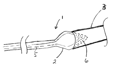
Figure 1 is illustrates the diagnostic catheter designed
for use in the pulmonary vein.
Figure 2 is cross-sectional view of the left atrium with
a guide wire entering the target pulmonary vein through a hole
in the atrial septum.
Figure 3 illustrates the diagnostic balloon inserted into
the target pulmonary vein where the balloon is in the expanded
state thereby in contact with the ostium of the target
pulmonary vein.
4
CA 02403279 2002-09-19
WO 01/26727 PCT/US00/28306
Detailed Description of the Inventions
Figure 1 shows the preferred embodiment of the diagnostic
catheter 1 with the balloon 2 in its inflated state within the
pulmonary vein 3. The catheter body 4 includes a fluid supply
lumen 5 which extends from the proximal end of the catheter to
the balloon, providing a fluid pathway from a fluid reservoir
(not shown) at the proximal end of the catheter to the
balloon.
The diagnostic catheter includes a catheter having a
balloon 2 located at the distal end of the catheter. The
balloon is adapted to contact the pulmonary vein 3 when the
balloon is inflated. The balloon is provided in a conical or
frustoconical shape, allowing it to seat against the flared
shape of the ostium 14 (the ostium is the junction between the
pulmonary vein and the atrium) with the tip of the cone
penetrating into the pulmonary vein and the base of the cone
° having a larger diameter than the pulmonary vein. The base of
the cone should have a diameter chosen in relation to the
estimated predetermined size of the ostium so that it is about
the same or slightly larger than the ostium, and prevents
insertion of the base segment into the pulmonary vein. Pores
6 are located on the distal end of the balloon, on the distal
tip of the conical segment of the balloon. This distal tip is
of the conical segment of the balloon is sized to fit within
the pulmonary vein, such that the fluid is only injected into
the pulmonary vein when the balloon porous distal end is
disposed within the pulmonary vein and fluid is provided to
the balloon. The proximal portion of the balloon is water-
tight and non-porous, so that it does not permit fluid to exit
the balloon near the base of the conical portion of the
balloon. The fluid being injected interrupts electrical
signals, and identification of a pulmonary vein which contains
5
CA 02403279 2002-09-19
WO 01/26727 PCT/US00/28306
an arrythmogenic area is facilitated when the only electrical
signals being interrupted are those of the pulmonary vein and
not of the atrium itself. If the electrical signals of the
atrium are inhibited then it is difficult to diagnose whether
or not the atrial fibrillation has been prevented because the
triggering signal from the pulmonary vein has been prevented
or because the fluid has interrupted the atrial fibrillation
in the atrium itself.
Figure 2 illustrates the method of accessing the
pulmonary vein for placement of the diagnostic catheter. The
left atrium 7 laid open to show the openings to the pulmonary
veins 8, 9 and 10. The left atrium contains inlets of
pulmonary veins which may be accessed from the right atrium
(not shown) by passing a catheter 1 through the atrial septum
11 (with the catheter passing through an access hole cut into
the atrial septum to allow passage of the catheter). To
insert the diagnostic device into the pulmonary vein, access
to the left atrium is first gained by percutaneous insertion
of a catheter into the left atrium. To accomplish this, a
needle catheter is placed through the venous system into the
right atrium, and then penetrates the fossa ovalis (the atrial
septum) to gain access to the left atrium. Access to the
right atrium can be through the femoral vein in the thigh and
the inferior vena cava, or may be through the subclavian vein,
brachial vein or cephalic vein, etc., in the shoulder and arm,
and then through the superior vena cava. A catheter sheath
12, such as a guiding catheter, is advanced over the needle
catheter and is inserted into the left atrium. The needle
catheter is then removed, and the diagnostic catheter is
inserted. The distal end of the catheter is then maneuvered
into the left atrium and into the target pulmonary vein. Once
the catheter is passed through the access hole, then a guide
wire 13 is inserted to locate the target pulmonary vein 8.
6
CA 02403279 2002-09-19
WO 01/26727 PCT/US00/28306
After the target pulmonary vein is located the diagnostic
catheter is inserted into the target pulmonary vein, as in
Figure 3. Once the balloon 2 is placed within the ostium 14
of the target pulmonary vein it is expanded to contact the
pulmonary vein walls. Location of the diagnostic balloon is
confirmed and the balloon is expanded to the point where it
engages the wall of the pulmonary vein. Preferably, the
distal tip of the conical segment of the balloon will be
disposed within the pulmonary vein, as shown, and the base of
the conical segment will be located in the ostium and, when
urged distally, will seat itself within the ostium. Once the
balloon engages the wall of the target pulmonary vein, the
fluid is administered to the target pulmonary vein. After the
fluid has been administered it can be determined whether or
not treatment of the pulmonary vein is necessary.
The method for diagnosing whether treatment of a target
pulmonary vein will prevent atrial fibrillation in a patient
includes several steps. The first step includes monitoring
the patient's EKG to identify an atrial fibrillation. If
atrial fibrillation is not occurring, then the physician
performing the diagnosis may induce an atrial fibrillation.
The second step includes inserting a diagnostic catheter into
the heart and then into the target pulmonary vein, as
described above. The third step includes injecting a
diagnostic fluid into the target pulmonary vein through the
pores of the balloon located at the distal end of the
catheter. The diagnostic fluid is a fluid which disrupts
electrical impulses of heart tissue, and preferably has a very
short-lived effect, so the any disruption in naturally
occurring arrythmia dissipates within an intra-operative time
frame (several second to several minutes) and allows the
physician to test another site. The last step includes
determining whether the target pulmonary vein is where the
7
CA 02403279 2002-09-19
WO 01/26727 PCT/US00/28306
atrial fibrillation is triggered. This is accomplished by
monitoring the EKG of the patient and evaluating whether
atrial fibrillation resolves after injection of the fluid.
After one or more catheters have been swapped out through
the catheter sheath, the catheter sheath is removed, and the
patient is closed. In most cases the opening through the
fossa ovalis is very small and it heals up on its own.
However, it is conceivable that a repair may be required in
some patients using catheter techniques developed for closing
septal defects.
The primary advantage of this device and method is that
no tissue is unnecessarily damaged because the purpose of the
diagnostic catheter is to diagnose the efficacy of a proposed
pulmonary vein treatment prior to performing it. Such
proposed pulmonary vein treatments could include ablation or
stenting procedures.
A number of fluids may be used a diagnostic fluids in
this method. Many pharmacologic agents prevent or slow
conduction and can be used as the fluid which is administered
to the target pulmonary vein. Cold saline or water may be
used, since rapid cooling of electrically active heart tissue
stops conduction of the tissue. Antiarrhythmic agents can
also be used. It is preferred that the antiarrhythmic agent
have a short pharmacodynamic half-life. Drugs that
predominantly affect slow pathway conduction include
digitalis, calcium channel blockers, and beta-blockers. Drugs
that predominantly prolong refractoriness, or time before a
heart cell can be activated, produce conduction block in
either the fast pathway or in accessory AV connections
including the class IA antiarrhythmic agents (quinidine,
procainimide, and disopyrimide) or class IC drugs (flecainide
and propafenone). The class III antiarrhythmic agents
8
CA 02403279 2002-09-19
WO 01/26727 PCT/US00/28306
(sotolol or amiodorone) prolong refractoriness and delay or
block conduction over fast or slow pathways as well as in
accessory AV connections. Temporary blockade of slow pathway
conduction is usually achieved by intravenous administration
of adenosine or verapamil. [Scheinman, Melvin:
Supraventricular Tachycardia: Drug Therapy Versus Catheter
Ablation, Clinical Cardiology Vol. 17, Supp. II -11-II-15
(1994)]. Other agents such as encainide, diltiazem, and
nickel chloride are also available.
While the preferred embodiments of the devices and
methods have been described in reference to the environment in
which they were developed, they are merely illustrative of the
principles of the inventions. Other embodiments and
configurations may be devised without departing from the
spirit of the inventions and the scope of the appended claims.
9