Note: Descriptions are shown in the official language in which they were submitted.
CA 02404041 2002-09-26
WO 01/72123 PCT/USO1/10118
METHODS FOR INCREASING A CYTOTOXIC T LYMPHOCYTE
RESPONSE IN VIVO
GOVERNMENT RIGHTS
The United States Government may have certain rights in this application
pursuant
to National Institutes of Health Grant Nos. AI40682 and AI47078.
FIELD OF THE INVENTION
This invention relates to methods of modulating an immune response, and in
particular to methods of increasing an antigen-specific cytotoxic T lymphocyte
response.
BACKGROUND OF THE INVENTION
Immunostimulatory nucleic acid molecules were initially discovered in the
mycobacterial genome as DNA sequences that selectively enhance NIA cell
activity
(Yamamoto, et al. (1992) Microbiol. Inamunol. 36:983-997). Uptake of
mycobacterial
DNA or has been shown to activate cells of the innate immune system, such as
NK cells
and macrophages and stimulating a type-1 like response (Roman, et al. (1997)
Nature
Med.. 3:849-854). Further, administration of immunostimulatory nucleic acid
molecules
has been shown to induce B cell proliferation (Messina, et al. (1991) J.
Immunol.
147:1759-1764), stimulate production of cytokines, such as interferons (IFNs),
IL-12, IL-
18 and TNF-a (Sparwasser, et al. (1998) Eu~. J. Immunol. 28:2045-2054;
Sparwasser, et
al. (1997) Eur. J. Immunol. 27:1671-1679; Stacey, et al. (1996) J. Immunol.
157:2116-
2122) and up-regulate co-stimulatory receptors (Martin-Orozco, et al. (1999)
Int. Iframun.
11:111-118; Sparwasser, et al. (1998) Eu~. J. Immuraol. 28:2045-2054) by these
cells.
Cytotoxic T Lymphocytes (CTL) axe critical effector cells in the control of
cells
infected with intracellular pathogens and in the control of MHC class I+
tumors. Induction
of CTL is a primary goal of many vaccine strategies. Accumulating evidence
indicates that
one of the pathways of CTL priming in vivo is through "cross-priming," which
involves the
uptake and re-presentation of soluble, exogenous antigens by bone marrow-
derived
antigen-presenting cells (ADCs), e.g., dendritic cells. Depending on the
activation state of
the "cross-presenting" APC, responding T cells can either be activated or
tolerized. The
1
CA 02404041 2002-09-26
WO 01/72123 PCT/USO1/10118
nature of the specific requirements for these disparate outcomes is currently
a topic of
intense interest, as elucidation of such would aid in the design of vaccines
as well as in the
modulation of anti-tumor CTL responses. Current models of cross-priming
consist of two
steps; a "licensing" interaction between antigen presenting cells (APC) and
helper T cells
(T,,), followed by an activating interaction between "licensed" APC and
cytotoxic T
lymphocytes (CTL). Thus, in current models, there is a requirement for T,,
cells in cross-
priming of CTL.
Immunodeficiency can arise from a variety of causes, including primary
immunodeficiencies, e.g., due to a heritable defect; and acquired
immunodeficiencies, e.g.,
due to cancer chemotherapy, or due to infection with a pathogen, e.g., human
immunodeficiency virus. Immunodeficient individuals are more vulnerable to
infectious
diseases than individuals with healthy immune systems. Antibiotics can control
bacterial
infections, but long-term treatment with antibiotics is not without risk of
adverse side
effects. Control of intracellular pathogens, including viruses, bacteria, and
protozoans,
poses a greater challenge for treatment. Immunodeficient individuals may also
be more
vulnerable to growth of cancer cells than individuals with healthy immune
systems.
Treatment of these individuals with conventional anti-cancer therapeutic
agents is not
always feasible.
The current methodologies for inducing a CTL response include vaccines which
use
' attenuated viruses or DNA vaccines. There is a need in the art for more
effective ways of
increasing an antigen-specific CTL response in an individual. Furthermore,
there is a need
in the art for alternative methods of enhancing immune functions in
immunodeficient
individuals. The present invention addresses these needs by providing methods
for
increasing cytotoxic T lymphocyte (CTL) activity. The methods are useful for
increasing
an antigen-specific CTL response in an individual to any soluble antigen. The
methods are
also useful for increasing an antigen-specific CTL response in CD4+-deficient
individuals
and individuals at risk for becoming CD4+ deficient.
SUMMARY OF THE INVENTION
The invention provides methods for T helper-independent activation of an
antigen-
specific cytotoxic T lymphocyte response in an individual. The methods
generally involve
2
CA 02404041 2002-09-26
WO 01/72123 PCT/USO1/10118
administering to an individual an immunostimulatory nucleic acid molecule in
an amount
effective to increase an antigen-specific CTL response in the individual. The
invention
further provides methods for increasing chemokine secretion, which can block
HIV
infection.
The methods are useful for generating both a CTL response and a humoral
response
to a soluble exogenous antigen. Thus, an immunostimulatory nucleic acid
molecule, when
administered together with a soluble, exogenous antigen, results in cross-
priming of CTLs.
Therefore, the methods are useful in generating an immune response,
particularly a CTL
response, to a cell infected with an intracellular pathogen, or to a tumor
cell expressing a
tumor-specific or tumor-associated antigen.
The methods are also useful in treating individuals with a reduced number of
functional CD4+ T cells ("CD4+-deficient individuals" or "CD4+-low
individuals")
compared to normal individuals, e.g. individuals affected by an acquired or
primary
immunodeficiency, as well as those at risk for becoming immunodeficient.
The immunostimulatory nucleic acid molecules may be administered in a
formulation alone, or together with an antigen, e.g., admixed or linked or
conjugated to an
antigen or antigenic epitope. In many embodiments, the antigen is a soluble,
exogenous
antigen. The methods are useful in stimulating, or increasing antigen-specific
CTL activity
to any of a variety of target antigens, e.g., an antigen expressed in a cell,
or an antigen
expressed on the surface of a cell or cell population. In some embodiments,
methods are
provided for increasing CTL activity toward pathogen-infected cells. In other
embodiments, methods are provided for increasing CTL activity toward a tumor
cell.
The invention further provides methods for increasing tumor-specific immunity
in
an individual. The methods generally involve administering to an individual an
immunostimulatory nucleic acid molecule in an amount effective to increase
tumor-specific
immunity in an individual. The methods are useful to treat cancer, e.g., to
inhibit the
growth of cancer cells. The methods are also useful as a preventive measure,
e.g., to inhibit
the probability that cancerous cell growth will occur, or that a previously
treated cancer
will recur. The methods are particularly useful for decreasing a tumor load in
a CD4+ T-
cell deficient individual, and in individuals at risk for becoming CD4+
deficient.
CA 02404041 2002-09-26
WO 01/72123 PCT/USO1/10118
The invention further provides methods of immunizing against and/or treating
an
infectious disease in an individual. The methods generally involve
administering to an
individual an immunostimulatory nucleic acid molecule in an amount effective
to increase
antigen-specific CTL activity. The methods are particularly useful in
immunizing against
and/or treating infectious diseases due to intracellular pathogens. The
methods are also
useful for treating infectious disease in a CD4+ T-cell deficient individual,
and in
individuals at risk for becoming CD4~ deficient.
The present invention further provides compositions and methods for increasing
secretion of a chemokine from a eukaryotic cell, which in turn inhibits
infection of a cell by
pathogens that establish infection in a host, or cause disease by, interaction
with a
chemokine receptor. The methods generally involve contacting a cell with a
composition
comprising an immunostimulatory nucleic acid molecule. Accordingly, the
invention
further provides methods of reducing infection of a cell by a pathogen,
comprising
contacting a cell with an immunostimulatory nucleic acid molecule such that
chemokine
secretion is increased, and infection is reduced. Chemokine secretion may be
antigen
specific, where both immunostimulatory nucleic acid molecule and antigen are
administered, or antigen non-specific, where immunostimulatory nucleic acid
molecule is
administered in the absence of exogenously provided antigen.
Immunostimulatory nucleic acid molecules induce secretion of chemokines that
bind to chemokine receptors. Certain chemokine receptors are used by
pathogenic
microorganisms to enter and infect a cell. Increasing synthesis of such
chemokines serves
to competitively inhibit binding of the pathogenic microorganism to the
chemokine
receptor. Accordingly, in further aspects, the present invention provides
compositions and
methods for increasing secretion of a chemokine from a eukaryotic cell, which
in turn
inhibits infection of a cell by pathogens that establish infection in a host,
or cause disease
by, interaction with a chemokine receptor. The methods generally involve
contacting a cell
with a composition comprising an immunostimulatory nucleic acid molecule.
Accordingly,
the invention further provides methods of reducing infection of a cell by a
pathogen,
comprising contacting a cell with an immunostimulatory nucleic acid molecule
such that
chemokine secretion is increased, and infection is reduced. Chemokine
secretion may be
antigen specific, where both immunostimulatory nucleic acid molecule and
antigen are
4
CA 02404041 2002-09-26
WO 01/72123 PCT/USO1/10118
administered, or antigen non-specific, where immunostimulatory nucleic acid
molecule is
administered in the absence of exogenously provided antigen.
These and other objects, advantages, and features of the invention will become
apparent to those persons skilled in the art upon reading the details of the
invention as more
fully described below.
BRIEF DESCRIPTION OF THE DRAWINGS
Figure 1 is a graph depicting the effect of vaccination with protein-ISS
conjugates
on antigen-specific CTL activity. Animals were injected intradermally with
protein-ISS
conjugate (open squares), using hen egg ovalbumin (OVA) as a model antigen;
OVA + ISS
(open diamonds); pACB-OVA (closed circles); OVA alone (closed triangles);
protein-
mutated ISS conjugate (open diamonds); or target control (open squares). Open
circles
indicate no treatment.
Figures 2A-C are graphs depicting the effects of vaccination with protein-ISS
conjugates on the Thl immune response. Total splenocytes were restimulated as
described
in Example 2, and IFN~y levels (Figure 2A) were measured. IgGl titers (Figure
2B), and
IgG2a titers (Figure 2C) were measured in serum.
Figures 3A-C are graphs depicting MHC Class-I restricted CTL activation in CD4
-
/- by protein-ISS conjugates in wild type (Figure 3A), CD4-l- (Figure 3B), and
MHC class
II-l- (Figure 3C) mice. Mice were injected intradermally on days zero and 14
with either
protein-ISS conjugate (squares); OVA + ISS (diamonds); or OVA alone (circles).
Figures 4A-C are graphs depicting protective immunity conferred by vaccination
with protein-ISS conjugates in preventive and therapeutic models of cancer.
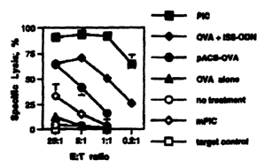
Figure 5 is a graph depicting specific lysis versus effectoraarget ratio for
CTL from
chimeric mice made from wild-type mice and TAP-~- bone-marrow injected with
ISS +
OVA (TAP-~- -~ wt); wild-type mice with wild-type bone marrow injected with
ISS + OVA
(wt -~ wt); wild-type mice injected with ISS + OVA (wt); and wild-type mice
not injected
with ISS + OVA (no treatment).
Figure 6 is a graph depicting specific lysis versus effectoraarget ratio for
CTL from
wild-type injected with ISS + OVA (wt); CD40-j- mice injected with ISS + OVA;
wild-type
mice pre-treated with anti-CD40 ligand and injected with ISS + OVA (wt, anti-
CD40 L);
5
CA 02404041 2002-09-26
WO 01/72123 PCT/USO1/10118
wild-type mice injected with OVA alone (OVA); and wild-type mice not injected
with ISS
+ OVA (no treatment).
Figure 7 is a graph depicting specific lysis versus effectoraarget ratio for
CTL from
wild-type injected with ISS + OVA (wt); wild-type mice pre-treated with anti-
B7-1/-2 and
injected with ISS + OVA (anti B7-1/-2); wild-type mice pre-treated with anti-
B7-1/-2 and
anti-CD40 ligand antibody, and injected with ISS + OVA (anti B7-1/-2; anti
CD40 L);
CD28-'- mice injected with ISS + OVA; and CD28-'- mice pre-treated with anti-
CD40 ligand
antibody, and injected with ISS + OVA (CD28-'-; anti-CD40L).
Figure 8 is a graph depicting specific lysis versus effectoraarget ratio for
CTL from
wild-type injected with ISS + OVA (wt); IL-12-'- mice injected with ISS + OVA;
IL-12-'-
mice pre-treated with anti-CD40L antibody and anti-B7-1/-2 antibody, then
injected with
ISS + OVA (IL-12-'-; anti-CD40L; anti-B7-1/-2); wild-type mice injected with
OVA alone
(wt, OVA alone); and wild-type mice not injected with ISS + OVA (no
treatment).
Figure 9 is a bar graph depicting production of MIP 1 a by mouse splenocytes
in
response to immunization with ISS and gp120.
Figure 10 is a bar graph depicting production of MIP1~3 by mouse splenocytes
in
response to immunization with ISS and gp120.
Figure 11 is a bar graph depicting production of RANTES by mouse splenocytes
in
response to immunization with ISS and gp120.
Figures 12A-D are graphs depicting antigen-specific immunoglobulin (Figure
12A),
cytokine (Figure 12B), and chemokine (Figures 12C and 12D) responses in mice
injected
intradermally with ISS-based gp120 vaccines.
Figures 13A-E are graphs depicting systemic antigen-specific immunoglobulin
(Figure 13A), mucosal antigen-specific immunoglobulin (Figure 13B), cytokine
(Figure
13C), and chemokine (Figures 13D and 13E) responses in mice immunized
intranasally
with ISS-based gp120 vaccines.
Figures 14A-D are graphs depicting splenic and mucosal CTL activity in mice
immunized intradermally (Figure 14A) or intranasally (Figure 14B). Mucosal CTL
activity
from lamina propria (Figure 14C) and Peyer's patch (Figure 14D) lymphocytes
was
determined 12 weeks after initiation of i.n. or i.d. immunization.
6
CA 02404041 2002-09-26
WO 01/72123 PCT/USO1/10118
Figures 15A-C are graphs depicting MHC Class I-restricted IFNy (Figure 15A)
and
chemokine (Figures 15B and 15C) responses in mice immunized intradermally with
ISS-
based gp120 vaccines.
Figures 16A-E are graphs depicting MHC Class I-restricted cytokine (Figures
16A
and 16D), chemokine (Figures 16B, C, and D) and CTL activity (Figure 16E)
elicited by
gp120:ISS vaccination in normal (untreated) or CD4-depleted (treated with anti-
CD4 Ab)
mice.
DETAILED DESCRIPTION OF THE INVENTION
The immune system can react to the presence of a foreign antigen by generating
antigen-specific CD4+ (helper) T cells and CD8+ (cytotoxic) T cells. CD4+ T
cells are
sometimes classified as Thl or Th2, depending on the cytokine profile
produced. The
present invention relates to the observation that an immunostimulatory nucleic
acid
molecule can stimulate an antigen-specific cytotoxic T lymphocyte (CTL)
response even in
the absence of CD4+T helper cells. This observation is counter to the accepted
model of a
requirement for CTL activation. Current models posit that an antigen-
presenting cell
(APC), must have an initial "licensing" interaction with Th cells before the
Th cells can
activate CTL. Previous work describing the APC response to immunostimulatory
nucleic
acid molecule stimulation (e.g., upregulation of cytokines and co-stimulatory
molecules)
suggested that APCs deliver the stimulatory signal to T helper cells. The
present inventors
have made the surprising discovery that, contrary to this model,
immunostimulatory nucleic
acid molecules are capable of increasing an antigen-specific CTL response,
even in the
absence of in a CD4+ T lymphocytes. In addition, immunostimulatory nucleic
acid
molecules increase chemokine secretion, which chemokines are competitive
inhibitors of
HIV for binding to HIV receptors.
Without wishing to be bound by theory, immunostimulatory nucleic acid
molecules
may replace some or all of the "licensing" effects on APCs, indicating that
the Thl
phenotype and CTL activation are independent, rather than linked. Thus, the
immunostimulatory nucleic acid molecule allows the APC to activate directly
antigen-
specific CTL activity. The present inventors' observations thus make it
possible, for the
7
CA 02404041 2002-09-26
WO 01/72123 PCT/USO1/10118
first time, to use immunostimulatory nucleic acid molecules to increase a CTL
response in
CD4+T helper cell-deficient individuals.
Accordingly, the present invention provides methods of inducing or increasing
antigen-specific CTL activity in an individual via cross-presentation,
comprising
administering an immunostimulatory nucleic acid molecule and a soluble
exogenous
antigen to the individual. The methods generally involve administering to an
individual an
immunostimulatory nucleic acid molecule, which may optionally be administered
with an
antigen, particularly a soluble exogenous antigen. The methods can be used to
increase or
induce a CTL response to various undesired cells or cell populations, e.g.,
pathogen-
infected cells, and tumor cells.
The invention further provides methods of inducing CTL activity in CD4-
deficieint
individuals or to individuals with a healthy, intact immune system, but who
are at risk for
becoming CD4+ deficient. The methods generally involve administering to an
individual
an immunostimulatory nucleic acid molecule (which may optionally be
administered with
an antigen, particularly a soluble exogenous antigen), and are useful in
increasing or
inducing a CTL response to various undesired cells or cell populations, e.g.,
pathogen-
infected cells, and tumor cells.
The present invention further provides compositions and methods for increasing
chemokine secretion from a eukaryotic cell, particularly to inhibit infection
of the cell by a
pathogen that establishes infection or otherwise causes disease or symptoms of
disease in a
host by interaction with a chemokine receptor. This aspect of the invention is
based on the
unexpected discovery that certain polynucleotides, termed immunostimulatory
nucleic acid
molecules, can increase secretion of chemokines from cells that normally
produce
chemokines. For example, increased chemokine production, particularly of
chemokines
that bind HIV co-receptors, can reduce the incidence of HIV entry into a cell.
Thus, the
invention further provides methods of reducing susceptibility to infection of
a susceptible
eukaryotic cell by a pathogen, as well as methods for treating an infection by
a pathogen.
The methods involve administering an immunostimulatory nucleic acid molecule
to an
individual to increase secretion of a chemokine that binds to a chemokine
receptor which
serves as a co-receptor for infection by a pathogen. The secreted chemokine
binds to the
8
CA 02404041 2002-09-26
WO 01/72123 PCT/USO1/10118
chemokine receptor and reduces pathogen entry into the cell, or otherwise
reduces the
undesirable effects of pathogen interaction with,the cell.
Before the present invention is described, it is to be understood that this
invention is
not limited to particular embodiments described, as such may, of course, vary.
It is also to
be understood that the terminology used herein is for the purpose of
describing particular
embodiments only, and is not intended to be limiting, since the scope of the
present
invention will be limited only by the appended claims.
Unless defined otherwise, all technical and scientific terms used herein have
the
same meaning as commonly understood by one of ordinary skill in the art to
which this
invention belongs. Although any methods and materials similar or equivalent to
those
described herein can be used in the practice or testing of the present
invention, the preferred
methods and materials are now described. All publications mentioned herein are
incorporated herein by reference to disclose and describe the methods andlor
materials in
connection with which the publications are cited.
It must be noted that as used herein and in the appended claims, the singular
forms
"a", "and", and "the" include plural referents unless the context clearly
dictates otherwise.
Thus, for example, reference to "an immunostimulatory nucleic acid molecule"
includes a
plurality of such molecules and reference to "the tumor cell" includes
reference to one or
more tumor cells and equivalents thereof known to those skilled in the art,
and so forth.
The publications discussed herein are provided solely for their disclosure
prior to
the filing date of the present application. Nothing herein is to be construed
as an admission
that the present invention is not entitled to antedate such publication by
virtue of prior
invention. Further, the dates of publication provided may be different from
the actual
publication dates which may need to be independently confirmed.
Definitions
The terms "oligonucleotide," "polynucleotide," and "nucleic acid molecule",
used
interchangeably herein, refer to a polymeric forms of nucleotides of any
length, either
ribonucleotides or deoxyribonucleotides. Thus, this term includes, but is not
limited to,
single-, double-, or mufti-stranded DNA or RNA, genomic DNA, cDNA, DNA-RNA
hybrids, or a polymer comprising purine and pyrimidine bases or other natural,
chemically
9
CA 02404041 2002-09-26
WO 01/72123 PCT/USO1/10118
or biochemically modified, non-natural, or derivatized nucleotide bases. The
backbone of
the polynucleotide can comprise sugars and phosphate groups (as may typically
be found in
RNA or DNA), or modified or substituted sugar or phosphate groups.
Alternatively, the
backbone of the polynucleotide can comprise a polymer of synthetic subunits
such as
phosphoramidites, and/or phosphorothioates, and thus can be an
oligodeoxynucleoside
phosphoramidate or a mixed phosphoramidate-phosphodiester oligomer. Peyrottes
et al.
(1996) Nucl. Acids Res. 24:1841-1848; Chaturvedi et al. (1996) Nucl. Acids
Res. 24:2318-
2323. The polynucleotide may comprise one or more L-nucleosides. A
polynucleotide
may comprise modified nucleotides, such as methylated nucleotides and
nucleotide
analogs, uracyl, other sugars, and linking groups such as fluororibose and
thioate, and
nucleotide branches. The sequence of nucleotides may be interrupted by non-
nucleotide
components. A polynucleotide may be further modified after polymerization,
such as by
conjugation with a labeling component. Other types of modifications included
in this
definition are caps, substitution of one or more of the naturally occurring
nucleotides with
an analog, and introduction of means for attaching the polynucleotide to
proteins, metal
ions, labeling components, other polynucleotides, or a solid support.
The terms "polypeptide," "peptide," and "protein", used interchangeably
herein,
refer to a polymeric form of amino acids of any length, which can include
coded and non-
coded amino acids, chemically or biochemically modified or derivatized amino
acids, and
polypeptides having modified peptide backbones. The term includes polypeptide
chains
modified or derivatized in any manner, including, but not limited to,
glycosylation,
formylation, cyclization, acetylation, phosphorylation, and the like. The term
includes
naturally-occurring peptides, synthetic peptides, and peptides comprising one
or more
amino acid analogs. The term includes fusion proteins, including, but not
limited to, fusion
proteins with a heterologous amino acid sequence, fusions with heterologous
and
homologous leader sequences, with or without N-terminal methionine residues;
immunologically tagged proteins; and the like.
The term "tumor-associated antigen" is a term well understood in the art, and
refers
to surface molecules that are differentially expressed in tumor cells relative
to non-
cancerous cells of the same cell type. As used herein, "tumor-associated
antigen" includes
not only complete tumor-associated antigens, but also epitope-comprising
portions
CA 02404041 2002-09-26
WO 01/72123 PCT/USO1/10118
(fragments) thereof. A tumor-associated antigen (TAA) may be one found in
nature, or
may be a synthetic version of a TAA found in nature, or may be a variant of a
naturally-
occurnng TAA, e.g., a variant which has enhanced immunogenic properties.
"A peptide associated with a pathogenic organism," as used herein, is a
peptide (or
fragment or analog thereof) that is normally a part of a pathogenic organism,
or is produced
by a pathogenic organism. Generally, a peptide associated with a pathogenic
organism is
one that is recognized as foreign by a normal individual with a healthy,
intact immune
system, e.g., the peptide is displayed together with an MHC Class I molecule
on the surface
of a cell, where it is recognized by a CD8+ lymphocyte.
The terms "antigen" and "epitope" are well understood in the art and refer to
the
portion of a macromolecule which is specifically recognized by a component of
the
immune system, e.g., an antibody or a T-cell antigen receptor. As used herein,
the term
"antigen" encompasses antigenic epitopes, e.g., fragments of an antigen which
are antigenic
epitopes. Epitopes are recognized by antibodies in solution, e.g. free from
other molecules.
Epitopes are recognized by T-cell antigen receptor when the epitope is
associated with a
class I or class II major histocompatibility complex molecule.
The terms "preventing," "reducing," and "inhibiting," used interchangeably
herein
in the context of pathogen infection refer to reducing the incidence of
pathogen infection of
a cell which is susceptible to infection by the pathogen. Reducing pathogen
infection refers
to reducing any parameter or event which leads to pathogen entry into a cell,
including, but
not limited to, reducing co-receptor-mediated fusion; reducing entry of the
pathogen into
the cell; reducing binding of the pathogen to a cell-surface chemokine
receptor; and
reducing binding of the pathogen to a cell-surface CD4 molecule. The terms
also refer to
reducing susceptibility of a cell to infection by a pathogen. The terms also
refer to
reducing any undesired effect of binding of a pathogen to the cell. As used
herein, "a cell
which is susceptible to infection by a pathogen" is a cell which can be
infected by a
pathogen that establishes infection or otherwise causes disease or symptoms of
disease in a
host by interaction with a chemokine receptor.
As used herein the term "isolated" is meant to describe a compound of interest
(e.g.,
a virus, a peptide, etc.) that is in an environment different from that in
which the compound
naturally occurs. "Isolated" is meant to include compounds that are within
samples that are
11
CA 02404041 2002-09-26
WO 01/72123 PCT/USO1/10118
substantially enriched for the compound of interest and/or in which the
compound of
interest is partially or substantially purified.
As used herein, the term "substantially purified" refers to a compound that is
removed from its natural environment and is at least 60% free, preferably 75%
free, and
most preferably 90% free from other components with which it is naturally
associated.
The terms "immunomodulatory nucleic acid molecule," "immunostimulatory
nucleic acid molecule," "ISS," "ISS-PN," and "ISS-ODN," are used
interchangeably herein
to refer to a polynucleotide that comprises at least one immunomodulatory
nucleic acid
moiety. The terms "immunomodulatory," and "immunostimulatory," as used herein
in
reference to a nucleic acid molecule, refer to the ability of a nucleic acid
molecule to
modulate an immune response in a vertebrate host. In particular, these terms
refer to the
ability of an immunostimulatory nucleic acid molecule to increase an immune
response in a
vertebrate host, particularly to increase a CTL response, particularly an
antigen-specific
CTL response.
The terms, "increasing," "inducing," and "enhancing," used interchangeably
herein
with reference to a CTL response, refer to any increase in a CTL response over
background, and include inducing a CTL response over an absence of a
measurable CTL
response, or increasing CTL response over a previously measurable CTL
response.
The terms "CD4+-deficient" and "CD4+-low" are used interchangeably herein,
and,
as used herein, refer to a state of an individual in whom the number of CD4'~
T lymphocytes
is reduced compared to an individual with a healthy, intact immune system. CD4
deficiency includes a state in which the number of functional CD4+T
lymphocytes is less
than about 600 CD4+T cells/mm3 blood; a state in which the number of
functional CD4+T
cells is reduced compared to a healthy, normal state for a given individual;
and a state in
which functional CD4+ T cells are completely absent.
As used herein, a "CD4+-deficient individual" is one who has a reduced number
of
functional CD4~-T cells, regardless of the reason, when compared to an
individual having a
normal, intact immune system. In general, the number of functional CD4''~-T
cells that is
within a normal range is known for various mammalian species. In human blood,
e.g., the
number of functional CD4k-T cells which is considered to be in a normal range
is from
about 600 to about 1500 CD4+-T cells/mm3 blood. An individual having a number
of
12
CA 02404041 2002-09-26
WO 01/72123 PCT/USO1/10118
CD4+-T cells below the normal range, e.g., below about 600/mm3, may be
considered
"CD4+-deficient." Thus, a CD4+-deficient individual may have a low CD4+ T cell
count, or
even no detectable CD4+ T cells. A CD4+-deficient individual includes an
individual who
has a lower than normal number of functional CD4+-T cells due to a primary or
an acquired
immunodeficiency.
A "functional CD4+-T cell" is a term well understood in the art and refers to
a
CD4~-T cell which is capable of providing T cell help, directly or indirectly,
to effect one
or more of the following responses: CTL activation; antibody production;
macrophage
activation; mast cell growth; and eosinophil growth and differentiation.
As used herein, the terms "immunodeficient," "immunosuppressed," and
"immunocompromised," used interchangeably herein, refer to a state of a CD4+-
deficient
individual.
As used herein, the term "soluble exogenous antigen" refers to an antigen that
a cell
takes up from its environment, and processes intracellularly. A "soluble
exogenous
antigen" is distinguished from an antigen that is synthesized intracellularly
(e.g., translated
in the cell cytoplasm).
As used herein, the terms "treatment", "treating", and the like, refer to
obtaining a
desired pharmacologic and/or physiologic effect. The effect may be
prophylactic in terms
of completely or partially preventing a disease or symptom thereof and/or may
be
therapeutic in terms of a partial or complete cure for a disease and/or
adverse affect
attributable to the disease. "Treatment", as used herein, covers any treatment
of a disease
in a mammal, particularly in a human, and includes: (a) preventing the disease
from
occurring in a subject which may be predisposed to the disease but has not yet
been
diagnosed as having it; (b) inhibiting the disease, i.e., arresting its
development; and (c)
relieving the disease, e.g., causing regression of the disease, e.g., to
completely or partially
remove symptoms of the disease.
The term "biological sample" encompasses a variety of sample types obtained
from
an organism and can be used in a diagnostic or monitoring assay. The term
encompasses
blood and other liquid samples of biological origin, solid tissue samples,
such as a biopsy
specimen or tissue cultures or cells derived therefrom and the progeny
thereof. The term
encompasses samples that have been manipulated in any way after their
procurement, such
13
CA 02404041 2002-09-26
WO 01/72123 PCT/USO1/10118
as by treatment with reagents, solubilization, or enrichment for certain
components. The
term encompasses a clinical sample, and also includes cells in cell culture,
cell
supernatants, cell lysates, serum, plasma, biological fluids, and tissue
samples.
The terms "cancer", "neoplasm", "tumor", and "carcinoma", are used
interchangeably herein to refer to cells which exhibit relatively autonomous
growth, so that
they exhibit an aberrant growth phenotype characterized by a significant loss
of control of
cell proliferation. Cancerous cells can be benign or malignant.
By "individual" or "host" or "subject" or "patient" is meant any mammalian
subject
for whom diagnosis, treatment, or therapy is desired, particularly humans.
Other subjects
may include cattle, dogs, cats, guinea pigs, rabbits, rats, mice, horses, and
so on.
METHODS OF INCREASING AN ANTIGEN-SPECIFIC CTL RESPONSE IN VIVO
The invention provides methods for induction of a CTL response to any
exogenous
soluble antigen via a process of cross-presentation. In addition, the
invention provides
methods for T helper-independent activation of an antigen-specific cytotoxic T
lymphocyte
response in an individual; methods for decreasing the number of infectious
pathogens in an
individual; methods for decreasing tumor load in an individual; and methods of
treating an
infectious disease in an individual. The methods generally involve
administering to an
individual an immunostimulatory nucleic acid molecule (either alone or in
combination
with one or more antigens) in an amount effective to increase an antigen-
specific CTL
response in the individual and/or to decrease a tumor load in an individual
andJor to prevent
and/or reduce an infectious disease in an individual.
During an immune response, an antigen presenting cell (APC) presents antigen
to T
lymphocytes, and the result may be production of antigen-specific antibody,
and activation
of antigen-specific cytotoxic cells which serve to destroy cells displaying
foreign antigen
on their cell surface. It was previously believed that CD4+ cells were
required for CTL
activation. Without wishing to be bound by theory, immunostimulatory nucleic
acid
molecules may bypass the requirement for CD4+ cells, and may induce APC's to
activate a
CTL response directly, even in the absence of CD4+ cells, or in the presence
of an
insufficient number of functional CD4+ cells. The present invention provides a
means for
increasing antigen-specific CTL activity even in the absence of CD4+cells.
14
CA 02404041 2002-09-26
WO 01/72123 PCT/USO1/10118
The results presented in the Examples also demonstrate that an
immunostimulatory
nucleic acid molecule can, when administered together with a soluble exogenous
antigen,
increase both an antigen-specific CTL response to the soluble antigen and
cross-reacting
epitopes, as well as an antigen-specific humoral response to the soluble
antigen and cross-
reacting epitopes.
T lymphocytes capable of antigen recognition are generally classified as
"CD4''~" or
"CD8+," depending on whether a CD4 or a CD8 molecule is displayed on the cell
surface.
CD4~ cells recognize exogenously-produced antigen which has been taken up by
an antigen,
presenting cell (APC), processed, and displayed on the APC cell surface
together with a
major histocompatibility complex (MHC) class II molecule. In general, CD4+ T
cells
provide the signals to activate other cells, e.g., CD4+ T cells activate
CD8+cells, to induce
B cell to produce antibodies, or to activate macrophages. In contrast, CD8+
cells are
cytotoxic, and recognize antigen produced from within a cell and displayed on
the cell
surface together with an MHC Class I molecule.
In general, CD4''- helper T (Th) cells are divided into broad groups based on
their
specific profiles of cytokine production: Thl, Th2, and ThO. "Thl" cells are T
lymphocytes that release predominantly the cytokines IL-2 and IFN-y, which
cytokines in
turn promote T cell proliferation, facilitate macrophage activation, and
enhance the
cytolytic activity of natural killer (NIA) cells and antigen-specific
cytotoxic T cells (CTL).
In contrast, the cytokines predominantly released by Th2 cells are IL-4, IL-5,
and IL-10.
IL-4 and IL-5 are known to mediate antibody isotype switching towards IgE or
IgA
response, and promote eosinophil recruitment, skewing the immune system toward
an
"allergic" type of response. Th0 cells release a set of cytokines with
characteristics of both
Thl-type and Th2-type responses. While the categorization of T cells as Thl,
Th2, or Th0
is helpful in describing the differences in immune response, it should be
understood that it
is more accurate to view the T cells and the responses they mediate as forming
a
continuum, with Thl and Th2 cells at opposite ends of the scale, and Th0 cells
providing
the middle of the spectrum. Therefore, it should be understood that the use of
these terms
herein is only to describe the predominant nature of the immune response
elicited, and is
not meant to be limiting to an immune response that is only of the type
indicated. Thus, for
CA 02404041 2002-09-26
WO 01/72123 PCT/USO1/10118
example, reference to a "type-1" or "Thl" immune response is not meant to
exclude the
presence of a "type-2" or "Th2" immune response, and vice versa.
The immunostimulatory nucleic acid molecule may be administered before,
simultaneously with (e.g., in admixture with antigen, or covalently or non-
covalently
bound, directly or via a linker, to an antigen or antigenic epitope), or after
the subject is
exposed to antigen. Exposure to antigen may occur by intentionally introducing
the
antigen into the subject via a systemic or mucosal route, e.g., intranasally,
intrarectally,
intravenously, subcutaneously, intradermally, or intraperitoneally, and the
like, e.g., by a
clinician. Alternatively, exposure to antigen may occur accidentally or
naturally (e.g., by
happenstance), e.g., by the usual routes of exposure of a subject to plant,
animal, and other
antigens, such as by inhalation, accidental skin exposure, ingestion, and the
like.
Methods of T helper-independent activation of an antigen-specific CTL
response
The present invention provides methods of increasing an antigen-specific CTL
response in an individual, comprising administering a formulation comprising
an
immunostimulatory nucleic acid molecule to the individual.
An antigen-specific CTL response may be directed to an intracellular pathogen,
such as a virus, an intracellular bacterium, fungus, or protozoan; or may be
directed to a
tumor-associated antigen. Pathogens include microorganisms that are commonly
pathogenic in healthy individuals with an intact immune system, as well as
microorganisms
that cause opportunistic infections in individuals who are immunocompromised.
In general, the methods for increasing an antigen-specific CTL response are
effective to increase an antigen-specific CTL response by at least about 10%,
at least about
20%, at least about 25%, at least about 50%, at least about 75%, at least
about 100% (or
two-fold), at least about 5-fold, at least about 10-fold, at least about 20-
fold, at least about
50-fold, or at least about 100-fold or more, when compared to a suitable
control. Thus, in
these embodiments, an "effective amount" of an immunostimulatory nucleic acid
molecule
is an amount sufficient to increase an antigen-specific CTL response in an
individual by at
least about 10%, at least about 20%, at least about 25%, at least about 50%,
at least about
75%, at least about 100% (or two-fold), at least about 5-fold, at least about
10-fold, at least
16
CA 02404041 2002-09-26
WO 01/72123 PCT/USO1/10118
about 20-fold, at least about 50-fold, or at least about 100-fold or more,
when compared to
a suitable control. In an experimental animal system, a suitable control may
be a
genetically identical animal not treated with the immunostimulatory nucleic
acid molecule.
In non-experimental systems, a suitable control may be the level of antigen-
specific CTL
present before administering the immunostimulatory nucleic acid molecule.
Other suitable
controls may be a placebo control.
In some embodiments, an immunostimulatory nucleic acid molecule is co-
administered with a soluble exogenous antigen. In certain embodiments, the
immunostimulatory nucleic acid molecule and soluble exogenous antigen are
admixed with
one another; in certain other embodiments, the immunostimulatory nucleic acid
molecule
and soluble exogenous antigen are linked to one another (e.g., either
covalently or non-
covalently, e.g., to place the antigen and the immunostimulatory nucleic acid
molecule in
spatial proximity at a distance sufficient to provide for the desired
immunomodulatory ,
effect). Co-administration of an immunostimulatory nucleic acid molecule and a
soluble
exogenous antigen results in an increase in both antigen-specific CTL response
and
antigen-specific humoral response. An antigen-specific CTL response to a
soluble
exogenous antigen encompasses a CTL response to an epitope that is shared
between the
soluble exogenous antigen and another protein.
Whether an antigen-specific CTL response is increased can be determined using
any
of a number of assays known in the art, including, but not limited to,
measuring specific
lysis by CTL of target cells expressing antigen on their surface, which target
cells have
incorporated a detectable label which is released from target cells upon
lysis, and can be
measured, using, e.g., an assay such as that described in the Examples, a S'Cr-
release assay,
a lanthanide fluorescence-based cytolysis assay, and the like.
An immunostimulatory nucleic acid molecule can also elicit production of IFNy
in
CD4-deficient individuals. Thus, in some embodiments, the invention provides
methods of
increasing IFNy production in a CD4+ T cell deficient individual, comprising
administering a formulation comprising an immunostimulatory nucleic acid
molecule to the
individual. In many embodiments, an immunostimulatory nucleic acid molecule is
administered together with (e.g., in admixture, as a conjugate, etc.) an
antigen. In these
embodiments, IFNy is produced in an antigen-specific manner, e.g., IFNy is
produced in
17
CA 02404041 2002-09-26
WO 01/72123 PCT/USO1/10118
response to the antigen administered, to an epitope contained on the
administered antigen,
or to a cross-reactive antigen or epitope, but not to an unrelated antigen.
IFN~y is produced
in an antigen-specific manner by CD8+ cells in CD4~-deficient individuals. In
the context
of IFNy production, an "effective amount" of an immunostimulatory nucleic acid
molecule
is an amount sufficient to increase production of IFNy in an individual by at
least about
10%, at least about 20%, at least about 25%, at least about 50%, at least
about 75%, at least
about 100% (or two-fold), at least about 5-fold, at least about 10-fold, at
least about 20-
fold, at least about 50-fold, or at least about 100-fold or more, when
compared to a suitable
control, as described above.
Whether IFNy production is increased can be determined using any known assay.
A non-limiting example of such an assay is an enzyme-linked immunosorbent
assay, using
antibody specific for IFNy.
Methods of decreasing tumor load in an individual
The present invention fizrther provides methods for decreasing tumor load in
an
individual, comprising administering a formulation comprising an
immunostimulatory
nucleic acid molecule to the individual, in an amount effective to reduce the
tumor load.
The methods are effective to reduce a tumor load by at least about 5%, at
least
about 10%, at least about 20%, at least about 25%, at least about 50%, at
least about 75%,
at least about 85%, or at least about 90%, up to total eradication of the
tumor, when
compared to a suitable control. Thus, in these embodiments, an "effective
amount" of an
immunostimulatory nucleic acid molecule is an amount sufficient to reduce a
tumor load by
at least about 5%, at least about 10%, at least about 20%, at least about 25%,
at least about
50%, at least about 75%, at least about 85%, or at least about 90%, up to
total eradication
of the tumor, when compared to a suitable control. In an experimental animal
system, a
suitable control may be a genetically identical animal not treated with the
immunostimulatory nucleic acid molecule. In non-experimental systems, a
suitable control
may be the tumor load present before administering the immunostimulatory
nucleic acid
molecule. Other suitable controls may be a placebo control.
Whether a tumor load has been decreased can be determined using any known
method, including, but not limited to, measuring solid tumor mass; counting
the number of
18
CA 02404041 2002-09-26
WO 01/72123 PCT/USO1/10118
tumor cells using cytological assays; fluorescence-activated cell sorting
(e.g., using
antibody specific for a tumor-associated antigen); computed tomography
scanning,
magnetic resonance imaging, and/or x-ray imaging of the tumor to estimate
and/or monitor
tumor size; measuring the amount of tumor-associated antigen in a biological
sample, e.g.,
blood; and the like.
Methods of preventing or treating an infectious disease in an individual
The present invention further provides methods for preventing or treating an
infectious disease in an individual, comprising administering a formulation
comprising an
immunostimulatory nucleic acid molecule to the individual, in an amount
effective to
prevent or treat the disease. The methods are particularly useful for
preventing or treating
infectious diseases caused by intracellular pathogens, such as viruses,
intracellular bacteria,
fungi and parasites (e.g. protozoans). In particular, opportunistic infections
can be treated
using the methods of the invention.
"Preventing an infectious disease," as used herein, refers to reducing the
likelihood
that an individual, upon infection by a pathogenic organism, will exhibit the
usual
symptoms of a disease caused by a pathogenic organism.
"Treating an infectious disease," as used herein, encompasses reducing the
number
of pathogenic agents in the individual (e.g., reducing viral load) and/or
reducing a
parameter associated with the infectious disease, including, but not limited
to, reduction of
a level of a product produced by the infectious agent (e.g., a toxin, an
antigen, and the like);
and reducing an undesired physiological response to the infectious agent
(e.g., fever, tissue
edema, and the like).
The methods are effective to treat an infectious disease by at least about 5%,
at least
about 10%, at least about 20%, at least about 25%, at least about 50%, at
least about 75%,
at least about 85%, or at least about 90%, up to total eradication of the
infecting pathogen
and/or an associated parameter, when compared to a suitable control. Thus, in
these
embodiments, an "effective amount" of an immunostimulatory nucleic acid
molecule is an
amount sufficient to treat an infectious disease, e.g., to reduce the number
of pathogens
and/or reduce a parameter associated with the presence of a pathogen, by at
least about 5%,
at least about 10%, at least about 20%, at least about 25%, at least about
50%, at least about
19
CA 02404041 2002-09-26
WO 01/72123 PCT/USO1/10118
75%, at least about 85%, or at least about 90%, up to total eradication of the
infectious
disease, when compared to a suitable control. In an experimental animal
system, a suitable
control may be a genetically identical animal not treated with the
immunostimulatory
nucleic acid molecule. In non-experimental systems, a suitable control may be
the
infectious disease present before administering the immunostimulatory nucleic
acid
molecule. Other suitable controls may be a placebo control.
Whether an infectious disease has been treated can be determined in any of a
number of ways, including but not limited to, measuring the number of
infectious agents in
the individual being treated, using methods standard in the art; measuring a
parameter
caused by the presence of the pathogen in the individual, e.g., measuring the
levels of a
toxin produced by the pathogen; measuring body temperature; measuring the
level of any
product produced by the pathogen; measuring or assessing any undesired
physiological
parameter associated with the presence of an infectious agent in an
individual. Measuring
the number of infectious agents can be accomplished by any conventional assay,
such as
those typically used in clinical laboratories, for evaluating numbers of
pathogens present in
a biological sample obtained from an individual. Such methods have been amply
described
in the literature, including, e.g., Medical Microbiology 3rd Ed., (1998) P.R.
Murray et al.,
eds. Mosby-Year Book, Inc. A level of a product, including a toxin, produced
by a
pathogen can be measured using conventional immunological assays, using
antibody which
detects the product, including, but not limited to enzyme-linked immunosorbent
assays
(ELISA), radioimmunoassays. Other assays, include in vivo assays for toxins.
Subiects suitable for treatment with the methods of the invention
Subj ects suitable for treatment with the methods of the invention include an
individual who has been infected with a pathogenic microorganism; an
individual who is
susceptible to infection by a pathogenic microorganism, but who has not yet
been infected;
and an individual who has a tumor.
Subjects particularly suitable for treatment with the methods of the invention
include CD4+-deficient individuals, e.g., individuals who have lower than
normal numbers
of functional CD4+ T lymphocytes. As used herein, the term "normal individual"
refers to
an individual having CD4+ T lymphocyte levels and functions) within the normal
range in
CA 02404041 2002-09-26
WO 01/72123 PCT/USO1/10118
the population, for humans, typically 600 to 1500 CD4+ T lymphocytes per mm3
blood.
CD4''--deficient individuals individuals who have an acquired
immunodeficiency, or a
primary immunodeficiency. An acquired immunodeficiency may be a temporary CD4+
deficiency, such as one caused by radiation therapy, or chemotherapy, as
described below.
Also suitable for treatment with the methods of the invention are individuals
with
healthy, intact immune systems, but who are at risk for becoming CD4+
deficient ("at-risk"
individuals). At-risk individuals include, but are not limited to, individuals
who have a
greater likelihood than the general population of becoming CD4~ deficient.
Individuals at
risk for becoming CD4''- deficient include, but are not limited to,
individuals at risk for HIV
infection due to sexual activity with HIV-infected individuals; intravenous
drug users;
individuals who may have been exposed to HIV-infected blood, blood products,
or other
HIV-contaminated body fluids; babies who are being nursed by HIV-infected
mothers;
individuals who were previously treated for cancer, e.g., by chemotherapy or
radiotherapy,
and who are being monitored for recurrence of the cancer for which they were
previously
treated; and individuals who have undergone bone marrow transplantation or any
other
organ transplantation.
A reduction of normal levels and/or function of CD4~ T lymphocytes compared to
a
normal individual can result from a variety of disorders, diseases infections
or conditions,
including immunosuppressed conditions due to leukemia, renal failure;
autoimmune
disorders, including, but not limited to, systemic lupus erythematosus,
rheumatoid arthritis,
auto-immune thyroiditis, scleroderma, inflammatory bowel disease; various
cancers and
tumors; viral infections, including, but not limited to, human
immunodeficiency virus
(HIV); bacterial infections; and parasitic infections.
A reduction of normal levels and/or function of CD4+ T lymphocytes compared to
a
normal individual can also result from an immundeficiency disease or disorder
of genetic
origin, or due to aging. Examples of these are immunodeficiency diseases
associated with
aging and those of genetic origin, including, but not limited to,
hyperimmunoglobulin M
syndrome, CD40 ligand deficiency, IL-2 receptor deficiency, y-chain
deficiency, common
variable immunodeficiency, Chediak-Higashi syndrome, and Wiskott-Aldrich
syndrome.
A reduction of normal levels and/or function of CD4+ T lymphocytes compared to
a
normal individual can also result from treatment with specific pharmacological
agents,
21
CA 02404041 2002-09-26
WO 01/72123 PCT/USO1/10118
including, but not limited to chemotherapeutic agents to treat cancer; certain
immunotherapeutic agents; radiation therapy; immunosuppressive agents used in
conjunction with bone marrow transplantation; and immunosuppressive agents
used in
conjunction with organ transplantation.
Accordingly, individuals who may benefit from treatment using the methods of
the
present invention include, but are not limited to, individuals with various
cancers,
including, but not limited to, leukemia, Hodgkin's disease, lung cancer, colon
cancer,
gliomas, renal cell carcinoma, etc.; individuals with various bacterial,
protozoan, and viral
infections, including, but not limited to, patients with acquired
immunodeficiency
syndrome (AIDS), cytomegalovirus infections, malaria, Epstein Barn Virus,
etc.;
individuals infected with intracellular pathogens, including, but not limited
to, individuals
with leprosy, tuberculosis, leishrnania; individuals with autoimmune diseases,
including,
but not limited to systemic lupus erythematosus, rheumatoid arthritis,
sclerodenna,
autoirnmune thyroiditis; and individuals who have undergone stem cell
replacement
therapy, organ transplantation, bone marrow transplant, chemotherapy,
radiotherapy and
the like.
METHODS OF INCREASING CHEMOKINE SECRETION
The present invention provides methods for increasing chemokine production and
secretion by a cell. The methods are useful for treating various disorders
which are
mediated by cells expressing chemokine receptors. In some embodiments, the
methods are
carried out in vitro or ex vivo. In these embodiments, the methods generally
involve
contacting the cell with an immunostimulatory nucleic acid molecule in an
amount
sufficient to increase secretion of a chemokine. In other embodiments, the
methods are
earned out iya vivo. In these embodiments, the methods generally involve
administering to
an individual an immunostimulatory nucleic acid molecule in an amount
sufficient to
increase secretion of a chemokine. In some embodiments, the invention provides
methods
for increasing chemokine production and secretion in an antigen non-specific
manner. In
these embodiments, cells are contacted with, or individuals are administered
with,
immunostimulatory nucleic acid molecule without antigen. In other embodiments,
the
invention provides methods for increasing chemokine production and secretion
in an
22
CA 02404041 2002-09-26
WO 01/72123 PCT/USO1/10118
antigen-specific manner. In these;embodiments, immunostimulatory nucleic acid
molecule
and antigen are brought into contact with cells, or administered to an
individual.
The methods of the invention increase secretion of a chemokine from a cell
that
normally produces a chemokine, particularly those cells that are susceptible
to infection by
a pathogen. Cells that normally produce chemokines include, but are not
limited to, T
lymphocytes, macrophages, monocytes, dendritic cells and related antigen-
presenting cells
(APCs), B lymphocytes, epithelial cells, fibroblasts, endothelial cells,
basophils,
eosinophils, neutrophils, natural killer cells, and bone marrow stem cells.
Chemokines whose secretion is increased by contacting a cell that normally
produces a chemokine with an immunostimulatory nucleic acid molecule include,
but are
not limited to, MIP-la, and MIP-1(3. Other chemokines which may have increased
secretion in response to immunostimulatory nucleic acid include, but are not
necessarily
limited to, RANTES, SDF-1, MCP-1, MCP-2, MCP-3, MCP-4, eotaxin, eotaxin-
2,I-309/TCA3, ATAC, HCC-1, HCC-2, HCC-3, LARC/MIP-3a, PARC, TARC, CK(34,
CK(36, CK/37, CK[38, CK(39, CK(311, CK(312, and CK(313, C10, an interleukin-8
(IL-8)
family member; GROa, GROG, GROy, mouse KC, mouse MIP-2, ENA-78, GCP-2,
PBP/CTAPIII/(3-TG/NAP-2, IP-10/mouse CRG, Mig, PBSF/SDF-1, a member of the
platelet factor 4 (PF4) family, lymphotactin, or an equivalent in any
mammalian species of
any of the foregoing.
In some embodiments, the cells are susceptible to infection with a pathogen
that
exploits a chemokine receptor to establish infection and/or cause disease
symptoms, e.g., an
immunodeficiency virus. In some of these embodiments, the cells are
macrophages and/or
monocytes and/or T cells. In particular embodiments, the cells are macrophages
and/or
monocytes, and/or T lymphocytes, and the chemokines are MIP-1 a, and/or MIP-1
(3, and/or
RANTES.
In some embodiments, methods are provided for increasing chemokine secretion
in
an antigen non-specific manner. In these embodiments, an immunostimulatory
nucleic acid
molecule is brought into contact with a cell, or administered to an
individual, in the absence
of exogenously provided antigen, i.e., antigen is not intentionally introduced
into the
individual, either before, simultaneously with, or after introduction of the
immunostimulatory nucleic acid molecule into the individual.
23
CA 02404041 2002-09-26
WO 01/72123 PCT/USO1/10118
In particular embodiments, production and secretion of a chemokine is antigen-
specific. The term "antigen-specific" is one well understood in the art, and
refers to
chemokine production in response to the antigen with which the individual is
immunized,
or to closely related ("cross-reactive") antigens, e.g., antigens that share
one or more
epitopes with the immunizing antigen. In in vivo embodiments, the method
generally
involves administering to an individual an immunostimulatory nucleic acid
molecule and
an antigen, wherein the immunostimulatory nucleic acid molecule is
administered in an
amount sufficient to increase secretion of a chemokine in an antigen-specific
manner. In ita
vitro or ex vivo embodiments, the method generally involves contacting a cell
with an
immunostimulatory nucleic acid molecule and an antigen, wherein the cell is
contacted
with immunostimulatory in an amount sufficient to increase secretion of a
chemokine in an
antigen-specific manner.
The immunostimulatory nucleic acid molecule and the antigen may be
administered
substantially simultaneously, or the immunostimulatory nucleic acid molecule
may be
administered before or after the antigen. Generally, the immunostimulatory
nucleic acid
molecule and the antigen are administered within about 72 hours, about 48
hours, about 24
hours, about 12 hours, about 8 hours, about 4 hours, about 2 hours, about 1
hour, or about
30 minutes or less, of each other.
Antigen may be administered separately from the immunostimulatory nucleic acid
molecule, in admixture with immunostimulatory nucleic acid molecule, or the
immunostimulatory nucleic acid and antigen can be proximately associated with
(e.g.,
conjugated or brought into spatial proximation by other means, as described in
more detail
below) to one or more immunostimulatory nucleic acid molecules. Generally, and
most
preferably, an immunomodulatory nucleic acid and an antigen are proximately
associated at
a distance effective to enhance the immune response generated compared to the
administration of the ISS and antigen as an admixture. For a detailed
discussion of method
for proximate association of a polynucleotide and an antigen see, e.g., PCT
Publication WO
00/21556, incorporated herein by reference.
Whether chemokine secretion is increased in an antigen-specific manner can be
readily determined by those skilled in the art using standard methods. As one
non-limiting
example, splenocytes from an individual immunized with immunostimulatory
nucleic acid
24
CA 02404041 2002-09-26
WO 01/72123 PCT/USO1/10118
molecule plus antigen are cultured in the presence of the immunizing antigen,
and secretion
of chemokines measured using any known method, as described below.
In vitro and ex vivo methods of the invention comprise contacting a cell that
normally produces a chemokine with an immunostimulatory nucleic acid molecule.
In
these embodiments, contacting a cell that normally produces a chemokine with
an
immunostimulatory nucleic acid molecule increases chemokine secretion from the
cell by
at least about 10%, at least about 25%, at least about 30%, at least about
50%, at least about
75%, at least about 100% (or two-fold), at least about five fold, at least
about 10 fold, at
least about 15 fold, at least about 25 fold, at least about 50 fold, at least
about 75 fold, at
least about 100 fold, at least about 200 fold, at least about 300 fold, at
least about 400 fold,
at least about 500 fold, at least about 600 fold, at least about 700 fold, at
least about 800
fold, at least about 900 fold, at least about 1000 fold, at least about 2000
fold, at least about
3000 fold, at least about 4000 fold, at least about 5000 fold, or at least
about 10,000 fold or
more, when compared the level of secretion of the chemokine from the cell not
contacted
with the immunostimulatory nucleic acid molecule.
In vivo methods of the invention comprise administering to an individual an
immunostimulatory nucleic acid molecule in an amount sufficient to increase
secretion of a
chemokine from a cell that normally produces a chemokine. A "sufficient
amount," used
interchangeably in this context with "an effective amount," is an amount of
immunostimulatory nucleic acid molecule sufficient to increase chemokine
secretion such
that the level of chemokine produced is increased by at least about 10%, at
least about 25%,
at least about 30%, at least about 50%, at least about 75%, at least about
100% (or two-
fold), at least about five fold, at least about 10 fold, at least about 15
fold, at least about 25
fold, at least about 50 fold, at least about 75 fold, at least about 100 fold,
at least about 200
fold, at least about 300 fold, at least about 400 fold, at least about 500
fold, at least about
600 fold, at least about 700 fold, at least about 800 fold, at least about 900
fold, at least
about 1000 fold, at least about 2000 fold, at least about 3000 fold, at least
about 4000 fold,
at least about 5000 fold, or at least about 10,000 fold or more, when compared
the level of
chemokine in the individual before being administered with the
immunostimulatory nucleic
acid molecule.
CA 02404041 2002-09-26
WO 01/72123 PCT/USO1/10118
Whether, and to what extent, an immunostimulatory nucleic acid molecule
increases
chemokine secretion from a cell that normally produces (e.g., is capable of
producing) can
be readily determined using any known assay method. The amount of chemokine
secreted
from a cell can be determined quantitatively (e.g., the amount secreted
measured) or semi-
s quantitatively (e.g., the amount secreted relative to a control determined).
Levels of
chemokine can be determined using any method known in the art, including a
biochemical
assay, an immunological assay, or a biological assay. Immunological assays
include, but
are not limited to, radioimmunoassays, and enzyme-linked immunosorbent assays
(ELISA), a number of which are commercially available. Assays can be conducted
in vitro,
e.g., by adding an immunostimulatory nucleic acid molecule to the cell culture
medium of
an in vitro cell culture, and, after a suitable time (e.g., about 10 minutes
to about 24 hours),
determining the level of chemokine in the cell culture supernatant.
Biological assays include, but are not limited to, in vitro assays to detect
pathogen
binding to and/or entry into a cell bearing a chemokine receptor on its
surface, which
receptor serves as a receptor or co-receptor for infection by the pathogen or
as a receptor or
co-receptor for a pathogen-derived ligand that elicits disease symptoms or
causes disease.
Any known assay to determine infection of a cell with a pathogen can be used.
For
example, binding or infection by an immunodeficiency virus can be detected by
syncitia
formation, cytopathic effects, production of an immunodeficiency virus-encoded
polypeptide, e.g. p24, and/or reverse transcriptase, and/or gp120.
As one non-limiting example, the following protocol can be used. Peripheral
blood
mononuclear cells (PBMC) cultures are infected with a virus stock. Virus is
harvested
when p24 or reverse transcriptase (RT) is detected in the supernatant.
Dilutions of a
solution (e.g., a cell culture supernatant) are mixed with target
phytohemagglutinin- (PHA-)
and IL-2-stimulated PBMCs and incubated at 37°C for 30 minutes, and are
then exposed to
an equal volume of virus supernatant containing 1000 times the median tissue
culture
infectious dose (TCID50), and reincubated at 37°C for 3 hours. Input
virus is then washed
out before adding growth medium containing appropriate chemokine
concentrations. The
cultures are incubated at 37 C for up to 12 days with medium changes twice
weekly but
without further addition of chemokine. Virus production into the supernatant
is assessed by
measurement of RT activity using a sensitive nonradioactive method (e.g., a
commercially
26
CA 02404041 2002-09-26
WO 01/72123 PCT/USO1/10118
available assay, e.g., the Retrosys RT activity kit from Innovagen AB, Lund,
Sweden).
Simmons et al. (1997) Science 276:276-279.
DISORDERS AMENABLE TO TREATMENT BY THE METHODS OF THE
S INVENTION
Diseases or conditions of humans or other mammals which are amenable to
treatment by increasing chemokine secretion include, but are not limited to,
immunosuppression, such as that in individuals with immunodeficiency syndromes
such as
acquired immunode~ciency syndrome (AIDS); infection by an immunodeficiency
virus,
including, but not limited to human immunodeficiency virus (HIV) (including
any known
subtype), simian immunodeficiency virus, and feline immunodeficiency virus;
radiation
therapy, chemotherapy, immunosuppressive therapy for an autoimmune disease, or
other
drug therapy which causes immunosuppression; immunosuppression due to
congenital
deficiency in receptor function or other causes; chronic infectious diseases,
including, but
not limited to, hepatitis B and hepatitis C infections; and infectious
diseases, such as
parasitic diseases, including but not limited to, leshmaniasis, helminth
infections, such as
nematodes, trematodes, cestodes, visceral worms, visceral Iarva migrans, and
the like.
METHODS OF REDUCING ENTRY OF A PATHOGEN INTO A CELL
The present invention further provides methods for reducing entry of a
pathogen,
e.g., an immunodeficiency virus, into a cell. The methods generally involve
contacting the
cell with an immunostimulatory nucleic acid molecule. The methods are useful
for
reducing infection with an immunodeficiency virus in an individual.
In the context of methods of reducing pathogen entry into a susceptible cell,
an
effective amount of an immunostimulatory nucleic acid molecule is one that
increases
chemokine secretion from a cell and reduces infection by the pathogen into the
same cell or
cells in the vicinity of the chemokine-producing cell. The cell secreting
chemokine and the
cell susceptible to infection by the pathogen may be the same cell, but need
not be.
As used herein, "reducing pathogen entry into a cell susceptible to pathogen
infection" encompasses reducing pathogen entry into a cell susceptible to
pathogen
infection, reducing pathogen binding to a cell susceptible to pathogen
infection. In this
27
CA 02404041 2002-09-26
WO 01/72123 PCT/USO1/10118
context, the terms "reducing" and "inhibiting" and "preventing" are used
interchangeably
herein.
Methods of the invention for reducing pathogen entry into a cell susceptible
to
pathogen infection are also useful for treating a pathogen infection.
"Treating a pathogen
infection," as used herein, includes, but is not limited to, preventing an
infection in an
individual who does not yet have a clinically detectable infection; reducing
the probability
of an infection in an individual who does not yet have a clinically detectable
infection;
reducing spread of pathogen from an infected cell to a cell not yet infected
but susceptible
to infection; improving one or more indicia of an infection. For example,
treating an HIV
infection, includes, but is not limited to, preventing HIV infection, reducing
the probability
of HIV infection, reducing the spread of HIV from an infected cell to a
susceptible cell,
reducing viral load in an HIV-infected individual, reducing an amount of
virally-encoded
polypeptide(s) in an HIV-infected individual, and increasing CD4 T cell count
in an HIV-
infected individual.
Methods of determining whether the methods of the invention are effective in
reducing pathogen-induced disease in a susceptible cell include any known test
for
infection by a given pathogen, including, but not limited to, measuring the
number of
pathogens in a biological sample from a host, e.g., by using a PCR with
primers specific for
a nucleotide sequence in the pathogen; counting the number of pathogens in the
host;
. detecting or measuring a polypeptide or other product produced by the
pathogen; and
measuring an indicia of pathogen infection.
For example, methods of determining whether the methods of the invention are
effective in reducing HIV entry into a cell, and/or treating an HIV infection,
are any known
test for indicia of HIV infection, including, but not limited to, measuring
viral load, e.g., by
measuring the amount of HIV in a biological sample, e.g., using a polymerase
chain
reaction (PCR) with primers specific for an HIV polynucleotide sequence;
detecting and/or
measuring a polypeptide encoded by HIV, e.g., p24, gp120, reverse
transcriptase, using,
e.g., an immunological assay with an antibody specific for the polypeptide;
and measuring
CD4 cell count in the individual. Methods of assaying an HIV infection (or any
indicia
associated with an HIV infection) are known in the art, and have been
described in
28
CA 02404041 2002-09-26
WO 01/72123 PCT/USO1/10118
numerous publications such as HIV Protocols (Methods in Molecular Medicine,
17) N.L.
Michael and J.H. Kim, eds. (1999) Humana Press.
Subjects suitable for treatment with the methods of the invention include, but
are
not limited to, individuals infected with a pathogen; and individuals not yet
infected with
the pathogen, but at risk for becoming infected. For example, subjects
suitable for
treatment with the methods of the invention include, but are not limited to,
individuals who
have been diagnosed as having an HIV infection; individuals at risk for HIV
infection due
to sexual activity with HIV-infected individuals; intravenous drug users;
individuals who
may have been exposed to HIV-infected blood, blood products, or other HIV-
contaminated
body fluids; babies who are being nursed by HIV-infected mothers.
IMMUNOSTIMULATORY NUCLEIC ACID MOLECULES SUITABLE FOR USE IN THE
METHODS OF THE INVENTION
The term "polynucleotide," as used in the context of immunostimulatory nucleic
acid molecules, is a polynucleotide as defined above, and encompasses, inter
alia, single-
and double-stranded oligonucleotides (including deoxyribonucleotides,
ribonucleotides, or
both), modified oligonucleotides, and oligonucleosides, alone or as part of a
larger nucleic
acid construct, or as part of a conjugate with a non-nucleic acid molecule
such as a
polypeptides. Thus immunostimulatory nucleic acid molecules may be, for
example,
single-stranded DNA (ssDNA), double-stranded DNA (dsDNA), single-stranded RNA
(ssRNA) or double-stranded RNA (dsRNA). Immunostimulatory nucleic acid
molecules
also encompasses crude, detoxified bacterial (e.g., mycobacterial) RNA or DNA,
as well as
enriched plasmids enriched for immunostimulatory nucleic acid molecules. In
some
embodiments, an "immunostimulatory nucleic acid molecules-enriched plasmid"
refers to a
linear or circular plasmid that comprises or is engineered to comprise a
greater number of
CpG motifs than normally found in mammalian DNA. Exemplary immunostimulatory
nucleic acid molecules-enriched plasmids are described in, for example, Roman
et al.
(1997) Nat Med. 3(8):849-54. Modifications of oligonucleotides include, but
are not
limited to, modifications of the 3'0H or 5'0H group, modifications of the
nucleotide base,
modifications of the sugar component, and modifications of the phosphate
group.
29
CA 02404041 2002-09-26
WO 01/72123 PCT/USO1/10118
An immunostimulatory nucleic acid molecule may comprise at least one
nucleoside
comprising an L-sugar. The L-sugar may be deoxyribose, ribose, pentose,
deoxypentose,
hexose, deoxyhexose, glucose, galactose, arabinose, xylose, lyxose, or a sugar
"analog"
cyclopentyl group. The L-sugar may be in pyranosyl or furanosyl form.
Immunostimulatory nucleic acid molecules generally do not provide for, nor is
there any requirement that they provide for, expression of any amino acid
sequence
encoded by the polynucleotide, and thus the sequence of a immunostimulatory
nucleic acid
molecule may be, and generally is, non-coding. Immunostimulatory nucleic acid
molecules may comprise a linear double or single-stranded molecule, a circular
molecule,
or can comprise both linear and circular segments. Immunostimulatory nucleic
acid
molecules may be single-stranded, or may be completely or partially double-
stranded.
In some embodiments, an immunostimulatory nucleic acid molecule of the
invention is an oligonucleotide, e.g., consists of a sequence of from about 6
to about 200,
from about 10 to about 100, from about 12 to about 50, or from about 15 to
about 25,
nucleotides in length.
In other embodiments, an immunostimulatory nucleic acid molecule is part of a
larger nucleotide construct (e.g., a plasmid vector, a viral vector, or other
such construct).
A wide variety of plasmid and viral vector are known in the art, and need not
be elaborated
upon here. A large number of such vectors has been described in various
publications,
including, e.g., Current Protocols in Molecular Biology, (F. M. Ausubel, et
al., Eds. 197,
and updates). Many vectors are commercially available.
Immunostimulatorv nucleic acid molecules comurising a CnG motif
In some embodiments, the immunostimulatory nucleic acid molecules used in the
invention comprise at least one unmethylated CpG motif. In general, these
immunostimulatory nucleic acid molecules increase a Thl response in an
individual. The
relative position of any CpG sequence in a polynucleotide having
immunostimulatory
activity in certain mammalian species (e.g., rodents) is 5'-CG-3' (i.e., the C
is in the 5'
position with respect to the G in the 3' position). Immunostimulatory nucleic
acid
molecules can be conveniently obtained by substituting the cytosine in the CpG
dinucleotide with another nucleotide, particularly a purine nucleotide.
CA 02404041 2002-09-26
WO 01/72123 PCT/USO1/10118
Exemplary imrnunostimulatory nucleic acid molecules useful in the invention
include, but are not necessarily limited to, those comprising the following
core nucleotide
sequences: 1) hexameric core sequences comprising "CpG" motifs or comprising
XpY
motifs, where X cannot be C if Y is G and vice-versa; 2) octameric core
sequences
comprising "CpG" motifs or comprising XpY motifs, where X cannot be C if Y is
G and
vice-versa; and 3) inosine and/or uracil substitutions for nucleotides in the
foregoing
hexameric or octameric sequences for use as RNA immunostimulatory nucleic acid
molecule (e.g., substituting uracil for thymine and/or substituting inosine
for a purine
nucleotide). As used herein, "core sequence" in the context of an
immunostimulatory
nucleic acid molecule refers to a minimal sequence that provides for,
factilitates, or confers
the immunostimulatory activity of the nucleic acid molecule.
Exemplary consensus CpG motifs of immunostimulatory nucleic acid molecules
useful in the invention include, but are not necessarily limited to:
5'-Purine-Purine-[C]-[G]-Pyrimidine-Pyrimidine-3', in which the
immunostimulatory nucleic acid molecule comprises a CpG motif
flanked by at least two purine nucleotides (e.g., GG, GA, AG, AA,
II, etc.,) and at least two pyrimidine nucleotides (CC, TT, CT, TC,
IJLT, etc. );
5'-Purine-TCG-Pyrimidine-Pyrimidine-3 ;
5'-[TCG]p 3', where n is any integer that is 1 or greater, e.g., to provide a
poly-TCG immunostimulatory nucleic acid molecule (e.g., where
n=3, the polynucleotide comprises the sequence 5'-TCGTCGTCG-
3'); and
5'-Purine-Purine -CG-Pyrimidine-Pyrimidine-CG-3'.
The core structure of immunostimulatory nucleic acid molecules useful in the
invention may be flanked upstream and/or downstream by any number or
composition of
nucleotides or nucleosides. In some embodiments, the core sequence of
immunostimulatory nucleic acid molecules are at least 6 bases or 8 bases in
length, and the
complete immunostimulatory nucleic acid molecules (core sequences plus
flanking
sequences 5', 3' or both) are usually between 6 bases or 8 bases, and up to
about 200 bases
in length to enhance uptake of the immunostimulatory nucleic acid molecules.
Those of
31
CA 02404041 2002-09-26
WO 01/72123 3, PCT/USO1/10118
ordinary skill in the art will be familiar with, or can readily identify,
reported nucleotide
sequences of known immunostimulatory nucleic acid molecules for reference in
preparing
immunostimulatory nucleic acid molecules, see, e.g., Yamamoto, et al., (1992)
Microbiol.
Immunol., 36:983; Ballas, et al., (1996) J. Immunol., 157:1840; Klinman, et
al., (1997) J.
Inamunol., 158:3635; Sato, et al., (1996) Science, 273:352, each of which are
incorporated
herein by reference. In addition, immunostimulatory nucleic acid molecules
useful in the
°invention have been described in, for example, PCT publication nos. WO
98116427, WO
98155495, and WO 99111275.
Exemplary DNA-based immunostimulatory nucleic acid molecules useful in the
invention include, but are not necessarily limited to, polynucleotides
comprising the
following nucleotide sequences: AGCGCT, AGCGCC, AGCGTT, AGCGTC, AACGCT,
AACGCC, AACGTT, AACGTC, GGCGCT, GGCGCC, GGCGTT, GGCGTC, GACGCT,
GACGCC, GACGTT, GACGTC, GTCGTC, GTCGCT, GTCGTT, GTCGCC, ATCGTC,
ATCGCT, ATCGTT, ATCGCC, TCGTCG, and TCGTCGTCG.
Exemplary DNA-based immunostimulatory nucleic acid molecules useful in the
invention include, but are not necessarily limited to, polynucleotides
comprising the
following octameric nucleotide sequences: AGCGCTCG, AGCGCCCG, AGCGTTCG,
AGCGTCCG, AACGCTCG, AACGCCCG, AACGTTCG, AACGTCCG, GGCGCTCG,
GGCGCCCG, GGCGTTCG, GGCGTCCG, GACGCTCG, GACGCCCG, GACGTTGG,
and GACGTCCG.
Immunostimulatory nucleic acid molecules useful in the invention can comprise
one or more of any of the above CpG motifs. For example, immunostimulatory
nucleic
acid molecules useful in the invention can comprise a single instance or
multiple instances
(e.g., 2, 3, 5 or more) of the same CpG motif. Alternatively, the
immunostimulatory
nucleic acid molecules can comprises multiple CpG motifs (e.g., 2, 3, 5 or
more) where at
least two of the multiple CpG motifs have different consensus sequences, or
where all CpG
motifs in the immunostimulatory nucleic acid molecules have different
consensus
sequences.
Immunostimulatory nucleic acid molecules useful in the invention may or may
not
include palindromic regions. If present, a palindrome may extend only to a CpG
motif, if
32
CA 02404041 2002-09-26
WO 01/72123 PCT/USO1/10118
present, in the core hexamer or octamer sequence, or may encompass more of the
hexamer
or octamer sequence as well. as flanking nucleotide sequences.
Modifications
Immunostimulatory nucleic acid molecules of the invention can be modified in
a.
variety of ways. For example, the immunostimulatory nucleic acid molecules can
comprise
backbone phosphate group modifications (e.g., methylphosphonate,
phosphorothioate,
phosphoroamidate and phosphorodithioate internucleotide linkages), which
modifications
can, for example, confer inherent- anti-microbial activity on the
immunostimulatory nucleic
acid molecule and enhance their stability ifa vivo, making them particularly
useful in
therapeutic applications. A particularly useful phosphate group modification
is the
conversion to the phosphorothioate or phosphorodithioate forms of an
immunostimulatory
nucleic acid molecule. In addition to their potentially anti-microbial
properties,
phosphorothioates and phosphorodithioates are more resistant to degradation
ifa vivo than
their unmodified oligonucleotide counterparts, increasing the half lives of
the
immunostimulatory nucleic acid molecules and making them more available to the
subject
being treated.
Other modified immunostimulatory nucleic acid molecules encompassed by the
present invention include immunostimulatory nucleic acid molecules having
modifications
at the 5' end, the 3' end, or both the 5' and 3' ends. For example, the 5'
and/or 3' end can
be covalently or non-covalently associated with a molecule (either nucleic
acid, non-
nucleic acid, or both) to, for example, increase the bio-availability of the
immunostimulatory nucleic acid molecules, increase the efficiency of uptake
where
desirable, facilitate delivery to cells of interest, and the like. Exemplary
molecules for
conjugation to the immunostimulatory nucleic acid molecules include, but are
not
necessarily limited to, cholesterol, phospholipids, fatty acids, sterols,
oligosaccharides,
polypeptides (e.g., immunoglobulins), peptides, antigens (e.g., peptides,
small molecules,
etc.), linear or circular nucleic acid molecules (e.g., a plasmid), and the
like. Additional
immunostimulatory nucleic acid conjugates, and methods for making same, are
known in
the art and described in, for example, WO 98/16427 and WO 98/55495. Thus, the
term
"immunostimulatory nucleic acid molecule" includes conjugates comprising an
immunostimulatory nucleic acid molecule. The immunostimulatory nucleic acid
molecule
33
CA 02404041 2002-09-26
WO 01/72123 PCT/USO1/10118
and the antigen may be administered substantially simultaneously, or the
immunostimulatory nucleic acid molecule may be administered before or after
the antigen.
Generally, the immunostimulatory nucleic acid molecule and the antigen are
administered
within about 72 hours, about 48 hours, about 24 hours, about 12 hours, about 8
hours,
about 4 hours, about 2 hours, about 1 hour, or about 30 minutes or less, of
each other.
Immunostimulatory nucleic acid molecule may be administered separately from
antigen, in admixture with antigen, or the immunostimulatory nucleic acid can
be
proximately associated with (e.g., conjugated or brought into spatial
proximation by other
means, as described in more detail below) one or more antigens (or the antigen
can be
proximately associated with one or more immunostimulatory nucleic acid
molecules).
Generally, and most preferably, an immunomodulatory nucleic acid and an
antigen are
proximately associated at a distance effective to enhance the immune response
generated
compared to the administration of the ISS and antigen as an admixture. For a
detailed
discussion of method for proximate association of a polynucleotide and an
antigen see, e.g.,
PCT Publication WO 00/21556, incorporated herein by reference.
In one embodiment, the immunostimulatory nucleic acid molecule and the antigen
are provided as conjugates. Pat~icular conjugates which may be useful in the
methods of
the present invention include conjugates of an immunostimulatory nucleic acid
molecule
and a polypeptide associated with a tumor; and conjugates of an
immunostimulatory
nucleic acid molecule and a peptide associated with an pathogenic organism.
The polypeptide may be a naturally-occurring polypeptide associated with a
tumor
or with a pathogenic organism; or a synthetic analog of a naturally-occurring
polypeptide
associated with a tumor or with a pathogenic organism. A peptoid corresponding
to a
naturally-occurring polypeptide associated with a tumor or with a pathogenic
organism.
Peptoid compounds and methods for their preparation are described in WO
91/19735.
Any of a variety of known tumor-specific antigens or tumor-associated antigens
(TAA) can be used in a conjugate with an immunostimulatory nucleic acid
molecule. The
entire TAA may be, but need not be, used. Instead, a portion of a TAA, e.g.,
an epitope,
may be used. Tumor-associated antigens (or epitope-containing fragments
thereof) which
may be used into YFV include, but are not limited to, MAGE-2, MAGE-3, MUC-1,
MUC-
2, HER-2, high molecular weight melanoma-associated antigen MAA, GD2,
34
CA 02404041 2002-09-26
WO 01/72123 PCT/USO1/10118
carcinoembryonic antigen (CEA), TAG-72, ovarian-associated antigens OV-TL3 and
MOV18, TUAN, alpha-feto protein (AFP), OFP, CA-125, CA-50, CA-19-9, renal
tumor-
associated antigen 6250, EGP-40 (also known as EpCAM), S 100 (malignant
melanoma-
associated antigen), p53, and p2lras. A synthetic analog of any TAA (or
epitope thereof),
including any of the foregoing, may be used. Furthermore, combinations of one
or more
TAAs (or epitopes thereof) may be included in the conjugate. For example, two
or more
TAA epitopes may be conjugated in tandem to an immunostimulatory nucleic acid
molecule, with or without an intervening linker molecule.
Any of a variety of polypeptides associated with intracellular pathogens may
be
used in a conjugate with an immunostimulatory nucleic acid molecule.
Polypeptides and
peptide epitopes associated with intracellular pathogens are any polypeptide
associated
with (e.g., encoded by) an intracellular pathogen, fragments of which are
displayed together
with MHC Class I molecule on the surface of the infected cell such that they
are recognized
by, e.g., bound by a T-cell antigen receptor on the surface of, a CDB~
lymphocyte.
Polypeptides and peptide epitopes associated with intracellular pathogens are
known in the
art and include, but are not limited to, antigens associated with human
immunodeficiency
virus, e.g., HIV gp120, or an antigenic fragment thereof; cytomegalovirus
antigens;
Mycobactef°ium antigens (e.g., Mycobacteriuna avium, Mycobacterium
tz~bey~culosis, and the
like); Pneunaocystic carifaii (PCP) antigens; malarial antigens, including,
but not limited to,
antigens associated with Plasmodium falciparum or any other malarial species,
such as 41-
3, AMA-1, CSP, PFEMP-1, GBP-130, MSP-1, PFS-16, SERF, etc.; fungal antigens;
yeast
antigens (e.g., an antigen of a Candida spp.); toxoplasma antigens, including,
but not
limited to, antigens associated with Toxoplasma gondii, Toxoplasma
encephalitis, or any
other Toxoplasma species; Epstein-Barr virus (EBV) antigens; and the like.
A polypeptide may be conjugated directly or indirectly, e.g., via a linker
molecule,
to an immunostimulatory nucleic acid molecule. A wide variety of linker
molecules are
known in the art and can be used in the conjugates. The linkage from the
peptide to the
oligonucleotide may be through a peptide reactive side chain, or the N- or C-
terminus of
the peptide. Linkage from the oligonucleotide to the peptide may be at either
the 3' or 5'
terminus, or internal. A linker may be an organic, inorganic, or semi-organic
molecule, and
CA 02404041 2002-09-26
WO 01/72123 PCT/USO1/10118
may be a polymer of an organic molecule, an inorganic molecule, or a co-
polymer
comprising both inorganic and organic molecules.
If present, the linker molecules are generally of sufficient length to permit
oligonucleotides and/or polynucleotides and a linked polypeptide to allow some
flexible
movement between the oligonucleotide and the polypeptide. The linker molecules
are
generally about 6-50 atoms long. The linker molecules may also be, for
example, aryl
acetylene, ethylene glycol oligomers containing 2-IO monomer units, diamines,
diacids,
amino acids, or combinations thereof. Other linker molecules which can bind to
oligonucleotides may be used in light of this disclosure.
I0 Peptides may be synthesized chemically or enzymatically, may be produced
recombinantly, may be isolated from a natural source, or a combination of the
foregoing.
Peptides may be isolated from natural sources using standard methods of
protein
purification known in the art, including, but not limited to, HPLC, exclusion
chromatography, gel electrophoresis, affinity chromatography, or other
purification
technique. One may employ solid phase peptide synthesis techniques, where such
techniques are known to those of skill in the art. See Jones, The Chemical
Synthesis of
Peptides (Clarendon Press, Oxford)(1994). Generally, in such methods a peptide
is
produced through the sequential additional of activated monomeric units to a
solid phase
bound growing peptide chain. Well-established recombinant DNA techniques can
be
employed for production of peptides.
Formulations
In general, immunostimulatory nucleic acid molecules are prepared in a
pharmaceutically acceptable composition for delivery to a host.
Pharmaceutically
acceptable carriers preferred for use with the immunostimulatory nucleic acid
molecules of
the invention may include sterile aqueous of non-aqueous solutions,
suspensions, and
emulsions. Examples of non-aqueous solvents are propylene glycol, polyethylene
glycol,
vegetable oils such as olive oil, and injectable organic esters such as ethyl
oleate. Aqueous
carriers include water, alcoholic/ aqueous solutions, emulsions or
suspensions, and
microparticles, including saline and buffered media. Parenteral vehicles
include sodium
chloride solution, Ringer's dextrose, dextrose and sodium chloride, lactated
Ringer's or
36
CA 02404041 2002-09-26
WO 01/72123 PCT/USO1/10118
fixed oils. Intravenous vehicles include fluid and nutrient replenishers,
electrolyte
replenishers (such as those based on Ringer's dextrose), and the like. A
composition
comprising a immunostimulatory nucleic acid molecule may also be lyophilized
using
means well known in the art, for subsequent reconstitution and use according
to the
invention.
In general, the pharmaceutical compositions can be prepared in various forms,
such
as granules, tablets, pills, suppositories, capsules, suspensions, salves,
lotions and the like.
Pharmaceutical grade organic or inorganic Garners and/or diluents suitable for
oral and
topical use can be used to make up compositions comprising the therapeutically-
active
compounds. Diluents known to the art include aqueous media, vegetable and
animal oils
and fats. Stabilizing agents, wetting and emulsifying agents, salts for
varying the osmotic
pressure or buffers for securing an adequate pH value, and skin penetration
enhancers can
be used as auxiliary agents. Preservatives and other additives may also be
present such asp
for example, antimicrobials, antioxidants, chelating agents, and inert gases
and the like. In
one embodiment, as discussed above, the immunostimulatory nucleic acid
molecule
formulation comprises an additional anti-mycobacterial agent.
Immunostimulatory nucleic acid molecules can be administered in the absence of
agents or compounds that might facilitate uptake by target cells (e.g., as a
"naked"
polynucleotide, e.g., a polynucleotide that is not encapsulated by a viral
particle, a
liposome, or any other macromolecule). Immunostimulatory nucleic acid
molecules can be
administered with compounds that facilitate uptake of immunostimulatory
nucleic acid
molecules by target cells (e.g., by macrophages) or otherwise enhance
transport of an
immunostimulatory nucleic acid molecule to a treatment site for action.
Absorption
promoters, detergents and chemical irntants (e.g., keratinolytic agents) can
enhance
transmission of an immunostimulatory nucleic acid molecule composition into a
target
tissue (e.g., through the skin). For general principles regarding absorption
promoters and
detergents which have been used with success in mucosal delivery of organic
and peptide-
based drugs, see, e.g., Chien, Novel Drug Delivery Systems, Ch. 4 (Marcel
Dekker, 1992).
Examples of suitable nasal absorption promoters in particular are set forth at
Chien, supra
at Ch. 5, Tables 2 and 3; milder agents are preferred. Suitable agents for use
in the method
of this invention for mucosal/nasal delivery are also described in Chang, et
al., Nasal Drug
37
CA 02404041 2002-09-26
WO 01/72123 PCT/USO1/10118
Delivery, "Treatise on Controlled Drug Delivery", Ch. 9 and Tables 3-4B
thereof, (Marcel
Dekker, 1992). Suitable agents which are known to enhance absorption of drugs
through
skin are described in Sloan, Use of Solubility Parameters from Regular
Solution Theory to
Describe Partitioning-Driven Processes, Ch. 5, "Prodrugs: Topical and Ocular
Drug
Delivery" (Marcel Dekker, 1992), and at places elsewhere in the text. All of
these
references are incorporated herein for the sole purpose of illustrating the
level of
knowledge and skill in the art concerning drug delivery techniques.
A colloidal dispersion system may be used for targeted delivery of the
imrnunostimulatory nucleic acid molecules to specific tissue. Colloidal
dispersion systems
include macromolecule complexes, nanocapsules, microspheres, beads, and lipid-
based
systems including oil-in-water emulsions, micelles, mixed micelles, and
lipo5omes.
Liposomes are artificial membrane vesicles which are useful as delivery
vehicles irc
vitYO and ira vivo. It has been shown that large unilamellar vesicles (LUV),
which range in
size from 0.2-4.0 Fm can encapsulate a substantial percentage of an aqueous
buffer
1S comprising large macromolecules. RNA and DNA can be encapsulated within the
aqueous
interior and be delivered to cells in a biologically active form (Fraley, et
al., (1981) Trends
Biocl2efra. Sci., 6:77). The composition of the liposome is usually a
combination of
phospholipids, particularly high-phase-transition-temperature phospholipids,
usually in
combination with steroids, especially cholesterol. Other phospholipids or
other lipids may
also be used. The physical characteristics of liposomes depend on pH, ionic
strength, and
the presence of divalent cations. Examples of lipids useful in liposome
production include
phosphatidyl compounds, such as phosphatidylglycerol, phosphatidylcholine,
phosphatidylserine, phosphatidylethanolamine, sphingolipids, cerebrosides, and
gangliosides. Particularly useful are diacylphosphatidylglycerols, where the
lipid moiety
contains from 14-18 carbon atoms, particularly from 16-18 carbon atoms, and is
saturated.
Illustrative phospholipids include egg phosphatidylcholine,
dipalmitoylphosphatidylcholine
and distearoylphosphatidylcholine.
Where desired, targeting of liposomes can be classified based on anatomical
and
mechanistic factors. Anatomical classification is based on the level of
selectivity, for
example, organ-specific, cell-specific, and organelle-specific. Mechanistic
targeting can be
distinguished based upon whether it is passive or active. Passive targeting
utilizes the
38
CA 02404041 2002-09-26
WO 01/72123 PCT/USO1/10118
natural tendency of liposomes to distribute to cells of the reticulo-
endothelial system (RES)
in organs which contain sinusoidal capillaries. Active targeting, on the other
hand,
involves alteration of the liposome by coupling the liposome to a specific
ligand such as a
monoclonal antibody, sugar, glycolipid, or protein, or by changing the
composition or size
of the liposome in order to achieve targeting to organs and cell types other
than the
naturally occurring sites of localization.
The surface of the targeted delivery system may be modified in a variety of
ways.
In the case of a liposomal targeted delivery system, lipid groups can be
incorporated into
the lipid bilayer of the liposome in order to maintain the targeting ligand in
stable
association with the liposomal bilayer. Various well known linking groups can
be used for
joining the lipid chains to the targeting ligand (see, e.g., Yanagawa, et al.,
(1988) Nuc.
Acids Symp. Ser., 19:189; Grabarek, et al., (1990) Anal. Biochem., 185:131;
Status, et al.,
(1986) Anal. Biochem. 156:220 and Boujrad, et al., (1993) Proc. Natl. Acad.
Sci. USA,
90:5728). Targeted delivery of immunostimulatory nucleic acid molecules can
also be
achieved by conjugation of the immunostimulatory nucleic acid molecules to a
the surface
of viral and non-viral recombinant expression vectors, to an antigen or other
Iigand, to a
monoclonal antibody or to any molecule which has the desired binding
specificity.
An immunostimulatory nucleic acid molecule can be administered to an
individual
in combination (e.g., in the same formulation or in separate formulations)
with another
therapeutic agent ("combination therapy"). The immunostimulatory nucleic acid
molecule
can be administered in admixture with another therapeutic agent or can be
administered in a
separate formulation. When administered in separate formulations, an
immunostimulatory
nucleic acid molecule and another therapeutic agent can be administered
substantially
simultaneously (e.g., within about 60 minutes, about 50 minutes, about 40
minutes, about
30 minutes, about 20 minutes, about 10 minutes, about 5 minutes, or about 1
minute of
each other) or separated in time by about 1 hour, about 2 hours, about 4
hours, about 6
hours, about 10 hours, about 12 hours, about 24 hours, about 36 hours, or
about 72 hours,
or more.
Therapeutic agents that can be administered in combination therapy, such as
anti-
inflammatory, anti-viral, anti-fungal, anti-mycobacterial, antibiotic,
amoebicidal,
trichomonocidal, analgesic, anti-neoplastic, anti-hypertensives, anti-
microbial and/or
39
CA 02404041 2002-09-26
WO 01/72123 PCT/USO1/10118
steroid drugs, to treat antiviral infections. In some embodiments, patients
with a viral or
bacterial infection are treated with a combination of one or more
immunostimulatory
nucleic acid molecules with one or more of the following; beta-lactam
antibiotics,
tetracyclines, chloramphenicol, neomycin, gramicidin, bacitracin,
sulfonamides,
nitrofurazone, nalidixic acid, cortisone, hydrocortisone, betamethasone,
dexamethasone,
fluocortolone, prednisolone, triamcinolone, indomethacin, sulindac, acyclovir,
amantadine,
rimantadine, recombinant soluble CD4 (rsCD4), anti-receptor antibodies (e.g.,
for
rhinoviruses), nevirapine, cidofovir (VistideTM), trisodium phosphonoformate
(FoscarnetTM), famcyclovir, pencyclovir, valacyclovir, nucleic
acid/replication inhibitors,
interferon, zidovudine (AZT, RetrovirTM), didanosine (dideoxyinosine, ddI,
VidexTM),
stavudine (d4T, ZeritTM), zalcitabine (dideoxycytosine, ddC, HividTM),
nevirapine
(ViramuneTM), lamivudine (EpivirTM, 3TC), protease inhibitors, saquinavir
(InviraseTM,
FortovaseTM), ritonavir (NorvirTM), nelfinavir (ViraceptTM), efavirenz
(SustivaTM), abacavir
(ZiagenTM), amprenavir (AgeneraseTM) indinavir (CrixivanTM), ganciclovir,
AzDU,
delavirdine (RescriptorTM), kaletra, trizivir, rifampin, clathiromycin,
erythropoietin, colony
stimulating factors (G-CSF and GM-CSF), non-nucleoside reverse transcriptase
inhibitors,
nucleoside inhibitors, adriamycin, fluorouracil, methotrexate, asparaginase
and
combinations thereof.
Routes of administration
Immunostimulatory nucleic acid molecules are administered to an individual
using
any available method and route suitable for drug delivery, including irz vivo
and ex vivo
methods, as well as systemic, mucosal, and localized routes of administration.
Conventional and pharmaceutically acceptable routes of administration include
intranasal, intramuscular, intratracheal, intratumoral, subcutaneous,
intradermal, topical
application, intravenous, rectal, nasal, oral and other parenteral routes of
administration.
Routes of administration may be combined, if desired, or adjusted depending
upon the
immunostimulatory nucleic acid and/or the desired effect on the immune
response. The
immunostimulatory nucleic acid composition can be administered in a single
dose or in
multiple doses, and may encompass administration of booster doses, to elicit
and/or
maintain the desired effect on the immune response.
CA 02404041 2002-09-26
WO 01/72123 PCT/USO1/10118
Immunostimulatory nucleic acid molecules can be administered to a host using
any
available conventional methods and routes suitable for delivery of
conventional drugs,
including systemic or localized routes. In general, routes of administration
contemplated
by the invention include, but are not necessarily limited to, enteral,
parenteral, or
inhalational routes. Inhalational routes may be preferred in cases of
pulmonary
involvement, particularly in view of the activity of certain immunostimulatory
nucleic acid
molecules as a mucosal adjuvant.
Inhalational routes of administration (e.g., intranasal, intrapulmonary, and
the like)
are particularly useful in stimulating an immune response for prevention or
treatment of
infections of the respiratory tract. Such means include inhalation of aerosol
suspensions or
insufflation of the polynucleotide compositions of the invention. Nebulizer
devices,
metered dose inhalers, and the like suitable for delivery of polynucleotide
compositions to
the nasal mucosa, trachea and bronchioli are well-known in the art and will
therefore not be
described in detail here. For general review in regard to intranasal drug
delivery, see, e.g.,
Chien, Novel Drug Delivery Systems, Ch. 5 (Marcel Dekker, 1992).
Parenteral routes of administration other than inhalation administration
include, but
are not necessarily limited to, topical, transdermal, subcutaneous,
intramuscular,
intraorbital, intraspinal, intrasternal, and intravenous routes, i.e., any
route of
administration other than through the alimentary canal. Parenteral
administration can be
carried to effect systemic or local delivery of immunostimulatory nucleic acid
molecules.
Systemic administration typically involves intravenous, intradermal,
subcutaneous,
or intramuscular administration or systemically absorbed topical or mucosal
administration
of pharmaceutical preparations. Mucosal administration includes administration
to the
respiratory tissue, e.g., by inhalation, nasal drops, ocular drop, etc.; anal
or vaginal routes
of administration, e.g., by suppositories; and the like.
Immunostimulatory nucleic acid molecules can also be delivered to the subject
by
enteral administration. Enteral routes of administration include, but are not
necessarily
limited to, oral and rectal (e.g., using a suppository) delivery.
Methods of administration of immunostimulatory nucleic acid molecules through
the skin or mucosa include, but are not necessarily limited to, topical
application of a
suitable pharmaceutical preparation, transdermal transmission, injection and
epidermal
41
CA 02404041 2002-09-26
WO 01/72123 PCT/USO1/10118
administration. For transdermal transmission, absorption promoters or
iontophoresis are
suitable methods. For review regarding such methods, those of ordinary skill
in the art may
wish to consult Chien, supra at Ch. 7. Iontophoretic transmission may be
accomplished
using commercially available "patches" which deliver their product
continuously via
electric pulses through unbroken skin for periods of several days or more. An
exemplary
patch product for use in this method is the LECTRO PATCHTM (manufactured by
General
Medical Company, Los Angeles, CA) which electronically maintains reservoir
electrodes at
neutral pH and can be adapted to provide dosages of differing concentrations,
to dose
continuously and/or to dose periodically.
Epidermal administration can be accomplished by mechanically or chemically
irritating the outermost layer of the epidermis sufficiently to provoke an
immune response
to the irritant. An exemplary device for use in epidermal administration
employs a
multiplicity of very narrow diameter, short tyres which can be used to scratch
immunostimulatory nucleic acid molecules coated onto the tyres into the skin.
The device
included in the MONO-VACCTM tuberculin test (manufactured by Pasteur Merieux,
Lyon,
France) is suitable for use in epidermal administration of immunostimulatory
nucleic acid
molecules.
The invention also contemplates opthalmic administration of immunostimulatory
nucleic acid molecules, which generally involves invasive or topical
application of a
pharmaceutical preparation to the eye. Eye drops, topical cremes and
injectable liquids are
all examples of suitable formulations for delivering drugs to the eye.
Dosages
Although the dosage used will vary depending on the clinical goals to be
achieved,
a suitable dosage range is one which provides up to about 1 p,g to about 1,000
p,g or about
10,000 ~g of immunostimulatory nucleic acid molecule can be administered in a
single
dosage. Alternatively, a target dosage of immunostimulatory nucleic acid
molecule can be
considered to be about 1-10 ~M in a sample of host blood drawn within the
first 24-48
hours after administration of immunostimulatory nucleic acid molecules. Based
on current
studies, immunostimulatory nucleic acid molecules are believed to have little
or no toxicity
at these dosage levels.
42
CA 02404041 2002-09-26
WO 01/72123 PCT/USO1/10118
It should be noted that the immunotherapeutic activity of immunostimulatory
nucleic acid molecules is generally dose-dependent. Therefore, to increase
immunostimulatory nucleic acid molecules potency by a magnitude of two, each
single
dose is doubled in concentration. Increased dosages may be needed to achieve
the desired
therapeutic goal. The invention thus contemplates administration of "booster"
doses to
provide and maintain a desired immune response. For example, immunostimulatory
nucleic acid molecules may be administered at intervals ranging from at least
every two
weeks to every four weeks (e.g., monthly intervals) (e.g., every four weeks).
1O EXAMPLES
The following examples are put forth so as to provide those of ordinary skill
in the
art with a complete disclosure and description of how to make and use the
present
invention, and are not intended to limit the scope of what the inventors
regard as their
invention nor are they intended to represent that the experiments below axe
all or the only
experiments performed. Efforts have been made to ensure accuracy with respect
to
numbers used (e.g. amounts, temperature, etc.) but some experimental errors
and deviations
should be accounted for. Unless indicated otherwise, parts are parts by
weight, molecular
weight is weight average molecular weight, temperature is in degrees Celsius,
and pressure
is at or near atmospheric.
Example 1: Conjugates of OVA and an immunostimulatory nucleic acid
molecule induce high antigen-specific CTL activity
Methods
Protein immunostimulatory sequence oligonucleotide (ISS-ODI~ (PIC) conjugate
synthesis
All chemicals were purchased from Sigma (St. Louis, MO) unless otherwise
noted.
Ovalbumin (chicken egg albumin, Grade VI) was activated with 20-fold molar
excess of
sulfosuccinimidyl-4-(N-maleimidomethyl) cyclohexane-1-carboxylate (sulfo-SMCC,
Pierce, Rockford, IL) at room temperature for one hour. This modified the
amino side
chains of L-lysine residues by the addition of maleimide groups. Residual
reagents were
removed by chromatography on a G-25 desalting column (Amersham Pharmacia
Biotech,
43
CA 02404041 2002-09-26
WO 01/72123 PCT/USO1/10118
Piscataway, NJ). 5'-disulfide-ISS-ODN were reduced with 200 mM TRIS (2-
carboxyethyl) phosphine (TCEP, Pierce) at room temperature for one hour, and
residual
reagents were removed by chromatography on a G-25 desalting column. The
resulting 5'-
thio-ISS-ODN were mixed with the modified OVA at a 5:1 molar ratio (ISS-
ODN:OVA)
and incubated overnight at room temperature. 5' Disulfide-linked
phosphorothioate ISS-
ODN, sequence 5'-disulfide-TGACTGTGAACGTTCGAGATGA-3' (SEQ ID NO:1) and
mutated ODN, sequence 5'-disulfide-TGACTGTGAACCTTCGAGATGA-3' (SEQ ID
N0:2) were purchased from Tri-Link Biotechnology (San Diego, CA). Non-reducing
SDS-PAGE was performed using Novex (San Diego, CA) 10-20% Tricine mini-gels
run at
a constant voltage of 100 V. Protein concentration was determined by Bradford
assay
(Bio-Rad, Hercules, CA). PIC samples were determined to be LPS-free by Limulus
amebocyte lysate assay (BioWhittaker, Walkersville, MD).
Traccines
Single stranded phosphorothioate ISS-ODN, sequence 5'-TGACTGTGAACGT
TCGAGATGA-3' (SEQ ID N0:3) were purchased from Tri-Link Biotechnology. Plasmid
pACB-OVA was as described. Corr et al. (1997) J. Imnaunol. 159:4999-5004; and
Raz et
al. (1996) Proc. Natl. Acad. Sci. USA 93:5141-5145.
Peptides
H-2b MHC class I-restricted peptides were purchased from Peptido Genics
Research
(Fullerton, CA). OVA peptide: NHZ SIINFEKL-COON (SEQ ID N0:4). Influenza virus
nucleoprotein (NP) peptide (negative control): NHz ASNENMETM-COOH (SEQ ID
NO:S).
CTL assay
The CTL assay was conducted as follows. Briefly, 2 x 10⁶ effector splenocytes
were restimulated in culture for five days with 1.8 x 10' OVA peptide-pulsed
stimulator
splenocytes and 50 U/ ml recombinant human IL-2 (PharMingen) in RPMI culture
medium
(Irvine Scientific, Santa Ana, CA) supplemented with 10% heatBinactivated
fetal calf
serum (FCS), SO mM (3-mercaptoethanol (Sigma), 2 mM L-glutamine, 100 U/ ml
penicillin,
and 100 ~,g/ m1 streptomycin (RP 10). After restimulation, viable lymphocytes
were
recovered by centrifugation over Ficoll lympholyte M (Cedarlane Laboratories,
Ltd.,
Ontario, Canada) at room temperature for 20 minutes. Cells were washed once in
RP2
44
CA 02404041 2002-09-26
WO 01/72123 PCT/USO1/10118
(RPMI + 2% FCS) and then serially-diluted to several effector to target cell
ratios (E:T) in
96-well U-bottom culture plates (Costar, Cambridge, MA) in colorless RPMI
(Irvine
Scientific) supplemented with 2% bovine serum albumin (Sigma), 2 mM L-
glutamine, 100
U/ml penicillin, and 100 p,g/ml streptomycin. Target EL4 cells were pulsed
with OVA or
NP peptide at 37°C for one hour, then washed three times with colorless
RPMI. Plates
were incubated for 4 hours, and supernatants recovered. Specific lysis was
assayed with
the CytoTox 96 kit (Promega, Madison, WI) according to the manufacturer's
instructions.
Results
Synthesis of OVA-ISS cofajugates
Hen egg ovalbumin (OVA) was used as a model antigen in the series of
experiments described herein. The OVA-ISS-conjugates were prepared as
described above.
PIC was qualitatively evaluated by non-reducing sodium dodecyl sulfate-
polyacrylamide
gel electrophoresis (SDS-PAGE). Bands corresponding to PIC were visualized by
short-
wave UV shadowing on a silica gel thin layer chromatography plate, followed by
Coomassie G-250 staining to detect protein bands. A ladder is visible
corresponding to
increasing ISS-ODN:OVA ratios in the conjugate. PIC at ISS-ODN:OVA ratios
higher
than l :l stain poorly with Coomassie blue due to the high concentration of
acidic ODN.
The average molar ratio of ISS-ODN:OVA in this series of experiments was
2.4:1.
Conjugate was also synthesized with mutated ODN that do not contain CpG
dinucleotides,
and this mutated PIC (designated "mPIC") was used as a control.
PIC vaccination induces high aratigen-specific CTL activity
To determine if PIC is more efficient than co-administration of OVA and ISS-
ODN
or plasmid DNA vaccine, wild type (wt) C57B1/6 mice were immunized with PIC
and the
resultant CTL activity was evaluated. For comparison, test groups were
immunized with
mPIC, OVA + ISS-ODN co-administration, pACB-OVA (a plasmid DNA vaccine that
contains ISS motifs), and OVA alone. An untreated group was also included. In
secondary
CTL assays, PIC vaccination resulted in remarkably high activity, with 91 + 5%
specific
lysis observed at a 25:1 effector to target (E:T) ratio and 92 + 4% at a 1:l
ratio, indicating
that activity was at a plateau even at a high dilution.
These results are shown in Figure 1. Test animals were immunized intradermally
(i.d.) at the tail base on days zero and 14 with the following vaccines: PIC
(50 ~,g, closed
CA 02404041 2002-09-26
WO 01/72123 PCT/USO1/10118
square), OVA + ISS-ODN co-administration (50 ~,gg each, closed diamond), mPIC
(50 ~,g,
open diamond), pACB-OVA (50 ~,g, closed circle), or OVA alone (50 ~,g, closed
triangle).
Vaccines were diluted in sterile normal saline solution. At six weeks, total
splenocytes
were isolated and restimulated in culture for five days. Secondary CTL
activity was
determined by LDH release. Targets were EL-4 cells loaded with either MHC
class I-
restricted OVA peptide or influenza virus nucleoprotein peptide (target
control, open
square). Error bars indicate the standard error of the mean. Data are averaged
from four to
five mice per group, and are representative of four experiments.
At a 0.2:1 E:T ratio, specific lysis was 64 ~ 10%. This activity was
significantly
higher than that observed in OVA + ISS co-administration, despite the higher
molar ratio of
ISS-ODN:OVA protein in the latter treatment (6:1 in the co-administration vs.
2.4:1 in the
PIC).
PIC also elicited higher levels of CTL activity than pACB-OVA vaccination. .
Animals immunized with mPIC exhibited low CTL activity (33 ~ 2% specific lysis
at 25:1
ratio), which is comparable to the non-specific adjuvant activity of mutated
oligonucleotide. OVA administration alone did not stimulate CTL activity.
Target cells
loaded with an irrelevant MHC class I-restricted peptide were not lysed by
splenocytes
from OVA PIC-immunized mice, indicating that the observed response was antigen-
specific. These results show that vaccination with PIC resulted in higher
antigen-specific
CTL activity than other ISS-based vaccines.
Example 2: Conjugates of OVA and an immunostimulatory nucleic acid
molecule induces a Thl-like response
Methods
Cytokirze ELISA
Purified rat anti-mouse IFNy capture antibody and purified, biotinylated rat
anti-
mouse IFNr detecting antibody were purchased from PharMingen (San Diego, CA).
IL-4
capture and detecting antibodies (Duoset) were purchased from Genzyme
(Cambridge,
MA). Briefly, splenocytes were isolated as described in the previous section,
and 5 x 10⁵
splenocytes were aliquoted in triplicate into 96 well culture plates (Costar)
in a total
volume of 200 ~,l RP10 with and without 50 ~,g/ ml ovalbumin (Sigma). Cultures
were
46
CA 02404041 2002-09-26
WO 01/72123 PCT/USO1/10118
incubated at 37°C with 5% COZ for three days, and then aliquots of
tissue culture
supernatant were removed for cytokine ELISA. Half area 96 well plates (Costar)
were
coated with capture antibody diluted 1:1000 in carbonate buffer (15 xnM
NazC03, 35 mM
NaHC03, pH 9.6), overnight at 4° C. Plates were washed with 1X BBS (160
mM NaCI, 40
mM NaOH, 200 mM Boric acid, pH 8.0) and then blocked for two hours at
37°C with
blocking buffer (1% BSA in BBS). Plates were washed and incubated with tissue
culture
supernates diluted 1:2 in blocking buffer overnight at 4° C. Plates
were washed and
incubated with detecting antibody diluted 1:1000 in blocking buffer at room
temperature
for one hour. Plates were washed and incubated with streptavidin-HRP conjugate
(Zymed,
South San Francisco, CA) diluted 1:2000 in blocking buffer at room temperature
for one
hour. Plates were washed and incubated with TMB substrate (Moss, Incorporated,
Hanover, MD). The reaction was stopped with 1M phosphoric acid (Sigma) and the
plates
were read at 450 nm on a Molecular Devices ThermoMax microplate reader
(Sunnyvale,
CA).
Ig ELISA
Alkaline phosphatase-conjugated goat anti-mouse IgG, and IgG2a were purchased
from Southern Biotechnology Associates (Birmingham, AL). Plates were coated
with
serum serially diluted in blocking buffer overnight at 4°C. Plates were
washed and
incubated with detecting antibody diluted 1:2000 in blocking buffer. Plates
were washed
and incubated with 4-nitrophenyl phosphate substrate (Roche Diagnostics,
Basel,
Switzerland). Plates were read at 405 nm as described above.
Results
PIC vaccination ira~luces a T,,1-like immune response
Total splenocyte cytokine production and antigen-specific isotype switching to
2,5 IgGza were examined to assess the T helper response to PIC vaccination.
Splenocytes from
the groups described in the previous section were re-stimulated in culture,
and tissue
culture supernatants were collected at day 3 for cytokine ELISA. PIC and OVA +
ISS-
ODN co-administration induced comparable levels (3800 ~ 1800 and 3600 ~ 1100
pg/ml,
respectively) of IFNy, a Thl-associated cytokine, in response to OVA
stimulation. The
results are shown in Figure 2A. By comparison, pACB-OVA and mPIC vaccination
47
CA 02404041 2002-09-26
WO 01/72123 PCT/USO1/10118
resulted in IFNr production approximately 20% and 10% of this amount,
respectively. IL-
4, a Th2-associated cytokine, was not detected in any of the groups.
IFN~y is a switch factor for IgG2a, and isotype switching to IgG2a is also a
marker
for Thl-biased immune responses. Serum was collected at week 6 and assayed for
OVA
specific IgG, and IgG2a by immunoglobulin ELISA. The results are shown in
Figures 3B
and 3C. PIC induced a substantial isotype switch to IgG2a, similar to co-
administration of
OVA + ISS-ODN. PIC vaccination also produced a higher titer of IgG, + IgG2a,
suggesting
that the magnitude of the T,, and concomitant B cell response was higher.
Immunization
with mPIC resulted in antigen-specific IgG, production without isotype
switching to IgGZa.
These data indicate that PIC vaccination promoted a Thl-like helper phenotype
as
measured by IFNr production and IgGZa isotype switching.
Total splenocytes were isolated as described in Example 1. Splenocytes were
restimulated as described above, and IFN~y concentration in day three
supernatants was
determined by cytokine ELISA. Values for IFNy concentration are shown in
Figure 2A.
IgG, and IgG~a titers in week six serum from immunized mice are shown in
Figures 2B and
2C, respectively. Relative titer was determined by isotype-specific ELISA.
Error bars
indicate the standard error of the mean. Data are averaged from four to five
mice per
group, and are representative of four experiments.
Example 3: Induction of CTL activity by a conjugate of OVA and an
immunostimulatory nucleic acid molecule is independent of MHC class II-
restricted T cell help
Results
Ifaduction of CTL activity by PIC is independent of MHC class II restricted
help
To test the hypothesis that CTL induction and T,,1-bias are independent in PIC
vaccination, CD4''- and MHC class II-'- gene-deficient mice were vaccinated
according to
the protocol described in Example 1. MHC class II-'- mice were included to
assess the
contribution of MHC class II-restricted T cell help by CD4-/ CD8- lymphocytes.
The CTL
responses of wt mice to PIC, OVA and ISS-ODN co-administration, and OVA alone
are
shown in Figure 3A. Vaccination with either PIC or OVA + ISS-ODN elicited high
antigen-specific CTL activity (61 ~ 3% and 54 ~ 2% specific lysis at a 25:1
E:T ratio,
48
CA 02404041 2002-09-26
WO 01/72123 PCT/USO1/10118
respectively) from CD4-'- mice, as shown in Figure 3B. These vaccines also
stimulated
CTL activity (83 ~ 3% and 65 ~ 4% specific lysis, respectively) from MHC class
II-'- mice,
as shown in Figure 3C. OVA alone did not elicit CTL activity in these groups.
Plasmid
pACB-OVA did not stimulate antigen-specific CTL activity in CD4-'- mice.
Neither CD4-'-
nor MHC class II-'- mice generated a Thl-biased immune response to PIC
vaccination as
measured by IFNy and IgG2a production. Therefore, activation of CTL activity
by ISS
adjuvant was independent of MHC class II-restricted T cell help.
Interestingly, effector
function from CD4-'- and MHC class II-'- mice immunized with OVA + ISS-ODN was
more
rapidly diluted at 5:1 and 1:1 E:T ratios compared to PIC vaccination,
suggesting that co-
administration was less efficient than PIC under conditions where T cell help
was not
available.
Figures 3A-C show the results of the above-described experiments performed in
wild type (Figure 3A), CD4-'-(Figure 3B), and MHC class II-'- (Figure 3C) gene-
deficient
animals. Animals were vaccinated i.d. at the tail base on days zero and 14
with either PIC
(50 ~,g, square), OVA + ISS-ODN (50 ~,g each, diamond), or OVA alone (50 ~,g,
circle).
CTL activity was determined as described in Example 1. Error bars indicate the
standard
error of the mean. Data are averaged from four mice per group, and are
representative of
two experiments.
Example 4: Vaccination with a conjugate of OVA and an immunostimulatory
nucleic acid molecule results in protective immunity in both preventive and
therapeutic models of cancer
Results
Iraccinatiof2 with PIC results ifa protective immufzity in mouse models of
caracer
To assess the in vivo effectiveness of PIC vaccination, two mouse models of
cancer
were examined. In a preventive model of tumor vaccination, C57B1/6 mice were
vaccinated with PIC and other ISS-based vaccines, as well as controls. The
test animals
were vaccinated twice, then received a lethal tumor challenge of E.G7-OVA or
EL-4 cells,
and tumor growth was followed for six Weeks. Porgador et al. (1996). J.
Immunol.
156:2918-2926.
49
CA 02404041 2002-09-26
WO 01/72123 PCT/USO1/10118
Vaccination with PIC suppressed tumor growth, as shown in Figure 4A.
Vaccination with pACB-OVA also inhibited tumor growth, but to a lesser degree.
OVA +
ISS-ODN initially appeared to slow growth of tumor, but at later time points,
this effect
was reduced. Neither ISS-ODN nor OVA protein alone appeared to significantly
retard
tumor growth. Similarly, vaccination with mPIC, which does not contain CpG
dinucleotides, did not confer protection. Immunization with ISS-based vaccines
did not
prevent the growth of EL-4 cells, the parental line that does not express OVA,
indicating
that the protective effect was antigen-specific. Treatment with ISS-based
vaccines also did
not appear to affect overall survival, in that there was no evidence of
increased, non-tumor-
related morbidity or mortality among groups that demonstrated tumor immunity
over the
susceptible groups.
PIC appeared to be the most effective vaccine for stimulating resistance to
tumor
growth in the preventive model, so it was also tested in a therapeutic model
of cancer. Test
animals received tumor challenge on day zero, and were subsequently immunized
either on
days zero, 6, and 11 (early), or on days 6, 11, and 15 (late). Early
vaccination with PIC
resulted in profound suppression of tumor growth relative to controls, as
shown in Figure
4B. Late vaccination with PIC induced tumor regression by 14 days, with
subsequent
suppression of tumor growth. These results showed that vaccination with PIC
resulted in
protective immunity against tumor expressing OVA in both preventive and
therapeutic
models of cancer.
Preventive model (Figure 4A). Test animals were vaccinated i.d. at the tail
base on
days zero and 14 with the following vaccines: PIC (50 ~,g, closed square),
mPIC (50 ~,g,
open square), OVA + ISS-ODN co-administration (50 ~,g each, closed diamond),
pACB-
OVA (50 fig, closed triangle), or OVA alone (50 ~,g, closed circle). On day
28, each group
received a lethal challenge of 20 X 106 E.G7-OVA cells sub-cutaneously (s.c.)
in the right
flank, and tumor growth was followed over the subsequent six weeks. The
observed
difference in tumor growth is statistically significant between the PIC and
xnPIC groups
(two-tailed t test, p=0.05), PIC and OVA alone (p<0.02), and PIC and no
treatment
(p<0.025).
Therapeutic model (Figure 4B). Test animals received s.c. tumor challenge on
day
zero and early (days zero, 6, and 11, open square) or late (days 6, 11, and
15, closed square)
CA 02404041 2002-09-26
WO 01/72123 PCT/USO1/10118
i.d. vaccination with PIC (50 ~,g). The observed difference in tumor growth
between the
PIC treatment groups and untreated controls is statistically significant
(p<0.005).
PIC=induced anti-tumor immunity is depezzdent on CD~+ CTL and
independefzt of CD4+ cell help
PIC efficiently promotes CTL activity and Thl-biased immune responses. To
assess
the roles of CD8+ CTL activity and CD4~ Th dependent mechanisms in anti-tumor
immunity, CD4-'- and CD8-'- gene-deficient mice received subcutaneous tumor
challenge on
day zero and were immunized with PIC on days zero, 3, and 7. CD4-'- and wt
control
animals exhibited similar suppression of tumor growth, whereas CD8-'- mice did
not
suppress tumor growth, as shown in Figure 4C. As expected, CD4-'- animals did
not exhibit
a T,,1-biased immune response, while CD8-'- mice had a response similar to wt
animals.
These results showed that protective anti-tumor immunity induced by PIC is
mediated by
CD8* CTL activity, rather than Th dependent mechanisms.
Therapeutic model in CD4-'- (closed square) and CD8-'- (open square) gene-
deficient
mice (Figure 4C). Gene-deficient animals received s.c. tumor challenge on day
zero and
were immunized i.d. with PIC (50 ~,g) on days zero, 3, and 7. The observed
difference in
tumor growth between CD4-'- and CD8-'- groups is statistically significant
(p<0.005).
Tumor growth in all three plots is expressed as tumor index = square root
(length x width).
Data are averaged from six mice per group and are representative of two
experiments each.
Example 5: Characterization of the requirements for ISS-mediated
activation of CTL
Protocols
Mice were vaccinated with ISS + OVA as described in Example 1. CTL assays
were conducted, as described in Example 1.
Bone marrow chimeras were created using a standard protocol. Briefly, femurs
were harvested from TAP-/- and C57BL/6 wt donors. Bone marrow was flushed out,
and a
single-cell suspension in cell culture medium was made. After allowing debris
to settle, the
top layer of the suspension was transferred to a fresh tube. Cells were
subsequently treated
with anti-Thyl, anti-CD4, and anti-CD8 antibodies plus complement. The final
cell culture
was pelleted, the re-suspended in cell culture medium at about 10$ cells/ml.
51
CA 02404041 2002-09-26
WO 01/72123 PCT/USO1/10118
Results
ISS-based vaccines require TAP activity for priming of CTL
Transporters associated with Antigen Processing (TAP) are heterodimeric
proteins
associated with the endoplasmic reticulum membrane, and are required for
antigen
presentation by MHC Class I molecules.
Bone marrow from TAP-'- mice were introduced into wild type mice to create
chimeras. The TAP- wt bone marrow chimeras were vaccinated, and CTL assays
were
conducted. As shown in Figure 5, TAPS wt chimeras failed to generate antigen-
specific
CTL, whereas wt-~ wt chimeras did generate antigen-specific CTL, demonstrating
that
TAP is required for cross-presentation promoted by ISS + OVA immunization.
CD40 signaling is raot esserztial for ISS-mediated CTL activation
CD40 is a molecule found on the surface of activated macrophages and B cells,
and
interacts with CD40 ligand, which is expressed on the surface of effector T
cells. Current
models of cross-priming to soluble protein antigens suggest that APC require
an initial
"licensing" interaction with Th cells before they can prime naive CTL, and
this interaction
requires CD40 signaling.
CD40''- mice, CD40 ligand-'- mice, and wild-type mice pre-treated with anti-
CD40
ligand antibody were vaccinated as described in Example 1. As shown in Figure
6, wt mice
immunized with ISS + OVA demonstrated antigen-specific CTL activity of 80~2%
specific
lysis at a 25:1 effectoraarget (E:T) ratio. Lytic activity from splenocytes of
CD40 -/- mice
(68~2% at 25:1 E:T) and wt mice pre-treated with anti-CD40L monoclonal
antibody (mAb)
(78~7% at 25:1 E:T ratio) did not differ significantly from that of untreated
wt mice. Thus,
in contrast to current models for cross-priming to soluble protein antigens,
CD40 signaling
is not essential for direct activation of CTL by ISS-based vaccines.
Activation of CTL by ISS vaccine requires B7-CD28 signaling
Effector T cells are activated when their antigen-specific receptors and
either the
CD4 or CD8 co-receptors bind to peptide:MHC complexes. However, stimulation of
naive
T cells to proliferate and differentiate into armed effector T cells requires
a co-stimulatory
signal. Binding of a B7 molecule on the surface of an APC to a CD28 molecule
on the
surface of T cells is an example of molecular interaction which provides the
required co-
stimulatory signal.
52
CA 02404041 2002-09-26
WO 01/72123 PCT/USO1/10118
Wild-type mice were pre-treated with blocking antibodies to the co-stimulatory
molecules B7-1 and -2, then vaccinated as described in Example 1. CD28-'- mice
were
vaccinated as described in Example 1. As shown in Figure 7, vaccination of
wild-type
mice pre-treated with blocking antibodies to B7-1 and B7-2, or vaccination of
CD28-'-
mice, resulted in a 52-80% reduction in CTL activity, compared to wild-type
mice. These
data indicate that the co-stimulatory signal provided by the B7-1/-2-CD28
interaction is
required for ISS-mediated CTL activation.
Addition of anti-CD40 ligand (anti-CD40L) to the anti-B7-1/-2-treated mice, or
to
the CD28-'- mice had no effect on CTL activation, supporting the above
conclusion that
CD40/CD40-ligand interactions are not essential for ISS-mediated CTL
activation.
IL-12 contributes to ISS-mediated priming of CTL
TL-12 is a pro-inflammatory cytokine produced by activated macrophages and
other
APC, and has been shown to promote priming of CTL.
IL-12-'- mice, wild-type mice, and IL-12-'- mice pre-treated with anti-B7-1l-2
antibody were vaccinated as described in Example 1. As shown in Figure 8, IL-
12-'- mice
showed an 35% reduction in CTL activation, while IL-12-'- mice pre-treated
with anti-B7-
1/-2 antibody showed a 70% reduction in CTL activation, compared to wild-type
mice.
These data indicate that IL-12 contributes to ISS-mediated priming of CTL, but
does not
synergize with B7 signaling at the activation step.
The above results indicate that ISS-based vaccines bypass T cell help in this
system
by providing both co-stimulation and cross-presentation.
Example 6: Induction of cbemokines by immunostimulatory sequences
(ISS)
Bone marrow derived macrophages (BMDM) were obtained from femurs of
BALB/c mice and grown in tissue culture for one week. After one week in
culture, BMDM
were stimulated with ISS (1 ~.g/ml), M-ODN (control-ODN, 1 ~/ml), or
lipopolysaccharide
(LPS) (10 ~,g/ml). ISS has the sequence 5'-TGACTGTGAACGTTCGAGATGAB3' (SEQ
ID N0:3); and mutated, control ODN (M-ODN) has the sequence 5'-
TGACTGTGAACCTTCGAGATGAB3' (SEQ ID N0:6).
53
CA 02404041 2002-09-26
WO 01/72123 PCT/USO1/10118
Twenty-four hours after stimulation, supernatants were collected and chemokine
levels were detected and measured using an enzyme-linked immunosorbent assay
(ELISA)
from Pharmingen. The results are shown in Table 1, below.
Table 1
MIP-1 a MIP-1 (3 RANTES
(pg/ml) (pg/ml) (pg/ml)
Media <100 <200 <2
LPS 31055 2810210 215
ISS-ODN (1 4222350 45601230 208
p,g)
M-ODN (1 ~,g) <100 <200 <2
The data presented in Table 1 show that BMDM from BALB/c mice exposed in
culture to ISS secrete high levels of MIP-la and MIP-1 (3, and lower levels of
R.ANTES.
The level of MIP-la and MIP-1 (3 secretion exceeded that from
lipopolysacchaxide (LPS)-
treated BMDM. The level of RANTES secretion from ISS- and LPS-treated BMDM was
about the same. A mutant oligonucleotide lacking a critical CpG motif did not
stimulate
secretion of any of the chemokines tested.
Example 7: Induction of a gp120-specific chemokine response
Female BALB/c mice aged 4-6 weeks were irmnunized with gp120 (10 ~,g) alone,
with gp120 with ISS (50 ~,g), or M-ODN (50 ~,g), or with gp120:ISS conjugate
(10 ~,g
based on gp120 content). Immunizations were given by either an intradermal
(i.d.) or
intranasal (i.n.) routes on 3 occasions spaced 2 weeks apart. Mice were
sacrificed and
splenocytes were isolated by routine methods at week 12. Gp120-specific
chemokine
responses by CD4~ cells were evaluated by incubation of 5 x 10⁵ splenocytes in
96-well
plates in a final volume of 200 ~,1 of RPMI 1640 supplemented with gp 120 at
10 ~,g/ml. In
control cultures, splenocytes were incubated in RPMI 1640 without added gp
120. Culture
supernatants were harvested at 72 hours and analyzed by ELISA. The data are
shown in
Figures 9-11. MIP 1 a, MIP 1 (3, and RANTES were detected at the indicated
levels in
supernatants of splenocytes isolated from mice administered with ISS mixed
with gp120,or
54
CA 02404041 2002-09-26
WO 01/72123 PCT/USO1/10118
in supernatants of splenocytes from mice administered with ISS:gp120
conjugates, when
the isolated splenocytes were cultured in the presence of gp 120. Only very
low levels of
MIP 1 a, MIP 1 [3, and RANTES could be detected in splenocytes isolated from
mice
administered with gp 120 alone, or from mice administered with gp 120 mixed
with M-
ODN. Neither MIP 1 a, MIP 1 (3, nor RANTES was detected in supernatants of
splenocytes
cultured in the absence of gp120. The data demonstrate that splenocytes
derived from mice
immunized with ISS and gp120 (either as a mixture or as a conjugate) and
cultured with
gp 120 produce CCRS-binding chemokines MIP 1 a, MIP I (3, and RANTES in a gp
120
specific manner. In contrast to the gp120-specific, CCRS binding chemokine
response
(i.e., MIPla, MIPl[3, and RANTES) presented in Figures 9-11, the stem cell-
derived
factor-1 (SCF-l; a CXCR4-binding chemokine) was not produced by splenocytes
cultured
with gp120.
Example 8: Immunostimulatory DNA-based vaccines elicit multi-
faceted immune responses against HIV at systemic and mucosal sites
Materials and Methods
Reagefzts: HIV gp120 protein was obtained from Quality Biological, Inc.
(Gaithersburg, MD). ISS and mutated phosphorothioate oligodeoxynucleotides
(mODN)
were purchased from Trilink Biotechnologies (San Diego, CA). The sequence of
the ISS
used in these studies is 5'-TGACTGTGAACGTTCGAGATGA-3' (SEQ ID N0:3). The
mODN has the sequence S'-TGACTGTGAACCTTAGAGATGA-3' (SEQ ID N0:7).
gp120:ISS and gp120:mODN conjugates were produced in a three-step process as
previously described. Cho et al. (2000) Nat. Biotech. 18:509-514; and Gallucci
et al.
(1999) Nat. Med. 5:1249-1255. Introduction of maleimido groups onto gp120
molecules
was achieved by incubation with a 20 molar excess of sulfo-SMCC
(sulfosuccinimidyl 4-
(N-maleimidomethyl)-cyclohexane-1-carboxylate) (Pierce Chemicals, Rockford,
IL) for 2
hours followed by purification on a NAP-25 Column (Amersham-Pharmacia,
Uppsala,
Sweden). 5' activation of oligodeoxynucleotides was carried out by incubation
with 0.2M
TCEP (tricarboxyethylphosphine, Pierce Chemicals) and activated
oligodeoxynucleotides
were subsequently purified on a NAP-10 column. Maleimido-modified gp120 and
thiol
activated oligodeoxynucleotides were then incubated together overnight, and
free
CA 02404041 2002-09-26
WO 01/72123 PCT/USO1/10118
oligodeoxynucleotides were removed by filtration using an Amicon-50 spin
column
(Amicon, Inc., Beverly, MA). The conjugate was analyzed by SDS-PAGE. After
electrophoresis, the samples were transferred onto nitrocellulose membranes
and visualized
by chemiluminescent detection of anti-gp120 antibody (Western blotting) or by
autoradiography after hybridization with complementary 32P-y-ATP-labeled
oligodeoxynucleotides (Southwestern blotting).
ImmufZizatioh protocols: Female BALB/c mice (Jackson Laboratories, Bar Harbor,
ME) aged 4-6 weeks were immunized with gp120 (10 ~,g) alone or with ISS (50
fig) or
mODN (50 ~,g). Alternatively, mice were vaccinated with gp120:ISS or
gp120:mODN
conjugate (10~g based on gp120 content). For intradermal (i.d.) immunization,
reagents
were delivered in SOp,l of saline by injection into the base of the tail. For
intranasal (i.n.)
vaccination, reagents were applied topically in 30,1 of saline divided equally
and delivered
to each nare of lightly anesthetized mice. Immunizations were delivered on 3
occasions
spaced 2 weeks apart. For CD4 T cell depletion, mice received 1 mg of GIs 1.5
mAb (Bio
Express, West Lebanon, NH) intraperitoneally (i.p.) on 3 occasions, 4 weeks
apart. With
the use of flow cytometry, we determined that mice receiving GIs 1.5 mAb, had
<1 % of the
peripheral blood and splenic CD4 T cell counts of untreated mice throughout
the course of
these experiments. All animal procedures followed UCSD's animal care
guidelines.
Sample collection and processing: Serum was obtained by retro-orbital bleeds
during week 12. Vaginal washes were obtained during week 12 by lavage with 50
pL of
PBS. Samples were spun to remove cellular debris, and frozen at -70~C until
the IgA assay
was performed. Feces were collected at week 12 and IgA extracted by routine
methods.
Briefly, 3-6 pieces of freshly voided feces were collected and subsequently
dried in a Speed
Vac Concentrator. After drying, net dry weights were recorded, and the
material was
resuspended in PBS with 5% nonfat dry milk and protease inhibitors at a ratio
of 20 ~.L/mg
of feces to standardize for variability in the amount of fecal material
collected. The solid
matter was resuspended by vortexing for 12 hours followed by centrifugation at
16,OOOxg
for 10 minutes to separate residual solids from supernatant. Supernatants were
frozen at -
70 C until the IgA assay was performed.
Splenocytes (1x10$/mouse), Peyer's patch lymphocytes (1x10'/mouse), and lamina
propria lymphocytes (l.Sx106/moue) were recovered 12 weeks after the
initiation of
56
CA 02404041 2002-09-26
WO 01/72123 PCT/USO1/10118
immunization, by routine methods. Briefly, spleens were harvested and teased
to make
single cell suspensions. Intestines were isolated, stripped of mesenteric fat,
and Peyer's
patches excised. The tissue was washed, and incubated in digestion media
(collagenase
VIII-300 U/ml, Sigma, St. Louis, MO; Dnase 1-1.5 p,g/mL, Sigma) for 1 hour.
Single cell
suspensions were obtained by pouring the digestion mixture over a fine nylon
sieve. Cells
were subsequently washed and Peyer's patch lymphocytes separated on a 75%/40%
Percoll
gradient. Lamina propria lymphocytes were isolated by opening residual
intestinal tissue
longitudinally, washing extensively, cutting intestines into short segments,
and incubating
in 1 mM EDTA to remove the epithelial layer. After EDTA treatment, the tissue
was
washed in RPMI (Irvine Scientific, Irvine, CA) supplemented with 10% heat-
inactivated
fetal calf serum (Gibco BRL, Gaithersburg, MD), 2 mM L-glutamine (Cellgro,
Natham,
VA), 100 U/mL penicillin and 100 ~,g/mL streptomycin (Pen/Strep, Cellgro), and
fungizone (Gibco BRL). The tissue was poured over a coarse sieve and residual
tissue was
incubated with digestion media. The lamina propria lymphocyte digestion
mixture was
poured over a fine nylon sieve to obtain a single cell suspension and then
lymphocytes
were purified on a 75%/40% Percoll gradient. These procedures resulted in >90%
viability
of all lymphocyte preparations.
Izzzznuzzologic assays: Antibody levels were determined by routine ELISA
techniques. Antibody levels are expressed in units/mL based on pooled high
titer anti
gp 120 standards. The undiluted IgG and IgG2a standards and the IgA standard
were given
arbitrary concentrations of 200,000, and 80,192 U/ml, respectively. Samples
were
compared to the standard curve on each plate using the DeltaSOFT II v. 3.66
program
(Biometallics, Princeton, NJ).
For CTL assays, 7x10⁶ splenocytes, Peyer's patch lymphocytes, or lamina
propria
lymphocytes were cultured in supplemented RPMI with 6x10⁶ mitomycin C-treated
naive
splenocytes in the presence of recombinant human IL-2 (50 ILT/mL, PharMingen,
San
Diego, CA) and a HIV-1 class I (H2d)-restricted gp120 peptide, (pl8-I10; R-G-P-
G-R-A-F-
V-T-I; 4 ~g/mL) (SEQ ID N0:8). After 5 days, restimulated cells were harvested
and
specific lysis of target cells was measured using the Cytotox 96 assay kit
according to the
manufacturer's instructions (Promega, Madison, WI).
57
CA 02404041 2002-09-26
WO 01/72123 PCT/USO1/10118
IFNy, MIPla, and MIPl(3 responses were evaluated by incubation of splenocytes,
Peyer's patch lymphocytes or lamina propria lymphocytes at 5 x 10⁵ cells/ml in
96 well
plates in a final volume of 200~L of supplemented RPMI with gp120 (10~g/mL) or
p18-
I10 (4p.g/mL). Culture supernatants were harvested at 72 hours and analyzed by
ELISA for
TFNy (PharMingen), MIP 1 a, MIP 1 (3, or RANTES content (R&D Systems, Inc.,
Minneapolis, MN), according to the manufacturers's recommendations. Each
culture
supernatant was compared to the standard curve on the plate using the
DeltaSOFT II v.
3.66 program.
ELISPOT assays were performed using nitrocellulose-backed 96 well plates
(Millipore, Bedford, MA). Plates were coated with 50,1 of PBS containing rat
anti-mouse
IFNy antibody (PharMingen) at 10 ~g/mL or goat anti-mouse MIPla antibody (R&D)
at 5
~,g/mL and incubated overnight at 4° C. Wells were washed with
BBS/0.05% Tween-20
and then blocked with 200 p,L of supplemented RPMI for one hour at 37°
C. Serial dilutions
of splenocytes from each mouse starting at 2x 10⁶ cells/well were then plated
and incubated
in triplicate wells in media alone or with gp120 (lOpg/mL) or P18-I10 (4
~,g/mL). After 24
hours, wells were washed and biotinylated anti-IFNy (PharMingen) or
biotinylated anti-
MIPla,(R~ZD) was added to the appropriate wells for two hours at room
temperature.
Wells were then washed and horseradish peroxidase-streptavidin conjugate
(Zymed, South
San Francisco, CA) was added for one hour at room temperature. Plates were
then
developed by adding TMB Membrane Substrate (Kierkegaard and Perry
Laboratories,
Gaithersburg, MD) per the manufacturer's instructions. Plates were dried and
spots counted
using a dissecting microscope. The number of peptide-specific cytokine-
secreting cells
was determined as a frequency of total CD8 T cells by using a correction
factor based on
the fraction of CD8 T cells present in spleens of untreated and CD4-depleted
mice as
determined by flow cytometry.
Statistical analyses: Statistical analyses were performed using the GraphPad
Prism
program (GraphPad Software, Inc., San Diego, CA). The significance of
differences in
means between multiple groups was determined using one-way analysis of
variance
(ANOVA) with Bonferroni's post-test analysis. When only two groups were
compared, the
significance of differences in means between the two groups was determined by
unpaired t-
test. Significant differences were defined as p<0.05.
58
CA 02404041 2002-09-26
WO 01/72123 PCT/USO1/10118
Results
Synthesis of the gpl ~O:ISS conjugate
Because the optimal antigenic targets for HIV vaccine development have not yet
been established, the present studies with gp120 represent a proof of
principle for the
application of ISS-based immunization strategies to the generation of improved
irmnunity
to other and better HIV target antigens as they are identified. We generated
the gp120:ISS
conjugate to determine whether ISS conjugation might generate an improved
immune
response to this relatively poorly immunogenic HIV antigen. ODNs containing
ISS (7.5
kD) were conjugated to gp120 protein (120 kD) as described in the methods.
Coomasie
blue staining after SDS-PAGE of the gp120:ISS conjugate revealed a 140 kD
band,
reflecting a protein:ODN ratio of approximately 1:3. Western blot analysis
with anti-gp 120
antibody and southwestern blot analysis with radioactively labeled ODNs
complementary
to the ISS confirmed successful conjugation. Conjugation of gp120 to a non-
stimulatory,
mutated ODN (mODN) for use as a control was also performed and verified by the
same
methods.
Intradermal immunization with ISS-based gp120 vaccines elicits a Thl-
biased immune response and chemokirze secretion.
To determine if ISS could improve humoral and cytokine responses to gp120,
BALB/c mice were immunized intradennally (i.d.) with co-administered gp120 +
ISS or
with gp120:ISS conjugate. For comparison, control mice were immunized with
gp120
alone, gp120 + mODN, or gp120:mODN conjugate. In pilot experiments, ISS co-
administered with gp120 at a dose similar to that present in the conjugate
(1.3 ~g of ISS per
mouse), led to immune responses similar to those seen after immunization with
gp120
alone. Therefore, in subsequent studies where unconjugated ISS was co-
delivered with
gp120, a 40-fold higher dose of ISS (50 p,g) was used.
gp 120 is a poor antigenic target for the generation of HIV neutralizing
antibodies.
Therefore, humoral immune responses to the vaccination reagents under study
were
determined by measuring antigen-specific total IgG and IgG2a (Thl dependent)
levels from
serum collected twelve weeks after initiation of immunization (Figure 12A).
Compared
with controls, mice i.d. immunized with gp120 + ISS or gp120:ISS conjugate
showed
59
CA 02404041 2002-09-26
WO 01/72123 PCT/USO1/10118
significantly higher levels of total IgG and IgG2a (p<0.001). In addition,
both gp120 + ISS
co-administration and gp120:ISS immunization improved IgGl responses relative
to
control immunizations.
IFNy production is a hallmark feature of Thl biased immunity and contributes
to
protection against many viral infections. Therefore, the CD4 T cell IFNy
response of
immunized mice was determined by culture of splenocytes with gp 120 and
analysis of
supernatants by ELISA (Figure 12B). IFNy production was significantly higher
for mice
immunized with gp120 + ISS (p<0.05) or gp120:ISS conjugate (p<0.001) vs.
control
immunized mice. Furthermore, gp120:ISS conjugate was more effective at
inducing an
IFNy response than gp120 + ISS (p<0.001).
The CCRS chemokine receptor acts as a co-receptor for HIV entry into cells and
competitive inhibition of this virus/co-receptor interaction by (3-chemokines
(MIP 1 a,
MIP1[3, and RANTES) inhibits the intercellular spread of HIV and the natural
progression
of the infection to AIDS. The ability of ISS to induce CCRS specific (3-
chemokine
production from macrophages in an antigen-independent manner led us to
investigate
whether ISS-based vaccines could elicit their antigen-specific production.
Antigen-specific
secretion of MIP 1 a, MIP 1 (3, and RANTES was assessed by ELISA of
supernatants from
splenocytes cultured with gp120 (Figure 12C and 12D). Mice immunized with
gp120:ISS
conjugate demonstrated significantly stronger MIP 1 a and MIP 1 (3 responses
than controls
(p<0.001 ) or gp 120 + ISS-immunized mice (p<0.001 for MIP 1 a and p<0.05 for
MIP 1 (3).
However, while less effective then gp120:ISS conjugate, gp120 + ISS co-
administration
also elicited significant MIP 1 (3 but not MIP 1 a production. gp 120-specific
RANTES
production was not appreciably induced above background levels in this series
of
experiments.
Intt-anasal immunization with ISS-based gp120 vaccines elicits systemic and
mucosal immune f-esponses.
Protection against HIV infection is likely to require immunity at mucosal
sites, as 1)
its spread is principally by sexual transmission, 2) the intestinal mucosa
represents an
important site for the initial replication of the virus, and 3) mucosal but
not systemic
immunity provides protection against mucosal challenge in models of viral
infection. As
mucosal immunity is best elicited by vaccine delivery to mucosal sites, the
immunization
CA 02404041 2002-09-26
WO 01/72123 PCT/USO1/10118
reagents described in the previous sections were administered intranasally
(i.n.) to mice at
the same doses used for i.d. immunization, and both systemic and mucosal
immune
parameters were measured. Similar to i.d. immunization, i.n. immunization with
gp120 +
ISS or gp120:ISS conjugate elicited significantly higher levels of serum IgG
and IgG2a
than controls (p<0.001) (Figure 13A). However, unlike i.d. immunization, i.n.
immunization with gp120 + ISS induced a weaker IgG (p<0.05) and IgG2a
(p<0.001)
response than the conjugate. Furthermore, i.n. vaccination with gp120/ISS or
gp120:ISS
conjugate also induced a vigorous secretory IgA response detected in vaginal
washes and
fecal samples (p<0.001 vs. controls) (Figure 13B). In contrast, i.d.
vaccination with these
reagents failed to elicit a significant mucosal IgA response. Finally, i.n.
immunization with
either gp120 + ISS or gp120:ISS conjugate elicited significantly more gp120-
specific IFNy,
MIP 1 a, and MIP 1 (3 production than control vaccinations (p<0.001 for IFNy,
p<0.05 for
MIP 1 a and MIP 1 [3; Figures 13C-E).
ISS based gp120 vaccines elicit systemic and mucosal CTL activity.
Since an effective CD8 CTL response is important in preventing and controlling
HIV infection, the ability of ISS-based gp120 immunization to elicit antigen-
specific CTL
activity was next determined. Both i.d. (Figure 14A) and i.n. (Figure 14B)
administration
of gp120:ISS conjugate elicited similar levels of high specific lysis in
splenic CTL assays.
However, while i.d. administration of gp120 + ISS elicited CTL activity that
was similar to
the conjugate, i.n. administration of gp120 + ISS elicited a significantly
lower CTL
response (p<0.05). In addition to systemic CTL activity, i.n. gp120:ISS
conjugate delivery
and to a much lesser extent gp120 + ISS co-administration induced mucosal CTL
responses
as measured with lamina propria (Figure 14C) and Peyer's patch lymphocytes
(Figure
14D). However, consistent with the poor secretory IgA response seen after
systemic
vaccination, both i.d. gp120 + ISS and gp120:ISS conjugate immunizations
induced only
weak CTL responses at these mucosal sites.
ISS-based gp120 vaccines elicit MHC class I restricted cytokirae and
chemokifae responses.
The cytokine and chemokine data described in the previous sections reflect MHC
class II-dependent responses, as intact gp120 protein was used to stimulate
cells. Cytokine
and chemokine secretion by CD 8 T cells, in addition to CTL responses, are
important for
61
CA 02404041 2002-09-26
WO 01/72123 PCT/USO1/10118
controlling HIV infection, while CD4 T cell deficiency is a characteristic
feature of AIDS.
Therefore, the ability of ISS-based vaccines to induce cytokine and chemokine
responses
from CD8 T cells was investigated. Splenocytes from immunized mice were
restimulated
in vitro with MHC class I (H2d)-restricted gp120 peptide and cytokine and
chemokine
production in culture supernatants was subsequently determined by ELISA
(Figures 15A-
C). Mice immunized i.d. with either gp120 + ISS or gp120:ISS conjugate
demonstrated
significant CD8 T cell production of IFNy, MIP 1 a, and MIP 1 (3 compared to
control
immunized mice. Similar results were seen with i.n. gp120 + ISS and
gp120:conjugate
vaccination.
The class I restricted cytokif~e, chernokine, and CTL responses elicited by
gp120:ISS immunization are CD4 T cell-independent.
During the course of HIV infection, CD4 T cells are depleted. Therefore, it
would
be important for a therapeutic AIDS vaccine to elicit robust immunity in the
absence of
CD4 T cells. The ability of ISS-based vaccines to elicit cytokine, chemokine,
and CTL
responses from CD8 T cells led us to investigate whether these responses
required CD4 T
cell help. Previous investigations have demonstrated that ovalbumin:ISS
(OVA:ISS)
conjugate vaccination induces equivalent CTL responses in CD4 knockout and
wild type
mice, while the CTL response of OVA + ISS vaccinated CD4 knockout mice is
compromised. Therefore, gp120:ISS conjugate was used to i.d. immunize mice
depleted of
CD4 T cells with anti-CD4 mAb and non-CD4 T cell depleted mice, to compare
their CD8
T cell responses. Splenocytes from gp120:ISS conjugate immunized CD4 T cell-
depleted
mice that were restimulated with a class I restricted gp120 peptide
demonstrated a retained
ability to secrete antigen-speciF c IFNy (Figure 16A), MIP 1 a (Figure 16B),
and MIP 1 [3
(Figure 16C) relative to splenocytes from immunized mice that were not CD4 T
cell
depleted.
Furthermore, by ELISPOT analysis, gp120:ISS conjugate immunized CD4 T cell
depleted and non-CD4 T cell depleted mice had equivalent frequencies of CD8 T
cells
producing IFNy and MIP I a in response to incubation with a class I restricted
gp 120
peptide (Figure 16D). Consistent with these results, antigen-specific CTL
activity was also
retained in CD4 T cell depleted mice (Figure 16E). As expected, restimulation
of
splenocytes from immunized CD4 T cell depleted mice with gp 120 protein failed
to elicit
62
CA 02404041 2002-09-26
WO 01/72123 PCT/USO1/10118
cytokine or chemokine responses. Furthermore, CD4 T cell depleted mice were
unable to
generate a detectable antibody response after gp120:ISS conjugate immunization
in spite of
their development of CD8 T cell immunity. Similar to i.d. immunized mice, CD4
T cell
depleted mice immunized i.n. with gp120:ISS conjugate also showed retained CD8
T cell
responses.
While the present invention has been described with reference to the specific
embodiments thereof, it should be understood by those skilled in the art that
various
changes may be made and equivalents may be substituted without departing from
the true
spirit and scope of the invention. In addition, many modifications may be made
to adapt a
particular situation, material, composition of matter, process, process step
or steps, to the
objective, spirit and scope of the present invention. All such modifications
are intended to
be within the scope of the claims appended hereto.
63
CA 02404041 2002-09-26
WO 01/72123 PCT/USO1/10118
SEQUENCE LISTING
<110> Raz, Eyal
Cho, Hearn Jay
Richman, Douglas
Homer, Anthony A.
<120> Methods for Increasing a Cytotoxic T
Lymphocyte Response in vivo.
<130> 06510-188W01
<150> US 60/192,537
<151> 2000-03-28
<150> US 60/203,567
<151> 2000-05-11
<150> US 60/215,895
<151> 2000-07-05
<160> 8
<170> FastSEQ for Windows Version 4.0
<210> 1
<211> 22
<212> DNA
<213> Artificial Sequence
<220>
<223> Disulfide-linked phosphorothioate ISS-ODN
<221> modified_base
<222> (1)...(1)
<223> disulfide thymine
<400> 1
tgactgtgaa cgttcgagat ga 22
<210> 2
<211> 22
<212> DNA
<213> Artificial Sequence
<220>
<223> mutated ODN
<221> modified_base
<222> (1)...(1)
<223> disulfide thymine
<400> 2
tgactgtgaa ccttcgagat ga 22
<210> 3
<211> 22
<212> DNA
<213> Artificial Sequence
<220>
-1-
CA 02404041 2002-09-26
WO 01/72123 PCT/USO1/10118
<223> phosphorothioate ISS-ODN
<400> 3
tgactgtgaa cgttcgagat ga 22
<210> 4
<211> 8
<212> PRT
<213> Artificial Sequence
<220>
<223> OVA peptide
<400> 4
Ser Ile Ile Asn Phe Glu Lys Zeu
1 5
<210> 5
<211> 9
<212> PRT
<213> Artificial Sequence
<220>
<223> Influenza virus nucleoprotein peptide
<400> 5
Ala Ser Asn Glu Asn Met Glu Thr Met
1 5
<210> 6
<211> 22
<212> DNA
<213> Artificial Sequence
<220>
<223> mutated control ODN
<400> 6
tgactgtgaa ccttcgagat ga 22
<210> 7
<211> 22
<212> DNA
<213> Artificial. Sequence
<220>
<223> mODN
<400> 7
tgactgtgaa ccttagagat ga 22
<210> 8
<211> 10
<212> PRT
<213> Artificial Sequence
<22 0>
<223> HIV-1 class I-restricted gp120 peptide
<400> 8
Arg Gly Pro Gly Arg Ala Phe Val Thr Ile
1 5 10
_2_