Note: Descriptions are shown in the official language in which they were submitted.
CA 02404352 2002-10-08
WO 01/073461 PCT/USO1/09692
-1-
ENDOLUMINAL MRI PROBE
Background of the Invention
1. Field of the Invention
The invention relates in general to magnetic resonance imaging (MRI), and in
particular to devices for in vivo MRI.
2. Related Art
Minimally invasive surgical techniques often involve introducing a medical
device
to e.g. an endoscope in any body lumen (natural or man-made) to provide an
optical view of
anatomy of interest. Surgical tools such as biopsy needles, incision/suturing
devices, etc are
used under optical guidance of the endoscope. The limitation of this technique
is that the
field of view (FOV) is limited in front of the device, in some cases by the
end of the cavity.
In particular, nothing can be seen beyond the surface of the tissue
surrounding the
15 endoscope. This poses a limitation for the operating surgeon, limiting the
efficacy of the
procedure. One approach to circumvent this problem is to employ imaging
systems relying
on signals other than visible light to generate an image of surrounding
tissue. One such
system is magnetic resonance imaging (MRI).
MRI is a well known, highly useful technique for imaging matter. It has
particular
2o use with imaging the human body or other biological tissue without invasive
procedures or
exposure to the harmful radiation or chemicals present with x-rays or CT
scans. MRI uses
changes in the angular momentum or "spin" of atomic nuclei of certain elements
to show
locations of those elements within matter. In an MRI procedure, a subject is
usually
inserted into an imaging machine that contains a large static magnetic field
generally on the
25 order of 0.2 to 4 Tesla although machines with higher and lower strength
fields are being
developed and used. This static magnetic field tends to cause the vector of
the
magnetization of the atomic nuclei placed therein to align with the magnetic
field. The
subject is then exposed to pulses of radio frequency (RF) energy in the form
of a second,
oscillating, RF magnetic field having a particular frequency referred to in
the art as a
3o resonant or Larmor frequency. This frequency is equal to the rate that the
spins rotate or
precess.
SUBSTITUTE SHEET (RULE 26)
CA 02404352 2002-10-08
WO 01/073461 PCT/USO1/09692
-2-
This second field is generally oriented so that its magnetic field is oriented
in the
transverse plane to that of the static magnetic field and is generally
significantly smaller.
The second field pulls the net magnetism of the atomic nuclei off the axis of
the original
magnetic field. As the second magnetic field pulses, it pulls the spins off
axis. When it is
turned off, the spins "relax" back to their position relative to the initial
magnetic field. The
rate at which the spins relax is dependent on the molecular level environment.
During the
relaxation step, the precessing magnetization at the Larmor frequency induces
a signal
voltage that can be detected by antennas tuned to that frequency. The magnetic
resonance
signal persists for the time it takes for the spins to relax. Since different
tissues have
1o different molecular level environments, the differences in relaxation times
provides a
mechanism for tissue contrast in MRI. The magnetic resonance signal is
detected in the
form of a voltage that the precessing magnetization induces in an antenna
placed nearby.
In order to image the magnetic resonance signal it is necessary to encode the
locations of the resonant spins. This is performed by applying pulses of
gradient magnetic
fields to the main magnetic field in each of the three dimensions. By creating
these fields,
the location of resonant nuclei can be determined because the nuclei will
resonate at a
different Larmor frequencies since the magnetic field they experience differs
from their
neighbors. The magnetic resonance (MR) image is a representation of the
magnetic
resonance signal on a display in two or three dimensions. This display usually
comprises
2o slices taken on an axis of interest in the subject, or slices in any
dimension or combination
of dimensions, three-dimensional renderings including computer generated three-
dimensional "blow-ups" of two-dimensional slices, or any combination of the
previous, but
can comprise any display known to the art.
MR signals are very weak and therefore the antenna's ability to detect them
depends
on both its size and its proximity to the source of those signals. In order to
improve the
signal of an MRI, the antenna may be placed near or inside the subject to be
imaged. Such
improvements can enable valuable increases in resolution sensitivity and
reduction of scan
time. It may be desirable to have evidence of the MRI antenna itself on the
MRI image to
allow the individual inserting the MRI antenna to direct where it is going and
to maneuver
3o it with aid from the MR image. Such a benefit could be useful in medical
procedures where
MRI is used simultaneously to track the position of an intraluminal device and
to evaluate
SUBSTITUTE SHEET (RULE 26)
CA 02404352 2002-10-08
WO 01/073461 PCT/USO1/09692
-3-
the structures surrounding the lumen. For example, an intravascular catheter
could be
directed through a vessel using MRI to reach a targeted area of the vessel,
and the MRI
apparatus could further be used to delineate the intravascular anatomy or
nearby tissue to
determine whether a particular therapeutic intervention would be required.
Using MRI to
guide the catheter and using MRI further to map out the relevant anatomy could
complement conventional angiographic imaging technology within an
interventional
radiology or cardiology or minimally invasive imaging suite. Once the catheter
is directed
to the desired anatomic target under MR guidance, and once the topography or
other
relevant anatomy of the target lesion is depicted using MRI, the clinician can
make
l0 decisions about what type of intervention would be indicated, if any, and
where the
intervention should be delivered.
Many conventional vascular interventional procedures use X-ray imaging
technology in which guidewires and catheters are inserted into a vein or
artery and
navigated to specific locations in the heart for diagnostic and therapeutic
procedures.
~ 5 Conventional X-ray guided vascular interventions, however, suffer from a
number of
limitations, including: (1) limited anatomical visualization of the body and
blood vessels
during the examination, (2) limited ability to obtain a cross-sectional view
of the target
vessel, (3) inability to characterize important pathologic features of
atherosclerotic plaques,
(4) limited ability to obtain functional information on the state of the
related organ, and (5)
2o exposure of the subject to potentially damaging x-ray radiation.
MRI techniques offer the potential to overcome these deficiencies. However,
many
conventional intraluminal tools are not suitable for use in MRI machines since
they contain
steel or magnetic materials that can cause significant image artifacts in an
MRI machine and
can cause injury to a patient from unintended motion due to effects of the
magnetic fields or
25 induced Ohmic heating. Additionally, intraluminal devices made of non-
magnetic materials
(e.g., polymers) cannot easily be visualized by MRI. Even those antennae which
have been
fabricated for use inside a human body are not useful for many types of
interventional
procedures. Many of these devices are simply too large to be sufficiently
miniaturized to
allow the placement of an interventional device simultaneously with the
antenna in a small
3o vessel without causing injury to the subject. Furthermore, many of these
devices are not
useful because the antenna cannot work in conjunction with the range of
interventional
SUBSTITUTE SHEET (RULE 26)
CA 02404352 2002-10-08
WO 01/073461 PCT/USO1/09692
-4-
tools that are widely used in many types of procedures due to space and design
considerations of the antenna. Such devices include, but are not limited to,
such tools as
balloon catheters for dilatation angioplasties, for stmt placements, for drug
infusions, and
for local vessel therapies such as gene therapies; atherotomes and other
devices for plaque
resection and debulking; stmt placement catheters; drug delivery catheters;
intraluminal
resecting tools; electrophysiologic mapping instruments; lasers and radio
frequency and
other ablative instruments. Conventional antennas fail in this regard because
they have no
method for allowing the loading and use of these devices concurrent with image
acquisition
by the antenna.
1o Various imaging coils for interventional MRI are known in the art. U.S.
Patent No.
5,738,632 to Karasawa, discloses an endoscope/rigidoscope with MRI coils
located in the
distal section of the device. U.S. Patent No. 5,699,801 to Atalar et al
(hereafter "Atalar
'801") describes a loop antenna for interventional MRI and spectroscopy
applications. The
distance between the two sides of the loop is fixed and is approximately 2-3
mm. This
separation is relatively small, which results in a received signal having a
lower signal-to-
noise ratio (SNR) than could be achieved with a larger separation. The caliber
of such a
device is limited, however, by the size of the smallest bodily structure
through which it
might be advanced. For example, if device according to Atalar ' 801 were to be
advanced
through a vein with a diameter of S mm into a second vein with a diameter of
15 mm and
finally into a heart chamber with a diameter to 40 mm, the device, its coil,
and any other
parts must all be less than 5 mm in caliber. If a device with a caliber of,
for example, 25
mm were practiced according to Atalar '801, it could not be used in the
preceding example
because its size is fixed, and it could not fit through the smallest structure
in the desired
path of the device.
In applications of such MRI coils, it would be desirable to introduce adjacent
to the
MRI antenna other devices including PTCA catheters, endoscopes, trocars, other
minimally
invasive surgical equipment or MRI antennae for the purpose of diagnosis or
therapeutic
intervention. The prior art does not provide for such a capability.
Also in applications of such MRI coils, it is desirable to introduce the MRI
antenna
3o into a cavity, access to which is available only through very narrow
lumens. For example,
access to chambers of the heart is limited by the caliber of blood vessels
entering and
SUBSTITUTE SHEET (RULE 26)
CA 02404352 2002-10-08
WO 01/073461 PCT/USO1/09692
-5-
exiting the heart. Thus, a low profile device is needed to gain access to such
cavities. This
necessity introduces all the limitations of existing low profile devices,
primarily diminished
SNR. In addition, if the narrow-lumen access pathway is a vascular structure,
a device
completely occluding that lumen might not be usable in that lumen since
tissues whose
blood supply depends on the patency of that vessel would be starved of oxygen.
The prior
art does not provide a means for an MRI antenna to make use of additional
available space
once the antenna has been fully advanced into a cavity with a lumen larger
than its access
structures, or for positioning an MRI antenna in a structure while leaving
that structure at
least partly patent throughout its length.
1o Catheters have long been used in the art as sleeves through which other
medical
devices may be.advanced to an anatomical point of interest for examination,
diagnosis, and
intervention. However, advancement of the catheter requires constant
monitoring to ensure
that the catheter is being advanced through the correct structures, without
kinking, causing
injury, failing mechanically, and for other reasons known to one skilled in
the art. Methods
existing in the art for such monitoring include X-ray visualization of the
catheter, and MRI
tracing of a component of the catheter designed to be visible to an MRI
antenna. These
methods are of limited usefulness because, in the case of the X-ray method,
the subject and
the persons operating the device are exposed to potentially harmful X-rays. In
the case of
MRI tracing, the catheter cannot be used for imaging but only for catheter
location.
2o Therefore if an unexpected obstruction is encountered by the individual
threading the
catheter, additional interventional tools or imaging techniques must be used.
This can result
in increased possibility of injury for a patient, and increased difficulty of
the procedure.
U.5. Patent No. 5,348,010 to Schnall et al. discloses an inflatable MRI
receiver coil
employing a balloon. The tuning matching components in the Schnall device are
placed
outside the patient, thereby reducing the SNR of the received signal. Further,
the balloon
must be inflated during image acquisition, thereby occluding the entire
diameter of the
vessel in which it is placed, limiting or precluding its use in vascular
applications where
blood flow is desired during image acquisition, or, for extended periods of
time, the
airways. The distance between the receiver coil conductors in the Schnall
device is also not
3o fixed at any point along its inflation, which limits the tuning matching
and decoupling
components as they cannot be predetermined for a loop of a particular size
while imaging.
SUBSTITUTE SHEET (RULE 26)
CA 02404352 2002-10-08
WO 01/073461 PCT/USO1/09692
-6-
There remains a need in the art for an MRI imaging device sleeve incorporating
a
flexible elongated MRI antenna suitable for a wide variety of interventional
applications.
Summary
In accordance with the embodiments of the invention, systems and methods are
provided herein for imaging using magnetic resonance imaging.
As used herein, the following terms generally encompass the following
meanings,
although these definitions do not limit the meaning of these words as would be
understood
by one of skill in the art.
"Internally imaging" generally denotes the acquisition of data interpretable
as an
1o image from an antenna situated within the confines of a structure to be
imaged or within a
body containing the structure to be imaged.
"Adj acent" generally denotes the condition of being inside of, next to, or in
proximity of an object of reference. It may also denote the condition of being
within the
same body that contains the object of reference.
15 "Detector coil," "imaging coil," and "coil" are synonymous terms that
generally
denote any arrangement of an electrically conductive and magnetic resonance
compatible
material acting as an antenna to receive and convey magnetic resonance data.
"Sleeve" generally denotes an object which surrounds a lumen or may be
considered
hollow by one of ordinary skill in the art. It may be of any shape. However, a
sleeve will
20 often refer to a tubular shape herein.
"Imaging sleeve" generally denotes a sleeve attached to a detector coil for
internally
imaging.
"MRI sleeve" generally denotes an imaging sleeve dimensionally and/or
constitutionally adapted for use in magnetic resonance imaging.
25 "Dimensionally different" generally denotes the condition in which one
state of an
object of reference differs from another state by the shape of the volume of
space occupied
by the object.
"Probe" generally denotes any object that is adapted for passage through a
substantially tubular member.
30 Certain embodiments comprise an apparatus for internally imaging using
magnetic
resonance imaging, having a first substantially tubular member including a
distal and a
SUBSTITUTE SHEET (RULE 26)
CA 02404352 2002-10-08
WO 01/073461 PCT/USO1/09692
7_
proximal end and an interior and exterior surface, and a detector coil
attached to the tubular
member for internally imaging using MRI. In an embodiment, the detector coil
is attached
in proximity to the distal end of the tubular member. In another embodiment,
the detector
coil is located on the exterior surface.
In yet another embodiment, the detector coil is embedded within the tubular
member. In another embodiment, the apparatus further comprises an electrical
transmission
member for electrically connecting the detector coil to an MRI scanner. In an
embodiment,
the electrical transmission member is located on the exterior surface of the
first tubular
member. In an embodiment, the electrical transmission member is a coaxial
cable. In an
1o embodiment, the electrical transmission member is a triaxial cable.
In one embodiment, the apparatus further comprises a second substantially
tubular
member placed coaxially with the first substantially tubular member. In an
embodiment,
the second tubular member is slideably related to the first tubular member.
In an embodiment, the detector coil includes at least one of a loop coil, a
quadrature
15 loop coil, a loopless coil, a loop expandable coil, a quadrature loop
expandable coil, or a
loopless expandable coil. In an embodiment, the first tubular member is
dimensionally
adapted for insertion into a body. In an embodiment, the first tubular member
is
dimensionally adapted for passage of medical devices therein.
In an embodiment, the detector coil resides on a flexible circuit board. In an
20 embodiment, the detector coil comprises a solenoid.
In an embodiment, the apparatus further comprises a probe. In an embodiment,
the
probe includes a probe detector coil. In an embodiment, the probe detector
coil includes at
least one of a loop coil, a quadrature loop coil, a loopless coil, a loop
expandable coil, a
quadrature loop expandable coil, or a loopless expandable coil.
25 In an embodiment, the apparatus further comprises an attachment point
disposed at
the distal end of the first tubular member to affix the tubular member to an
attached device.
In an embodiment, the attached device includes a medical device. In an
embodiment, the
attached device is permanently affixed to the first tubular member. In an
embodiment, the
attached device is temporarily attached to the first tubular member. In an
embodiment, the
30 apparatus may further comprise a connector hub disposed at the proximal end
of the first
tubular member. In an embodiment, the connector hub includes strain relief.
SUBSTITUTE SHEET (RULE 26)
CA 02404352 2002-10-08
WO 01/073461 PCT/USO1/09692
_g_
In an embodiment, the apparatus further comprises an interface system having a
tuning/matching circuit and a decoupling circuit, and is interposed between
the detector coil
and an MRI imaging system.
In an embodiment, the exterior surface and interior surface are coated with a
lubricious material. In an embodiment, the lubricious material includes at
least one of
polyvinylpyrrolidone, polyacrylic acid, or silicone.
An embodiment comprise an apparatus for imaging using magnetic resonance
imaging (MRI) including a substantially tubular member having a distal end, a
proximal
end, and a lumen extending between said distal and said proximal end, and a
detector coil
to for imaging, using magnetic resonance imaging (MRI), wherein the tubular
member is
moveable between at least two states relative to the detector coil, such that
in the first state
the detector coil is positioned within the lumen and in the second state the
detector coil is
extended beyond the lumen to permit imaging.
In an embodiment, the detector coil includes at least one of a loop expandable
coil,
15 a quadrature loop expandable coil, or a loopless expandable coil. In an
embodiment, the
detector coil in the second state is expanded. In an embodiment, the detector
coil in the first
state is dimensionally different from the detector coil in the second state.
In an
embodiment, the detector coil is placed in a subject in the first state and
detects magnetic
resonance in the subject in the second state. In an embodiment, the detector
coil is
2o dimensionally adapted for insertion into and advancement through a
catheter. In an
embodiment, the detector coil can image in the first state.
In certain embodiments, the apparatus may further comprise a body lumen
obstruction device. In an embodiment, the apparatus may further comprise an
interface
system having a tuning/matching circuit and a decoupling circuit, and the
interface system
25 is interposed between the detector coil and an MRI imaging system.
Another embodiment provides a method for imaging using magnetic resonance
imaging comprising placing a first and a second detector coil internal to a
subject and
adjacent to an area for imaging, generating magnetic resonance in the area,
and moving the
first detector coil relative to the second detector coil so that the coils in
combination detect
30 the magnetic resonance.
SUBSTITUTE SHEET (RULE 26)
CA 02404352 2002-10-08
WO 01/073461 PCT/USO1/09692
-9-
In an embodiment, wherein the step of placing, at least one of the first
detector coil
and the second detector coil can detect the magnetic resonance. In an
embodiment, wherein
the step of placing, magnetic resonance is generated.
Another embodiment provides a system for imaging using magnetic resonance
imaging, comprising a first detector coil for internally detecting magnetic
resonance, a
second detector coil for internally detecting magnetic resonance, and a
controller for using
the first detector coil in combination with the second detector coil for
detecting magnetic
resonance in an area to be imaged.
Another embodiment provides a system for imaging using magnetic resonance
l0 imaging, comprising means for placing a first and a second detector coil
internal to a
subject and adjacent to an area for imaging, and means for moving the first
detector coil
relative to the second detector coil so that the coils in combination detect
magnetic
resonance.
Another embodiment provides an apparatus for internally imaging using MRI,
comprising a detector coil for internally imaging using MRI, and a trigger
mechanism in
communication with the detector coil, wherein activation of the trigger
mechanism causes
the detector coil to change from a collapsed state to an expanded state. In an
embodiment,
the trigger mechanism comprises a pull wire. In an embodiment, the detector
coil in the
collapsed state is dimensionally different from the detector coil in the
expanded state.
Brief Description of the Drawings
The foregoing and other embodiments, features, and advantages of the invention
will be apparent from the following more particular description of preferred
embodiments
as illustrated in the accompanying drawings, in which reference characters
refer to the same
parts throughout the various views. The drawings are not necessarily to scale,
emphasis
instead being placed upon illustrating principles of the invention.
FIG. 1 shows a cross-sectional view illustrating an imaging sleeve according
to a
first embodiment having a loopless imaging antenna.
FIG. 1A shows a proximal end view of the embodiment depicted in FIG. 1.
FIG. 1B shows a cross-sectional view illustrating an embodiment having two
tubular members.
SUBSTITUTE SHEET (RULE 26)
CA 02404352 2002-10-08
WO 01/073461 PCT/USO1/09692
- 1~ -
FIG. 2 shows a cross-sectional view illustrating an imaging sleeve according
to a
second embodiment having a loop antenna imaging coil.
FIG. 2A shows a proximal end view of the embodiment depicted in FIG. 2.
FIG. 3 shows a cross-sectional view illustrating one embodiment of a loop
imaging
coil.
FIG. 4 shows a cross-sectional view illustrating an imaging sleeve according
to an
embodiment having a quadrature loop imaging coil.
FIG. 4A shows a cut-section view of the embodiment depicted in FIG. 4 taken
through the line A-A indicated in FIG. 4.
1o FIG. 5 shows a cross-sectional view illustrating an imaging sleeve
according to an
embodiment adapted for use with a second medical device.
FIG. 5A shows a proximal end view of the embodiment depicted in FIG. 5.
FIG. 6 shows a cross-sectional view illustrating an imaging sleeve according
to an
embodiment having an expandable loop imaging coil with the expandable loop
imaging coil
15 in its expanded state.
FIG. 6A shows a cross-sectional view illustrating an imaging sleeve according
to an
embodiment having an expandable loop imaging coil with the expandable loop
imaging coil
in its collapsed state.
FIG. 6B shows a cross-sectional view of the imaging loop coil of FIG. 6.
20 FIG. 6C shows a cross-sectional view of the embodiment depicted in FIG. 6
taken
through the line C-C indicated in FIG. 6.
FIG. 6D shows a cross-sectional view illustrating an imaging sleeve according
to an
embodiment having an expandable quadrature loop imaging coil in its expanded
state.
FIG. 6E shows a right-end cross-section view illustrating an imaging sleeve
25 according to an embodiment having an expandable quadrature loop imaging
coil in its
collapsed state.
FIG. 7 shows a cross-sectional view illustrating an imaging probe according to
an
embodiment having an expandable loop imaging coil in its expanded state.
FIG. 7A shows a cross-sectional view illustrating an imaging probe according
to an
3o embodiment having an expandable loop imaging coil in its collapsed state.
SUBSTITUTE SHEET (RULE 26)
CA 02404352 2002-10-08
WO 01/073461 PCT/USO1/09692
-11-
FIG. 8 shows a cross-sectional view illustrating a combination imaging device
according to an embodiment having a loopless imaging coil embedded in the
sleeve and a
probe insert having an expandable loop imaging coil in its collapsed state.
FIG. 8A shows a cross-sectional view illustrating a combination imaging device
according to an embodiment having a loopless imaging coil embedded in the
sleeve and a
probe insert having an expandable loop imaging coil in its expanded state.
FIG. 9 shows a cross-sectional view illustrating a combination imaging device
according to an embodiment having a loopless imaging coil embedded in the
sleeve and a
probe insert having a loop imaging coil.
1o FIG. 9A shows a cross-sectional view illustrating a combination imaging
device
according to an embodiment having a loop imaging coil embedded in the sleeve
and a probe
insert having a loopless imaging coil.
FIG. 10A, l OB, l OC are schematic representations of signal strength as a
function of
position along loop, loopless, and combination imaging loops, respectively.
15 FIG. 11 shows a cross-sectional view illustrating an MRI sleeve according
to an
embodiment having a loop imaging coil and a loopless imaging coil embedded in
the
sleeve.
FIG. 12 shows a cross-sectional view illustrating an arrangement of the
capacitors of
a tuning/mating circuit of the invention according to an embodiment having a
loop imaging
2o coil.
FIG. 13 shows a cross-sectional view illustrating an arrangement of the series
capacitor of a tuning/matching circuit of the invention according to an
embodiment having
a loop imaging coil.
25 Detailed Description
The invention will now be described with reference to certain illustrated
embodiments and certain exemplary practices. However, it should be understood
that the
following description is only meant to be illustrative of the invention and is
not meant to
limit the scope of the invention which is applicable to other forms of
anatomic evaluation,
3o diagnosis and treatment, as will be evident to practitioners in the art.
The below described
embodiments primarily refer to the use of apparatuses for imaging internally
to a structure
SUBSTITUTE SHEET (RULE 26)
CA 02404352 2002-10-08
WO 01/073461 PCT/USO1/09692
-12-
using magnetic resonance imaging (MRI). To image the subject internally the
device
performing the imaging is placed within the subject and the image is recorded
from this
device. One of skill in the art would understand that the principles disclosed
herein could
also be used for external imaging. In the embodiments below the magnetic
resonance is
generally imposed by an external MRI scanner such as those manufactured by
Siemens or
GE and understood to one of skill in the art. However, the magnetic resonance
may be
generated in any fashion, including by the apparatuses themselves. Further,
the below
embodiments are primarily directed to the imaging of the human body in a
living subject.
However, one of skill in the art would understand that the principles could be
extended to
any subject including, but not limited to, human beings or parts of human
beings, non-
human animals or parts of non-human animals, biological matter, or any other
type of
matter which would be desirable to image, such as, for example, imaging the
interior of the
walls of a building.
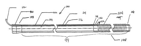
With reference to FIG. 1, an MRI imaging apparatus (100) according to one
embodiment includes a substantially tubular member (99) having a distal end
(105) and a
proximate end (103) with a lumen (101) therebetween. The substantially tubular
member
(99) generally has an exterior surface (102) and an interior surface (116).
There is also
included an imaging coil (104) which may be of any design capable of receiving
and/or
transmitting magnetic resonance signals. The coil pictured in FIG. 1 is a
loopless design.
2o Loopless designed coils are known in the art, and a loopless coil could
include, but is not
limited to, designs such as those described by Ocali et al in US Pat.
#5,928,145 and by
Lardo et al in US Pat. Application #09/536,090 "Magnetic resonance imaging
guidewire
probe," filed Mar. 24, 2000 (hereafter "Lardo '090"), the entire disclosures
of which are
herein incorporated by reference.
The apparatus shown in FIG. 1 shows the coil (104) embedded within the tubular
member (99), but such a construction is by no means necessary. In other
embodiments the
coil could be on the interior surface (116) or the exterior surface (102) of
the tubular
member (99).
In one embodiment, an attachment point (108) to affix the sleeve to another
device,
3o such as a medical device (such as but not limited to a PTCA catheter,
endoscope, balloon
device for dilatation angioplasty, stmt placement tool, drug delivery tool,
intraluminal
SUBSTITUTE SHEET (RULE 26)
CA 02404352 2002-10-08
WO 01/073461 PCT/USO1/09692
-13-
resecting tool, guidewires, electrophysiologic mapping instrument, atherotome
for
atherosclerotic plaque removal and debulking, another imaging device such as
an MRI coil,
and any other device designed for use within a catheter or sleeve) may be
included at the
distal end (105), and a connector hub (110) possibly with strain relief may be
included at
the proximal end (103). Attachment point (108) may be of any type for
temporary or
permanent attachment, and may comprise any type of connector for interfacing
with the
attached device known to one of skill in the art. An electrical transmission
member, in this
case a coaxial cable (114), connects the coil (104) to an MRI scanner (Not
shown) for the
transmission of signals between the scanner and the coil. In the embodiment in
FIG. 1 the
1o electrical transmission member is also embedded within the tubular member
(99). In an
embodiment, the coaxial cable (114) is connected to a decoupling circuit
connector (112)
and connects the coil (104) to a decoupling circuit (not shown). An example of
a
decoupling circuit to which the decoupling circuit connector could be attached
is described
in Lardo '090. In one embodiment, the connector hub (110) and decoupling
circuit
connector (112) are located at the proximate end, while the imaging coil (104)
is located at
the distal end. However, other arrangements of these elements relative to the
ends (103,
105) will be readily apparent to one skilled in the art.
An embodiment of the apparatus of the following construction is shown in FIG.
14.
The coaxial cable (114) may be built in the walls of the tubing in form of a
inner (95) and
2o an outer (96) braid where the inner braid (95) acts as a core of the
coaxial cable and the
outer braid (96) acts as a primary shielding. This design may leave the lumen
(101) entirely
patent for delivering various devices e.g. guidewires, therapeutic catheters,
contrast agents,
and the like.
The antenna can be a loop, quadrature loop, loopless with the whip coiled or,
as
shown in FIG. 14A, loopless where the coil (104) comprises an extension of the
inner braid
(95) extending to the distal end (105) of the sleeve. In an embodiment,
depicted by way of
example in FIG. 14B, another layer of braiding can be provided over the
primary shielding
to act as a bazooka balun (97). Also another braiding connected to the ground
(not shown)
can be added below the core-braiding to prevent coupling/change loading
conditions when
3o devices are inserted and moved inside the sleeve.
In an embodiment, the braidings comprise copper, tanatalum or any other
SUBSTITUTE SHEET (RULE 26)
CA 02404352 2002-10-08
WO 01/073461 PCT/USO1/09692
-14-
nonmagnetic material which will give a low susceptibility artifact under MR.
In another
embodiment, the braidings comprise gold, silver or any other metal plating on
a polymeric
surface or applied using different techniques such as, but not limited to,
sputtering. In an
embodiment, the metallic conductive layers may be electrically continuous, but
need not be
physically continuous.
The impedance of the coaxial cable created this way may generally be anywhere
from 10-50 ohms. Also the distal end of the imaging sleeve can be formed in
various
shapes, for instance, for forming different guide catheters.
In an embodiment, the imaging sleeve further comprises a contrast agent to
enhance
the active tracking ability of the coil. The contrast agent is incorporated
into the tubular
member or the coil, for example, by applying a coating containing the contrast
agent,
blending the contrast agent with the material of the sleeve during or before
extrusion, or
other means readily apparent to one of ordinary skill in the art. This
contrast agent may be
incorporated throughout the entire sleeve or confined in a portion thereto. In
active
tracking, the sleeve images the anatomy around the device, including a broad
signal from
the coil, and the coil outline is bigger than the actual device. The contrast
agent may reduce
the outline so that the size of the device as seen on the image will
approximate its true size.
Examples of contrast materials include, but are not limited to gadolinium and
dysprosium
oxide, and any other MRI contrast materials known to one of skill in the art.
Data acquisition during imaging may occur in different modes. In an
embodiment,
high-speed data acquisition and display techniques may be employed when the
coil is being
used to locate the position of the sleeve relative to an anatomical structure
of interest. Use
of a contrast agent may be especially beneficial in this situation because the
contrast
material will generate a very intense signal in the MRI image. Image sampling
may then
occur at a faster rate. In another embodiment, high-resolution imaging mode is
employed to
generate the highest-quality image possible, and the speed of acquisition may
be slower
than in high-speed mode. . Our aim is to generate the best quality image.
In another embodiment, shown by way of example in FIG. 16, the apparatus
3o comprises a rapid exchange or a monorail catheter, having an imaging sleeve
(100) and a
guidewire lumen (65) with 2 wire ports are provided below the imaging coil
(104). The
SUBSTITUTE SHEET (RULE 26)
CA 02404352 2002-10-08
WO 01/073461 PCT/USO1/09692
-15-
imaging antenna can be a single loop, fixed or expandable, quadrature loop or
a loopless
design.
In an embodiment, the apparatus may further comprise additional substantially
tubular members. For example, a second tubular member may be the guidewire
lumen (65)
as shown in FIG. 16. In another embodiment, a lumen is provided for deployment
of
additional medical devices, such as a balloon catheter or basket device. In an
embodiment,
the proximal end (103) has a plurality of ports providing access to, for
example, the volume
enclosed by the tubular member, a connection through which water or any other
fluid may
be discharged into the sleeve, a connector to the detector coil to change its
shape, and other
l0 uses as will be apparent to one of skill in the art.
In an embodiment exemplified by FIG. 18, the sleeve may take the form of a
guide
catheter (64) similar to that used in typical angioplasty and angiography
procedures. The
guide catheter has a preformed shape to facilitate access into the right or
left coronary artery
systems. The sleeve may further comprise a lumen obstruction device, such as a
balloon, to
perform angioplasty. The sleeve may further comprise an embedded braid
providing
stiffness and torque control. The stiffness of the braid may vary from
position to position in
the sleeve.
In an embodiment, the tubular member is constructed of polymer. This could be
a
single polymer, or multiple polymers could be used. The reasons for selecting
a particular
2o polymer or combination of polymers would be apparent to one of skill in the
art but could
include controlling particular mechanical or electrical properties for any
portion of the
tubular member (99). Examples of suitable polymers are nylon, PEBAX,
polyurethane,
polyethylene, silicone polymers, fluoro-polymers, or other similar polymers
known to those
skilled in the art. Some or all of the length of the tubular member can be
made up of single
or multiple polymers so as to control mechanical properties over the length of
the member.
The apparatus can be coated on interior surface (116) and/or exterior surface
(102) with
appropriate coatings, e.g., hydrophilic coatings on the exterior surface and
silicone on the
inner surface to achieve further desired mechanical or electrical properties.
Examples of
suitable coatings include PVP, poly acrylic acid, and other hydrophilic-based
polymers.
In an embodiment, the tubular member may be constructed so as to have varying
stiffness at different positions. For instance, the distal end could be more
flexible than the
SUBSTITUTE SHEET (RULE 26)
CA 02404352 2002-10-08
WO 01/073461 PCT/USO1/09692
-16-
proximal end so as to help prevent injury to subject during insertion and
placement of the
sleeve.
In FIG. 1, the coil is created in a manner so as to allow for it to be able to
image
structure surrounding the distal end (105) of tubular member (99). One method
of creating
such a coil is described as follows. At a transition point (118), the coaxial
cable (114) is
terminated and its core (120) is extended onward and is coiled forming the
coil (104). The
coil (104) is depicted in FIG. 1 as a helical wound conductor by way of
example. A
secondary shielding (122) which in one embodiment is in the form of a braiding
may be
provided and is connected to the shielding of the coaxial cable at the distal
end (105). The
to braiding may comprise a suitable electrical conductor at the MRI/MRS radio
frequencies.
Examples of suitable materials include copper, or a nickel titanium alloy
commonly known
as Nitinol plated with gold, silver (or alternate layers of gold, silver, or
copper, and/or gold
on nitinol), or copper, or may comprise an MR compatible stainless steel, or
aluminum, or
gold or silver coated MR compatible stainless steel.
The secondary shielding (122) can prevent the electrical and imaging
properties of
the coil from changing when the coil is attached to the tubular member. In
addition, the
braiding may provide electrical isolation from the devices used inside the
sleeve. For
example, an imaging guidewire inserted inside the sleeve may couple with the
detector coil
in the sleeve and cause imaging artifacts. In an embodiment, the secondary
shielding (122)
is electrically grounded and may thus prevent changes in loading conditions
which might
occur due to having another coil inside the imaging sleeve.
FIG. 1A depicts a proximal end (103) view of the assembly of FIG. 1, showing
the
relationship of the tubular member (99) with the coaxial cable (114) and the
lumen (101)
therein.
In an embodiment shown in FIG. 1B the coil is attached to the exterior surface
(102)
of the first tubular member (99) and a second tubular member (98) is placed co-
axially with
the first tubular member (98) . This may be placed so as to provide an
exterior covering of
the coil (104) as is shown in FIG. 1B. This second tubular member (98) may be
loose or
may be bonded on the first tubular member (99). In an embodiment, the second
tubular
3o member (98) is loose and may move slideably along at least a portion of the
length of the
first tubular member (99).
SUBSTITUTE SHEET (RULE 26)
CA 02404352 2002-10-08
WO 01/073461 PCT/USO1/09692
-17-
FIG. 2 shows another embodiment employing a loop imaging coil (224) as coil
(104). The exterior surface (202) and inner surface (216), connector hub
(210), connector
(212), and clip (208) may be as described in FIG. 1. The loop imaging coil
(224) is similar
to that described above, except that the loopless imaging component is
replaced by the loop
components, e.g., an imaging loop (226), tuning matching capacitors (228a,
228b), and a
triaxial cable (214) to conduct the received signals to a scanner and
incorporating a balun
circuit. The secondary shielding (222) may be included in the loop antenna
imaging sleeve.
Tuning/matching capacitors can be distributed around the loop to improve
performance, as
for example depicted in the embodiment of FIG. 3 with a tuning/matching
capacitor (340)
l0 at the distal end. A tuning/matching capacitor can also be added to the
proximal end of the
loop, or one tuning capacitor added at the distal end and one at the proximal
end as depicted
in FIG. 2 with tuning/matching capacitors (228a, 228b). FIG. 2A depicts a
proximal end
view of the instant embodiment, showing the relationship between the exterior
(202) and
interior (216) surfaces with the triaxial cable (214).
~5 The loop imaging coil (224) may be of any design known in the art,
including those
described by Atalar et al in US Pat. #5,699,801 (hereafter "Atalar '801"), the
entire
disclosure of which is herein incorporated by reference, and by Atalar, US.
Pat. Application
#09/191,563, entitled "Miniature magnetic resonance catheter coils and related
methods,"
filed Nov. 13, 1998 (hereafter "Atalar '563") the entire disclosure of which
is herein
2o incorporated by reference. FIG. 3 shows one embodiment of a loop imaging
coil which
may be used. In this embodiment, the detector coil resides on a flexible
circuit board. The
detector coil may reside on any substrate (330), made for instance of Kapton
or other
material known to one of skill in the art, and may be applied, for example by
etching,
depositing, or by some other process known to one of skill in the art. A
copper conductor
25 (332), distal pads (334a, 334b) for a tuning/matching capacitor and
decoupling circuit
(340), and proximal pads (338a, 338b) for connecting the coaxial cable (214)
may also be
present. In an embodiment, the copper conductor may have dimensions of at
least 5
micrometers thick and 0.1 millimeters wide. In another embodiment, the copper
conductor
may have the dimensions of 18 micrometers thick and 0.7 millimeters wide.
3o FIG. 4 shows yet another embodiment employing another type of loop imaging
coil,
in this case a quadrature loop imaging coil (404). Two substantially
orthogonal loops are
SUBSTITUTE SHEET (RULE 26)
CA 02404352 2002-10-08
WO 01/073461 PCT/USO1/09692
-18-
used to improve the homogeneity of the coil reception in a substantially
quadrature mode.
One skilled in the art would understand that the coils may also be situated at
angles other
than substantially orthogonal. The tuning/matching capacitors (428a, 428b) may
similarly
be incorporated into the quadrature loop embodiments. The dimensions of the
loop and the
device will vary as according to the particular application, i.e. the
procedure and anatomy of
interest, and the image resolution desired. Quadrature loops are described in
Atalar'S63.
FIG. 4A is a cut-section through line A-A of FIG. 4 and shows one arrangement
of the two
loop coils (407a, 407b) of the quadrature loop imaging coil (404).
FIG. 5 shows another embodiment which may be used in conjunction with a second
1o medical device to be deployed within the lumen (501). The interior surface
(516) of the
present embodiment can be coated with a lubricious coating (542) as described
above to
facilitate fitting of the apparatus over another medical device, such as but
not limited to a
PTCA catheter, endoscope, balloon device for dilatation angioplasty, stmt
placement tool,
drug delivery tool, intraluminal resecting tool, electrophysiologic mapping
instrument,
atherotome for atherosclerotic plaque removal and debulking, another imaging
device such
as an MRI coil, or any other device capable of deployment within a sleeve. The
detector
coil (504) may comprise a loopless imaging coil or a loop imaging coil of any
type known
in the art, including those types described above and by Ocali et al in US
Pat. #5,928,145,
by Atalar '801, and by Atalar '563.
2o Such arrangement may be used whenever imaging of an anatomical region or
structure is desired while advancing a device to the region or structure or
while using the
device to examine, characterize, sample, diagnose, treat, ablate, resect, or
otherwise
manipulate the structure or region in ways readily apparent to one of skill in
the art. Use of
MRI instead of visible light visualization may be particularly advantageous. A
visible light
camera requires an unblocked optical light path for visualization. Any devices
in the lumen
of a sleeve may themselves block this path and prevent visualization of the
anatomical
structure or region being manipulated. An MRI antenna, such as those disclosed
herein, has
no such requirement and thus may provide a complete and unimpaired image
regardless of
what device, if any, is present in the lumen of the sleeve. MRI may also
provide imaging
data of anatomical structure beneath the surface of the structure or region of
interest. This
additional data may be of considerable value to an operator of a device
according to this
SUBSTITUTE SHEET (RULE 26)
CA 02404352 2002-10-08
WO 01/073461 PCT/USO1/09692
-19-
embodiment. It may show, for example, evidence of tissue damage that would not
be
apparent by visible light visualization.
FIG. 6 shows yet another embodiment designed to provide an expandable loop
imaging coil (644). A second tubular member (698) is slideably displaceable
along the
longitudinal axis of the sleeve between an extended position and a retracted
position. When
the second tubular member (698) is in its retracted state, the expandable loop
imaging coil
(604) is in its expanded state. When the second tubular member (698) is in its
extended
state, the expandable loop imaging coil (604) is in its collapsed state. The
second tubular
member (698) is depicted in the retracted position in FIG. 6 and in the
extended state in
1o FIG. 6A. Although in FIGS. 6,6A the loop is shown as being dimensionally
different in the
two states, that is not a necessary part of the design. The exterior (602) and
interior (616)
surfaces of the first tubular member (699) remain fixed relative to each
other, and the
interior surface (616) defines the lumen of the sleeve into and through which
other devices
may be inserted. As shown in FIG. 6B, the expandable imaging loop (644) can
comprise a
core (650) surrounded and encased by an insulator (648). In one embodiment,
the insulator
(648) comprises polymeric tubing. The core (650) is a pre-shaped superelastic
electrically -
conducting material or metal such as a nickel titanium alloy commonly known as
Nitinol.
However, other known superelastic conducting materials including beryllium-
copper alloy,
and non-magnetic stainless steel are examples of materials that may be used.
The pre-
2o shaped superelastic material that forms the expandable loop is plated with
gold, silver (or
alternate layers of gold, silver and gold on nitinol) or other conductive
metal to increase RF
conductivity of the loop. It will be recognized that tuning capacitors may be
incorporated in
the distal or proximal or both ends of the loop as discussed for the
embodiments of FIG. 2.
FIG. 6C is a cut-section through line C-C of FIG. 6 and shows two ports (652a,
652b)
which house the ends of the expandable imaging loop (644). Refernng again to
FIG. 6,
even in its fully retracted state, the second tubular member (698) may house
the ends of the
expandable imaging loop (644), tuning/matching capacitor (628), ports (652a,
652b), and
coaxial cable (614). This embodiment may further comprise a connector (612),
which may
be a BNC connector or mini-BNC connector for connection to an MRI machine, a
3o decoupling circuit, or other apparatus (not shown). The expandable imaging
loop (604) .
may comprise any loop imaging coil design known to the art, including all
described above
SUBSTITUTE SHEET (RULE 26)
CA 02404352 2002-10-08
WO 01/073461 PCT/USO1/09692
-20-
and all others described by Ocali et al in US Pat. #5,928,145, in Atalar '801,
and in Atalar
'563. An expandable loop antenna can also be of a loopless design in an
embodiment.
FIG. 6D shows an embodiment of the sleeve in which the expandable imaging loop
(604) comprises a quadrature loop coil. The two loops (607a, 607b) of the
expandable
imaging loop (604) may be nested in their collapsed state in a substantially
orthogonal
manner similar to that illustrated in FIG. 4A for the two loop coils (407a,
407b). As shown
in FIG. 6E, the loop coils (607a, 607b) may also be nested side-by-side in
their collapsed
state. When the second tubular member (698) is retracted, one of the two loop
coils (607a,
607b), for example loop coil (607a) is mounted, spring-loaded, or otherwise
attached in
1o such a way that it rotates to or otherwise assumes a substantially
orthogonal orientation
relative to, for example, loop coil (607b) as the quadrature loop coil (604)
transitions to its
expanded state. Other arrangements of the two loop coils will be readily
apparent to one
skilled in the art.
Considering once again FIG. 6, to place the expandable imaging loop (604) in
its
15 collapsed state, the second tubular member (698) may be slid into its
extended position over
the expandable imaging loop (604) so that the loop is caused to contract. The
collapsed state
of the expandable imaging loop (604), as shown in FIG. 6A, may be used during
insertion
of the sleeve and advancement of the sleeve to a point or anatomy of interest.
This may
provide the advantage of having a low-profile device during advancing and
retracting from
20 the anatomy of interest, and an expanded imaging loop once the apparatus is
situated in the
anatomy of interest for improved imaging for improved diagnostic value. In one
embodiment, the expandable imaging loop (604) comprises a superelastic
material, such as
Nitinol, having a very high degree of "memory." This allows for the loop to
have a precise,
predetermined separation when the loop is expanded again. Because this
separation remains
25 essentially constant throughout many cycles of loop expansion and
contraction, the tuning
and matching components can be set to constant, finely tuned settings.
The expanded state may be used during image acquisition, and provides improved
SNR over other low-profile coils. To place the expandable imaging loop (604)
in its
expanded state, as shown in FIG. 6, the second tubular member (698) is slid to
the retracted
3o position at which it may cover only the proximal ends of the expandable
imaging loop
(604).
SUBSTITUTE SHEET (RULE 26)
CA 02404352 2002-10-08
WO 01/073461 PCT/USO1/09692
-21 -
For the loop coils the area in the loop and therefore the distance of
separation
between the parallel conductors determines the image quality or SNR. In
general, the
greater the separation, the greater the SNR, which provides an SNR advantage
for the
expandable loop compared to a fixed loop (FIG. 2) if the location of interest
is suitable for
its deployment. The expandable loop can be made in various configurations e.g.
to open to a
specific dimensions, expand depending on the anatomical cavity available, or
within the
lumen of another device or vessel.
The expandable loop and any of the other coils known in the art or disclosed
herein
may be encased in a body lumen obstruction device, for example, a balloon, or
some other
1o similar device known to one of ordinary skill in the art. Such an
obstruction device may be
used to prevent flow of any material through the lumen in which the apparatus
is situated.
For example, an obstruction device may be deployed while the apparatus is in a
blood
vessel. In this case, the obstruction device would prevent flow of blood
through the blood
vessel. Specifically, the device may be used in any of the coronary arteries
or principal
divisions thereof to guide, with the detector coil, a angioplasty means such
as a lumen
obstruction device to a diseased artery. The balloon can be circular or
elliptical with
variable or fixed diameter as per inflation pressure. However, since tuning
matching is
specific for a particular separation, if the separation varies, the device may
require retuning
for optimum performance.
2o The expandable loop may also be employed in an MRI imaging probe designed,
for
example, to be deployed within the MRI sleeve as a guidewire, or as any of the
probes
described in Atalar '801. As shown in FIG. 7, such a probe can comprise an
detector coil
(704), the ends (754a, 754b) of which are connected by ports (752a, 752b), to
a
tuning/matching circuit (762) coupled to a coaxial cable (714) that conducts
signals
2s received by the expandable imaging loop (704) to an MRI scanner or the
like, via a BNC
connector or other connector (764). An interface system, being for example a
flexible
circuit board, may be used to mount the tuning/matching circuit (762) and a
decoupling
circuit. Flexible polymeric tubing (766) houses the ends (754a, 754b) of the
expandable
imaging loop (704), ports (752a, 752b), tuning/matching circuit (762), and
coaxial cable
30 (714).
SUBSTITUTE SHEET (RULE 26)
CA 02404352 2002-10-08
WO 01/073461 PCT/USO1/09692
-22-
A tubular member (702) having a lumen (701 ) encases the assembly and in one
embodiment comprises a polymeric tubing for access to areas some distance from
the point
of entry. However, the material may be metallic for use as a trocar or
introducer to guide
placement of interventional tools through it. To place the detector coil (704)
in its expanded
state, as shown in FIG. 7, the tubular member (702) is slid to a position at
which it may
cover only the proximal ends (754a, 754b) of the detector coil (704).
Therefore, the at least
part of the detector coil (704) is positioned outside the lumen (701) of the
tubular member
(702) when in its expanded state. The expanded state may be used during image
acquisition, and provides improved signal-to-noise ratio over other low-
profile probes.
to FIG. 7A shows a device according to the embodiment of FIG. 7 but wherein
the expandable
imaging loop (704) is in its collapsed state and is wholly or partially
contained within the
lumen (701).
To place the expandable imaging loop (704) in its collapsed state, the tubular
member (702) is slid over the expandable imaging loop (704) so that the loop
is caused to
contract. The collapsed state may be used during insertion of the device and
advancement of
the device to a point or anatomy of interest. In one embodiment, the
expandable imaging
loop (704) comprises a superelastic material, such as Nitinol, having a very
high degree of
"memory." This allows for the loop to have a precise, predetermined separation
when the
loop is expanded again. Because this separation remains essentially constant
throughout
2o many cycles of loop expansion and contraction, the tuning and matching
components can be
set to constant, finely tuned settings.
The imaging probe featuring the expandable imaging loop may be used in
conjuction with any of the MRI sleeves herein using any of the imaging coil
designs
described herein and in the above given references. The expandable imaging
probe, in its
collapsed state, may be inserted into an MRI sleeve as shown in FIG. 8 with a
loopless
sleeve coil. One skilled in the art would understand that any type of imaging
coil known in
the art may be employed in the MRI sleeve component of the combination device.
The
combination device comprises an MRI sleeve (868) and an expandable probe
(870). The
combination device may be advanced to the anatomy of interest, perhaps through
narrow-
lumened structures such as blood vessels, esophagus, small intestine, biliary
tree members,
and others that are obvious to practitioners of the art. Once the combination
device is in
SUBSTITUTE SHEET (RULE 26)
CA 02404352 2002-10-08
WO 01/073461 PCT/USO1/09692
- 23 -
position, the expandable probe (870) may be advanced so that the coil region
(804)
protrudes from the sleeve. As depicted in FIG. 8A, the coil region (804) may
be brought
into its expanded state by retracting the tubular member (802) to expose the
coil region
(804). In another embodiment, the sliding sheath (802) may be omitted, with
the interior
s surface (816) of the sleeve holding the expandable probe (870) in its
collapsed state. The
expandable probe (870) may also be placed in its expanded state by advancing
the
expandable probe (870) so that the coil (804) protrudes from the sleeve (868).
The use of an expandable probe with an MRI sleeve provides advantages over the
use of either alone. For example, the imaging sleeve may be used to provide
visualization
of surrounding tissue and of itself as it is introduced into a body and
advanced to the
structure of interest. Once the combination probe is in place, the expandable
probe insert
may be advanced and expanded, providing increased SNR over lower-profile coils
during
image acquisition. Alternatively, the expandable probe may be advanced through
a
structure of such limited dimensions that the sleeve itself is excluded. In
this case, the inner
1s surface of the sleeve is used to maintain the collapsed state.
Probe inserts used in combination with an MRI sleeve may also comprise
nonexpandable MRI probes dimensionally adapted to be inserted into a sleeve or
catheter.
The probe insert coil and the MRI sleeve coil may both be of any type known in
the art,
including those described in the above-named references. FIG. 9 shows an
embodiment in
2o which a loop imaging coil probe (972) is inserted in a loopless imaging
coil sleeve (974).
FIG. 9A shows another embodiment in which a loopless imaging coil probe (978)
is
inserted in a loop imaging coil sleeve (976). The MRI sleeve and MRI probe may
both
comprise one or more imaging coils of any types known in the art or disclosed
herein.
Other combinations will be readily apparent to one skilled in the art.
2s Combinations of MRI coils such as those described above, and such as
certain
embodiments of which are depicted in FIGS 8, 8A, 9, 9A may offer superior SNR
and
imaging sensitivty along the length of the imaging coil combination compared
to a single
coil alone. Loop imaging coils offer near field high resolution imaging, while
loopless coils
provide broad field imaging at lower resolution. FIG. 10A depicts
schematically the
3o sensitivity profile of a loopless imaging coil. Signal stength reaches a
peak in a fixed
diameter region along the length of the imaging coil. This provides excellent
visualization
SUBSTITUTE SHEET (RULE 26)
CA 02404352 2002-10-08
WO 01/073461 PCT/USO1/09692
-24-
in only a confined area. In contrast, FIG. lOB shows a schematic sensitivity
profile of a
loop imaging coil. While not approaching the peak signal strength achieved by
the loop
design anywhere along its length, the loopless coil design provides limited
sensitivity
distributed along its length. By combining two imaging coils, one of each
design, (i.e., a
loop sleeve with a loopless probe, or a loopless sleeve with a loop probe) a
sensitivity
profile combining the strength properties of each design is achieved, as
depicted
schematically in FIG. l OC. Combinations of a loopless probe and a loopless
sleeve and of a
loop probe and loop sleeve accentuate the signal sensitivity properties of the
respective
designs. In all combinations, any loop or loopless coils may be of any types
and designs
to known in the art or disclosed herein, including but not limited to,
loopless coil, helical coil,
solenoid loop, loop, quadrature loop, expandable loop, or expandable
quadrature loop.
As described above, in an embodiment, all coils may be located inside a
subject to
be imaged. In another embodiment, at least one coil of a combination may be
situated
outside the subject to be imaged, and at least one other coil may be inside
the subject. In
yet another embodiment, all coils may be located outside the subject to be
imaged.
The signals from the imaging coils may be combined through the use of a
controller,
such as, but not limited to, a computer, computer software, image acquisition
systems on
the MRI scanner, or any other systems known to one of skill in the art.
FIG. 15 depicts one embodiment of an interface circuit. The interface circuit,
when
2o used in conjunction with a loop detector coil enables the loop coil to
perform as a combined
loop+ loopless antenna. The interface circuit may comprises, for example, a
BNC connector
(68), a micro BNC receptacle (67), balun cable trap (94), decoupling capacitor
(93), DC
regulating circuit (92), PIN diode (71), and a tuning/matching circuit having
an inductor (70)
and capacitor (69). The interface circuit may be connected to any loop coil.
This changes the
SNR characteristics of the coil so that it behaves similar to a loop +
loopless coil (combined
coil). The loop coils have matching tuning and decoupling circuits on the coil
itself. The
circuit described above makes it perform as a loopless antenna + a loop
antenna. The cable trap
(94) acts as a balun for both the loop and the loopless. The decoupling
circuit in the box as
described above decouples the loopless antenna and allows the DC current to
flow through to
3o decouple the loop antenna. This DC flows through the resistor or an
inductor in the circuit (92)
activating the PIN diode (71) on the coil. The output of both the coils is
then matched and
SUBSTITUTE SHEET (RULE 26)
CA 02404352 2002-10-08
WO 01/073461 PCT/USO1/09692
-25-
tuned by the matching tuning circuit in the box (inductor 70, capacitor 69).
Combinations of loop and loopless imaging coils may be incorported directly
within
the MRI sleeve itself. Such a combination provides the advantages of improved
signal
strength and imaging sensitivity as depicted schematically in FIG. 10C, but
also provides
for the simulatneous use of another medical instrument deployed in the lumen
of the MRI
sleeve. FIG. 11 depicts one exemplary embodiment. A loop imaging coil (1180)
connected
to a loop coil coaxial cable (1184) and a loopless imaging coil (1182)
connected to a
loopless coil coaxial cable (1114) may both be embedded in a tubular member
(99),
similarly as described for the embodiments of FIGS 1,2. The lumen (101) may
remain
patent for the passage of medical devices as described above. FIG. 11A depicts
a left-end
view of the embodiment of FIG. 10, showing this relationship. In all
combinations, any
loop or loopless coils may be of any types and designs known in the art or
disclosed herein,
including but not limited to, loopless helical coil, solenoid coil loop, loop,
quadrature loop,
expandable loop, or expandable quadrature loop.
15 In another embodiment, an MRI sleeve comprises at least one loop imaging
coil and
at least one loopless imaging coil embedded in the tubular member (99). The
imaging coils
may each be of any type known in the art or disclosed herein, including but
not limited to
the loop coil, quadrature loop coil, expandable imaging loop coil, and
loopless imaging coil.
In yet another embodiment, a combination device comprises an MRI sleeve having
2o at least one loop imaging coil and at least one loopless imaging coil
embedded in the
tubular member (99) and an MRI probe of any design known in the art or
disclosed herein.
This results in a combination having at least three coils. Combinations having
greater than
three coils may also be fashioned and are readily apparent to one of skill in
the art.
In general, it is useful, for the purposes of optimizing SNR and minimizing
25 electromagnetic interactions between the imaging sleeve antennae and other
coils and
antennae to interface the imaging sleeve to the MRI scanner via one or more
decoupling
tuning/matching circuits and/or a balun. The tuning and matching capacitors
can be placed
in a variety of locations that are apparent to those skilled in the art and
can be determined
without undue experimentation. One embodiment is shown in FIG. 12, in which
the ends
30 (644a, 654b) of any type of loop imaging coil are attached to the coaxial
cable (614) by a
tuning/matching circuit comprising a capacitor in parallel (658) and a
capacitor in series
SUBSTITUTE SHEET (RULE 26)
CA 02404352 2002-10-08
WO 01/073461 PCT/USO1/09692
-26-
(660). The capacitor in series (660) may also be placed anywhere along the
loop, for
example, at the distal end of the loop. FIG. 13 depicts an embodiment in which
the
capacitor in series (660) is placed at the distal end of the coil. Such a
positioning can
improve imaging performance of the sleeve.
Each such configuration provides unique SNR properties, which will be apparent
to
those of ordinary skill in the art. The decoupling circuit (diode) in one
embodiment is
placed at the proximal end of the probe or in a suitable position with respect
to the antenna
to achieve maximum decoupling.
The MRI sleeve in an embodiment offers physicians and surgeons the opportunity
to
1o gather MR images for examination of anatomy, diagnosis, image-guided
biopsy, and for
guiding therapies such as minimally-invasive intervention, and surgery. Other
applications
will be readily apparent to one skilled in the art. The sleeve can be used
with any MRI
compatible surgical device of the physician or surgeon's choice, including
additional MRI
devices. Any inserted devices can be easily withdrawn and replaced by other
devices as
needed, for example, if a biopsy is followed by a surgical procedure during a
single
intervention. The metallic properties of the antenna in the sleeve renders it
visible under x-
ray which can also be used to determine its location in the body, if desired.
For example, if
the MRI apparatus to which a device according to the present invention is
connected were
to fail during use, MRI sleeves according to the invention could still be
localized using X-
2o ray imaging. The MRI sleeve may also be used as a locatable catheter in
circumstances in
which the use of MRI is inappropriate. For example, in subjects who have
contraindications for MRI use (such as pacemakers or implanted prostheses
containing
ferromagnetic elements) the MRI sleeve may still be of utility because its
location may be
determined using X-ray imaging without actually exposing the subject to the
magnetic
fields required in MRI acquisition.
An MRI sleeve according to an embodiment may also be used in conjunction with
any of the imaging guidewires disclosed in Lardo '080.
In one typical application, the sleeve is mounted on a commercially available
MRI
compatible surgical device, for example, an endoscope or laparoscope, which is
then
inserted into the body and advanced, for example, into the gastro-intestinal
(GI) tract for
examination, image-guided biopsy, or minimally-invasive surgery. The imaging
sleeve can
SUBSTITUTE SHEET (RULE 26)
CA 02404352 2002-10-08
WO 01/073461 PCT/USO1/09692
-27-
be used with a trocar or other surgical device for minimally-invasive surgical
procedures.
The sleeve may also be used in combination to introduce another instrument or
be used
within the lumen of an endoscope or laparoscope to allow viewing through the
wall, not
attached to the end of an instrument.
The imaging sleeve offers the advantage of being useful with many medical
devices
e.g. MRI compatible endoscopes, laparoscopes, minimally invasive surgical tool
(for
example, trocar), and a single sleeve can be usable with multiple devices. It
can be used
independently as an access device for introducing surgical devices to the site
of interest.
MRI and endoscopy can be done simultaneously, thus providing a direct
correlation and
1o correspondence between visual surface information and the underlying
anatomy and
function detectable by MRI. Devices according to the invention can also be
coupled with
computer-integrated and guided surgical techniques. The invention has the
capability to
provide the minimally-invasive surgeon with a real-time three-dimensional view
of the area
of surgery. Other particular applications of the present invention include,
esophageal
15 imaging of the coronary arteries, imaging the prostate, urinary tract,
bladder, GI tract,
vasculature etc. The field of view possible by use of the sleeve or
combinations of antennas
of the invention is generally much larger than that provided by surface coils
or other
imaging modalities.
The present invention provides significant advantages over other devices. The
low
2o profile of the antennae according to the invention allow placement in small
or narrow
anatomies of interest, e.g., vasculature and GI tract. A high SNR can be
obtained, using the
invention, which provides for improved resolution and image quality. For
vascular
applications where an uninterrupted supply of blood is important to prevent
hypoxic
damage to tissues supplied by the vascular member in question, the device of
the invention
25 can be used without blocking the flow of blood, thereby allowing it to be
held in vascular
locations for relatively long periods of time without causing or risking
tissue damage or
necrosis. In addition, devices according to the invention may be used in
combination with
or function as the principal coronary or peripheral interventional tools, such
as introducers,
guide catheters, PTCA balloons, plaque removing devices such as atherectormy,
drug
3o delivery catheters, gene delivery catheters, radiation catheters, stmt
placement, and other
SUBSTITUTE SHEET (RULE 26)
CA 02404352 2002-10-08
WO 01/073461 PCT/USO1/09692
-28-
applications readily apparent to one of ordinary skill in the art. The balun,
matching, tuning
and decoupling circuit can be placed close to the loop, thereby reducing
signal loss.
Referring now to Figure 17 which depicts an embodiment of a multi-channel MRI
system suitable for exciting the exciting and receiving MRI systems detected
by the
aforementioned inventive devices. The figure depicts in greater detail the "RF
source",
"patient", "magnetic field", "receiver", "A/D converter", and "computer"
sections
numbered 2, 4, 6, 8, 10 and 12 of Figure 1 of Atalar ' 801. The magnetic field
6, is
produced by magnet means with field strength in the range 0.1 to 4 Tesla, but
more
typically from 0.3 Tesla to 3 Tesla as used for whole body clinical MRI. We
now describe
to the mufti-channel MRI system for using the aforementioned MRI probes in
conjunction
with conventional external coils, and how it is preferably used.
In an embodiment, patient or object to be studied 4 is placed in the magnetic
field
generated by the MRI system 6. MRI signals are excited by RF source 2, which
includes
RF power amplifier means 22 and transmit coil means 21 known to those skilled
in the art.
External MR detector coil means 85a, and/or 85b and/or 85c and the like are
initially used
to detect said excited signals. The external MR detector coil can be a single
surface coil
85a, or multiple coils for detecting signals either serially or in a parallel
imaging
configuration such as that known as a phased-array as described by P.B. Roemer
et al in the
journal "Magnetic Resonance in Medicine", Volume 16, pages 192-225, 1990. The
MRI
2o signals from the coils pass through tuning and transmitlreceive switches
82a, 82b, 82c and
the like and are fed to the receiver 8, which includes preamplifier means 84a,
84b, 84c and
the like and analog or digital receiver means 87a, 87b, 87c etc. The low noise
preamplifiers
86a, 86b, 86c etc and 87a, 87b, 87c etc of the MRI system each have gain and
noise-figures
substantially equivalent or comparable to individual preamplifiers
conventionally used in
MRI and known to those skilled in the art. The signals are digitized in 10 and
fed to
computer 12 where the image signals are reconstructed and displayed using
Fourier
reconstruction or other techniques known to those in the art, and displayed on
display
means 16 of Fig 1 of Atalar '801, which can be a cathode ray tube. In the
context of an
MRI exam employing the inventive devices for internal MRI described above and
in
3o Figures 1-13 of the present application, the displayed images are used .for
visualizing the
internal anatomy for the purpose of placement and introduction of the internal
MRI coils.
SUBSTITUTE SHEET (RULE 26)
CA 02404352 2002-10-08
WO 01/073461 PCT/USO1/09692
-29-
At or before the time that it is desirable to visualize images generated from
the
internal MRI coils, the one or more outputs of the aforementioned internal MRI
coils are
input to tuning and matching means and transmit/receive switching means 81 a,
81b, 81 c
etc, examples of component designs which are described in Figs 4, and 5 of
Atalar '801,
and Figs 3 and 7 of Ocali et al, US patent 5,928,145, and included herein by
reference in its
entirety. The outputs of each device is then each fed to a separate channel of
the multi-
channel receiver which includes preamplifier means 83a, 83b, 83c etc, receiver
means 86a,
86b etc, and is digitized by 90a, 90b etc before being fed to computer means
12. While it is
envisaged that the gains of preamplifiers 83a, 83b, 83 c etc, used for
amplifying the internal
1o coils will generally be comparable to that of 84a, 84b etc for the external
coils, in some
circumstances, the inherent voltage signal strength between internal and
external coils and
between different internal coils may differ and it may be desirable to adjust
the gain of
amplifiers 83a, 83b, 83c etc by automatic or fixed means, for example, under
computer
control, or by inserting an additional amplifying means in the appropriate
receiver channel,
in order to improve image quality. The signals presented in the computer are
then
reconstructed in one of 3 ways. Either the signals from one of the input
channels is selected
and reconstructed individually, or the signal from one of the coils and a
desired signal from
one of the other coils are alternately reconstructed and displayed, or the
signals from 2 or
more of the coils, including a possible choice of both an internal and an
external coil are
reconstructed in parallel by treating them, for the purpose of image
reconstruction, as
phased-array signals.
Imaging by this means may proceed in a rapid fashion while the internal coils
are
being introduced into the object being studied, for example, employing a
phased-array
reconstruction of all coils that provide sensitivity to a region of interest
in the body. Once
at a region of interest, it may be desirable to switch to an internal coil
with a smaller field of
view to acquire high resolution information and/or to perform an
interventional procedure at
the local site. After visualizing the desired structures, it may then be
desired to reposition
the internal coil under image guidance, at which point one may switch back to
using
multiple coils in parallel with a larger field of view, before switching again
to one or more
of the internal coils to provide high-resolution imaging. Accordingly it is a
desirable
feature of the MRI computer means 12, to include means of switching and
selecting
SUBSTITUTE SHEET (RULE 26)
CA 02404352 2002-10-08
WO 01/073461 PCT/USO1/09692
-30-
between one and more of the various input devices 80a, b, c... and 85 a, b,
c... etc via
software under operator control.
Another embodiment provides an apparatus for internally imaging using MRI,
comprising a detector coil for internally imaging using MRI, and a trigger
mechanism in
communication with the detector coil, wherein activation of the trigger
mechanism causes
the detector coil to change from a collapsed state to an expanded state. In an
embodiment,
the trigger mechanism comprises a pull wire. In an embodiment, the detector
coil in the
collapsed state is dimensionally different from the detector coil in the
expanded state.
Various alternative embodiments are envisioned and within the scope of the
1o invention. The imaging sleeve of the invention can be used with different
puncture needles
used to access the cranial anatomy, with a minimally invasive device for vein
harvesting,
and sleeves fabricated to fit over endoscopes for GI imaging, trocars, devices
used for
robotic guided surgery, devices for minimally invasive cardiac surgery (valve
replacement,
bypass grafts, etc.), orthopedic surgical devices, urethral catheters, and
linear extrusion
catheters for colonoscopy and lower GI tract diagnosis.
Therefore, while the invention has been particularly shown and described with
reference to a number of embodiments thereof, it will be understood by those
skilled in the
art that various changes in form and details may be made therein without
departing from the
spirit and scope of the invention.
SUBSTITUTE SHEET (RULE 26)