Note: Descriptions are shown in the official language in which they were submitted.
CA 02405093 2002-10-03
Implant
The invention concems an implant for mounting a connection pin of a medical
device to a
longitudinal axis, a distal end and a proximal end from which extends a
removal recess' for the
connection pin in the direction of the longitudinal axis into the inside of
the implant, whereby the
outer generated surface of the implant can be connected either by force or
adaptation to the
inner generated surface of a mounting hole in a bone of a human or animal
body, and whereby
the connection pin, which is adapted to the mounting recess, can be connected
by force or
adaptation to the inner generated surface of the mounting recess.
Implant systems of this type are widely used, for example, in the field of
dentistry. In this case,
the medical device to be mounted consists, for example, of a false tooth, a
cap, a post, or a
Gum Shaper. The false tooth known from WO 99/29255 consists of a connection
pin with a
bottom part, a central part and a top part, as well as a crown which slides
onto the top part. The
false tooth is inserted as an integral unit, by means of its conical bottom
part, into a similariy
conical mounting hole, adapted thereto, in an implant, which is set into the
jawbone in advance
and must be sufficiently healed therein. In the connection between the
connecting pin at the
mounting recess, the principal of a chuck cone is used. The possibility of
rotation of the false
tooth around the longitudinal axis of the connection pin admittedly enables
exact adjustment of
the angular position of the false tooth by rotating it around its vertical
axis during the insertion
process; this possibility, however, leads to the disadvantage that, especially
when the load
involved is considerable, it becomes impossible to guarantee, with a
sufficient degree of
security, that the false tooth will not rotate in the implant.
Of the altematives to the implant system described above, a screw connection
between the
false tooth and the implant is the most commonly used type of connection; this
connection is
characterized by its ease of reversibility. A disadvantage of this type of
connection, however, is
the large amount of time required for the insertion of the fastening screws -
especially in cases
involving a large number of false teeth. Frequently, only a temporary
connection between the
medical device and the implant is required, so that the device must again be
removed from the
implant after certain period of time.. In the case of a screw connection, a
large amount of time is
also required for the removal of the screw during the removal process.
' Translator's note: as written in the original German document; this is
probably an error for "a" moun6ng
recess
CA 02405093 2002-10-03
2
Moreover, especially in cases involving a temporary connection of medical
devices to the
implant, a smaller degree of stability of the connection is required than with
regard to final
insertions - such as, for example, when implanting false teeth. This being the
case, it is
desirable to differentiate between the connections for devices only
temporarily inserted in the
implant and those used for devices which are to remain therein over a long
period of time.
The objective of the invention is to propose an implant for mounting the
connection pin of a
medical device, in which the connection pin can be attached to the mounting
recess of the
implant in a simple manner requiring only a brief period of time. In addition,
the connection as
set forth above should also enable very simple and rapid disassembly.
Starting with an implant of the type described at the beginning of this
document, the task of the
invention is to provide the inner generated surface of the mounting recess
with at least one
depression extending perpendicular to the longitudinal axis of the implant and
constituting an
undercut with which an outwardly protruding projection of an elastic clip
device of the
connection pin can be radially engaged.
In the invention's implant the connection between the medical device and the
implant can be
most simply created by plugging in the connection pin of the device. This plug-
in automatically
engages the projection or projections of the at least one clip device, thus
resulting in a positive
fit and generating a radial force through the spring tension of the clip; said
radial force - as a
function of the downward-directed curvature of the depression in the implant -
draws the
connection pin axially downward until the top part of the pin rests completely
against the front
surface of the implant. At the same time, a resistance is created which
opposes the backward-
directed movement of the connection pin out of the mounting recess. This
resistance may be
adjusted as required, according to the rigidity of the clip and the shape and
depth of the
undercut. If force is exerted upon the medical device in a direction opposite
to that of the
insertion, the device will be held in its inserted position by the effect of
the positive fit of the
projection which is engaged in the depression, until a predetermined amount of
force is
reached. When the predetermined amount of force is reached, the connection
between the
medical device and the implant will be disassembled; at that point, the
elastic clip, including the
projection thereof, will be radially and inwardly displaced by a precise
amount, and will thus
come forward out of the undercut of the depression in the inner generated
surface of the
CA 02405093 2002-10-03
3
mounting recess. As soon as the projection has completely come out of the
depression, the
device may be removed from the mounting recess in an axial direction without
the exertion of
any great amount of force.
The invention's implant is thus characterized as a type of connection to a
medical device which
may be inserted, thus providing a very simple and rapid method of creating or
breaking of a
connection. For this purpose, the degree of force in a direction opposite that
of insertion will
result in the breaking of the connection and may be adjusted as desired by
suitable selection of
the geometric parameters and materials. In the invention's implant, a medical
device may
accordingly be inserted and again removed in an extremely brief period of
time; this has the
effect - especially in cases involving multiple, simultaneously handled
implants - of reducing the
time required for treatment, and accordingly lower treatment costs as well, to
a not insignificant
degree.
According to a preferred embodiment of the invention, several clip devices are
arranged at the
distal end of the connection pin. In this way, the radial elasticity of the
clip devices can be most
easily produced, whereby the plurality of the clip devices accomplishes an
especially secure
fixation of the medical device.
Even when multiple clips are present on the medical device, the effort and
expense required to
prepare the depression in the implant may be kept to a minimum by providing
the inner
generated surface of the mounting recess with a circular groove, into which
the projections of
the clip devices engage.
When the inner generating surface is provided with several circular grooves,
arranged parallel
and at a distance to each other, the medical device may either be attached to
various positions
in the implant, or, altematively, it is possible to insert several medical
devices in the same
implant, by means of clip devices at varying distances from the top area of
the implant.
According to an additional embodiment, the invention provides for the clip
devices and the
connection pin to be constructed as a single unit, to which a top part of the
medical device is
removably attached.
CA 02405093 2002-10-03
4
In order to enable the breaking of the connection between the implant and the
medical device to
be accomplished in a simple manner, it is proposed that a top part of the
medical device - said
top part being located outside the implant when the medical device is
connected to the implant -
to be provided with at least one depression, into which suitable gripping
devices of a tool may
be introduced.
An especially simple embodiment of such depression consists of providing the
top part of the
medical device with a circular groove, encompassing the entire circumference
of its outer
generating surface. If this circular groove has a V-shaped cross-section, then
(for example) the
V-shaped jaws of a pair of pliers may be introduced into it, in order to pull
the medical device out
of the implant.
An especially advantageous application of the implant according to the
invention consists of
making the implant so that it may be introduced into a jawbone and making the
medical device
in the form of a cap, a Gum Shaper, a post or false tuna.
The implant according to the invention is described in greater detail below by
means of one
embodiment, which is shown in the figures. They show:
Figure 1: Implant with a clipped-in cap, in longitudinal section;
Figure 2: Top view of the cap;
Figure 3: Top view of the implant following removal of the cap;
Figure 4: Cross-section along line IV-IV through the implant according to
Figure 1;
Figure 5: Cross-section along line V-V through the implant according to Figure
1;
Figure 6: Enlarged cutaway view of the engaging area of a clip device;
Figure 7: Similar to Figure 1, but with a clipped-on Gum Shaper;
Figure 8: Similar to Figure 7, but with a slot in the Gum Shaper to allow
trans-guml healing;
Figure 9: Blank for a temporary false tooth in a laboratory implant;
Figure 10: Similar to Figure 9, but showing a finished temporary false tooth;
Figure 11: Similar to Figure 10, but in the implant according to the
invention.
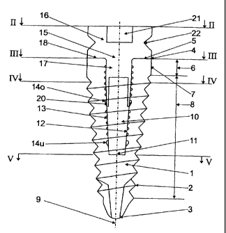
Figures 1 to 5 show an implant I made of titanium, which has an approximately
conical extemal
shape, and which is provided with an extemal screw thread 2. The implant 1 has
a rounded
CA 02405093 2002-10-03
distal end 3 and a proximal end 4, which consists of a basically annular
contact surface 5. In a
section 6 which is adjacent to the contact surface 5, the implant 1 has a
cylindrical shape with a
mirror-finished generating service 7. In a subsequent screw thread section 8,
the implant 1 is
conical in shape. Starting from contact surface 5 and extending parallel to a
longitudinal axis 9
of the implant 1 is a mounting recess 10, which runs over the entire length of
section 6 and over
part of the length of the screw thread section 8.
As may be seen in Figure 3, the cross-section of the mounting recess 10,
throughout the area of
section 6, exhibits the shape of a rounded rectangle. Starting at the screw
thread section 8, the
cross-section of the mounting recess 10 continually tapers off, so that, at
the base 11 of the
mounting recess 10, the cross-section is in the shape of a rounded square (cf.
Figure 5). The
transition from the rounded rectangular shape to the rounded square shape of
the cross-section
is continuous and without steps.
As may especially be seen in Figure 1, the wall 12 of the mounting recess 10
is provided with a
pluraiity of circular grooves 13, which are directed perpendicular to the
longitudinal axis 9. In
addition, the wall 12 is provided with a top circular groove 14o and a bottom
circular groove 14u,
whose function will be explained later this document.
Installed in the implant as shown in Figure 1 is a cap 15, which consists of
an approximately
symmetrical top part 16 and a connection pin 17, said connection pin being
arranged coaxially
with the top part 16 and extending into the mounting recess 10. A contact
surface 18 of the top
part 16 abuts against the contact surface 5 of implant 1 in a non-positive
manner.
As may be seen in Figure 3, the connection pin 17, in its upper part, has an
approximately
rectangular cross-section, whereby the comers of said cross-section are
broken, so that, in the
rounding area of the cross-section of mounting recess 10, between the
connection pin 17 and
the wall 12 of the mounting recess 10, four ventilation channels 19a are
created. Accordingly,
when the connection pin 17 is inserted into the mounting recess 10, displaced
air can be
conducted upward without necessitating a pressure structure which would
interfere with the
disassembly process; the air can escape through four radially outward-directed
ventilation
grooves 19b, which are formed in the front surface of the implant 1 and
communicate with the
ventilation channels 19a.
CA 02405093 2002-10-03
6
In the installed state, the outer generating surfaces of the connection pin 17
abut against the
wall 12 of the mounting recess 10 and against the two contact surfaces 5 and
18 in a non-
positive manner.
Because the cap 15 only temporarily remains on the implant 1 following the
implantation, it is
connected to the implant only by means of four clip devices 20, which engage
into the top
circular groove 14o.
As may especially be seen in Figure 6, the rod-shaped clip devices 20, in the
vicinity of their
distal end, have a radially outward-protruding projection 20a, which is placed
entirely inside the
circular groove 14o. The projection 20a tapers off increasingly, up to the
distal end of the elastic
clip device 20, so that the cap can be introduced directly into the mounting
recess 10.
The positive fit created by means of the clip devices 20 between the implant 1
and the cap 15,
or between the implant I and any other medical device, may be disassembled by
pulling the cap
15 axially upward, starting from the installed state shown in Figure 1. When
an upward-directed
axial force is exerted on the cap 15, the latter, due to the positive nature
of the connection, is
initially retained in the installed position. However, when the force exceeds
a predetermined
amount, the clip devices 20 are radially and inwardly displaced, because the
projections 20a are
guided into the top section of the wall of the circular groove 14o, by means
of a component
which is perpendicular to the longitudinal axis 11 [Translator's note: as
written in the original
German document; this is probably an error for "longitudinal axis 9"] of the
implant 1. As soon as
the radially outermost part of the projection 20a has reached the top edge 14'
of the circular
groove 14o, the positive fit is disassembled and the cap 15 may be fully
removed from the
mounting recess 10 of the implant 1 by means of only a slight amount of force.
Instead of the engagement of the clip devices 20 in the top circular groove
14o as shown in
Figure 1, it is possible, provided that the connection pin 17 and/or the clip
devices 20 are
sufficiently elongated, to have them engage in the bottom circular groove 14u.
The cap 15 is installed in the implant 1 by the manufacturer thereof and
serves, on one hand, to
screw in the implant 1, fotlowing the preparation of a suitable hole in the
bone, by means of a
CA 02405093 2002-10-03
7
screwdriver which engages into the slot 21 shown in Figure 2. By virtue of the
approximately
rectangular cross-section of the connection pin 17 and the adjusted mounting
recess 10, it is
possible to exert torque upon the implant 1 by means of the cap 15. Following
the
implementation, the cap 15 remains on the implant 1, in order, on the other
hand, to protect the
mounting recess 10 from contamination from the outside.
About three to six months after the insertion of the implant in the jawbone,
the healing has
progressed so far that the mucous membrane which covers the cap 15 can be
opened by
means of a second operation. The cap 15 is removed by means of a tool
resembling a pair of
pliers, which engages into a V-shaped circular groove 22 in the top part 16
and, by means of a
slight axial tug in an upward direction, removes the entire cap 15 from the
implant 1. The
positive fit created by the clip devices 20 is thus disassembled, thanks to
the elasticity of the clip
devices 20.
As shown in Figure 7, a connection pin 17 of a Gum Shaper 23 is now inserted
into the
mounting recess 10. The fastening principle is the same as for the cap 16. By
way of example,
Figure 8 shows that the clip devices 20 of the Gum Shaper 23 engage into the
bottom circular
groove 14u. However, in a similar manner, it is conceivable to have a Gum
Shaper 23 in which
the clip devices 20 engage into the top circular groove 14o.
Figure 8 shows a Gum Shaper with a slot, in the manner used for trans-gumi
healing. In this
case, no cap is used; rather, the implant with the Gum Shaper is screwed
directly into the
jawbone.
Figure 9 shows a laboratory implant 1 L, whose mounting recess corresponds to
that of the
implant 1, and into which a blank 24 for a temporary false tooth is inserted,
similarly by means
of a connection pin 17. The blank 24 is shaped like a truncated cone and
flares out, starting
from the front surface 5L of the laboratory implant 1 L, at an angle a of 15 .
In this way, any
oblique positioning of the implant I or 1 L, relative to neighboring teeth or
additional implants,
may be adjusted in additional angle ranges in all directions. The connection
pin 17 of the blank
24 is similarly provided with clip devices 20, which ensure an uncomplicated
fixation and
removal of the blank 24.
CA 02405093 2002-10-03
8
Figure 10 shows a completely finished temporary false tooth 25, which was
produced in a dental
laboratory by means of chip-producing processing of the blank 24. An outer
ceramic layer 26 is
bumed onto the ground blank 24. The finished temporary false tooth 25 can then
be introduced
into the implant 1 in the patient's jaw (Figure 11) and can be attached there
by means of clip
devices 20, where it will remain until the patient can be fitted with the
final false tooth 28.