Note: Descriptions are shown in the official language in which they were submitted.
CA 02405715 2002-10-09
WO 01/79479 PCT/USO1/11811
1
METHODS FOR SELECTIVE TARGETING
BACKGROUND OF THE INVENTION
The present invention is directed to methods for the selection and
identification of
compounds capable of binding specifically to a target in the presence of
undesired
background targets (anti-targets) using libraries of similar compounds. In one
particular
aspect, the present invention is related to the selection of ligands from
peptide libraries.
Ligand peptides identified according to the method of the invention have a
binding affinity
and a selectivity to a target similar to the binding affinity and selectivity
of antibodies.
The literature is replete with examples of recent advances in methods for
screening
large library pools of compounds, especially peptides. Methods for screening
these
compounds to identify molecules that bind to a preselected target have also
been
advanced. One well-known method is biopanning which was originally developed
by Smith,
G.P., (1985), Science 228:1315. Biopanning in its simplest form is an in vitro
selection
process in which a library of phage-displayed peptides is incubated with a
target. The
target and phage are allowed to bind and unbound phage are washed away. The
specifically bound phage are then acid eluted. The eluted pool of phage is
amplified in vivo
and the process is repeated. After a number of rounds individual clones are
isolated and
sequenced.
A number of variations of the biopanning technique first introduced by Smith
have
been described and reference is made to Christian et al., (1992) J. Mol.
Biol., 227:711;
Cwirla et al., (1990) Proc. Natl. Acad. Sci. USA, 87:6378; Cull et al., (1992)
Proc. Natl.
Acad. Sci. USA, 89:1865; Huls et al., (1996) Nature Biotechnol., 7:276; and
Bartoli et al.,
(1998) Nature Biotechnol., 16:1068.
Huls et al., 1996 supra, describe a method comprising flow cytometry-based
subtractive selection of phage antibody on intact tumor cells. The phage-
displayed
antibodies remain bound to the target during the flow-cytometric selection.
However, prior
to amplification the cell-bound phages are eluted from the target. WO 98/54312
discloses
selection of antibodies under mild conditions with high affinities for
antigens using antibody
libraries displayed on ribosomes.
In many prior art methods it is generally assumed that elution of target bound
ligands is sufficient to identify the tightest binding ligands in a library.
However, a number of
research papers report on low affinity binders using elution techniques (U.S.
Patent No.
5,582,981). Nevertheless, physical separation of the ligands from the target
prior to
amplification or identification is the standard method for selecting ligands
that bind to a
preselected target.
CA 02405715 2002-10-09
WO 01/79479 PCT/USO1/11811
2
Balass et at., (1996) Anal. Biochem., 243:264, describe the selection of high-
affinity
phage-peptides from phage-peptide libraries using a biotinylated target
immobilized on a
nitrostreptavidin matrix. The interacting phage particles were released under
conventional
acid elution. Further, after acid elution, the target complex was analyzed for
bound phage.
These particles were exposed to alkaline solutions or free biotin to release
the target bound
phage particles from the solid support. The affinity of the isolated phage was
found to be
higher than the phage released by traditional acid elution methods. However,
the
synthetically prepared peptides exhibited a lower affinity for the target than
the peptides
prepared from sequences obtained by acid-eluted phage.
Other targeting methods include, for example, SELEX. This is a procedure in
which
an oligonucleotide from a library of randomized sequences is embedded in a
pool of nucleic
acids. Many cycles of affinity selection to a target of the oligonucleotide
from the
heterologous RNA or DNA population occurs. The target and annealed nucleic
acids are
partitioned and amplified. In order to proceed to the amplification step,
selected nucleic acids
must be released from the target after partitioning. (U.S. Patent No.
5,475,096)
While various methods for screening and selecting libraries of compounds
exist,
improved methods that do not require multiple rounds of selection are
particularly needed for
compounds that a) bind tightly and specifically to targets that are not well-
defined at the
chemical, biochemical or genetic level but have macroscopic properties that
are desirable to
target, b) bind tightly and specifically to targets that cannot be easily
physically separated
from a large background of undesirable targets (anti-targets), and c) bind to
targets under
harsh conditions, such as acidic pH, high detergent concentration or high
temperature.
The selective targeting method according to the invention overcomes some of
the
above deficiencies of the prior art methods and in particular offers an
advantage in rapidly
identifying compounds, particularly peptides, that bind with a high affinity
and selectively to
a target.
SUMMARY OF THE INVENTION
In one aspect, the invention concerns a method for screening a ligand library
comprising contacting the ligand library with an anti-target to allow the
ligands to bind with
the anti-target; separating unbound ligands and contacting said unbound
ligands with the
selected target to allow said unbound ligands to bind with the target to form
a target-bound
ligand complex; separating said target-bound ligand complex from ligands which
do not bind
to said target; and identifying the target-bound ligands on the target-bound
ligand complex.
In another aspect, the invention concerns a method for screening a ligand
library
comprising contacting the ligand library essentially simultaneously with a
selected target and
an anti-target to allow the ligands to bind with the target forming a target-
bound ligand
CA 02405715 2002-10-09
WO 01/79479 PCT/USO1/11811
3
complex; separating the target-bound ligand complex from the anti-target, anti-
target bound
ligands and free ligands; and identifying the ligands of the target-bound
ligand complex. The
contacting step may be accomplished either in vivo or in vitro.
In one preferred embodiment, the selectivity of ligand binding to a target
compared to
ligand binding to an anti-target is about at least 10:1. In a second preferred
embodiment, the
ligand is a peptide but not an antibody and is bound to the target with a KD
at least about 10"'
M and preferably in the range of about 10-7 M to 10"10 M. In a third preferred
embodiment, the
ligand library is a peptide library. Preferably the peptides identified
according to the method
are less than 25 amino acids in length and more preferably between 4 to 15
amino acids in
length. In a fourth embodiment, the koff is about 10-4 sec -1 or less. In a
fifth embodiment, the
target is a stain, and particularly a stain on fabric, wherein the stain is a
porphyrin derived
stain, a tannin derived stain, a carotenoid pigment derived stain, an
anthocyanin pigment
derived stain, a soil-based stain, oil-based stain, or human body soil stains.
In yet a further aspect, the invention is directed to the ligands,
particularly peptide
ligands, which are identified by the selective targeting method of the
invention.
Another embodiment of the invention concerns a method for identifying peptides
useful in a cleaning composition comprising, contacting a peptide library with
an anti-target to
allow the peptides to bind with the anti-target, wherein the anti-target is
selected from the
group consisting of fabric, ceramic, glass, stainless steel, and plastic;
separating unbound
anti-target peptides, contacting the unbound anti-target peptides with a
target wherein the
target is a stain selected from the group consisting of porphyrin derived
stains, tannin derived
stains, carotenoid pigment derived stains, anthocyanin pigment derived stains,
soil-based
derived stains, oil-based derived stains and human body soil stains to allow
the unbound
peptides to bind with the stain to form a stain-bound peptide complex; and
identifying the
stain-bound peptide on the stain-bound peptide complex. In at least one
embodiment the
peptide binds to the stain with a KD in the range of about 10-7M to 10"10M.
BRIEF DESCRIPTION OF THE DRAWINGS
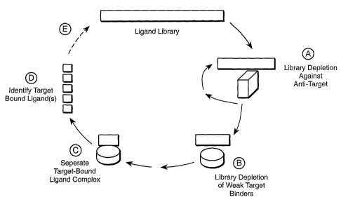
Figure 1 is a general schematic diagram of the selective targeting method
disclosed
herein. The method comprises the steps of, a) selection against anti-targets
which provides a
library of ligands depleted of anti-target bound ligands, b) selection for the
target by formation
of a target-bound ligand complex, c) separation of the target-bound ligand
complex, d)
identification of the target-bound ligands, and e) optionally sequencing the
target-bound
ligands, exposing the target-bound ligands to additional rounds of selective
targeting, and/or
diversification.
CA 02405715 2002-10-09
WO 01/79479 PCT/USO1/11811
4
Figures 2A and 2B are photographs of a gel of PCR amplified DNA fragments
after
lysis of target bound phage. Figure 2A illustrates TNF-a bound phage and
Figure 2B
illustrates IL-6 and IL-8 bound phage.
Figure 3 is a photograph of a gel of PCR amplified DNA fragments for soil-
targeted
peptides.
Figure 4 illustrates binding, dissociation and attempted elution of phage
peptide clone
Al corresponding to RYWQDIP (SEQ ID NO: 3) from immobilized TNF-a on an lAsys
biosensor cuvette.
Figures 5A and 5B are images of collar soils and the corresponding polyester
fabric
as viewed by digital imaging and autoradiography, respectively.
Figures 6A and 6B illustrate the fractional percent 14C labeled peptide
binding to collar
soils on polyester cotton fabric. Fig. 6A illustrates a soil-targeted peptide,
SISSTPRSYHWT,
(SEQ ID NO: 20) which is terminally labeled with 14C-glycine wherein o depicts
stain #1,
^ depicts stain #2, and ^ depicts blue polycotton and Fig. 6B illustrates a
random peptide,
NFFPTWILPEHT (SEQ ID NO: 78) which is terminally labeled with 14C-glycine.
Figure 7 illustrates the kinetics of dissociation of the Ni-chelated peptide
GGHTFQHQWTHQTR (SEQ ID NO: 28) from collar soil (=) and the corresponding
cotton
fabric (o). The slope of the lines correspond to rate constants koff = 1 x 10-
3 sec 1.
Figure 8 is a photograph of a gel of PCR amplified fragment for egg soil
targets and
stainless steel or glass bead anti-targets.
Figure 9 illustrates ELISA assay results for binding of 3 peptides. LESTPKMK
(SEQ
ID NO: 115) binds to hair and FTQSLPR (SEQ ID NO: 116) selectively targets
skin and not
hair (^ depicts hair and ^ depicts skin).
DETAILED DESCRIPTION OF.THE INVENTION
A. Unless defined otherwise, all technical and scientific terms used herein
have the
same meaning as commonly understood by one of ordinary skill in the art to
which this
invention pertains. For the purposes of the present invention, the following
terms are used to
describe the invention herein.
The term "ligand" refers to a molecule or compound that is recognized by a
particular
target or anti-target. The term is independent of molecular size or
compositional feature. The
ligand may serve as a substrate for an enzyme-catalyzed reaction, as an
agonist, as an
antagonist, act as a signal messenger, or stimulate or inhibit metabolic
pathways. Ligands
may be nucleic acids, peptides, peptide derivatives, peptidomimetics,
polypeptides, small
organic molecules, carbohydrates and other molecules that are isolated from a
candidate
mixture that acts on a target in a desirable manner. Preferably the desirable
manner is
binding the target, but could include for example, catalytically changing the
target or reacting
CA 02405715 2009-11-24
I =
WO 01/79479 PCT/USO1/11811
with the target that modifies or alters the target. In one preferred
embodiment, the ligand has
a binding affinity for the target in the range of an antibody binding affinity
for a selected
receptor.
The term "library" refers to a collection of chemical or biological entities
that can be
5 created in a single reservoir and simultaneously screened for a desired
property. As used
herein a library can have a minimum size of at least two members and may
contain as many
as 1015 members. In one aspect, the library has at least 102 members. In
another aspect, the
library has at least 103 members. In yet another aspect, the library has at
least 106 members.
In a further aspect, the library has at least 109 members. The size of a
library refers to the
total number of entities comprising the library whether the members are the
same or different.
A "peptide library" refers to a set of peptides and to the peptides and any
fusion
proteins containing those peptides. Stochastic or random processes may be used
to
construct random peptides. The term "random" does not mean that the library
composition is
not known.
The term "peptide" refers to an oligomer in which the monomeric units are
amino
acids (typically, but not limited to L-amino acids) linked by an amide bond.
Peptides may be
two or more amino acids in length. Peptides identified according to the
invention are
preferably less than 50 amino acids in length, more preferably less than 30
amino acids in
length, also preferably less than 25 amino acids in length, and preferably
less than 20 amino
acids in length. In one preferred embodiment the peptides identified according
to the method
of the invention are between 4 and 15 amino acids in length. However, in
general peptides
may be up to 100 amino acids in length. Peptides that are longer than 100
amino acids in
length are generally referred to as polypeptides. Standard abbreviations for
amino acids are
used herein. (See Singleton et al.,. (1987) Dictionary of Microbiology and
Molecular Biology,
Second Ed., page 35).
The peptides or polypeptides may be provided as a fusion peptide or protein.
Peptides include synthetic peptide analogs wherein the amino acid sequence is
known. The
term peptide does not include molecules structurally related to peptides, such
as peptide
derivatives or peptidomimetics whose structure cannot be determined by
standard
sequencing methodologies, but rather must be determined by more complex
methodologies
such as mass spectrometric methods. Peptidomimetics.(also known as peptide
mimetics) are
peptide analogs but are non-peptide compounds. Usually one or more peptide
linkages are
optionally replaced. (Evans et al., (1987) J. Med. Chem. 30:1229). The term
"protein" Is well
known and refers to a large polypeptide.
- The term "nucleic acid" means DNA, RNA, single-stranded or double-stranded
and
chemical modifications thereof. Modifications may include but are not limited
to modified
bases, backbone modifications, methylations, unusual base pairing
modifications, and
CA 02405715 2002-10-09
WO 01/79479 PCT/USO1/11811
6
capping modifications. When a nucleic acid library is used in the selective
targeting method
of the invention, the nucleic acid ligand is generally between 4 and 250
nucleotides in length,
and preferably between 4 and 60 nucleotides in length.
The invention further includes ligands, preferably nucleic acid, peptide or
polypeptide
ligands and more preferably peptide ligands that have substantially the same
ability to bind to
a target as the nucleic acid, peptide or polypeptide identified by the
selective targeting
method described herein. Substantially the same ability to bind a target means
the affinity
and selectivity is approximately the same as the affinity and selectivity of
the ligands selected
by the method herein claimed.
to Additionally a ligand having substantially the same ability to bind to a
target will be
substantially homologous to the Iigand identified by the disclosed selective
targeting method.
With respect to a nucleic acid sequence, substantially homologous to an
identified ligand
means the degree of primary sequence homology is in excess of 80%, preferably
in excess
of 85%, more preferably in excess of 90%, further preferably in excess of 95%,
even more
preferably in excess of 97%, and most preferably in excess of 99%. It will be
appreciated by
those skilled in the art that as a result of the degeneracy of the genetic
code, a multitude of
peptide encoding nucleotide sequences may be produced. A peptide or
polypeptide is
substantially homologous to a reference peptide or polypeptide if it has at
least 85%
sequence identity, preferably at least 90% to 95% sequence identity, more
preferably at least
97%, and most preferably at least 99% identical or equivalent to the reference
sequence
when optimally aligned. Optimal alignment of the sequences may be conducted by
various
known methods and computerized implementation of known algorithims (e.g.
TFASTA,
BESTFIT, in the Wisconsin Genetics Software Package, Release 7.0, Genetics
Computer
Group, Madison, WI). General categories of equivalent amino acids include 1)
glutamic acid
and aspartic acid; 2) lysine, arginine, and histidine; 3) alanine, valine,
leucine, and isoleucine;
4) asgaragine and glutamine; 5) threonine and serine; 6) phenylalaine,
tyrosine and
tryptophan; and 7) glycine and alanine. It is well within the ordinary skill
of those in the art to
determine whether a given sequence substantially homologous to those
identified herein
have substantially the same ability to bind a target.
A small organic molecule as defined herein is a molecule, preferably a
nonpolymeric
molecule, having a molecular weight of approximately 1000 daltons or less and
more
preferably 500 daltons or less. A "peptoid" is defined herein as an
enzymatically resistant
peptide analog.
The term "target" or "anti-target" refers to molecules or heterogeneous
molecules that
have a binding affinity as defined herein, for a given ligand. Both target and
anti-targets may
be naturally occurring or synthetic molecules or heterogeneous molecules.
CA 02405715 2009-11-24
WQ 01/79479 PCT/USO1/11811
The binding affinity of a ligand for its target or anti-target may be
described by the
dissociation constant (K0), concentration needed for 50% effective binding
(EC50), or
concentration needed for 50% inhibition of binding of another compound that
binds to the
target (ICso). K D is defined by k ko,,. The k value defines the rate at which
the target-
ligand complex breaks apart or separates. This term is sometimes referred to
in the art as
the kinetic stability of the target-ligand complex or the ratio of any other
measurable
quantity that reflects the ratio of binding affinities, such as an enzyme-
linked
immunosorbent assay (ELISA) signal or radio-active label signal. Selectivity
is defined by
the ratio of binding affinities or k for dissociation of the ligand-complex
(target Kpl anti-
,0 target Q. The k,,, value describes the rate at which the target and ligand
combine to form
the target-ligand complex.
The term "contacting" is broadly defined to mean placing a library of ligands
and a
target or anti-target in immediate proximity or association and includes in
vitro and in vivo
contact. The term includes touching, associating, joining, combining,
intravenous injection,
is oral administration, intraperitoneally, topical application, Intramuscular,
inhalation,
subcutaneous application and the like. The term "separating" as used herein
means to
select, segregate, partition, isolate, collect, keep apart and disunite.
"Amplifying" means a process or combination of process steps that increases
the
amount or number of copies of a molecule or class of molecules. In one aspect,
20 amplification refers to the production of additional copies of nucleic acid
sequences that is
carried out using polymerase chain reaction (PCR) technology well known in the
art. In
another aspect, amplification refers to production of phage virions by
Infection of a host.
As used in the specification and claims, the singular "a", "an" and "the"
include the
plural references unless the context clearly dictates otherwise. For example,
the term "a
25 protease" may include a plurality of proteases.
The following references describe the general techniques employed herein:
Sambrook et at., (1989) Molecular Cloning: A Laboratory Manual, Cold Spring
Harbor
Laboratory Press, Cold Spring Harbor, NY; Innis et al., PCR Protocols--A Guide
to Methods
and Applications (1990), Academic Press, Inc.; Kay et al., (1996) Phage
Display of Peptides
so and Proteins, Academic Press; Ausubel et al., (1987) Current Protocols in
Molecular Biology,
Greene-Publishing & Wiley Interscience NY (Supplemented through 1999); Berger
and
Kimmel, (1987) Methods in Enzymology, Vol. 152. Academic Press Inc., San
Diego, CA.
CA 02405715 2009-11-24
S =
WQ 01/79479 PCT/USO1/11811
8
B. General Method
Described herein is a selective targeting method for screening a library of
ligands
having a binding affinity and selectivity for a selected target. In its most
basic form the
selective targeting method may be defined as follows: Preparing or obtaining a
library of
ligands, preferably peptides of different sequences and more preferably a
random peptide
library. Deselecting ligands that bind with an anti-target by contacting the
ligand library with
an anti-target under conditions favorable for binding between the ligands of
the library and
the anti-target; allowing the anti-target to bind with the ligands; and
separating the anti-target
non-binders (unbound Iigands) from the anti-target ligand bound molecules and
any free
ligands. Contacting the anti-target non-binders with a selected target under
suitable
conditions and allowing them to bind. Ligands with an affinity for the target
will bind to form a
target-bound ligand complex. The removal of ligands bound to the anti-target
and removal of
weak target-bound ligands may generally be referred to as library depletion.
The target-
bound ligand complex is then separated from the remaining mixture including
the unbound
is ligands, and the target-bound ligands are identified. The target-bound
ligand complex or the
target-bound ligands may then optionally be subjected to amplification,
sequencing or further
rounds of selection (Figure 1). The invention further comprises the ligands
identified
according to the selective targeting method of the invention.
In the practice of the invention, a library of compounds to be tested will
generally be
provided. A library of Iigands may include, but is not limited to, random
peptide iibraries,
synthetic peptide or peptidomimetic combinatorial libraries, peptide loop
libraries,
combinatorial chemical libraries, and oligonucleotide libraries. These
libraries are well known
to those in the art as well as methods for making said libraries. Reference is
made to Barbas,
C.F. (1993) Current Opinion in Biotech., 4:526; Cwirla et at., (1990) supra;
Scott and Smith,
(1990) Science, 249:386; Cull et al., (1992) supra; Pinilla et al., (1994)
Biochem. J. 301:847;
Sambrook et al., (1989) supra; Ausubel et al., (1987) supra; and Gubler and
Hoffman, (1983)
Gene 25:263.
One preferred type of library includes random peptide libraries (also
sometimes
referred to in the art as epitope libraries). These libraries may include cell-
surface display
libraries, for example yeast display (Boder and Wittrup (1997) Nat.
Biotechnol., 15:553);
peptide libraries inserted into proteins (Lenstra et al., (1992) J. Immunoi.
Methods, 152:149
and U.S. Pat. No. 5,837,500); direct screening of peptides on polysomes (Tuerk
et al., (1990)
Science 249:505) and phage display libraries (Delvin et at., (1990) Science
249:404;
W091/18980; Dower et at. W091/1 9818; and Parmley at at., (1988) Gene 73:305).
Phage
display libraries are particularly preferred. A phage display library is a
library in which
numerous peptides are displayed on the surface of a bacteriophage, such as a
filamentous
phage. The peptide or protein is expressed as a fusion with a coat protein of
the
CA 02405715 2002-10-09
WO 01/79479 PCT/USO1/11811
9
bacteriophage resulting in display of the fusion protein on the surface of the
virion while the
DNA encoding the fusion resides within the virion. Suitable non-limiting
examples of vectors
for construction of phage libraries include fAFFI; the fUSE series, such as
fUSE5; lamba
phage vectors; and T7select (non-filamentous) phage vectors. (Smith and Scott
(1993)
Methods Enzymol. 217:228; and Cwirla et al., (1990) Proc. Natl. Acad. Sci. USA
87 :6378).
Phage-peptide library kits are available and reference is made to Chiron Corp.
(Emeryville,
CA), New England BioLabs Inc., Catalog No.8100 (Beverly, MA), and Novagen
Catalog No.
70550-3 (Madison WI). While various antibody libraries are known, including
antibody display
libraries on phage (de Bruin et al., (1999) Nat. Biotechnol., 17:397), in one
preferred aspect
of the present invention, the library of ligands used in the selective
targeting method
according to the invention will not include antibodies.
Another type of peptide library encoded by nucleic acids includes a library
wherein
the peptide is expressed as a fusion with another protein, for example, either
a cell-surface
protein or an internal protein of a host. The nucleotides encoding the peptide
are inserted into
Is a gene encoding the internal protein. Various examples of this type of
library include the
fusion of peptides to a lac repressor, GAL4, thioredoxin, and various
antibodies (U.S. Patent
Nos. 5,283,173; 5,270,181; and 5,292,646). Cull et al. (1992) Proc. Natl.
Acad. Sci. USA
89:1865 teach the construction of a fusion gene encoding a fusion protein of
peptide library
members and Lacl. Nucleic acids encoding a library of peptides are inserted
into a gene
encoding Lacl. The fusion protein and the fusion plasmid encoding the fusion
protein are
physically linked by binding of the peptides to the lac operator sequence in a
plasmid. Host
cells may be transformed with the library plasmids. The cells expressing the
fusion protein
are lysed releasing the fusion protein and associated DNA (see for example
U.S. Pat. No.
5,733,731). The library can then be screened or selected. DNA shuffled
libraries are also
known which are constructed by homologous exchange of DNA fragments during DNA
recombination methods or by synthetic methods (see for example U.S. Pat. No.
5,605,793
and Stemmer (1994), Proc. Natl. Aca. Sci. USA 91:10747).
So called anchor libraries have been described in PCT US96/09383 and WO
97/22617. This is a peptide library wherein peptides have non-continuous
regions of random
ao amino acids separated by specifically designated amino acids. These
libraries are made by
genetic or chemical means.
A combinatorial chemical library and particularly a peptide library may also
be
synthesized directly by methods known in the art including, but not limited to
synthesis by
arrays (Foder et al., (1991) Science 251:767); synthesis on solid supports
(W097/35198);
and other chemical methods such as those disclosed in Lam et al., (1993)
Bioorg. Med.
Chem. Lett., 3:419, Tjoeng et al., (1990) Int. J. Pept. Protein Res. 35:141,
and W096/33010.
CA 02405715 2002-10-09
WO 01/79479 PCT/USO1/11811
Methods for creating combinatorial chemical libraries are also known in the
art.
Combinatorial libraries include large numbers of chemical variants for
peptides,
oligonucleotides, peptoids, carbohydrates, small organic molecules and even
solid-state
materials (Schultz et al., (1995) Science, 268:1738). A core structure will be
varied by adding
5 substituents or by linking different molecular building blocks. Libraries
may include molecules
free in solution, linked to solid particles or beads, or arrayed on surfaces
of modified
organisms. Virtually any class of compounds may be modified by varying
substituents around
the core molecule. Various non-limiting examples of classes of compounds for
combinatorial
libraries include benzodiazepines; mercaptoacyl prolines; carbamates; chalcone
libraries;
to ketoamide conjugates; polyketones; paclitaxel libraries; anilides;
aryloxyphenoxypropionates;
oxazolidinones; carbohydrates; and numerous other classes. While methods for
making
combinatorial libraries are well documented in the literature, these methods
may be very time
consuming. Various companies now make instrumentation to generate
combinatorial libraries
from both solution and solid phase synthesis (CombiChem Inc. (San Diego, CA);
Advanced
is ChemTech (Louisville); Zymark Corp. (MA); and Hewlett Packard (CA)). Once a
library has
been generated it can optionally be purified for example by high performance
liquid
chromatography (HPLC). Once a small organic molecule is screened and
identified
according to the selective targeting method of the invention, it may be
produced on a larger
scale by means of organic synthesis known in the art.
As taught herein not only are standard methods for generating libraries of
ligands well
known, but also ligand libraries may be obtained commercially, for example
from Sigma (St.
Louis Mo.) or from various public sources such as American Type Culture
Collection (ATCC)
and the National Institute of Health (NIH).
Suitable targets and anti-targets used in the selective targeting method
according to
the invention include, but are not limited to, proteins, peptides, nucleic
acids, carbohydrates,
lipids, polysaccharides, glycoproteins, hormones, receptors, antigens,
antibodies, viruses,
pathogens, toxic substances, metabolites, inhibitors, drugs, dyes, nutrients,
growth factors,
cells or tissues.
Sources of cells or tissues include human, animal, bacterial, fungal, viral
and plant.
Tissues are complex targets and refer to a single cell type, a collection of
cell types or an
aggregate of cells generally of a particular kind. Tissue may be intact or
modified. General
classes of tissue in humans include but are not limited to epithelial,
connective tissue, nerve
tissue, and muscle tissue.
Preferred human cellular targets or anti-targets include hematopoietic cells,
cancer
cells and retroviral-mediated transduced cells. Hematopoietic cells encompass
hematopoietic
stem cells, erythrocytes, neutrophils, monocytes, platelets, mast cells,
eosinophils, basophils,
B and T cells, macrophages, and natural killer cells.
CA 02405715 2002-10-09
WO 01/79479 PCT/USO1/11811
11
Non-limiting examples of protein and chemical targets encompassed by the
invention
include chemokines and cytokines and their receptors. Cytokines as used herein
refer to any
one of the numerous factors that exert a variety of effects on cells, for
example inducing
growth or proliferation. Non-limiting examples include interleukins (IL), IL-
2, IL-3, IL-4 IL-6, IL-
10, IL-12, IL-13, IL-14 and IL-16; soluble IL-2 receptor; soluble IL-6
receptor; erythropoietin
(EPO); thrombopoietin (TPO); granulocyte macrophage colony stimulating factor
(GM-CSF);
stem cell factor (SCF); leukemia inhibitory factor (LIF); interferons;
oncostatin M(OM); the
immunoglobulin superfamily; tumor necrosis factor (TNF) family, particularly
TNF-a; TGFP;
and IL-1 a; and vascular endothelial growth factor (VEGF) family, particularly
VEGF (also
referred to in the art as VEGF-A), VEGF-B, VEGF-C, VEGF-D and placental growth
factor
(PLGF).
Chemokines are a family of small proteins that play an important role in cell
trafficking
and inflammation. Members of the chemokine family include, but are not limited
to, IL-8,
stomal-derived factor-1(SDF-1), platelet factor 4, neutrophil activating
protein-2 (NAP-2) and
monocyte chemo attractant protein-1 (MCP-1).
Other protein and chemical targets include: immunoregulation modulating
proteins,
such as soluble human leukocyte antigen (HLA, class I and/or class II, and non-
classical
class I HLA (E, F and G)); surface proteins, such as soluble T or B cell
surface proteins;
human serum albumin; arachadonic acid metabolites, such as prostaglandins,
leukotrienes,
thromboxane and prostacyclin; IgE, auto or alloantibodies for autoimmunity or
allo- or
xenoimmunity, Ig Fc receptors or Fc receptor binding factors; G-protein
coupled receptors;
cell-surface carbohydrates; angiogenesis factors; adhesion molecules; ions,
such as calcium,
potassium, magnesium, aluminum, and iron; fibril proteins, such as prions and
tubulin;
enzymes, such as proteases, aminopeptidases, kinases, phosphatases, DNAses,
RNAases,
lipases, esterases, dehydrogenases, oxidases, hydrolases, sulphatases,
cyclases,
transferases, transaminases, carboxylases, decarboxylases, superoxide
dismutase, and their
natural substrates or analogs; hormones and their corresponding receptors,
such as follicle
stimulating hormone (FSH), leutinizing hormone (LH), thyroxine (T4 and T3),
apolipoproteins,
low density lipoprotein (LDL), very low density lipoprotein (VLDL), cortisol,
aldosterone,
3o estriol, estradiol, progesterone, testosterone, dehydroepiandrosterone
(DHBA) and its sulfate
(DHEA-S); peptide hormones, such as renin, insulin calcitonin, parathyroid
hormone (PTH),
human growth hormone (hGH), vasopressin and antidiuretic hormone (AD),
prolactin,
adrenocorticotropic hormone (ACTH), LHRH, thyrotropin-releasing hormone
(THRH),
vasoactive intestinal peptide (VIP), bradykinin and corresponding prohormones;
catechcolamines such as adrenaline and metabolites; cofactors including
atrionatriutic factor
(AdF), vitamins A, B, C, D, E and K, and serotonin; coagulation factors, such
as prothrombin,
thrombin, fibrin, fibrinogen, Factor VIII, Factor IX, Factor XI, and
vonWillebrand factor;
CA 02405715 2002-10-09
WO 01/79479 PCT/USO1/11811
12
plasminogen factors, such as plasmin, complement activation factors, LDL and
ligands
thereof, and uric acid; compounds regulating coagulation, such as hirudin,
hirulog, hementin,
hepurin, and tissue plasminigen activator (TPA); nucleic acids for gene
therapy; compounds
which are enzyme antagonists; and compounds binding ligands, such as
inflammation
factors.
Non-human derived targets and anti-targets include without limitation; drugs,
especially drugs subject to abuse, such as cannabis, heroin and other opiates,
phencyclidine
(PCP), barbiturates, cocaine and its derivatives, and benzadiazepine; toxins,
such as heavy
metals like mercury and lead, arsenic, and radioactive compounds;
chemotherapeutic
agents, such as paracetamol, digoxin, and free radicals; bacterial toxins,
such as
I ipopo lysacch a rides (LPS) and other gram negative toxins, Staphylococcus
toxins, Toxin A,
Tetanus toxins, Diphtheria toxin and Pertussis toxins; plant and marine
toxins; snake and
other venoms, virulence factors, such as aerobactins, or pathogenic microbes;
infectious
viruses, such as hepatitis, cytomegalovirus (CMV), herpes simplex virus (HSV
types 1, 2 and
6), Epstein-Barr virus (EBV), varicella zoster virus (VZV), human
immunodeficiency virus
(HIV-1, -2) and other retroviruses, adenovirus, rotavirus, influenzae,
rhinovirus, parvovirus,
rubella, measles, polio, pararyxovirus, papovavirus, poxvirus and
picornavirus, prions,
plasmodia tissue factor, protozoans, such as Entamoeba histolitica, Filaria,
Giardia,
Kalaazar, and toxoplasma; bacteria, gram-negative bacteria responsible for
sepsis and
nosocomial infections such as E. coli, Acynetobacter, Pseudomonas, Proteus and
Klebsiella,
also gram-positive bacteria such as Staphylococcus, Streptococcus,
Meningococcus and
Llycobacteria, Chlamydiae Legionnella and Anaerobes; fungi such as Candida,
Pneumocystis, Aspergillus, and Mycoplasma.
In one aspect the target includes an enzyme such as proteases,
aminopeptidases,
kinases, phosphatases, DNAses, RNAases, lipases, esterases, dehydrogenases,
oxidases,
hydrolases, sulphatases, cellulases, cyclases, transferases, transaminases,
carboxylases,
decarboxylases, superoxide dismutase, and their natural substrates or analogs.
Particularly
preferred enzymes include hydrolases, particularly alpha/beta hydrolases;
serine proteases,
such as subtilisins, and chymotrypsin serine proteases; cellulases; and
lipases.
In another aspect the target is a stain on a fabric or other surface material
such as
ceramic, glass, silica, wood, paper, metal and alloys, and living tissue, such
as skin. The
stain may be selected from the following non-limiting group of stains;
porphyrin derived
stains, tannin derived stains, carotenoid pigment derived stains, anthocyanin
pigment derived
stains, soil-based stains, oil-based stains, and human body derived stains.
Particularly the
stain may be a blood-derived stain or a chlorophyll-derived stain. More
specifically the stain
may be grass; paprika; a tea-derived stain; or a fruit or vegetable derived
stain, such as from
CA 02405715 2002-10-09
WO 01/79479 PCT/USO1/11811
13
wine, tomato and berries. A particularly preferred stain is human body soil,
and more
specifically stains referred to as collar soil.
In yet another aspect the target includes hematopoietic stem cells (HSCs). A
particularly preferred surface antigen expression profile of HSCs is CD34+Thy-
l , and
preferably CD34+Thy-1 {Lin . Lin" refers to a cell population selected on the
basis of the lack
of expression of at least one lineage specific marker. Methods for isolating
and selecting
HSCs are well known in the art and reference is made to U.S. Patent Nos.
5,061,620;
5,677,136; and 5,750,397.
In a further aspect, preferred targets include cytokines, particularly IL-2,
IL-3, IL-6, IL-
10, IL-12, IL-13, IL-14 and IL-16; EPO; GM-CSF; the TNF family; the VEGF
family, GFR; and
IL-1 a. Cytokines are commercially available from several vendors including
Amgen
(Thousand Oaks, CA), Immunex (Seattle, WA) and Genentech (South San Francisco,
CA).
Particularly preferred are VEGF and TNF-a. Antibodies against TNF-a show that
blocking
interaction of the TNF-a with its receptor is useful in modulating over-
expression of TNF-a in
several disease states such as septic shock, rheumatoid arthritis, or other
inflammatory
processes. VEGF is an angiogenic inducer, a mediator of vascular permeability,
and an
endothelial cell specific mitogen. VEGF has also been implicated in tumors.
Targeting
members of the VEGF family and their receptors may have significant
therapeutic
applications, for example blocking VEGF may have therapeutic value in ovarian
hyper
stimulation syndrome (OHSS). Reference is made to N. Ferrara et al., (1999)
Nat. Med.
5:1359 and Gerber et al., (1999) Nat. Med. 5:623. Other preferred targets
include cell-
surface receptors, such as T-cell receptors.
It is preferred that the target and anti-target are characterized in some
detail at the
structural, chemical or genetic level to allow some control over the purity,
stability and
concentration of the target. However, targets and anti-targets may be used
that are not well
characterized. Non-limiting examples of potentially not well-characterized
targets include
collar soil, tumor cells, human skin and hair.
A preferred anti-target includes fabric selected from the group consisting of
cotton,
wool, silk, polyester, rayon, linen, nylon and blends thereof.
In another aspect, when the target is damaged cells, tissue, or organs, the
anti-target
is healthy normal (non-damaged) cells, tissue, organs or combinations thereof.
Specific non-
limiting anti-target examples include healthy normal whole blood, skin, hair,
teeth, and nails.
In some applications, the target and anti-target can be reversed depending
upon the
specific application of interest. For example there may be multiple
applications where it is
desirable to target human skin and not hair. Therefore the anti-target would
be hair. In a
similar application it may be desirable to target human hair and not the
corresponding anti-
target, skin.
CA 02405715 2002-10-09
WO 01/79479 PCT/USO1/11811
1414-
The following general examples of target/anti-target used in the same
application are
provided for illustrative purpose only and are not meant to limit the
selective targeting method
disclosed herein: tumor cell/normal cell; receptor cell/cell not expressing
the receptor;
neoplastic cell/ normal cell; soil stain/cotton fabric; food stain/ceramic;
specific protease/other
protease; serine protease/whole blood; hematopoietic stem cell/whole blood;
specific enzyme
variant/other forms of the enzyme; virus in a cell/cell; TNF-alpha/blood
components; specific
insect enzyme/homologous enzymes in animals; hematopoietic stem cell/other
hematopoietic
cells; hair/skin; nucleus/mitochondria; cytoplasm/nucleus; alpha/beta
hydrolases/other
hydrolases; and a specific enzyme involved in photosynthesis/leaf tissue.
Both the target and anti-target concentrations to be used in the selective
targeting
method will vary depending on the type of ligand library, anti-target and
target used. As
discussed herein, the disclosed method has wide applicability to many
different targets and
anti-targets, therefore the concentration useful in the method may vary from
about 1.0 M to
10-15 M, preferably the concentration is in the 10-9 M range. In general an
excess amount of
anti-target relative to the amount of target is required. While not meant to
limit the invention,
this excess amount may be in the range of at least 10 fold greater to more
than 1000 fold
greater. An initial target concentration may be preferably provided in the
range of 10-3 M to
10-15 M. In one preferred embodiment, when the target is an enzyme, the target
concentration may be provided in the range of about 10-3 M to 10-12 M. In
another preferred
embodiment, when the target is a cytokine, the target may be provided in the
concentration
range of about 10-3 M to 10-12 M. In yet another embodiment, when the target
is a
hematopoietic cell, the target concentration may be provided in the range of
about 10 to 109
cells.
In one preferred embodiment, when the anti-target is a blood protein or an
enzyme,
the anti-target concentration may be provided in a concentration range of
about 1.0 M to 10-
12 M.
In certain preferred embodiments, the anti-target or target may be a material
or
surface, such as a fabric, ceramic or micro-fluidic chip. In this instance the
area of the target
or anti-target will be important. While not intended to limit the invention in
any manner, in
general the size of the anti-target or target material will be about 1.0 mm to
1.5 cm; more
preferably about 25.0 mm to 0.5 cm; however, the diameter or area may be more
or less than
these values.
In one aspect, the invention is directed to the screening and identification
of ligands
that bind to a selected target to form a non-covalent target-ligand complex
with a binding
affinity in the range of antibody affinities for antigens. The ligand binding
affinity according to
the present invention for KD, EC50 or IC50 is in the range of between about
10"7 M to 10-15 M,
although higher or low binding affinities may be achieved. In one aspect, the
affinity is in the
CA 02405715 2002-10-09
WO 01/79479 PCT/USO1/11811
1510-
range of at least about 1 0-7M, also at least about 10-8M, preferably at least
about 1.0"9 M and
also preferably at least about 10"12 M. In another embodiment, the affinity is
less than about
10-7M. In another aspect, koff values for the ligand-target complex will be
less than about 10-3
sec 1, less than about 10' sec 1, and also less than about 10"5 sec -1. The
ligands identified
according to the selective targeting method of the invention will not bind
with any significance
to the anti-target. While not meant to limit the invention, a preferred ligand
identified
according to the selective targeting method described herein may have a Ko for
the anti-
target greater than about 10 -4M, and preferably greater than about 10-1 M.
The selective targeting method according to the invention may be characterized
not
only by the binding affinity of a ligand to the target, but also may be
characterized by the
selectivity of the ligand-target complex. The selectivity of ligand binding
for a target
compared to ligand binding to an anti-target can be defined by a ratio of KD,
EC50 or IC50 in
the range of about 3:1 to 500:1. In one aspect, selectivity is at least about
5:1, preferably at
least about 10:1, more preferably at least about 20:1, even more preferably at
least about
30:1, even more preferably at least about 50:1, and yet more preferably at
least about 100:1.
In another aspect, the selective targeting method may be used to select
ligands with a
low affinity for the target but with a high selectivity for the target. In
this aspect, the selectivity
of ligand binding affinity for the target compared to said ligand binding to
an anti-target would
be at least about 5:1, preferably at least about 10:1, also preferably at
least about 20:1, more
preferably at least about 50:1, and even preferably at least about 100:1.
However, the target
binding affinity would be in the range of about 10"3 M tol0-7 M.
Methods for measuring binding affinities and selectivity are well known in the
art, and
these methods include but are not limited to measurement by radio-labeled
release and
competition assay; by isothermal titration calorimetry; biosensor binding
assays (Morton &
Myszka, (1998) Methods Enzymol. 295:268-294); by fluorescence and chemi-
luminescence
spectroscopy; and by mass spectrophotometry (Gao et al., (1996), J. Med,
Chem., 39:1949).
In one aspect, the anti-target is combined with the library of ligands and
allowed to
incubate prior to exposing the library of ligands to the target. In another
aspect, the anti-
target and target are combined with the library of ligands essentially
simultaneously.
Essentially simultaneously means at the same time or very close in time
wherein the ligand
library is exposed to both the anti-target and the target prior to any
separation step.
The selective targeting method as described herein may be performed in vitro
or in
vivo. When performed in vitro, the library of ligands and the anti-target (and
optionally the
target), are combined in or on a vessel. The vessel may be any suitable
material or
receptacle such as a plate, culture tube, microtiter plate, micro-fluidic
chip, petri dish and the
like.
CA 02405715 2009-11-24
S
WQ 01/79479 PCT/USO1/11811
Preferably, the anti-target and the target are available in an environment
where non-
specific binding events are minimized. This may be accomplished by various
means
including, but not limited to, 1) by coating a vessel containing the ligand
library and the
target/anti-target with BSA, skim-milk or other adsorbing protein to block non-
specific binding,
2) by labeling the target molecule with a capture agent such as a biotinylated
compound, for
example biotin, avidin, or mutated form thereof which can be subsequently
trapped by
streptavidin or a streptavidin derivative, such as nitrostreptavidin, 3)
displaying the
target/anti-target on magnetic beads that can be physically separated from the
library, or 4)
by using library display vectors with low background adsorption properties.
These methods
io are known in the art and reference is made to Parmley et at. (1988) supra;
and Bayer et al.,
(1990) Methods Enzymol. 184 :138.
A composition including a library of ligands and an anti-target may be
combined
together with additional compounds such as buffers and optionally detergents
and organic
solvents under suitable conditions to allow binding of the ligands with the
anti-target. One
Is skilled in the art is well aware of useful buffers. Non-limiting examples
include;
tris(hydroxymethyl)aminomethane (Tris) buffers; N-2-hydroxyethylpiperazine-N'-
2-
ethanesulfonic acid (HEPES) buffers; morphololino-ethanesulfonic acid (MES)
buffers;
buffered saline solutions, such as N,N-bis[2-hydroxyethyl]2-
aminoethanesulfonic acid (BES),
Tris, and phosphate-buffered saline (PBS), preferably buffered saline
solutions (Sambrook et
20 al., (1989) supra). Commercial buffers are available for example
SuperBlockT" (Pierce,
Rockford, IL). Other ingredients such as detergents, for example TweenTM and
Triton TM can be
used in the solutions.
Depending on the target, the composition including the ligand library and anti-
target is
incubated for a period of about 1 minute to about 96 hours to allow the
ligands to bind with
25 the anti-target. However, longer time periods may be used depending on the
stability of the
target or anti-target. The component containing the unbound anti-target
ligands Is separated
from the anti-target bound ligands after incubation. While not essential, the
separated
component including the unbound anti-target ligands may optionally be
transferred to a new
vessel including the anti-target, incubated and then the component containing
the unbound
30 anti-target ligands can again be separated from the bound anti-target
ligands. This transfer
process may be repeated numerous times, for example it may be repeated between
2 to 10
times or more. The repeated transfer step further reduces the number of
ligands that bind to
the anti-target. However, the contacting of the library of ligands with the
anti-target and the
separating of the anti-target bound ligands from the unbound ligands may be
accomplished
35 in one round. The contacting including incubation, and the separation
steps, whether
completed in one round or in multiple rounds may generally be referred to as
deselection.
CA 02405715 2002-10-09
WO 01/79479 PCT/USO1/11811
1711
In general, the temperature conditions during deselection may be between 2 and
30 C. The temperature is limited by the stability of the components and is
well within the skill
of one of ordinary skill in the art to determine.
The unbound anti-target ligands may be separated from the anti-target bound
ligands
by methods well known in the art. Some of these methods include liquid
transfer, washing,
centrifugation, filtration, chromatography, micro-dissection and fluorescence
activator cell
sorting (FAGS).
The ligand library, depleted of anti-target binding ligands and containing
unbound
ligands is transferred to a vessel including the target under suitable
conditions which will
allow one or more members of the ligand library to bind with the target
thereby forming a
target-bound ligand complex. In one aspect the ligands may be contacted with
the same
target. In another aspect the ligands may be contacted with an array of
targets at the same
time. One non-limiting example of an array of targets includes the contacting
of a ligand with
multiple stains on a surface. The ligands are incubated under conditions that
allow binding to
the target and generally for a period of time ranging from about 1 minute to
about 96 hours.
The incubation time depends on the stability of the target. When the target is
a stain, the
incubation period will generally range from about 5 minutes to about 90
minutes. The vessel
may further include buffers as described herein above. The temperature range
is generally
between about 2 and 30 C, and preferably about 18 to 25 C.
One skilled in the art is well aware of references describing cell, organ, and
tissue
culture, and reference is made to Atlas and Parks (eds) (1993), The Handbook
of
Microbiological Media, CRC Press, Boca Raton FL; Gamborg and Phillips (eds)
(1995) Plant
Cell Tissue and Organ Culture, Fundamental Methods, Springer Lab Manual
Springer-
Verlag.
The target-bound ligand complex may be subject to one or more wash steps. The
washing compounds may include buffers (such as TBS and PBS), detergents, acids
(glycine), organic solvents, bases, enzymes, sonication, or combinations
thereof, wherein
unbound ligands are washed. When the target-bound ligand complex is subject to
an acid
elution, the pH of the acid elution may be in the range of about 1.5 to 4.5,
preferably in the
range of about 2.0 to 3.5. The acid elution may take place for between 2 to 20
minutes and
generally no longer than about 10 minutes. The wash step may be repeated
numerous times
and in general can be repeated between 2 - 6 depending on the specific target
and ligand
library. Particularly when the washing step is with an acid, washing will
generally be followed
by neutralization with various well-known compounds and buffers, such as TRIS-
HCL. The
washing step results in a target-bound ligand complex comprising tight binding
ligands
having a KD, koff and selectivity values as herein defined.
CA 02405715 2002-10-09
WO 01/79479 PCT/US01/11811
181ti -
When the ligand library is contacted with the anti-target and target
essentially
simultaneously as opposed to sequentially the ligand library, anti-target and
target
composition may further include all materials described above for the
sequential exposure of
the anti-target and target.
Further when the ligand library is contacted with the anti-target and target
essentially
simultaneously, the method may also be performed in vivo. In this aspect, the
library of
ligands may be administered by means well known in the art, but preferably by
injection into
a host. If the library is a phage-peptide library, the number of transducing
units may be in the
range of 104 -1010. The host may be any animal, such as a human, mouse,
chicken, or pig,
preferably mouse. The target for example may be whole organs or damaged or
tumor tissue,
more specifically tumor blood vessels. If the target is a tissue or cells
found in the blood, the
library of ligands may be circulated in the blood for a period of about 1
minute to 10 minutes
and allowed to bind with the target. The target-bound ligand complex may be
recovered after
perfusion and the tissue dissected (Koivunen et al., (1999) Nature Biotech.
17:768 and Arap
et al., (1998) Science 279:377).
Separation of the target-bound ligands from the anti-target unbound ligands or
free
ligands in the mixture may also be accomplished by well-known means in the art
and these
methods include affinity chromatography; centrifugation; high-performance
liquid
chromatography (HPLC); filtration, such as gel filtration; enzyme-linked
immunosorbent
assays (ELISA); and fluorescence-activator cell sorting (FACS). The choice of
the
separating method will depend on various factors such as the target, anti-
target and ligand
molecules. The choice of the separation method is well within the skill of one
in the art and
a variety of instruments used for these separation methods are commercially
available.
(See Kenny and Fowell (eds) (1992) Practical Protein Chromatography Methods in
Molecular Biology, vol. 11, Humana Press, Totowa NJ)
The target-bound ligand on the target-bound ligand complex may be identified
by
various techniques including polymerase chain reaction (PCR), mass
spectrophotometry
(MS), surface plasmon resonance, immunoprecipitation and nuclear magnetic
resonance
(NMR) spectroscopy (U.S. Patent No. 4,683,202; Szabo et al., (1995) Curr.
Opin. Struct.
3o Bio.) 5:699; Harlow et al., (1999) Using Antibodies, A Laboratory Manual,
Cold Spring
Harbor Press; and Hajduk et al., (1999) J. Med Chem., 42:2315). Asymmetric PCR
may also
be used for identification of the target-bound ligand wherein a single primer
species or
primers in differential concentration may be used. As well known to those in
the art, when the
library members are genetically linked to the peptide or protein, DNA or mRNA
can be
amplified by PCR and the corresponding sequence subcloned into a vector for
sequencing
and identification.
CA 02405715 2002-10-09
WO 01/79479 PCT/USO1/11811
19113
During the process of the identifying step, the target-bound ligand may
separate from
the target-bound ligand complex, but the identifying step does not require
separation, and
preferably the target-bound ligand is not separated from the target-bound
ligand complex
prior to identification of the ligand. For example, in mass spectrophotometry
(MS), once the
target-bound ligand complex is injected into the mass spectrophotometer the
target-bound
ligand may be separated from the target complex. Additionally, PCR may be
directly carried
out on the target-bound ligand complex.
The selective targeting method according to the invention preferably includes
PCR to
identify target-bound peptides. According to the invention use of PCR results
in the recovery
of peptides not recovered by conventional biopanning methods which utilize
acid-elution. In
general, a ligand encoding a DNA is amplified by PCR with appropriate primers.
The presence of specific PCR products indicates that the target-bound ligand
encoding DNA is present. The amount of the target-bound ligand is determined
by
quantitative PCR. The degree of wash stringency can be monitored to a desired
level and to
95 very low detection levels for example to attomole levels. Nonspecific
ligand binders may be
competed out for example by adding wild type phage and designing primers that
only amplify
the ligand library. To prevent deterioration of signal-to-noise ratio, the
sequences flanking the
ligand encoding DNA may be changed frequently during rounds of selection.
Sensitivity for
the analysis of target-bound ligands may be controlled by changing target
concentration, the
number of PCR amplification cycles, the specificity of the PCR primers, and
the detection
method for PCR products.
In one embodiment, when the target is a tumor antigen, tumor tissue including
the
target-bound phage ligands may be excised from a tumor and addition of
appropriate PCR
primers, nucleotides, and polymerase may yield the amplified PCR product.
Various inhibitory
reactions of PCR may be alleviated by the addition of excipents including
bovine serum
albumin, cationic amines, and organic solvents and reference is made to Roux,
(1995)
"Optimization and Troubleshooting in PCR" in PCR Primer: A Laboratory Manual,
Cold
Spring Harbor Press. DMSO and glycerol may be used to improve amplification
efficiency
and specificity of PCR. The DNA of the target-bound ligand may also be
extracted and
purified using standard techniques.
To facilitate sequencing of desired clones or separation from undesired non-
specific
phage, the polynucleotide products generated by PCR may be labeled for example
with
biotinyl or fluorescent label moieties by incorporation during polymerase
mediated catalysis.
When the desired PCR product is to be cloned into a vector for additional
rounds of selective
targeting according to the method of the invention, it may be desirable to
introduce diversity
by mutagenic PCR methods, (See Stemmer, in Kay et al., supra). These include
cassette
mutagenesis, error prone FOR, DNA shuffling, ITCHY-SCRATCY and the like as is
well
CA 02405715 2002-10-09
WO 01/79479 PCT/USO1/11811
20ZU -
known by those in the art. Also reference is made to Tillett and Neilan,
(1999) "Enzyme-free
Cloning: A Rapid Method to Clone PCR Products Independent of Vector
Restriction Enzyme
Sites": Nucl. Acids. Res., 27:26e.
As mentioned above and as well known in the art, the PCR fragments may be
cloned
into various vectors for sequencing, they may be used in the formation of
peptide protein
fusions, or cloned into additional display vectors.
The target bound library members may also be identified preferably by mass
spectrometric methods. This is a rapid and accurate identification of the
structure of a
compound based on the mass of the compound and on fragments of the compound
generated in the mass spectrometry. The use of mass spectrometry to identify
the structure
of compounds has been reported in Cao et al., (1997) Techniques in Protein
Chemistry VIII,
Academic Press pages 177 - 184; and Youngquist et al., (1995) J. Am. Chem.
Soc.
117:3900. Also reference is made to Cheng et al., (1995) J. Am. Chem. Soc.,
117:8859 and
Walk et al., (1999) Angew. Che. Int. Ed., 38 :1763. One mass spectrometric
technique is
tandem mass spectrometry (MS/MS) wherein mass spectrometry is performed in
tandem
with liquid chromatography. To purify and separate the ligand of interest,
this type of MS is
preferably used to screen target-bound (igands other than phage-type peptides
because of
the need to separate and purify target-bound ligands from a biological system
prior to
injection of the ligands into a mass spectrometer. Various recently developed
MS techniques
are available for identification of the target-bound ligands. (See Wu et al.,
(1997) in
Chemistry and Biology, vol. 14(9):653, Marshall et al., (1998), Mass
Spectrometry Reviews
17:1, and Nelson et al., (1999) J. Mol. Recognition, 12:77).
Following the screening of one or more ligand members, particularly peptide
ligands,
the amino acid sequence of the peptides may be determined according to
standard
techniques known by those in the art such as direct amino acid sequencing of
the selected
peptide by using peptide sequencers, MS/MS, or manually or by determining the
nucleotide
sequence that encodes the peptide. The invention further includes the target-
bound ligands,
particularly the target-bound peptides identified according to the selective
targeting method.
Preferred target-bound peptides identified according to the method include
peptides having
the amino acid sequence of SEQ ID NOs: 3 -17; SEQ ID NOs: 18 - 26; SEQ ID NOs:
29 -
49; SEQ ID NOs: 50 - 63; SEQ ID NOs: 64 - 77 and SEQ ID NOs: 79 - 102.
When multiple (igands are selected from the initial ligand library, and the
library is a
peptide library, the amino acid sequences of the ligands when aligned do not
necessarily
exhibit a conserved region or a peptide motif, which is herein defined as an
amino acid
consensus sequence that represents preferred amino acid sequences in all of
the selected
peptides.
CA 02405715 2002-10-09
WO 01/79479 PCT/USO1/11811
21
In a particular embodiment, the method concerns selecting peptides from a
peptide
library having a binding affinity for a target of between about 10-7M to
aboutl0"10 M which
comprises, contacting a peptide library with an anti-target to allow the
peptides in the library
to bind with the anti-target; separating unbound peptides from the anti-target
bound peptides;
contacting the separated unbound peptides with a target under conditions
allowing binding of
the unbound peptides with the target to form a target-bound peptide complex;
separating the
target-bound peptide complex from the peptides that do not bind to the target;
and identifying
the bound peptides on the target-bound peptide complex wherein the peptides
are less than
about 50 amino acids in length, are not antibodies, and have a selectivity in
the range of
about 10:1 to about 50:1. Preferably the peptides identified on the target-
bound peptide
complex are less than 25 amino acids in length with selectivity in the range
of about 20:1.
Once the target-bound ligands are identified, the ligands may be exposed to
repeated
rounds of the selective targeting method and reference is made to Figure 1.
The target-
bound ligands may be subject to diversification. Diversification including
chemical diversity
may include a number of mutagenesis techniques. See Saiki et al., (1988)
Science 239:487;
Zoller et al., (1982) Nucl. Acids. Res. 10:6487; and Smith (1985) Ann Rev.
Genetics. 19:423.
The target-bound ligands may be sequenced to determine the identity of the
bound ligands
and then oligonucleotides may be made based on the sequences but which include
small
variations. PCR may be used to make small changes in the nucleotide coding
sequences for
the ligands. This PCR mutagenesis can result in a mutation at any position in
the coding
sequence. Diversification may also take place by mutagenesis of a small subset
of identified
ligands. In general diversified ligands will have at least 80%, 85%, 90%, 95%,
97% or 99%
sequence identity at the nucleotide level to the target-bound ligand. When the
ligand is a
peptide the diversified peptide will have at least 80%, 85%, 90%, 95%, 97% or
99% amino
acid sequence identity to the identified target-bound peptide. The diversified
ligands may be
exposed to one or more rounds of the selective targeting method of the present
invention.
The diversified ligands may be screened with other identified target-bound
ligands from
which they were derived and assayed in appropriate applications for which the
ligands were
originally screened.
The selective targeting method of the current invention for screening a
library of
ligands that bind to a target has wide utility for many applications. In one
particular
application, the selective targeting method described herein may be used to
identify ligands
that bind to a target under harsh conditions. A harsh condition may include
but is not limited
to acidic pH, high temperature, and exposure to detergents, such as those
found in
ss household laundry detergents. In this respect, one exemplary application
according to the
invention is screening and identification of a ligand, particularly a peptide,
which is useful in
cleaning applications. Cleaning applications include but are not limited to
detergent
CA 02405715 2002-10-09
WO 01/79479 PCT/USO1/11811
22
compositions, stain removal compositions, and textile treatment compositions.
Particular
stain targets include human body soil stain, a porphyrin derived stain, a
tannin derived stain,
a carotenoid pigment derived stain, an anthocyanin pigment derived stain, a
soil-based stain,
or an oil-based stain. Components of various cleaning compositions and
particularly
detergent compositions, are well known in the art and are not repeated herein
in any detail.
The compositions may include, but are not limited to one or more of the
following
components; surfactants, such as; anionic, nonioinc, cationic, amphoteric,
soaps and
mixtures thereof; builders, such as; phosphate builders, for example
triphosphates, sodium
aluminosilicate builders, for example zeolites; organic builders, for example
polycarboxylate
polymers; enzymes, such as proteases, cellulases, lipases and others; enzyme-
stabilizers;
bleaching agents; dyes; masking agents; softening agents; and others.
Reference is made
to the following references U.S. Pat. Nos. 3,929,678; 4,760,025; 4,800,197;
5,011,681; and
McCutheon's Detergents and Emulsifiers, North American Edition (1986) Allured
Publishing
Co.
In another particular application, selective targeting according to the
invention may be
used to screen and identify a ligand useful for therapeutic intervention. In
this respect a
library of ligands may be screened to identify a tumor-bound ligand. The tumor
may be a
carcinoma, sarcoma or melanoma. While one skilled in the art could envisage
any number of
anti-targets one preferred anti-target is a normal cell. Once a tumor-bound
ligand is identified
the ligand may be used to prevent tumor cell migration, tumor cell
establishment and/or
tumor cell growth in vivo.
In yet another particular therapeutic intervention application a library of
ligands may
be screened according to the invention to identify a cytokine and in
particular a TNF or a
VEGF. A cytokine-bound ligand may prevent the cytokine from binding with its
corresponding
receptor. This inhibition could render the cytokine inactive and inhibit
downstream signal
transduction that controls various disease states. While one skilled in the
art could envisage
any number of anti-targets, one preferred anti-target is blood. Another
preferred anti-target is
the corresponding receptor or an isoform.
In a further application, the selective targeting method according to the
invention may
be used to identify ligands, particularly peptides, useful in personal care
applications for
example skin care or hair care.
In another application, the selective targeting method according to the
invention may
be used to identify cell type specific surface molecules. Preferred anti-
targets include one or
more different cell types, cells in different states, or cells that do not
display the surface
molecule.
The selective targeting method and the ligands identified according to the
method
may be used in broad applications. In addition to the applications discussed
herein above,
CA 02405715 2002-10-09
WO 01/79479 PCT/USO1/11811
23
other non-limiting applications, particularly for peptide ligands include: 1)
for mapping
antibody epitopes; 2) in providing new ligands for important binding
molecules, such as
enzymes and hormone receptors; 3) in providing potential agricultural
compounds with
pesticidial properties; 4) for developing new drug leads and exploiting
current leads; 5)
s identifying industrial catalysts; 6) in identifying highly sensitive in vivo
and in vitro diagnostic
agents; 7) for increasing the efficiency of enzyme catalysts by binding metals
and other
cofactors; 8) for controlling protease action in vivo; 9) to change inhibitory
properties of
targeted proteins; 10) use in developing a targeted enzyme; 11) use in
selective delivery of
gene therapy vectors to specific tissues or cell types; and 12) use in drug
delivery or targeted
actives.
Accordingly, the following examples are offered by way of illustration, and
are not
meant to limit the invention in any manner. Those skilled in the art will
recognize or be able to
ascertain using no more than routine experimentation, many equivalents to the
specific
embodiments of the invention described herein.
EXAMPLES
The procedures for restriction digest, ligation, preparation of competent
cells using
calcium chloride, preparation of 20 mg/ml isopropyl (IPTG), preparation of 20
mg/ml 5-bromo-
4-chloro-3-indolyl-(3-D-galactoside (X-gal), and preparation of phosphate-
buffered saline
(PBS) were according to well-known methods in the art and can be found in
Sambrook et a).
(1989) supra. Phage-displayed libraries (cyclic 7-mer, linear 7-mer and linear
12-mer) were
supplied by New England Biolabs ((NEB; Beverly, MA). Restriction endonucleases
Eagl and
Acc651, 10X NEBuffer 3, T4 DNA ligase, alkaline calf intestinal phosphatase,
E. coil ER2537
host strain, and M13KE gill cloning vector were supplied by NEB and used
according to the
manufacturer's instructions unless stated otherwise. Taq polymerase, 1OX PCR
Buffer, and
dNTP mix were supplied by Roche Molecular Biochemicals (Indianapolis, IN). PCR
was
carried out using a HYBAID Omn-E Thermocycler from E&K Scientific Products
(Campbell,
CA).
Both the QlAquick Gel Extraction Kit and QlAquick PCR Purification Kit were
obtained
from QIAGEN (Valencia, CA). AmpliWaxTM PCR Gems were obtained from Perkin
Elmer.
Phenol/chloroform extractions were carried out using Phase Lock GeISTM I
(light) from 5
Prime 3 Prime, Inc. (Boulder, CO). Nondenaturing Polyacrylamide Gels (8%) and
D-15 DNA
Markers were obtained from Novex (San Diego, CA).
Example I
Selection of Phage-Peptides that Bind to Tumor Necrosis Factor a (TNF-a )
Using PCR for
Identification of High Affinity Phage-Peptide Clones:
CA 02405715 2002-10-09
WO 01/79479 PCT/USO1/11811
24--
A thin-walled PCR tube was coated with the target human (h)TNF-a (BioSource
International; Camarillo, CA) by incubating 100 pl of 0.5 mg/ml purified TNF-a
in PBS
overnight at 4 C in the PCR tube. Excess unbound TNF-a was removed, and the
tube was
coated overnight at 4 C with 100 pl of SuperBlockTM blocking buffer (Pierce:
Rockford, IL) in
Tris buffered saline (TBS). The anti-target (SuperBlockTM blocking buffer) was
prepared in
separate PCR tubes coated overnight at 4 C with 100 pl of SuperBlockTM. A
phage-
displayed 7-mer random peptide library (10 pl of 2x1 013 plaque forming units
(pfu)/ml) was
diluted in 50 pl of PBS and incubated at 4 C with shaking for 30 minutes in
the anti-target
PCR tube. The supernatant was transferred to another anti-target PCR tube and
this
procedure was repeated 3 times to greatly reduce the number of phage-displayed
peptides
that bind to the anti-target.
The supernatant containing the phage-peptide library (depleted of anti-target
binders)
was transferred to the target PCR tube coated with TNF-a and incubated for 4
hours at 4 C
with shaking to allow the phage-displayed peptides to bind to the target.
Unbound phage
were removed by washing the tube 5 times with 150 pl PBS containing 0.1 %
Tween-20 at
room temperature. Low affinity binders were washed away by incubating with 60
pl of 0.2 M
Glycine (pH 2.2) for 6 minutes followed by neutralization with 9 pl 1 M Tris-
Cl (pH 9.1). The
acid washed population was retained for further analysis. The tube was then
washed again 3
times with 150 pl of PBS.
To the remaining phage-peptides bound to the TNF-a, 54 pl of Lysis Buffer A
(10 mM
Tris-CI, pH 8.4, 0.1% Triton-X100) and an AmpliWaxTM PCR gem (Perkin Elmer,
Norwalk,
USA.) was added. The tube was heated at 95 C for 15 min and then allowed to
cool. The
following PCR reagents were then added:
10 mM dNTPs 2.5 pl
50 pM CMM13-01 primer 10 pl
50 pM CMM13-02 primer 10 PI
1OX PCR Buffer 7.5 pl
Taq Polymerase (5 U/ml) 1 PI
PCR amplification was performed using 20 cycles of denaturation at 94 C for 15
sec,
annealing at 55 C for 20 sec, and extension at 72 C for 30 sec. The sequences
of the
primers (synthesized by GIBCO BRL) were:
CMM13-01 5' CCTCGAAAGCAAGCTGATAAC 3' SEQ ID NO: I
CMM13-02 5' CATTCCACAGACAACCCTCATAG 3' SEQ ID NO: 2
CA 02405715 2002-10-09
WO 01/79479 PCT/USO1/11811
The PCR product (267 base pairs (bp)) was analyzed on an 8% polyacrylamide gel
along with the PCR product from a single phage peptide clone (positive
control) and
molecular weight markers (Figure 2). A slower running product that appeared as
a diffuse
band was observed at around 500 - 700 bp. This was due to too much template
(i.e. phage)
5 in the PCR reaction, and can be alleviated by decreasing the phage
concentration or by
decreasing the number of PCR cycles (Figure 3). To decrease the amount of the
500 - 700
bp diffuse band, the PCR product was diluted appropriately for subsequent PCR
reactions
from this starting material to generate more products for sub-cloning
purposes. Once the
desired product (267 bp) was amplified, it was digested with Eagl and Acc651
restriction
10 endonucleases to produce a 45 bp fragment containing the DNA coding for the
random
peptide. The 45 bp fragment was then sub-cloned into the M13KE vector (New
England
Biolabs; Beverly, MA) at the Eagl and Acc651 restriction sites using standard
techniques
(Sambrook, et al., (1989) supra). After ligating the 45 bp fragment into the
MI3KE vector, the
ligation reaction was transformed into chemically competent ER2537 E. coli
cells. The cells
15 were made competent with calcium chloride using a standard protocol
(Sambrook, et al.,
(1989) supra). The M13 DNA was isolated from various transformants using a
modified
protocol from New England Biolab's protocol for M13 DNA preparation and then
sequenced.
The modification includes the use of 96-well plates as opposed to tubes. The
corresponding
peptide sequences are shown in Table 1.
Table 1
Amino Acid sequences that bind to TNF-alpha and not to SuperBlockTM
Clone ID Amino Acid Sequence Frequencya
T1 RYWQDIP 8 SEQ ID NO: 3
T2 APEPILA 7 SEQ ID NO: 4
T3 DMIMVSI 3 SEQ ID NO: 5
T4 WTPKPTQ 2 SEQ ID NO: 6
T5 ATFPNQS 2 SEQ ID NO: 7
T6 ASTVGGL 2 SEQ ID NO: 8
T7 TMLPYRP 2 SEQ ID NO: 9
T8 AWHSPSV SEQ ID NO: 10
T9 LTQSFSS SEQ ID NO: 11
T10 THKNTLR SEQ ID NO: 12
T11 GQTHFHV SEQ ID NO: 13
T12 LPILTQT SEQ ID NO: 14
T13 SILPVSH SEQ ID NO: 15
CA 02405715 2002-10-09
WO 01/79479 PCT/USO1/11811
26
T14 LSQPIPI SEQ ID NO: 16
T15 QPLRKLP SEQ ID NO: 17
'Number of multiple times this amino acid sequence occurred out of 24 clones
sequenced
Example 2:
Characterization of Binding Affinity & Selectivity of Pha e-Peptides that Bind
to TNF-a:
The binding and dissociation of phage clone Al, amino acid sequence: RYWQDIP;
Table 1, (SEQ ID NO: 3) to TNF-a was monitored using an lAsys AutoPlus
Biosensor
following the Labsystems Affinity Sensors lAsys Protocol 2.4 `Immobilization
to Protein Layer:
Thiol coupling to avidin' (Thermo BioAnalysis Corp. Franklin, MA). Two
cuvettes were first
coated with avidin and one (the control) was then blocked with biotin. This
was followed by
activation of the lysine groups. A 15 pl aliquot of a 1 mg/ml (h)TNF-a
solution was added to
each cuvette. No binding of the protein to the surface was observed in the
control cuvette,
but (h)TNF-a was clearly immobilized on the unblocked avidin-coated cuvette
(not shown).
This complex was stable and did not dissociate over a 10 minute time period.
After blocking
and washing, phage clone Al, RYWQDIP (SEQ ID NO: 3) was added to give a final
titer of 5
x 1011 pfu/ml. As shown in Figure 4, there is significant binding of the phage
to the TNF-a in
the sample cuvette, while very little phage bound to the control cuvette.
The dissociation of the phage from the TNF-a is very slow with the
dissociation
constant estimated as k,,ff <10"4 sec' (10 mM HEPES/0.05% Tween). Washing with
a 1 OX
buffer concentrate (at the 70 min time point) only removed a small portion of
the phage.
Additional washes with 10 mM HCI failed to completely remove the phage peptide
from the
target. Binding of phage-displayed peptide sequence RYWQDIP (SEQ ID NO: 3) is
specific
since wild-type phage lacking the insert did not bind to immobilized TNF.
Example 3:
Selection of Phage-Peptides that Bind to IL-6 and IL-8:
Using the same method as described in Example 1, human IL-6 and IL-8 were used
as targets and SuperBlockTM blocking buffer was used as an anti-target. The
PCR tubes were
coated with recombinant human IL-6 (0.1 mg/ml) and IL-8 (0.25 mg/ml)
(Biosource
International). Selections yielded PCR bands of the expected size (267 bp)
even after acid
elution of phage from the target (Figure 2B).
Example 4
Selection of Phage-Peptides that bind to VEGF:
A sterile microtitre plate (5 wells/sample) was coated with 200 pl 1 % PBS/BSA
(PBS + 1% Bovine Serum Albumin) followed by washing 3 X with 200 pl 0.25%
PBST. The
CA 02405715 2002-10-09
WO 01/79479 PCT/USO1/11811
27
wells were left filled. A library of phage peptides were deselected against
whole human
blood as the anti-target by mixing 100 pl fresh, whole human blood with 10 pl
phage library,
adding to the first coated well, and incubating for 30 minutes at room
temperature (RT).
Following 30 minutes, the solution was aspirated and delivered to the next
coated well.
This procedure was repeated 4 times to generate the library of anti-target non-
binding
phage. For the target, 5 mg of 200 pm polystyrene beads were coated with human
VEGF,
by incubating with 100 pl of 100 pg/ml recombinant human VEGF (Biosource
International;
Camarillo, CA) overnight at 4 C with gentle agitation. Excess unbound VEGF was
removed
by washing 3 times with PBST (0.25% Tween-20 in 1 x PBS). The beads were then
blocked with 2% Tween-20 a x PBST for two hours at room temperature (RT). A
phage-
displayed cyclic 7-mer random peptide library was used. The selection
procedure is
essentially the same as described in Example 1. After the first round of
selection, the PCR
fragment from the target-bound ligand was purified, digested with Eagl and
Acc651, and the
restriction enzymes were heat denatured. The fragments were ligated directly
into M13KE
cut vector using the Takara ligation Kit (Promega Corp). Ligation mixes were
transformed,
and amplified according to standard procedures (Sambrook et al. (1989) supra).
A second
round of selection was carried out to further enrich phage-peptides that bind
to VEGF. The
corresponding peptide sequences are shown in the following table:
Table 2
Amino Acid sequences that bind to VEGF using selective targeting
Clone ID Amino Acid Sequence SEQ ID NO:
V -1 CSKHSQITC SEQ ID NO: 79
V- 2 CKTNPSGSC SEQ ID NO: 80
V- 3 CRPTGHSLC SEQ ID NO: 81
V- 4 CKHSAKAEC SEQ ID NO: 82
V- 5 CKPSSASSC SEQ ID NO: 83
V- 6 CPVTKRVHC SEQ ID NO: 84
V- 7 CTLHWWVTC SEQ ID NO: 85
V- 8 CPYKASFYC SEQ ID NO: 86
V- 9 CPLRTSHTC SEQ ID NO: 87
V-10 CEATPRDTC SEQ ID NO: 88
V-11 CNPLHTLSC SEQ ID NO: 89
V-12 CKHERIWSC SEQ ID NO: 90
V-13 CATNPPPMC SEQ ID NO: 91
V-14 CSTTSPNMC SEQ ID NO: 92
CA 02405715 2002-10-09
WO 01/79479 PCT/USO1/11811
28
V-15 CADRSFRYC SEQ ID NO: 93
V-16 CPKADSKQC SEQ ID NO: 94
V-17 CPNQSHLHC SEQ ID NO: 95
V-18 CSGSETWMC SEQ ID NO: 96
V-19 CALSAPYSC SEQ ID NO: 97
V-20 CKMPTSKVC SEQ ID NO: 98
V-21 CITPKRPYC SEQ ID NO: 99
V-22 CKWIVSETC SEQ ID NO: 100
V-23 CPNANAPSC SEQ ID NO: 101
V-24 CNVQSLPLC SEQ ID NO: 102
To compare the selective targeting method of the current invention with the
conventional biopanning method, a parallel experiment using conventional acid-
elution
method was performed. Three rounds of biopanning according to methods
described by
Smith and Scott (1990) Science 249:386 yielded the sequence profiles
summarized in
Table 3. These sequences do not overlap with the sequences identified the
selective
targeting method according to the current invention (Table 2).
Table 3
Amino Acid sequences that bind VEGF using conventional biopanning method
Clone ID Amino Acid Sequence SEQ ID NO.
BP 81 CYNLYGWTC SEQ ID NO: 103
BP 82 CTLWPTFWC SEQ ID NO: 104
BP 83 CNLWPHFWC SEQ ID NO: 105
BP 84 CSLWPAFWC SEQ ID NO: 106
BP 85 CSLWPHFWC SEQ ID NO: 107
BP 86 CAPWNSHIC SEQ ID NO: 108
BP 87 CAPWNLHIC SEQ ID NO: 109
BP 96 CLPSWHLRC SEQ ID NO: 110
BP 97 CPTILEWYC SEQ ID NO: 111
BP 02 CTLYPQFWC SEQ ID NO: 112
BP 04 CHLAPSAVC SEQ ID NO: 113
Example 5:
Selection of Phage-Peptides that Bind to Collar Soil:
CA 02405715 2002-10-09
WO 01/79479 PCT/USO1/11811
29
Soiled shirt collars on cotton or 65% polyester:35% cotton containing the
target (collar
soil) and anti-target EMPA 213 polyester cotton fabric (Test Fabrics,
Freehold, NJ)) were cut
to a diameter of 7/32" using a die with an expulsion to fit an NAEF punch
press (MS
Instrument Company, Stony Creek, NY). A 96-well flat bottom microtiter plate
(Costar, cat #
3598) was coated overnight with SuperBlockTM blocking buffer and then washed
with 200-
250 pl TBST (0.1 % Tween-20) using an EL403 auto plate washer (Bio-Tek
Instruments,
Winooski, VT). The fabric pieces were placed in the wells and a 10 pl stock
solution of a
phage-peptide 12-mer library displayed on M13 filamentous phage was added to
100 pl of
detergent (3.4 g/L European Detergent) in a well containing polyester-cotton
as the anti-
target. After a 20-minute incubation, the supernatant containing unbound phage
(anti-target
non-binders) was transferred to a second well containing the polyester-cotton
fabric. This
was repeated once more. The supernatant was then transferred to the well
containing the
soiled shirt collar fabric, and the remaining phage peptide population was
selected for "stain
binders" by incubation with the stain for 10-60 min. The stain was subjected
to a series of
wash steps with either TBST containing 0.1 - 2% Tween-20 or 3.4 g/L detergent.
The wash
step can be manipulated toward the desired stringency. After an initial wash
in the original
well containing the stain, the stained fabric piece containing any remaining
bound phage was
transferred to the next well for a second wash step.
A portion of the stained fabric containing the bound phage was transferred to
a PCR
tube with 60 pl of Lysis Buffer B (10 mM Tris-CI, pH 8.4, 1 % Triton-X100; 10
mM EDTA) and
an AmpliWaxTM PCR gem (Perkin Elmer, Norwalk, USA.). The tube was heated at 95
C for
20 min and then allowed to cool. PCR amplification of target-bound phage was
carried out as
described in Example 1 with minor modification. Figure 3 shows amplification
of a single
band of homoduplex DNA requires less than 20 PCR cycles (lane 4). Longer cycle
times
(lane 3) yield substantial fractions of heteroduplex DNA formation whereas
shorter cycle
times (lane 5) do not yield measurable PCR products. The correct size PCR
product was gel
purified on a 8% polyacrylamide gel, subcloned back into MI3KE and sequenced
as
described in Example 1. The amino acid sequences corresponding to phage
peptide clones
that bind to collar soils and not to polyester cotton fabric in detergent are
summarized in
Table 4.
CA 02405715 2002-10-09
WO 01/79479 PCT/USO1/11811
Table 4
Amino Acid sequences that bind to collar soils and not to polyester-cotton
Clone ID Amino Acid Sequence Frequencya
5
C1 HPASQTFTFTRT 2 SEQ ID NO: 18
C2 NSDVLFKPYPMF 7 SEQ ID NO: 19
C4 SISSTPRSYHWT 8 SEQ ID NO: 20
C8 TPSTMPPSLPLR SEQ ID NO: 21
10 C14 TPDKDTMSPPVP SEQ ID NO: 22
C16 HLPVRITDWFHH SEQ ID NO: 23
C19 EPILMRASPFRE SEQ ID NO: 24
C20 ESSAFTALSGQP SEQ ID NO: 25
C21 SSPNMITLLSSL SEQ ID NO: 26
15 a Number of multiple times this amino acid sequence occurred out of 24
clones sequenced.
Example 6:
Selective Binding of a Radiolabeled Peptide to Target Collar Soils on Fabric:
20 Soiled shirt collars (cotton or 65% polyester:35% cotton) containing the
target (collar
soil) and anti-target EMPA 213 polyester cotton (Test Fabrics, Freehold, NJ))
were cut to a
diameter of 1/2" and placed into a Costar 24-well plate. The soil targeting
peptide
SISSTPRSYHWT (SEQ ID NO: 20) identified according to Example was N-terminally
labeled
with 14C-glycine. 10 pL of a 400 pM solution of [1-14C-G]SISSTPRSYHWT (SynPep,
Dublin,
25 CA) (SEQ ID NO: 114) was added to 4 mL of 50 mM CAPS buffer, pH 10.4,
containing
0.002% Tween-20. 950 uL aliquots of the radiolabled peptide were added to each
well and
samples were shaken on a rotary shaker at 30 C for 30 minutes. Samples were
removed
and washed with 4 mL of buffer, followed by 4 mL of milliQ H2O for 20 min.
Samples were
air dried on Whatman filter paper, and digitally scanned with an Hewlett
Packard scanner
30 (Palo Alto, CA). The radioactively labeled swatches were then exposed to a
phosphor
screen (Molecular Dynamics; Sunnyvale, CA) for 30 hours at -70 C. The
resulting
phosphorimage was scanned using a Molecular Dynamics Storm system. Figure 5
illustrates the visual image of the target (stain) and anti-target along with
the corresponding
phosphorimage of stained and control fabric. The relative intensity of the
phosphorimage was
quantitated using the ImageQuant image analysis software (Molecular Dynamics;
CA 02405715 2002-10-09
WO 01/79479 PCT/USO1/11811
31
Sunnyvale, CA) and shows that the selectivity ratio of stain binding to fabric
binding is > 15
:1.
Example 7:
s Demonstration of slow koff rate constant for release of Stain Targeted
Peptide:
Soiled shirt collars (65% polyester: 35% cotton) containing the target (collar
soil) and
anti-target along with the control anti-target (unstained polyester: cotton
from the same shirt)
were cut to a diameter of 7/32" and placed into a 96-well microtiter plate
(Millipore Corp.,
0.22 pM Durapore membrane; Cat. No. MAGV N22 50). Serial dilutions of a 400 pM
stock
solution of a soil targeting peptide SISSTPRSYHWT (SEQ ID NO: 20) and a
peptide control
NFFPTWILPEHT (SEQ ID NO: 78) both terminally labelled with 14C-glycine were
added to 1
g/L Tide detergent solutions (Procter and Gamble, Cincinnati, OH) and 60 pL
aliquots were
placed into the wells of the microtiter plate. The plate was incubated with
shaking for 30
minutes at 32 C followed by suction filtering of excess unbound radiolabeled
peptide
(Vacuum manifold; Millipore Corp. Cat. No. MAVM 096 OR). The samples were
rinsed three
times with 200 pL of distilled water, with shaking in between rinses, over a
period of about 40
minutes. The remaining radioactivity bound to the samples was quantitated by
liquid
scintillation counting in a Wallac microbeta counter. Figure 6A shows that
greater than 50%
of the total radiolabel remains bound to the stained fabric for the stain
targeted peptide, even
after rinsing for 40 minutes. This corresponds to a rate constant for release
of the soil
targeting peptide koff <_ 2 x 10⁻⁴ sec'. In contrast, the control peptide
shows no affinity or
selectivity in the same assay, Figure 6B.
Example 8:
Selectivity and affinily of peptides for acid elution compared to the
selective targeting method
of the invention:
A phage peptide sequence HTFQHQWTHQTR, (SEQ ID NO: 27 ) that binds to collar
soil on cotton was identified after five rounds of biopanning as described in
example 5 except
that phage peptides were eluted by acid after each round using the methods
described in
Scott and Smith (1990) Science 249:386. The selectivity (stain vs. cotton
binding) and affinity
(koff) were measured for the corresponding peptide binding to collar soil as
follows: A 1 mM
solution of Ni chelated GGHTFQHQWTHQTR (SEQ ID NO: 28) was incubated with
collar soil
on cotton or cotton alone for 90 minutes at room temperature with shaking in a
microtiter
plate. A control peptide chelate Ni GGH was also tested under the same
conditions. After
pipeting off the incubation solution from the well, fabric swatches were
rinsed in 200 pL water
with shaking for 3 minutes. The residual bound peptide was assayed by adding
200 pL o-
CA 02405715 2002-10-09
WO 01/79479 PCT/USO1/11811
32
phenylenediamine (OPD) and 50 pL of 100 mM H202 and measuring the absorbance
of the
oxidized OPD at 420 nm. As summarized in Table 5 and Figure 7, the selectivity
ratio of stain
binding to fabric is less than or equal to < 3: 1 and the affinity as measured
by koff = I x 10 "3
sec "1. These data demonstrate specific and selective tight binding peptides
are preferably
s identified using the selective targeting methods according to the present
invention.
Table 5
Summary of Collar Soil Binding Peptide Selectivity and Affinity
Sequence Method of Rounds of Selectivity Affinity
identification Selection Ratio 10-4 koff (sec-1)
GSISSTPRSYHWT Selective 1 > 15:1 < 2
SEQ ID NO: 114 Targeting
(PCR)
GGHTFQHQWTHQTR Biopanning 5 < 3:1 10
SEQ ID NO: 28
Example 9:
Stability of Phage Displayed Libraries in a Detergent Matrix:
To examine the effect of household laundry detergents on the stability of
phage
peptide libraries, a stock solution of a peptide 12-mer library displayed on
M13 filamentous
phage (New England Biolabs, Beverly MA, USA) containing 1013 pfu/mL was
diluted to 1012
pfu/mL in a) 100 mM Tris HCI, pH 7.5, 0.1 % Tween-20 (TBST control) b) 0.7 g/L
of Ariel
Futur (Procter & Gamble, Cincinnati, OH) containing 3 grams per gallon (gpg)
hardness, and
c) 3.4 g/L of Ariel Futur containing 15 gpg hardness. Aliquots (100 pL) were
added to the
wells of a 96-well flat bottom microtiter plate (Costar, cat # 3598) that was
blocked with
Superblock blocking buffer in TBS (Pierce, cat # 37535). The samples were
incubated at 25
C with gentle rocking for 90 minutes. 10 pL aliquots were removed and serially
diluted into
Luria Broth for phage titering according to standard procedures (Kay et al.,
(1996) supra).
No loss in phage titer was observed in detergent solution, relative to the
control phage library
in TBST.
CA 02405715 2002-10-09
WO 01/79479 PCT/USO1/11811
33
Example 10:
Selection of Phage-Peptides that Bind to Polyurethane and not to cotton,
polyester, or
polyester-cotton fabrics.
1.5 mL microfuge tubes were blocked overnight with Blocking buffer-PBS, washed
with 1 mL 3.4g/L detergent and drained. 500 pL 3.4g/L detergent was added to 4
tubes,
along with one piece each cotton, polycotton, and polyester fabric. Phage
peptide libraries
were added as follows:
Tube 1 10 pL Ph.D.-C7C library
Tube 2 10 pL Ph.D.-12 library
Tube 3 10 pL wild-type phage control
Tube 4 no phage control
The tubes were incubated at room temperature for 20 minutes at 1000 rpm in an
Eppendorf thermomixer and fabric pieces were removed. This deselection step
was repeated
at total of 3 times, followed by incubation of the phage libraries with a
polyurethane plug
wetted by squeezing with a clean pipet tip for 30 minutes.
The supernatant was aspirated from the plugs using a clean pipet tip attached
to a
vacuum line and 1 mL of 3.4 g.L detergent solution was added to the tube. The
plug was
rewetted by squeezing with clean inoculation loop, and tubes were placed in
the Eppendorf
thermomixer for a total of 10 washes. Plugs were transferred to clean 100 mL
disposable
filter systems(Corning) and 3X 40 mL PBST (0.25% v/v Tween-20) washes were
performed
by delivering the wash solution to the filter system. Plugs were rewetted by
squeezing with a
clean pipet tip, incubated momentarily with the PBST, then dried by
aspiration. Ten I mL PBS
washes were performed by pipeting wash solution directly onto the plug while
the filter
system was under vacuum.
The plugs were transferred to clean 0.5 mL microfuge tubes. 100 pL lysis
buffer
(0.1 % Triton X-100, 10mM Tris pH 8.4) was added, and the tubes were incubated
at 95 C for
20 minutes to lyse the phage. Lysed phage were PCRed in the same tube as
follows:
50 pL HotStarTaq master mix (QIAGEN)
25 pL lysis buffer
5 pL BSA (10 pg/pL)
1.25 pL CMM13-01 primer (50 pM)
1.25 pL CMM13-02 primer (50 pM)
17.5 pL H2O
PCR amplification was performed using 30 cycles of denaturation at 95 C for 15
sec,
annealing at 58 C for 30 sec, and extension at 72 C for 30 sec followed by a
single cycle at
72 C for 5 min. PCR products were cloned into the TOPO -10 vector (Invitrogen,
San
CA 02405715 2002-10-09
WO 01/79479 PCT/USO1/11811
34
Diego, CA) by zero blunt cloning according to the manufacturer's instructions.
Clones were
sequenced using standard sequencing methods and summarized in Table 6.
Table 6
Amino Acid sequences that bind to polyurethane and not to fabrics
Clone ID Amino Acid Sequence
P 39 HPSWAPVSSTLR SEQ ID NO: 29
P 40 STPHQPCATAPH SEQ ID NO: 30
P 41 LDQILTSSRIWP SEQ ID NO: 31
P 42 HYLKNVEATGPR SEQ ID NO: 32
P 43 SSRMYPSPDSFM SEQ ID NO: 33
P 44 SMATQLQGNITM SEQ ID NO: 34
P 45 YMHASLMWAFG SEQ ID NO: 35
P 46 KALPPNSTLSRA SEQ ID NO: 36
P 47 LELPNNIQSITS SEQ ID NO: 37
P 48 QVFHIAGVRDQV SEQ ID NO: 38
P 49 REPAPSCTTTCL SEQ ID NO: 39
P 50 YPHHPRLHYTFS SEQ ID NO: 40
P 52 KVTEFQKAHCSS SEQ ID NO: 41
P 53 GITLHNTMVPWT SEQ ID NO: 42
P 54 EAGLSPTRPYMF SEQ ID NO: 43
P 56 SHHTHYGQPGPV SEQ ID NO: 44
P 57 FYPSPSTAKMWR SEQ ID NO: 45
P 58 SGFQSAYAFPYS SEQ ID NO: 46
P 59 MVSQPDPRATLR SEQ ID NO: 47
P 61 IKSKILIPXSAP SEQ ID NO: 48
P 62 TNVSTQNIVQPL SEQ ID NO: 49
Peptide sequences were synthesized and the ability of the peptides to protect
against
polyurethane oxidation was determined.
CA 02405715 2002-10-09
WO 01/79479 PCT/USO1/11811
Example 11
Selective Binding of a Peptide Selected to Target Baked-On Egg Soil on
Stainless Steel or
Glass:
Egg soil was prepared by using the yolks from fresh eggs. The yolks were
rinsed in
5 cold water, then forced through a strainer into a beaker. The beaker was
placed into a 140
F water bath, and the egg yolk cooked for 30 minutes with constant stirring.
After 30
minutes, the beaker was placed into an ice bath to cool the yolks to room
temperature with
constant stirring. #316 Stainless steel foil disks were cut to a diameter of
7/32" using a die
with an expulsion to fit an NAEF punch press (MS Instrument Company, Stony
Creek, NY).
10 These were used as both the substrate for the target, baked-on egg soil,
and the anti-
target, unsoiled disks. Before use, the disks were washed in mild detergent,
and rinsed
thoroughly in deionized water.
Egg soiled 316 stainless steel disks and egg soiled glass beads were placed
into a
Costar 96-well flat bottom plate. For each peptide library, three clean
stainless steel (SS)
15 disks or glass beads (anti-targets) were placed into adjacent wells in a 96
well plate. An
egg soiled (target) disk or bead was placed in the adjacent well. Into the
first well (A)
containing a clean disk or bead was added 150 pL detergent and 10 pL phage
library--
C7C, linear 7-mer, or wild type phage. The samples were incubated at room temp
for 20
min with gentle agitation and the supernatant containing unbound phage
peptides was
20 transferred to the next well and the process repeated a total of three
times. The
supernatant was then transferred to the egg soiled coupon or bead and
incubated for 30
minutes with gentle mixing. The samples were then transferred to a fresh well
and washed
a total of 38 times as follows: 3x in 200 pL detergent solution, 3.5 g/I
powder automatic
dishwashing detergent, 30x in 250 pL PBST, and 5x in 200 pL PBS.
25 The washed disks or glass beads were transferred to a 0.5 mL PCR tube. The
PCR
reaction was run directly on the egg soiled disks or beads using 200 pL of
reaction mixture
using the Qiagen HotStart kit and 50 pL of mineral oil. PCR amplification was
performed
using 1 cycle at 95 C for 15 min to initiate the reaction, followed by 30
cycles of
denaturation at 94 C for 30 sec, annealing at 58 C for 30 sec, and extension
at 72 C for 30
30 sec, and concluding with 1 cycle for 10 min at 72 C for elongation. The 278
bp product as
analyzed on an 2% agarose gel along with molecular weight markers. As shown in
Figure
8 for stainless steel as the anti-target, a PCR product is visible for the
linear 7-mer library,
and there was no visible signal for wild-type (WT) phage control. A second PCR
amplification was conducted and the PCR product was cloned into a TOPO TA
vector
35 (Invitrogen) for sequencing as summarized in Table 7.
CA 02405715 2002-10-09
WO 01/79479 PCT/USO1/11811
36
Table 7
Amino Acid sequences that bind to egg-soil on stainless steel
and not to stainless steel
Clone ID Amino Acid Sequence Frequencya
E 1 LSPHLAR 4 SEQ ID NO: 50
E2 THRPDWD 3 SEQ ID NO: 51
E3 APKSFKT 2 SEQ ID NO: 52
E4 AYSQWKY 2 SEQ ID NO: 53
E5 DFSPQLD 2 SEQ ID NO: 54
E6 GLFEWRV 2 SEQ ID NO: 55
E7 ILNHPPN 2 SEQ ID NO: 56
E8 LNQKNVT 2 SEQ ID NO: 57
E9 LPSEFLR 2 SEQ ID NO: 58
E10 MPGATSL 2 SEQ ID NO: 59
E 11 QMSAQWR 2 SEQ ID NO: 60
E 12 SNTAIWR 2 SEQ ID NO: 61
E13 TASPMPL 2 SEQ ID NO: 62
E14 VALPTLT 2 SEQ ID NO: 63
aNumber of multiple times this amino acid sequence occurred out of 118 clones
The sequences were cloned into a subtilisin protease gene and the affinity for
egg solid
was determined in a proteolytic assay.
Example 12
Specific and selective binding of a selected peptide to target tea stains on
ceramic:
Using the methods described in Example 1, peptides that bind to tea on ceramic
in
the presence of automatic dishwashing detergent were identified after two
rounds of
selective targeting. The target bound peptide sequences are summarized in
Table 8.
CA 02405715 2002-10-09
WO 01/79479 PCT/USO1/11811
37
Table 8
Amino Acid sequences that bind to tea on ceramic
Clone ID Amino Acid Sequence
T 1 LDYKHDL SEQ ID NO: 64
T2 SAAADYL SEQ ID NO: 65
T3 TPGPLFL SEQ ID NO: 66
T4 DXQDNIW SEQ ID NO: 67
T5 MPQPSSM SEQ ID NO: 68
T6 LTITIQE SEQ ID NO: 69
T7 XPGPLFL SEQ ID NO: 70
T8 TNFATXL SEQ ID NO: 71
T9 DARNALF SEQ ID NO: 72
T 10 WTSLISN SEQ ID NO: 73
T11 ACWLRPXLHC SEQ ID NO: 74
T12 NLSSSNKHAVGN SEQ ID NO: 75
T13 YVHRPNA SEQ ID NO: 76
T 14 GSYDPKEFHHPQ SEQ ID NO: 77
Example 13
Screening for Peptides Selected to Target Human Skin and not Hair:
Two 3 inch strands of dark human hair (international Hair Importers &
Products,
White Plains, NY) were placed in BSA blocked 50 ml conical tubes containing 10
ml of a
2% Neutrogena body wash (Neutrogena Corp.) solution in DI water. 10 pL of
cyclic 7-
mer or linear 12-mer peptide libraries (1010 pfu/ l), or wild type phage (109
pfu/ l) were
added and the samples mixed at room temperature for 15 min with rotatory
shaking (30
rpm). The unbound supernatent was transferred to a new tube containing an
additional two
3 inch strands of dark hair, and incubated at room temperature for 15 min with
rotary
shaking. After this second hair incubation, 500 l of the solution was
transferred to the
surface of human skin tissues (EpiDermTM, MatTek Corp. Ashland, MA) in a 6
well culture
plate containing 0.9 mL tissue culture media (MatTek Corp) for 30 minutes at
room
temperature with gentle agitation. The skin tissues were removed and washed 2X
in 50 mis
of 2% body wash for 5 min each and 3X in 50 mis of PBS for 5 min each in
blocked 50 mL
conical tubes. After the final PBS wash, the skin tissues were frozen at -20 C
followed by
PCR of the target bound ligand phage.
CA 02405715 2002-10-09
WO 01/79479 PCT/USO1/11811
38
Example 14
Screening for Peptides Selected to Target Human Hair and Not Skin:
Pre-equilbrated skin tissues were placed into a 6 well culture plate
containing fresh
0.9 mL tissue culture media and 300 gl of a 2% Neutrogena body wash
containing, 10 pL
of cyclic 7-mer or linear 12-mer peptide libraries (1010 pfu/ l), or wild type
phage (109 pfu/ l)
were added to the skin surface. The samples were incubated at room temperature
for 15
min with gentle agitation. The unbound supernatent was transferred to a new
well
containing skin tissue and the procedure was repeated. The incubation solution
was
transferred to nine 3 inch dark hair (International Hair Importers & Products,
White Plains,
NY) strands in 50 ml tubes containing 10 ml of 2% body wash for 30 minutes at
room
temperature with rotatory shaking (30 rpm). The hair samples were then washed
with 1X
50 mis, 2 X 50 mis, or 4 X 50 mis of 2% body wash; Wash cycles in PBS followed
(1 X 25
mis for 5 min, 1 X 25 mis for 2 min, 2 X 50 mis for 5 min each, 150 mis
total). After the final
PBS wash the hair samples containing bound phage peptides were frozen at -20
C.
PCR amplification of target-bound phage was carried out as described in
Example I
with minor modifications. PCR reactions contained 50 g of BSA to prevent
inhibition of the
PCR reactions by hair or skin.
Example 15
ELISA Assay for Selective Binding of Peptides that Target Human Hair and Not
Skin or
Target Skin and Not Hair.
Peptide sequences identified in Examples 13 and 14 along with a random control
peptide were C-terminally labeled with the sequence GGGK(biotin). The sequence
LESTPKMK (SEQ ID NO: 115) contains the consensus sequence LEST and was
isolated
on hair. FTQSLPR (SEQ ID NO: 116) contains the consensus sequence TQSL and was
isolated on skin. YGGFMTSE (SEQ ID NO: 117) is a control peptide.
Dark brown hair (3" long, 4 each), moistened with 2% body wash and pre-
equilibrated
human skin tissues, were placed in the wells of a 24 well plate. I ml of a 200
pM solution of
the biotinylated peptide in 2% Neutrogena body wash was added to the hair and
skin
samples and incubated 30 min at room temperature with gentle agitation. The
solution was
then pipetted off and the hair and skin samples transferred with clean
tweezers to a 50 ml
conical tube, washed once with 50 ml of 2% body wash, twice with 50 ml of
water, and once
with 50 ml of PBS; each wash step took 5 min and was performed on a rotary
shaker at 20
CA 02405715 2002-10-09
WO 01/79479 PCT/USO1/11811
39
rpm. The hair and skin samples were then transferred with clean tweezers to a
fresh 24 well
plate where 1 ml of streptavidin conjugated horseradish peroxidase (diluted
1/1000 in PBS)
was added for 1 hr at room temperature under gentle rocking. Excess
streptavidin HRP was
removed by washing twice with 50 ml of PBS (5 min, 20 rpm each) in a 50 mL
conical tube.
s The hair and skin samples were transferred to fresh wells and 1 ml of
H202/OPD solution was
added and the color left to develop at room temperature. Figure 9 shows that
peptide
binding is selective for the respective targets, relative to the control
peptide.
CA 02405715 2003-03-06
SEQUENCE LISTING
<110> Genencor International, Inc.
<120> METHODS FOR SELECTIVE TARGETING
<130> 11816-39
<140> CA 2,405,715
<141> 2001-04-11
<150> US 60/197,259
<151> 2000-04-14
<160> 117
<170> FastSEQ for Windows version 4.0
<210> 1
<211> 21
<212> DNA
<213> Artificial Sequence
<220>
<223> primer
<400> 1
cctcgaaagc aagctgataa c 21
<210> 2
<211> 23
<212> DNA
<213> Artificial Sequence
<220>
<223> primer
<400> 2
cattccacag acaaccctca tag 23
<210> 3
<211> 7
<212> PRT
<213> Artificial Sequence
<220>
<223> peptides screened from a phage display random
peptide library
<400> 3
Arg Tyr Trp Gln Asp Ile Pro
1 5
<210> 4
<211> 7
<212> PRT
<213> Artificial Sequence
<220>
<223> peptides screened from a phage display random
CA 02405715 2003-03-06
41
peptide library
<400> 4
Ala Pro Glu Pro Ile Leu Ala
1 5
<210> 5
<211> 7
<212> PRT
<213> Artificial Sequence
<220>
<223> peptides screened from a phage display random
peptide library
<400> 5
Asp Met Ile Met Val Ser Ile
1 5
<210> 6
<211> 7
<212> PRT
<213> Artificial Sequence
<220>
<223> peptides screened from a phage display random
peptide library
<400> 6
Trp Thr Pro Lys Pro Thr Gln
1 5
<210> 7
<211> 7
<212> PRT
<213> Artificial Sequence
<220>
<223> peptides screened from a phage display random
peptide library
<400> 7
Ala Thr Phe Pro Asn Gln Ser
1 5
<210> 8
<211> 7
<212> PRT
<213> Artificial Sequence
<220>
<223> peptides screened from a phage display random
peptide library
<400> 8
Ala Ser Thr Val Gly Gly Leu
1 5
<210> 9
<211> 7
<212> PRT
CA 02405715 2003-03-06
42
<213> Artificial Sequence
<220>
<223> peptides screened from a phage display random
peptide library
<400> 9
Thr Met Leu Pro Tyr Arg Pro
1 5
<210> 10
<211> 7
<212> PRT
<213> Artificial Sequence
<220>
<223> peptides screened from a phage display random
peptide library
<400> 10
Ala Trp His Ser Pro Ser Val
1 5
<210> 11
<211> 7
<212> PRT
<213> Artificial Sequence
<220>
<223> peptides screened from a phage display random
peptide library
<400> 11
Leu Thr Gln Ser Phe Ser Ser
1 5
<210> 12
<211> 7
<212> PRT
<213> Artificial Sequence
<220>
<223> peptides screened from a phage display random
peptide library
<400> 12
Thr His Lys Asn Thr Leu Arg
1 5
<210> 13
<211> 7
<212> PRT
<213> Artificial Sequence
<220>
<223> peptides screened from a phage display random
peptide library
<400> 13
Gly Gln Thr His Phe His Val
1 5
CA 02405715 2003-03-06
43
<210> 14
<211> 7
<212> PRT
<213> Artificial Sequence
<220>
<223> peptides screened from a phage display random
peptide library
<400> 14
Leu Pro Ile Leu Thr Gln Thr
1 5
<210> 15
<211> 7
<212> PRT
<213> Artificial Sequence
<220>
<223> peptides screened from a phage display random
peptide library
<400> 15
Ser Ile Leu Pro Val Ser His
1 5
<210> 16
<211> 7
<212> PRT
<213> Artificial Sequence
<220>
<223> peptides screened from a phage display random
peptide library
<400> 16
Leu Ser Gln Pro Ile Pro Ile
1 5
<210> 17
<211> 7
<212> PRT
<213> Artificial Sequence
<220>
<223> peptides screened from a phage display random
peptide library
<400> 17
Gln Pro Leu Arg Lys Leu Pro
1 5
<210> 18
<211> 12
<212> PRT
<213> Artificial Sequence
<220>
<223> peptides screened from a phage display random
peptide library
CA 02405715 2003-03-06
44
<400> 18
His Pro Ala Ser Gln Thr Phe Thr Phe Thr Arg Thr
1 5 10
<210> 19
<211> 12
<212> PRT
<213> Artificial Sequence
<220>
<223> peptides screened from a phage display random
peptide library
<400> 19
Asn Ser Asp Val Leu Phe Lys Pro Tyr Pro Met Phe
1 5 10
<210> 20
<211> 12
<212> PRT
<213> Artificial Sequence
<220>
<223> peptides screened from a phage display random
peptide library
<400> 20
Ser Ile Ser Ser Thr Pro Arg Ser Tyr His Trp Thr
1 5 10
<210> 21
<211> 12
<212> PRT
<213> Artificial Sequence
<220>
<223> peptides screened from a phage display random
peptide library
<400> 21
Thr Pro Ser Thr Met Pro Pro Ser Leu Pro Leu Arg
1 5 10
<210> 22
<211> 12
<212> PRT
<213> Artificial Sequence
<220>
<223> peptides screened from a phage display random
peptide library
<400> 22
Thr Pro Asp Lys Asp Thr Met Ser Pro Pro Val Pro
1 5 10
<210> 23
<211> 12
<212> PRT
<213> Artificial Sequence
CA 02405715 2003-03-06
<220>
<223> peptides screened from a phage display random
peptide library
<400> 23
His Leu Pro Val Arg Ile Thr Asp Trp Phe His His
1 5 10
<210> 24
<211> 12
<212> PRT
<213> Artificial Sequence
<220>
<223> peptides screened from a phage display random
peptide library
<400> 24
Glu Pro Ile Leu Met Arg Ala Ser Pro Phe Arg Glu
1 5 10
<210> 25
<211> 12
<212> PRT
<213> Artificial Sequence
<220>
<223> peptides screened from a phage display random
peptide library
<400> 25
Glu Ser Ser Ala Phe Thr Ala Leu Ser Gly Gln Pro
1 5 10
<210> 26
<211> 12
<212> PRT
<213> Artificial Sequence
<220>
<223> peptides screened from a phage display random
peptide library
<400> 26
Ser Ser Pro Asn Met Ile Thr Leu Leu Ser Ser Leu
1 5 10
<210> 27
<211> 12
<212> PRT
<213> Artificial Sequence
<220>
<223> peptides screened from a phage display random
peptide library
<400> 27
His Thr Phe Gln His Gln Trp Thr His Gln Thr Arg
1 5 10
CA 02405715 2003-03-06
46
<210> 28
<211> 14
<212> PRT
<213> Artificial Sequence
<220>
<223> peptides screened from a phage display random
peptide library
<400> 28
Gly Gly His Thr Phe Gln His Gln Trp Thr His Gln Thr Arg
1 5 10
<210> 29
<211> 12
<212> PRT
<213> Artificial Sequence
<220>
<223> peptides screened from a phage display random
peptide library
<400> 29
His Pro Ser Trp Ala Pro Val Ser Ser Thr Leu Arg
1 5 10
<210> 30
<211> 12
<212> PRT
<213> Artificial Sequence
<220>
<223> peptides screened from a phage display random
peptide library
<400> 30
Ser Thr Pro His Gln Pro Cys Ala Thr Ala Pro His
1 5 10
<210> 31
<211> 12
<212> PRT
<213> Artificial Sequence
<220>
<223> peptides screened from a phage display random
peptide library
<400> 31
Leu Asp Gln Ile Leu Thr Ser Ser Arg Ile Trp Pro
1 5 10
<210> 32
<211> 12
<212> PRT
<213> Artificial Sequence
<220>
<223> peptides screened from a phage display random
peptide library
CA 02405715 2003-03-06
47
<400> 32
His Tyr Leu Lys Asn Val Glu Ala Thr Gly Pro Arg
1 5 10
<210> 33
<211> 12
<212> PRT
<213> Artificial Sequence
<220>
<223> peptides screened from a phage display random
peptide library
<400> 33
Ser Ser Arg Met Tyr Pro Ser Pro Asp Ser Phe Met
1 5 10
<210> 34
<211> 12
<212> PRT
<213> Artificial Sequence
<220>
<223> peptides screened from a phage display random
peptide library
<400> 34
Ser Met Ala Thr Gln Leu Gln Gly Asn Ile Thr Met
1 5 10
<210> 35
<211> 11
<212> PRT
<213> Artificial Sequence
<220>
<223> peptides screened from a phage display random
peptide library
<400> 35
Tyr Met His Ala Ser Leu Met Trp Ala Phe Gly
1 5 10
<210> 36
<211> 12
<212> PRT
<213> Artificial Sequence
<220>
<223> peptides screened from a phage display random
peptide library
<400> 36
Lys Ala Leu Pro Pro Asn Ser Thr Leu Ser Arg Ala
1 5 10
<210> 37
<211> 12
<212> PRT
<213> Artificial Sequence
CA 02405715 2003-03-06
48
<220>
<223> peptides screened from a phage display random
peptide library
<400> 37
Leu Glu Leu Pro Asn Asn Ile Gln Ser Ile Thr Ser
1 5 10
<210> 38
<211> 12
<212> PRT
<213> Artificial Sequence
<220>
<223> peptides screened from a phage display random
peptide library
<400> 38
Gln Val Phe His Ile Ala Gly Val Arg Asp Gln Val
1 5 10
<210> 39
<211> 12
<212> PRT
<213> Artificial Sequence
<220>
<223> peptides screened from a phage display random
peptide library
<400> 39
Arg Glu Pro Ala Pro Ser Cys Thr Thr Thr Cys Leu
1 5 10
<210> 40
<211> 12
<212> PRT
<213> Artificial Sequence
<220>
<223> peptides screened from a phage display random
peptide library
<400> 40
Tyr Pro His His Pro Arg Leu His Tyr Thr Phe Ser
1 5 10
<210> 41
<211> 12
<212> PRT
<213> Artificial Sequence
<220>
<223> peptides screened from a phage display random
peptide library
<400> 41
Lys Val Thr Glu Phe Gln Lys Ala His Cys Ser Ser
1 5 10
<210> 42
CA 02405715 2003-03-06
49
<211> 12
<212> PRT
<213> Artificial Sequence
<220>
<223> peptides screened from a phage display random
peptide library
<400> 42
Gly Ile Thr Leu His Asn Thr Met Val Pro Trp Thr
1 5 10
<210> 43
<211> 12
<212> PRT
<213> Artificial Sequence
<220>
<223> peptides screened from a phage display random
peptide library
<400> 43
Glu Ala Gly Leu Ser Pro Thr Arg Pro Tyr Met Phe
1 5 10
<210> 44
<211> 12
<212> PRT
<213> Artificial Sequence
<220>
<223> peptides screened from a phage display random
peptide library
<400> 44
Ser His His Thr His Tyr Gly Gln Pro Gly Pro Val
1 5 10
<210> 45
<211> 12
<212> PRT
<213> Artificial Sequence
<220>
<223> peptides screened from a phage display random
peptide library
<400> 45
Phe Tyr Pro Ser Pro Ser Thr Ala Lys Met Trp Arg
1 5 10
<210> 46
<211> 12
<212> PRT
<213> Artificial Sequence
<220>
<223> peptides screened from a phage display random
peptide library
<400> 46
CA 02405715 2003-03-06
Ser Gly Phe Gln Ser Ala Tyr Ala Phe Pro Tyr Ser
1 5 10
<210> 47
<211> 12
<212> PRT
<213> Artificial Sequence
<220>
<223> peptides screened from a phage display random
peptide library
<400> 47
Met Val Ser Gln Pro Asp Pro Arg Ala Thr Leu Arg
1 5 10
<210> 48
<211> 12
<212> PRT
<213> Artificial Sequence
<220>
<223> peptides screened from a phage display random
peptide library
<221> VARIANT
<222> (1)...(12)
<223> Xaa = Any Amino Acid
<221> VARIANT
<222> (1)...(12)
<223> Xaa = Any Amino Acid
<400> 48
Ile Lys Ser Lys Ile Leu Ile Pro Xaa Ser Ala Pro
1 5 10
<210> 49
<211> 12
<212> PRT
<213> Artificial Sequence
<220>
<223> peptides screened from a phage display random
peptide library
<400> 49
Thr Asn Val Ser Thr Gln Asn Ile Val Gln Pro Leu
1 5 10
<210> 50
<211> 7
<212> PRT
<213> Artificial Sequence
<220>
<223> peptides screened from a phage display random
peptide library
<400> 50
Leu Ser Pro His Leu Ala Arg
CA 02405715 2003-03-06
51
1 5
<210> 51
<211> 7
<212> PRT
<213> Artificial Sequence
<220>
<223> peptides screened from a phage display random
peptide library
<400> 51
Thr His Arg Pro Asp Trp Asp
1 5
<210> 52
<211> 7
<212> PRT
<213> Artificial Sequence
<220>
<223> peptides screened from a phage display random
peptide library
<400> 52
Ala Pro Lys Ser Phe Lys Thr
1 5
<210> 53
<211> 7
<212> PRT
<213> Artificial Sequence
<220>
<223> peptides screened from a phage display random
peptide library
<400> 53
Ala Tyr Ser Gln Trp Lys Tyr
1 5
<210> 54
<211> 7
<212> PRT
<213> Artificial Sequence
<220>
<223> peptides screened from a phage display random
peptide library
<400> 54
Asp Phe Ser Pro Gln Leu Asp
1 5
<210> 55
<211> 7
<212> PRT
<213> Artificial Sequence
<220>
<223> peptides screened from a phage display random
CA 02405715 2003-03-06
52
peptide library
<400> 55
Gly Leu Phe Glu Trp Arg Val
1 5
<210> 56
<211> 7
<212> PRT
<213> Artificial Sequence
<220>
<223> peptides screened from a phage display random
peptide library
<400> 56
Ile Leu Asn His Pro Pro Asn
1 5
<210> 57
<211> 7
<212> PRT
<213> Artificial Sequence
<220>
<223> peptides screened from a phage display random
peptide library
<400> 57
Leu Asn Gln Lys Asn Val Thr
1 5
<210> 58
<211> 7
<212> PRT
<213> Artificial Sequence
<220>
<223> peptides screened from a phage display random
peptide library
<400> 58
Leu Pro Ser Glu Phe Leu Arg
1 5
<210> 59
<211> 7
<212> PRT
<213> Artificial Sequence
<220>
<223> peptides screened from a phage display random
peptide library
<400> 59
Met Pro Gly Ala Thr Ser Leu
1 5
<210> 60
<211> 7
<212> PRT
CA 02405715 2003-03-06
53
<213> Artificial Sequence
<220>
<223> peptides screened from a phage display random
peptide library
<400> 60
Gln Met Ser Ala Gln Trp Arg
1 5
<210> 61
<211> 7
<212> PRT
<213> Artificial Sequence
<220>
<223> peptides screened from a phage display random
peptide library
<400> 61
Ser Asn Thr Ala Ile Trp Arg
1 5
<210> 62
<211> 7
<212> PRT
<213> Artificial Sequence
<220>
<223> peptides screened from a phage display random
peptide library
<400> 62
Thr Ala Ser Pro Met Pro Leu
1 5
<210> 63
<211> 7
<212> PRT
<213> Artificial Sequence
<220>
<223> peptides screened from a phage display random
peptide library
<400> 63
Val Ala Leu Pro Thr Leu Thr
1 5
<210> 64
<211> 7
<212> PRT
<213> Artificial Sequence
<220>
<223> peptides screened from a phage display random
peptide library
<400> 64
Leu Asp Tyr Lys His Asp Leu
1 5
CA 02405715 2003-03-06
54
<210> 65
<211> 7
<212> PRT
<213> Artificial Sequence
<220>
<223> peptides screened from a phage display random
peptide library
<400> 65
Ser Ala Ala Ala Asp Tyr Leu
1 5
<210> 66
<211> 7
<212> PRT
<213> Artificial Sequence
<220>
<223> peptides screened from a phage display random
peptide library
<400> 66
Thr Pro Gly Pro Leu Phe Leu
1 5
<210> 67
<211> 7
<212> PRT
<213> Artificial Sequence
<220>
<223> peptides screened from a phage display random
peptide library
<221> VARIANT
<222> (1)...(7)
<223> Xaa = Any Amino Acid
<221> VARIANT
<222> (1)...(7)
<223> Xaa = Any Amino Acid
<400> 67
Asp Xaa Gln Asp Asn Ile Trp
1 5
<210> 68
<211> 7
<212> PRT
<213> Artificial Sequence
<220>
<223> peptides screened from a phage display random
peptide library
<400> 68
Met Pro Gln Pro Ser Ser Met
1 5
CA 02405715 2003-03-06
<210> 69
<211> 7
<212> PRT
<213> Artificial Sequence
<220>
<223> peptides screened from a phage display random
peptide library
<400> 69
Leu Thr Ile Thr Ile Gln Glu
1 5
<210> 70
<211> 7
<212> PRT
<213> Artificial Sequence
<220>
<223> peptides screened from a phage display random
peptide library
<221> VARIANT
<222> (1) ... (7)
<223> Xaa = Any Amino Acid
<221> VARIANT
<222> (1)...(7)
<223> Xaa = Any Amino Acid
<400> 70
Xaa Pro Gly Pro Leu Phe Leu
1 5
<210> 71
<211> 7
<212> PRT
<213> Artificial Sequence
<220>
<223> peptides screened from a phage display random
peptide library
<221> VARIANT
<222> (1)...(7)
<223> Xaa = Any Amino Acid
<221> VARIANT
<222> (1)...(7)
<223> Xaa = Any Amino Acid
<400> 71
Thr Asn Phe Ala Thr Xaa Leu
1 5
<210> 72
<211> 7
<212> PRT
<213> Artificial Sequence
<220>
CA 02405715 2003-03-06
56
<223> peptides screened from a phage display random
peptide library
<400> 72
Asp Ala Arg Asn Ala Leu Phe
1 5
<210> 73
<211> 7
<212> PRT
<213> Artificial Sequence
<220>
<223> peptides screened from a phage display random
peptide library
<400> 73
Trp Thr Ser Leu Ile Ser Asn
1 5
<210> 74
<211> 10
<212> PRT
<213> Artificial Sequence
<220>
<223> peptides screened from a phage display random
peptide library
<221> VARIANT
<222> (1)...(10)
<223> Xaa = Any Amino Acid
<221> VARIANT
<222> (1)...(10)
<223> Xaa = Any Amino Acid
<400> 74
Ala Cys Trp Leu Arg Pro Xaa Leu His Cys
1 5 10
<210> 75
<211> 12
<212> PRT
<213> Artificial Sequence
<220>
<223> peptides screened from a phage display random
peptide library
<400> 75
Asn Leu Ser Ser Ser Asn Lys His Ala Val Gly Asn
1 5 10
<210> 76
<211> 7
<212> PRT
<213> Artificial Sequence
<220>
<223> peptides screened from a phage display random
CA 02405715 2003-03-06
57
peptide library
<400> 76
Tyr Val His Arg Pro Asn Ala
1 5
<210> 77
<211> 12
<212> PRT
<213> Artificial Sequence
<220>
<223> peptides screened from a phage display random
peptide library
<400> 77
Gly Ser Tyr Asp Pro Lys Glu Phe His His Pro Gln
1 5 10
<210> 78
<211> 12
<212> PRT
<213> Artificial Sequence
<220>
<223> peptides screened from a phage display random
peptide library
<400> 78
Asn Phe Phe Pro Thr Trp Ile Leu Pro Glu His Thr
1 5 10
<210> 79
<211> 9
<212> PRT
<213> Artificial Sequence
<220>
<223> peptides screened from a phage display random
peptide library
<400> 79
Cys Ser Lys His Ser Gln Ile Thr Cys
1 5
<210> 80
<211> 9
<212> PRT
<213> Artificial Sequence
<220>
<223> peptides screened from a phage display random
peptide library
<400> 80
Cys Lys Thr Asn Pro Ser Gly Ser Cys
1 5
<210> 81
<211> 9
<212> PRT
CA 02405715 2003-03-06
58
<213> Artificial Sequence
<220>
<223> peptides screened from a phage display random
peptide library
<400> 81
Cys Arg Pro Thr Gly His Ser Leu Cys
1 5
<210> 82
<211> 9
<212> PRT
<213> Artificial Sequence
<220>
<223> peptides screened from a phage display random
peptide library
<400> 82
Cys Lys His Ser Ala Lys Ala Glu Cys
1 5
<210> 83
<211> 9
<212> PRT
<213> Artificial Sequence
<220>
<223> peptides screened from a phage display random
peptide library
<400> 83
Cys Lys Pro Ser Ser Ala Ser Ser Cys
1 5
<210> 84
<211> 9
<212> PRT
<213> Artificial Sequence
<220>
<223> peptides screened from a phage display random
peptide library
<400> 84
Cys Pro Val Thr Lys Arg Val His Cys
1 5
<210> 85
<211> 9
<212> PRT
<213> Artificial Sequence
<220>
<223> peptides screened from a phage display random
peptide library
<400> 85
Cys Thr Leu His Trp Trp Val Thr Cys
1 5
CA 02405715 2003-03-06
59
<210> 86
<211> 9
<212> PRT
<213> Artificial Sequence
<220>
<223> peptides screened from a phage display random
peptide library
<400> 86
Cys Pro Tyr Lys Ala Ser Phe Tyr Cys
1 5
<210> 87
<211> 9
<212> PRT
<213> Artificial Sequence
<220>
<223> peptides screened from a phage display random
peptide library
<400> 87
Cys Pro Leu Arg Thr Ser His Thr Cys
1 5
<210> 88
<211> 9
<212> PRT
<213> Artificial Sequence
<220>
<223> peptides screened from a phage display random
peptide library
<400> 88
Cys Glu Ala Thr Pro Arg Asp Thr Cys
1 5
<210> 89
<211> 9
<212> PRT
<213> Artificial Sequence
<220>
<223> peptides screened from a phage display random
peptide library
<400> 89
Cys Asn Pro Leu His Thr Leu Ser Cys
1 5
<210> 90
<211> 9
<212> PRT
<213> Artificial Sequence
<220>
<223> peptides screened from a phage display random
peptide library
CA 02405715 2003-03-06
<400> 90
Cys Lys His Glu Arg Ile Trp Ser Cys
1 5
<210> 91
<211> 9
<212> PRT
<213> Artificial Sequence
<220>
<223> peptides screened from a phage display random
peptide library
<400> 91
Cys Ala Thr Asn Pro Pro Pro Met Cys
1 5
<210> 92
<211> 9
<212> PRT
<213> Artificial Sequence
<220>
<223> peptides screened from a phage display random
peptide library
<400> 92
Cys Ser Thr Thr Ser Pro Asn Met Cys
1 5
<210> 93
<211> 9
<212> PRT
<213> Artificial Sequence
<220>
<223> peptides screened from a phage display random
peptide library
<400> 93
Cys Ala Asp Arg Ser Phe Arg Tyr Cys
1 5
<210> 94
<211> 9
<212> PRT
<213> Artificial Sequence
<220>
<223> peptides screened from a phage display random
peptide library
<400> 94
Cys Pro Lys Ala Asp Ser Lys Gln Cys
1 5
<210> 95
<211> 9
<212> PRT
<213> Artificial Sequence
CA 02405715 2003-03-06
61
<220>
<223> peptides screened from a phage display random
peptide library
<400> 95
Cys Pro Asn Gln Ser His Leu His Cys
1 5
<210> 96
<211> 9
<212> PRT
<213> Artificial Sequence
<220>
<223> peptides screened from a phage display random
peptide library
<400> 96
Cys Ser Gly Ser Glu Thr Trp Met Cys
1 5
<210> 97
<211> 9
<212> PRT
<213> Artificial Sequence
<220>
<223> peptides screened from a phage display random
peptide library
<400> 97
Cys Ala Leu Ser Ala Pro Tyr Ser Cys
1 5
<210> 98
<211> 9
<212> PRT
<213> Artificial Sequence
<220>
<223> peptides screened from a phage display random
peptide library
<400> 98
Cys Lys Met Pro Thr Ser Lys Val Cys
1 5
<210> 99
<211> 9
<212> PRT
<213> Artificial Sequence
<220>
<223> peptides screened from a phage display random
peptide library
<400> 99
Cys Ile Thr Pro Lys Arg Pro Tyr Cys
1 5
CA 02405715 2003-03-06
62
<210> 100
<211> 9
<212> PRT
<213> Artificial Sequence
<220>
<223> peptides screened from a phage display random
peptide library
<400> 100
Cys Lys Trp Ile Val Ser Glu Thr Cys
1 5
<210> 101
<211> 9
<212> PRT
<213> Artificial Sequence
<220>
<223> peptides screened from a phage display random
peptide library
<400> 101
Cys Pro Asn Ala Asn Ala Pro Ser Cys
1 5
<210> 102
<211> 9
<212> PRT
<213> Artificial Sequence
<220>
<223> peptides screened from a phage display random
peptide library
<400> 102
Cys Asn Val Gln Ser Leu Pro Leu Cys
1 5
<210> 103
<211> 9
<212> PRT
<213> Artificial Sequence
<220>
<223> peptides screened from a phage display random
peptide library
<400> 103
Cys Tyr Asn Leu Tyr Gly Trp Thr Cys
1 5
<210> 104
<211> 9
<212> PRT
<213> Artificial Sequence
<220>
<223> peptides screened from a phage display random
peptide library
CA 02405715 2003-03-06
63
<400> 104
Cys Thr Leu Trp Pro Thr Phe Trp Cys
1 5
<210> 105
<211> 9
<212> PRT
<213> Artificial Sequence
<220>
<223> peptides screened from a phage display random
peptide library
<400> 105
Cys Asn Leu Trp Pro His Phe Trp Cys
1 5
<210> 106
<211> 9
<212> PRT
<213> Artificial Sequence
<220>
<223> peptides screened from a phage display random
peptide library
<400> 106
Cys Ser Leu Trp Pro Ala Phe Trp Cys
1 5
<210> 107
<211> 9
<212> PRT
<213> Artificial Sequence
<220>
<223> peptides screened from a phage display random
peptide library
<400> 107
Cys Ser Leu Trp Pro His Phe Trp Cys
1 5
<210> 108
<211> 9
<212> PRT
<213> Artificial Sequence
<220>
<223> peptides screened from a phage display random
peptide library
<400> 108
Cys Ala Pro Trp Asn Ser His Ile Cys
1 5
<210> 109
<211> 9
<212> PRT
<213> Artificial Sequence
CA 02405715 2003-03-06
64
<220>
<223> peptides screened from a phage display random
peptide library
<400> 109
Cys Ala Pro Trp Asn Leu His Ile Cys
1 5
<210> 110
<211> 9
<212> PRT
<213> Artificial Sequence
<220>
<223> peptides screened from a phage display random
peptide library
<400> 110
Cys Leu Pro Ser Trp His Leu Arg Cys
1 5
<210> 111
<211> 9
<212> PRT
<213> Artificial Sequence
<220>
<223> peptides screened from a phage display random
peptide library
<400> 111
Cys Pro Thr Ile Leu Glu Trp Tyr Cys
1 5
<210> 112
<211> 9
<212> PRT
<213> Artificial Sequence
<220>
<223> peptides screened from a phage display random
peptide library
<400> 112
Cys Thr Leu Tyr Pro Gln Phe Trp Cys
1 5
<210> 113
<211> 9
<212> PRT
<213> Artificial Sequence
<220>
<223> peptides screened from a phage display random
peptide library
<400> 113
Cys His Leu Ala Pro Ser Ala Val Cys
1 5
<210> 114
CA 02405715 2003-03-06
<211> 13
<212> PRT
<213> Artificial Sequence
<220>
<223> peptides screened from a phage display random
peptide library
<400> 114
Gly Ser Ile Ser Ser Thr Pro Arg Ser Tyr His Trp Thr
1 5 10
<210> 115
<211> 8
<212> PRT
<213> Artificial Sequence
<220>
<223> peptides screened from a phage display random
peptide library
<400> 115
Leu Glu Ser Thr Pro Lys Met Lys
1 5
<210> 116
<211> 7
<212> PRT
<213> Artificial Sequence
<220>
<223> peptides screened from a phage display random
peptide library
<400> 116
Phe Thr Gln Ser Leu Pro Arg
1 5
<210> 117
<211> 8
<212> PRT
<213> Artificial Sequence
<220>
<223> peptides screened from a phage display random
peptide library
<400> 117
Tyr Gly Gly Phe Met Thr Ser Glu
1 5