Note: Descriptions are shown in the official language in which they were submitted.
CA 02406684 2002-10-04
Ultrasound Transducer Array
Related Applications
This application claims the benefit of the filing date of U.S. Provisional
Patent
Application Serial No. 60/326,995, filed on October 5, 2001, the contents of
which are
incorporated herein by reference in their entirety.
Field of the Invention
This invention relates generally to an ultrasound transducer and more
specifically
to an ultrasound transducer array.
Background of the Invention
Ultrasound imaging is a well established tool for medical diagnosis and non-
destructive testing and inspection of materials. Ultrasound imaging systems
use
transducers to create short high frequency acoustic pulses. The acoustic
pulses
propagate into the object under investigation. At locations where there is a
change in
acoustic properties, such as the boundary between two different tissue layers,
part of
the ultrasound energy is reflected. The reflected echos are detected by the
transducer
and processed to produce a two dimensional image of the underlying structures.
The
ability of the imaging system to detect small or subtle structures is
primarily determined
by the ability of the transducer to focus the ultrasound energy. Focusing the
ultrasound
beam can be achieved by shaping the transducer, by using an acoustic lens, or
by using
an array of transducers. Most modern ultrasound imaging systems employ
transducer
arrays.
The ultrasound energy produced by an array is focused by introducing time
delays to the signals delivered to (or received from) individual array
elements so that
signals transmitted to (or received from) the desired region in space
constructively
interfere while signals outside this region destructively interfere. How well
an array
achieves this constructive interference is determined by the radiation pattern
of the
array. The radiation pattern can be visualized as a plot of the amplitude of
the signal
transmitted or received by the array as a function of position in space. An
example of a
radiation pattern is shown in Figure 1, where the radiation pattern has been
plotted as a
contour plot using a logarithmic scale (dB) normalized to the peak pressure.
The peak
in the radiation pattern corresponds to the desired region in space over which
the
CA 02406684 2002-10-04
-2-
energy will be transmitted and from which the energy will be received. The
width of the
main peak in the radiation pattern is inversely proportional to the width of
the array and
determines the resolution of the imaging system. The non-zero amplitude of the
radiation pattern away from the peak is caused by imperfect destructive
interference and
results in transmission and reception of unwanted energy. Transducer arrays
are
designed with the goal of obtaining a peak in the radiation pattern that is as
narrow as
possible while minimizing the amplitude of energy in the radiation pattern
away from the
main peak.
Ultrasound transducer arrays are fabricated by cutting a series of narrow
grooves or kerfs through a bulk transducer substrate such as lead zirconate
titanate
(PZT). The grooves are used to mechanically and electrically isolate the array
elements. To provide mechanical support to the narrow array elements, the
grooves are
often filled with a soft polymer material. The resulting array is essentially
a composite
structure consisting of alternating layers of PZT ceramic and polymer.
Transducer
arrays have also been fabricated by forming an electrode pattern on the
surface of a
ceramic-polymer composite fabricated using other methods, or on a polymer
transducer
substrate such as poly(vinylidene fluoride) (PVDF)
The grooves or kerfs of most ceramic arrays are machined using a thin diamond
wheel. Other machining techniques such as laser machining or ultrasonic
machining
have also been used to separate the array elements. Although these techniques
work
well for arrays designed to operate below 10 MHz, they are not suitable for
machining
the extemely small and closely spaced elements required for high frequency
imaging.
They are also difficult to use with new single crystal relaxor ferroelectric
materials such
as lead zirconate niobate-lead titanate (PZN-PT) and lead magnesium niobate-
lead
titanate (PMN-PT) which are brittle and prone to chipping and cracking.
Summary of the Invention
According to a first aspect of the invention there is provided a transducer
for
transmitting and/or receiving ultrasound energy, comprising a hard substrate
having first
and second opposed faces; a first electrode disposed on said first face, said
first
electrode comprising an array of electrode elements; and a second electrode
disposed
on said second face; wherein said first electrode lacks grooves in said
substrate
CA 02406684 2002-10-04
-3-
between said electrode elements. In various embodiments, the substrate is of a
material selected from piezoelectric ceramic, ferroelectric ceramic, and
single crystal
relaxor ferroelectric materials. In a preferred embodiment, the substrate is
of PZT
material.
In further embodiments, the transducer further comprises at least one matching
layer disposed over said second electrode. In one embodiment, the matching
layer is
silver epoxy. In a preferred embodiment, the second electrode is also a
matching layer.
In further embodiments, the transducer further comprises at least one backing
layer disposed over said first electrode. In a preferred embodiment, the
backing layer is
tungsten-loaded epoxy.
In one embodiment, said second electrode comprises an array of electrode
elements. In a further embodiment, said second electrode further comprises
grooves in
the substrate between said electrode elements. In a preferred embodiment, said
first
electrode comprises a linear array and said second electrode comprises a
linear phased
array.
In accordance with a second aspect of the invention there is provided a method
of producing a transducer for transmitting and/or receiving ultrasound energy,
comprising providing a hard substrate having first and second opposed faces;
disposing
a first electrode on said first face, said first electrode comprising an array
of electrode
elements; and disposing a second electrode on said second face; wherein said
first
electrode lacks grooves in the substrate between said electrode elements.
In accordance with a third aspect of the invention there is provided an
ultrasonic
imaging system comprising a transducer as described herein and ultrasound
transmitting andlor receiving circuitry.
In accordance with a further aspect of the invention there is provided a
method
of producing an ultrasound image of a material under investigation,
comprising: (a)
transmitting at least one ultrasonic pulse into a material under investigation
using a
transducer comprising a hard substrate having first and second opposed faces;
a first
electrode disposed on said first face, said first electrode comprising an
array of
electrode elements; and a second electrode disposed on said second face;
wherein said
first electrode lacks grooves in said substrate between said electrode
elements; (b)
receiving an echo of said at least one pulse with said transducer; and (c)
processing
information corresponding to said pulse and said echo to generate an image of
said
CA 02406684 2002-10-04
-4-
material.
In another embodiment, the invention provides a method of producing a 3-D
ultrasound image of a material under investigation, comprising: (a) providing
a
transducer wherein said first electrode comprises a linear array and said
second
electrode comprises a linear phased array, as described above; (b) activating
a first
subset of said linear array elements to create a first 2-D image; (c)
activating a second
subset of said linear array elements to create a second 2-D image, wherein
said second
2-D image is spatially adjacent to said first 2-D image; (d) repeating steps
(b) and (c) n
times to create n spatially adjacent 2-D images; and (e) assembling said n 2-D
images
to produce a 3-D image of said material under investigation.
Brief Description of Drawings
Embodiments of the invention are described below, by way of example, with
reference to the accompanying drawings, wherein:
Figure 1 shows a hypothetical radiation patten for a transducer array;
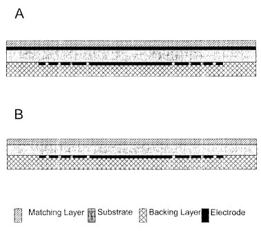
Figures 2A and 2B are schematic cross-sectional side views of transducer
arrays
according to two embodiments of the invention;
Figure 3 shows an electrode pattern for an annular array operating at 50 MHz
according to the invention;
Figure 4 shows the impedance response (a) amplitude and (b) phase of the
central and outermost elements of a 7 element kerfless annular array;
Figure 5 shows the pulse on-axis at (a) 0.1 mm and (b) 5 mm from the
transducer and (c) spectrum of the pulse at 5 mm, for a 7 element kerfless
annular
array;
Figure 6 shows one-way radiation patterns of a focussed transducer using (a to
c) finite element models and (d to f) electrically and acoustically isolated
elements, of an
annular array with (from top) 5, 7, and 10 elements;
Figure 7 shows relative amplitude of the 2-way radiation pattern at a 5 mm
radius
from a 64 element, 50 MHz kerfless linear array (solid line) and diced linear
array
(dashed line), focussed to 5 mm;
Figure 8 is a schematic diagram showing a perspective view of a linear-linear
phased array according to an embodiment of the invention;
CA 02406684 2002-10-04
-5-
Figure 9 shows pulses at 5 mm from an annular array with the central element
excited, and opposing electrode (a) allowed to float and (b) set to ground
Figure 10 is a schematic diagram of a section of a linear-linear phased array
transducer according to an embodiment of the invention, showing top and bottom
electrode configurations for (a) transmit and (b) receive sequences; and
Figure 11 is a schematic diagram illustrating how a transducer can image a
volume by taking many sector scans.
Detailed Description of the Invention
According to one aspect of the invention there is provided a transducer array
for
transmitting and receiving ultrasonic energy, suitable for use in non-
destructive imaging
in, for example, bio-medical, engineering, and manufacturing applications. A
transducer
array according to the invention comprises a substrate having two opposed
faces, a first
electrode comprising an array of electrode elements formed on a first face of
the
substrate, a second electrode formed on the second face of the substrate, and
further
optional layers such as one or more matching layers and an acoustically lossy
backing
layer. The invention provides ultrasonic transducer arrays that can operate at
frequencies as low as 100 kHz and at least as high as 500 MHz. It is expected
that a
transducer array of the invention can be made to operate at higher frequencies
(e.g., 1
GHz); the ability to achieve such high frequency being limited by available
production
capabilities. The ability to operate at such high frequency makes the
transducer arrays
valuable for applications where high resolution imaging is required.
In accordance with the invention, the transducer substrate is a hard substrate
that is an efficient resonator, and is unlike polymer substrates that are
generally much
softer and less efficient resonators. Suitable substrate materials include,
but are not
limited to, piezoelectric and ferroelectric ceramics, and single crystal
materials having
regular crystalline structure. Examples of such ceramic materials are lead
zirconate
titanate (PZT), lead titanate (PT), barium titanate (BT), barium strontium
titanate (f3ST),
sodium-potassium niobate, lithium tantalate, lead metaniobate, zinc oxide,
aluminum
nitride, bismuth titanate, barium strontium titanate, barium magnesium
fluoride, and
potassium nitrate. Examples of suitable single crystal materials are quartz
and lithium
niobate, and single crystal relaxor ferroelectric materials such as lead
zirconate niobate-
CA 02406684 2002-10-04
-6-
lead titanate (PZN-PT) lead magnesium niobate. (PMN), and lead magnesium
niobate-
lead titanate (PMN-PT). The thickness of the substrate is chosen to resonate
at a
desired operating (e.g., imaging) frequency, as would be apparent to one of
ordinary
skill in the art. For example, a PZT substrate operating at 50 MHz is about 40
um to
about 50 um thick, preferably about 45 Nm thick. The resonant frequency of a
transducer will of course also depend on characteristics (e.g., thickness,
type of
material) of the matching layers) and electrodes, described below.
The first electrode, which comprises an array of electrode elements, is formed
on
one of the opposed sides of the substrate by applying a suitable material
(e.g., gold) to
the substrate. One or more of the elements of the electrode array is provided
with a
suitable electrical connection to signal transmitting and/or receiving
circuitry. For
example, elements can be connected to bonding pads, and gold bond wires used
to
connect bonding pads to the drive/receive electronics. In some embodiments the
second electrode is formed on the second side of the substrate by applying a
material
such as gold to the substrate (Figure 2A). In other embodiments the second
electrode
is formed by applying a conductive matching layer such as silver epoxy to the
substrate
(Figure 2B).
Although optional, it is preferable to apply at least one matching layer, as
such
layer enhances coupling of the acoustic energy to the medium under
investigation and
hence improves the bandwidth and efficiency of the array. The matching layer
can be
any suitable material such as, for example, silver epoxy, gold epoxy, or
epoxy, and is
applied at an appropriate thickness to achieve the desired matching for a
given material
and operating frequency, generally about 5 um to about 200 um. The matching
layer is
applied to the electrode (either the first or second electrode) which is
facing the material
under investigation, preferably the second electrode. As noted above, use of a
conductive material for the matching layer on the second electrode obviates
the need for
a metallic electrode applied to the substrate (Figure 2B). This simplifies
production in
such embodiments. In embodiments where the matching layer is applied to the
first
electrode (i.e., the electrode array), conductive materials should not be
used.
An acoustically lossy backing layer is optionally applied to the electrode not
facing the material under investigation, preferably the first electrode. The
backing layer
material absorbs acoustic energy radiating from the surface of the electrode
to which it
is applied, such that most of the radiated acoustic energy is radiated from
the electrode
CA 02406684 2002-10-04
7-
facing the material under investigation. In embodiments lacking a backing
layer, air
advantageously provides this function, but has the drawback that the pulse
ring-down
time is increased (e.g., 10 to 12 cycles vs. 1 to 2 cycles with a backing
layer}. The
longer pulse compromises the resolution of the transducer array. For the
backing layer,
a material such as, for example, tungsten-loaded epoxy or alumina-loaded
epoxy, with a
thickness of about 0.5 to about 15 mm, is suitable.
The first electrode, which comprises the element array, is formed from a thin
layer (e.g., about 500 A to about 1.5 trm thick) of a material such as gold or
an alloy of
metals such as chromium and gold (Cr-Au). The array pattern is applied to a
first side
of the substrate using any suitable means, such as, for example, conventional
evaporating or sputtering and photolithography. The second electrode can also
be
formed by applying a layer of electrode material to the second side of the
substrate by
such sputtering or evaporating techniques.
In one embodiment the transducer array is configured as a sparse array, in
which certain elements of the array are used for transmitting ultrasound, and
other
elements of the array are used to receive ultrasound. In further embodiments
the
transmit and receive elements in a sparse array have different structure
(e.g.,
geometry), each optimized for their respective transmitting and receiving
functions. In a
further embodiment, a transducer is configured solely for transmitting
ultrasound, for use
with a second transducer configured solely to receive ultrasound. The transmit
and
receive transducers can have the same or different array geometry. In such
embodiment, a material under investigation is placed between the transmit and
receive
transducers.
It will be appreciated that a transducer array according to the invention
lacks
grooves between adjacent array elements. As used herein, the term "groove" is
intended to mean a recess, channel, or kerf between electrodes of an array.
Such
grooves are formed by, for example, mechanical, ultrasonic, or laser
machining, or
chemical etching, and penetrate at least part of the way through the
substrate, and
usually all of the way through the substrate. A transducer array according to
the
invention is therefore "grooveless" or "kerfless", meaning that the substrate
of the
transducer lacks grooves or kerfs between electrode elements.
Rather, in a transducer array according to the invention, the elements are
defined by simply the pattern of the electrodes disposed on the substrate.
This
CA 02406684 2002-10-04
-$_
represents a substantial departure from prior ultrasonic transducer arrays.
That is, it is
generally accepted by those skilled in the art that an array cannot be
fabricated on a
ceramic substrate without mechanically and electrically isolating the array
elements, by
providing grooves through the array substrate. It is believed that if this is
not done, the
signal transmitted or received by one element will influence the signal on an
adjacent
element and unless this unwanted coupling between elements is very small, the
radiation pattern of the array will be degraded [1,2]. The present inventors
have
discovered that although this is true for an array that is electronically
focussed and
steered, it is not necessarily true for an array that is only focussed
electronically. In fact,
the invention demonstrates that a satisfactory radiation pattern can be
achieved using a
grooveless array in which the array elements are defined by the electrode
pattern alone.
Accordingly, the invention is applicable to all arrays that are focussed
electronically and
have a fixed steering angle. Examples of such arrays are annular arrays,
linear arrays
(i.e., 1-D arrays), curved linear arrays, 1.5-D arrays, and 2-D arrays.
As used herein, the term "1-D array" refers to an array having (N x 1)
discrete
elements, the term "2-D array" refers to an array having (N x M) discrete
elements, and
the term "1.5 D array" refers to an array having (N x M) discrete elements
where N>M.
An advantage of the transducer array of the invention is that the fabrication
process is substantially simplified, which in turn permits the fabrication of
transducer
arrays with very small geometry and capable of operating at very high
frequency.
While not holding to one theory at the exclusion of others, it is believed
that the
performance of the inventive transducer array can be explained as follows:
When a
single element in an array having no grooves is excited, the neighbouring
elements will
also be excited, although to lesser extent. The major effect of this unwanted
coupling
between elements will be to make the array element appear wider than the
specified
electrode pattern. As the width of the radiating surface increases, the
resulting
ultrasound beam becomes more directed in front of the array and less energy is
radiated
at oblique angles. This is a serious problem for a linear phased array where
the
ultrasound beam is steered over a range of angles from --45 to +45 degrees.
However,
for an array where the steering angle is fixed at about 0 +/- 5 degrees, the
loss of
directivity is not a problem and appears to be an advantage.
In one embodiment of a transducer array of the invention, the first electrode
and
the second electrode each comprises an array of electrode elements.
Preferably, the
CA 02406684 2002-10-04
_g_
two arrays are of different patterns or configurations, such as, for example,
two linear
arrays oriented at 90 degrees to one another. In such an embodiment, the two
electrode arrays are grooveless arrays as described above. Such a transducer
is used
to obtain multiple 2-D images of a study material, using beamforming
techniques in
which subsets of array elements are individually addressed to acquire each
image.
In a further embodiment, the first electrode and the second electrode of the
transducer each comprises an array of electrode elements, wherein one of the
arrays is
a grooveless array. Preferably, the two arrays are of different patterns or
configurations.
In a preferred embodiment, one of the arrays is a grooveless linear array, and
the
second array is a linear phased array having grooves or kerfs between
electrode
elements. An advantage of such a "hybrid" linear-linear phased array
transducer is that
it provides focussing and steering of the ultrasound beam in the elevation and
azimuthal
directions. Accordingly, this embodiment is used to create 3-D images by
stepping the
beam through a study material so as to obtain multiple adjacent 2-D images
(i.e., sector
format slices), and assembling the 2-D images to create a 3-D image. This is
described
in detail in Example 3, below.
Traditional ultrasound imaging systems used in applications such as bio-
medical
imaging provide images in a two-dimensional (2-D) format. Although synthesis
of a
three-dimensional (3-D) image of the anatomy based on the acquisition of
multiple two-
dimensional images has been attempted, the resulting images are often of poor
quality.
The main problem is that conventional ultrasound systems are too slow to
acquire a full
3-D data set in real-time. For example, a 3-D image of the heart requires a
collection of
approximately 90 two-dimensional images acquired at a rate of 20 images/s.
Slow
image acquisition introduces the problem of how to align adjacent 2-D images
collected
at different times over many different cardiac cycles. The patient and imaging
probe
can be immobilized to minimize movement between adjacent images and cardiac
and
respiratory gating can also be used [11 ]. Unfortunately, even in a carefully
controlled
situation the resulting 3-D data set is often badly distorted and is of little
use.
The ability of an imaging system to detect small or subtle structures is
primarily
determined by the ability of a transducer to focus the ultrasound energy. Many
beamforming techniques have been developed to optimally focus and steer the
ultrasound beam to produce high resolution images. A conventional sector
format
image is created with a linear phased array by beamforming along each line in
the
CA 02406684 2002-10-04
-10-
image. On transmit the ultrasound is steered to a certain angle, and focussed
to a
single depth in the tissue by applying appropriated delays across the array.
On receive,
a delay pattern is applied again to steer the beam along the same angle as for
transmit.
As the pulse is reflected at different depths in the sample material, the
delays applied to
the receive signal are dynamically changed to sweep the focus through the
material.
This dynamic focussing produces a scan line focussed along its complete
length. To
improve the resolution of the image, multiple transmit focal zones can be used
for each
scan line.
The time to create an image depends on the number of scan lines, the number
of transmit focal zones, and the time for one transmit-receive event. For a
typical
imaging system, the image is composed of 200 scan lines in a 90 degree sector
image,
and uses a single transmit focal zone. For example, for typical cardiac
imaging the
ultrasound penetrates to a depth of about 15 cm and the speed of sound in
tissue is
about 1500 mls, giving a round trip transit time of 200 ms. Therefore, the
time required
to acquire a single 2-D image is 200 x 200 ms = 40 ms, which limits the frame
rate to 25
frames/s. This is fast enough for real-time 2-D imaging, but too slow to image
a volume
a volume of interest in real-time. Images can be created more rapidly by
reducing the
number of scan lines, however the resolution is then reduced.
The time to produce an image can be reduced while retaining sufficient
resolution by using a synthetic aperture approach. The imaging speed of such a
system
can be increased by using a sparse transmit array, which minimizes the number
of
transmit pulses and maximizes the amount of information that can be collected
[12].
Such a system having only 5 transmit elements can provide a frame rate up to
1000
frames/s. Alternatively, the imaging speed can be increased by forming
multiple
simultaneous transmit beams. By mechanically rocking or translating the array,
it is
possible to create a 3-D image of aligned 2-D slices in real-time; however,
the motion
creates unwanted noise and vibration.
An alternative approach to real-time 3-D imaging uses a 2-D transducer array
to
scan and focus the ultrasound beam through the tissue volume [13,14]. A wide
transmit
beam is used and many receive scan fines can be formed simultaneously. While
there
is still a trade-off between acquisition time and resolution, the beams are
formed over a
volume, permitting real-time beamforming. A major problem with this approach
is the
large number of elements associated with a 2-D array. For example, over 16,000
CA 02406684 2002-10-04
-11-
elements are required to obtain the same 2-D resolution of a conventional 128
element
linear phased array. This requires extensive electronics for beamforming the
signal
from each element. Another problem with a full 2-D array is that the elements
are'/x
wavelength, which is, for example, 150 mm x 150 mm for a 5 MHz array. The
electrical
impedance of the elements is very high and the efficiency low because of the
minute
size of the elements.
In contrast, the invention can provide a transducer array that permits real-
time 3-
D imaging using far fewer elements than the 2-D array approach while avoiding
the
need to mechanically translate the transducer. Real-time 3-D imaging is
possible
because the array is able to electronically scan and focus the ultrasound beam
through
a complete imaging volume in the time normally required to collect a single 2-
D image
using a conventional system. According to this embodiment, the invention
comprises a
2-D array for real-time 3-D imaging wherein a kerfless linear array is
combined with a
conventional diced linear phased array.
In accordance with another aspect of the invention there is provided a method
of
producing a transducer array. The method comprises providing a hard substrate
having
two opposed faces, disposing an electrode array having electrode elements on
one of
the faces of the substrate, and disposing an electrode on the second face of
the
substrate, wherein the substrate lacks grooves between electrode elements.
Heretofore, methods used to produce ultrasound transducer arrays on hard
substrates
included providing grooves between adjacent electrode elements, which is the
most
difficult part of the fabrication process. By eliminating this step, the
invention provides a
simplified method of producing ultrasound transducer arrays. Further, the
provision of
grooves on hard substrates is considered to be the limiting factor in
providing very high
frequency transducers. The invention overcomes this limitation, thus allowing
the
fabrication of transducer arrays operating at much higher frequencies than was
previously possible. In fact, the maximum operating frequency of a transducer
array
according to the invention is limited only by the available technology for
disposing on a
substrate electrode elements having very small geometries.
In accordance with another aspect of the invention there is provided a
nondestructive ultrasonic imaging system comprising an ultrasonic transducer
array as
described herein, and appropriate ultrasound transmit/receive circuitry. The
system may
further comprise means for processing signal information. The imaging system
can be
CA 02406684 2002-10-04
-12-
configured for operation at low frequencies, e.g., 100 kHz, up to very high
frequencies,
e.g., 500 MHz or higher, subject to the ability to provide an ultrasonic
transducer array
capable of operating at such high frequency. As discussed above, the invention
provides the basis for such high frequency transducers, the production of
which might
be limited by the available technology. Of course, the operating frequency of
the
imaging system is chosen to provide the desired resolution for the material
under
investigation. In many biomedical imaging applications, for example, a
resolution of less
than 20 Nm is desirable, and can be achieved with an imaging system in
accordance
with the invention.
All cited publications are incorporated herein by reference in their entirety.
The invention is further described below by way of the following non-limiting
Examples.
Example 1.
With reference to Figure 2B, a high frequency (50 MHz) transducer array
comprises a PZT substrate (Motorola 3203HD) having a thickness of
approximately 47
Nm. An electrode layer of Cr-Au about 1000 ~ thick is deposited on the first
face of the
substrate. An electrode layer of aluminum about 2 Nm thick is deposited on the
second
face. The second face or electrode is patterned using photolithography to
define the
array geometry as shown in Figure 3, which figure depicts an electrode pattern
suitable
for a 50 MHz annular array. A matching layer of silver epoxy at a thickness of
approximately 9 Nm is applied to the first face of the array. Electrical
connections are
made to the array elements and a backing layer of tungsten-loaded epoxy (EPO-
TEK
301-2), at a thickness of approximately 1 mm, is applied to the second face of
the array.
The performance of several embodiments of a 50 MHz kerfless annular array
was evaluated using a finite element model (FEM) (PZFIex, Weidlinger
Associates, CA)
of the arrays. The software has been shown to accurately model the electrical
and
mechanical response of a transducer [3-9], including non-idealized behaviour
such as
CA 02406684 2002-10-04
-13-
mechanical or electrical coupling between elements and unwanted vibrationai
modes.
Three performance parameters were investigated: 1 ) the pulse shape, which is
important for axial resolution, 2) the electrical impedance of the array
elements, which is
important for electronics design, and 3) the radiation pattern, which defines
the lateral
resolution and image contrast. The radiation pattern from the finite element
analysis
was compared to the ideal radiation pattern for each array. The ideal
radiation pattern
was generated using the impulse-response method reported by Arditi et al.
[10]. Arditi
derived analytical expressions for the impulse response of an annular array
based on
the array geometry. The radiation pattern is calculated by convolving the
impulse
response at each location in the field with a pulse representing the normal
particle
velocity at the surface of the array. The resulting radiation pattern is used
as the ideal
pattern because the entire array is assumed to vibrate in an ideal piston
(i.e., single)
mode with no electrical and mechanical coupling between the array elements. By
comparing the ideal radiation and the radiation pattern from the FEM, the
effect of non-
ideal vibration modes and coupling between elements can be determined.
The results of the modelling are summarized in Figures 4 to 6. The magnitude
and phase of the electrical impedance for the central element and outer
element of a 7
element kerfless annular array are shown in Figure 4. The electrical impedance
for the
two elements is remarkably similar, and very flat over the bandwidth of
interest from 40
to 60 MHz. The impedance shows no evidence of lateral modes even though the
width
of the outer electrode (64 um) is similar to the thickness of the transducer
substrate (4?
Nm).
Figure 5a shows the pulse very close (0.1 mm) to the face of the 7 element
array. The elements were excited using a monocycle 50 MHz impulse. Appropriate
time delays were introduced to focus the beam at 5 mm. The resulting acoustic
pulse is
longer than desired. Since the elements are not mechanically isolated, energy
is
coupled freely through the transducer substrate and a long (e.g., more than 5
cycles)
ringing pulse is produced. However, the pulse at the focal region, shown in
Figure 5b, is
very different. At the focal region, only the thickness mode vibrations are
correlated and
consequently, the long tail in the pulse is almost entirely eliminated. The
pulse
spectrum at the focal region, shown in Figure 5c, is centred at approximately
45 MHz
with a -6 dB bandwidth of 55%.
CA 02406684 2002-10-04
-14-
The one-way radiation patterns for 5, 7, and 10 element annular arrays are
shown in Figure 6. Two radiation patterns are shown for each array geometry,
one
representing the finite element prediction (left) and the other the ideal
response (right).
In each case the array was focused at 5 mm (f/2.5). The radiation patterns are
plotted
as contour plots in dB normalized to the peak pressure. The FEM and ideal
radiation
patterns are very similar. Both radiation patterns show significant
improvement in
suppression of off-axis energy with increasing number of elements. However,
the
positive effect of increasing the number of elements is less in the FEM
radiation pattern
than in the ideal pattern. This shows that the relative importance of coupling
between
elements increases with increasing number of elements. However, for an array
with
less than 10 elements, coupling between elements in a kertless design does not
appear
to be the limiting factor.
Example 2.
The pertormance of 50 MHz kertless and sub-diced arrays was evaluated using
finite element modeling (PZFIex, Weidlinger Associates, CA.). Both arrays had
64
elements, 30 Nm element spacing, a single front quarter-wavelength matching
layer,
and a high loss, high impedance backing layer. No kerf filler was used for the
diced
array and each element was sub-diced once to suppress lateral modes. The -6 dB
width of the directivity (single element) calculated at a 5 mm radius was 16
degrees for
the kerfless array, compared with 72 degrees for the sub-diced array. Cross-
talk
between adjacent elements was -7.5 dB for the kertless array, and -28 dB for
the sub-
diced array. As shown in Figure 7, the radiation pattern (5 mm focal distance)
had a -6
dB width of 1.5 degrees for both arrays. At an angle of 7.5 degrees, the
radiation
pattern was below -60 dB for both arrays. The pulse shape and pulse amplitude
at the
focal region were remarkably similar for both arrays.
The finite element model predictions were verified experimentally using a
lower
frequency (2.0 MHz) kertless linear array. The array had 24 elements with 670
Nm
center-to-center element spacing. The elements were defined by evaporating a
chrome-gold electrode through a thin stainless steel mask onto the back
surface of 1.0
mm thick PZTSH disk. A gap approximately 150 um wide was used to separate
adjacent electrodes. A continuous electrode was evaporated on the top surface
of the
CA 02406684 2002-10-04
-15-
ceramic and a thin layer of tungsten-loaded epoxy was cast onto the front
surface and
lapped to thickness corresponding to a quarter of a wavelength at 2 MHz.
Electrical
connections were then made to individual electrodes on the back surface and a
2 cm
thick backing layer of tungsten loaded epoxy was applied. The array was tested
in a
water bath by recording the reflections from a line target placed a fixed
distance in front
of the array, oriented at 90 degrees to the array elements. The radiation
pattern of
individual array elements was measured by scanning the array across the target
and
recording the maximum amplitude of the reflected signal as a function of
position. Good
agreement was obtained between the model predictions and the experimental
results.
Thus, the results indicate that kertless linear arrays can be produced with
little or no
degradation of the radiation pattern compared to a conventional diced array.
Example 3.
A hybrid linear-linear phased array is shown schematically in Figure 8, based
on
a substrate 10, where the phased array portion is a conventional diced array
with half-
wavelength spacing and a kerf filler 20 between elements, and with the
electrodes 15 on
the top face of the transducer. On the bottom face of the transducer,
electrodes 25 are
patterned perpendicular to those of the phased array, creating a kerfless
linear array.
The hybrid array is made from the same materials as described previously
(e.g.,
PZT substrate, chrome-gold electrodes, tungsten-loaded epoxy backing layer,
one or
more polymer matching layers), and the geometry of the elements is preferably
rectangular. The linear phased array elements has an aspect ratio (height /
width) of at
least 2 to avoid coupling unwanted lateral modes into the bandwidth of the
array. The
linear array elements have about 1/2 to about 2 wavelength spacing, with a gap
of about
5% to about 30% of the element width between adjacent electrodes, and the
length of
the array elements ranges from about 16 to about 200 wavelengths. The
impedance of
the elements is similar to a conventional array, assuming similar element
size. The
pulse shape and bandwidth of such a hybrid array are similar to whatcould be
achieved
using a conventional array, for example, about 40% to 100% factional
bandwidth.
Preferably, the array provides a narrow main beam, with secondary lobes less
than -60
dB with respect to the main lobe.
CA 02406684 2002-10-04
-16-
According to this embodiment, a single element is activated by setting one
electrode to ground and placing a signal on an opposing electrode. If all the
other
electrodes are allowed to 'float', an acoustic pulse is only emitted from the
region where
the grounded and active electrodes overlap.
Preliminary tests of a such transducer with floating electrodes have been
performed using a finite element model (FEM). One electrode of an array was
excited,
and the opposing, normally grounded electrode was allowed to float. Pulses at
a
distance of 5 mm in front of the array from a non-grounded and a grounded
transducer
are shown in Figure 9. The amplitude of the pulse excited using a floating
electrode
was 3 orders of magnitude smaller. Therefore, the acoustic signal emitted from
a non-
grounded element has minimal effect on the acoustic signal resulting from an
excited
element with one electrode set to ground.
A linear-linear phased array can create one 2-D slice of a 3-D image in a
similar
manner to a linear phased array. An example of the sequence of transmit and
receive
events with a linear-linear phased array is shown in Figure 10. To transmit a
pulse, one
of the edge electrodes of the phased array is set to ground, while a subset of
the linear
array electrodes is excited with appropriate delays to focus the pulse over
the desired
depth. To receive a signal, the linear array electrodes that had just been
driven are set
to ground, and the electrical signals resulting from the reflected ultrasound
pulse are
taken from the phased array electrodes. As for the 1-D phased array, 3 to 5
transmit
events are used, and the image is created using synthetic aperture
beamforming.
Alternatively, the image could be formed using multiple simultaneous transmit
beams
and synthetic aperture beamforming. Those of ordinary skill in the art will
appreciate
that many other configurations, including other combinations of elements
excited and
set to ground, are possible.
The full 3-D image from a linear-linear phased array is acquired using linear
array beamforming techniques. A subset of the linear array elements is
activated,
creating a 1-D linear phased array in one region which images a slice of the
tissue. The
subset of linear array electrodes activated is shifted to create many (i.e.,
"n") sequential,
spatially adjacent sector format slices (i.e., 2-D images), denoted by
reference numerals
40a to 40n in Figure 11. The n sector format slices are assembled to make a 3-
D
image. The number of 2-D images required to produce a 3-D image will depend on
each particular situation, including system constraints such as amount of
memory
, CA 02406684 2002-10-04
-17-
available for image storage and manipulation of the data. Practically, a 3-D
image can
be produced from as few as 20 2-D images, or as many as 200 2-D images. It
should
be noted that by repeating this process using different subsets of the linear
array
elements, synthetic aperture beamforming can also be used to focus the
ultrasound
beam in the elevation direction.
An advantage of a linear-linear phased array over a full 2-D array for real-
time 3-
D imaging is that a 128x128 linear-linear phased array has only 256 elements,
while a
full 2-D array has over 16,000 elements. Consequently, the amount of
electronics
required can be greatly reduced for the same image quality. Because the linear-
linear
phased array elements are much larger than those of the 2-D array, the
impedance is
much lower, and electrical matching simplified. Further, using the linear
array to focus
the transmit pulse in the third dimension for each image slice eliminates the
need to
mechanically move the transducer to image the third dimension, reducing noise
and the
overall size of the transducer head.
Equivalents
Those skilled in the art will recognize or be able to ascertain, using no more
than
routine experimentation, variants of the embodiments disclosed herein. Such
variants
are understood to be within the scope of the invention and are covered by the
appended
claims.
References
1. G.F. Guesse, C.G. Oakley, S.J. Douglas, R.D. Morgan, "Cross-talk Paths in
Array Transducers," Proc. 1995 IEEE Ultrasonic Symp., vol. 2, 1279-1282, 1995.
2. P.A. Payne, J.V. Hatfield, A.D. Armitage, Q.X. Chen, P.J. Hicks and N.
Scales,
"Integrated Ultrasound Transducers," Proc. 1994 IEEE Ultrasonics Symp, vol. 2,
1523-1526, 1994.
3. G.L. Wojcik, D.K. Vaughn, N. Abboud, and J. Mould, Jr. "Electromechanical
modeling using explicit time-domain finite elements," Proc. 1993 IEEE
Ultrasonic
Symp., vol. 2, 1107-1112, 1993.
4. G.L. wojcik, D.D. Vaughn, V. Murray, and J. Mould, Jr., "Time-domain
modeling
of composite arrays for underwater imaging," Proc. 1994 IEEE Ultrasonic Symp.,
CA 02406684 2002-10-04
-18-
vol. 2, 1027-1032, 1994. ," Proc. 1996 IEEE Ultrasonic Symp., vol. 2, 1509-
1512,
1996
5. G. Wojcik, J. Mould, Jr., F. Lizzi, N. Abboud, M. Ostromogilsyk, and D.
Vaughn,
"Nonlinear modeling of therapeutic ultrasound," Proc. 1995 IEEE Ultrasonic
Symp., vol. 2, 1617-1622, 1995.
6. D.M. Mills, and S.W. Smith, "Combining multi-layers and composites to
increase
SNR for medical ultrasound transducers," Proc. 1996 IEEE Ultrasonic Symp.,
vol. 2, 1509-1512, 1996.
7. D.J. Powell, G.L. Wojcik, C.S. Desilets, T.R. Gururaja, K. Guggenberger, S.
Sherrit, and B.K. Mukherjee, "Incremental 'model-build-test' validation
exercise
for a 1-D biomedical ultrasonic imaging array," Proc. 1997 IEEE Ultrasonic
Symp., vol. 2, 1669-1674, 1997.
8. R.L. Goldberg, M.J. Jurgens, D.M. Mills, C.S. Henriquez, D. Vaughn, and
S.W.
Smith, "Modeling of piezoelectric multiplayer ceramics using finite element
analysis," IEEE Trans. Ultrason., Ferroelect., Freq. Contr., vol. 44, no. 6,
1204-
1213, 1997.
9. N.N. Abboud, G.L. Wojcik, D.K. Vaughn, J. Mould, D.J. Powell, and L.
Nikodym,
"Finite Element Modeling for Ultrasonic Transducers", SPIE Int. Symp. Medical
Imaging, Vol. 3341 p.19-42, 1998.
10. M Arditi, F.S. Foster, and J.W. Hunt, "Transient fields of cancave annular
arrays", Ultrasonic Imaging, Vol. 3, pp. 37-61, 1981
11. G. Stetten and R. Tamburo, "Real-time Three-Dimensional Ultrasound Methods
for Shape Analysis and Visualization," Methods, vol. 25, 221-230, 2001.
12. G.R. Lockwood, P.C. Li, M. O'Donnel, and F.S. Foster, "Optimizing the
Radiation
Pattern of Sparse Periodic Linear Arrays," IEEE Trans. Ultrason., Ferroelect.,
Freq. Contr., vol. 43, no. 1, 7-14, 1996.
13. S.W. Smith, H.G. Pavy, Jr., and O.T. von Ramm, "High-Speed Ultrasound
Volumetric Imaging System - Part I: Transducer Design and Beam Steering,"
IEEE Trans. Ultrason., Ferroelect., Freq. Contr., vol. 38, no. 2, 100-108,
1991
14. 0.T. von Ramm, S.W. Smith, and H.G. Pavy, Jr., "High-Speed Ultrasound
Volumetric Imaging System - Part II: Parallel Processing and Image Display,"
IEEE Trans. Ultrason., Ferroelect., Freq. Contr., vol. 38, no. 2, 109-115,
1991.