Note: Descriptions are shown in the official language in which they were submitted.
CA 02407004 2002-10-18
1
D E S C R I P T I 0 N
X-RAY CT APPARATUS
Technical Field
The present invention relates to an X-ray CT
apparatus capable of obtaining a tomographic image of
an object of inspection in a short time.
Background Art
An X-ray CT (Computed Tomography) apparatus
comprises an X-ray tube and a detector that are located
on either side of an object of inspection. The X-ray
tube emits the X rays toward the object of inspection,
and the detector detects the X rays emitted from the
X-ray tube. Based on the difference in the rate of
X-ray absorption between parts in the object of
inspection, the X-ray CT apparatus analyzes, by using
a computer, transmitted X-ray data that the detector
detects each time when X rays are applied to the object
of inspection in many directions, and obtains
a tomographic image of the object of inspection.
There is an X-ray CT apparatus of an R-R
(rotate-rotate) type in which the X-ray tube and
detector rotate around the object of inspection while
maintaining their relative positions. In the case
where the X-ray CT apparatus of this type is used for
diagnosis in the medical field, however, it involves
CA 02407004 2002-10-18
2
mechanical movement such that the X-ray tube and
the detector rotate around a patient, the object of
inspection, so that the taking time (scanning time),
which is necessary to obtain one tomographic image, is
long.
There is also an X-ray CT apparatus of another
type. This X-ray CT apparatus comprises an electron
gun, a magnetic field coil, and a target ring instead
of the X-ray tube. The electron gun is located on the
central axis of the target ring. An electron beam shot
from the electron gun toward the center of the target
ring is bent by the magnetic field coil, whereupon it
hits the target ring. When the target ring is hit by
the electron beam, it emits an X-ray toward an object
of inspection located inside the target ring. The
X-ray CT apparatus changes the position for the impact
of the electron beam in the circumferential direction
of the target ring by changing the direction of the
magnetic field that the magnetic field coil generates.
By doing this, the X-ray CT apparatus applies X rays to
the object of inspection in many directions, and
obtains a tomographic image in accordance with the
resulting transmitted X-ray data. In the X-ray CT
apparatus of this type, the X-ray source is moved
electrically, so that the taking time can be made
shorter than in the case of the X-ray CT apparatus of
the R-R type. However, there is a limit to shorten
CA 02407004 2002-10-18
3
the time for acquiring a tomographic image, because
the X-ray source is only one in number at the same time
and a detector takes time to detect the necessary dose
of X rays.
In the industrial field, there is proposed
an X-ray CT apparatus for observing the movement of
air bubbles in two-phase flow, for example. As an
example of this X-ray CT apparatus, there is an X-ray
CT apparatus comprising a large number of X-ray sources
as described in Jpn. Pat. Appin. KOKAI Publication
No. 9-248300, 10-75944, or 10-295682.
The X-ray CT apparatuses described in Jpn. Pat.
Appln. KOKAI Publications Nos. 9-248300 and 10-75944
use a large number of X-ray tubes as X-ray sources.
In the case described in Jpn. Pat. Appln. KOKAI
Publication No. 10-75944, X rays are simultaneously
applied from two or more X-ray tubes on condition that
regions on the detector upon which X rays are incident
never overlap one another.
The X-ray CT apparatus described in Jpn. Pat.
Appln. KOKAI Publication No. 10-295682 comprises
a vacuum chamber, a large number of X-ray sources, and
a detector. The vacuum chamber is in the shape of
a ring that surrounds the object of inspection.
The numerous X-ray sources are arranged in
a circumferential direction in the vacuum chamber, and
emit X rays in fan beam that crosses the object of
CA 02407004 2002-10-18
4
inspection toward the object of inspection. The
detector is in the shape of a ring that surrounds the
object of inspection in a position on the inner
peripheral side of the vacuum chamber, and serves to
detect the X rays that are emitted from the X-ray
sources and passed through the object of inspection.
The X-ray sources are actuated one after another in the
order of arrangement and emit the fan-shaped X rays
toward the object of inspection. The emitted X rays
are passed through the object of inspection and
detected by the detector on the opposite side.
A tomographic image can be obtained in a short time by
quickly switching signals that serve to actuate the
X-ray sources.
Accordingly, application of industrial X-ray CT
apparatuses to an examination in the medical field is
under investigation. When a patient is performed
diagnosis by using an industrial X-ray CT apparatus,
the taking time for the acquisition of a tomographic
image can be shortened considerably, so that
improvement of the efficiency of diagnosis can be
expected. Since the industrial X-ray CT apparatus can
acquire a tomographic image in a short time, moreover,
an image can be obtained corresponding to change that
is occurred in a short time.
In the case where X rays are generated by
utilizing an electron beam, X rays are emitted in all
CA 02407004 2002-10-18
directions from the spot that is hit by the electron
beam. However, no consideration is given to the
spread angle of the fan-beams that are emitted from the
X-ray sources of the industrial X-ray CT apparatus.
5 Thus, the object of inspection is irradiated with
unnecessary X rays that are not detected by means of
the detector.
Further, no consideration is given to the spread
angle of X rays in the industrial X-ray CT apparatus.
When the X-ray CT apparatus irradiates the fan-beams
from three or more X-ray sources to the object of
inspection at a time, the X rays inevitably interfere
with one another in some regions on the detector and
a high-precision tomographic image cannot be obtained.
Therefore, the X rays must be emitted from opposite
positions at 180 from each other in order to apply the
X rays without interference. In the case where the X-
ray CT apparatus irradiates the fan-beams from
two X-ray sources to the object of inspection at
a time, the two X-ray sources must be switched so that
they always maintain their symmetrical positions with
respect to the object of inspection. The time for
taking the acquisition of one tomographic image is
restricted by the time that is necessary to switch
each X-ray source around the object of inspection by
a half turn.
The X-ray CT apparatus for diagnosis in
CA 02407004 2002-10-18
6
the medical field must apply the X rays to the object
of inspection in as many directions as possible to
acquire transmitted X-ray data in order to obtain
a fine tomographic image. In the case where the
industrial X-ray CT apparatus is applied to the medical
field, therefore, the X-ray sources must be increased
in number.
When the directions in which X rays are applied is
increased, however, the dose of X rays applied to the
patient inevitably increases, and the taking time is
prolonged, so that the load on the patient increases.
The purpose of the present invention is to provide
an X-ray CT apparatus designed so that the dose of X
rays applied to the object of inspection can be
reduced, and that the time, which is necessary to
acquire a tomographic image of the object of
inspection, can be shortened.
Disclosure of Invention
An X-ray CT apparatus according to one embodiment
of the present invention is designed to reduce the dose
of X rays applied to an object of inspection and to
shorten the time that is necessary to acquire
a tomographic image of the object of inspection.
An X-ray CT apparatus according to one embodiment
of the present invention includes a large number of
X-ray sources, a detector, and a collimator. The X-ray
sources are arranged around the object of inspection.
CA 02407004 2002-10-18
7
The detector detects X rays emitted from the X-ray
sources. The collimators are located between the X-ray
sources and the object of inspection, thereby
restricting those X rays that, among the X rays emitted
from the X-ray sources, are not applied to the
detection surface of the detector.
An X-ray CT apparatus according to another
embodiment of the present invention includes a main
body, a large number of X-ray sources, a vacuum
chamber, a collimator, a detector, a bed, and a beam
limiter. The main body has a hole in which an object
of inspection is located. The X-ray sources are
concentrically arranged around the hole. The vacuum
chamber is in the form of a ring surrounding the hole
and holds the X-ray sources. The collimator is mounted
along the inner peripheral wall of the vacuum chamber
and has through holes corresponding to the individual
X-ray sources. The detector includes a large number of
detection elements for detecting the X rays emitted
from the X-ray sources. The detection elements are
arranged densely in the shape of a cylinder having the
same central axis with a concentric circle composed of
the X-ray sources, with the detection surface thereof
facing toward the central axis. The bed has a slide
mechanism and a lift device and serves to position
the object of inspection in the hole of the main body.
The beam limiter is located between the X-ray sources
CA 02407004 2002-10-18
8
and the object of inspection and serves to restrict the
spread of the X rays in the direction along the central
axis of the concentric circle composed of the X-ray
sources within the width of the detector in the
direction along the central axis.
Brief Description of Drawings
FIG. 1 is a sectional side view typically showing
an X-ray CT apparatus according to one embodiment of
the present invention;
FIG. 2 is a sectional view as taken from the front
of the X-ray CT apparatus, typically showing a vacuum
chamber, X-ray sources, and a detector of FIG. 1;
FIG. 3 is a perspective view typically showing the
vacuum chamber and the detector of FIG. 1;
FIG. 4 is a view typically showing a profile of
the vacuum chamber of FIG. 1;
FIG. 5 is a view typically showing the positional
relations between X rays emitted from an optional X-ray
source of the X-ray CT apparatus of FIG. 1, a
collimator for restricting the X rays, an object of
inspection, and the detector;
FIG. 6 is a view typically showing a state in
which X rays are simultaneously emitted from four X-ray
sources of the X-ray CT apparatus of FIG. 1 toward the
object of inspection;
FIG. 7 is a perspective view showing a state in
which the shape of X rays is changed by a beam limiter
CA 02407004 2002-10-18
9
of FIG. 1; and
FIG. 8 is a diagram typically showing the
positional relation between X-ray sources 13 and the
detector 12 of FIG. 1.
Best Mode for Carrying Out of the Invention
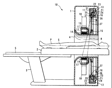
An X-ray CT apparatus 10 for diagnosis that is
utilized in the medical field will be described as an
example according to one embodiment of the present
invention with reference to FIGS. 1 to 8. The X-ray CT
apparatus 10 shown in FIG. 1 uses X-ray sources 13 that
emit X rays R by applying electron beams e from an
electron gun to a target. The X-ray CT apparatus 10
shown in FIG. 1 comprises a main body 1 and a bed 2
that carries thereon a patient P as an object of
detection. The main body 1 is in the form of a
doughnut that has a horizontal central axis S. A hole
4 that opens in the central portion of the main body 1
is of a size such that it allows the patient P on the
bed 2 to be horizontally inserted therein. The bed 2
is provided with a lift device 3 and a slide
mechanism 5. The lift device 3 can make the body axis
of the patient P incline at an angle to or extend
parallel to the central axis S of the main body 1. The
slide mechanism 5 moves the patient P along the body
axis. Thus, the bed 2 can allow the patient P to be
inserted into the hole 4 and hold the patient P in any
desired position. Since the bed 2 is expected only to
CA 02407004 2002-10-18
be able to hold the patient P relative to the hole 4 of
the main body 1, the main body 1 may be moved with
respect to the bed 2 or both may be moved individually.
As shown in FIG. 2, the main body 1 is provided
5 with a large number of X-ray sources 13 concentrically
arranged around the hole 4 and a detector 12 for
detecting the X rays R emitted from the X-ray
sources 13. The X-ray sources 13 are held in a vessel
(vacuum chamber) 11 inside which a vacuum is
10 maintained. The vacuum chamber 11 is in the form of a
ring that has its center on the central axis S.
As shown in FIG. 4, the profile of the vacuum
chamber 11 is square. The detector 12 is in the form
of a ring that is located inside the vacuum chamber 11
and has its center on the central axis S. The vacuum
chamber 11 and the detector 12 are offset with respect
to each other along the central axis S. The X-ray
sources 13 are arranged at equal distance along the
vacuum chamber 11.
Each X-ray sources 13, like a triode, comprises a
cathode 21, an anode 22, and a gate 23. A high voltage
is applied between the cathode 21 and the anode 22.
The cathode 21 has a filament that emits thermions when
heated. The filament is a coiled one, for example, and
generates heat attributable to resistance heating when
energized. As a potential that is opposite to the
thermions in polarity is applied to anode 22, the
CA 02407004 2002-10-18
11
thermions are attracted to the target on a part of the
anode and accelerated. The accelerated thermions form
electron beams, which run against the target. The
target, which is formed of, e.g., tungsten, emits X
rays from that area which is hit by the thermions.
Since the target is heated with collision energy of the
thermions, the anode 22 is made of a material such as
copper that has high heat conductivity. The gate 23
prevents the thermions from being emitted toward the
anode 22 as it is located between the cathode 21 and
the anode 22 and is supplied with potential of the same
polarity with the thermions. The cathode 21 and gate23
function as the electron gun. The cathode 21 and the
gate 23 are mounted on a holding member 24. A screw 25
is formed on the outer peripheral surface of the
holding member 24.
The sidewall of the vacuum chamber 11 is provided
with a hole 26 to which the holding member 24 is
attached. A mounting member 27 for positioning the
holding member 24 is attached to the edge portion of
the hole 26. A screw 28 is formed on the inner
peripheral surface the mounting member 27. Further, a
sealing member 29 is fitted in a recess that is formed
in the edge of the hole 26 which is in contact with the
holding member 24 so as to surround the hole 26. The
sealing member 29 may be fitted in a recess that is
formed in the holding member 24 so that the vacuum
CA 02407004 2002-10-18
12
chamber 11 can be kept airtight.
The holding member 24 is fixed to the vacuum
chamber 11 in a manner such that the screw 25 of the
holding member 24 and the screw 28 of the mounting
member 27 mesh with each other. The holding member 24
is attached to the sidewall of the vacuum chamber 11 so
that the direction of emission of the electron beams is
parallel to the central axis S. The cathode 21 and the
gate 23 project inside the vacuum chamber 11. The
mounting member 27 is provided so that the position of
irradiation of the target with the electron beams that
are emitted from the cathode 21 can be shifted.
A support member 31 is attached to the inner
surface of the vacuum chamber 11 by an insulating
member 32. The support member 31 is in the form of a
ring that extends along the inner periphery of the
vacuum chamber 11 and has its center on the central
axis S. The anode 22 is attached to the support member
31 so as to receive the electron beams that are emitted
from the cathode 21. The surface of the anode 22 which
receives the electron beams slightly tilts toward the
central axis S so that the X rays R can be emitted
toward the detector 12.
The support member 31 is formed of a material
with high heat conductivity and is provided internally
with a cooling water channel 33 throughout the
circumference. A cooling water pipe 34 is connected to
CA 02407004 2002-10-18
13
the cooling water channel 33. Cooling water is fed
through the cooling water pipe 34 and circulated in the
cooling water channel 33 by a cooling device (not
shown). Thus, the anode 22 that is heated with the
energy of the electron beams can be cooled indirectly.
Although only one cooling water pipe 34 is shown in
FIG. 4, at least two are provided for water supply and
drainage in order to circulate the cooling water.
A window 35 through which the X rays R generated
from the X-ray sources 13 are transmitted toward the
detector 12 is provided in the inner peripheral wall of
the vacuum chamber 11 so as to extend continuously
throughout the circumference. Outside the inner
peripheral wall of the vacuum chamber 11, a collimator
36 is mounted along the window 35. The collimator 36
has a width greater than that of the window 35 in the
direction along the central axis S. The collimator 36
is made of a metallic material such as tungsten or zinc
that absorbs the X rays R at a high rate. The
collimator 36 is provided with through holes 37, which
correspond individually to the X-ray sources 13 and are
directed toward the central axis S. Each through hole
37 is of a shape and a size such that the emitted X
rays R never projects beyond the width of the detector
12 in the direction along the central axis S and that X
rays R that spread in the circumferential direction
without interfering with the X rays R emitted from the
CA 02407004 2002-10-18
14
X-ray sources 13 in different directions. Thus, the
through holes 37 restrict other X rays than the X rays
R that are detected by the detector 12. The collimator
36 may be provided corresponding to the X-ray sources
13 in a one-to-one relation or provided collectively in
a circular arc for each angular range.
The detector 12 detects the X rays R emitted from
the X-ray sources 13 in positions symmetrical with
respect to the central axis S. As shown in FIG. 3, the
detector 12 detects the X rays R on its cylindrical
inner peripheral surface. On the inner peripheral
surface, detection elements 14 are arranged in a
lattice in the circumferential direction (8-direction)
and the direction (L-direction) along the central
axis S. In a specific example, 2,048 detection
elements 14 are arranged in the B-direction, and 200
in the L-direction.
A beam limiter 15 is mounted in a position on the
detector 12 near the vacuum chamber 11. The beam
limiter 15 is designed to restrict those X rays R from
the X-ray sources 13 which project beyond the width of
the detector 12. If the X rays R are restricted to
prevent from spreading beyond the width of the inner
peripheral surface of the detector 12 by the collimator
36 having the round through holes 37, the spread in
the 6-direction is also restricted, inevitably. Since
the beam limiter 15 can restrict the X rays R in
CA 02407004 2002-10-18
the L-direction, as shown in FIG. 7, the X rays R
that never spread beyond the width of the detector 12
can be obtained without restricting the spread in the
6-direction.
5 The X-ray CT apparatus 10 constructed in this
manner delivers a command signal for the acquisition of
a tomographic image from a measurement control device
(not shown) to an irradiation control element (not
shown). The irradiation control element settles the
10 direction of irradiation and the order of irradiation
of the X rays R and delivers a control signal to an X-
ray generation control device 16 shown in FIG. 1. The
X-ray generation control device 16 controls the
emission of X rays by controlling the gate 23 of each
15 X-ray source 13 in accordance with the control signal.
Patterns for the activation the X-ray sources 13
include a single-slice mode, serial-slice mode, sector-
slice mode, single-shot mode, video mode, etc. In the
single-slice mode, an optional tomographic image of a
patient is picked up with the X-ray sources 13 switched
for a revolution around the patient. In the serial-
slice mode, a plurality of tomographic images are
obtained for a patient who requires a volume inspection
while the bed 2 is slid and the X-ray sources 13 are
switched. In this case, the volume for the width of
the detector 12 can be inspected by only switching the
X-ray sources 13 for a revolution around the patient,
CA 02407004 2002-10-18
16
because the detector 12 of the X-ray CT apparatus 10
has its width in the direction along the central
axis S. In the sector-slice mode, a tomographic image
of an optional part of the patient P is obtained while
the X-ray sources 13 that are located within an
optional angular range are switched. In the single-
shot mode, the X-ray sources 13 in a desired
irradiation position are selected out of the numerous
X-ray sources, and X rays are applied. Since the
detector 12 has its width in the direction along the
central axis S, a X-ray radiographic image
corresponding to the width of the detector can be
obtained. Thus, the X-ray CT apparatus 10 can be used
as if it were a X-ray radiographic apparatus. The X-
ray CT apparatus 10 can take the X-ray radiographic
image in a desired direction with the patient P lying,
that is, without moving the patient P. In the video
mode, a continuous stereoimage for the width
corresponding to the detector 12 or a continuous image
of optional tomographic images can be obtained by
electrically switching the X-ray sources 13 at speed.
Further, single-ray irradiation shown in FIG. 5 or
synchronized multi-ray irradiation shown in FIG. 6 can
be selected for each mode. When the single-ray
irradiation is selected, the dose of X rays applied at
a time is small. When the synchronized multi-ray
irradiation is selected the load on the patient P is
CA 02407004 2002-10-18
17
small, because the tomographic image can be obtained in
a short time.
An electron beam, which emitted from the cathode
21 as the X-ray generation control device 16 controls
the gate 23, runs against the target of the anode 22.
The X rays R are radiated in an isotropic manner from
the spot of the target that is hit by the electron
beam. The radiated X rays R are restricted by the
collimator 36 that is attached to the inner peripheral
wall of the vacuum chamber 11, and the X rays R having
passed through the through holes 37 are emitted toward
the patient P. The X rays R emitted from the through
holes 37 are further restricted by the beam limiter 15
so as to match the width of the detector 12. After the
X rays R are absorbed and attenuated depending on the
part of the patient P, they are detected by the
detector 12. The detector 12 outputs a signal
proportional to the dose of transmitted X rays detected
by the detection elements 14 to a preamplifier 17 shown
in FIG. 1. The signal output to the preamplifier is
sent, as transmitted X-ray information associated with
the X-ray sources 13 from which the X rays R are
emitted when it is detected, to a data processor (not
shown) through a main amplifier (not shown), data
recorder (not shown), etc.. The data processor
analyzes each piece of transmitted X-ray information
and forms a tomographic image of the patient P, based
CA 02407004 2002-10-18
18
on the difference in the rate of absorption between the
X rays R that depends on the density of each part of
the patient P.
The X-ray CT apparatus 10 restricts irradiation
regions for the X rays R by the collimator 36 and the
beam limiter 15, and never irradiates the patient P
with X rays that are not detected by the detector 12,
so that the dose of X rays to which the patient P is
exposed can be minimized.
The X-ray CT apparatus 10 restricts the spread of
the X-ray irradiation regions in the 8-direction of
the detector 12 by the collimator 36. Accordingly, the
X-ray sources 13 that can simultaneously apply the X
rays R without causing the X-ray irradiation regions
formed on the detection surface of the detector 12 to
interfere with one another can be increased.
In the case where the X rays R are simultaneously
applied to the detector 12 from n number of X-ray
sources 13 that are arranged at equal distances, an
irradiation angle 24) of the X rays R emitted from each
X-ray source 13 through each through hole 37 is settled
as follows. Let it now be supposed that the distance
from the central axis S to a starting point A at which
the X-ray source 13 emits the X rays R is K, the
distance from the central axis S to the detection
surface of the detector 12 is k, the detection angle of
the detector 12 obtained when the irradiation range of
CA 02407004 2002-10-18
19
the X rays R is viewed from the central axis S is 20
,
and the distance from the starting point A to the end
of the irradiation range of the X rays R is B, as shown
in FIG. 8. The irradiation angle 24) of the X rays R
emitted from each X-ray source 13 must satisfy:
B=sin0_:!!~ k=sine
to prevent the detection angle 26 being exceeded
when the angle to the central axis S is uniform. Since
B is written as:
B2 =K2 +k2 -2Kk=cos(7r -8)=K2 +k2 +2Kk=cos6
B= KZ +k2 +2Kk=cos9
according to the second cosine formula, we get
k=sin8
sin
K2 + k2 +2Kk=cos6
The detection angle 29 of the detector that
prevents the X rays R emitted from the n number of X-
ray sources 13 from interfering with one another is
written as:
2A = 2~ , and therefore, 6= 7t
n n
Accordingly, we obtain
k=sin ("r-
n
sin 0~
jK2 + kZ + 2Kk = cos(~)
n
When the angle of the detector 12, which is
CA 02407004 2002-10-18
covered by the irradiation region for the X rays R from
one X-ray source 13 on the detection surface of the
detector 12, is settled as ninety degrees as shown in
FIG. 6, for example, X rays can be synchronously
5 applied from four positions. Thus, when the collimator
36 is arranged so that the X-ray irradiation regions
on the detection surface of the detector 12 can cover
360 /n, corresponding to the number n of the X-ray
sources 13, the X-ray sources 13 can be switched for
10 one revolution in a time corresponding to 1/n.
Therefore, the load on the patient P can be reduced.
Thus, the X-ray CT apparatus 10 obtains a
tomographic image by applying the X rays R to the
patient P in many directions in a manner such that the
15 numerous X-ray sources 13 that are arranged in a ring
around the patient P are switched electrically. Since
the X-ray CT apparatus 10 can lessen indistinctness,
what is called "blur", of a tomographic image that is
attributable to the movement of the patient P, the X-
20 ray CT apparatus 10 can acquire a fine tomographic
image in a short time. Since the X-ray CT apparatus 10
restrains the X rays R that are not detected by the
detector 12 from being applied to the patient P by the
collimator 36 and the beam limiter 15, the dose of X
rays to which the patient P is exposed can be
minimized. Thus, the X-ray CT apparatus 10 relives the
load on the patient P.
CA 02407004 2002-10-18
21
The present invention is not limited to the
embodiment described above, and various modifications
may be effected therein. Although the X-ray CT
apparatus described according to the present invention
is applied to diagnosis in the medical field in
connection with the present embodiment, it may be also
applied to the industrial field and the investigation
field. Further, the X-ray sources are not limited to
the ones described in connection with the present
embodiment, X-ray tubes may be adopted for the X-ray
sources.
Industrial Applicability
An X-ray CT apparatus according to the present
invention is applicable to the fields of medicine,
industry, and investigation, and can acquire X-ray
radioscopic images, tomographic images, and three-
dimensional stereoimages of objects of inspection and
their dynamic images.