Note: Descriptions are shown in the official language in which they were submitted.
CA 02407377 2002-10-24
WO 01/81375 PCT/USO1/13023
DNA & PROTEIN BINDING MINIATURE PROTEINS
INVENTORS
Alanna Schepartz Shrader, Jason W. K. Chin, Reena Zutshi, Stacey E. Rutledge,
Joanne D. Kehlbeck Martin, Neal J. Zondlo
CROSS REFERENCE TO RELATED APPLICATIONS
This application claims the benefit of U.S. Provisional Applications
60/199,408
filed April 24, 2000; 60/240,566 filed October 13, 2000; and U.S. Provisional
Applications entitled "Small Polypeptide Molecules that Bind DNA or Proteins
with High
Affinity and Specificity" filed January 13, 2001 and February 23, 2001 these
applications
herein incorporated by reference in their entirety.
STATEMENT OF GOVERNMENT SUPPORT
This invention was partially made with government support under National
Institute of Health Grant 5-RO1-GM59483.
FIELD OF THE INVENTION
The present invention relates to a polypeptide scaffold, such as an avian
pancreatic
polypeptide, that is modified by substitution of at least one amino acid
residue that is
exposed on the alpha helix domain of the polypeptide when the polypeptide is
in a tertiary
form. The invention also relates to phage display libraries for such
scaffolds.
BACKGROUND OF THE INVENTION
Many proteins recognize nucleic acids, other proteins or macromolecular
assemblies using a partially exposed alpha helix. Within the context of a
native protein
fold, such alpha helices are usually stabilized by extensive tertiary
interactions with
residues that may be distant in primary sequence from both the alpha helix and
from each
other. With notable exceptions (Armstrong et al., (1993) J. Mol. Biol. 230,
284-291),
removal of these tertiary interactions destabilizes the alpha helix and
results in molecules
that neither fold nor function in macromolecular recognition (Zondlo &
Schepartz, (1999)
J. Am. Chem. Soc. 121, 6938-6939). The ability to recapitulate or perhaps even
improve
-1-
CA 02407377 2002-10-24
WO 01/81375 PCT/USO1/13023
on the recognition properties of an alpha helix within the context of a small
molecule
should find utility in the design of synthetic mimetics or inhibitors of
protein function
(Cunningham et al., (1997) Curr. Opin. Struct. Biol. 7, 457-462) or new tools
for
proteomics research.
Two fundamentally different approaches have been taken to bestow alpha helical
structure on otherwise unstructured peptide sequences. One approach makes use
of
modified amino acids or surrogates that favor helix initiation (Kemp et al.,
(1991) J. Org.
Chem. 56, 6683-6697) or helix propagation (Andrews & Tabor, (1999) Tetrahedron
55,
11711-11743; Blackwell & Grubbs, (1998) Angew. Chem. Int. Ed. Eng. 37, 3281-
3284;
Schafineister et al., (2000) J. Am. Chem. Soc. 122, 5891-5892). Perhaps the
greatest
success has been realized by joining the i and i+7 positions of a peptide with
a long-range
disulfide bond to generate molecules whose helical structure was retained at
higher
temperatures (Jackson et al., (1991) J. Am. Chem. Soc. 113, 9391-9392). A
second
approach (Cunningham et al., (1997) Curr.. Opin. Struct. Biol. 7, 457-462;
Nygren, (I997)
Curr. Opin. Struct. Biol. 7, 463-469), is to pare the extensive tertiary
structure surrounding
a given recognition sequence to generate the smallest possible molecule
possessing
function. This strategy has generated minimized versions of the Z domain of
protein A
(fifty-nine amino acids) and atrial natriuretic peptide (twenty-eight amino
acids). The two
minimized proteins, at thirty-three and fifteen amino acids, respectively,
displayed high
biological activity (Braisted & Wells, (1996) Proc. Natl. Acad. Sci., USA 93,
5688-5692;
Li et al., (1995) Science 270, 1657-1660). bespite this success, it is
difficult to envision a
simple and general application of this truncation strategy in the large number
of cases
where the alpha helical epitope is stabilized by residues scattered throughout
the primary
sequence.
2S In light of this limitation, a more flexible approach to protein
minimization called
protein grafting has been employed. Schematically, protein grafting involves
removing
residues required fox molecular recognition from their native alpha helical
context and
grafting them on the scaffold provided by small yet stable proteins. Numerous
researchers
have engineered protein scaffolds to present binding residues on a relatively
small peptide
carrier. These scaffolds are small polypeptides onto which residues critical
for binding to
a selected target can be grafted. The grafted residues are arranged in
particular positions
such that the spatial arrangement of these residues mimics that which is found
in the
_2_
CA 02407377 2002-10-24
WO 01/81375 PCT/USO1/13023
native protein. These scaffolding systems are commonly referred to as
miniproteins. A
common feature is that the binding residues are known before the miniprotein
is
constructed.
Examples of these miniproteins include the thirty-seven amino acid protein
chaxybdotoxin (Vita et al., (1995) Proc. Natl. Acad. Sci. USA 92, 6404-6408;
Vita et al.,
(1998) Biopolymers 47, 93-I00) and the thirty-six amino acid protein, avian
pancreatic
peptide (Zondlo & Schepartz, (1999) Am. Chem. Soc. 121, 6938-6939). Avian
pancreatic
polypeptide (aPP) is a polypeptide in which residues fourteen through thirty-
two form an
alpha helix stabilized by hydrophobic contacts with an N-terminal type II
polyproline
(PPII) helix formed by residues one through eight. Because of its small size
and stability,
aPP is an excellent scaffold for protein grafting of alpha helical recognition
epitopes
(Zondlo & Schepartz, (1999) J. Am. Chem. Soc. 121, 6938-6939).
SUMMARY OF THE INVENTION
The invention encompasses an avian pancreatic polypeptide modified by
substitution of at least one amino acid residue, this residue being exposed on
the alpha
helix domain of the polypeptide when the polypeptide is in a tertiary form. In
some
embodiments, the modified polypeptide contains at least six substituted
residues, while in
other embodiments it contains eight substituted residues, while in another
embodiment it
contains ten substituted residues, while in yet another embodiment it contains
at least
twelve substituted residues.
The substituted residues are selected from any site on a known protein through
which interaction with another molecule occurs. Known proteins include, but
are not
limited to, GCN4, CEBP, Max, Myc, MyoD, double minute two, Bcl-2, protein
kinase A,
2S Jun and Fos. In a preferred embodiment, the site on the known protein is a
binding site.
In some embodiments the modified avian pancreatic polypeptide is capable of
inhibiting
the interaction between the known protein and another molecule while in other
embodiments it is capable of enhancing the interaction. In some embodiments,
the
binding site is a DNA binding site while mothers it is a protein binding site.
Preferred
DNA binding sites include, but are not limited to the CRE half site, the CEBP
site, the
MyoD half site and the Q50 engrailed variant site.
The invention also encompasses a phage-display library comprising a plurality
of
-3-
CA 02407377 2002-10-24
WO 01/81375 PCT/USO1/13023
recombinant phage that express any of the aforementioned modified avian
pancreatic
polypeptides of the invention. Tn a related embodiment, the invention
encompasses a
phage-display library comprising a plurality of recombinant phage that express
a protein
scaffold modified by substitution of at least one amino acid residue, this
residue being
exposed on the polypeptide when the polypeptide is in a tertiary form. In some
embodiments, the protein scaffold of the phage-display library comprises the
avian
pancreatic polypeptide. The invention also encompasses an isolated phage
selected from
the phage library of the invention.
The invention further encompasses an isolated polypeptide selected from the
group
comprising: an isolated polypeptide comprising the amino acid sequence of SEQ
ID NO:
8, 9, 10, 11, 12, 13, 17, 18, 19, 20, 21, 22, 23, 24, 2S, 26, 27, 28, 29, 31,
33, 34, 3S, 36, 37,
47, 48, 49, S0, S1, S2, S3, S4, SS, 56, S7, S8, S9, 60, 61, 62, 63, 64, 70, 71
or 72; an
isolated polypeptide comprising a fragment of at least twelve (12) amino acids
of SEQ ID
NO: 8, 9, 10, 11, 12, 13, 17, 18, 19, 20, 21, 22, 23, 24, 2S, 26, 27, 28, 29,
31, 33, 34, 3S,
36, 37, 47, 48, 49, S0, S1, S2, S3, S4, SS, S6, S7, S8, S9, 60, 61, 62, 63,
64, 70, 71 or 72; an
isolated polypeptide comprising the amino acid sequence of SEQ ID NO: 8, 9,
10, 11, 12,
13, 17, 18, 19, 20, 21, 22, 23, 24, 2S, 26, 27, 28, 29, 31, 33, 34, 3S, 36,
37, 47, 48, 49, S0,
SI, S2, S3, S4, SS, S6, S7, S8, S9, 60, 6I, 62, 63, 64, 70, 7I or 72;
comprising one or more
conservative amino acid substitutions; an isolated polypeptide comprising the
amino acid
sequence of SEQ ID NO: 8, 9, 10, 11, 12, 13, 17, 18, 19, 20, 21, 22, 23, 24,
2S, 26, 27, 28,
29, 31, 33, 34, 3S, 36, 37, 47, 48, 49, S0, Sl, S2, S3, S4, SS, S6, S7, S8,
S9, 60, 61, 62, 63,
64, 70, 71 or 72; comprising one or more naturally occurring amino acid
sequence
substitutions; and an isolated polypeptide with at least ninety-five (9S)
percent amino acid
homology to SEQ ID NO: 8, 9, 10, 11, 12, 13, 17, 18, 19, 20, 21, 22, 23, 24,
2S, 26, 27,
28, 29, 31, 33, 34, 3S, 36, 37, 47, 48, 49, 50, SI, S2, S3, S4, SS, S6, S7,
S8, S9, 60, 6I, 62,
63, 64, 70, 71 or 72. In a related embodiment, the invention also encompasses
a nucleic
acid encoding any one of the polypeptides aforementioned polypeptides of the
invention.
The invention also encompasses a method of preparing a miniprotein that
modulates the interaction between a known protein and another molecule,
comprising the
steps of identifying at least one amino acid residue responsible for the
association between
a known protein and another molecule; and modifying an avian pancreatic
polypeptide by
substitution of said at least one amino acid iresidue, such that it is exposed
on the alpha
-4-
CA 02407377 2002-10-24
WO 01/81375 PCT/USO1/13023
helix domain of the polypeptide when the polypeptide is in a tertiary form.
The invention further encompasses a method of identifying a miniprotein that
modulates the interaction between a known protein and another molecule,
comprising the
step of isolating at least one recombinant phage clone from the phage display
library of the
invention that displays a protein scaffold that modulates the association
between a known
protein and another molecule.
BRIEF DESCRIPTION OF THE DRAWINGS
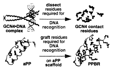
Fi ure 1- Protein grafting strategy for the design of DNA-binding miniature
x0 proteins.
Fi ure 2 - (A) Alignment of the aPP and the GCN4 basic-spacer segment
sequences used to guide protein design. Essential DNA-contact residues within
GCN4 are
in pink; essential folding residues within aPP are in yellow or blue. Conflict
positions are
indicated by a dashed line. (B) Peptides used and their affinities for
hsCRE24.
3
Equilibrium dissociation constants of stable PPBRsR-hsCRE complexes are listed
at right.
All peptides except G56 and G27 contained GGC sequences at their carboxyl
termini. Ga7
contained a single cysteine. The carboxy-terminal cysteine was alkylated with
bromoacetamide to study protein monomers (PPBRsR & Ga7) or oxidized to study
disulfide-linked dimers (PPBRss).
Figure 3 - (A) Residues of PPBR4 targeted for variation mapped onto the
crystal
structure of aPP. Side chains varied in library A are in yellow, those varied
in library B
are in green. (B) Sequences of PPBR4 and the two libraries. Residues varied
are
indicated by an X. Each position was randomized at the DNA level using the NNS
codon
scheme. (C) Sequences of the N-terminal amino acids deduced from the DNA
sequences
of the library B clones after three selection rounds. Peptides containing the
boxed
sequences followed by the remaining residues of PPBR4 were synthesized and
their
properties investigated ih vitro.
Figure 4 - Seven distinct sequences isolated from BAKLIB phage library.
Dissociation constants for miniature protein binding to Bcl-2 are shown on the
right.
Figure 5 - Sequences of the p53 miniature proteins which inhibit p53 binding
to
hDM2. Residues that stabilize the aPP core are in yellow or blue, residues
that contribute
-5-
CA 02407377 2002-10-24
WO 01/81375 PCT/USO1/13023
to binding hDM2 are in purple, residues identified by phage display are in
red.
Equilibrium dissociation constants of stable PPBRsR-hsCRE complexes are listed
at right.
Figure 6 - Two views of the universal library that illustrate the relative
orientation
of the six residues chosen for variation (in beige) on the aPP solvent-exposed
face (top).
The image on the left sites along the alpha helix axis; the image on the right
sites
perpendicular to the alpha helix axis. Residues in blue contribute to forming
the aPP
hydrophobic core. Alignment of aPP and the universal library (bottom).
Residues in blue
stabilize the aPP hydrophobic core; residues in red are targeted for
variation.
DETAILED DESCRIPTION
Definitions
As used herein, the term "binding" refers to the specific association or other
specific interaction between two molecular species, such as, but not limited
to, protein-
DNA interactions and protein-protein interactions. For examples, the specific
association
between proteins and their DNA targets, receptors and their ligands, enzymes
and their
substrates. It is contemplated that such association is mediated through
specific sites on
each of the two interacting molecular species. Binding is mediated by
structural and/or
energetic components, the latter comprising the interaction of molecules with
opposite
charges.
As used herein, the term "binding site" refers to the reactive region or
domain of a
macromolecule that directly participate in its specific binding with another
molecule. For
example, when referring to the binding site on a protein or nucleic acid,
binding occurs as
a result of the presence of specific amino acids or nucleotide sequence,
respectively, that
interact with the other molecule and, collectively, are referred to as a
"binding site."
As used herein, the term "exposed on the alpha helix domain" means that an
amino
acid substituted, for example, into the avian pancreatic polypeptide is
available for
association or interaction with another molecule and are not otherwise bound
to or
associated with another amino acid residue on the avian pancreatic
polypeptide. This
term is used interchangeably with the term-"solvent-exposed alpha helical
face"
throughout the specification.
-6-
CA 02407377 2002-10-24
WO 01/81375 PCT/USO1/13023
As used herein, the terms "miniature protein" or "miniprotein" refers to a
relatively
small protein containing at least a protein scaffold and one or more
additional domains or
regions that help to stabilize its tertiary structure.
As used herein, the term "modulate" refers to an alteration in the association
between two molecular species, for example, the effectiveness of a biological
agent to
interact with its target by altering the characteristics of the interaction in
a competitive or
non-competitive manner.
As used herein, the term "protein" refers to any of a group of complex organic
compounds which contain carbon, hydrogen, oxygen, nitrogen and usually
sulphur, the
characteristic element being nitrogen and which are widely distributed in
plants and
animals. Twenty different amino acids are commonly found in proteins and each
protein
has a unique, genetically defined amino acid sequence which determines its
specific shape
and function. The term "protein" is generally used herein interchangeably with
the terms
peptide and polypeptide.
As used herein, the term "protein scaffold" refers to a region or domain of a
relatively small protein, such as a miniature protein, that has a conserved
tertiary structural
motif which can be modified to display one or more specific amino acid
residues in a fixed
conformation.
Miniature Proteins
The present invention provides engineered miniature proteins that associate
with
(i.e., or bind to) specific sequences of DNA or other proteins and also
provides methods
fox designing and making these miniature proteins. These miniature proteins
bind, for
example, to DNA or other proteins with high affinity and selectivity.
Schematically, the
invention involves a technique that the inventors have designated as protein
grafting (see,
e.g., Fig. 1). Tn one aspect, this technique identifies critical binding site
residues from a
globular protein that participate in binding-type association between that
protein and its
specific binding partners, then these residues are grafted onto a small but
stable protein
scaffold. The preferred protein scaffolds of the invention comprise members of
the
pancreatic fold (PP fold) protein family, particularly the avian pancreatic
polypeptide.
The PP fold protein scaffolds of the invention generally contain thirty-six
amino
acids and are the smallest known globular proteins. Despite their small size,
PP fold
CA 02407377 2002-10-24
WO 01/81375 PCT/USO1/13023
proteins are stable and remain folded under physiological conditions. The
preferred PP
fold protein scaffolds of the invention consist of two anti-parallel helices,
an N-terminal
type II polyprbline helix (PPII) between amino acid residues two and eight and
an alpha-
helix between residues 14 and 31 and/or 32. The stability of the PP fold
protein scaffolds
of the invention derives predominantly from interactions between hydrophobic
residues on
the interior face of the alpha-helix at positions 17, 20, 24, 27, 28, 30 & 31
and the residues
on the two edges of the polyproline helix at positions 2, 4, 5, 7 & 8. . In
general, the
residues responsible for stabilizing it tertiary structure are not substituted
in order to
maintain the tertiary structure of the miniature protein or are compensated
for using phage
display.
In certain embodiments, two or more of the critical binding site residues of,
for
example, a selected globular protein are grafted onto the protein scaffold in
positions
which are not essential in maintaining tertiary structure, preferably on the
solvent-exposed
alpha helical face. In one preferred embodiment, six or more of such binding
site residues
~5 are grafted onto the protein scaffold. In a more preferred embodiment,
eight or more of
such binding site residues are grafted onto the protein scaffold. In an even
more preferred
embodiment, ten or more of such binding site residues are grafted onto the
protein
scaffold. In a most preferred embodiment, 'twelve or more of such binding site
residues
are grafted onto the protein scaffold. Preferred positions for grafting these
binding site
residues on the protein scaffold include, but are not limited to, positions on
the solvent-
exposed alpha-helical face of aPP. Substitutions of binding site residues may
be made,
although they are less preferred, for residues involved in stabilizing the
tertiary structure
of the miniature protein.
The skilled artisan will readily recognize that it is not necessary that
actual
substitution of the grafted residues occur on the protein scaffold. Rather it
is necessary
that a peptide be identified, through, for example, phage display, that
comprises a
polypeptide constituting a miniature protein having the association
characteristics of the
present invention. Such peptides may be pioduced using any conventional means,
including, but not limited to synthetic and recombinant techniques.
Members of the PP fold family of protein scaffolds which are contemplated by
the
present invention include, but are not limited to, avian pancreatic
polypeptide (aPP),
Neuropeptide Y, lower intestinal hormone polypeptide and pancreatic peptide.
In the most
_g_
CA 02407377 2002-10-24
WO 01/81375 PCT/USO1/13023
preferred embodiment, the protein scaffold comprises the PP fold protein,
avian pancreatic
polypeptide (SEQ ID NO: 06) (see, e.g., Blundell et al., (1981) Proc. Natl.
Acad. Sci.
USA 78, 4175-4179; Tonan et al., (1990) Biochemistry 29, 4424-4429). aPP is a
PP fold
polypeptide characterized by a short (eight residue) amino-terminal type II
polyproline
helix linked through a type I beta turn to an eighteen residue alpha-helix.
Because of its
small size and stability, aPP is an excellent protein scaffold for, e.g.,
protein grafting of
alpha-helical recognition epitopes.
DNA-binding Miniature Proteins
In another aspect, the present invention encompasses miniature proteins that
bind
to specific DNA sequences and further encompasses methods for making and using
such
miniature proteins. In some embodiments,"these DNA sequences comprise sites
for
known proteins that bind to that specific DNA sequence (contemplated known
proteins
would be, e.g., a promotor or regulator). For example, in the design of a DNA-
binding
miniature protein, the amino acid residues of a known protein that participate
in binding or
other association of the protein to that particular DNA sequence are
identified.
In some embodiments of the present invention, the relevant binding residues
are
identified using three-dimensional models of a protein or protein complex
based on
crystallographic studies while in other embodiments they are identified by
studies of
deletion or substitution mutants of the protein. The residues that participate
in binding of
the protein to the specific DNA sequence are then grafted onto those positions
of the
miniature protein that are not necessary to maintain the tertiary structure of
the protein
scaffold to form the DNA-binding miniature protein. The identification of such
positions
can readily be determined empirically by persons skilled in the art. Other
embodiments of
the present invention involve the screening of a library of modified
miniproteins that
contain peptide species capable of specific association or binding to that
specific DNA (or,
in other cases, protein) sequence or motif.
Generally, it is contemplated that any potential binding site on a DNA
sequence
can be targeted using the DNA binding miniature proteins of the invention.
Preferred
embodiments include helical structures which bind to the DNA binding site. In
some
embodiments, the binding involves a basic region leucine zipper (bZIP)
structure (Konig
& Richmond, (1995) J. MoI. Biol. 254, 657-667) while in other embodiments the
structure
CA 02407377 2002-10-24
WO 01/81375 PCT/USO1/13023
involves a basic-helix-loop-helix (bHLH) structure (Shimizu et al., (1997)
EMBO J. 16,
4689-4697). In another embodiment, the binding involves a structure like those
found in
homeodomain proteins (Scott & Weimer, (1984) Proc. Natl. Acad. Sci. 81, 4115-
4119).
Preferred bZIP structures include, but are not limited to, those found in GCN4
and C/EBP
-delta (Suckow et al., (1993) EMBO J. 12, 1193-1200) while preferred bHLH
structures
include, but are not limited to, those found in Max (Ferre-D'Amare et al.,
(1993) Nature
363, 38-45), Myc and MyoD (Ma et al., (1994) Cell 77, 451-459). Preferred
homeodomain structures include, but are not Limited to, those found in the Q50
engrailed
variant protein (Kissinger et al., (1990) Cell 63, 579-590).
In one embodiment, the invention encompasses a DNA-binding miniature protein
that binds to the cAMP Response Element (CRE) half site promotor DNA sequence
(ATGAC) (SEQ ID NO: 65). Essential residues for binding are identified from
the protein
GCN4 which is a bZIP protein which binds to this sequence. These residues are
identified
by utilizing the three-dimensional structure of the GCN4 protein which bind to
the hsCRE
I5 and grafting these residues onto the protein. scaffold. By grafting various
combinations of
residues on the solvent-exposed alpha-helical face or domain of aPP which are
essential to
binding of GCN4 (SEQ ID NO: 7) to the CRE half site (hsCRE), a series of
polyproline
helix-basic region (PPBRsR) molecules containing most or all of the DNA-
contact residues
of GCN4 and most or all of the folding residues of aPP is generated (Fig. 2).
This
procedure generated three positions (Tyr27, Leu28 and Va130) where essential
DNA-
contact and aPP-folding residues occupied a single position on the helix (Fig.
2).
Examples of the DNA-binding miniature proteins which bind to hsCRE include,
but are not Limited to, the amino acid sequences depicted in SEQ ~ NO: 11
(PPBR2sR),
12 (PPBR4sR), 13 (Ga7) & 14 (PPBR4~SR)..
In another embodiment, protein grafting was used for the design of a miniature
protein whose DNA binding properties mimic those of the CCAAT/enhancer protein
C/EBP-delta. C/EBP-delta is a member ofvthe C/EBP sub-family of bZIP
transcription
factors that includes C/EBP-alpha, C/EBP-beta, C/EBP-gamma, C/EBP-delta and
C/EBP-
epsilon. Although C/EBP proteins are members of the bZIP superfamily, they
differ from
CGN4 at several residues within the DNA recognition helix. In particular,
D/EBP-delta
and GCN4 differ at two of six residues that contact bases or sugars and three
of six
residues that contact phosphates in all published structures of GCN4 DNA
complexes.
-10-
CA 02407377 2002-10-24
WO 01/81375 PCT/USO1/13023
These changes, as well as the substitution of tyrosine or alanine at position
fifteen,
contribute to the preferred interaction of C/EBP proteins with the C/EBP site
(ATTGCGCAAT) (SEQ ID NO: 67) over the CRE site (ATGACGTCAT) (SEQ ID NO:
68) recognized by GCN4.
For the design of PPEBP (polyproline-enhancer binding protein) according to
the
present invention, the first step in the grafting protocol is alignment of the
alpha-helix of
aPP (residues 14-36) with the alpha-helical region of the protein of interest.
Alignment of
the aPP alpha-helix with residues 187-221 (the DNA-binding basic segment) of
human
C/EBP-delta identified three conflict positions (27, 28 & 30 according to the
aPP
numbering system) where DNA-contact residues within C/EBP-delta and folding
residues
within aPP occupied the same position on the helix. The PPEBPIsR (SEQ ZD NO:
47)
miniature protein of the invention contains.arginine residues derived from
C/EBP-delta at
positions 27, 28 & 30 to preserve binding affinity because high-affinity DNA
recognition
by PPEBP miniature proteins is enhanced by retention of DNA-contact residues
at these
positions despite the concomitant loss in folding energy. In addition,
tyrosine, asparagine
and valine residues are substituted at positions 15, 23 & 26, respectively to
foster specific
recognition of the C/EBP half site ATTGC (hsCEBP). Finally an alanine residue
is
inserted at position 31 in place of the potentially core-disrupting and
complex-
destabilizing aspartate found in C/EBP-delta and in place of the helix
destabilizing valine
present at this position of aPP.
Examples of the DNA-binding miniature proteins which bind to the C/EBP site
include, but are not limited to, the amino acid sequences depicted in SEQ ID
NO: 47
(PPEBP 1 sR), 48 (PPEBP2SR) and 49 (EBP I sR).
Production of Miniature Proteins Using Phase DisPlay
In some embodiments, a miniature protein is produced and selected using a
phage
display method (McCafferty et al., (1990) Nature 348, 552-554). In such a
method,
display of recombinant miniature proteins on the surface of viruses which
infect bacteria
(bacteriophage or phage) make it possible to produce soluble, recombinant
miniature
proteins having a wide range of affinities and kinetic characteristics. To
display the
miniature proteins on the surface of phage, a synthetic gene encoding the
miniature
protein is inserted into the gene encoding a~'phage surface protein (pII1) and
the
-11-
CA 02407377 2002-10-24
WO 01/81375 PCT/USO1/13023
recombinant fusion protein is expressed on the phage surface (McCafferty et
al., (1990)
Nature 348, 552-554; Hoogenboom et al., (1991) Nucleic Acids Res. 19, 4133-
4137).
Variability is introduced into the phage display library to select for
miniature proteins
which not only maintain their tertiary, helical structure but which also
display increased
affinity for a preselected target because the critical (or contributing but
not critical)
binding residues are optimally positioned on the helical structure.
Since the recombinant proteins on the surface of the phage are fiulctional,
phage
bearing miniature proteins that bind with high-affinity to a particular target
DNA or
protein can be separated from non-binding or lower affinity phage by antigen
affinity
chromatography. Mixtures of phage are allowed to bind to the affinity matrix,
non-
binding or lower affinity phage are removed by washing, and bound phage axe
eluted by
treatment with acid or alkali. Depending on the affinity of the miniature
protein for its
target, enrichment factors of twenty-fold to a million-fold are obtained by a
single round
of affinity selection. By infecting bacteria with the eluted phage, however,
more phage
can be grown and subjected to another round of selection. In this way, an
enrichment of a
thousand-fold in one round becomes a million-fold in two rounds of selection.
Thus, even
when enrichments in each round are low (Marks et al., (1991) J. Mol. Biol,
222, 581-597),
multiple rounds of affinity selection leads to the isolation of rare phage and
the genetic
material contained within which encodes the sequence of the domain or motif of
the
recombinant miniature protein that binds or otherwise specifically associates
with it
binding target.
Tn various embodiments of the invention, the methods disclosed herein are used
to
produce a phage expression library encoding miniature proteins capable of
binding to a
DNA or to a protein that has already been selected using the protein grafting
procedure
described above. In such embodiments, phage display can be used to identify
miniature
proteins that display an even higher affinity for a particular target DNA or
protein than
that of the miniature proteins produced without the aid of phage display. In
yet another
embodiment, the invention encompasses a universal phage display library that
can be
designed to display a combinatorial set of epitopes or binding sequences to
permit the
recognition of nucleic acids, proteins or small molecules by a miniature
protein without
prior knowledge of the natural epitope or specific binding residues or motifs
natively used
for recognition and association.
-12-
CA 02407377 2002-10-24
WO 01/81375 PCT/USO1/13023
Various structural modifications also are contemplated for the present
invention
that, for example, include the addition of restriction enzyme recognition
sites into the
polynucleotide sequence encoding the miniature protein that enable genetic
manipulation
of these gene sequences. Accordingly, the re-engineered miniature proteins can
be ligated,
for example, into an M13-derived bacteriophage cloning vector that permits
expression of
a fusion protein on the phage surface. These methods allow for selecting phage
clones
encoding fusion proteins that bind a target ligand and can be completed in a
rapid manner
allowing for high-throughput screening of miniature proteins to identify the
miniature
protein with the highest affinity and selectivity for a particular target.
According to the methods of the invention, a library of phage displaying
modified
miniature proteins is incubated with the immobilized target DNA or proteins to
select
phage clones encoding miniature proteins that specifically bind to or
otherwise
specifically associate with the immobilized DNA or protein. This procedure
involves
immobilizing a oligonucleotide or polypeptide sample on a solid substrate. The
bound
IS phage are then dissociated from the immobilized oligonucleotide or
polypeptide and
amplified by growth in bacterial host cells. Tndividual viral plaques, each
expressing a
different recombinant miniature protein, are expanded to produce amounts of
protein
sufficient to perform a binding assay. The DNA encoding this recombinant
binding
protein can be subsequently modified for ligation into a eukaryotic protein
expression
vector. The modified miniature protein, adapted for expression in eukaryotic
cells, is
ligated into a eukaryotic protein expression vector.
Phage display methods that can be used to make the miniature proteins of the
present invention include those disclosed iri Brinkman et al., (1995) J.
linmunol. Methods
182, 41-S0; Ames et al., (1995) J. Tmmunol. Methods 184:177-186; Kettleborough
et al.,
(1994) Eur. J. Immunol. 24, 952-958; Persic et al., (1997) Gene 187, 9-18;
Burton et al.,
(1994) Adv. Immunol. 57, 191-280; U.S. Patents 5,698,426; 5,223,409;
5,403,484;
5,580,717; 5,427,908; 5,750,753; 5,821,047; 5,571,698; 5,427,908; 5,516,637;
5,780,225;
5,658,727; 5,733,743, 5,837,500 & 5,969,108.
Protein-Binding Miniature Proteins
The invention encompasses miniatt~xe proteins that bind to other proteins and
methods for making these miniature proteins. The binding of the miniature
proteins
-13-
CA 02407377 2002-10-24
WO 01/81375 PCT/USO1/13023
modulates protein-protein and/or protein-ligand interactions. Thus, in some
embodiments
the binding blocks the association (or specific binding) of ligands and
receptors. The
ligand can be either another protein but also can be any other type of
molecule such as a
chemical substrate. In one embodiment of~the present invention, making the
protein-
s binding miniature protein of the invention involves identifying the amino
acid residues
which are essential to binding of the ligand.protein to its taxget receptor
protein. In some
embodiments, these essential residues are identified using three-dimensional
models of a
protein or protein complex which binds to or interacts with another protein
based on
crystallographic studies while in other embodiments they are identified by
studies of
deletion or substitution mutants of the protein. The residues that participate
in binding of
the protein to are then grafted onto those positions which are not necessary
to maintain the
tertiary structure of the protein scaffold to form the protein-binding
miniature protein.
The structure of any protein which binds to another protein can be used to
derive
the protein-binding miniature proteins of the invention. Preferred embodiments
include
helical structures such as those involved in protein-protein interactions
between Fos and
Jun (Kouzarides & Ziff, (1988) Nature 336, 646-651), Bcl -2 and Bak (Sattler
et al.,
(1997) Science 275, 983-986), CBP-KIX and CREB-KID (Radhakrishnan et al.,
(1997)
Cell 91, 741-752) and p53 binding to DM2 (Kussie et al., (1996) Science 274,
948-953).
Tn some embodiments, the binding involves coiled coil protein structures
and/or leucine
zippers.
In one embodiment of the invention, the methods disclosed herein are used to
produce a miniature protein that binds to the Bcl-2 or Bcl-XL proteins
(Sattler et al.,
(1997) Science 275, 983-986). In this method, the protein grafting procedure
described
herein was applied to the Bak-BH3 binding domain to design a miniature protein
capable
of binding to Bcl-XL. In this procedure, thd primary sequence of a protein of
interest is
aligned with residues in the alpha helix of a.PP. All possible alignments of
the primary
sequence of positions 74-92 of Bak with aPP are assessed in two ways. First,
the number
of conflicts in a primary sequence alignment between residues important for
hydrophobic
core formation or maintenance of aPP helix dipole, and residues in Bak
important for
binding Bcl-XL was considered. Alignments with a large number of conflicts are
eliminated as they would force selection between sequences that were well
folded or have
high affinity, but make it difficult to isolate a molecule with both these
properties.
-14-
CA 02407377 2002-10-24
WO 01/81375 PCT/USO1/13023
Structural models of the aPP based peptides that are associated or complexed
with
the BH3 domain of Bcl-XL in each of the alignments are evaluated for
unfavorable
interactions or steric clashes between the VanderWaals surface of Bcl-XL and
the
backbone of the aPP scaffold. Structural models with multiple unfavorable
interactions or
steric clashes are eliminated from further consideration.
An alignment is identified with only a single conflict where structural
modeling
suggested no steric clashes. A phage display expression library of chimeric
peptides
ultimately was based on this alignment. The resulting library of peptides was
displayed on
the surface of M13 phage and used in selection and isolation of miniature
proteins that
bind Bcl with high-affinity. Examples of the protein-binding miniature
proteins isolated
from the phage display library which bind to Bcl include, but are not limited
to, the amino
acid sequences depicted in SEQ ID NO: 23 (4100), 24 (4101), 25 (4099) & 26
(4102).
In another embodiment of the invention, the methods of the invention are used
to
produce a miniature protein that binds to the human oncoprotein double minute
two
(hDM2). The alpha-helical segments of p53 and aPP were aligned to identify
three critical
hDM2 contact residues (positions 22, 26 & 29) on the exposed alpha-helical
face of aPP
without substituting any aPP residues important for folding. Because many p53
residues
within the p53 activation domain that interacts with hDM2 display phi and psi
angles
outside the ideal alpha-helical range, this application of protein grafting
introduced
ZO diversity at five positions along the alpha-helix and the highest affinity
ligands were
selected using phage display
Examples of the protein-binding miniature proteins isolated from the phage
display
library which bind to hDM2 include, but are not limited to, the amino acid
sequences
depicted in SEQ >D NO: 31 (p53AD), 33 (p3254), 34 (p3255), 35 (p354~), 36
(p3559) &
37 (p3257).
Ndiniature Protein Variants
The miniature proteins of the present invention further include conservative
variants of the miniature proteins herein described. As used herein, a
conservative variant
refers to alterations in the amino acid sequence that do not substantially and
adversely
affect the binding or association capacity of the protein. A substitution,
insertion or
deletion is said to adversely affect the miniature protein when the altered
sequence
-15-
CA 02407377 2002-10-24
WO 01/81375 PCT/USO1/13023
prevents or disrupts a function or activity associated with the protein. For
example, the
overall charge, structure or hydrophobic-hydrophilic properties of the
miniature protein
can be altered without adversely affecting an activity. Accordingly, the amino
acid
sequence can be altered, for example to render the peptide more hydrophobic or
hydrophilic, without adversely affecting the activities of the miniature
protein.
These variants, though possessing a slightly different amino acid sequence
than
those recited above, will still have the same or similar properties associated
with the
miniature proteins depicted in SEQ ID NO: 8, 9, 10, 11, 12, 13, 17, 18, 19,
20, 21, 22, 23,
24, 25, 26, 27, 28, 29, 31, 33, 34, 35, 36, 37, 47, 48, 49, 50, 51, 52, 53,
54, 55, 56, 57, 58,
59, 60, 61, 62, 63, 64, 70, 71 or 72.
Ordinarily, the conservative substitution variants, will have an amino acid
sequence having at least ninety percent amino acid sequence identity with the
miniature
sequences set forth in SEQ ID NO: 8, 9, 10, 11, 12, 13, 17, 18, 19, 20, 21,
22, 23, 24, 25,
26, 27, 28, 29, 31, 33, 34, 35, 36, 37, 47, 4$, 49, 50, 51, 52, 53, 54, 55,
56, 57, 58, 59, 60,
61, 62, 63, 64, 70, 71 or 72, more preferably at least ninety-five percent,
even more
preferably at least ninety-eight percent, and most preferably at least ninety-
nine percent.
Identity or homology with respect to such sequences is defined herein as the
percentage of
amino acid residues in the candidate sequence that are identical with the
known peptides,
after aligning the sequences and introducing gaps, if necessary, to achieve
the maximum
percent homology, and not considering any conservative substitutions as part
of the
sequence identity. N-terminal, C-terminal or internal extensions, deletions,
or insertions
into the peptide sequence shall not be construed as affecting homology.
Thus, the miniature proteins of the present invention include molecules
comprising
the amino acid sequence of SEQ ID NO: 8,.9, 10, 11, 12, 13, 17, 18, 19, 20,
21, 22, 23, 24,
25, 26, 27, 28, 29, 31, 33, 34, 35, 36, 37, 47, 48, 49, 50, 51, 52, 53, 54,
55, 56, 57, 58, 59,
60, 61, 62, 63, 64, 70, 71 or 72; fragments thereof having a consecutive
sequence of at
least about 20, 25, 30, 35 or more amino acid residues of the miniature
proteins of the
invention; amino acid sequence variants of such sequences wherein at least one
amino acid
residue has been inserted N- or C-terminal to, or within, the disclosed
sequence; amino
acid sequence variants of the disclosed sequences, or their fragments as
defined above,
that have been substituted by another residue. Contemplated variants further
include those
derivatives wherein the protein has been covalently modified by substitution,
chemical,
-16-
CA 02407377 2002-10-24
WO 01/81375 PCT/USO1/13023
enzymatic, or other appropriate means with a moiety other than a naturally
occurring
amino acid (for example, a detectable moiety such as an enzyme or
radioisotope).
Nucleic Acid Molecules Encoding Miniature Proteins
The present invention further provides nucleic acid molecules that encode the
miniature proteins comprising the amino acid sequence of SEQ m NO: 8, 9, 10,
11, 12,
13, 17, 18, 19, 20, 21, 22, 23, 24, 25, 26, 27, 28, 29, 31, 33, 34, 35, 36,
.37, 47, 48, 49, 50,
51, 52, 53, 54, 55, 56, 57, 58, 59, 60, 61, 62, 63, 64, 70, 71 or 72 and the
related miniature
proteins herein described, preferably in isolated form. As used herein,
"nucleic acid"
includes cDNA and mRNA, as well as nucleic acids based on alternative
backbones or
including alternative bases whether derived from natural sources or
synthesized.
As used herein, a nucleic acid molecule is said to be "isolated" when the
nucleic
acid molecule is substantially separated from contaminant nucleic acid
encoding other
polypeptides from the source of nucleic acid.
The present invention further provides fragments of the encoding nucleic acid
molecule. As used herein, a "fragment of an encoding nucleic acid molecule"
refers to a
portion of the entire protein encoding sequence of the miniature protein. The
size of the
fragment will be determined by the intended use. For example, if the fragment
is chosen
so as to encode an active portion of the protein, the fragment will need to be
large enough
to encode the functional regions) of the protein. The appropriate size and
extent of such
fragments can be determined empirically by persons skilled in the art.
Modifications to the primary structure itself by deletion, addition, or
alteration of
the amino acids incorporated into the protein sequence during translation can
be made
without destroying the activity of the miniature protein. Such substitutions
or other
alterations result in miniature proteins having an amino acid sequence encoded
by a
nucleic acid falling within the contemplated scope of the present invention.
The present invention further provides recombinant DNA molecules that contain
a
coding sequence. As used herein, a recombinant DNA molecule is a DNA molecule
that
has been subjected to molecular manipulation. Methods for generating
recombinant DNA
molecules are well known in the art, for example, see Sambrook et al., (1989)
Molecular
Cloning - A Laboratory Manual, Cold Sprig Harbor Laboratory Press. In the
preferred
recombinant DNA molecules, a coding DNA sequence is operably linked to
expression
_ ~7 ,
CA 02407377 2002-10-24
WO 01/81375 PCT/USO1/13023
control sequences and vector sequences.
The choice of vector and expression control sequences to which one of the
protein
family encoding sequences of the present invention is operably linked depends
directly, as
is well known in the art, on the functional properties desired (e.g., protein
expression, and
the host cell to be transformed). A vector of the present invention may be at
least capable
of directing the replication or insertion into the host chromosome, and
preferably also
expression, of the structural gene included in the recombinant DNA molecule.
Expression control elements that are used for regulating the expression of an
operably linked miniature protein encoding sequence are known in the art and
include, but
are not limited to, inducible promoters, constitutive promoters, secretion
signals, and other
regulatory elements. Preferably, the inducible promoter is readily controlled,
such as
being responsive to a nutrient in the host cell's medium.
In one embodiment, the vector containing a coding nucleic acid molecule will
include a prokaryotic replicon, i. e., a DNA' sequence having the ability to
direct
autonomous replication and maintenance of the recombinant DNA molecule extra-
chromosomal in a prokaryotic host cell, such as a bacterial host cell,
transformed
therewith. Such replicons are well known in the art. In addition, vectors that
include a
prokaryotic replicon may also include a gene whose expression confers a
detectable
marker such as a drug resistance. Typical of bacterial drug resistance genes
are those that
confer resistance to ampicillin or tetracycline.
Vectors that include a prokaryotic replicon can further include a prokaryotic
or
bacteriophage promoter capable of directing the expression (transcription and
translation)
of the coding gene sequences in a bacterial host cell, such as E. coli. A
promoter is an
expression control element formed by a DNA sequence that permits binding of
RNA
polyrneras~e and transcription to occur. Promoter sequences compatible with
bacterial
hosts are typically provided in plasmid vectors containing convenient
restriction sites for
insertion of a DNA segment of the present invention. Any suitable prokaryotic
host can
be used to express a recombinant DNA molecule encoding a protein of the
invention.
Expression vectors compatible with eukaryotic cells, preferably those
compatible
with vertebrate cells, can also be used to form a recombinant DNA molecules
that contains
a coding sequence. Eukaryotic cell expression vectors are well known in the
art and are
available from several commercial sources. Typically, such vectors are
provided
-18-
CA 02407377 2002-10-24
WO 01/81375 PCT/USO1/13023
containing convenient restriction sites for insertion of the desired DNA
segment.
Eukaryotic cell expression vectors used to construct the recombinant DNA
molecules of the present invention may further include a selectable marker
that is effective
in an eukaryotic cell, preferably a drug resistance selection marker. A
preferred drug
resistance marker is the gene whose expression results in neomycin resistance,
i.e., the
neomycin phosphotransferase (neo) gene. (Southern et al., (1982) J. Mol. Anal.
Genet. 1,
327-341). Alternatively, the selectable marker can be present on a separate
plasmid, the
two vectors introduced by co-transfection of the host cell, and transfectants
selected by
culturing in the appropriate drug for the selectable marker.
Transformed Host Cells
The present invention further provides host cells transformed with a nucleic
acid
molecule that encodes a miniature protein of the present invention. The host
cell can be
either prokaryotic or eukaryotic. Eukaryotic cells useful for expression of a
miniature
protein of the invention axe not limited, so long as the cell line is
compatible with cell
culture methods and compatible with the propagation of the expression vector
and
expression of the gene product.
Transformation of appropriate cell hosts with a recombinant DNA molecule
encoding a miniature protein of the presenfinvention is accomplished by well
known
methods that typically depend on the type of vector used and host system
employed. With
regaxd to transformation of prokaryotic host cells, electroporation and salt
treatment
methods can be employed (see, for example, Sambrook et al., (1989) Molecular
Cloning -
A Laboratory Manual, Cold Spring Harbor Laboratory Press; Cohen et al., (1972)
Proc.
Natl. Acad. Sci. USA 69, 2110-2114). With regard to transformation of
vertebrate cells
with vectors containing recombinant DNA, electroporation, cationic lipid or
salt treatment
methods can be employed (see, for example, Graham et al., (1973) Virology 52,
456-467;
Wigler et al., (1979) Proc. Natl. Acad. Sci. USA 76, 1373-1376).
Successfully transformed cells (cells that contain a recombinant DNA molecule
of
the present invention), can be identified by"well known techniques including
the selection
fox a selectable marker. For example, cells resulting from the introduction of
a
recombinant DNA of the present invention 'can be cloned to produce single
colonies.
Cells from those colonies can be harvested, lysed and their DNA content
examined for the
-19-
CA 02407377 2002-10-24
WO 01/81375 PCT/USO1/13023
presence of the recombinant DNA using a method such as that described by
Southern,
(1975) J. Mol. Biol. 98, 503-517 or the proteins produced from the cell
assayed via an
immunological method.
Production of Recombinant Miniature Proteins
The present invention fizrther provides methods for producing a miniature
protein
of the invention using nucleic acid molecules herein described. In general
terms, the
production of a recombinant form of a protein typically involves the following
steps: a
nucleic acid molecule is obtained that encodes a protein of the invention,
such as the
nucleic acid molecule encoding any of the miniature proteins depicted in SEQ m
NO: 8,
9, 10, 11, 12, 13, 17, 18, 19, 20, 21, 22, 23, 24, 25, 26, 27, 28, 29, 31, 33,
34, 35, 36, 37,
47, 48, 49, 50, 5I, 52, 53, 54, 55, 56, 57, 58, 59, 60, 61, 62, 63, 64, 70, 71
or 72. The
nucleic acid molecule is then preferably placed in operable linkage with
suitable control
sequences, as described above, to form an expression unit containing the
protein open
reading frame. The expression unit is used to transform a suitable host and
the
transformed host is cultured under conditions that allow the production of the
recombinant
miniature protein. Optionally the recombinant miniature protein is isolated
from the
medium or from the cells; recovery and purification of the protein may not be
necessary in
some instances where some impurities may be tolerated.
Each of the foregoing steps can be done in a variety of ways. The construction
of
expression vectors that are operable in a variety of hosts is accomplished
using appropriate
replicons and control sequences, as set forth above. The control sequences,
expression
vectors, and transformation methods are dependent on the type of host cell
used to express
the gene. Suitable restriction sites, if not normally available, can be added
to the ends of
the coding sequence so as to provide an excisable gene to insert into these
vectors. A
skilled artisan can readily adapt any host/expression system known in the art
for use with
the nucleic acid molecules of the invention to produce a recombinant miniature
protein.
Methods to Identify Binding Partners
The present invention provides methods for use in isolating and identifying
binding partners of the miniature proteins of the invention. In some
embodiments, a
miniature protein of the invention is mixed with a potential binding partner
or an extract or
-ZO-
CA 02407377 2002-10-24
WO 01/81375 PCT/USO1/13023
fraction of a cell under conditions that allow the association of potential
binding partners
with the protein of the invention. After mixing, peptides, polypeptides,
proteins or other
molecules that have become associated with a miniature protein of the
invention are
separated from the mixture. The binding partner bound to the protein of the
invention can
then be removed and further analyzed. To identify and isolate a binding
partner, the entire
miniature protein can be used. Alternatively, a fragment of the miniature
protein which
contains the binding domain can be used. ;
As used herein, a "cellular extract" refers to a preparation or fraction which
is
made from a Iysed or disrupted cell. A variety of methods can be used to
obtain an extract
of a cell. Cells can be disrupted using either physical or chemical disruption
methods.
Examples of physical disruption methods include, but are not limited to,
sonication and
mechanical shearing. Examples of chemical lysis methods include, but are not
limited to,
detergent lysis and enzyme lysis. A skilled artisan can readily adapt methods
for
preparing cellular extracts in order to obtain extracts for use in the present
methods.
Once an extract of a cell is prepared, the extract is mixed with the a
miniature
protein of the invention under conditions in which association of the
miniature protein
with the binding partner can occur. A variety of conditions can be used, the
most
preferred being conditions that closely resemble conditions found in the
cytoplasm of a
human cell. Features such as osmolarity, pH, temperature, and the
concentration of
cellular extract used, can be varied to optimize the association of the
protein with the
binding partner.
After mixing under appropriate conditions, the bound complex is separated from
the mixture. A variety of techniques can be utilized to separate the mixture.
For example,
antibodies specific to a protein of the invention can be used to
immunoprecipitate the
binding partner complex. Alternatively, standard chemical separation
techniques such as
chromatography and density-sediment centrifugation can be used.
After removal of non-associated cellular constituents found in the extract,
the
binding partner can be dissociated from the, complex using conventional
methods. For
example, dissociation can be accomplished ~by altering the salt concentration
or pH of the
mixture.
To aid in separating associated binding partner pairs from the mixed extract,
the
miniature protein of the invention can be immobilized on a solid support. For
example,
_21_
CA 02407377 2002-10-24
WO 01/81375 PCT/USO1/13023
the miniature protein can be attached to a nitrocellulose matrix or acrylic
beads.
Attachment of the miniature protein to a solid support aids in separating
peptide-binding
partner pairs from other constituents found in the extract. The identified
binding partners
can be either a single DNA molecule or protein or a complex made up of two or
more
proteins. Alternatively, binding partners may be identified using the
Allcaline Phosphatase
fusion assay according to the procedures of Flanagan & Vanderhaeghen, (1998)
Annu.
I~ev. Neurosci. 21, 309-345 or Takahashi et al., (1999) Cell 99, 59-69; the
Far-Western
assay according to the procedures of Takayama et al., (1997) Methods Mol.
Biol. 69, 171-
184 or Sauder et al., J. Gen. Virol. (1996) 77, 991-996 or identified through
the use of
epitope tagged proteins or GST fusion proteins.
Alternatively, the nucleic acid molecules encoding a miniature protein of the
invention can be used in a yeast two-hybrid system. The yeast two-hybrid
system has
been used to identify other protein partner pairs and can readily be adapted
to employ the
nucleic acid molecules herein described (see, e.g., Stratagene Hybrizap~ two-
hybrid
system).
Screening,~Dia~nostic & Therapeutic Uses
The miniature proteins of the invention are particularly useful for drug
screening to
identify agents capable of binding to the same binding site as the miniature
proteins. The
miniature proteins are also useful for diagnostic purposes to identify the
presence and/or
detect the levels of DNA or protein that binds to the miniature proteins of
the invention.
In one diagnostic embodiment, the miniature proteins of the invention are
included in a kit
P
used to detect the presence of a particular DNA or protein in a biological
sample. The
miniature proteins of the invention also haee therapeutic uses in the
treatment of disease
2S associated with the presence of a particular DNA or protein. In one
therapeutic
embodiment, the miniature proteins can be used to bind to DNA to promote or
inhibit
transcription, while in another therapeutic embodiment, the miniature proteins
bind to a
protein resulting in inhibition or stimulation of the protein.
Without further description, it is believed that a person of ordinary skill in
the art
can, using the preceding description and the following illustrative examples,
make and
utilize the compounds of the present invention and practice the claimed
methods. The
-~2-
CA 02407377 2002-10-24
WO 01/81375 PCT/USO1/13023
following working examples therefore, specifically point out preferred
embodiments of
the present invention, and are not to be construed as limiting in any way the
remainder of
the disclosure.
EXAMPLES
Example 1- Synthesis of DNA-binding miniature roteins
Polypeptides constituting miniature~proteins were prepared using solid phase
methodology and contain a carboxy-terminal amide and a free amino terminus
unless
otherwise indicated. High performance liquid chromatography (HPLC) was
performed on
either a Waters 600E Multisolvent Delivery System with a Waters 490E
multiwavelength
detector or a Rainin Dynamax SD-200 Solvent Delivery System with a Rainin
Dynamax
PDA-2 Diode Array Detector.
Solid phase peptide synthesis was performed on a Perseptive BioSearch 9600
peptide synthesizer. Standard research grade argon (Connecticut AirGas) was
passed
through an OxyClear oxygen scrubber before introduction to the synthesizer.
HATU (O-
(7-benzotrizol-1-yl)-1,1,3,3,-tetramethyl uronium hexafluorophosphate) was
used as the
activating reagent without addition of supplemental benzotrizole.
Dimethylformamide,
piperidine and methylene chloride (Baker) were fresh and stored under
nitrogen.
Anhydrous dimethylformamide was mixed with diisopropylethylamine (DIPEA,
redistilled 0.46 M) to prepare the base activator solution. 9-
Fluorenylmethoxycarbonyl
(F-moc)-protected amino acids utilized the following side chain protecting
groups: O-t-
butyl (Asp, Glu); t-butyl (Tyr, Thr, Ser); 2,2,4,6,7-
pentamethyldihydrobenzofuran-5-
sulfonyl (Pbf) (Arg); t-butoxycarbonyl (Lys); and triphenyhnethyl (Cys, His,
Asn, Gln).
Synthesis was performed on a 0.10 mmol scale using PAL (peptide amide linker)
resin
(Fmoc-NH2-CHa-(di-m-methoxy,p-O-(CH2)4C(O)-polystyrene) which resulted in an
amidated carboxy-terminus. Fmoc-amino acid and HATU were used in four-fold
excess
(0.4 mmol per coupling). After the final coupling was completed, the Fmoc-
protecting
group was removed and the resin was washed for the last time. The resin was
dried and
stored in a desicator until cleavage and deprotection were initiated.
Reverse phase HPLC was performed using eluents composed of mixtures of Buffer
A (98% HPLC water, 2% acetonitrile, 0.05% trifluoroacetic acid) and Buffer B
(20%
HPLC water, 80% acetonitrile, 0.06% trifluoroacetic acid). All HPLC solvents
were
-23-
CA 02407377 2002-10-24
WO 01/81375 PCT/USO1/13023
filtered through a 0.2 micron filter prior to use. Solvents and chemicals for
peptide
synthesis were obtained from Aldrich and Perseptive Biosearch unless stated
otherwise.
Peptides were lyophilized using a Savant SC100 Speed Vacuum instrument.
Denaturing
sodium dodecyl sulfate-polyacryalmide gel electrophoresis (SDS-PAGE) analysis
was
performed with a Pharmacia PhastGel system using High Density gels (20%
acrylamide
soaked in glycerol). Amino acid analysis was assayed on a Beckman Analyzer.
For deprotection and purification of PPEBP1SH, PAL resin (15 mg) containing
protected PPEBP1SH was allowed to react for five hours at room temperature in
a
deprotection cocktail (84% trifluoroacetic acid, 4% phenol, 4% ethanedithiol,
4%
thioanisole and 4% water). The solvent was removed by blowing a stream of
nitrogen
over the solution until the volume reached approximately 0.25 ml. Diethylether
(1 ml) and
dithiothreitol (20 mg) were added to precipitate the peptide and stabilize the
cysteine. The
supernatant was removed after centrifugation and the precipitate dried. The
crude peptide
was dissolved in 1 ml phosphate-buffered saline (pH 7.5) with added
dithiothreitol (5 mg)
and filtered with a 0.2 micron filter. The peptide was purified by reverse
phase HPLC
(Vydac semipreparative 300 ~ C18, 5 microns, 10.0 x 250 mm) using a 120 minute
linear
gradient of 100 - 30% Buffer A in Buffer B. The peptide eluted at 49.3 minutes
using a
flow rate of 4 ml/min and was analyzed by electrospray ionization mass
spectrometry.
The predicted and observed masses were 4729.4 and 4730.0, respectively.
For preparation of PPEBPIsR, 0.080 mg of PPEBPIsH was dissolved in 0.50 ml of
2 mg/ml (15 mM) 2-bromoacetamide in 20 mM sodium phosphate buffer (pH 7.5).
The
reaction was allowed to proceed for thirty minutes at room temperature. The
peptide was
purified by reverse phase HPLC (Rainin analytical 1001 C18, 5 microns, 4.6 x
250 mm)
using a forty minute linear gradient of 100 - 30% Buffer A in Buffer B. The
peptide
eluted at 23.3 minutes using a flow rate of 1 ml/min and was characterized by
electrospray
ionization mass spectrometry and amino acid analysis. AAA expected: Alas AsxS
CmCysl Glx2 Phel Gly4 HisO LleO Lys3 Leu2 MetO Pro4 ArgB Ser2 Thrl Val2 Tyr2,
found A1a5.2 Asx4.8 CmCys0.6 G1x2.0 Phel.O G1y4.1 HisO LleO Lys2.9 Leu2.0 MetO
Pro3.7 Arg6.9 Serl.8 Thr0.8 Va12.0 Tyrl.B; mass predicted 4786.4, found
4787.1.
For deprotection and purification of PPEBP2sH, PAL resin (10 mg) containing
protected PPEBP2sH was allowed to react for seven hours at room temperature in
the
deprotection cocktail and the solvent was removed. Diethylether (I ml) and
dithiothreitol
_24_
CA 02407377 2002-10-24
WO 01/81375 PCT/USO1/13023
(20 mg) were added, the supernatant was removed after centrifugation and the
precipitate
dried. The crude peptide was dissolved in 1 rnl phosphate-buffered saline (pH
7.5)
containing 5 mg fresh dithiothreitol and filtered. The peptide was purified by
reversed
phase HPLC (Vydac semipreparative 300 ~ C18, 5 microns, I0.0 x 250 mm) using a
linear 120 minute gradient of 100 - 50% Buffer A in Buffer B. The peptide
eluted at 67.8
minutes using a flow rate of 4 ml/min and was characterized by electrospray
ionization
mass spectrometry: mass predicted 4654.2, found 4653.6.
For preparation of PPEBP2sR, 0.070 mg of PPEBP2sH was dissolved in 0.50 m1 of
2 mg/ml (15 mM) 2-bromoacetamide in 20 mM sodium phosphate buffer (pH 7.5).
The
reaction was allowed to proceed forty minutes at room temperature. The peptide
was
purified by reverse phase HPLC using a four minute linear gradient of 100 -
30% Buffer A
in Buffer B (Rainin analytical 100 A C18, 5 microns, 4.6 x 250 mm). PPEBP2sH
eluted at
24.9 minutes using a flow rate of 1 ml/min,. and was characterized by
electrospray
ionization mass spectrometry and amino acid analysis. AAA expected: Alas Asx6
CmCys1 GIx3 Phel Gly4 HisO LleO Lys3 Leu2 MetO Pro4 Arg7 Ser2 Thrl Val2 Tyrl,
found A1a5.0 Asx5.8 CmCys0.9 G1x3.0 Phel.O G1y4.0 HisO L1e3.0 Lys3.0 Leu2.1
MetO
Pro4 Arg7 Ser2 Thr1 Val2 Tyrl; mass predicted 4711.3, found 4710.8.
For deprotection and purification of EBPIsH, PAL resin (12 mg) containing
protected EBPIsH was allowed to react for six hours at room temperature in the
deprotection cocktail and treated as described for PPEBPIsR. The crude peptide
was
dissolved in 1 m1 phosphate-buffered saline (pH 7.5) with added dithiothreitol
(5 mg) and
filtered. The peptide was purified by reversed phase HPLC (Vydac
semipreparative 300 ~
C18, 5 microns, 10.0 x 250 mm) using a 72 minute linear gradient of 100 - 70%
Buffer A
in Buffer B. EBPIsH eluted at 49.6 minutes using a flow rate of 1. ml/min and
was
characterized by electrospray ionization mass spectrometry: mass predicted
3346.9, found
3346.2.
For preparation of EBP 1 SR, 150 micrograms of EBP 1 sH was dissolved in 0.50
ml
of 2 mg/ml (15 mM) 2-cromoacetamide in~20 mM sodium phosphate buffer (pH 7.5).
The
reaction was allowed to proceed thirty minutes at room temperature. The
peptide was
purified by reverse phase HPLC (Rainin analytical 1001 C18, 5 microns, 4.6 x
250 mm)
using a 40 minute linear gradient of 100 - 30% Buffer A in Buffer B. EBPIsR
eluted at
17.0 minutes using a flow rate of 1 ml/min and was characterized by
electrospray
-25-
CA 02407377 2002-10-24
WO 01/81375 PCT/USO1/13023
ionization mass spectrometry and amino acid analysis. AAA expected: Ala4 Asx3
.
CmCys1 Glxl Phel Gly2 HisO LleO Lys3 Leu2 MetO ProO Arg8 Serl ThrO Vall Tyrl,
found A1a3.9 Asx3.0 CmCys0.9 61x1.0 Phel.O G1y2.1 HisO LleO Lys2.8 Leu2.0 MetO
ProO Arg6.9 Ser0.9 ThrO Va11.0 Tyrl.O; mass predicted 3404.0; found 3403.7.
For C/EBPISa, a stock solution of the purified C/EBP peptide was prepared by
dissolution in phosphate-buffered saline with 10 mM dithiothreitol. The
solution was
heated to 95°C and allowed to slowly cool to room temperature in order
to assure
reduction of the cysteine near the carboxy terminus of the peptide. The
peptide was then
used immediately for EMSA analysis. The peptide was characterized by amino
acid
analysis. AAA expected: Ala8 Asxl8 61x18 PheS Gly6 HisO Lle4 Lysl4 Leul2 Met3
Pro6 Argl3 SerlS Thr7 Val9 Tyr2, found A1a9.2 Asx16.9 61x18.0 Phe4.5 G1y7.0
HisO
L1e3.8 Lys14.2 Leu11.3 Met2.7 Pro6.0 Arg10.8 Ser13.0 Thr7.0 Va18.0 Tyrl.7.
Example 2 - Binding of miniature proteins'to DNA
Miniature protein-binding to DNA was measured using a electrophoretic mobility
shift assay performed in a Model SE600 Dual-Controller Vertical Slab Unit
(Hoefer) using
14 x 16 cm gel plates. Temperature was controlled using a constant temperature
bath.
Reactions were performed in a binding buffer composed of 137 mM NaCI, 2.7 mM
KCI,
4.3 mM NaZHP04, 1.4 mM NaH2P04 (pH 7.4), 1 mM EDTA, 0.1% NP-40, 0.4 mg/ml
BSA (non-acetylated) and 5% glycerol. For experiments involving the bZIP
peptide
C/EBPISa, the binding buffer was supplemented with 2 mM dithiothreitol. Serial
peptide
dilutions were performed as 1:1 dilutions with binding buffer. In general,
0.002 ml of
gamma 32P-labeled, double-stranded DNA (CRE24, hsCRE24, C/EBP24 or hsCEBPa4;
final
concentration <_ 50 pM in binding buffer; final concentration <_ 5 pM for
peptides with Kap~
< 500 pM) in binding buffer were added to 0.008 ml of a serial peptide
dilution on ice.
Peptide-DNA mixtures were incubated for thirty minutes on ice and then applied
to a pre-
equilibrated, native polyacrylamide gel (8% acrylamide:bisacrylamide) prepared
in 10
mM Tris buffer (pH. 8.1). Gels were allowed to run 0.75 to 1.5 hours at 500 V
and were
dried on a Model SE1160 Drygel Sr. gel dryer (Hoefer). The gels were analyzed
using a
Storm 840 Phosphorimager (Molecular Dynamics). Amounts of free and bound DNA
were quantified and analyzed using the program KaleidaGraph 3.0 (Synergy
Software).
Dissociation constants were determined byfitting the data to the Langmuir
equation =
-26-
CA 02407377 2002-10-24
WO 01/81375 PCT/USO1/13023
c[(1+ (KapplpeptideT ))-1] where n =1 for PPEBPsR and EBPsR and n = 2 for
C/EBPISZ. In
these equations, theta = cpm in protein-DNA complex/(cpm in protein-DNA
complex +
cpm free DNA); peptideT = the total peptide concentration and c is an
adjustable parameter
representing the maximum value of theta (c _< 1; for many peptides c was
defined as 1).
Values reported represent the average of at least three independent trials ~
the standard
error. Error bars on the plots represent the standard error for each data
point.
Fox determination of binding stoichiometry, binding reactions were performed
in
the same buffer used for EMSA experiments. Each reaction contained 200 nM
hsCREa4
and between 25 nM to 1600 nM PPEBPIsR. The hsCEBP24 concentration was
determined
by measuring the absorbance of each single stranded oligonucleotide at 260 nm.
One
strand of each duplex was labeled with gamma-32P. A small amount (0.010 ml) of
labeled
DNA was added to a 0.002 mM stock of the same strand. The ensure that the
labeled
strand annealed completely to its complement, an excess of cold complementary
strand
was added and the mixture was allowed to anneal by heating to 95°C for
two minutes and
slowly cooling to room temperature. Labeled hsCEBPa4 was added to the PPEBPIsR
solution and the reaction incubated at 4°C for thirty minutes before
being applied to a
native 8% (80:1 acrylamide:bisacrylamide) prepaxed in 10 mM Tris buffer (pH =
8.0 at
4°C). The gels were suspended in a chamber containing 10 mM Tris buffer
that was kept
at 4°C by immersion in a water circulating temperature bath. The gels
were dried and
quantified with a Phosphorimager (Molecular Dynamics).
No significant DNA binding was detected with peptides PPBROsR (SEQ m NO:
8), PPBRIOsR (SEQ ID NO: 9) and PPBRIIsR (SEQ m~NO: 10) which lacked one or
more of these DNA-contact residues. High=affinity DNA binding was observed
with a
peptide that contained these three residues: The equilibrium dissociation
constant (Ka) of
the PPBR2sR (SEQ ID NO: 11) binding to hsCRE was 5 nM under conditions of
physiological ionic strength. DNA affinity was enhanced further by selective
alanine
substitutions that increased the overall alpha-helical propensity of the
peptide, producing
the PPBR4sR-hsCRE24 complex whose Ka was 1.5 nM under identical conditions.
Formation of the PPBR4sR-hsCRE2Q complex was unaffected by high concentrations
of
poly (dIdC)-(dTdC) (Garner & Revzin, (1981) Nucl. Acids Res. 9, 3047-3048;
Fried &
Crothers, (1981) Nucl. Acids Res. 9, 6505-6506) or a scrambled CRE site (NON)
indicating that the high stability of PPBR4sR-hsCRE24 was not due primarily to
-27-
CA 02407377 2002-10-24
WO 01/81375 PCT/USO1/13023
nonspecific ionic interactions. Circular dichroism experiments indicated that
like bZIP
peptides (Weiss et al., (1990) Nature 347, 575-578; O'Neil, (1990) Science
249, 774-778),
no detectable changes in secondary structure occurred. PPBR4sR (SEQ >D NO: 12)
attained a fully alpha-helical conformation only in the presence of specific
DNA (The CD
spectrum of PPBR4sR was unchanged between 0.001 and 0.020 mM, indicating that
no
detectable changes in secondary structure occurred in this range. Addition of
hsCRE
DNA significantly increased the alpha-helix content of PPBR4sR while smaller
changes
were observed upon addition of hsCEBP DNA.
Although others have described monopartite DNA recognition by basic segment
peptides, the affinities reported have been only moderate (60 nM-0.003mM), and
the
complexes are stable only in very low ionic strength buffers (Park et al.,
(1996) J. Am.
Chem. Soc. 118, 4235-4239; Morii et al., (1996) J. Am. Chem. Soc. 118, 10011-
10012).
PPBR4sR represents the first example of high affinity, monopartite, major
groove
recognition at physiological ionic strength.
Example 3 - Role of h,~ophobic core in miniature protein-binding to DNA
The contribution of hydrophobic core formation on PPBR4sR-hsCRE24 complex
stability was examined utilizing UV circular dichroism experiments. Circular
dichroism
spectra were recorded in PBS on an Aviv-202 CD spectrometer and were
background
corrected but not smoothed. Wavelength scans were performed at 4°C
between 200 and
260 nm at 1 nm intervals with a recording time of five seconds at each
interval. Thermal
denaturation curves were measured at 222 i1m between 4°C and
98°C with 2°C steps and
one minute equilibration at each temperature. Mean residue ellipticity and
percent helicity
were calculated from the value at 222 nm after background correction.
Ga7 lacked the polyproline helix and turn, whereas PPBR4-deltasR contained D-
tryptophan at position four. and leucine at position thirty-one. Modeling
studies suggested
that these substitutions would disrupt core formation by kinking the
polyproline or the
alpha-helix. The stability of the G27-hsCRE24, and PPBR4-deltasR-hsCRE24
complexes
were 3.1 and 3.2 kcal-mol-1 lower, respectively, than that of PPBR4sR-hsCRE24
complex.
These data indicate that hydrophobic core formation stabilized the PPBR4sR-
hsCRE24
complex by as much as 3 kcal~mol-1.
-28-
CA 02407377 2002-10-24
WO 01/81375 PCT/USO1/13023
Example 4 - DNA seduence specificity of miniature protein binding
The sequence specificity of PPBR4sR was examined by comparing its affinity for
hsCRE24 (SEQ m NO: 13) to that for hsCEBP24 (SEQ m NO: 4), a sequence
containing
the half site recognized by C/EBP bZIP proteins (Fig. 2) (Afire et al., (1989)
Science 246,
922-926) using the electrophoretic mobility shift assay described above. This
half site
(ATTGC) differs from the CRE half site (ATGAC) by two base pairs and provides
an
excellent measure of base pair specificity (Suckow et al., (1993) EMBO J. 12,
1193-1200;
Johnson, (1993) Mol. Cell. Biol. 13, 6919-6930). PPBR4sR displayed remarkable
specificity for hSCRE24. The specificity ratio Kre~ (I~(hsCRE)/Ka(hsCEPB))
describing
preferred recognition of hsCRE24 by PPBR4sR was 2600 (delta,delta-G = -4.4
kcal~mol-1).
By contrast, G56 which comprised the bZIP element of GCN4, displayed low
specificity.
Specificity ratios of 118 and 180 were observed for binding of CRE24 (SEQ ID
NO: 3) by
Gs6 in preference to CEBP24 (SEQ m NO: 4) and hsCRE24 in preference to
hsCEBP24
(delta,delta-G = -2.6 and -2.9 kcal~mol-1, re'spectively). The relative
specificities of Gss
and PPBR4sR were most recognizable when one considered the concentration of
each
protein required to bind one-half of the two DNA. For PPBR4sR, this difference
corresponded to a ratio of 2600, whereas for G56, it corresponded to a ratio
of eleven.
PPBR4sR more readily distinguished the two base pair difference between
hsCRE24 and
hsCEBPa4 than G56 distinguished CRE24 from hsCEBP24, two sequences that
differed by
six of ten base pairs. These comparisons emphasize that PPBR4sR was
considerably more
selective than was GCN4, the protein on which its design was based.
Example 5 - Construction of s,~nthetic genes encoding a miniature protein
As described into detail below, the phage display vector pJC20 was derived
from
7S the mnnnvalant nhaaa rlienlav vaetnr n(~'AT.TTARSF. lPharmar.ial mT(-'!7ll
~z~ae i,ra~,arara by
CA 02407377 2002-10-24
WO 01/81375 PCT/USO1/13023
pJC20 and pJC21 were confirmed by automated DNA sequencing
A synthetic gene for aPP was constructed using codons chosen to optimize
expression in E. coli and incorporated four unique restriction sites to
facilitate cassette
mutagenesis. The 142 base pair duplex insert was generated by use of mutually
primed
synthesis and the oligonucleotides APP.TS (CTA TGC GGC CCA GCC GGC CGG TCC
GTC CCA GCC GAC CTA CCC GGG TGA CGA CGC ACC GGT TGA AGA TCT
GAT CCG TTT CTA CAA CGA CCT GCA GCA GTA CCT GAA CGT TGT TAC CCG
TCA CCG TTA CGC GGC CGC AGG TGC G) (SEQ ID NO: 39) and APP.BS (CTA
TGC GGC CCA GCC GGC CGG TCC GTC CCA GCC GAC CTA CCC CGG GTG
ACG ACG CAC CGG TTG AAG ATC TGA TCC GTT TCT ACA ACG) (SEQ ID NO:
40) which overlap at nineteen base pairs. The reaction mixture (20 ml)
contained 8 pmol
APP.TS, 8 pmol APP.BS, lx ThermoPol buffer (New England Biolabs), 2 mg BSA, 1
mM dNTPs, 25 mCi jgamma-32P] ATP, 5 mM MgSO4 and 2 ml Vent(exo-) DNA
polymerase and was incubated at 94°C for thirty seconds, 60°C
for thirty seconds and
72°C for one minute. The major reaction product was purified from a
denaturing (8 M
urea) 10% acrylamide (29:1 acrylamide:bis-acrylamide) gel and amplified by PCR
in a
0.100 ml volume containing 1,500 pmol of the primers CTA TGC GGC CCA GCC GGC
CGG (SEQ ID NO: 41) and CGC ACC TGC GGC CGC GTA ACG (SEQ TD NO: 42),
0.010 ml template, 0.25 mM dNTPs, 5 mlVt MgS04, lx ThermoPol buffer (New
England
Biolabs) and 2 ml Vent(exo-) (New England Biolabs). The PCR reaction was
subjected to
thirty cycles of denaturation (94°C for thirty seconds), annealing
(60°C for thirty seconds)
and extension (72°C for one minute). The insert was digested with Sfi I
at 50°C in NEB
buffer two for four hours. This buffer was then supplemented with NaCI to a
final
concentration of 100 mM and with Tris-HCl to a final concentration of 50 mM
before
digestion with Not I for four hours at 37°C. The resulting insert was
ligated into the
vector pCANTAB-SE (Pharmacia) in a reaction containing 800 units T4 DNA ligase
(New
England Biolabs), 50 mM Tris-HCl (pH 7.8), 10 mM MgCl2, 10 mM DTT, 25 mg/ml
BSA, 1 mM ATP, 250 ng pCANTABSE at 16°C for one and a half hours. The
ligation
products were transformed by electroporation into TGl E. coli and the
resulting plasmid
designated pJC20. A synthetic gene for PPBR4 was generated by replacing fifty-
seven
base pair at the 3' end of the aPP synthetic gene (in pJC20) with the sequence
encoding
-30-
CA 02407377 2002-10-24
WO 01/81375 PCT/USO1/13023
the C-terminal twenty-five amino acids of PPBR4.
The oligonucleotides PPBR4TS (GAT CTG AAG CGC TTT CGT AAC ACC CTG
GCT GCG CGC CGT TCC CGT GCA CGT AAA GCT GCA CGT GCT GCA GCT
GGT GGT TGC GC) (SEQ ID NO: 43) and PPBR4BS (CGC ACC TGC GGC CGC GCA
ACC ACC AGC TGC AGC ACG TGC AGC TTT ACG TGC ACG GGA ACG GCG
CGC AGC CAG GGT GTT ACG AAA GCG CTT CAG ATC TTC AAC C) (SEQ ID
NO: 44) were annealed and phosphorylated on the 5' end to form the PPBR4
insert. The
PPBR4 insert was ligated into pJC20 that had been previously digested with Bgl
II and
Not I and dephosphorylated with enzyme. The ligation reaction mixture
contained 800
units T4 DNA ligase in 50 mM Tris-HCl (pH 7.8), 10 mM MgCl2, 10 mM DTT, 25
mg/ml
BSA, 1 mM ATP, 90 ng digested pCANTAB-SE and 8 ng annealed insert. After
reaction,
the ligation mixture was transformed into electro-competent TGl E. coli. The
plasmid
was designated pJC21. The sequences of all final constructs were confirmed by
automated sequencing.
Example 6 - DNA-binding miniature protein pha a 1_,;,~ ibrary construction
A 10 ml volume of 2xYT containing 100 mg/ml ampicillin and 2% glucose was
innoculated with a 500 ml overnight culture of TG-1 E. coli containing the
plasmids
pJC20 or pJC21 and shaken at 37°C to an OD6oo = 0.8. 4 x 101°
pfu of M13 K07 helper
phage were added and shaking continued for an additional one hour. Cells were
pelleted
for fifteen minutes at 5000 x g and resuspended in an equal volume of 2xYT
containing
100 mg/ml ampicillin and 50 mg/ml kanamycin and grown for ten hours with
shaking.
Cells were pelleted by centrifugation at 5000 x g for twenty minutes and the
phage
supernatant filtered through a 0.45 micron filter before precipitation with
PEG/NaCI (20%
w/v PEG-8000, 2.5 M NaCI in ddH20) on ice for forty-five minutes. Phage were
pelleted
at 13000 x g for thirty minutes at 4°C and resuspended in binding
buffer.
Example 7 - Expression of miniature proteins b,~phag_e
As a first step towards displaying miniature proteins on the surface of phage,
the
inventors sought to verify that aPP was expressed from the synthetic gene,
which is under
the control of a lac promoter. To this end, TG-1 E. coli harboring pJC20 were
induced
with isopropylthiogalactoside (IPTG), lysed and the cell lysates probed with a
rabbit anti-
-31-
CA 02407377 2002-10-24
WO 01/81375 PCT/USO1/13023
aPP antibody (Peninsula Laboratories #RGG-7194) as described below.
TGl cells containing pJC20 were grown for one hour at 30°C in 2xYT
containing
ampicillin at 100 mg/ml and 2% glucose. Cells were pelleted by centrifugation
at 5000 x
g and resuspended in an equal volume of 2xYT containing 100 mg/ml ampicillin
and 1
mM IPTG, grown for three hours at 30°C and then lysed by boiling in SDS
sample buffer.
Aliquots were loaded onto a Pharmacia Phast HOMO 20 gel and electrphoresed at
95 V
until the solvent front ran off the gel. Proteins in the gel were transferred
to an
Immobilon-P membrane at 65°C for one hour. The membrane was blocked for
thirty
minutes with TBST (20 mM Tris-HCl (pH 8.0), 150 mM NaCI, 0.05% Tween-20)
containing 0.5% BSA and then incubated with a 1:10000 dilution of rabbit anti-
aPP
(Peninsula Laboratories RGG-7194) provided at 4 mg/ml. The membrane was then
washed three times (five minutes per wash) with TBST and then incubated with
TBST
containing a goat anti-rabbit alkaline phosphatase conjugate (Santa Cruz sc-
2007) at a
1:1000 dilution. After three five minute washes with TBST and a single wash
with TBS
(TBST lacking Tween-20), the membrane was stained with VISTRA ECF (Pharmacia)
and visualized at 405 nm on a STORM 850 Phosphoimager (Molecular Dynamics).
For Western blots on phage particles, 10 ml of phage were produced and
precipitated with PEG/NaCI as described above. The phage were then resuspended
in 1
ml ddH~O, precipitated with 200 ml of PEG/NaCI, resuspended in 100 ml ddH20
and
heated to 95°C in SDS sa~nnple buffer for ten minutes. The phage
proteins were then
applied to a 10% SDS gel (29:1 acrylamide:bisacrylamide) and subjected to
electrophoresis at 20 mA in Tris-glycine electrophoresis buffer until the
solvent front ran
off the gel. The separated proteins were transferred to an Immobilon-P
membrane
(Millipore) at 20 V for four hours using a TE62 unit (Pharmacia) containing
Towbin
buffer (20% MeOH, 25 mM Tris-HCl (pH 8), 192 mM glycine, 0.1 % SDS (w/v)) at
4°C.
After blocking with 5% nonfat milk in TBST for sixteen hours and washing twice
(five
minutes per wash) with TBST, the membrane was probed for thirty minutes with
anti-aPP
in TBST supplemented with 2.5% nonfat milk. The membrane was washed three
times
(five minutes per wash) with TBST, then exposed to a goat anti-rabbit antibody-
alkaline
phosphatase conjugate (Santa Cruz sc-2007) at a 1:5000 dilution in TBST
supplemented
with 2.5% nonfat milk for fifteen minutes. After washing three times (five
minutes per
-32-
CA 02407377 2002-10-24
WO 01/81375 PCT/USO1/13023
wash) with TBST and two times (five minutes per wash) with TBS the membrane
was
stained with VISTRA ECF (Pharmacia) and visualized at 405 nm on a STORM 850
phosphorimager (Molecular Dynamics). ~~
These experiments demonstrate clear evidence for IPTG-inducible expression of
aPP fused to the minor capsid protein III of M13 bacteriophage. To investigate
whether
this fusion protein was assembled into viable phage particles, purified phage
were, phage
proteins resolved using SDS-PAGE and probed with the rabbit anti-aPP antibody.
The
Western blot clearly shows that the fusion protein containing aPP and protein
III is
incorporated into fully assembled M13 phage particles. No signal was observed
when
phage produced from pJC21 bearing cells were probed with the rabbit anti-aPP
antibody
Example 8 - Functional selection of DNA-binding_miniature proteins on phag_e
As a first step towards the optimization of PPBR4, the inventors confirmed
that
phage displaying PPBR4 could be selected over phage bearing aPP when sorted on
the
basis of specific DNA-binding. Phage displaying either PPBR4 or its progenitor
aPP were
panned against magnetic beads coated with a twenty-four base pair duplex
oligonucleotide
containing the five base pair sequence recognized by PPBR4, half site CRE
(hsCRE,
ATGAC). The DNA was attached to streptavidin coated beads through a 3' biotin
TEG
(triethyleneglycol) linker (Glen Research). Panning was performed essentially
as
previously described and as set forth beloov (Choo & Klug, (1994) Proc. Natl.
Acad. Sci.
USA 91, 11163-11167).
For panning experiments, 0.5 mg of streptavidin-coated M-280 magnetic beads
(Dynal) were washed six times with 50 ml bf 2x B+W buffer (10 mM Tris-HCl (pH
7.5),
1 mM EDTA, 2.0 M NaCl). Each wash step was performed for two minutes. The
beads
were blocked by incubation in 50 ml of 1x B+W containing 6% nonfat milk for
fourteen
hours. The beads were then washed five times with 50 ml of 1 x B+W and
resuspended in
50 ml of lx B+W containing approximately 1 mM duplex hsCRE242 carrying a 3'
biotin
label on one stxand for twelve minutes. This procedure loaded approximately 75
pmol
DNA per mg bead. The beads were then washed five times with 50 ml of phage
binding
buffer (phosphate buffered saline supplemented with 0.4 mg/ml BSA, 0.1 % NP-40
and
2.5 mg of poly-dIdC). 1010 phage in a volume of 0.4 ml were added to the beads
at 4°C
and incubated with rotation on a Labquake shaker rotisserie for two hours.
Beads were
-33-
CA 02407377 2002-10-24
WO 01/81375 PCT/USO1/13023
washed five times for five minutes at 4°C with wash buffer (phage
binding buffer lacking
poly-dIdC). Bound phage were eluted by the addition of wash buffer containing
4 M
NaCl and an increase in temperature to 25°C for two hours. 200 ml of
the elution and 200
ml of phage not subj ect to panning were used to infect 7 ml of log phase TG-1
E. coli.
After one hour, serial dilutions of infected cells were plated on SOBAG (SOB
media
supplemented with ampicillin to 100 mg/ml and 2% glucose) and grown for twelve
hours
at 30°C. Values of percent retention were calculated where percent
retention = (output
titer/input titer) x 100.
In the present experiments, wash conditions were optimized to maximize
differential retention of phage displaying PPBR4 and phage displaying aPP. In
phosphate
buffered saline (PBS) supplemented with 0.1% NP-40, 0.4 mg/ml BSA and 2.5
~,g/ml
poly-dIdC, the percent retention of PPBR4 phage on hsCRE beads was teri times
greater
than that of aPP phage. This result indicates that miniature proteins
generated by protein
grafting can be functionally selected on M13 phage.
Example 9 - Isolation of hig_hly selective DNA-bindins miniature proteins
Two phage libraries were created essentially as described in the previous
examples
to identify appropriately folded PPBR4 analogs that would bind with higher
affinity and
specificity (Fig. 3). The members of libraries A and B differ from PPBR4 at
three (library
A) or four (library B) positions on the PPII Helix. The proline residues
retained at
positions two and five of library A are highly conserved among PP-fold
proteins. It was
anticipated that retention of these two prolines would effectively constrain
the
conformational space available to library A members and that most would
contain N-
terminal PPII helices. Such conformational constraints are absent in library
B,
acknowledging that there may be many ways to stabilize DNA-bound alpha-
helices.
Since the amino acids at positions two and five of library B are not
restricted to
proline, it was anticipated that this library would sample a larger fraction
of available phi-
psi space. Phage were sorted for three rounds on the basis of their ability to
bind an
oligonucleotide duplex containing the sequence ATGAC (hsCRE). To favor
identification
of sequences that bound hsCRE with high affinity at ambient temperature, two
rounds of
selection at 4°C were followed by a single round at room temperature.
By the final round,
library A phage were retained at a level only comparable to PPBR4 phage and
were not
-34~
CA 02407377 2002-10-24
WO 01/81375 PCT/USO1/13023
considered further. Library B phage were retained at a level comparable to
PPBR4 phage
after the first round, but at levels fifteen to~'sixteen times better than
PPBR4 phage after the
subsequent two rounds. Twelve library B clones were sequenced (Fig. 3c) after
round
three. Six sequences (p007, p009, p011, p012, p013, and p016) were synthesized
and the
DNA-binding properties of four analyzed in detail.
Quantitative electrophoretic mobility shift experiments were performed as
described in the previous examples to assess the DNA affinities of p007, p011,
p012, and
p016. All peptides tested bound hsCRE as well or better than did PPBR4 or G27
(the
isolated basic region of GCN4). At 4°C, p0I 1 and p012 bound hsCRE with
affinities of
1.5 ~ 0.2 nM and 2.5 ~ 0.5 nM, whereas p0I6 bound hsCRE with an affinity of
300 ~ 60
pM. Of particular interest is p007, which bound hsCRE to form an exceptionally
stable
complex with a dissociation constant of 23 .~ ~1.2 pM. This peptide bound
specific DNA
approximately 100-times better than did PPBR4 (Kd =1.9 ~ 0.2 nM) and
approximately
20,000 times better than did Ga7 (Ka = 410 ~ 53 nM). Moreover, at 25°C
p007 bound
hsCRE with an affinity of 1.6 ~ 0.1 nM. Neither PPBR4 nor G27 showed evidence
of
DNA binding at this temperature. P007 binds specific DNA considerably more
tightly
than two fingers from the Tramtrack zinc finger protein, which binds five base
pairs of
DNA with an affinity of 400 nM (Segal & Barbas, (2000) Curr.. Op. Chem. Biol.
4, 34-
35).
Example 10 - ~necifici , of highly selective miniature protein DNA-binding
The specificity of DNA binding was investigated by determining the affinity of
p007 for several duplex oligonucleotides containing two base pair changes
within the five
base pair hsCRE sequence using quantitative electrophoretic mobility shift
assays as
described in the previous examples. p007 was extremely discriminating,
exhibiting a
specif city ratio R (defined as the ratio of the dissociation constants of
specific and
mutated complexes) between 200 and 800 (delta,delta-G = -3.3 to 4.0 kcal mol-
1). This
high level of discrimination was observed across the entire five base pair
hsCRE sequence,
indicating that no single interaction dominated the free energy of the p007-
hsCRE
complex and that the binding energy is partitioned across the entire protein-
DNA
interface. By contrast, at 4°C PPBR4 discriminates poorly (delta,delta-
G = -1.7 kcal
mol-1) against sequences possessing mutations at the 5' terminus of hsCRE.
-3.5-
CA 02407377 2002-10-24
WO 01/81375 PCT/USO1/13023
To investigate the possibility that DNA sequences other than these four might
bind
p007 tightly, the affinity of p007 for calf thymus DNA (CT DNA) which
possesses a
potential binding site in every register on either DNA strand was measured.
The average
specificity ratio for recognition of hsCRE in preference to any site in CT DNA
was 4169.
This ratio is considerably greater than the number of potential competitor
sites (45 =
1024). Whereas the triple zinc finger construct Zif268 and variants thereof
selected by
phage display fail to uniquely specify one to two base pairs of their nine
base pair binding
sites (Li et al., (1992) Biochemistry 31, 1245-1253), p007 completely
specifies all five
base pairs of its target sequence. In fact, even if each possible five base
pair competitor
site were present at equal molarity to the target site, 80% of the p007
molecules would be
bound to hsCRE, despite the effects of mass action.
Example 11- NMR characterization of miniature protein structure
For NMR Spectroscopy, p007 was dissolved in 90% H20/10% D20 containing 4
mM KCI, 205 mM NaCI, 6.5 mM NaZHP04, 2.1 mM KHaPO4 (pH 7.4). Peptide
concentration was approximately.I .5 mM. Chemical shifts were referenced in
ppm from
internal 3-(trimethylsilyl)propionic-2,2,3,3=d4 acid, sodium salt. All spectra
were
recorded on a Varian 800 MHz Inova instrument at 2°C with a sweep width
of 9000 Hz.
NOESY experiments were performed using a waterflip-watergate pulse sequence
for
water suppression with 4096t2 x 50011 complex points. Mixing times of 50, I50
and 300
ms were acquired. DQF-COSY spectra (60 ms mixing time) were acquired with
2048t2 x
300t1 complex points. Data was processing was performed on a Silicon Graphics
Workstation using Felix 98 (MSI). Prior to Fourier transform of the free
induction decays,
a gaussian window function was applied to NOESY spectra, while a Kaiser window
function was applied to DQF-COSY spectra. The digital resolution of the NOESY
spectra
was 2.2 Hzlpt. DQF COSY data was zero filled to yield a 8192 x 8192 matrix
with a
digital resolution of 1.1 Hz. Spectra were assigned by standard methods.
Multidimensional NMR experiments allowed for characterization of the structure
of p007 in greater detail. The backbone and side-chain connectivities in p007
were
assigned on the basis of reasonably disperse NOESY spectra. The presence of
amide-
amide cross peaks between residues at positions i and i+3 and i and i+4
defined an alpha-
helical conformation for residues 14-30. Eleven long range NOES between
residues 8 and
-36-
CA 02407377 2002-10-24
WO 01/81375 PCT/USO1/13023
17, 8 and 20, 7 and 20, 5 and 20, 4 and 27,,2 and 29, 2 and 30 specify a
folded structure
that superimposes on residues 5-8 and 15-28 of aPP with a backbone rmsd of
1.61.. Thus,
the main chain folds of p007 and aPP are remarkably similar, with residues 5,
7 and 8
proximal to residue 20 and residues 1 and 2 proximal to residue 30. As in
previous studies
of pancreatic fold polypeptides (Blundell et al., (1981) 78, 4175-4176), the
PPII helix
proposed for residues 1-8 of p007 is under-defined by the NMR data. However,
in Light of
the similarity between the aPP and p007 folds, p007 must contain a structure
similar to a
PPII helix.
Example 12 - Protein-bindin mg iyature protein pha a l~ ibrary_construction
For construction of the aPPBAK library, mutagenesis was carried out using the
NNS codon scheme, where N = any base and S = G/C. This scheme codes for all
twenty
amino acids and the amber stop codon TAG which is suppressed by insertion of
glutamine
in the E. coli SupE strains used. The oligonucleotides BAI~LIB: GGT GAC GACGCA
CCG GTT GAA GAT CTG ATC CGC TTT GTT NNS CGT CTG CTG NNS TAC ATC
NNS GAC NNS ATC AAC CGT CGT GCG GCC GCA GGT GCG (SEQ ID NO: 45)
and PBAKLIB: CGC ACC TGC GGC GGCACG ACG (SEQ ID NO: 46) were
synthesized and purified by denaturing gel electrophoresis. 400 pmol of each
oligonucleotide were annealed in 1x Sequenase buffer (USB) in a total volume
of 0.20 ml.
The annealed oligonucleotides were converted to duplex DNA by primer extension
upon
addition of 2.5 mM dNTPs, 1 mg/ml BSA and 50 units Sequenase (USB) and
incubation
D
at 37°C for thirty minutes. The duplex DNA was digested in lx buffer 3
(New England
Biolabs) by the addition of 0.015 ml Bgl II, 0.015 ml Not I, 2.5 mM DTT, 0.1
mg/ml BSA
in a total volume of 0.430 ml. The reaction mixture was extracted twice with
an equal
volume of Tris buffered phenol (pH 8.0) arid applied to a 15% acrylamide (29:1
acrylamide:bisacrylamide) gel in 1 x TBE at 500 V. The doubly digested product
was
visualized by ethidium staining, excised and extracted in 1 x TE. The insert
was ethanol
precipitated. 0.12 mg of the vector pJC20 was digested with 0.05 ml of Bgl II,
Not I and
Pst I in a total volume of 0.60 ml. The digested vector was purified by
Chromaspin 1000
size exclusion chromatography (Clonetech) and phenol chloroform extraction
followed by
ethanol precipitation. Ligations were performed using the Ligation express kit
(Clontech)
with 830 ng of vector (pJC20) and 14 ng of insert. Transformation by
electroporation in
_37_
CA 02407377 2002-10-24
WO 01/81375 PCT/USO1/13023
to TG-1 E. coli yielded 3 x 10⁶ transformants. The number of transformants is
greater
than the theoretical diversity of the library (324 =1.05 x 10⁶) and the
library is statistically
greater than 90% complete. Automated DNA sequencing of twenty clones showed
the
mutant genes were inserted correctly in all cases.
Example 13 - Functional selection of protein-binding miniature proteins on
phase,
For biopanning of the aPPBAK library, a glutathione coated microtiter plate
(Reacti-bind glutathione coated plate #15140, Pierce) was washed three times
with 0.20
ml of PBS per wash. Human recombinant Bcl-2 (I-205) was obtained as a soluble
GST-
fusion from Santa Cruz Biotechnology. 9.0 pmol of Bcl-2 in 0.20 ml of PBS was
added to
each well and incubated at 4°C for twelve hours with shaking. The wells
were then
blocked for three hours with 0.20 ml of TBST containing 5% nonfat dry milk.
Before use,
the well was washed three times with TBST for five minutes per wash.
Phage were produced, harvested and propagated as described in the previous
examples, with the exception that, in rounds three through five, XL1-blue
cells were used
instead of TG-1 cells to propagate phage particles. This change eliminated
problems
encountered previously with deletions in later rounds of selection, which are
attributed to
the Rec A+ nature of TG-1 E. coli. Phage particles were resuspended in 2 ml of
TBST.
0.20 ml of phage (1 x 101° particles) were added to each well and
incubated for three hours
at 4°C in the first two rounds of selection and at 25°C in the
final three rounds. The wells
were then washed ten times with 0.20 ml of TBST, two minute washes in the
first round
and five minute washes in subsequent rounds. Washes were performed at the same
temperature in the binding reaction. After five rounds of selection, sixteen
clones were
sequenced by automated DNA sequencing."
The phage library BAI~LIB was subjected to five rounds of panning against
immobilized GST-Bcl-2. The percent retention of the phage library increased
225-fold
over the course of the selection from 0.01 °f in the first round to
2.25% in the fifth round.
This increase in retention underestimates the improvement of library retention
because the
final round was carried out at 25°C while the first round was performed
at 4°C. After five
rounds sixteen phagemid library clones were sequenced. The selected sequences
(Fig. 4)
show a high degree of convergence. Seven distinct sequences were isolated with
four
sequences represented multiple times. Interestingly, residue 28 in the
library, which
-38-
CA 02407377 2002-10-24
WO 01/81375 PCT/USO1/13023
corresponds to I81 of Bak, is mutated to F iii eleven of sixteen round five
clones, although
it was fixed in the initial pool. This result indicates that within the
context of the scaffold,
F28 is better at binding into the hydrophobic pocket of Bcl-2 than IzB. Eleven
of sixteen
sequences contain glycine at positions 75 and 82 as in Bak. Indeed, one
sequence that was
represented two of sixteen times contained residues identical to those of Bak
at all four
randomized positions, this sequence however, also contained the I-F mutation
at position
28. Comparison of the selected sequences to other BH3-containing proteins
reveals
further similarities. For example, at position 26 of the library, R occurred
in seven of the
sixteen sequences and R or K is the preferred amino acid at this position
(residue 79 in
Bak) in most BH3 domains. Similarly, an E at position 31 of the library was
selected in
six of sixteen sequences, where E/D is the preferred amino acid at the
corresponding
position of most known BH3 domains.
The similarities of selected amino acids at these positions to those in Bak
and other
BH3 domains indicates that the sequences of BH3 domains arose from the
requirement to
bind Bcl-2 family proteins and not for other biological function. Further, it
also indicates
that the selected peptides bind Bcl-2 in the same hydrophobic pocket as does
Bak.
Interestingly, one sequence represented twice contained a threonine at
position 31 of the
library. This residue provides both the methyl group of a valine which could
contribute to
hydrophobic core formation and a hydroxyl group that could provide a hydrogen
bond
acceptor like the native D/E residue in BH3 domains. One sequence that
appeared twice
in the round five clones sequenced contained a single amino acid deletion with
respect to
the library design that places both the aPP folding residues and the Bcl-2
residues out of
,,
register.
Example 14 - Synthesis of protein-binding miniature proteins
Peptides were synthesized on a 0.10 mM scale using Fmoc chemistry. Each
peptide contained a free N-terminal amine and a C-terminal amide. Peptides
were purif ed
by reverse phase HPLC as described in the previous examples. Two sets of
peptides were
prepared, peptides 4099-4102 and the Bak peptide (SEQ a7 NO: 73). Peptides for
fluorescent labeling and subsequence Kd determinations contained an additional
carboxy-
terminal YC sequence (the Y is derived from the native sequence of Bak), the
cysteine of
which was labeled with 5-iodoacetamidofluorescein (SIAF). Peptides at a final
_39_
CA 02407377 2002-10-24
WO 01/81375 PCT/USO1/13023
concentration of 200-400 mM were alkylated on the sulfur atom of C-terminal
cysteines
by incubation with ten equivalents of SIAF~ (Molecular Probes) in 0.20 ml of a
SO/SO
mixture of DMF and PBS. The labeling reaction was performed in the dark for
six hours
at room temperature. Alkylation was essentially quantitative as judged by
HPLC.
Labeled peptides were purified by reverse phase C-18 HPLC. The identifies of
the
peptides were verified by MALDI-TOF mass spectrometry (Voyager, Perseptive
Biosystems). The molecular weights were as expected: p4099 theoretical [MH+] =
3907,
observed [MH+] = 3907; p4100 theoretical [MH+] = 4020, observed [MH+] = 4020;
p4101 theoretical [MH+] = 3921, observed [MH+~ = 3922; p4102 theoretical [MH+]
_
3901, observed [MH+] = 3902; Bak 72-94 theoretical [MH+] =1724, observed [MH+]
_
1723; p4121-flu theoretical [MH+] = 4562, observed [1VIH+] = 4560; p4122
theoretical
[MH+] = 4675, observed [MH+] = 4766; p4123 theoretical [MH+] = 4576, observed
[MH+] = 4577; p4124 theoretical [MH+~ = 4SS6, observed [MH+~ = 4SS6; Bak-flu
theoretical [MH+] = 2S3S, observed [MH+] = 2S3S. Peptide concentrations were
determined by amino acid analysis.
Example 1 S - Binding of miniature proteins to other proteins
To measure the equilibrium dissociation constant of Bcl-2 binding to the
selected
peptides or the Bak BH3 peptide, Bcl-2 was serially diluted from 0.0036 mM in
PBS with
the fluorescently labeled peptide added at a constant concentration between
0.020 - 0.040
mM. After equilibration for forty minutes at 4°C, the fluorescein was
excited at 492 nm
using a PS-220B lamp power supply (Photon Technologies) and the fluorescence
emission
spectra between SOS and S60 nm recorded bn an 814 photomultiplier detection
system
(Photon Technologies) with a 2 nm stepsize and a one second equilibration
time, using S
nm slit widths. The fluorescence emission maxima at S 1 S nm for three
independent trials
were averaged and the dissociation constants calculated as previously
described (need ref).
Similar experiments were used to determine the dissociation constants for the
Bak peptide
or selected peptides binding carbonic anhydrase II (Sigma) or calinodulin
(Sigma). The
calmodulin binding was measured in a buffer composed of 20 nM HEPES (pH. 7.2),
130
mM KCl, 1 mM CaCl2 while carbonic anhydrase binding was measured in PBS.
The Bak peptide along with four sequences represented multiple times in the
sixteen sequenced clones from round five were chemically synthesized. Bcl-2
binding
-40-
CA 02407377 2002-10-24
WO 01/81375 PCT/USO1/13023
affinity of the peptides was determined by measuring the change in
fluorescence emission
of a carboxy-terminal fluorescein label on the peptide as a function of Bcl-2
concentration.
To validate this assay the Kd for the Bak peptide binding to Bcl-2 was
measured. This Ka
was 363 nM ~ 56 nM, consistent with a Kd of 340 nM previously reported for the
Bak
peptide Bcl-XL interaction (measured by fluorescence quenching of intrinsic
tryptophan in
Bcl-XL) and a I~ of about 200 nM reported for the Bak Bcl-2 interaction
(measured by
fluorescence polarization of a fluorescein labeled Bak peptide). The I~ for
the selected
peptides were: p4099 Kd = 352 ~ 33 nM, p4100 Ka = 401 ~ 40 nM, p4101 Ka = 811
~ 20
nM, p4102 3700 ~ 1400 nM. The I~ for all the peptides without deletions
indicate that
they bind significantly better than the mutant p4102 that contains a deletion
in the alpha-
helix. Within this series of peptides, p4099 (GAGT) binds about two-fold
better than
p4101 (GAGD), that differs in only a D to T mutation at position 31. p4100
(GRGE)
binds with comparable affinity to p4099 indicating that these two peptides
represent
convergent and equal solutions to forming a protein-protein interface.
In order to compare the specificity of 4099 to the Bak peptide, their
interaction
with Calmodulin was investigated. Calmodulin is known to bind a range of alpha
helices
and Carbonic anhydrase II, which has a large hydrophobic cavity. p4099 bound
Calmodulin with a I~ of 0.025 ~ 0.004 mM, while the Bak peptide bound
Calmodulin
with a Kd of 0.025 ~ 0.004 mM. p4099 bound Carbonic anhydrase II with a Ka of
0.0086
~ 0 mM, the Bak peptide bound Carbonic anhydrase with a Ka of 0.022 ~ 0.0046
mM.
p4099 discriminates well against these nori-specific proteins indicating that
the interaction
between the peptide and Bcl-2 results from a stereospecific set of VanderWaals
contacts.
Example 16 - Structure of protein-binding miniature proteins
Circular dichroism spectra were recorded in PBS on an Aviv 202 CD Spectrometer
and were background corrected but not smoothed. Wavelength scans were
performed at
4°C between 200 and 260 nm at 1 nm intervals with a recording time of
five seconds at
each interval. Bak (72-94), 4099, 4100, 4101, 4102 were used at concentrations
of 0.028
mM, 0.0069 mM, 0.0119 mM, 0.014 mM and 0.016 mM respectively. Thermal
denaturation curves were measured at 222 nm between 4 - 9~°C with
2°C steps and one
minute equilibration at each temperature. peptides were used at the highest
concentrations
-41-
CA 02407377 2002-10-24
WO 01/81375 PCT/USO1/13023
used for the wavelength scans described above. Mean residue elliptcity and
percent
helicity were calculated from the value at 222 nm after background correction.
The structure of peptides was investigated by far UV circular dichroism as
described above. Wavelength scans reveal~ahe previously reported random coil
signature
for the Bak peptide. In contrast the selected peptides 4099, 4100, 4101, 4102
show
minima at 208 and 222 nm, characteristic of alpha-helical content. The mean
ellipticity of
peptide 4099 was shown to be concentration independent down to the lowest
concentration measurable 0.0011 mM. The percentage helicity of p4099 is
approximately
60%, consistent with an aPP-like tertiary fold in which residues 14-35 adopt a
helical
confirmation. This helicity is comparable to that seen for p007, a peptide
evolved to bind
DNA with high affinity and specificity as described in the previous examples.
Thermal
denaturation of the peptides was monitored by far UV circular dichroism at 222
nm.
p4099 had a cooperative thermal melt with 'a Tm of approximately 65°C,
comparable to the
Tm reported for aPP.
Example 17 - Miniature proteins for inhibiting hDM2-p53 interactions
hDM2 inhibits p53 by binding to the p53 activation domain (p53AD), inhibiting
interaction of this domain with the transcriptional apparatus and targeting
p53 for
degradation. As few as fifteen amino acids of the p53AD support high-affinity
interaction
with hDM2. The alpha-helical segments of p53 and aPP are aligned in Fig. 5.
This
alignment positions the three critical hDM2 contact residues (Phe22, Leu29,
and Trp26)
on the exposed alpha-helical face of aPP without forsaking any aPP residues
important for
folding. Because many p53 residues within the p53AD-hDM2 structure display phi
and
psi angles outside the ideal alpha-helical range, diversity at five positions
along the alpha-
helix was introduced and selected for the highest affinity ligands using phage
display. The
library of M13 phage generated contained 6 x 10⁷ transformants, a value that
exceeds the
theoretical diversity (3.4 x 10⁷). Phage were sorted for three rounds on the
basis of their
affinity for GST-hDM2 (residues 1-188) that had been immobilized on
glutathione-coated
96-well plates. Weakly bound phage were removed by extensive washes and the
bound
phage eluted at low pH. Three selection rounds led to a 100-fold enrichment in
affinity
for GST-hDM2. Several peptides from round two and round three were synthesized
and
labeled at the C-termini with fluorescein for fluorescence polarization
analysis.
-42-
CA 02407377 2002-10-24
WO 01/81375 PCT/USO1/13023
To determine the affinity of each peptide for hDM2, varying concentrations of
GST-hDM2 (50 nM to 0.002 mM) were incubated with a fixed concentration (25 nM)
of
labeled peptide at 4°C for twenty minutes. The sample was irradiated at
492 nm and the
fluorescence measured at 515 nm. A peptide containing p53AD (residues 15-33)
was used
as a positive control. Under the conditions,of this assay, the p53AD-hDM2
complex was
characterized by a Kd = 261 nM when measured directly and 1.2 mM when measured
by
competition, verifying that the fluorescein moiety had no measurable effect on
the stability
of this interaction. When measured directly, each of the selected peptides
displayed a high
affinity for GST-hDM2, with dissociation constants in the nanomolar
concentration range.
One of the selected peptides, pZutshi (SEQ ID NO: 36), was significantly more
potent
than p53AD itself, binding GST-hDM2 with a I~ = 99 nM ~ 11 nM. Thus, pZutshi
(p3559) which contains 31 amino acids, displays an activity similar to that of
evolved
protein antagonists in which the p53AD peptide (and variants thereof) is
incorporated into
the active site loop of the 109 residue thioredoxin.
In order to probe the specificity of the interaction between pZutshi and hDM2,
we
monitored the affinity of the miniature protein for a series of receptors and
enzymes that
bind helical or hydrophobic peptides or small molecules. Calmodulin, an EF
hand protein
notorious for its ability to bind many alpha=helical peptides and proteins,
bound pZutshi
modestly with an affinity in the millimolar'concentration range (I~ ~ 2.5 mM).
Similar Kd
values were measured in analogous experirn' ents performed with the bZIP
region of Fos,
which forms dimeric complexes with other bZIP proteins (42 ~M), carbonic
anhydrase,
which binds COZ (0.298 mM) and protein kinase A (0.016 mM). The large
difference
between the stability of these complexes and that of the complex formed
between pZutshi
and GST-hDM2 (99 nM) suggests that the latter complex is specific and is
stabilized by a
highly stereo-specific set of van der Waals contacts.
A competition experiment was performed to establish whether pZutshi bound
hDM2 in a manner that would inhibit the simultaneous binding of p53 and the
concentration dependence of this inhibition. 400 nM GST-hDM2 and 10 nM p53AD-
Flu
was incubated with varying concentrations of pZutshi and monitored the
fraction of
p53AD-Flu bound at equilibrium. In the absence of pZutshi, approximately 60%
of
p53AD is bound under these solution conditions. Addition of pZutshi led to a
concentration-dependent decrease (I~; = 722 nM) in the fraction of p53AD bound
to GST-
-43-
CA 02407377 2002-10-24
WO 01/81375 PCT/USO1/13023
hDM2. Similar K; values were determined at shorter and longer incubation
times,
indicating that equilibrium had been reached.
The secondary structure of pZutshi in the absence of hDM2 was investigated
using
circular dichroism spectroscopy. The CD spectrum of pZutshi was characterized
by
considerable negative ellipticity at 208 and 222 nm, as expected for a protein
containing
an a-helix. Temperature-dependent experiments showed that pZutshi undergoes a
cooperative melting transition characterized by a Tm of 47°C. The CD
spectra at 0.00275
and 0.00675 mM were identical, suggesting that pZutshi undergoes no
concentration-
dependent conformational changes in this range and providing support that it
exists as a
well-folded monomer in solution. By contrast, the CD spectrum of p53AD showed
little
evidence of helical structure at 25°C.
Example 18 - Miniature proteins for inhibiting_nrotein kinase A
Three different potential miniature protein inhibitors of PKA (aPKIl, aPKI2,
aPKI3) were designed by grafting residues from PKI, a known alpha helical
peptide
inhibitor of PKA, onto the exposed alpha helical surface of aPP. These
potential miniature
proteins differed in terms of how the residues important for binding PKA and
folding aPP
were aligned, and in terms of which type of residue was retained at positions
of conflict.
One miniature protein (aPKI2) bound and inhibited PKA and displayed a Kd = 99
nM and
an ICso = 8 nM, values similar to those measured for PKI itself (Ka = 31.2 nM;
ICso = 8
nM). In addition aPKI2 selectively inhibited PKA, unlike many small molecule
inhibitors
which mimic ATP. Work is in progress to characterize the inhibitory potential
of aPKI2
tethered to such a small molecule kinase inhibitor, K252a, through an eight-
carbon linker.
K252a alone does not discriminate between PKA and PKC and displays an ICSO
value of
35 nM in experiments with PKA.
Example 19 - Miniature proteins for activating transcription through
interactions with the
co-activator protein CREB-binding~orotein ~CBPI
In the first step of the grafting protocol, the region of CREB encompassing
both
the protein kinase A (PKA) recognition site and helix B (residues 130-146) was
aligned
with the alpha helix of aPP such that no conflicts occurred between residues
required for
phosphorylation by PKA, binding by CBP or folding of aPP. To facilitate
identification of
-44-
CA 02407377 2002-10-24
WO 01/81375 PCT/USO1/13023
folded miniature proteins, a library of peptides for phage display that
included (with one
exception) all of these residues and all twenty amino acids at five positions
along the aPP
PPII helix was created. These positions are indicated by 'X' in the sequence
GXS XXT
XXG DDA PVR RLS FFY ILL DLY LDA P (SEQ ID NO: 69). The residue
corresponding to Tyr134 of CREB was fixed as a Phe residue in the library; in
the context
of the CREB KID domain, the Tyr to Phe mutation does not affect affinity for
KIX, yet
lowers the Km for phosphorylation by PKA. It was reasoned that the Phe residue
would
play a similar role in the context of our graifted peptides and enhance their
ability to be
phosphorylated on the phage surface. Residues 2, 4, 5, 7 & 8 of the grafted
peptides were
randomized to all twenty amino acids plus the amber TAG stop codon in the
library. The
corresponding residues in the polyproline helix of aPP contribute to the
hydrophobic core.
Our library contained 5 x 10⁷ independent transformants, greater than the
theoretical
diversity of 325 = 3.3 x 10⁷; statistically, the library was greater than 75%
complete. The
library phage were treated with protein kinase A and then sorted on the basis
of binding to
immobilized GST-KIX. Eight rounds of selection were performed, two rounds at
4°C and
six rounds at 25°C.
Twenty clones were sequenced from rounds six and seven, and thirty-eight
clones
were sequenced from round eight. One sequence (PPKID1): GAS DMT YWG DDA PVR
RLS FFY ILL DLY LDA P (SEQ ID NO: 70) was found once in round six and once in
round seven. Another sequence (PPKID2): GMS RVT PGG DDA PVR RLS FFY ILR
DLY LDA P (SEQ ID NO: 72) was found once in round six, four times in round
seven
and nineteen times in round eight. Note this sequence contains a single amino
acid
mutation (Leu to Arg) as compared to the original library. A third sequence
(PPKID3):
GAS PHT SSG DDA PVR RLS FFD ILL DLY LDA P (SEQ ID NO: 73) was found twice
in round seven and fourteen times in round eight. This sequence also contained
a single
amino acid mutation (Tyr to Asp) as compared to the original library, but a
different
mutation from that of PPKID2.
Synthetic peptides corresponding to each of these three sequences were
prepared in
both phosphorylated and unphosphorylated forms, labeled with
acetamidofluorescein on a
C-terminal Cys, and their affinities for the KIX domain of CBP measured by
fluorescence
polarization. Two peptides were synthesized for use as positive controls in
these binding
experiments. One, KID31, contained residues 119-I48 of CREB, and was used to
ensure
-45-
CA 02407377 2002-10-24
WO 01/81375 PCT/USO1/13023
that the assay provided an accurate measure of KIX-binding affinity.
Phosphorylated
Km31 bound GST-KIX with a Kd of 0.0012 mM, a value similar to the reported
value of
between 550 and 750 nM. A second peptide KID20, containing residues 130-148 of
CREB (i.e., the grafted residues), was used to measure KIX-binding affinity of
the isolated
helix B. Phosphorylated KID20 bound GST-KIX with a Ka of 0.048 mM. In
contrast, all
three selected peptides bound GST-KTX_ with much higher affinity, both when
phosphorylated, and albeit more weakly, also when
unphosphorylated:phosphorylated
PPKID 1: Ka = 31 nM, phosphorylated PPKID2: I~ = 280 nM, unphosphorylated
PPKID2:
I~ = 0.0076 mM, phosphorylated PPKID3: Kd = 73 nM, unphosphorylated PPKm3: Kd
=
681 nM.
Example 20 - Preparation of a universal miniature protein phag_e displa~brar~
A combinatorial library designed to be used generally in the discovery and
engineering of miniature proteins can also be constructed using the methods of
the
invention. This universal library is designed to display a combinatorial set
of epitopes to
enable the recognition of nucleic acids, proteins or small molecules by a
miniature protein
without prior knowledge of the natural epitope used for recognition. The
universal library
optimally is formed by varying (at least about) six residues on the solvent-
exposed face of
aPP which do not contribute to the formation of the hydrophobic aPP core (Fig.
6). These
residues of aPP include Tyr2l, Asn22, Asp22, G1n23 and Asn26. All members of
this
universal library will retain the remarkable stability and compact structure
of avian
pancreatic polypeptide while introducing a diverse, functional, solvent-
exposed surface
available for recognition. The number of independent transformants (2.5 x 10⁹
clones)
required to cover sequence space of a six-ri~embered library is experimentally
feasible.
Although the present invention has been described in detail with reference to
examples above, it is understood that various modifications can be made
without departing
from the spirit of the invention. Accordingly, the invention is limited only
by the
following claims. All patents and publications referred to in this application
are herein
incorporated by reference in their entirety. The results of some of the
experiments
disclosed herein have been published (Zondlo & Schepartz, (1999) J. Am. Chem.
Soc.
121, 6938-6939; Chin & Schepartz, (2001) 123, 2929-2930).
-46-
CA 02407377 2002-10-24
WO 01/81375 PCT/USO1/13023
SEQUENCE LISTING
<110> Schepartz Shrader, Alanna
Chin, Jason W. K.
Zutshi, Reena
Rutledge, Stacey E.
Kehlbeck Martin, Joanne D.
Zondlo, Neal J.
<120> DNA and Protein Binding Miniature Proteins
<130> 44574-5099-WO
<140>
<141>
<150> US 60/199,408
<151> 2000-04-24
<150> US 60/240,566
<151> 2000-10-13
<150> US PROVISIONAL
<151> 2001-Ol-13
<150> US PROVISIONAL
<151> 2001-02-23
<160> 73
<170> PatentIn Ver. 2.1
<210> 1
<211> 24
<212> DNA
<213> Artificial Sequence
<220>
<223> Description of Artificial Sequence: Recognition
site of hsCRE24 protein
<400> 1
agtggagatg acagctactc gtgc 24
<210> 2
<21I> 24
<212> DNA
<213> Artificial Sequence
<220>
<223> Description of Artificial Sequence: Recognition
site of hsCEBP24 protein
<400> 2
agtggagatt gcagctactc gtgc 24
1
CA 02407377 2002-10-24
WO 01/81375 PCT/USO1/13023
<210> 3
<211> 24
<212> DNA
<213> Artificial Sequence
<220>
<223> Description of Artificial Sequence: Recognition
site of CRE24 protein
<400> 3
agtggagatg acgtcatctc gtgc 24
<210> 4
<211> 24
<212> DNA
<213> Artificial Sequence
<220>
<223> Description of Artificial Sequence: Recognition
site of CEBP24 protein
<400> 4
agtggagatt gcgcaatctc gtgc 24
<210> 5
<211> 24
<212> DNA
<213> Artificial Sequence
<220>
<223> Description of Artificial Sequence: Competitor
site in recognition studies
<400> 5
agtggagtaa ggcctatctc gtgc 24
<210> 6
<211> 36
<212> PRT
<213> Artificial Sequence
<220>
<223> Description of Artificial Sequence: Segment of
avian pancreatic polypeptide
<400> 6
Gly Pro Ser Gln Pro Thr Tyr Pro Gly Asp Asp Ala Pro Val Glu Asp
1 5 10 15
Leu Ile Arg Phe Tyr Asn Asp Leu Gln Gln Tyr Leu Asn Val Val Thr
20 25 30
2
CA 02407377 2002-10-24
WO 01/81375 PCT/USO1/13023
Arg His Arg Tyr
<210> 7
<211> 27
<212> PRT
<213> Artificial Sequence
<220>
<223> Description of Artificial Sequence: Segment of
GCN4 protein
<400> 7
Asp Pro Ala Ala Leu Lys Arg Ala Arg Asn Thr Glu Ala Ala Arg Arg
1 5 10 15
Ser Arg Ala Arg Lys Leu Gln Arg Met Lys Gln
20 25
<210> 8
<211> 39
<212 > PRT
<213> Artificial Sequence
<220>
<223> Description of Artificial Sequence: Pancreatic
polypeptide basic region PPBRO
<400> 8
Gly Pro Ser Gln Pro Thr Tyr Pro Gly Asp Asp Ala Pro Val Glu Asp
1 5 10 15
Leu Lys Arg Phe Arg Asn Thr Leu Ala Ala Tyr Leu Ser Val Val Arg
20 25 30
Lys Leu G1n Arg Met Lys Gln
<210> 9
<211> 39
<212> PRT
<213> Artificial Sequence
<220>
<223> Description of Artificial Sequence: Pancreatic
polypeptide basic region PPBR10
<400> 9
Gly Pro Ser Gln Pro Thr Tyr Pro Gly Asp Asp Ala Pro Val Glu Asp
1 5 10 15
Leu Lys Arg Phe Arg Asn Thr Leu Ala Ala Tyr Leu Ser Arg Leu Arg
20 25 30
3
CA 02407377 2002-10-24
WO 01/81375 PCT/USO1/13023
Lys Ala Ala Arg Ala Ala Ala
<210> 10
<211> 39
<212 > PRT
<213> Artificial Sequence
<220>
<223> Description of Artificial Sequence: Pancreatic
polypeptide basic region PPBR11
<400> 10
Gly Pro Ser Gln Pro Thr Tyr Pro Gly Asp Asp Ala Pro Val Glu Asp
1 5 10 l5
Leu Lys Arg Phe Arg Asn Thr Leu Ala Ala Arg Leu Ser Arg Leu Arg
20 25 30
Lys Ala Ala Arg Ala Ala Ala
<210> 11
<211> 39
<212> PRT
<213> Artificial Sequence
<220>
<223> Description of Artificial Sequence: Pancreatic
polypeptide basic region PPBR2
<400> 11
Gly Pro Ser Gln Pro Thr Tyr Pro Gly Asp Asp Ala Pro Val Glu Asp
1 5 10 15
Leu Lys Arg Phe Arg Asn Thr Leu Ala Ala Arg Arg Ser Arg Ala Arg
20 25 30
Lys Leu Gln Arg Met Lys Gln
<210> 12
<211> 39
<212 > PRT
<213> Artificial Sequence
<220>
<223> Description of Artificial Sequence: Pancreatic
polypeptide basic region PPBR4
<400> 12
Gly Pro Ser Gln Pro Thr Tyr Pro Gly Asp Asp Ala Pro Val Glu Asp
1 5 10 15
4
CA 02407377 2002-10-24
WO 01/81375 PCT/USO1/13023
Leu Lys Arg Phe Arg Asn Thr Leu Ala Ala Arg Arg Ser Arg Ala Arg
20 25 30
Lys Ala Ala Arg Ala Ala Ala
<210> 13
<211> 27
<212> PRT
<213> Artificial Sequence
<220>
<223> Description of Artificial Sequence: G27
<400> 13
Asp Pro Ala Ala Leu Lys Arg Ala Arg Asn Thr Glu Ala Ala Arg Arg
1 5 l0 l5
Ser Arg Ala Arg Lys Leu Gln Arg Met Gln Cys
20 25
<210> 14
<211> 39
<212> PRT
<213> Artificial Sequence
<220>
<223> Description of Artificial Sequence: Pancreatic
polypeptide basic region PPBR4-delta
<400> 14
Gly Pro Ser Gln Pro Thr Tyr Pro Gly Asp Asp Ala Pro Val Glu Asp
1 5 10 15
Leu Lys Arg Phe Arg Asn Thr Leu Ala Ala Arg Arg Ser Arg Leu Arg
20 25 30
Lys Ala Ala Arg Ala Ala Ala
<210> 15
<211> 35
<212> PRT
<213> Artificial Sequence
<220>
<223> Description of Artificial Sequence: Variant
pancreatic polypeptide basic region, Library A
<220>
<221> VARIANT
<222> (1) . . (7)
<223> Xaa at positions 1, 4 and 7 = any amino acid.
5
CA 02407377 2002-10-24
WO 01/81375 PCT/USO1/13023
<400> 15
Xaa Pro Ser Xaa Pro Thr Xaa Pro Gly Asp Asp Ala Pro Val Glu Asp
1 5 10 15
Leu Lys Arg Phe Arg Asn Thr Leu Ala Ala Arg Arg Ser Arg Ala Arg
20 25 30
Lys Ala Ala
<210> 16
<211> 35
<212> PRT
<213> Artificial Sequence
<220>
<223> Description of Artificial Sequence: Variant.
pancreatic polypeptide basic region, Library B
<220>
<221> VARIANT
<222> (2) . . (7)
<223> Xaa at positions 2, 4, 5 and 7 can be any amino
acid.
<400> 16
Gly Xaa Ser Xaa Xaa Thr Xaa Pro Gly Asp Asp Ala Pro Val Glu Asp
1 5 10 15
Leu Lys Arg Phe Arg Asn Thr Leu Ala Ala Arg Arg Ser Arg Ala Arg
20 25 30
Lys Ala Ala
<210> 17
<211> 35
<212> PRT
<213> Artificial Sequence
<220>
<223> Description of Artificial Sequence: Variant
pancreatic polypeptide basic region, Lib. B, clone
007
<400> 17
Gly Gly Ser Arg Ala Thr Met Pro Gly Asp Asp Ala Pro Val Glu Asp
1 5 10 15
Leu Lys Arg Phe Arg Asn Thr Leu Ala Ala Arg Arg Ser Arg Ala Arg
20 25 30
Lys Ala Ala
6
CA 02407377 2002-10-24
WO 01/81375 PCT/USO1/13023
<210> 18
<211> 35
<212> PRT
<213> Artificial Sequence
<220>
<223> Description of Artificial Sequence: Variant
pancreatic polypeptide basic region, Lib. B, clone
012
<400> 18
Gly Val Ser Val Gly Thr Arg Pro Gly Asp Asp Ala Pro Val Glu Asp
1 5 10 15
Leu Lys Arg Phe Arg Asn Thr Leu Ala Ala Arg Arg Ser Arg Ala Arg
20 25 30
Lys Ala Ala
<210> 19
<211> 35
<212> PRT
<213> Artificial Sequence
<220>
<223> Description of Artificial Sequence: Variant
pancreatic polypeptide basic region, Lib. B, clone
011
<400> 19
Gly Thr Ser Thr Gly Thr Arg Pro Gly Asp Asp Ala Pro Val Glu Asp
1 5 10 15
Leu Lys Arg Phe Arg Asn Thr Leu Ala Ala Arg Arg Ser Arg Ala Arg
20 25 30
Lys Ala Ala
<210> 20
<211> 35
<212 > PRT
<213> Artificial Sequence
<220>
<223> Description of Artificial Sequence: Variant
pancreatic polypeptide basic region, Lib. B, clone
013
<400> 20
Gly Val Ser Ser Val Thr Trp Pro Gly Asp Asp Ala Pro Val Glu Asp
1 5 TO 15
7
CA 02407377 2002-10-24
WO 01/81375 PCT/USO1/13023
Leu Arg Lys Phe Arg Asn Thr Leu Ala Ala Arg Arg Ser Arg Ala Arg
20 25 30
Lys Ala Ala
<210> 21
<211> 35
<212> PRT
<213> Artificial Sequence
<220>
<223> Description of Artificial Sequence: Variant
pancreatic polypeptide basic region, Lib. B, clone
009
<400> 21
Gly Pro Ser Glu Gly Thr Glu Pro Gly Asp Asp Ala Pro Val Glu Asp
1 5 10 15
Leu Lys Arg Phe Arg Asn Thr Leu Ala Ala Arg Arg Ser Arg Ala Arg
20 25 30
Lys Ala Ala
<210> 22
<211> 35
<212> PRT
<213> Artificial Sequence
<220>
<223> Description of Artificial Sequence: Variant
pancreatic polypeptide basic region, Lib. B, clone
016
<400> 22
Gly Arg Ser His Gln Thr Trp Pro Gly Asp Asp Ala Pro Val Glu Asp
1 5 10 15
Leu Lys Arg Phe Arg Asn Thr Leu Ala Ala Arg Arg Ser Arg Ala Arg
20 25 30
Lys Ala Ala
<210> 23
<211> 15
<212> PRT
<213> Artificial Sequence
<220>
<223> Description of Artificial Sequence: Peptide 4100
8
CA 02407377 2002-10-24
WO 01/81375 PCT/USO1/13023
isolated from BakLib
<400> 23
Phe Val Gly Arg Leu Leu Arg Tyr Phe Gly Asp Glu Ile Asn Arg
1 5 10 15
<210> 24
<211> 15
<212> PRT
<213> Artificial Sequence
<220>
<223> Description of Artificial Sequence: Peptide 4101
isolated from BakLib
<400> 24
Phe Val Gly Arg Leu Leu Ala Tyr Phe Gly Asp Asp Ile Asn Arg
1 5 10 15
<210> 25
<211> 15
<212> PRT
<213> Artificial Sequence
<220>
<223> Description of Artificial Sequence: Peptide 4099
isolated from BakLib
<400> 25
Phe Val Gly Arg Leu Leu Ala Tyr Phe Gly Asp Thr Ile Asn Arg
1 5 l0 15
<210> 26
<211> 14
<212> PRT
<213> Artificial Sequence
<220>
<223> Description of Artificial Sequence: Peptide 4102
isolated from BakLib
<400> 26
Phe Val Ser Arg Leu Arg Tyr Ile Ala Asp Leu Ile Asn Arg
1 5 10
<210> 27
<211> 15
<212> PRT
<213> Artificial Sequence
<220>
<223> Description of Artificial Sequence: Peptide
9
CA 02407377 2002-10-24
WO 01/81375 PCT/USO1/13023
isolated from BakLib
<400> 27
Phe Val Arg Arg Leu Leu Gly Tyr Ile Asp Asp Ile Ile Asn Arg
1 5 10 15
<210> 28
<211> 15
<212> PRT
<213> Artificial Sequence
<220>
<223> Description of Artificial Sequence: Peptide
isolated from BakLib
<400> 28
Phe Val Leu Arg Leu Leu Trp Tyr Ile Pro Asp Gly Ile Asn Arg
1 5 10 15
<210> 29
<211> 15
<212> PRT
<213> Artificial Sequence
<220>
<223> Description of Artificial Sequence: Peptide
isolated from BakLib
<400> 29
Phe Val Arg Arg Leu Leu Val Tyr Ile Trp Asp Asp Ile Asn Arg
1 5 10 15
<210> 30
<2l1> 15
<212> PRT
<213> Artificial Sequence
<220>
<223> Description of Artificial Sequence: Consensus
sequence for peptides isolated from BakLib
<220>
<221> VARIANT
<222> (3) . . (12)
<223> Xaa at positions 3, 7, 10 and 12 can be any amino
acid.
<400> 30
Phe Val Xaa Arg Leu Leu Xaa Tyr Ile Xaa Asp Xaa Ile Asn Arg
1 5 10 15
<210> 31
CA 02407377 2002-10-24
WO 01/81375 PCT/USO1/13023
<211> 11
<212> PRT
<213> Artificial Sequence
<220>
<223> Description of Artificial Sequence: p53 miniature
protein p53AD
<400> 31
Glu Thr Phe Ser Asp Leu Trp Lys Leu Leu Pro
1 5 10
<210> 32
<211> 31
<212> PRT
<213> Artificial Sequence
<220>
<223> Description of Artificial Sequence: p53 miniature
protein, Library 1 consensus sequence
<220>
<221> VARIANT
<222> (21) . . (31)
<223> Xaa at positions 21, 23, 25, 30, 31 = any amino
acid.
<400> 32
Gly Pro Ser Gln Pro Thr Tyr Pro Gly Asp Asp Ala Pro Val Glu Asp
1 5 10 15
Leu Ile Arg Phe Xaa Phe Xaa Leu Xaa Trp Tyr Leu Leu Xaa Xaa
20 25 30
<210> 33
<211> 15
<212> PRT
<213> Artificial Sequence
<220>
<223> Description of Artificial Sequence: p53 miniature
protein, Lib. 1, clone p3254
<400> 33
Leu Ile Arg Phe Gln Phe Ala Leu Arg Trp Tyr Leu Leu Pro Met
1 5 10 15
<210> 34
<211> 15
<212> PRT
<213> Artificial Sequence
<220>
11
CA 02407377 2002-10-24
WO 01/81375 PCT/USO1/13023
<223> Description of Artificial Sequence: p53 miniature
protein, Lib. 1, clone p3255
<400> 34
Leu Ile Arg Phe Gln Phe Gly Leu Gly Trp Tyr Leu Leu Ala Met
1 5 10 15
<210> 35
<211> 15
<212> PRT
<213> Artificial Sequence
<220>
<223> Description of Artificial Sequence: p53 miniature
protein, Lib. 1, clone p3548
<400> 35
Leu Ile Arg Phe Gln Phe Pro Leu Arg Trp Tyr Leu Leu Trp Ala
1 5 10 15
<210> 36
<211> 15
<212> PRT
<213> Artificial Sequence
<220>
<223> Description of Artificial Sequence: p53 miniature
protein, Lib. 1, clone p3559
<400> 36
Leu Ile Arg Phe Lys Phe Leu Leu Gln Trp Tyr Leu Leu Ala Leu
1 5 10 15
<210> 37
<211> 15
<212> PRT
<213> Artificial Sequence
<220>
<223> Description of Artificial Sequence: p53 miniature
protein, Lib. 1, clone p3257 '
<400> 37
Leu Ile Arg Phe Ser Phe Ala Leu Gln Trp Tyr Leu Leu Gly Glu
1 5 10 15
<210> 38
<211> 31
<212> PRT
<213> Artificial Sequence
<220>
12
CA 02407377 2002-10-24
WO 01/81375 PCT/USO1/13023
<223> Description of Artificial Sequence: Universal
library 1 consensus sequence for pancreatic
peptide basic region
<220>
<221> VARIANT
<222> (21) . . (29)
<223> Xaa at positions 21-23, 25, 26, 29 = any amino
acid.
<400> 38
Gly Pro Ser Gln Pro Thr Tyr Pro Gly Asp Asp Ala Pro Val Glu Asp
1 5 10 15
Leu Ile Arg Phe Xaa Xaa Xaa Leu Xaa Xaa Tyr Leu Xaa Val Val
20 25 30
<210> 39
<211> 142
<212> DNA
<213> Artificial Sequence
<220>
<223> Description of Artificial Sequence: Cloning
primer APP. TS
<400> 39
ctatgcggcc cagccggccg gtccgtccca gccgacctac ccgggtgacg acgcaccggt 60
tgaagatctg atccgtttct acaacgacct gcagcagtac ctgaacgttg ttacccgtca 120
ccgttacgcg gccgcaggtg cg 142
<210> 40
<211> 87
<212> DNA
<213> Artificial Sequence
<220>
<223> Description of Artificial Sequence: Cloning
primer APP. BS
<400> 40
ctatgcggcc cagccggccg gtccgtccca gccgacctac cccgggtgac gacgcaccgg 60
ttgaagatct gatccgtttc tacaacg 87
<210> 41
<211> 21
<212> DNA
<213> Artificial Sequence
<220>
<223> Description of Artificial Sequence: PCR primer
<400> 41
13
CA 02407377 2002-10-24
WO 01/81375 PCT/USO1/13023
ctatgcggcc cagccggccg g 21
<210> 42
<211> 21
<212> DNA
<213> Artificial Sequence
<220>
<223> Description of Artificial Sequence: PCR primer
<400> 42
cgcacctgcg gccgcgtaac g 21
<210> 43
<211> 83
<212> DNA
<213> Artificial Sequence
<220>
<223> Description of Artificial Sequence: Cloning
primer PPBR4TS
<400> 43
gatctgaagc gctttcgtaa caccctggct gcgcgccgtt cccgtgcacg taaagctgca 60
cgtgctgcag ctggtggttg cgc 83
<210> 44
<211> 103
<212> DNA
<213> Artificial Sequence
<220>
<223> Description of Artificial Sequence: Cloning
primer PPBR4BS
<400> 44
CgC3CCtgCg gCCgCgCaaC CdCCagCtgC agCaCgtgCa gctttacgtg cacgggaacg 60
gcgcgcagcc agggtgttac gaaagcgctt cagatcttca acc 103
<210> 45
<211> 96
<212> DNA
<213> Artificial Sequence
<220>
<223> Description of Artificial Sequence:
Oligonucleotide for constructing library
<220>
<221> variation
<222> (40) . . (69)
<223> n at positions 40, 41, 52, 53, 61, 62, 67, 68 =
14
CA 02407377 2002-10-24
WO 01/81375 PCT/USO1/13023
any nucleotide; s at positions 42, 54, 63, 69 = c
or g.
<400> 45
ggtgacgacg caccggttga agatctgatc cgctttgttn nscgtctgct gnnstacatc 60
nnsgacnnsa tcaaccgtcg tgcggccgca ggtgcg 96
<210> 46
<211> 21
<212> DNA
<213> Artificial Sequence
<220>
<223> Description of Artificial Sequence:
Oligonucleotide for constructing library
<400> 46
cgcacctgcg gcggcacgac g 21
<210> 47
<211> 41
<212> PRT
<213> Artificial Sequence
<220>
<223> Description of Artificial Sequence: PPEBP1,
polyproline-enhancer binding protein
<400> 47
Gly Pro Ser Gln Pro Thr Tyr Pro Gly Asp Asp Ala Pro Val Tyr Asp
1 5 10 15
Leu Ile Arg Phe Arg Asn Asn Leu Ala Val Arg Lys Ser Arg Val Lys
20 25 30
Ala Lys Arg Arg Asn Gln Gly Gly Cys
35 40
<210> 48
<211> 41
<212> PRT
<213> Artificial Sequence
<220>
<223> Description of Artificial Sequence: PPEBP2,
polyproline-enhancer binding protein
<400> 48
Gly Pro Ser Gln Pro Thr Tyr Pro Gly Asp Asp Ala Pro Glu Tyr Arg
1 5 10 15
Leu Arg Arg Phe Arg Asn Asn Leu Ala Val Arg Lys Ser Arg Val Lys
20 25 30
CA 02407377 2002-10-24
WO 01/81375 PCT/USO1/13023
Ala Lys Arg Arg Asn Gln Gly Gly Cys
35 40
<210> 49
<211> 41
<212> PRT
<213> Artificial Sequence
<220>
<223> Description of Artificial Sequence: PPEBP3,
polyproline-enhancer binding protein
<400> 49
Gly Pro Ser Gln Pro Thr Tyr Pro Gly Asp Asp Ala Pro Val Tyr Asp
1 5 l0 15
Leu Ile Arg Phe Arg Asn Asn Leu Ala Val Tyr Leu Ser Val Val Lys
20 25 30
Ala Lys Arg Arg Asn Gln Gly Gly Cys
35 40
<2l0> 50
<211> 41
<212> PRT
<213> Artificial Sequence
<220>
<223> Description of Artificial Sequence: PPEBP4,
polyproline-enhancer binding protein
<400> 50
Gly Pro Ser Gln Pro Thr Tyr Pro Gly Asp Asp Ala Pro Val Ala Arg
1 5 10 15
Leu Arg Arg Phe Ala Ala Thr Leu Ala Ala Ala Ala Ser Ala Ala Lys
20 25 30
Ala Lys Arg Arg Asn Gln Gly Gly Cys
35 40
<210> 51
<211> 28
<212> PRT
<213> Artificial Sequence
<220>
<223> Description of Artificial Sequence: EBP1,
polyproline-enhancer binding protein
<400> 51
Val Tyr Asp Leu Ile Arg Phe Arg Asn Asn Leu Ala Val Arg Lys Ser
2 5 10 15
16
CA 02407377 2002-10-24
WO 01/81375 PCT/USO1/13023
Val Val Lys Ala Lys Arg Arg Asn Gln Gly Gly Cys
20 25
<210> 52
<211> 41
<212> PRT
<213> Artificial Sequence
<220>
<223> Description of Artificial Sequence: Delta-PPEBP1,
polyproline-enhancer binding protein
<400> 52
Gly Pro Ser Trp Pro Thr Tyr Pro Gly Asp Asp Ala Pro Val Tyr Asp
1 5 10 15
Leu Ile Arg Phe Arg Asn Asn Leu Ala Val Arg Lys Ser Val Val Lys
20 25 30
Ala Lys Arg Arg Asn Gln Gly Gly Cys
35 40
<210> 53
<211> 34
<212> PRT
<213> Artificial Sequence
<220>
<223> Description of Artificial Sequence: PPMyol, Myo D
peptide
<400> 53
Gly Pro Ser Gln Pro Thr Tyr P.ro Gly Asp Asp Ala Pro Val Glu Asp
1 5 10 15
Leu Arg Arg Phe Tyr Asp Thr Leu Arg Glu Arg Arg Arg Val Val Gly
20 25 30
Gly Cys
<210> 54
<211> 34
<212> PRT
<213> Artificial Sequence
<220>
<223> Description of Artificial Sequence: PPMyo2, MyoD
peptide
<400> 54
Gly Pro Ser Gln Pro Thr Tyr Pro Gly Asp Asp Ala Pro Val Glu Asp
1 5 10 15
17
CA 02407377 2002-10-24
WO 01/81375 PCT/USO1/13023
Leu Arg Arg Phe Tyr Asp Thr Leu Arg Glu Tyr Leu Arg Val Val Gly
20 ' 25 30
Gly Cys
<210> 55
<211> 34
<212> PRT
<213> Artificial Sequence
<220>
<223> Description of Artificial Sequence: PPMyo3, MyoD
peptide
<400> 55
Gly Pro Ser Gln Pro Thr Tyr Pro Gly Asp Asp Ala Pro Val Glu Asp
1 5 10 15
Leu Arg Arg Phe Tyr Asp Thr Leu Arg Glu Tyr Arg Arg Val Val Gly
20 25 30
Gly Cys
<210> 56
<211> 34
<212> PRT
<213> Artificial Sequence
<220>
<223> Description of Artificial Sequence: PPMyo4, MyoD
peptide
<400> 56
Gly Pro Ser Gln Pro Thr Tyr Pro Gly Asp Asp Ala Pro Val Glu Asp
1 5 10 15
Leu Arg Arg Phe Tyr Asp Thr Leu Arg Glu Arg Leu Arg Val Val Gly
20 25 30
Gly Cys
<210> 57
<211> 31
<212> PRT
<213> Artificial Sequence
<220>
<223> Description of Artificial Sequence: PPengl, Q50K
engrailed variant peptide
18
CA 02407377 2002-10-24
WO 01/81375 PCT/USO1/13023
<400> 57
Gly Pro Ser Gln Pro Thr Tyr Pro Gly Asp Asp Ala Pro Lys Ile Trp
1 5 10 15
Leu Lys Asn Phe Arg Asp Lys Leu Lys Lys Tyr Leu Asn Val Val
20 25 30
<210> 58
<211> 31
<212> PRT
<213> Artificial Sequence
<220>
<223> Description of Artificial Sequence: PPeng2, Q50K
engrailed variant peptide
<400> 58
Gly Pro Ser Gln Pro Thr Tyr Pro Gly Asp Asp Ala Pro Lys Ile Trp
1 5 10 15
Leu Lys Asn Phe Arg Ala Lys Leu Lys Lys Tyr Leu Asn Val Val
20 25 30
<210> 59
<211> 31
<212> PRT
<213> Artificial Sequence
<220>
<223> Description of Artificial Sequence: PPeng3, Q50K
engrailed variant peptide
<400> 59
Gly Pro Ser Gln Pro Thr Tyr Pro Gly Asp Asp Ala Pro Val Glu Asp
1 5 10 15
Leu Lys Ile Phe Tyr Lys Asn Leu Arg Gln Tyr Leu Lys Val Val
20 25 30
<210> 60
<211> 31
<212> PRT
<213> Artificial Sequence
<220>
<223> Description of Artificial Sequence: PPeng4, Q50K
engrailed variant peptide
<400> 60
Gly Pro Ser Gln Pro Thr Tyr Pro Gly Asp Asp Ala Pro Val Glu Asp
1 5 10 15
Leu Lys Ile Phe Phe Lys Asn Leu Arg Ala Lys Leu Lys Lys Val
19
CA 02407377 2002-10-24
WO 01/81375 PCT/USO1/13023
20 25 30
<210> 61
<211> 43
<212> PRT
<213> Artificial Sequence
<220>
<223> Description of Artificial Sequence: PPFosl, Fos
peptide
<400> 61
Gly Pro Ser Gln Pro Thr Tyr Pro Gly Asp Asp Ala Pro Val Leu Glu
1 5 10 15
Leu Glu Asn Phe Tyr Leu Asn Leu Glu Ile Tyr Leu Leu Val Val Glu
20 25 . 30
Lys Glu Lys Leu Glu Phe Ile Leu Ala Ala Tyr
35 40
<210> 62
<211> 43
<212> PRT
<213> Artificial Sequence
<220>
<223> Description of Artificial Sequence: PPFos2, Fos
peptide
<400> 62
Gly Pro Ser Gln Pro Thr Tyr Pro Gly Asp Asp Ala Pro Val Leu Glu
1 5 10 15
Leu Glu Lys Phe Tyr Leu Asn Leu Glu Ile Tyr Leu Leu Val Val Glu
20 25 30
Lys Glu Lys Leu Glu Phe Ile Leu Ala Ala Tyr
35 40
<210> 63
<211> 50
<212> PRT
<213> Artificial Sequence
<220>
<223> Description of Artificial Sequence: PPFos3, Fos
peptide
<400> 63
Gly Pro Ser Gln Pro Thr Tyr Pro Gly Asp Asp Ala Pro Val Leu Asp
1 5 10 15
CA 02407377 2002-10-24
WO 01/81375 PCT/USO1/13023
Leu Glu Thr Phe Tyr Leu Glu Leu Glu Asn Tyr Leu Leu Val Val Glu
20 25 30
Ile Ala Asn Leu Leu Lys Glu Lys Glu Lys Leu Glu Phe Ile Leu Ala
35 40 45
Ala Tyr
<210> 64
<211> 50
<212> PRT
<213> Artificial Sequence
<220>
<223> Description of Artificial Sequence: PPFos4, Fos
peptide
<400> 64
Gly Pro Ser Gln Pro Thr Tyr Pro Gly Asp Asp Ala Pro Val Leu Asp
1 5 10 15
Leu Glu Thr Phe Tyr Leu Glu Leu Glu Lys Tyr Leu Leu Val Val Glu
20 25 30
Ile Ala Asn Leu Leu Lys Glu Lys Glu Lys Leu Glu Phe Ile Leu Ala
35 40 45
Ala Tyr
<210> 65
<211> 5
<212> DNA
<213> Artificial Sequence
<220>
<223> Description of Artificial Sequence: CRE half-site
promoter
<400> 65
atgac 5
<210> 66
<211> 5
<212> DNA
<213> Artificial Sequence
<220>
<223> Description of Artificial Sequence: C/EBP
half-site promoter
<400> 66
21
CA 02407377 2002-10-24
WO 01/81375 PCT/USO1/13023
attgc 5
<210> 67
<211> 10
<212> DNA
<213> Artificial Sequence
<220>
<223> Description of Artificial Sequence: C/EBP protein
binding site
<400> 67
attgcgcaat 10
<210> 68
<211> 10
<212> DNA
<213> Artificial Sequence
<220>
<223> Description of Artificial Sequence: CRE protein
binding site
<400> 68
atgacgtcat 10
<210> 69
<211> 31
<212> PRT
<213> Artificial Sequence
<220>
<223> Description of Artificial Sequence:
Transcription-activating miniature protein,
consensus sequence
<220>
<221> VARIANT
<222> (2) . . (8)
<223> Xaa at positions 2, 4, 5, 7, 8 = any amino acid.
<400> 69
Gly Xaa Ser Xaa Xaa Thr Xaa Xaa Gly Asp Asp Ala Pro Val Arg Arg
1 5 10 15
Leu Ser Phe Phe Tyr Ile Leu Leu Asp Leu Tyr Leu Asp Ala Pro
20 25 30
<210> 70
<211> 31
<212> PRT
<213> Artificial Sequence
22
CA 02407377 2002-10-24
WO 01/81375 PCT/USO1/13023
<220>
<223> Description of Artificial Sequence: PPKID1,
transcription-activating miniature protein
<400> 70
Gly Ala Ser Asp Met Thr Tyr Trp Gly Asp Asp Ala Pro Val Arg Arg
1 5 10 15
Leu Ser Phe Phe Tyr Ile Leu Leu Asp Leu Tyr Leu Asp AIa Pro
20 25 30
<210> 71
<211> 31
<212> PRT
<213> Artificial Sequence
<220>
<223> Description of Artificial Sequence: PPKID2,
transcription-activating miniature protein
<400> 71
GIy Met Ser Arg Val Thr Pro Gly Gly Asp Asp Ala Pro Val Arg Arg
1 5 10 15
Leu Ser Phe Phe Tyr Tle Leu Arg Asp Leu Tyr Leu Asp Ala Pro
20 25 30
<210> 72
<211> 31
<212> PRT
<213> Artificial Sequence
<220>
<223> Description of Artificial Sequence: PPKID3,
transcription-activating miniature protein
<400> 72
Gly Ala Ser Pro His Thr Ser Ser Gly Asp Asp Ala Pro Val Arg Arg
1 5 l0 15
Leu Ser Phe Phe Asp Ile Leu Leu Asp Leu Tyr Leu Asp Ala Pro
20 25 30
<210> 73
<211> 16
<212> PRT
<213> Artificial Sequence
<220>
<223> Description of Artificial Sequence: BH3 domain of
Bak peptide
<400> 73
23
CA 02407377 2002-10-24
WO 01/81375 PCT/USO1/13023
Gly Gln Val Gly Arg Gln Leu Ala Ile Ile Gly Asp Asp Ile Asn Arg
1 5 10 15
24