Note: Descriptions are shown in the official language in which they were submitted.
CA 02407577 2002-10-28
WO 01/82995 PCT/USO1/11886
IDENTIFICATION AND QUANTIFICATION OF NEEDLE DISPLACEMENT
DEPARTURES FROM TREATMENT PLAN
Reference to Related Application
The present application claims the benefit of U.S. Provisional Application No.
60/200,493, filed April 28, 2000, whose disclosure is hereby incorporated by
reference in its
entirety into the present disclosure.
Field of the Invention
The present invention is directed to an improvement to treatment plans using
brachytherapy or the like and more specifcally to a technique for rapid and
accurate
i identification and quantification of needle placement departures from such a
treatment plan.
Description of Related Art
In the treatment of prostate cancer, a method is often employed to implant
numerous
radioactive seeds in a carefully preplanned pattern in three dimensions within
the prostate. That
procedure serves to deliver a known amount of radiation dosage concentrated
around the
prostate, while at the same time sparing radiation-sensitive tissues, such as
the urethra, the
bladder axed the rectum. Customarily, 60 to 120 seeds are placed through 15 to
30 needles in the
inferior (feet) to superior (head) direction. Those needle positions are
selected from a 13x13 grid
of 0.5 cm evenly spaced template holes, wluch are used to achieve precise
needle insertion. The
number of those holes which intersect with the prostate cross section, and
therefore are
potentially usable, is typically about 60. The number of mathematical
combinations is therefore
greatly in excess of 101, each of which is a potential treatment plan but is
associated with
different degrees of cancer control and a different likelihood of treatment
complications.
1
CA 02407577 2002-10-28
WO 01/82995 PCT/USO1/11886
In current clinical practice, the design of a suitable seed configuration
which is
customized to the anatomy of a patient is achieved by a highly trained medical
physicist or
dosimetrist by using trial-and-error manual iterations. The practitioner
usually starts with an
iutial needle configuration based on experience or rules of thumb, and then
adjusts the
radioactive strength per seed or the locations of certain needles or both,
until the calculated dose
intensity distribution satisfies a set of clinical considerations. That
process requires between 15
minutes and 2 hours, depending on the experience of the treatment plamler and
the geometric
complexity of the relationship between the prostate and the surrounding
anatomical structures.
Those known treatment planning processes are typically aided by one of several
available
commercial computerized treatment planning systems. Such treatment planning
systems enable
the user to outline the prostate in relation to a template grid, to turn on or
off any available needle
positions and seed positions within each needle, and to examine the resultant
dose distribution
in two or three dimensions. Examples of such planning systems include those
offered by
Multimedia Medical Systems (MMS) of Charlottesville, Virginia, SSGI Prowess,
of Chico,
California, Nucletron Plato, from Columbia, Maryland, Computerized Medical
Systems (CMS)
Focus, of St Louis, Missouri, Radiation Oncology Computer Systems (ROCS), of
Carlsbad,
California, ADAC Laboratory's Pinnacle, afMilpitas, California and Theraplan,
available from
Theratronics International Ltd. of I~anata, Ontario, Canada.
In a number of such known commercial treatment planning systems, for example,
those
available from MMS and SSGI, the initial needle configuration that otherwise
would have to be
turned on by the human treatment planner is automatically set up by the
computer system. That
initial setup is based on simple rules of thumb, such as uniform loading,
peripheral loading or
modified peripheral loading. In a number ofinstances, the manufacturer claims
that its planning
system offers "automatic planning", "geometric optimization", or "real-time
dosimetry".
2
CA 02407577 2002-10-28
WO 01/82995 PCT/USO1/11886
However, none of those commercial planning systems offer true optimization in
that the
automatically loaded seeds are not designed based on customized dosimetric
calculations.
Rather, they are designed to fill the space of the prostate in some
predetermined manner.
Therefore, such known automatic seed loading techniques are designed to save
between 15 and
30 mouse clicks by the operator (or about 1 minute of operation). However, the
user is still
required to apply his or her expert knowledge to iteratively improve upon that
initial design in
order to achieve customized planning for any individual patient. Thus, there
are two significant
drawbacks of the above-mentioned current techniques: First, the complete
treatment planning
process is under the manual guidance of a radiation planning expert using
trial and error
techniques; and second, the adjustment of the delivered dose is achieved by
varying the
radioactive strength per seed until an isodose surface with the desired shape
and size is scaled
up or down to the prescription dose, i.e., those techniques will suffer when
the activity per seed
is fixed, as at the time of surgical implantation in the operating suite.
Because of those two severe drawbacks, the currently available commercial
treatment
planning systems are not suitable for intraoperative treatment planning in the
surgical suite,
where the patient is placed under anesthesia in volatile conditions and where
the cost per minute
is very high. The variability of human performance, experience and stress, and
the general
inability of humans to manage large amounts of numerical data in 1 to 2
minutes are also factors
that deter current practitioners from performing intraoperative treatment
planning.
An optimization technique for treatment planning is taught by U.S. Patent No.
5,391,139
to Edmufzdson. More specifically, Edmundsoh is intended fox use with a high
dose rate (HDR)
source which is moved within a hollow needle implanted in a prostate or other
anatomical
portion. The medical personnel using the system of Edfyaundso~2 select a
needle location using
empirically predetermined placement rules. An image is taken of the prostate
with the hollow
3
CA 02407577 2002-10-28
WO 01/82995 PCT/USO1/11886
needles implanted in it, and the dwell time of the source at each dwell
position in the needle is
optimized. However, placement itself is not optimized, but must instead be
determined by a
human operator.
Another optimization technique is taught by WO 00/25865 to one of the
inventors of the
present invention. An implant planning engine plans implants for radiotherapy,
e.g., prostrate
brachytherapy. The system optimizes intraoperative treatment planning on a
real-time basis
using a synergistic formulation of a genetic algorithm, mufti-objective
decision theory and a
statistical sensitive analysis.
While the above techniques allow calculation of optimized dwell time,
placement or the
lilce, they do not provide for detection and correction of errors in needle or
seed placement.
4
CA 02407577 2002-10-28
WO 01/82995 PCT/USO1/11886
Summary of the Invention
It will be apparent from the above that a need exists in the art to detect and
correct errors
in implementation of a treatment plan.
It is therefore a primary object of the present invention to permit rapid and
accurate
identification and quantification of needle placement departures from a
treatment plan generated
prior to a brachytherapy implant based on real-time ultrasound.
It is another object of the invention to allow real-time correction to the
brachytherapy
dosimetry and iterative compensation of loss of dose coverage due to
misplacement of the
needles/catheters and seeds.
It is still another obj ect of the invention to permit such identification,
quantification and
correction without the need for CT or MR imaging during the interval between
needle/catheter
placement in the target organ and final deposition of radioactive sources for
irradiation of the
target organ.
To achieve the above and other objects, the present invention is directed to a
technique
for identifying and quantifying needle displacement departures from a
placement plan for the
placement of radioactive seeds in a prostrate or other internal organ for
brachytherapy or the like.
The placement plan is made available to an intraoperative tracking interface
which also shows
a live ultrasound image of the needle or catheter placement in the prostate.
The difference in the
x-y plane between the planned and actual locations of the needle or catheter
is calculated, and
from that difference, the error in position of each seed is calculated. The
seeds are moved, or the
operator changes the number of seeds, and the dose is recalculated. A small
column of
ultrasound images is taken, and each seed located in the column of images is
given a confidence
level. If the confidence level exceeds a threshold set by the operator, the
dosimetry is
recalculated. Periodically throughout the seed placement, fluoroscopic x-rays
are taken, and the
S
CA 02407577 2002-10-28
WO 01/82995 PCT/USO1/11886
seed coordinates are matched to the x-ray image. Seed locations with low
confidence levels are
adjusted based on the x-ray locations, and the dosimetry is recalculated.
In a preferred embodiment, the technique is carned out through the following
steps.
. 1. The needle/catheter placement plan is made available to an intraoperative
tracking
interface. That interface contains an electrouc worksheet of needle and seed
coordinates, a live
ultrasound image window into which real-time video image of needlelcatheter
placement is fed,
and a series of isodose dosimetry panels reflecting the current state of dose
coverage. Each of the
needles/catheters can be activated by highlighting the corresponding row in
the coordinates
worlcsheet, or by highlighting the corresponding grid location graphically.
2. Following insertion of each needle/catheter, a hyperechoic (i.e., bright)
spot appears
on the live ultrasound image. That location is manually identified by the
operator. The difference
in the x-y plane between the plamled location and the actual location of the
needle/catheter is
calculated to give errors ~x and ~y. The errors 0x and ~y are then reflected
on the grid location.
The errors of each seed, fix' and 0y', are calculated based on straight line
interpolation at the
planned z location of the seed; the said straight line is constructed by j
oiling two known points:
(a) the actual needle location shown on ultrasound at the known z plane, (b)
the template
coordinate outside the patient body, through which the needle is inserted
under precision
template guidance (therefore at that location dx and ~y shall be assumed to
equal zero). The dose
is then recalculated by moving the seeds along the activated needle/catheter
in x and y by
amounts fix' and Dy', which may be the same or different for each and every
seed. The dosimetry
updated by such feedback of seed placement errors is redisplayed on the series
of isodose panels.
In addition, the operator is permitted to change the number of seeds deposited
by the
needle/catheter in question. lil that case, the operator is required to enter
the seed locations along
6
CA 02407577 2002-10-28
WO 01/82995 PCT/USO1/11886
the needle/catheter, which overndes the original treatment plan. Seed
placement errors in such
a case are tracked identically to the procedure described above.
3. A small column of ultrasound images in 3D is acquired along the straight
line as
constructed above. That column can be perpendicular to the x-y plane, or in
fact may often
sustain an angle a and an angle (3 from the x and the y planes, respectively.
The exact number
of seeds as deposited is identified using image processing algorithms in that
column of 3D
ultrasound region of interest. Each seed identified in that manner is assigned
a confidence level,
wluch depicts the likelihood/uncertainty of seed localization. The size of
that column is initially
set small; if the total number of seeds found in that manner is not equal to
the number of seeds
deposited by the given needle/catheter, the width of the column is adjusted
(e.g., the width is
increased to find additional seeds).
Whereas the previous step quantifies the errors fix' and dy' for each seed,
the ultrasound
step quantifies ~z' for each seed and at the same time further corrects Ox'
and ~y'. If the
confidence level of a given seed's localization exceeds a threshold value (to
be set by the
operator), the dosimetry is re-calculated yet again using the updated seed
location and displayed
in the same isodose panels. The isodose calculated is assigned a confidence
level, which is a
numerical composite of the individual confidence levels of the seeds and the
dosimetric impact
ofpositional uncertainties at each seed location (e.g., in high dose region,
positional uncertainty
has low impact).
4. Periodically throughout the seed placement procedure and the end of seed
placement,
a fluoroscopic x-ray may be may be taken in the anterior-posterior direction
and at up to ~45
degrees on either side of the anterior-posterior directions. The seed
coordinates as determined
above are proj ected in the same orientations. A best match to the x-ray seed
proj ections is made
based on multiple point matching using those seed identifications with the
highest confidence
7
CA 02407577 2002-10-28
WO 01/82995 PCT/USO1/11886
levels. Subsequent to such matching, the seed locations with low confidence
levels are adjusted
based on the x-ray locations. As a result, the conf dente levels of those
latter seeds are increased
by a amount reflective of the best match quality. The dosimetry is
recalculated. The confidence
level of the dosimetry is updated using updated confidence levels of the
seeds.
8
CA 02407577 2002-10-28
WO 01/82995 PCT/USO1/11886
Brief Description of the Drawings
A preferred embodiment of the present invention will be set forth in detail
with reference
to the drawings, in which:
Fig. 1 shows a schematic diagram of a system for carrying out the pr eferred
embodiment
of the present invention;
Figs. 2A-2C show a flow chart of a process according to the preferred
embodiment;
Fig. 3 shows a user interface used in the preferred embodiment;
Fig. 4 shows the user interface of Fig. 3 after the calculation of a needle
offset and also
identifies certain process steps of Fig. 2A with certain components of the
user interface;
Fig. 5 shows a flow chart of an image processing technique used to identify
seeds in the
ultrasound images;
Figs. 6A and 6B show an image with a desired grayscale distribution and a
histogram of
the desired grayscale distribution, respectively;
Figs. 7A and 7B show an image with a typical grayscale distribution and a
histogram of
the typical grayscale distribution, respectively;
Figs. 8A and 8B show the image of Fig. 7A after preprocessing and a histogram
of the
resulting grayscale distribution, respectively;
Figs. 9A and 9B show a sequence of images taken in a column and an
identification of
those images having hyperechoic spots, respectively;
Fig. 10 shows a plot of a threshold used to locate the hyperechoic spots;
Figs.11A and 11B show ideal and typical plots, respectively, ofbrightness
along a needle
path;
Figs. 12A-12C show three types of peaks which may occur in image data; and
Figs. 13A-13D show the locations of seeds in image data.
9
CA 02407577 2002-10-28
WO 01/82995 PCT/USO1/11886
Detailed Description of the Preferred Embodiment
A preferred embodiment of the present invention will be set forth in detail
with reference
to the drawings, in which like reference numerals refer to like elements
throughout.
Fig. l shows a system 100 on which the preferred embodiment can be
implemented. The
system 100 includes a computer 102, which can be the same as the computer used
in either of
the above-cited Edmundson and Yu references or any other suitable device. The
computer uses
a display 104 and a user input device or devices such as a keyboard 106 and a
mouse 108. Other
input devices can be used; for example, the mouse 108 can be replaced by a
light pen for use with
the display 104. The computer also receives input from an ultrasound device
110 and a
fluoroscopic x-ray device 112.
The system also includes components for administering the brachytherapy to the
patient.
Those components include needles 114 having radioactive seeds 116 spaced
therealong in
accordance with a treatment plan. A. template 118 having a grid of holes 120
is used to position
the needles 114 for insertion into the patent's prostate. The specifics of the
needles 114, the
seeds 116 and the template 118 are known from the prior art cited above. The
needles 114 can
be replaced by hollow needles or catheters in accordance with the treatment
plan to be used.
The use of the system 100 will now be explained with reference to the flow
chart of Figs.
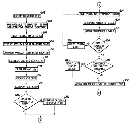
2A-2C. In step 202, a treatment plan is developed. Such a treatment plan can
be the one
developed in the above-cited Yu reference and can be developed either on the
computer 102 or
on a different device. In step 204, the treatment plan is made available to an
intraoperative
tracl~ing interface implemented on the computer 102. If the treatment plan is
not developed on
the computer 102, an appropriate communication medium can be provided to
supply the
treatment plan to the computer 102.
CA 02407577 2002-10-28
WO 01/82995 PCT/USO1/11886
The intraoperative tracking interface is displayed to the user on the display
104. As
shown in Fig. 3, the intraoperative tracking interface 300 includes the
following components.
An electronic worksheet 302 shows needle and seed coordinates, based on the
grid ofholes 120
in the template 118, and identifies needle locations with dots 304. A live
ultrasound image
window 306 shows a real-time image of a section of the prostate obtained from
the ultrasound
device 110 and allows a real-time view of needle placement in the prostate.
From the placement
of the seeds, the dosimetry is calculated, and a series of dosimetry panels
308 are shown, each
showing the dosimetry in a respective slice of the prostate from the base to
the apex. The
dosimetry in the panels 308 is shown by isodose lines 310. The electronic
worksheet 302 further
includes a spreadsheet 312 in which each row indicates one of the needles. The
spreadsheet 312
includes a column 314 indicating a needle by a number, a column 316
identifying the hole 120
in the template I I8 into which that needle is inserted by its coordinates
(letter and number), a
column 3I8 indicating an offset, a column 320 indicating the number of seeds
on the needle, a
column 322 indicating a ~x offset of the needle from its planned position, a
column 324
indicating a 0y offset of the needle from its planned position, a column 326
indicating the
number of currently selected seeds whose offsets have been calculated and a
column 328
indicating a total number of seeds whose offsets have been calculated. A
needle position 304
which the operator has selected is shown on the interface 300 as flashing, as
is the corresponding
row 330 in the spreadsheet 312.
Following the insertion of each needle or catheter in step 206, the live
ultrasouxld image
306 of the interface 300 displays a bright (hyperechoic) spot 332 in step 208.
In step 210, the
operator manually identifies the spot 332, e.g., by clicking on it with the
mouse 108. In step 212,
the he difference in the x-y plane between the planned location and the actual
location of the
needle or catheter is calculated to give errors dx and dy, which are shown
both on the grid 302
11
CA 02407577 2002-10-28
WO 01/82995 PCT/USO1/11886
and on the highlighted row 330 of the spreadsheet. The positional errors in
the x-y plane of each
seed, 0x' and by', are calculated in step 214 based on straight line
interpolation at the planned
z location of the seed. The straight line used in the interpolation is
constructed by joining two
knovm points: (a) the actual needle location shown on ultrasound at the known
z plane and (b)
the template coordinate outside the patient body through which the needle is
inserted under
precision template guidance. At the template 118, ~x and ~y are assumed to
equal zero. The
dose is then recalculated in step 216 by moving the seeds along the activated
needle or catheter
in the x and y directions by those amounts Ox' and Dy', which may be the same
or different for
every seed. The dosimetry updated by such feedback of seed placement errors is
redisplayed in
step 218 on the series of isodose panels 308. Fig. 4 shows the updated
interface 300 and also
identifies some of the above-mentioned method steps in association with the
corresponding
elements of the interface 300.
In addition, the operator is permitted to change the number of seeds deposited
by the
needle or catheter in question in step 220. In that case, the operator is
required to enter the seed
locations along the needle or catheter, which overndes the original treatment
plan in step 222.
Seed placement errors in such a case are tracked identically to the procedure
described above.
In step 224, a small column of 3D ultrasound images is acquired along the
straight line
constructed in step 214. That column can be perpendicular to the x-y plane or
may be at a non-
righ angle from the x and/or the y planes. The exact number of seeds as
deposited is identified
in step 226, using image processing algorithms to be described below, in the
column of 3D
ultrasound images. Each seed identified in the ultrasound images is assigned a
confidence level
in step 228, which indicates the likelihood or uncertainty of seed
localization.
The size of the column is initially set small. If it is determined in step 230
that the total
number of seeds found in step 226 is not equal to the number of seeds
deposited by the given
12
CA 02407577 2002-10-28
WO 01/82995 PCT/USO1/11886
needle or catheter, the width of the coluriln is adjusted in step 232; for
instance, the width is
increased to find additional seeds.
Thus, ~z' is quantified for each seed, and at the same time, fix' and ~y' are
further
corrected. If it is determined in step 234 that the confidence level of a
given seed's localization
exceeds a threshold value (set by the operator), the dosimetry is re-
calculated yet again in step
236 using the updated seed location and displayed in the same isodose panels.
The isodose
calculated is assigned a confidence level in step 238, which is a numerical
composite of the
individual confidence levels of the seeds and the dosimetric impact of
positional uncertainties
at each seed location. For example, in a high dose region, positional
uncertainty has low impact.
Periodically throughout the seed placement procedure and the end of seed
placement, a
fluoroscopic x-ray image may be may be taken in step 240 in the anterior-
posterior direction and
at up to ~45 degrees on either side of the anterior-posterior direction. The
seed coordinates as
determined above are projected in the same orientations in step 242. A best
match to the x-ray
seed projections is made in step 244 based on multiple point matclung using
those seed
identifications with the highest confidence levels. Subsequent to such
matching, the seed
locations with low confidence levels are adjusted in step 246 based on the x-
ray locations. As a
result, the confidence levels of those latter seeds are increased by a amount
reflective of the best
match quality. W step 248, the dosimetry is recalculated, and the confidence
level of the
dosimetry is updated using the updated confidence levels of the seeds.
The image processing algoritluns used in carrying out step 226 will now be
explained.
As shown in the flow chart of Fig. 5, there are three basic steps. In step
502, which is a
preprocessing step, the image brightness and contrast are adjusted to make the
hyperechoic spots
more distinct. In step 504, the seed pathway is tracked for further correcting
the offsets !1x' and
13
CA 02407577 2002-10-28
WO 01/82995 PCT/USO1/11886
0y' of the implanted seeds. In step 506, the seeds are identified for
correcting 0z' for each seed
along the tracking pathway.
Step 502 involves executing a grayscale transformation to each image in the
ultrasound
series from apex to base and is thus a pre-processing step. The purpose of
step 502 is to adjust
the brightness and contrast of the images so that the hyper-echoic spots will
be more distinct in
the transformed images. According to experience aquired from many actual OR
cases, an image
suitable for seed recognition processing has a grayscale histogram similar to
that shown in Figs.
6A and 6B, whereas in most cases, the images as taken have grayscale
histograms similar to that
shown in Figs 7A and 7B.
As shown in Fig. 6B, it is preferred that the background be very dark while
the
hyperechoic spots be very distinct. For that prefered case, 50% of the pixels
have grayscale levels
below 30, representing the background and dark issues; 90% of the pixels have
grayscale levels
below 60, with grayscale levels between 30 and 60 most likely representing the
brighter issues
of the gland; and 95% of the pixels have grayscale levels below 80, with
levels between 60 and
80 most likely representing some much brighter issues and some weaker airgaps.
The pixels
with the highest grayscale levels (from 80 to 255) are the hyper-echoic spots
of seeds and some
stronger air gaps.
Here, the images are assumed to have an eight-bit grayscale depth, namely,
with
grayscale values from zero to 255 inclusive. Of course, other grayscale depths
can be used
instead.
In the images as taken, the 50%, 90% and 95% grayscale levels are higher than
the
preferred ones set forth above. In the example of Figs. 7A and 7B, they are
60, 1 I O and 135,
respectively.
14
CA 02407577 2002-10-28
WO 01/82995 PCT/USO1/11886
To transform an image as taken into an image as preferred, the following
grayscale
transformation scheme can be used:
Original image Transformed image
Below median (050%) 1
50%~90% 150
90%~95% 5175
95%100% 76255
When the image of Figs. 7A and 7B is subj ected to such a transformation, the
result is
as shown in Figs. 8A and 8B. A comparison ofFigs 7A and 7B with Figs. 8A and
8B shows that
the hyper-echoic spots in transformed image of Figs. 8A and 8B axe more
distinct than they are
in the original image of Figs. 7A and 7B. Thus, it is easier for the
subsequent algoritluns to track
and identify the seeds. More importantly, it is possible for the algoritlnns
to use unified
parameters to process cases with different brightness and contrast settings.
Step 504, automatic tracking of the seeds along a same needle, is used to
correct Ox' and
~y' (displacement from the planned location) ofthe implanted seeds. Step 504
involves tracking
the pathway of the seeds, not just the seeds themselves. In other words, the
air gaps are also
included, and step 504 does not discriminate the seeds from the air gaps. Step
504 uses the
grayscale information the region of interest (ROI), such as the maximum value
of a hyper-echoic
spot, the mean and the standard deviation of the ROI, the contrast defined by
the maximum value
divided by the mean, etc.
In step 504, a center and the size of an ROI are preset. That operation can be
manually
done by the operator by clicking the mouse on the hyper-echoic spots at any z-
position or
automatically done by using the information from the treatment plan.
Thresholding and analysis
are then used to determine whether there is a hyper-echoic spot in the ROI. It
there is, the ROI
CA 02407577 2002-10-28
WO 01/82995 PCT/USO1/11886
center of the next image in the series is switched to the current center
position. If not, the
previous center is kept.
Fig. 9A shows a column of images taken along the pathway corresponding to grid
coordinates I2 in the grid 302 of Fig. 3. Fig. 9B shows the same column of
images, with boxes
identifying the images in which hyperechoic spots have been identified. As
shown in Fig. 9B,
each hyperechoic spot occupies five consecutive images because of the
dimensions of the seed
relative to the interval at which the images are taken; an illustrative
example of the relevant
dimensions will be given below.
The threshold measurement based on the grayscala analysis of the ROI can be
illustrated
by Fig. 10. For the salve of clarity of illustration, Fig. 10 shows only the
maximum, mean, and
contrast measurements because they can be shown in a 2-D plot. Fig. I O is not
drawn to scale,
and the parameters are examples only, used to make the illustration more
intuitive.
The ROI whose grayscale features fall in the shadow area of Fig. 10 is
identified as an
ROI containing a hyper-echoic spot. In the figure, the four borders of the
shadow area are
represented with four lines a, b, c, and d, respectively. The lines a and b
indicate that the
maximum value of the ROI should be between grayscale levels 75 and 255. The
line c indicates
that the mean value of the ROI should be greater than 5. The line d indicates
that the contrast
(the slope of the line in the 2-D coordinate system constructed by the mean
and maximum)
should be greater than 2.
In practice, the line d may be replaced by a curve a (the dotted curve in Fig.
10), which
delimits the border more accurately. That is because variations of the mean
and the contrast may
result in different thresholds. Generally speaking, the greater the mean, the
smaller the threshold.
As a result, curve a is in the form as shown in the figure. The curve a can be
implemented as a
curve equation or as a look-up table for correlating the threshold to the
mean.
16
CA 02407577 2002-10-28
WO 01/82995 PCT/USO1/11886
Extending the illustrative example of Fig.10 to more measurement parameters
results in
a mufti-dimensional space and a shadowed sub-space similar to the shadow area
in the 2-D space
in Fig. 10.
Step 506, detecting the real z-position of each seed placed along the needle
track, is in
fact a task of cutting the seed pathway into several segments by
discriminating the spots
representing seeds from any spots representing air gaps. The grayscale
information cannot be
used to achieve that goal because some stronger air gaps have greater
measurement values than
weak 'seeds, as will be explained below with reference to Fig. 11B. Therefore,
a wave form
analysis method is used instead.
To simplify the illustration, it is assumed that the distance between two
contiguous
images is 0.5 mm, so that one seed can occupy at most 10 images in the series,
and it usually
occupies fewer than 10 due to its slant. Thus, in a case in which the gland
has a length of 4.5 cm,
the offset is 5 mm, and there are 5 seeds with special spacing, i.e, no
spacer, at the apex, an ideal
waveform of a needle track should have the appearance shown in Fig. 1 lA,
having rectangular
peaks 1102,1 I04,1106, 1108 and 1110 indicating the seeds. However, a real
waveform is more
likely to have the appearance shown in Fig. 11B, having irregular peaks 1112,
1114, 1116, 1118
and 1120 indicating the seeds.
It can be seen in Fig. 11B that although the measured value (MV) of the second
peak
I 114 is Iess than that of the air gap 1122 between the peaks 1116 and 1118 or
that of the air gap
1124 between the peaks 11 I8 and 1120, the second peals 1114 has a wave form
of peak, while
each of the air gaps 1122 and 1124 has the wave form of valley. That
distinction between peaks
and valleys can be used to discriminate the seeds from the air gaps.
17
CA 02407577 2002-10-28
WO 01/82995 PCT/USO1/11886
Since it is already known how many seeds are placed in the needle track, the
positions
of the top several peaks are identified as the centers of seeds. In the case
of Fig. 11B, if the plan
has four seeds, their positions are taken as the peaks 1112, 1116, 1118 and
1120, but not 1 I 14.
That principle is simple, while the difficulty is the representation of the
MV. Since any
single grayscale measurement cannot reflect the whole feature ofthe ROI, it is
natural to use their
linear combination as the final MV, i.e.,
MV=EawZ,
in wluch v1 represents each feature such as maximum, contrast, and standard
deviation, etc, and
a~ represents the coefficient of each feature. Of course, the combination of
those features is not
constrained to the linear composition, which is the simplest one. Simple least
square statistics
will determine the value and the confidence interval for each coefficient.
Of course, the MV waveform should be smoothed before it can be processed
because the
raw signal may contain many noise peaks, as shown in Fig. 12A. Next, the false
peaks are
removed. For example, if two peaks have a distance less than 6 units along the
z-axis, they most
likely represent the same seed, so one of them will be absorbed by the other,
stronger one, as
shown in Fig. I2B. If a peak lies between two other higher peaks and has no
distinct drop off
before and after it, it is most likely noise, as shown in Fig. 12C.
After those adjustments to the waveform, the peaks are detected to determine
how many
peals there are. If the number is greater than the implanted number N of
seeds, only the highest
N peals are taken as the seeds, as explained above with reference to Fig. 1
IB. If the number is
less than N, either seed identification is forced using second-tier peaks
(with reduced
confidence), or the preset transverse size of the ultrasound column is changed
to process a larger
needle traclc that includes the exact number of the implanted seeds.
18
CA 02407577 2002-10-28
WO 01/82995 PCT/USO1/11886
Figs.13A-13D show sample seed identifications along grid location I2. In
Figs.13A and
13C, the seeds are identified by black marks M, while in Figs. 13B and 13D,
they are Left
unmarked.
Each seed identified in that manner is assigned a confidence level according
to the MV
level and the fall-off characteristics of the peak. The greater the MV level
and the fall off of the
peak, the more likely it is a seed. The locations of the seeds and their
confidence values are
convoluted into subsequent dosimetry calculations, which result in a
confidence level for each
of the dosimetry parameters arising from the dose-volume histogram, including,
D100, D95,
D90, D80 and D50.
If the confidence on the chosen dosirnetry parameter (currently D90) is
acceptably high,
seed localization is said to be reliable enough for re-planning and re-
optimization of dosimetry,
in order to compensate for the dosimetric impact of the aggregate seed
misplacements. If the
confidence on the chosen dosimetry parameter is not sufficiently high, simple
Baysian statistics
are used to determine which seed localizations require increased confidence to
achieve acceptable
confidence in dosimetry. Repeat ultrasound scans are acquired; imaging data
for the given needle
colunm(s) are fused using redundant information but with increased signal-to-
noise ratio. The
above-described process is repeated starting from active seed pathway traclang
and ending with
dosimetry confidence analysis.
If repeated application of the above process still cannot achieve acceptable
dosimetry
confidence, x-ray imaging of the seeds will be used to increase the
localization confidence of the
given seeds. Ultrasound-based seed identification of high confidence values
will be used as
"anchors" (fiducial marks) to register the ultrasound and x-ray spaces. The
coordinates of the low
confidence seed Iocalizations will thenbe corrected using the x-ray proj
ection(s). The confidence
19
CA 02407577 2002-10-28
WO 01/82995 PCT/USO1/11886
values are increased by a variable based on the degree of seed overlap on the
x-ray image, the
quality of the overall registration, and the quality of the x-ray itself for
seed localization.
While a preferred embodiment ofthe present invention has been set forth above
in detail,
those skilled in the art who have reviewed the present disclosure will readily
appreciate that other
embodiments can be realized within the scope of the present invention. For
example, the
numerical values set forth above should be construed as illustrative rather
than limiting. The
same is true of the arrangement of the user interface of Fig. 3. Therefore,
the present invention
should be construed as limited only by the appended claims.