Note: Descriptions are shown in the official language in which they were submitted.
CA 02407731 2002-10-29
WO 01/83813 PCT/EPO1/04871
IDENTIFICATION OF GENETIC MARKERS
The present invention relates to a method for the identification of the
presence of a genetic marker in a DNA sample, in particular by using a
oligonucleotide array. In particular, the method according to the invention
allows
for the identification and/or localization of genes) and/or mutations)
associated
with a distinguishable phenotype.
DEFINITIONS
By "complementary", it is referred to the topological compatibility or
matching together of interacting surfaces of a probe molecule and its target.
Thus,
the target and its probe can be described as complementary, and furthermore,
the
contact surface characteristics are complementary to each other. Although
perfect
complementarity is preferred, certain mismatch may be tolerated, as long as
the
specificity of hybridization is retained.
As used herein, "isolated" includes reference to material which is
substantially or essentially free from components which normally accompany or
interact with it as found in its naturally occurring environment. The isolated
material optionally comprises material not found with the material in its
natural
environment.
As used herein, "nucleic acid" or "oligonucleotide" includes reference to a
deoxyribonucleotide or ribonucleotide polymer in either single- or double-
stranded
form, and unless otherwise limited, encompasses known analogues of natural
nucleotides that hybridize to nucleic acids in a manner similar to naturally
occurring
nucleotides. In specific embodiments, the "nucleic acid" or "oligonucleotide"
can
be substituted by chemical substances that can form sequence specific
interactions
similar as for the natural phosphodiester "nucleic acid". Known and preferred
analogues include polymers of nucleotides with phosphorothioate or
methylphosphonate liaisons, or peptid nucleic acids. Unless otherwise
indicated, a
particular nucleic acid sequence includes the complementary sequence thereof.
Typical oligonucleotides are single-stranded nucleic acids of between 5 and
200
bases in length, more preferably of between 5 and 100, even more preferably of
CONFIRMATION COPY
CA 02407731 2002-10-29
WO 01/83813 PCT/EPO1/04871
2
between about 10 and 50 bases. Examples of such oligonucleotides are single
stranded DNA molecules of between 20 and 40 bases in length.
In the invention, a "probe" is a oligonucleotide that can be recognized by a
particular target. In particular, and in preferred embodiments, the "probe" is
immobilized on a surface. Depending on context, the term "probe" refers both
to
individual oligonucleotide molecules and to the collection of same-sequence
oligonucleotide molecules surface-immobilized at a discrete location.
The term "target" refers to a nucleic acid molecule that has an affinity for a
given probe. A target may be a naturally-occurring or a man-made nucleic acid
molecule. It can be employed in their unaltered state or as aggregates with
other
species. Targets may be attached, covalently or noncovalently, to a binding
member, either directly or via a specific binding substance. Targets may also
be
modified. In preferred embodiments, they harbor a fluorescent or radioactive
moiety, or groups or isotopes that can be identified by mass spectrometry.
A "feature" according to the invention is defined as an area of a substrate
having a collection of same-sequence, surface-immobilized oligonucleotide
molecules. One feature is different than another feature if the probes of the
different
features have different nucleotide sequences.
The term "oligonucleotide array" refers to a substrate having a two-
dimensional surface having at least two different features. Oligonucleotide
arrays
preferably are ordered so that the localization of each feature on the surface
is
spotted. In preferred embodiments, an array can have a density of at least
five
hundred, at least one thousand, at least 10 thousand, at least 100 thousand
features
per square cm. The substrate can be, merely by way of example, glass, silicon,
quartz, polymer, plastic or metal and can have the thickness of a glass
microscope
slide or a glass cover slip. Substrates that are transparent to light are
useful when the
method of performing an assay on the chip involves optical detection. As used
herein, the term also refers to a probe array and the substrate to which it is
attached
that form part of a wafer. The substrate can also be a membrane made of
polyester
or nylon. In this embodiment, the density of features per square cm is
comprised
between a few units to a few dozens.
The term "distinguishable phenotype" has to be understood as a phenotype
(i.e. a qualitative or quantitative measurable feature of an organism) that
can allow
CA 02407731 2002-10-29
WO 01/83813 PCT/EPO1/04871
3
the categorization of a given population. For exemple, a distinguishable
phenotype
encompasses the membership to a set of a given disease, or a peculiar feature
or
property (e.g. resistance or adverse effect when given a given drug).
The future sequence of the human will be finished in the next couple of
years. It will uncover the complete sequence of the 3 billion bases and the
relative
position of the 100 000 genes that constitute the genome. The enormous
information revealed by this project opens unlimited possibilities for the
elucidation
of gene function and interaction of different genes. It will also allow the
implementation of pharmacogenomics and pharmacogenetics.
Pharmacogenetics and pharmacogenomics aim at determining the genetic
determinants linked to different phenotypes, in particular diseases. Most of
the
disease are multigenic diseases, and the identification of the genes involved
therein
should allow for the discovery of new targets and the development of new
drugs.
Pharmacogenomics also encompasses the use of specific medications according to
the genotype of the patient. This should lead to a dramatic improvement of the
efficiency of the drugs.
Many physiological diseases are targeted by this novel pharmaceutical
approach. One can name the autoimmune and inflammatory diseases, for example
Addison's Disease, Alopecia Areata, Ankylosing Spondylitis, Behcet's Disease,
Chronic Fatigue Syndrome, Crohn's Disease and Ulcerative Colitis, Inflammatory
Bowel Disease, Diabetes, Fibromyalgia, Goodpasture Syndrome, Lupus, Meniere's,
Multiple Sclerosis, Myasthenia Gravis, Pelvic Inflammatory Disease, Pemphigus
Vulgaris, Primary Biliary Cirrhosis, Psoriasis, Rheumatic Fever, Sarcoidosis,
Scleroderma, Vasculitis, Vitiligo, Wegener's Granulomatosis.
Cancers are also believed to be multigenic diseases. Some oncogenes (for
exemple ras, c-myc) and tumor suppressor genes (for exemple p53) have
previously
been identified, as well as some genetic markers for predisposition (for
example the
genes BRCA1 and BRCA2 for breast cancer). The identification of new genes
involved in other kind of cancers should allow for a better information of the
patient
and the prevention of the development of the disease, an improved life
expectancy
as already observed with breast cancer (Schrag et al., JAMA, 2000; 23:617-24).
CA 02407731 2002-10-29
WO 01/83813 PCT/EPO1/04871
4
A necessary step for achieving these goals is therefore the characterization
of the genetic determinants specific of a given genotype in a population of
patients.
The determination of variability at the genome level can be achieved by
determining different markers and then refining the analysis to identify the
genes of
interest.
The major goal of genetics is indeed to link a phenotype (i.e. a qualitative
or
quantitative measurable feature of an organism) to a gene or a number of
genes.
Historically there are two genetics approaches that are applied to identify
genetic
loci responsible for a phenotype: familial linkage studies and association
studies.
Whatever the approach is, genetic studies are based on polymorphisms, i.e.
base
differences in the DNA sequence between two individuals at the same genetic
locus.
Currently two kinds of markers are used for genotyping: microsatellites and
single nucleotide polymorphisms (SNP). Microsatellites are highly polymorphic
markers where different alleles are made up of different numbers of repetitive
sequence elements between conserved flanking regions. On average, a
microsatellite is found every 100 000 bases. A complete map of microsatellites
markers covering the human genome was presented by the Centre d'Etude du
Polymorphisme Humain (Dib et al., Nature 1996; 380:152-4). Microsatellites are
genotyped by sizing PCR products generated over the repeat regions on gels.
The
most widely used systems are based on the use of fluorescently labeled DNA and
their detection in fluorescence sequencers.
Fewer SNP are in the public domain, and a SNP map is currently being
established by the SNP consortium which regroups pharmaceutical and
electronics
companies (Roberts, US News World Rep, 1999; 127:76-7).
Different analysis technologies have been developed for the genotyping of
these markers, for example gel based electrophoresis, DNA hybridization,
identification and characterization through mass spectrometry. The drawback of
all
these approaches is that they necessitate the amplification of many hundred of
thoushands of specific sequences, which makes these technologies both labor
intensive and expensive.
Linkage analysis has been the method of choice to identify genes implicated
in many diseases both monogenic and multigenic, but where only one gene is
CA 02407731 2002-10-29
WO 01/83813 PCT/EPO1/04871
implicated for each patient. In order to be reasonably powerful in the
statical
analysis the studied polymorphisms have to fulfill several criteria:
- high heterozygosity i.e. many alleles exist for a given locus (this
increases the informativity);
5 - genome wide representation;
- detectable with standard laboratory methods.
A type of polymorphisms fulfilling most of these criteria are microsatellite
markers. As already described, these are repetitive sequence elements of two,
three
or four bases. The number of repetitions is variable for a given locus,
resulting in a
high number of possible alleles, i.e. high heterozygosity (70-90 %).
Microsatellite
markers are still the genetic markers of choice for linkage analysis, and
genotyping
of these markers is performed by amplifying the alleles by PCR and size
separation
in a gel matrix (slab gel or capillary). For the study of complex human
diseases
usually 400-600 microsatellite markers are used that are distributed in
regular
distances over the whole genome (about 10-15 megabases).
Linkage studies follow alleles in families. However, each family might have
a different allele of a genetic locus linked to the phenotype of interest.
Association
studies in contrast follow the evolution of a given allele in a population.
The
underlying assumption is that at a given time in evolutionaary history one
polymorphism became fixed to a phenotype because:
a) it is itself responsible for a change in phenotype or;
b) it is physically very close to such an event and is therefore rarely
separated from the causative sequence element by recombination
(one says that the polymorphisms is in linkage disequilibrium
with the causative event).
As association studies postulate the existence of one given allele for a trait
of interest, it is therefore desirable that the markers for association
studies are
simple. Accordingly, the markers of choice are SNP, which show a simple base
exchange at a given locus, and are therefore bi-, rarely tri-allelic.
Association
studies can be carried out either in population samples (cases vs controls) or
family
samples (parents and one offspring, where the transmitted alleles constitute
the
"cases" and the non-transmitted the "controls").
CA 02407731 2002-10-29
WO 01/83813 PCT/EPO1/04871
6
In order to simplify the analysis and comparison of the genomes of two
people bearing the same phenotype, and the potential identification of the
genes
linked to this phenotype, it can be interesting to reduce the complexity of
the DNA
samples to analyze. Such a method, called genomic mismatch scanning (GMS) was
described by Nelson et al. (Nat Genet. 1993; 4:11-8). It allows the
identification of
all loci that are identical between two genomic DNA. This method will lead to
a
discrimination of the DNA samples, as only identical loci between two
individuals
will be present in solution after the GMS method is performed. The method of
the
invention will therefore be fully appreciated as it will allow the
identification of
said DNA samples, rather than their discrimination.
Other methods also lead to the reduction of the DNA complexity, for
example degenerate oligonucleotide primer PCR, ALU-PCR or amplified
restriction
fragment length polymorphism (AFLP). Indeed, these methods are often used on
genomic DNA to increase the amount of sample that would be needed for latter
studies. The drawback of these methods is that certain parts of genomic DNA
are
not amplified by these techniques. This explains why one can consider that
these
methods reduce the complexity of genomic DNA. The method according to the
present invention can be used to identify the regions of genomic DNA that have
been amplified, and therefore the representation of said DNA compared to the
whole genome.
Even with these methods, the analysis and comparison of the DNA samples
remain labor intensive, as they necessitate a large number of PCR reactions,
and gel
. analysis.
The invention provides a method which leads to the identification of specific
DNA sequences from a mixture of DNA fragments, which allows to perform
association and linkage studies. This method is simple, cheap and quick to
perform.
The invention is drawn to a method for the identification of the presence of a
genetic marker in a DNA sample comprising the following steps:
a) selection of sequences specific of said genetic marker;
b) fixation of oligonucleotides comprising said specific sequences
or the complementary sequences on a solid support;
CA 02407731 2002-10-29
WO 01/83813 PCT/EPO1/04871
7
c) addition of a mixture of DNA fragments representing the said
DNA sample to the solid support in a way that hybridization is
possible;
d) detection of the presence of the genetic marker in the DNA
sample by the presence of a signal corresponding to the
hybridization of a fragment of the DNA sample to the specific
oligonucleotide.
To perform the method of the invention, the sequences specific of the
genetic marker are the flanking regions of said genetic markers. Indeed, even
though the genetic marker is highly polymorphous in a population, its flanking
regions are conserved between two individuals. This ensures that the study of
the
polymorphism of the genetic marker will not be hampered by poor hybridization.
The genetic marker which is looked for in the method described in the
invention is preferably a SNP or a microsatellite, the latter being the most
preferred
case.
It has to be understood that the method of the invention is preferably to be
used in genotypage studies, and that the presence or absence of the genetic
marker
of interest will be investigated in many individuals. Also, it is preferred if
the
genetic markers that are sought are linked to a distinguishable phentoype.
It has also to be understood that the method of the invention is not primarily
intended to discriminate between multiple genetic markers, but rather to allow
for
the determination of the presence or the absence of said marker in a DNA
sample,
preferably a genomic DNA sample, the complexity of which has been reduced. In
this regard, this invention is particularly directed at characterizing the
content of
(e.g., determining the presence or absence of a genetic marker in) a nucleic
acid
sample after said sample has undergone a selection process in which the
complexity
of said sample is reduced.
Nevertheless, and as could be described later, some improvement can be
made to the current invention, that will further permit the identification of
the
genetic maxker, the presence of which has been detected.
CA 02407731 2002-10-29
WO 01/83813 PCT/EPO1/04871
g
The current invention is also drawn to a method for the identification of
genes) and/or mutations) associated with a distinguishable phenotype
comprising
the steps of
a) identifying genetic markers associated with said phenotype, by
applying the method described above to DNA samples from
individuals exhibiting said phenotype;
b) comparing the regions identified in step a) with the
corresponding regions in individuals that do not exhibit said
phenotype;
c) identifying the genes) and/or mutations) associated with said
phenotype.
The first step will allow to determine the shared genetic markers between
two individuals exhibiting a given phenotype (population A). It can therefore
be
postulated that the genetic marker linked to said phenotype can be isolated by
this
step. In order to refine the analysis, the step b) compares the genetic
markers
isolated in step a) with the markers harbored by individuals that do not
exhibit the
phenotype (population B). Therefore, any genetic marker shared between
population A and population B is not linked to the phenotype. The use of this
method with a sufficient number of individuals allows the restriction to a
small
number of genetic markers and the identification of the genes) and/or
mutations)
linked to the phenotype of interest.
It is as well very preferable to have reduced the complexity of the DNA
genomes to compare. It might be best to perform the method of GMS between two
individuals, as this method reduces the DNA samples to be analyzed to the DNA
fragments that are identical between the two individuals. But the other
methods of
reduction of complexity described above could also be used favorably.
This method is best performed on individuals that are related (i.e. from the
same family, in a large meaning, parents, cousins, uncles, aunts...). In fact,
this is
preferable, as related individuals share a certain percentage of DNA (on
average
50% between brothers and sisters, 16% between cousins). Therefore, it is more
likely that they will have identical genetic markers if they share the same
phenotype, and that these markers will be missing from the related individuals
that
do not exhibit the phenotype. By comparison of the missing hybridization
spots, it
CA 02407731 2002-10-29
WO 01/83813 PCT/EPO1/04871
9
will allow a very quick determination of the genetic markers linked to the
phenotype.
In a particular embodiment, this invention relates to a method of identifying
genes and/or mutations associated with a phenotype or trait, the method
comprising:
(a) preparing a composition enriched for identical nucleic acid fragments
from nucleic acid samples from individuals exhibiting said phenotype,
(b) characterizing said composition by contacting the same with a nucleic
acid array of oligonucleotides specific for flanking regions of selected
genetic
markers.
The present invention also includes methods of identifying genes related to a
phenotype, the methods comprising
(a) isolating nucleic acid fragments that are identical between two
individuals exhibiting said phenotype, and
(b) identifying genes contained in said nucleic acid fragments by contacting
said fragments with a nucleic acid array comprising, on a support,
nucleic acid sequences specific for regions flanking genetic markers.
Step (a) is preferably performed by a genomic mismatch scanning ("GMS")
approach, as described previously or by comparative genomic hybridisation
("CGH"). Alternatively, step (a) can be accomplished using the method
described in
WO00/53802. Most preferably, step (a) comprises treating the sample to produce
IBD fragments. The method is particularly suited to identify genes or
mutations
from genomic DNA from said individuals. In a particular embodiment, the
genomic
DNA or fragments may be amplified.
A preferred use of the above methods is to identify genes or mutations
related to a pathological condition, particularly a cardiovascular disease,
lipid-
metabolism disorder or central nervous system disorder.
Furthermore, in a particular embodiment, the method further comprises the
step of comparing the genes identified in (b) with the sequence of
corresponding
genes from individuals that do not exhibit the phenotype.
CA 02407731 2002-10-29
WO 01/83813 PCT/EPO1/04871
The present invention also relates to kits for implementing a method as
described above, comprising a nucleic acid array and reagents to isolate
identical
nucleic acid fragments from two samples.
The invention also relates to the use of a gene or mutation identified by a
5 method as described above, for diagnotic, therapeutic or screening purposes.
The
genes or mutations can be used to design probes or primers suitable to detect
the
presence of said gene or mutation in any sample. Identification of said gene
or
mutation in a sample from a subject may indicate the presence of or
predisposition
to a pathology. The gene or mutation may allow one to design a gene therapy
10 product incorporating the wild type version or any antisens product, to
correct the
deficiency associated with said gene or mutation. The gene or mutation also
allows
the implementation of screening methods to identify compounds that regulate
the
activity or expression of said gene.
In a preferred embodiment of the above methods according to this invention,
the oligonucleotides comprising the sequences specific of the genetic marker
are
further used for the amplification of said genetic marker. The
characterization of the
amplified product can be carried out with the usual methods known by the
person
skilled in the art (in particular electrophoresis, chromatography, sequencing,
or
mass spectrometry).
In order to improve the hybridization properties, it might be useful to modify
the oligonucleotides, in particular to substitute them by chemical substances
that
can form sequence specific interactions, as previously described.
One understands that the methods described in the current invention are best
performed by using DNA arrays. These arrays of oligonucleotides comprising
sequences specific of genetic markers, in particular the flanking sequences of
said
genetic marker, are also part of the invention. Most preferably, the genetic
marker is
a microsatellite marker.
It is highly preferable to prepare an array comprising all the flanking
sequences specific of the genetic markers the presence of which the
investigator
wants to determine. In particular, an array comprising oligonucleotides
comprising
the flanking sequences (or complementary sequences) of all the microsatellite
CA 02407731 2002-10-29
WO 01/83813 PCT/EPO1/04871
11
markers will be of choice for performing the methods of the invention. The
array
may comprise between 100 and 200 000 oligonucleotides specific for said
sequences. The array may comprise oligonucleotides specific for different
types of
genetic markers, e.g., SNPs and microsatellites.
The map of the microsatellite markers and their sequences can easily be
determined by the person skilled in the art (Dib et al., Nature 1996; 380:152-
4),
which can determine the flanking sequences specific of each microsatellite
that are
suitable for use on a DNA array, in the methods according to the invention. It
is
indeed important for the melting point of the oligonucleotides to be in the
same
range for each oligonucleotide, in order to improve the quality of
hybridization.
Preferred flanking regions of the genetic markers correspond to regions
located
within 500 by at the most on each side of the genetic marker.
The construction of the oligonucleotide array can be carried out by using
methods known by the one skilled in the art. In particular, the synthesis can
be
performed directly on the solid surface, in particular by a photochemical (US
5,424,186) or an ink jet technique. Alternatively, the oligonucleotides can be
synthesized ex situ and further bound to the solid surface. In this case, it
might be
useful for the oligonucleotide to carry a chemical modification that allows
the
binding to the solid surface. The addressing of the oligonucleotides on the
surface
can be performed mechanically, electronically or by ink jet.
The hybridization conditions will depend on the DNA sample to be
analyzed, but can be easily optimized by the person skilled in the art. The
conditions can be optimized by modifying the salinity, pH and temperature of
hybridization. They can also be electronically assisted (US 6,017,696), in
order to
improve the specificity.
The detection of the hybridization spots can be performed by radioisotopic
or fluorescent labeling, field effect measurement, opto-electrochemical
process,
piezzo-electrical process, or ellipsometry, optical fibers measurement, mass
spectrometry.
An alternative to oligonucleotide arrays can be the use of silicon microbeads
on which the oligonucleotides of the invention are bound. In this case, it is
advantageous to perform the detection of hybridization events by telemetry. It
is
CA 02407731 2002-10-29
WO 01/83813 PCT/EPO1/04871
12
preferable when each bead harbors a specific code, the reading of said code
allowing the identification of the hybridization events.
Prior to hybridization, it might be advantageous to label the DNA fragments
with fluorescent dyes or radioisotopes in order to facilitate the detection
with these
techniques. Alternatively, it can be interesting to label these fragments,
prior to
hybridization, with groups or isotopes that can be identified by mass
spectrometry,
in the case the detection is done by this method. The person skilled in the
art knows
the moieties and/or groups to use for such a purpose. It is highly desirable
to use
base specific labels.
In another embodiment, the DNA fragments are labeled subsequently to
hybridization, by the use of a proofreading DNA polymerase and labeled di-
desoxy
nucleotides (ddNTP), that leads to primer extension of the oligonucleotide.
The
person skilled in the art knows that this extra step increases the specificity
of the
reaction (Pastinen et al. Genome Res., 1997, 7, 606). The primer extension
reaction
is performed on the immobilized oligonucleotide if a DNA template is
hybridized to
it with nucleotides labeled with fluorescent dyes, radioactive isotopes, or
groups or
isotopes that can be identified by mass spectrometry. The use of different
fluorescent dyes or different masses of groups added to the ddNTP's in the
primer
extension reaction further increase the specificity and allow the unambiguous
identification of a specific fragment hybridization from background
hybridization,
and therefore to the presence of the genetic marker.
In the case the genetic marker the presence of which has been determined is
a SNP, this extra step of primer extension can also allow the identification
of said
SNP, as the use of ddNTPs labeled with different markers (preferably different
fluorescent dyes) can lead to the unambiguous determination of said SNP base.
The methods according to the invention are useful to determine the genes)
and/or mutations) responsible for a distinguishable phenotype. For example,
they
can be carried out on human beings, in order to quickly identify the genetic
markers) responsible for a given disease, or a susceptibility to a disease.
They can
also be carried out in the agricultural field, on animals or plants. The
investigator
can, with these methods, determine the genotype of animals or plants
presenting an
interest for the farmer and/or the industrial, and improve the quality of the
products.
CA 02407731 2002-10-29
WO 01/83813 PCT/EPO1/04871
13
For example, it could be interesting to determine the genes) responsible for a
high
casein concentration in dairy cattle.
The method can also be used on smaller organisms, like bacteria, viruses or
parasites, for example in order to quickly identify the mutations) in the
genes that
are linked to drug resistance. The person skilled in the art knows how to
choose the
oligonucleotides to perform this method in this case.
The methods described in the current invention offer obvious advantages
over the classical linkage and association methods.
The methods allow unambiguous detection of IBD fragments between
individuals, and is not dependent on allele frequencies or marker
heterozygosity;
These methods are not limited to the use of polymorphic markers, and can
be performed with any sequence, as long as some sequence and ampping
information is available:
The information given by these methods is based on the presence or absence
of a hybridization signal. This is an important advantage compared to the
methods
of the technique that necessitates allele discrimination.
After determination of a region of interest, for example by using the
microsatellites, the same methods can be applied to reduce the size of the
region
and identify the fragments of interest. This scaling to any density of the
genome is
very valuable.
Due to these advantages, it is necessary to screen less individuals to perform
the methods described in the current invention, and obtain usable results.
This is
particulary true when related individuals are tested, and when the GMS method
is
first performed on their DNA.
The following examples illustrate some preferred embodiments of the
invention, but shall not be considered as restricting the scope of the
invention.
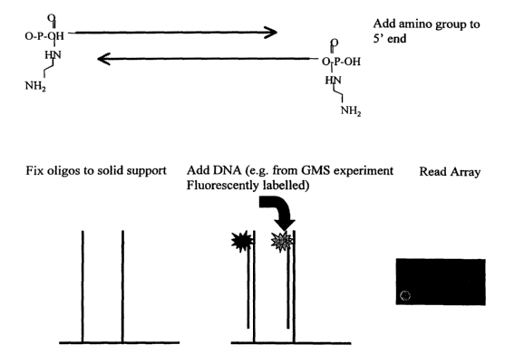
DESCRIPTION OF THE FIGURE
Figure 1 represents the microsatellite D1S2729 (underlined) and its flanking
regions
(SEQ ID N° 1). Two oligonucleotides that can be chosen in the flanking
regions in
order to perform the method according to the invention are represented by
arrows
(1.A.). Figure 1.B represents the chemical modifications that can be added to
the
CA 02407731 2002-10-29
WO 01/83813 PCT/EPO1/04871
14
oligonucleotides in order to fix them on a solid support. The presence of
microsatellite D1S2729 in the DNA sample after GMS reduction will lead to its
hybridization to the oligonucleotides and to the presence of a fluorescent
signal that
can be detected.
EXAMPLES
Example 1: Reduction of DNA complexity by GMS
Genomic DNA from subjects in a collection of families where at least two
related individuals show the same disease phenotype, is extracted by standard
methods e.g. phenol-chloroform extraction. The DNA's are separately cut with a
restriction enzyme (e.g. PstI) to create restriction fragments with an average
size
around 4 kilobases. To one of each of the restriction mixes from a pair of
individuals a solution containing dam methylase is added and the DNA is
methylated at adenin bases. The methylated products from one individual are
then
mixed with the non-methylated product of the second subject from the same
family.
The products are then heat denatured and allowed to re-anneal using stringent
hybridisation conditions (Casna et al. (1986) Nucleic Acids Res. 14: 7285-
7303).
This results in the formation of heteroduplexes from the DNA's from different
sources (individuals) which are hemimethylated (hybridisation of one
methylated
strand with one non-methylated. In addition homoduplexes are formed by
renaturation between the strands of each individulal with itself. These
homoduplexes are either completely methylated or completely non-methylated.
Using methylation sensitive enzymes like MboI (only cuts methylated
double stranded DNA) and DpuI (only cuts unmethylated double stranded DNA)
the homohybrids are digested. To this mixture a solution containing exo III
(or an
equivalent 3' recessed or blunt-end specific exonuclease) exonuclease is
added. The
exonuclease digests the blunt ended digested homoduplex fragments but not the
heteroduplexes with their 3' overhang, creating big single stranded gaps in
the
homoduplex fragments. These can be eliminated from the reaction mix through
binding to a single strand specific matrix (e.g. BND cellulose beads).
The remaining heteroduplexes comprise a pool of 100% identical fragments
and fragments with base pair mismatches (non-IBD fragments). A solution
CA 02407731 2002-10-29
WO 01/83813 PCT/EPO1/04871
containing the mismatch repair enzymes mutSHL is added to the mix resulting in
the nicking of mismatched heteoduplexes at a specific recognition site (GATC).
These nicks are further digested by adding exo III (or an equivalent 3'
recessed or
blunt-end specific exonuclease) exonuclease to the reaction mix, creating big
single
5 stranded gaps in the homoduplex fragments. These can be eliminated from the
reaction mix through binding to a single strand specific matrix (e.g. BND
cellulose
beads).
The remaining fragments in the reaction mix constitute a pool of 100%
identical DNA hybrids formed between the DNA's of different individuals
10 comprising the loci responsible for the disease phenotype.
Example 2: Manufacture of an oligonucleotide array
From the human genetic map which links over 5000 microsatellite markers
forward and reverse sequences flanking the repeat units are selected The
selection is
15 carried out from sequence information available through public data bases
especially the GENETHON database (figure 1 ). Critera for selection are the
uniqueness of the sequences in respect to each other, common primer selection
criteria for hybridization (no self complementarity, similar Tm etc.) and
sequence
stability (no known polymorphic sites in the oligonucleotide sequence.
The corresponding sequences are then synthesized in the form of
oligonucleotides that are typically between 25 and 35 bases long and are
activated
by the addition of an amino group to their 5' end (e.g. by addition and are
synthesized by standard procedures by a manufacturer providing salt free, high
quality oligonucleotides (e.g. MWG, Germany)).
These oligonucleotides are then applied to an amino-silane covered glass
slide using an appropriate automated arrayer (e.g. GMS 417 Arrayer, Genetic
Microsystems), through a specific reaction (see e.g. Urdea et al. Nucleic
Acids Res.
11 (1988)). An aminoester bridge is formed between the oligonucleotide and the
aminosilane and the oligonucleotide thus bound to the glass slide.
This array constitutes a representative selection of the whole human genome
with an average resolution of <1cM (sex averaged, about one marker every
1 megabase).
CA 02407731 2002-10-29
WO 01/83813 PCT/EPO1/04871
16
Example 3: Hybridization protocol
The remaining hybrid fragments are hybridized against the microsatellite
array in a hybridization chamber in a hybridization buffer (e.g. 6xSSC, Sx
Denhardt's solution), at temperatures between 45-62°C. After
hybridization several
washes with icreasing stringency (3-0.1 x SSC, 0.05% Tween 20 at 37-
45°C) are
carried out to wash out non-specific hybridizations. The person skilled in the
art can.
optimize the hybridization conditions, in particular with the teachings of
Sambrook
et al. (1989; Molecular cloning : a laboratory manual. 2"d Ed. Cold Spring
Harbor
Lab., Cold Spring Harbor, New York).
Example 4: Primer extension protocol
To increase the specificity a solution of fluorescently labelled
didesoxynucleotides is added where each of the four ddNTP's carnes a different
fluorophore. Through a polymerase the subsequent base following the last base
on
the oligonucleotide that is fixed to the chip is added. The DNA polymerase
used
(T7, Taq, Klenow fragment...) and the polymerization conditions will be chosen
by
the person skilled in the art depending on the DNA fragments to extend and
according to the teaching of Sambrook.
Example 5: Detection protocol
The result is the identification of fragments still present after the GMS
procedure by both position and fluorescent signal (colour). Statistical
analysis of the
signals from a sufficiently Iarge number of families identifies the loci
common to
affected individuals within a narrow interval of a few cMorgan.
CA 02407731 2002-10-29
WO 01/83813 PCT/EPO1/04871
1
SEQUENCE LISTING
<110> CNRS and INSERM
<120> Identification of genetic markers
<130> B0075
<160> 1
<170> PatentIn Ver. 2.1
<210> 1
<211> 389
<212> DNA
<213> Homo Sapiens
<223> Microsatellite D1S2729 and flanking regions
<400> 1
agctgctgag tttgtagtga tatggttaca cagcaataga tgaatatagt gaggaacagt 60
ctgtaaagca ctgagtccag tgctggcatg tggaggtgct ctgtaaggag ttgtgttatt 120
actgttgtat tgtnagtctg ctgattactt gcctaatgct gtgtggggcc tggctttgcc 180
ctgccccggt ccctagtggg gccaggttcc atggctctna ctagccctgc tggttctnat 240
accctggtac agaaagaaag attctatgac tcaaacacac acacacacac acacacacac 300
acacacacac acacacacac accccagagc cttaggcctt ggtctcccaa ggattgatat 360
cccagcccag tccacatgat tctgaattg 389