Note: Descriptions are shown in the official language in which they were submitted.
CA 02407872 2002-10-29
WO 01/090188
PCT/EP01/05916
RECEPTOR-BASED INTERACTION TRAP
The present invention relates to a recombinant receptor, comprising an
extracellular
ligand-binding domain and a cytoplasmic domain that comprises a heterologous
bait
polypeptide, which receptor is activated by binding of a ligand to said ligand
binding
domain and by binding of a prey polypeptide to said heterologous bait peptide.
The
present invention also relates to a method to detect compound-compound-binding
using said recombinant receptor.
Protein-protein interactions are an essential key in all biological processes,
from the
replication and expression of genes to the morphogenesis of organisms. Protein-
protein interactions govern amongst others ligand-receptor interaction and the
subsequent signaling pathway; they are important in assembly of enzyme
subunits, in
the formation of biological supramolecular structures such as ribosonies,
filaments and
virus particles and in antigen-antibody interactions.
Researchers have developed several approaches in attempts to identify protein-
protein
interactions. Co-purification of proteins and co-immunoprecipitation were
amongst the
first techniques used. However, these methods are tedious and do not allow
high
throughput screening. Moreover, they require lysis corrupting the normal
cellular
context. A major breakthrough was obtained by the introduction of the genetic
approaches, of which the yeast two-hybrid (Fields and Song, 1989) is the most
important one. Although this technique became widely used, it has several
drawbacks.
The fusion proteins need to be translocated to the nucleus, which is not
always
evident. Proteins with intrinsic transcription activation properties may cause
false
positives. Moreover, interactions that are dependent upon secondary
modifications of
the protein such as phosphorylation cannot be easily detected.
Several alternative systems have been developed to solve one or more of these
problems.
Approaches based on phage display do avoid the nuclear translocation.
W09002809
describes how a binding protein can be displayed on the surface of a genetic
package,
such as a filamentous phage, whereby the gene encoding the binding protein is
packaged inside the phage. Phages, which bear the binding protein that
recognizes
the target molecule are isolated and amplified. Several improvements of the
phage
display approach have been proposed, as described e.g. in W09220791, W09710330
and W09732017.
CONFIRMATION COPY
CA 02407872 2002-10-29
WO 01/090188
PCT/EP01/05916
However, all these methods suffer from the difficulties that are inherent at
the phage
display methodology: the proteins need to be exposed at the phage surface and
are so
exposed to an environment that is not physiological relevant for the in vivo
interaction.
Moreover, when screening a phage library, there will be a competition between
the
phages that results in a selection of the high affinity binders. Finally,
modification-
dependent phage display systems have not been described.
US5637463 describes an improvement of the yeast two-hybrid system, whereby can
be screened for modification dependent protein-protein interactions. However,
this
method relies on the co-expression of the modifying enzyme, which will exert
its
activity in the cytoplasm and may modify other enzymes than the one involved
in the
protein-protein interaction, which may on its turn affect the viability of the
host
organism.
An interesting evolution is described in US5776689, by the so-called protein
recruitment system. Protein-protein interactions are detected by recruitment
of a
guanine nucleotide exchange factor (Sos) to the plasma membrane, where Sos
activates a Ras reporter molecule. This results in the survival of the cell
that otherwise
would not survive in the culture conditions used. Although this method has
certainly the
advantage that the protein-protein interaction takes place under physiological
conditions in the submembranary space, it has several drawbacks. Modification-
dependent interactions cannot be detected. Moreover, the method is using the
pleiotropic Ras pathway, which may cause technical complications.
There is still a need for a selection system for protein-protein interactions
that can
study these interactions under physiological conditions, with a low and
controllable
background and by which modification-dependent protein-protein interactions
can be
isolated.
The present invention satisfies this need and provides additional advantages
as well.
A schematic representation of the invention is given in Figure 1.
It is one aspect of the present invention to provide a recombinant
transmembrane
receptor, comprising an extracellular ligand binding domain and a cytoplasmic
domain
that comprises a heterologous bait polypeptide, which receptor is activated by
binding
of a ligand to said ligand binding domain and by binding of a prey polypeptide
to said
heterologous bait polypeptide. The recombinant receptor can be a chimeric
receptor, in
which the ligand binding domain and the cytoplasmic domain are derived from
two
different receptors. Preferentially, the receptor is a multirnerizing
receptor; this can be
2
CA 02407872 2002-10-29
WO 01/090188
PCT/EP01/05916
a homomultimerizing receptor as well as a heteromultimerizing receptor. The
cytoplasmic domain of the recombinant receptor comprises a heterologous bait
polypeptide, which can be fused to the carboxyterminal end, or can replace a
part of
this carboxyterminal end or can be situated in the cytoplasmic domain itself,
as an
insertion or a replacement of an endogenous internal fragment. In case of a
heteromultimerizing receptor, not all the chains need to comprise the bait,
but it is
sufficient if one of the composing chains does comprise the bait in its
cytoplasmic
domain. At least one of the activation sites in the cytoplasmic domain of the
receptor
has been inactivated, so that the receptor is not activated and there is no
active
signaling pathway if only a ligand is binding to the ligand-binding domain of
said
recombinant receptor. Such inactivation can be obtained in several ways, such
as by
replacement of the amino acid, which can be activated, by another amino acid,
by
changing the amino acid context of the activation site or by deleting the
activation site.
Insertion of the heterologous bait polypeptide and inactivation of the
activation sites
may result in one or more deletions of the original cytoplasmic domain. The
only
limiting factor for the changes in the cytoplasmic domain is that said
cytoplasmic
domain should retain, directly or indirectly, its inherent modifying enzyme
activity
activity, either by retaining a modifying enzyme activity binding site such as
a Jak
binding site, or by incorporating an active modifying enzyme activity in the
cytoplasmic
domain itself. Activation of the receptor and of the signaling pathway is
achieved by
binding of a ligand to the ligand-binding domain and by binding of a prey
polypeptide to
the heterologous bait polypeptide comprised in the cytoplasmic domain of the
receptor.
The gene, encoding the recombinant receptor comprising the bait polypeptide
may be
placed downstream either a constitutive or an inducible promoter. The latter
construction may have some advantages in cases where there is a competition
for the
binding site between prey polypeptides and endogenous polypeptides. Induction
of the
recombinant receptor comprising the bait polypeptide in presence of the prey
polypeptides may facilitate the binding and avoid saturation of the binding
sites with
endogenous polypeptides
One preferred embodiment is a recombinant receptor according to the invention
whereby the activation site is a phosphorylation site and the modifying enzyme
activity
is a kinase.
Another preferred embodiment of the invention is a homomultimerizing
recombinant
leptin receptor, with a heterologous bait polypeptide fused into, or,
preferentially, at the
3
CA 02407872 2002-10-29
WO 01/090188 PCT/EP01/05916
carboxyterminal end of its cytoplasmic domain. Said heterologous bait
polypeptide
may replace part of said cytopmasmic domain. Preferentially, the three
conserved
tyrosine phosphorylation sites of the cytoplasmic domain are inactivated, more
preferentially by a replacement of tyrosine by phenylalanine. Another
preferred
embodiment is a homomultimerizing recombinant receptor in which an inactivated
cytoplasmic domain of the leptin receptor, comprising a heterologous bait
polypeptide,
as described above, is fused to the ligand binding domain of the
erythropoietin (EPO)
receptor. Still another embodiment is a heteromultimerizing recombinant
receptor in
which the inactivated cytoplasmic domain of the leptin receptor, comprising a
heterologous bait polypeptide is fused to the Interleukine-5 receptor a¨chain
ligand-
binding domain for one subunit, and to the interleukine-5 receptor p¨chain for
another
subunit. Still another embodiment is a heteromultimerizing recombinant
receptor in
which the inactivated cytoplasmic domain of the leptin receptor, comprising a
heterologous bait polypeptide is fused to the GM-CSFa¨chain ligand-binding
domain
for one subunit, and to the interleukine-5 receptor p¨chain for another
subunit.
It is another aspect of the invention to provide a recombinant receptor,
comprising a
ligand binding domain and a cytoplasmic domain that comprises a heterologous
bait
polypeptide which can be modified by modifications such as, but not limited to
phosphorylation, acetylation, acylation, methylation, ubiquitinilation,
glycosylation or
proteolytic processing, whereby said recombinant receptor is activated by
binding of a
ligand to said ligand binding domain and by binding of a prey polypeptide to
said
heterologous bait polypeptide and whereby said binding of the prey polypeptide
to the
heterologous bait polypeptide is dependent upon the modification state of the
heterologous bait polypeptide, i.e. either there is only binding with
modification, or
there is only binding without modification. Said modification state can be,
but is not
limited to presence or absence of phosphorylation, acetylation, acylation,
methylation,
ubiquitinilation or glycosylation, or occurrence of proteolytic cleavage or
not. The bait is
modified by the bait-modifying-enzyme activity which can be, but is not
necessarily
identical to the modifying enzyme activity which is modifying the activation
site. The
recombinant receptor can be a chimeric receptor, in which the ligand binding
domain
and the cytoplasmic domain are derived from two different receptors.
Preferentially, the
receptor is a multimerizing receptor. As described above, the cytoplasmic
domain of
the recombinant receptor comprises a heterologous bait polypeptide, which can
be
fused to the carboxyterminal end, or can replace a part of this
carboxyterminal end or
4
CA 02407872 2002-10-29
WO 01/090188
PCT/EP01/05916
can be situated in the cytoplasmic domain itself, as an insertion or a
replacement of an
endogenous internal fragment. In case of a heteromultimerizing receptor, not
all the
chains need to comprise the bait, but it is sufficient if one of the composing
chains
does comprise the bait in its cytoplasmic domain. At least one of the
activation sites in
the cytoplasmic domain of the receptor has been inactivated, so that the
receptor is not
activated and there is no active signaling pathway if only a ligand is binding
to the
ligand-binding domain of said recombinant receptor. Such inactivation can be
obtained
in several ways, such as by replacement of the amino acid, which can be
activated, by
another amino acid, or by changing the amino acid context of the activation
site or by
deleting the activation site. Insertion of the heterologous bait polypeptide
and
inactivation of the activation sites may result in one or more deletions of
the original
cytoplasmic domain. The only limiting factor for the changes in the
cytoplasmic domain
is that said cytoplasmic domain should retain, directly or indirectly, its
inherent
modifying enzyme activity, either by retaining a modifying enzyme binding
site, or by
incorporating an active modifying enzyme activity in the cytoplasmic domain
itself.
Preferentially, the activation site is a phosphorylation site, and the
modifying enzyme
activity is a kinase activity.
The modification of the bait may be either in cis or in trans, i.e. by an
enzymatic activity
that is situated on the same cytoplasmic domain, or by an enzymatic activity
that
comes from elsewhere. Preferentially, the modification of the bait is induced
by binding
of a ligand to the ligand-binding domain. One preferred embodiment is a
homodimerizing receptor in which the bait is phosphorylated by the inherent
kinase
activity of the cytoplasmic domain, preferentially a Jak kinase that is
binding to said
cytoplasmic domain. Another preferred embodiment is a heteromultimerizing
receptor
where the cytoplasmic domain of one chain comprises a bait to be modified, and
the
cytoplasmic domain of another chain comprises the bait-modifying enzyme
activity.
Activation of the receptor and of the signaling pathway is achieved by binding
of a
ligand to the ligand-binding domain and by binding of a prey polypeptide to
the
heterologous bait polypeptide situated in the cytoplasmic domain of the
receptor.
Binding of said prey polypeptide is dependent upon the modification state of
said
heterologous bait polypeptide, it means that binding occurs only in case the
bait is
modified or only in case the bait is not modified.
It is another aspect of the invention to provide a prey polypeptide, whereby
said prey
polypeptide is a fusion protein comprising a polypeptide that can interact
directly or
5
CA 02407872 2002-10-29
WO 01/090188
PCT/EP01/05916
indirectly with a bait polypeptide and another polypeptide that comprises at
least one
activation site. Said activation site is preferentially a phosphorylation
site, more
preferentially a tyrosine phosphorylation site. Even more preferentially, said
tyrosine
phosphorylation site is part of a Signal Transducer and Activator of
Transcription
(STAT) binding site, most preferentially part of a STAT1 and/or STAT3 binding
site.
Direct interaction means that there is a direct protein-protein contact
between the
heterologous bait polypeptide and the prey polypeptide; indirect interaction
means that
the heterologous bait polypeptide interacts with one or more other
polypeptides to form
a complex that interacts with said prey polypeptide or vice versa. In the
latter case, the
prey polypeptide may interact either with only one or with several
polypeptides from
the complex. The binding of the prey polypeptide to the bait polypeptide may
be
dependent upon the modification state of said bait polypeptide and/or of
proteins within
the binding complex.
In case that interactions of nuclear proteins are studied, the prey
polypeptide may
comprise a Nuclear Export Sequence (NES), to ensure that it is available in
the
cytosol. The NES signal (amino acids 37-46) of the heat-stable inhibitor of
the cAMP-
dependent protein kinase has been shown to override a strong nuclear
localisation
signal (Wiley et al., 1999). This NES will keep the prey polypeptide in the
cytoplasm
even if it has a strong nuclear localisation signal, facilitating the
interaction with the
bait.
One preferred embodiment is a prey polypeptide according to the invention,
whereby
said prey polypeptide interacts with the heterologous bait polypeptide of a
recombinant
receptor according to the invention. Upon binding of a ligand to the ligand
binding
domain of the recombinant receptor and upon direct or indirect interaction of
said
heterologous bait polypeptide with said prey polypeptide, the activation site
of the prey
polypeptide can be modified by the modifying enzyme activity inherent to the
cytoplasmic domain of the receptor. The modification of the activation site
will activate
the signaling pathway. Preferentially, said activation site is a
phosphorylation site and
the modifying enzyme activity is a kinase activity. More preferentially, this
activation
comprises binding of a STAT polypeptide to the phosphorylated phosphorylation
site,
followed by phosphorylation of said STAT polypeptide and subsequent
dimerization of
two phosphorylated STAT molecules.
Another aspect of the invention is a vector, encoding a recombinant receptor
according
to the invention and/or a vector, encoding a prey polypeptide according to the
6
CA 02407872 2002-10-29
WO 01/090188
PCT/EP01/05916
invention. Said recombinant receptor and said prey polypeptide may be situated
on
one or on separated vectors. The vector can be any vector, know to the person
skilled
in the art, including but not limited to episomal vectors, integrative vectors
and viral
vectors. A preferred embodiment is a bait vector whereby the bait may be
integrated in
the chromosome by a recombinase-assisted integration such as cre-lox or flp-
frt,
and/or a retroviral prey vector that allows retroviral integration in the
genonne.
Another aspect of the invention is an eukaryotic cell comprising a recombinant
receptor
according to the invention. Preferentially, the eukaryotic cell is obtained by
transformation or transfection with one or more vectors according to the
invention. Said
eukaryotic cell comprises, but is not limited to yeast cells, fungal cells,
plant cells,
insect cells and mammalian cells. Preferentially, the eukaryotic cell is a
mammalian
cell. A preferred embodiment is an eukaryotic cell line expression the mouse
retroviral
receptor, allowing safe retroviral work using retroviral cDNA libraries.
Still another aspect of the invention is a kit, comprising one or more cloning
vectors
allowing the construction of one or more vectors according to the invention.
It is clear
for the people skilled in the art that a cloning vector, encoding a
recombinant receptor
in which the part, encoding for the cytoplasmic domain comprises one or more
restriction sites allowing an "in frame" fusion of a nucleic acid fragment
encoding a
polypeptide can easily be used to construct a vector encoding a recombinant
receptor
according to the invention. In a similar way, a cloning vector encoding a
first
polypeptide comprising at least one activation site, comprising one or more
restriction
sites allowing an "in frame" fusion of a nucleic acid encoding a second
polypeptide with
said first polypeptide can easily be used to construct a vector encoding, a
prey
polypeptide according to the invention. Alternatively, both for the
construction of the
vector encoding the recombinant receptor and for the vector encoding the prey
polypeptide, other cloning strategies known to the person skilled in the art
may be
used.
Still another aspect of the invention is a method to detect compound-compound
binding using a recombinant receptor and/or a prey polypeptide according to
the
invention. In a preferred embodiment, an eukaryotic cell, carrying a
recombinant
receptor according to the invention is transformed or transfected with a
vector library
encoding prey polypeptides according to the invention. Bait-prey binding will
result in
an activation of the signaling pathway and can be detected by the use of a
reporter
system. Although it is not an essential feature, the use of a chimeric
receptor may
7
CA 02407872 2002-10-29
WO 01/090188
PCT/EP01/05916
represent an additional advantage for this method. A first advantage of the
use of a
chimeric receptor in this method is that it allows the elimination of a non
bait-specific
background. Indeed, by the use of two different receptors, a non bait-
comprising and a
bait-comprising receptor, a difference can be made between bait-specific and
non bait-
specific binding. This can be realized by the use of a host cell carrying at
least two
receptors, a first receptor, comprising a first ligand binding domain and a
cytoplasmic
domain that does not comprise an activation site neither a heterologous bait
polypeptide and a second receptor, comprising the same inactivated cytoplasmic
domain, however with a heterologous bait polypeptide now, and a second ligand
binding domain. Upon exogenous addition of the first ligand to the medium and
binding
of the first ligand to the receptor, a positive signal can only be detected
when there is a
non bait-specific interaction of a prey polypeptide fused to a polypeptide
comprising an
activation site with the cytoplasmic domain of the receptor; these cells can
be selected
and/or eliminated. After selection and/or elimination of the non bait-specific
interacting
preys, the second ligand can be added to the medium. Upon binding of the
Second
ligand to its ligand-binding domain, a positive signal will only be detected
upon specific
bait-prey interaction, as the preys binding to the cytoplasmic domain have
been
removed. Another advantage of the use of a chimeric receptor is that, in a
similar way,
a subtractive selection can be made for preys binding to closely related but
different
baits.
One specific embodiment of the method to detect compound-compound binding is a
method whereby said binding is a protein-protein interaction. Another specific
embodiment is a method to detect protein-protein interaction, whereby said
interaction
is modification state dependent. Still another specific embodiment is a method
to
detect compound-compound binding, whereby said binding is mediated by three or
more partners. In this case, one or more partners may not be or not completely
be of
proteineous nature. It is clear for a person skilled in the art that a
recombinant
receptor, according to the invention may, as a non-limiting example, bind to a
small
molecule. On the other hand, the prey polypeptide, according to the invention
may also
bind to the small molecule, so that bait and prey are linked together by said
small
molecule. Said small molecule may be present in the host cell, as a compound
produced by the cell itself, or as a compound that is taken up from the
medium.
Preferably, said method to detect compound-compound binding comprises the
construction of an eukaryotic cell comprising a recombinant receptor according
to the
8
CA 02407872 2013-07-17
29775-23
invention, followed by transformation or transfection of said cell by a
library of prey
polypeptide vectors according to the invention. The compound-compound binding
is
detected by the activation of the receptor, leading to an active signaling
pathway,
resulting in the induction of a reporter system. A reporter system can be any
system
that allows the detection and/or the selection of the cells carrying a
recombinant
receptor according to the invention. It is clear for the person skilled in the
art that
several reporter systems can be used. As a non-limiting example, a luciferase
gene,
an antibiotic resistance gene or a cell surface marker gene can be placed
after a
promoter that is induced by the signaling pathway. Alternatively, reporter
systems
may be used that are based on the change in characteristics of compounds of
the
signaling pathway, when said pathway is active, such as the phosphorylation
and/or
dimerisation of such compounds.
Accordingly, specific aspects of the invention include:
- a recombinant receptor comprising an extracellular ligand binding domain
and a
cytoplasmic domain that comprises a heterologous bait polypeptide, wherein at
least
one activation site of the cytoplasmic domain has been inactivated through
deletion
or mutation or both, and wherein the receptor is activated only by both
binding of a
ligand to said ligand binding domain and binding of a prey polypeptide
comprising an
activation site to said heterologous bait polypeptide;
- a vector encoding the recombinant receptor as described above;
- an eukaryotic cell comprising the recombinant receptor as described
above; and
- a method to detect protein-protein binding, comprising: providing an
eukaryotic cell
comprising the recombinant receptor as described above and a prey polypeptide
comprising an activation site, wherein the prey polypeptide binds to the
heterologous
bait polypeptide of the recombinant receptor; contacting said cell with the
ligand; and
screening for cells in which the receptor is activated which is indicative of
compound-compound binding.
9
CA 02407872 2011-07-11
29775-23
Definitions
The following definitions are set forth to illustrate and define the meaning
and scope of
various terms used to describe the invention herein.
Receptor as used here does not necessarily indicate a single polypeptide, but
may
indicate a receptor complex, consisting of two or more polypeptides, and
comprising a
ligand binding domain and a cytoplasmic domain. Recombinant receptor means
that at
least one of said polypeptides is recombinant. Preferably the polypeptide
comprising
the cytoplasmic domain is recombinant.
Activation site of a receptor is the site that, in the wild type receptor, is
modified after
binding of a ligand to the ligand binding domain, leading to a reorganization
of the
receptor and subsequent activation of the modifying enzyme activity, and to
which a
compound of the signaling pathway can bind after modification, or any site
that can
fulfill a similar function.
In the latter case, the activation site is not necessarily located on the same
polypeptide
as in the wild type receptor, but may be situated on another polypeptide of
the receptor
complex.
Modifying enzyme activity as used here means the enzymatic activity,
associated to or
incorporated in the cytoplasmic domain of the receptor that is normally
induced upon
binding of the ligand to the ligand binding domain and subsequent
reorganization of
the receptor (e.g. by a conformational change), and may modify the activation
site.
Preferably, the activation site is a phosphorylation site and the modifying
enzyme
=
9a
CA 02407872 2002-10-29
WO 01/090188
PCT/EP01/05916
activity is a kinase activity. The bait-modifying enzyme activity means the
activity which
modifies the bait. It can be, but is not necessarily identical to the
modifying enzyme
activity.
Activation of a receptor as used here means that the receptor is inducing a
signaling
pathway, by binding of a compound of the signaling pathway to the modified
activation
site, whereby said activation normally results in the induction or repression
of one or
more genes. Said gene is preferentially a reporter gene, which allows
monitoring the
activation of the receptor. An activated receptor is a receptor where the
binding of a
compound to the activation site has been enabled by modification of said site.
A
receptor in which the modifying enzyme activity has been induced, without
modification
of an activation site is not considered as activated.
Multimerizing receptor as used here means that the activated receptor
comprises
several polypeptides. It does not necessarily imply that the multimerization
is induced
by ligand binding: the receptor can exist as a preformed complex of which the
conformation is changed upon ligand binding.
Polypeptide as used here means any proteineous structure, independent of the
length
and includes molecules such as peptides, phosphorylated proteins and
glycosylated
proteins. Polypeptide as used herein is not necessarily indicating an
independent
compound but can also be used to indicate a part of a bigger compound, such as
a
domain of a protein.
Heterologous bait polypeptide, as comprised in the cytoplasmic domain of a
receptor
means that within the cytoplasmic domain, or fused to the cytoplasmic domain,
there is
a polypeptide that is not present in the cytoplasmic domain of the non-
recombinant
receptor. Said heterologous bait polypeptide may replace a part of said
cytoplasmic
domain. Bait herein means that this polypeptide can interact with other
polypeptides,
not belonging to the normal receptor complex.
Prey polypeptide as used here means a fusion protein comprising a polypeptide
that
can bind with the heterologous bait polypeptide and a polypeptide that
comprises at
least one activation site.
Ligand means every compound that can bind to the extracellular domain of a
receptor
and that is able to initiate the signaling pathway by binding to said
extracellular
domain. Initiating as used here means starting the events that normally
directly follow
the binding of the ligand to the extracellular domain of a receptor, e.g.
multimerization
CA 02407872 2002-10-29
WO 01/090188
PCT/EP01/05916
for a multimerizing receptor, but it does not imply activation of the receptor
and/or
accomplishing of the signaling pathway.
Compound means any chemical or biological compound, including simple or
complex
organic or inorganic molecules, peptides, peptido-mimetics, proteins,
antibodies,
carbohydrates, nucleic acids or derivatives thereof.
Bind(ing) means any interaction, be it direct or indirect. A direct
interaction implies a
contact between the binding partners. An indirect interaction means any
interaction
whereby the interaction partners interact in a complex of more than two
compounds.
This interaction can be completely indirect, with the help of one or more
bridging
compounds, or partly indirect, where there is still a direct contact that is
stabilized by
the interaction of one or more compounds.
Functional fragment of the inactivated leptin receptor cytoplasmic domain
means a
fragment of the leptin receptor cytoplasmic domain that still allows binding
of the Jak
kinases.
Inactivation of an activation site means any change, mutation or deletion that
is
inhibiting a modification at the position of the potentially modified residue
in the
polypeptide. In particular, inactivation of a tyrosine phosphorylation site
means any
change, mutation or deletion that is inhibiting a phosphorylation at the
position of the
potentially phosphorylated tyrosine residue in the polypeptide.
Preferentially, it is a
mutation at this position; more preferentially, it is a change of tyrosine
into
phenylalanine.
Cloning vector is a vector that is generally considered as an intermediate
step for the
construction of another vector. It is intended to insert one or more nucleic
acid
fragments, in order to obtain one or more new vectors that will be used to
transform or
transfect the host cell of interest, or as cloning vectors themselves.
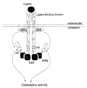
Brief description of the figures
Figure 1: Principle of the receptor-based interaction trap. Ligand binding
leads to
activation of a modifying enzyme activity (MA). Due to the inactivation of the
normal
receptor activation site (inactivated activation site, iAS), the activation of
modifying
enzyme activity does not result in an activation of the signaling pathway,
unless the
heterologous bait in the cytoplasmic domain of the recombinant receptor
(indicated as
'bait') is binding to a prey polypeptide (indicated as 'prey') which is fused
to a
polypeptide comprising an activation site (AS). The modifying enzyme activity
can now
11
CA 02407872 2002-10-29
WO 01/090188
PCT/EP01/05916
modify this activation site; modification (x) of this activation site results
in activation of
the signaling pathway and induction of a reporter system (indicated as
'detectable
activity').
Figure 2: Functionality of EpoR-LepR chimera in the Hek293T PAP21 cell line,
as
measured by luciferase light emission, measured in a chemiluminescence counter
(counts per second, cps). The cells were transfected with:
a. pSV-SPORT + pMET7mcs + pGL3-rPAP1-luci + pUT651
b. pSV-SPORT EpoR/LepR + pMET7mcs + pGL3-rPAP1-luci + pUT651
c. pMET7 LepRY985/1077F + pMET7mcs + pGL3-rPAP1-luci +pUT651
NC: non-stimulated negative control. Stimulations were carried out as
described in the
examples.
Figure 3: Functionality of p53-SV40 LargeT interaction trap, as measured by
luciferase light emission, measured in a chemiluminescence counter (cps).
The cells were transfected with:
a. pSV-SPORT + pMG1-SVT + pGL3-rPAP1-luci + pUT651
b. pSV-SPORT + pMG1-CIS + pGL3-rPAP1-luci + pUT651
c. pSV-SPORT + pMET7-SVT + pGL3-rPAP1-luci + pUT651
d. pSEL1-p53 + pMG1-SVT + pGL3-rPAP1-luci + pUT651
e. pSEL1-p53 + pMG1-CIS + pGL3-rPAP1-luci + pUT651
f. pSELl-p53 + pMET7-SVT + pGL3-rPAP1-luci + pUT651
NC: non-stimulated negative control. Stimulations were carried out as
described in the
examples.
Figure 4: Functionality of the EpoR-CIS phosphorylation-dependent interaction
trap,
as measured by luciferase light emission, measured in a chemiluminescence
counter
(cps).
The cells were transfected with:
a. pSV-SPORT + pMG1-CIS + pGL3-rPAP1-luci + pUT651
b. pSV-SPORT + pMG1-SVT + pGL3-rPAP1-luci + pUT651
c. pSV-SPORT + pEF-FLAG-I/mCIS + pGL3-rPAP1-luci + pUT651
d. pSEL1-EpoR + pMG1-CIS + pGL3-rPAP1-luci + pUT651
e. pSEL1-EpoR + pEF-FLAG-I/mCIS + pGL3-rPAP1-luci + pUT651
f. pSEL1-Ep0RY-F + pMG1-CIS + pGL3-rPAP1-luci + pUT651
g. pSEL1-EpoR + pMG1-SVT + pGL3-rPAP1-luci + pUT651
12
CA 02407872 2002-10-29
WO 01/090188
PCT/EP01/05916
NC: non-stimulated negative control. Stimulations were carried out as
described in the
examples.
Figure 5: Functionality of the IRS1-GRB2-Vav indirect interaction trap, as
measured
by luciferase light emission, measured in a chemiluminescence counter (cps).
The cells were transfected with:
a. pMET7mcs + pMG1-CIS + pGL3-rPAP1-luci + pUT651
b. pMET7mcs + pMG1-GRB2S + pGL3-rPAP1-luci + pUT651
c. pMET7mcs + pMG1-VavS + pGL3-rPAP1-luci + pUT651
d. pMET7 LepR-IRS1 + pMG1-CIS + pGL3-rPAP1-luci + pUT651
e. pMET7 LepR-IRS1 + pMG1-GRB2S + pGL3-rPAP1-luci + pUT651
f. pMET7 LepR-IRS1 + pMG1-VavS + pGL3-rPAP1-luci + pUT651
NC: non-stimulated negative control. Stimulations were carried out as
described in the
examples.
Figure 6: Functionality of the IRS1-GRB2-Vav indirect interaction trap, as
measured
by luciferase light emission, measured in a chemiluminescence counter (cps):
GRB2
dose dependent inhibition of the signal.
The cells were transfected with:
a. pMET7 LepR-IRS1 + 200 ng pMG1-VavS + pGL3-rPAP1-luci + pUT651
b. pMET7 LepR-IRS1 + 200 ng pMG1-VavS + 200 ng pMET7 GRB2SH3 + pGL3-
rPAP1-luci + pUT651
c. pMET7 LepR-IRS1 + 200 ng pMG1-VavS + 1000 ng pMET7 GRB2SH3 + pGL3-
rPAP1-luci + pUT651
NC: non-stimulated negative control. Stimulations were carried out as
described in the
examples.
Figure 7: Functionality of the IRS1-GRB2-Vav indirect interaction trap, as
measured
by luciferase light emission, measured in a chemiluminescence counter (cps):
VavS
dose dependent inhibition of the signal.
The cells were transfected with:
a. pMET7 LepR-IRS1 + 200 ng pMG1-VavS + pGL3-rPAP1-luci + pUT651
b. pMET7 LepR-IRS1 + 200 ng pMG1-VavS + 200 ng pMET7 VavS + pGL3-rPAP1-
luci + pUT651
c. pMET7 LepR-IRS1 + 200 ng pMG1-VavS + 1000 ng pMET7 VavS + pGL3-rPAP1-
luci + pUT651
13
CA 02407872 2002-10-29
WO 01/090188 PCT/EP01/05916
NC: non-stimulated negative control. Stimulations were carried out as
described in the
examples.
Figure 8: Layout of an optimised MAPPIT-based two-hybrid screening method
The procedure encompasses three successive steps, indicated with encircled
numbers. First, cells expressing the chimeric receptor with the C-terminal
"bait" (CR-
Bait) are generated upon recombinase-assisted genomic integration, followed by
hygromycin selection. Next, gp130-"prey" chimeras are expressed upon
retroviral gene
transfer. Finally, if cognate "bait"-"prey" interaction occurs, ligand binding
induces a
signalling cascade leading to induction of the puromycin resistance marker and
concomitant formation of cell colonies in selective medium. Direct RT-PCR
amplification of "prey" encoding transcripts from lysed cell colonies allows
rapid "prey"
identification.
Figure 9: the MAPPIT procedure for two-hybrid screening
(A) The HEK293-16 cell line shows ligand-induced puromycin resistance. HEK293-
16
cells were seeded in a 24 well plate, and were left untreated (top well), or
were subject
to puromycin selection (1 pg/ml) with (middle well) or without (bottom well)
prior
activation of gp130 using LIF for 48 hours. After 1 week, surviving cells were
stained
with crystal violet.
(B) Selection of isogenic HEK293-16 cells expressing the EpoR"bait". HEK293-16
cells
were co-transfected with the pcDNA5/FRT-EpoR"bait" and the Flp recombinase
expression vectors and selected for hygromycin resistance (100 pg/nr11) for 10
days.
Cells were stained using polyclonal antiserum recognising the extracellular
domain of
the EpoR and Alexa488-labelled secondary antibody. Solid and dotted lines show
parental or hygromycin-selected HEK293-16 cells, respectively.
(C) Selection of cells based on a cognate "prey"-"bait" interaction.
Hygrornycin-
resistant cells from (B) were seeded in a 24 well plate, infected with
CIS"prey"
expressing retrovirus (1/30 dilution of retroviral stock) for 48 hours, and
were either left
untreated (top well) or were stimulated with Epo for another 48hours (middle
well),
prior to treatment with puromycin (1 pg/ml). The bottom well shows Epo-
stimulated
cells selected as above, but expressing irrelevant lacZ protein (1/3 dilution
of retroviral
stock). Surviving cells were stained after 7 days with crystal violet.
(D) Puromycin-resistant cells express the "prey" chimera. Parental HEK293-16
cells or
puromycin-resistant cells from (C) were permeabilized, sequentially treated
with anti-
FLAG antibody and FITC-labelled secondary antibody and subjected to FACS
14
CA 02407872 2002-10-29
WO 01/090188
PCT/EP01/05916
analysis. Solid and dotted lines show parental or puromycin-selected HEK293-16
cells,
respectively. (INSET) RT-PCR detection of transcripts encoding the "prey"
chimera.
Cells from (C) were lysed and the "prey"-encoding transcript was amplified by
RT-
PCR. The arrow indicates the CIS-specific amplicon, which was verified by DNA
sequencing. A negative control on parental cells was also performed (middle
lane). M:
marker lane.
(E) Dose-dependent recovery of gp130-CIS"prey" expressing cell clones.
EpoR"bait"
expressing cells were seeded in 75 cm2 culture flasks and infected with
CIS"prey"
expressing retrovirus, 1/10 serially diluted in a complex retroviral HEK293
cDNA
library. After selection, purornycin-resistant colonies were stained using
crystal violet.
Panel 1 shows cells infected with a 1/10 dilution of CIS-"prey" but without
Epo
stimulation. Panels 2-5 show the 1/10 to 1/10,000 serial dilutions, while
panel 6 shows
the outcome of cells infected with the retroviral cDNA library in the absence
of
CIS"prey". In a parallel "spiking" experiment, 19 out of 21 analysed clones
contained
gp130-CIS"prey" transcripts.
(F) Functional analysis of the EpoR"bait" - SOCS-2"prey" interaction. MAPPIT
screening of a HEK293 cDNA library using the EpoR"bait" resulted in the
isolation of a
cell clone expressing a SOCS-2"prey" construct. This selected clone was
transiently
transfected with the pXP2d2-rPAP1-luci reporter alone, or in combination with
the
vector encoding the LR-F3 variant, and was stimulated with Epo or leptin for
24 hours,
respectively, or left unstimulated. The upper panel shows the corresponding
luciferase
inductions with a diagrammatic presentation of the activated receptors on top.
In the
lower panel, a graphic representation of the SOCS-2"prey" chimera and of
intact
SOCS-2 and CIS is shown.
(G) The EpoR"bait"- SOCS-2"prey" interaction is phosphorylation dependent.
(Upper
panel) HEK293-16 cells expressing chimeric receptors with (293-16 EpoR) or
without
(293-16 LR-F3) the EpoR"bait" were stimulated with Epo or were left untreated.
Lig and-dependent phosphorylation of the EpoR"bait" is observed, in contrast
to lack of
phosphorylation of the LR-F3 chimera.
(Middle and lower panels) HEK293T cells were transiently transfected with
expression
constructs for the EpoR"bait" (pSEL1-EpoR) and for the SOCS-2"prey". The
middle
panel and lower panel respectively show ligand-dependent "prey" and STAT3
phosphorylation which is only observed upon transfection with pSEL1-EpoR but
not
CA 02407872 2002-10-29
WO 01/090188 PCT/EP01/05916
with pSEL1-EpoRF. Expression controls for the SOCS-2"prey" and for STAT3 are
also
shown.
Figure 10: Functionality of IL3R-, IL5R- and GM-CSFR-LepR chimera in the Hek
293T cell line, as as measured by luciferase light emission, measured in a
chemiluminescence counter (cps).
The cells were transfected with:
a. pSV-SPORT-EpoR/LepR + pGL3-rPAP1-luci + pUT651
b. pSV-SPORT-IL-3Ra/LepR + pSV-SPORT-13c/LepR + pGL3-rPAP1-luci +
pUT651
c. pSV-SPORT-IL-5Ra/LepR + pSV-SPORT3c/LepR + pGL3-rPAP1-luci +
pUT651
d. pSV-SPORT-GM-CSFRa/LepR + pSV-SPORT-pc/LepR + pGL3-rPAP1-luci +
pUT651
e. pSV-SPORT-IL-3Ra/LepR + pGL3-rPAP1-luci + pUT651
f. pSV-SPORT-IL-5Ra/LepR + pGL3-rPAP1-luci + pUT651
g. pSV-SPORT-GM-CSFRa/LepR + pGL3-rPAP1-luci + pUT651
h. pSV-SPORT-13c/LepR + pGL3-rPAP1-luci + pUT651
NC: non-stimulated negative control. Stimulation was as described in the
examples.
Figure 11: Functionality of the Smad3-Smad4 phosphorylation-dependent
interaction
trap, as measured by luciferase light emission, measured in a
chemiluminescence
counter (cps).
The cells were transfected with:
a. pSV-SPORT-3c/LepR-F3-ALK4CA + pSV-SPORT-GM-CSFRa/LepR-F3-Smad3 +
pMG2-Smad4 + pXP2d2-rPAP1luci + pUT651.
b. pSV-SPORT-13c/LepR-F3-Srnad3 + pSV-SPORT-GM-CSFRa/LepR-F3-ALK4CA +
pMG2-Smad4 + pXP2d2-rPAP1luci + pUT651.
c. pSV-SPORT-pdLepR-F3 + pSV-SPORT-GM-CSFRa/LepR-F3-Smad3 + pMG2-
Smad4 + pXP2d2-rPAP1luci + pUT651.
d. pSV-SPORT-I3./LepR-F3-Smad3 + pSV-SPORT-GM-CSFRa/LepR-F3 + pMG2-
Snnad4 + pXP2d2-rPAP1luci + pUT651.
e. pSV-SPORT-13c/LepR-F3-ALK4CA + pSV-SPORT-GM-CSFRa/LepR-F3 + pMG2-
Smad4 + pXP2d2-rPAP1luci + pUT651.
16
= CA 02407872 2008-08-06
29775-23
f. pSV-SPORT-pdLepR-F3 + pSV-SPORT-GM-CSFRa/LepR-F3-ALK4CA + pMG2-
,
Smad4 + pXP2d2-rPAP1luci + pUT651.
g. pSV-SPORT-13dLepR-F3-ALK4CA + pSV-SPORT-GM-CSFRa/LepR-F3-Smad3 +
pMG2 + pXP2d2-rPAP1luci + pUT651.
h. pSV-SPORT-po/LepR-F3-Smad3 + pSV-SPORT-GM-CSFRa/LepR-F3-ALK4CA +
pMG2 + pXP2d2-rPAP1luci + pUT651.
I. pSV-SPORT-f3e/LepR-F3-ALK4CA + pSV-SPORT-GM-CSFRa/LepR-F3-Smad3 +
pMET7-Smad4 + pXP2d2-rPAP1luci + pUT651.
j. pSV-SPORT43JLepR-F3-Smad3 + pSV-SPORT-GM-CSFRa/LepR-F3-ALK4CA +
pMET7-Smad4 + pXP2d2-rPAP1luci + pUT651.
Examples
Materials and methods to the examples
Cell lines, transfection and infection procedures
Transfections were performed according to the calcium phosphate method (Graham
and van der Eb, 1973).
Recombinant mouse leptin, recombinant human leukemia inhibitory factor (LIF)
and
recombinant human erythropoietin (Epo) were all purchased from R&D Systems.
Typical stimulation conditions were 10Ong/mlleptin, 1 ng/ml LIF and 50 ng/ml
Epo.
For production of ecotropic retrovirus harbouring the gp130-CIS or LacZ coding
sequence, (l)NX-Eco cells were seeded at a density of 6x10⁶ cells/petridish
the day
prior to transfection. Cells were transfected with 50 pg of the retroviral
vector pBG1-
CIS according to the calcium phosphate procedure. 25 pM chloroquine was added
5
min. before transfection. Medium was harvested 24 and 48 hours post
transfection,
filtered over a 0.22 pm GV filter (Millipore) and stored at ¨80 C. Packaging
of the HEK
cDNA library was performed as described above with the exception that 1.6x10⁷
cells
in a 175cm2 falcon were transfected with 87 pg pBG1-HEK293cDNA. For titer
determination, 10% pMFG-EGFP (gift from Dr. Mulligan, Cambridge, MA) was
included
in the DNA in a parallel experiment. The virus titer was approx. 5x10⁶
infectious
units/ml as determined by FAGS analysis of EGFP expressing cells.
For infection with CIS"prey", target cells were seeded at a density of 2x10⁴
cells/well in
a 24-well plate, and 106 in 75cm2 culture flasks. The day after, cells were
incubated
for 24-48 hours with supernatant containing virus, diluted in medium as
indicated.
Polybrener(Sigma) was added at a final concentration of 2.5 pg/ml. After
infection, cells
*Trademark
17
CA 02407872 2002-10-29
WO 01/090188 PCT/EP01/05916
were stimulated with Epo (50 ng/ml) for 24-48 hours, followed by puromycin (1-
2 pg/ml
as indicated; Sigma) selection for 10 days.
Construction of bait, prey and reporter/selector constructs
Generation of the EpoR/LepR chimera
All polyrnerase chain reactions (PCR) were performed using Pfu polymerase
(Stratagene, typically 2,5-5U Pfu were used per reaction). The mouse leptin
receptor
(LepR) transmembrane and intracellular parts (amino acids 839-1162) were
amplified
by PCR using forward primer MBU-0-447 that contains a Pad l restriction enzyme
recognition site and the reverse primer MBU-0-448 that contains both a linker
sequence (Gly-Gly-Ser) and a multi cloning site (MCS) with Sall, Sad, Spel,
Notl and
Xbal recognition sites. Only one amino acid from the extracellular part of the
LepR was
included in the fragment (Gly). Primer design resulted in the insertion of an
Asn
between the Pad l generated Leu-Ile sequence and the extracellular Gly. The
amplicon
was gel-purified and ligated in the pCR -Blunt vector (Invitrogen). Pad-Sad l
digestion
on this pCR -Blunt construct results, after gel-purification, in the desired
LepR
fragment.
The pSV-SPORT-EpoR/IFNaR2-2 (Pattyn et al, 1999). vector expresses an
EpoR/IFNaR2-2 receptor chimera and was constructed as follows: RNA was
isolated
from 5x10⁶ IF-1 cells using the RNeasy kit (Qiagen). RT-PCR was performed as
follows: 2 pl (2pg) of oligodT (12-18 mer; Pharmacia) was added and incubated
at
70 C for 10 min., the reaction mixture was chilled on ice for 1 min., cDNA was
prepared by adding 4 pl of 10x RT buffer (Life Sciences), 1p1 20 mM dNTP's
(Pharmacia), 2p10,1M DTT, and 1p1 of MMLV reverse transcriptase (200U;
Superscript
RT; Life Technologies) to an end volume of 20p1. Incubations were as follows:
RT for
10 min., 42 C for 50 min., 90 C for 5 min., and 0 C for 10 min. Following
this, 0,5 pl
RnaseH (2U; Life Technologies) was added and the mixture was incubated at 37 C
for 20 min., followed by chilling on ice. PCR on this cDNA was performed using
Pfu
enzyme (5 U; Stratagene). Forward primer (MBU-0-167) and reverse primer (MBU-0-
308) were designed to amplify the extracellular part of the EpoR (amino acids
1-249)
between a Kpnl and Pad l site. A band of correct size was purified and the DNA
was
digested with Kpnl and Pad l and was inserted into the Kpnl-Pacl opened pSV-
SPORT-
IL-5Ra/IFNaR2-2 vector. This vector contains a chimeric receptor that has the
extracellular domain of the IL-5Ra receptor, fused to the transmembrane and
18
CA 02407872 2002-10-29
WO 01/090188
PCT/EP01/05916
intracellular domains of IFNaR2-2. By site-specific mutagenesis, a Pad l site
was
added to the fusion point by means of the QuikchangeTM site-directed
mutagenesis kit
(Stratagene, La Jolla) which resulted in the insertion of two amino acids (Leu-
11e)
before the most membrane-proximal, extracellular amino acid (Lys) of IFNaR2-2.
Hence, using the Kpnl site that precedes the coding sequence and the created
Padl
site on the extracellular/transmembrane domain fusion site, the extracellular
domain of
IL-5Ra could be exchanged by the one of EpoR, as described above.
The LepR fragment generated by Pad-Sad l digestion was ligated in the Pad-Sadl
digested and gel-purified pSV-SPORT-EpoR/IFNaR2-2 vector, resulting in pSV-
SPORT-EpoR/LepR.
Generation of 1L-3Ra/LepR, IL-5RaiLepR, GM-CSFRa/LepR and /3/LepR chimeras
The pSV-SPORT-IL-5Ra/IFNaR2-2 and pSV-SPORT-13c/IFNaR1 vectors express an
IL-5Ra/IFNaR2-2 and a pc/IFNaR1 chimera, respectively, composed of the
extracellular portion of the IL-5Ra or chain, and the transmembrane and
intracellular
parts of the IFNaR2-2 or IFNaR1. A Pad l site was used to generate the fusion
site just
preceding the transmembrane segment. The IFNaR2-2 or IFNaR1 parts in these
vectors were replaced by the same segments of the LepR, using the Pad l site
and an
Xbal site which is located just after the IFNaR2-2 or IFNaR1 stop codon.
Therefore,
the LepR fragment was generated by a Pacl-Xbal digest of the pSV-SPORT-
EpoR/LepR vector (see example 1), and was inserted into the Pacl-Xbal opened
and
gel-purified pSV-SPORT-IL-5Ra/IFNaR2-2 and pSV-SPORT-pc/IFNaR1 vectors,
resulting in the vectors pSV-SPORT-IL-5Ra/LepR and pSV-SPORT-r3dLepR.
The pSV-SPORT-IL-3Ra/LepR and pSV-SPORT-GM-CSFRa/LepR vectors were
constructed as follows: the extracellular portion of the IL-3Ra and GM-CSFRa
chains
were amplified using standard RT-PCR procedures with Pfu polymerase. 2 pl IF-1
cDNA was used as input. Forward primers were MBU-O-752 (IL-3Ra) and MBU-0-754
(GM-CSFRa), and generated a Kpnl site. Reverse primers MBU-0-753 (IL-3Ra) and
MBU-0-755 (GM-CSFRa), contain a Pad l site allowing in frame fusion to the
LepR.
After subcloning in the pCR -Blunt vector, the Kpnl-Pacl excised extracellular
fragments were ligated into the Kpnl-Pacl opened pSV-SPORT-IL-5Ra/LepR. For
the
GM-CSFRa construction, a partial Kpnl digest was applied since the
extracellular
portion contained an internal Kpnl site. The resulting vectors, pSV-SPORT-1L-
3Ra/LepR and pSV-SPORT-GM-CSFRa/LepR, contain chimeric receptors composed
19
CA 02407872 2002-10-29
WO 01/090188 PCT/EP01/05916
of the extracellular portion of the IL-3Ra or GM-CSFRa chain fused to the
transmembrane and cytoplasrnatic tail of the LepR.
Generation of the EpoR/LepR- F3 chimera
The mutant leptin receptors (Eyckerman et al., 1999) Y985-1077F and Y985-1077-
1138F (LepR-F3; previously called F-all) were generated using the Quikchange
TM site-
directed mutagenesis procedure using Pfu polymerase (Stratagene) on the pMET7-
LepR template. Mutagenic oligonucleotides were MBU-0-157, MBU-0-158, MBU-0-
159, MBU-0-160, MBU-0-161 and MBU-0-162. Each single mutation was coupled to
a change in restriction cleavage and was confirmed by restriction and DNA
sequence
analysis. The double and triple mutants were created using a sequential
approach.
Signalling properties of the generated mutants were investigated at the gene
induction
level using the rPAP1-luci reporter construct (see below) and Northern blot
analysis of
induction of Metallothionein II gene transcripts. The double Y985-1077F mutant
showed a higher stimulation of the relevant genes compared to the wild type
LepR,
which is probably due to loss of recruitment of a 5H2-module containing
tyrosine
phosphatase such as SHP-1 or SHP-2. The triple Y985-1077-1138F (LepR-F3)
showed almost complete loss of induction due to elimination of the Box3 or
STAT-3
association motif. This results in a receptor which still allows
phosphorylation and
activation of the associated JAK2 kinase but which cannot deliver a
stimulatory signal
to the studied genes.
PCR amplification on this pMET7-LepR-F3 vector template using MBU-0-447 and
MBU-0-448 as forward and reverse primers, respectively, resulted in a LepR-F3
amplicon spanning the transmembrane and intracellular domains of LepR-F3 (+1
extra
Gly of the extracellular part, see above), which was subcloned in the pCIRc)-
Blunt
vector. Pad-Sad l digestion of the resulting plasmid yielded a DNA fragment
containing
the LepR-F3 sequence, which was ligated into Pad-Sad l digested and gel-
purified
pSV-SPORT-EpoR/IFNaR2-2 vector (see above). This resulted in the pSV-SPORT-
EpoR/LepR-F3, which was renamed to pSEL1.
Generation of the prey vector
Prey constructs were generated in the pMET7 vector, which contains a strong
constitutive hybrid SRa promoter (Takebe etal., 1988).
Through site-directed mutagenesis (QuikchangeTM, Stratagene) a unique Apal
site was
introduced after the pMET7 promoter and before the unique EcoRI site in the
pMET7mcs vector resulting in pMET7mcsA (primers MBU-0-567 and MBU-0-568).
CA 02407872 2002-10-29
WO 01/090188
PCT/EP01/05916
The pMET7mcs vector is a modified version of pMET7 containing an expanded MCS
by insertion of the extra unique BgIII, EcoRV, BstEll, Agel and Xhol
restriction sites.
PCR amplification on the pSVL-gp130 template using the forward primer MBU-0-
586
and the reverse primer MBU-0-443 generated a DNA fragment encoding a 158 amino
acid-long intracellular fragment of the human gp130 chain, which contains 4
STAT-3
association motifs (amino acids 761-918, the stopcodon was not co-amplified).
The
forward primer contains from 5' to 3' an Apal restriction site, a Kozak
consensus
sequence, a flag-tag encoding sequence (Met-Asp-Tyr-Lys-Asp-Asp-Asp-Asp-Lys-
Ile),
and a BglIl restriction site. The reverse primer encodes an additional hinge
sequence
(Gly-Gly-Ser) and contains an EcoRI recognition site. Apal and EcoRI digestion
of the
PCR product (after subcloning in pCR -Blunt) and of pMET7-mcsA, allowed us to
ligate the gp130 fragment into the pMET7 vector, generating the pMET7-flag-
gp130
construct.
SV40 largeT antigen (SVT) was amplified using a vector from the HybriZAP-2.1
Two-
Hybrid cDNA synthesis kit (Stratagene, pSV40) as template. Primers MBU-0-445
and
MBU-0-446 were used to generate a DNA fragment encoding 448 amino acid between
residues 261 and 708. The N-terminal deletion eliminates the nuclear targeting
signal
in SVT. The forward primer contains an EcoRI recognition site that allows in-
frame
ligation to the gp130-hinge sequence. The reverse primer contains additional
Nrul,
Xhol, BgIII, Notl and Xbal restriction sites and also encodes the stop codon
after the
SVT coding sequence. Subcloning in pCR -Blunt, followed by recovery of the
cleaved
amplicon with EcoRI and Xbal, allowed ligation in the EcoRI-Xbal opened pMET7-
flag-
gp130 vector, yielding pMET7-flag-gp130-SVT, which was renamed to pMG1-SVT.
Digestion with EcoRI-Xhol or EcoRI-Notl allows the insertion of model preys or
of
cDNA libraries into this vector. In these cases, the SVT fragment acts as a
`stuffer.
Construction of the p53-SVT interaction trap vectors
A DNA fragment encompassing murine p53 was amplified with MBU-0-450 and MBU-
0-451 using the p53 control plasmid from the HybriZAP-2.1 Two-Hybrid cDNA
synthesis kit (Stratagene) as template. The forward primer contains a Sall
restriction
site that allows in-frame coupling to the EpoR/LepR-F3 hinge construct. The
reverse
primer contains a STOP codon and a Xbal restriction site. The 243 amino acid-
long
p53 fragment (amino acids 73-315) contains the interaction site with SVT, but
lacks the
nuclear targeting signal and the oligomerisation domain. Subcloning over
pCRQBlunt,
21
CA 02407872 2002-10-29
WO 01/090188
PCT/EP01/05916
digestion with Sall-Xbal and gel-purification yielded a fragment that was
ligated into
Sall-Xbal cut and gel-purified pSEL1 vector, resulting in pSEL1-p53.
Generation of the pMG1-SVT vector is described above.
Amplification using MBU-0-695 and MBU-0-696 as forward and reverse primer
Construction of EpoR-CIS interaction trap vectors
Through site directed mutagenesis (QuikchangeTM, Stratagene) a Tyr to Phe
mutation
on position 426 in the human EpoR was introduced in the pSEL1-EpoR construct
resulting in an inactive EpoR fragment. Forward primer MBU-0-717 and reverse
primer MBU-0-718 were used for the PCR-based mutagenesis which also resulted
in
The complete coding region for mouse Cytokine Inducible 5H2-containing protein
CIS
(amino acids 2-257) was amplified using MBU-0-677 and MBU-0-678 as forward and
22
CA 02407872 2002-10-29
WO 01/090188
PCT/EP01/05916
into an EcoRI-Xbal digested and gel-purified pMG1-SVT vector, leading to the
pMG1-
CIS vector.
Construction of the IRS1-Vav interaction trap vectors
Through a mutagenesis approach (QuikchangeTM, Stratagene) 4 amino acids (P1137-
Y-
M-P11) within the mutant leptin receptor Y985-1077F (pMET7 LepR Y985-1077F)
were exchanged for a phosphotyrosine encoding region from human IRS1 (Insulin
Receptor Substrate 1; S892-P-G-E-Y-V-N-I-E-F901). This removes the functional
STAT3 association motif within the leptin receptor. Primers MBU-0-515 and MBU-
0-
516 were used for the PCR-based mutagenesis. This construct was named pMET7
LepR-IRS1.
Using standard RT-PCR procedures with Pfu polymerase full size human GRB2
(Growth Receptor Bound 2; aa 1-217) was amplified using 2 pl HepG2 cDNA as
input,
and using MBU-0-467 and MBU-0-468 as forward and reverse primer respectively.
The forward primer contains an extra EcoRI site which allows in frame fusion
to the
gp130 chain in the pMG1 vector and the reverse primer contains an extra Xbal
site
after the stopcodon. After subcloning in the pCR -Blunt vector, the EcoRI-Xbal
excised fragment was ligated into the EcoRI-Xbal cut pMG1 vector, resulting in
pMG1-
GRB2. The SH2 domain of GRB2 (aa 60-158) was amplified in the same way using
primers MBU-0-469 and MBU-0-470 as forward and reverse respectively. The
forward primer contains an extra EcoRI recognition site which allows in frame
fusion to
the gp130 chain, and the reverse primer contains an extra stop codon and a
Xbal
enzyme recognition site. After subcloning of the PCR fragment in the pCle-
Blunt
vector, the EcoRI-Xbal generated fragment was ligated in the EcoRI-Xbal cut
pMG1
vector, resulting in the pMG1-GRB2S vector.
A fragment of human GRB2 comprising the C-terminal SH3 domain (aa 159-217) was
amplified using the pMG1-GRB2 construct as template and MBU-0-770 and MBU-0-
468 as forward and reverse primer respectively. MBU-0-770 allows in frame
fusion to
the flag tag by a BglIl site, MBU-0-468 is described above. After subcloning
this PCR
fragment in the pCle-Blunt vector, the GRB2 fragment was inserted in the pMG1
vector by a BgIII-Xbal based exchange, resulting in the pMET7-GRB2SH3 vector.
A fragment of human Vav (VavS: aa 259-789) was amplified using Pfu polyrnerase
from mRNA of the human TF1 cell line by standard RT-PCR techniques. Primers
were
MBU-0-737 and MBU-0-738 as forward and reverse respectively. MBU-0-737
contains an extra EcoRI allowing in frame fusion to gp130, and MBU-0-738
contains a
23
CA 02407872 2002-10-29
WO 01/090188
PCT/EP01/05916
Stop codon and a Xhol enzyme recognition site. The amplified fragment was
subcloned in the pCR -Blunt vector and ligated in the pMG1 vector through an
EcoRI-
Xhol based exchange. The VavS fragment was also amplified using forward primer
MBU-0-771, reverse primer MBU-0-741 and pMG1-VavS as template. The forward
primer contains a BamHI site, which allows in frame fusion to the flag tag,
and the
reverse primer contains a stop codon and a Xbal restriction site. The
arnplicon was
subcloned in the pCR -Blunt vector, and excised with BamHI and Xbal. The
purified
fragment was cloned in a BgIII-Xbal cut pMG1 vector, resulting in the pMET7-
VavS
construct.
Construction of the pGL3-rPAP1-luci and pSEAP-rPAP1 reporter constructs
Genomic DNA was isolated from the rat pheochromocytorna PC12 cell line using
the
DNAzol procedure (Gibco BRL). Optimal primers for PCR amplification of the rat
PAP1
promoter were selected using the "Oligo Primer 3" program (http://vvvvw-
genome.wi.mit.cdu/cgi-bin/primer3.cgi). Forward and reverse primers were MBU-0-
222 and MBU-0-223 respectively. Amplification was performed using Taq
polymerase
in 30 cycles: 2' at 94 C, 2' at 57 C, 2' at 72 C, followed by a 10' filling-in
reaction at
72 C. Optimal MgC12 concentration was determined to be 6mM. The promoter
fragment was cloned after polishing with Klenow polymerase in the pCR -Blunt
vector.
The promoter fragment was cut from this plasmid construct using a successive
Pstl
digest, Klenow treatment to polish this end, and BamHI digest, resulting in a
blunt-
sticky fragment. The gel-purified fragment was cloned into a Smal-Bg111 opened
and
gel-purified pGL3 control vector (Promega). Digestion with Sstl-Spel
restriction
enzymes and gel-purification resulted in a fragment that was cloned into a
Sstl-Nhel
and purified pGL3 basic vector, resulting in the pGL3- rPAP1-luci construct.
DNA
sequencing revealed 10 nucleotides differing from the published sequence, but
without
affecting the leptin-dependent induction of this promoter segment.
The full-length rPAP1 promoter fragment was excised from pGL3 rPAP1-luci using
partial digestion with Kpnl and Xhol and ligated into the Kpnl-Xhol opened
pXP2d2
vector (gift from Prof. S. Nordeen), resulting in pXP2d2-rPAP1-luci. The
coding
sequence for puromycin was amplified using primers MBU-0-719 and MBU-0-720 on
the pIRESpuro2 (Clontech) template. Combined Xhol-Xbal digestion allowed
insertion
in the pXP2d2-rPAP1-luci construct, resulting in pXP2d2-rPAP1-puroR.
Digestion of the pGL3-rPAP1-luci construct using Mlul and Xhol restriction
enzymes
resulted in a fragment spanning the full size rPAP1 promoter. This fragment
was gel-
24
CA 02407872 2002-10-29
WO 01/090188 PCT/EP01/05916
purified and ligated into a Mlul-Xhol cut and gel-purified pSEAP vector
(TROPIX,
Perkin Elmer) resulting in the pSEAP-rPAP1 construct. Functionality of this
construct
was assayed by transient co-transfection of pMET7 LepR and pSEAP-rPAP1 in PC12
cells using the Phospha-LightTM reporter assay kit from TROPIX (Perkin Elmer).
Construction of the pcDNA5/FRT-EpoR and pBG1-SVT, pBG1-CIS and pBG1-
ccdB vectors
Insertion of the EpoR-LR-F3-EpoR into the pcDNA5/FRT vector was obtained by re-
amplifying the complete chimeric construct using MBU-0-167 and MBU-0-769 on
the
pSEL1-EpoR template, followed by subcloning using Kpnl and Notl sites. This
construct was named pcDNA5/FRT-EpoR.
The SVT fragment was re-amplified from pMG1-SVT using forward primer MBU-0-766
and MBU-0-446. BamHI-Notl digestion allowed insertion in the pBMN-Z retroviral
vector (gift from G. Nolan), resulting in vector pBG1-SVT. EcoRI-Notl based
exchange
of the SVT fragment for CIS (pMG1-CIS) resulted in the pBG1-CIS vector.
To allow counter-selection for vector self-ligation in case of insertion of
cDNA libraries
in the pBG1 vector, the E. coli control of cell death gene (ccdB) was
amplified using
primers MBU-0-835 and MBU-0-836 and template pENTRY11 (Life Technologies),
and cloned in the pBG1-CIS vector by an EcoRI-Notl restriction-based
insertion,
resulting in the pBG1-ccdB vector.
Construction of the bait-modifying enzyme, substrate-bait and prey chimeras.
Generation of the pSV-SPORT-GM-CSFRalLepR-F3 and pSV-SPORT-I3c/LepR-F3
chimeras
The LepR fragment in pSV-SPORT-GM-CSFRa/LepR and pSV-SPORT-pc/LepR was
replaced by the LepR-F3 fragment of pSEL1 (pSV-SPORT-EpoR/LepR-F3) using the
Pad l and Notl site. Therefore, the LepR-F3 fragment was generated by a Pacl-
Notl
digest of the pSEL1 construct, gel-purified and inserted into the Pacl-Notl
opened and
gel-purified pSV-SPORT-GM-CSFRa or pSV-SPORT-Pc vectors, resulting in the
vectors pSV-SPORT-GM-CSFRa/LepR-F3 or pSV-SPORT-13c/LepR-F3.
Generation of the pSV-SPORT-GM-CSFRa/LepR-F3-modifying enzyme chimera and
pSV-SPORT-fickepR-F3-modifying enzyme chimera and generation of the pSV-
SPORT-GM-CSFRa/LepR-F3-bait chimera and pSV-SPORT-fickepR-F3-bait chimera
CA 02407872 2008-08-06
=
29775-23
The pSV-SPORT-GM-CSFRcc/LepR-F3 and pSV-SPORT-pc/LepR-F3 vectors were
digested with Sall, gel-purified and the ends were polished by Klenow fragment
(Boehringer Mannheim). These blunt ended vectors were incubated with Alkaline
Phosphatase (Boehringer Mannheim) to dephosphorylate the blunt ends. For the
modifying enzyme a construct containing the mouse cytoplasmic tail of ALK4
with
mutation T206D, resulting in a constitutive active kinase, in the vector pGBT9
was
obtained from Prof. D. Huylebroeck. The mutated cytoplasmic tail of ALK4 was
removed from the construct by an EcoRI-BamHI digestion, gel-purified and
incubated
with Klenow fragment to polish the ends. This insert was ligated in the opened
pSV-
SPORT-GM-CSFRa/LepR-F3 and pSV-SPORT-pdLepR-F3 vectors resulting in pSV-
SPORT-GM-CSFRa/LepR-F3-ALK4CA and pSV-SPORT-N/LepR-F3-ALK4CA.
For the bait a construct containing a human cDNA encoding the entire Smad3
protein
in vector pcdef was a kind gift of Prof. D. Huylebroeck. The Smad3 insert was
removed
with an EcoRI-Xhol digestion, gel-purified and the ends were polished by
Klenow
fragment (Boehringer Mannheim). This Smad3 insert was ligated into the opened
pSV-
SPORT-GM-CSFRakepR-F3 and pSV-SPORT-8c/LepR-F3 vectors (described above)
resulting in pSV-SPORT-GM-CSFRa/LepR-F3-Smad3 and pSV-SPORT-3iLepR-F3-
Smad3.
Generation of the pMG2-prey chimera
For construction of the pMG2 vector, the pMG1-SVT vector was digested with
EcoRI
and Notl, followed by incubation with Klenow fragment (Boehringer Mannheim) to
polish the ends. This blunt ended vector was incubated with Alkaline
Phosphatase
(Boehringer Mannheim) to dephosphorylate the blunt ends. Cassette rfB of the
Gateway Vector Conversion System (Life Technologies) was then ligated into the
opened vector leading to the pMG1-gateway vector. A PCR reaction using primers
MBU-0-1094 and MBU-O-1076 on the pMG1-SVT template was performed, resulting
in a fragment that contains gateway recombination sites. This fragment also
contains a
part (amino acids 905 - 918) of the gp130 chain. The fragment was then cloned
in the
pMG1-gateway vector using a two-step gateway reaction (Lifetechnologies),
resulting
in pMG2-SVT, a prey construct with a total of 6 STAT recruitment sites. The
pMG2-
SVT construct was digested by EcoRI-Xhol and the vector was gel-purified. For
the
prey we obtained a construct from Prof. D. Huylebroeck containing a cDNA
encoding
almost the entire human Smad4 protein, only lacking the first 3 amino acids.
The
*Trade-mark
26
CA 02407872 2008-08-06
=
29775-23
Smad4 insert was removed by an EcoRI-Xhol digestion, gel-purified and ligated
into
the opened pMG2 vector.
Construction of the reporter cell lines
Selection of the Hek293T PAP21 reporter cell line
The Blasticidin system (Invitrogen) was used to create a stable cell line with
an
endogenous pSEAP-rPAP1 reporter construct Sensitivity to the toxic agent
Blasticidirt
(Invitrogen) of Hek293T cells was estimated to be 3 pg/ml. 106 cells were
seeded in a
petridish and transfected the day after seeding using the Calcium Phosphate
Transfection System (Life Technologies) according to the manufacturers
instructions.
A total of 20 pg DNA was transfected (18 pg of pSEAP-rPAP1seap and 2 pg of
pcDNA6N5-HisA, which contains a Blasticidin resistance gene). After 48 hours
the
transfected cells were seeded in 96 well plates at 10 cells per well. After 24
hours,
BlastIcIdln was added at a concentration of 3 pg/ml and cells were maintained
under
selective conditions for 3-4 weeks. The resulting single cell clones were
screened by
stimulation for 24 hours with 20 ng/ml hyper-1L6 (fusion protein of IL-6 with
its specific
receptor IL6-Ra; Fischer et al., 1997). Hek293T PAP21 was selected as the best
responsive cell line.
Generation of the HEK293-16 cell line
Flp-ln-293 cells (Invitrogen) were stably transfected with a plasmid
containing
expression cassettes for the mouse ecotropic retroviral receptor (mEcoR) and
for
neomycin resistance. The pool of neomycin resistant cells (resistant to 400
pg/ml
geneticin, Life Technologies) were supertransfected, at a ratio of 5:1
respectively, with
the following two plasmids: i) a plasmid carrying the cDNA encoding the
puromycin
resistance marker (puromycin-N-acetyl-transferase) under control of promoter
sequences of rPAP1 (pXP2d2-rPAP1-puroR), ii) a plasmid carrying the cDNA for
the
blasticidin resistance marker (blasticidin S deaminase), under the control of
the EM7
promoter (pcDNA6N5-His, Invitrogen). After selection in blasticidin S (5
pg/ml,
Invitrogen), single colonies were picked and seeded in 24 well plates.
Puromycin
resistance (1 pg/ml, Sigma; added 48 hours after seeding) was monitored in the
absence or presence of LIF (1ng/m1). After another 5 days, surviving cells
were
stained with crystal violet using a standard procedure.
*Trade-mark
27
CA 02407872 2008-08-06
29775-23
RT-PCR analysis
Unless otherwise stated, cells were lysed in 100 pl RLT buffer (RNeasy
method,
* =
Qiagen) and chromosomal DNA was sheared using Qiashredder columns (Qiagen).
Beads were pre-treated according to the manufacturers' instructions (Dynabeads
M-
280 Streptavidin, Dynal). Briefly, Dynabeads were washed twice in a high salt
buffer
(1M NaCI, 10mM Tris HCI pH7,5 and 1mM EDTA), and were incubated with 200
pmoles of biotinylated oligonudeotide directed against the gp130 chain (5'
GGGCTGGGTAGACTCGGATCTTGAGAAGAC). Next, beads were washed three
times in the above mentioned high salt buffer and resuspended in a low salt
buffer
(0,15M NaCI, 10mM Tris NCI pH7,5 and 1mM EDTA) to a concentration of 10 pg/pl.
5
I of this suspension was added to 100 pl total lysate diluted 1/5 in the high
salt buffer.
15' minutes after gentle rotation at room temperature, beads were washed three
times
with low salt buffer and eluted in 30 pl water for 2' at 65 C. 15 pl of this
sample was
used as input for a standard RT-PCR reaction with the Qiagen OneStep RT-PCR
Kit.
Primers 5' GGCATGGAGGCTGCGACTG and 5' TCGTCGACCACT GTGCTGGC
were used for amplification of the "prey" fragment. In a pilot experiment
using CIS as
"prey" template, efficient amplification was obtained with lysate from less
than 103
cells.
Reporter assays, binding assays, cell survival assay and FA CS analysis
Luciferase was measured after lysis of the cells and addition of luciferase
substrate
(Luciferin, Duchefa). Light emission was measured using a TopCount
chemiluminescence counter (Canberra Packard). All luciferase measurements were
normalized using an expression construct constitutively expressing ri-
galactosidase
(pUT651), which was measured in triplicate for every transfection using the
GalactoStar kit (TROPIX).
Puromycin-resistant cell colonies were stained with crystal violet using a
standard
procedure.
Human erythropoietin receptor expression was monitored using goat anti-human
EpoR
polyclonal IgG (R&D Systems) at 2 pg/ml and Alexa488-conjugated donkey anti-
goat
IgG (Molecular Probes) at 4 pg/ml. For demonstration of the expression of the
FLAG-
tagged "prey" construct, cells were fixed and permeabilized with StarfiqsTM
according
to the manufacturers' protocol (Immuno Quality Products) and were stained with
an
anti-FLAG mouse mAb (Sigma) at 8 pg/ml and fluorescein-conjugated sheep anti-
*Trade-mark
28
CA 02407872 2008-08-06
29775-23
mouse IgG (Amersham), 1/50 diluted. All FACS analyses were performed on a
FACSCalibur (Becton Dickinson).
Generation of retroviral cDNA libraries and screening conditions
Construction of the HEK293 library was performed using standard procedures.
Briefly,
5 pg of HEK293 polyA+mRNA was used as input for both oligo-dT and random
primed
IC
first strand synthesis with Superscript II reverse transcriptase (Life
Technologies). Both
the oligo-dT and random primers contain a Notl site. After second strand
synthesis,
adaptors containing an EcoRI site were ligated. The cDNA was analyzed using
agarose gel electrophoresis and fragments between 0.5 and 2.5 Kbp were cloned
unidirectionally in the pBG1-ccdB vector opened with EcoRI-Notl.
For screening, a total of 6.107 "bait" expressing cells were seeded at a
density of 2.106
per 175 cm2 tissue culture flasks. 24 hours after seeding, cells were infected
with the
retroviral HEIK293"prey" cDNA library (complexity of 2.106) for another 24
hours. After
infection, cells were stimulated for 6,5 hours with 50 ng/ml Epo, and
puromycin was
added at a final concentration of 2 pg/ml for 20 days. Single cell colonies
were picked
and analysed using a functional assay and RT-PCR sequencing.
Imrnunoprecipitations and Western blot analysis
For demonstration of EpoR"bait" phosphorylation, we transiently transfected
approximately 3.106 HEK293T cells with plasmids encoding the EpoR"bait" or the
EpoR"bait" Y402F mutant, and the CIS/SOCS-2"preys". Cleared lysates (in
modified
RIPA buffer: 50 mM TrisHCI p118.0; 200 mM NaCI; 1% NP40; 0.5 % DOC; 0.05% SDS;
2 mM EDTA; 1mM Na3VO4; 1mM NaF; 20 mM 13-glycerophosphate; Complete"'
protease inhibitor cocktail [Roche]) of stimulated and unstimulated cells were
incubated
with 2 pg goat anti-human EpoR polyclonal IgG and Protein G Sepharosj
(Amersham
Pharmacia Biotech). After precipitation, polyacrylamide gel electrophoresis
and
blotting, phosphorylation was revealed using PY20 antibody (Transduction
Laboratories).
lmmunoprecipitation of "preys" was performed after transfection of
EpoR/EpoRF"baits"
and the SOCS-2"prey"in HEK293T cells. Incubation of cleared lysates (modified
RIPA)
with anti-FLAG M2 affinity gel, allowed SOCS-2"prey" precipitation.
Phosphorylation of
the "prey" was revealed using the PY20 antibody. "Prey" expression levels were
verified after stripping the blots and reprobing with anti-FLAG antibody.
*Trade-mark
29
CA 02407872 2008-08-06
=
=
29775-23
STAT3 phosphorylation was demonstrated using the Phospho-STAT3 (Tyr705)
Antibody (Cell Signaling) according to the manufacturers instructions. STAT3
expression was verified on the same blots using anti-STAT3 antibody
(Transduction
Laboratories).
Example 1: Functionality of EpoR-LepR chimera in the Hek293T PAP21 cell line
To determine the functionality of the EpoR/LepR chimera, 3 combinations of
plasmids
were transfected into Hek293T PAP21 cells:
a. pSV-SPORT + pMET7mcs + pGL3-rPAP1-luci + pUT651
b. pSV-SPORT EpoR/LepR + pMET7mcs + pGL3-rPAP1-luci + pUT651
c. pMET7 LepRY985/1077F + pMET7mcs + pGL3-rPAP1-luci +pUT651
Transfection was performed according to the calcium phosphate method (Graham
and
van der Eb, 1973). A precipitate was formed using 3,4 pg as total DNA input
(0.4 pg for
pUT651, 1 pg for each of the others) into a 300 pl total mixture. 200 pl of
this mixture
was added to 4x10⁵ Hek293T PAP21 cells seeded the day before transfection into
a
six well plate (Falcon). 6 hours after adding the mixture, the cells, were
washed once
with Dulbecco's PBS (Life Technologies) and new DMEM medium (Life
Technologies)
was added. Two days after transfection, medium was removed and cells were
resuspended using 200 pl Cell Dissociation Agent* (Life Technologies). After
neutralization with 1200 pl DMEM medium, 50 pl of the cell suspension was
seeded
into a 96 well plate (Costar) in triplicate for every condition, and
stimulated by adding
recombinant human leptin (R&D systems) to a final concentration of 100 ng/ml,
or
recombinant human erythropoietin (R&D systems) to a final concentration of 0.5
units/ml, or the combination of leptin (same concentration as above) plus
forskolin
(Sigma, 10 pM final concentration), or the combination of erythropoietin (same
concentration as above) plus forskolin (same concentration as above). A non
stimulated negative control was also incorporated in the experiment. Forskolin
is a
chemical agent that activates the adenylate cyclase that is present within the
cells,
which leads to heightened levels of the second messenger cAMP. Treatment of
transfected cells with forskolin alone did not result in a significant
induction of
luciferase activity. Through an undefined mechanism, cAMP elevation leads to
strong
co-stimulation with the leptin signal on PAP1 induction (Eyckerman et al.,
1999). 24
hours after stimulation, the cells were lysed in the wells and luciferase
substrate
'(Luciferin, Duchefa) was added. Light emission was measured using a TopCount
*Trade-mark
CA 02407872 2008-08-06
=
=
29775-23
chemiluminescence counter (Canberra Packard). The mock control transfection
(transfection a) showed no signal in all cases. Results are shown in figure 2.
The
transfection with the EpoR/LepR chimera resulted in a 3,7 fold induction with
erythropoietin, and a 6,5 fold induction with erythropoietin and forskolin. No
significant
signal was detected when stimulated with leptin, nor with leptin + forskolin.
In the cells
transfected with the LepR Y985/1077F mutant a 33,2 fold induction was detected
when
stimulated with leptin, and a 37,6 fold induction was detected when co-
stimulated with
forskolin. No signal was detected in both erythropoietin and erythropoietin +
forskolin
stimulated cells. All results were normalized using the internal transfection
control
vector pUT651 and the GalactoStar kit (See above).
The difference in induction between EpoR/LepR and LepR Y985/1077F is very
likely
due to the elimination in the latter receptor construct of tyrosines involved
in the
recruitment of tyrosine phosphatases and SOCS proteins to the complex, leading
to an
enhanced signal (Eyckemian etal., 1999).
Example 2: Functionality of p53-SV40 LargeT interaction trap
To investigate the functionality of this modification-independent interaction,
the
following plasmid combinations were transfected into 4x10⁵ Hek293T cells
seeded in a
6 well plate the day before transfection:
a. pSV-SPORT + pMG1-SVT + pGL3-rPAP1-luci + pUT651
b. pSV-SPORT + pMG1-CIS + pGL3-rPAP1-luci + pUT651
c. pSV-SPORT + pMET7-SVT + pGL3-rPAP1-luci + pUT651
d. pSEL1-p53 + pMG1-SVT + pGL3-rPAP1-luci + pUT651
e. pSEL1-p53 + pMG1-CIS + pGL3-rPAP1=luci + pUT651
f. pSEL1-p53 + pMET7-SVT + pGL3-rPAP1-luci + pUT651
A 300 pl precipitation mixture was prepared which contained 3.1 pg DNA (0.1 pg
of
pUT651, 1 pg of each of the others). 200 pl was added to the cells for 6 hours
after
which they were washed once with Dulbecco's PBS. After washing, DMEM medium
was added to the cells. After 24 hours cells were resuspended using 200 pl
Cell
Dissociation Agent which was neutralized with 2200 pl DMEM medium. Of this
cell
suspension 40 pl was brought into a 96 well plate for each transfection and
stimulation
was performed in triplicate. 60 pl DMEM was added to an end volume of 100 pl
and
24 hours later, cells were stimulated for 24 hours with erythropoietin or
erythropoietin
plus forskolin (same concentrations as described above). A non-stimulated
negative
*Trade-mark
31
CA 02407872 2002-10-29
WO 01/090188
PCT/EP01/05916
control was also included in the experiment. Luciferase measurements are shown
in
figure 3.
Transfected cells from transfections a, b and c showed no significant
induction of the
reporter construct under all conditions tested. A 9,4 fold induction and a
14,6 fold
induction was detected in transfected cells from transfection d, after
stimulation with
erythropoietin and erythropoietin plus forskolin respectively, implying an
interaction-
dependent signal. No signal was detected in transfection e and f. This implies
a
specific interaction, which leads to gp130-dependent STAT-3 activation. All
results
were normalized using the internal transfection control vector pUT651 and the
GalactoStar kit (See above).
Example 3: Functionality of the EpoR-CIS phosphorylation-dependent
interaction trap
To determine the functionality of the EpoR-CIS phosphorylation-dependent
interaction
trap, the following plasmid combinations were transfected in 4x10⁵ Hek293T
cells,
which were seeded the day before transfection:
a. pSV-SPORT + pMG1-CIS + pGL3-rPAP1-luci + pUT651
b. pSV-SPORT + pMG1-SVT + pGL3-rPAP1-luci + pUT651
c. pSV-SPORT + pEF-FLAG-I/mCIS + pGL3-rPAP1-luci + pUT651
d. pSEL1-EpoR + pMG1-CIS + pGL3-rPAP1-luci + pUT651
e. pSEL1-EpoR + pEF-FLAG-I/mCIS + pGL3-rPAP1-luci + pUT651
f. pSEL1-EpoRY-F + pMG1-CIS + pGL3-rPAP1-luci + pUT651
g. pSEL1-EpoR + pMG1-SVT + pGL3-rPAP1-luci + pUT651
A precipitation mixture of 300 pi was prepared which contained 3,1 pg DNA (0,1
pg of
pUT651, 1 pg of each of the others). 200 pl of this mixture was applied to the
cells.
After 6 hours, the cells were washed once with Dulbecco's PBS, and DMEM medium
was added. After 48 hours the cells were resuspended using 250 pl Cell
Dissociation
Agent. After neutralization with 2200 pl DMEM medium, 100 pl of this cell
suspension
was brought into a 96 well plate (Costar). The cells were stimulated with
erythropoietin
or erythropoietin plus forskolin (for final concentrations, see above). A non-
stimulated
negative control was also included in the experiment. Luciferase expression
was
measured 24 hours after stimulation using a TopCount chemilunninescense
counter
(Canberra Packard). Transfected cells from transfections a, b, c, e, f and g
showed no
significant induction of luciferase activity. Transfected cells from
transfection d showed
32
CA 02407872 2002-10-29
WO 01/090188 PCT/EP01/05916
a 6,2 fold and a 10,5 fold induction with erythropoietin or erythropoietin
plus forskolin,
respectively. This indicates an erythropoietin-dependent phosphorylation of
the EpoR
bait, resulting in interaction between the CIS protein and EpoR. Interaction
leads to
gp130 phosphorylation, STAT activation and thus signalling toward the rPAP1
promoter, leading to luciferase activity (Figure 4).
Example 4: Functionality of the IRS1-GRB2-Vav indirect interaction trap
In order to investigate the IRS1-GRB2-Vav indirect interaction trap, following
combinations of plasrnids were transfected in 4x10⁵ Hek293T cells, seeded the
day
before transfection:
a. pMET7mcs + pMG1-CIS + pGL3-rPAP1-luci + pUT651
b. pMET7mcs + pMG1-GRB2S + pGL3-rPAP1-luci + pUT651
c. pMET7mcs + pMG1-VavS + pGL3-rPAP1-luci + pUT651
d. pMET7 LepR-IRS1 + pMG1-CIS + pGL3-rPAP1-luci + pUT651
e. pMET7 LepR-IRS1 + pMG1-GRB2S + pGL3-rPAP1-luci + pUT651
f. pMET7 LepR-IRS1 + pMG1-VavS + pGL3-rPAP1-luci + pUT651
A 300 pl precipitation mixture was prepared containing 3,05 pg DNA (0,05 pg
for
pUT651, 1 pg for others). 200 pl of this mixture was added to the cells for 16
hours.
After transfection the cells were washed once with Dulbecco's PBS and DMEM
(both
from Gibco BRL) was added. After 48 hours the cells were resuspended using 200
pl
of Cell Dissociation Agent (Gibco BRL) which was neutralized by 1,8 ml DMEM
medium. 100 pl of cell suspension was seeded in a Costar 96 well plate and
stimulated in a final volume of 200p1 with final concentrations of 100 ng/ml
leptin, 100
ng/ml leptin plus 10 pM forskolin, 10 pM forskolin, or were left unstimulated.
24 hours
after stimulation, luciferase and galactosidase activity assays were performed
as
described above. From these results (Figure 5) we can conclude that the cells
from
transfections a, b, c, and d show no significant induction of the rPAP1
promoter. Cells
from transfection e show a slight induction with leptin and a moderate
induction when
co-stimulated with forskolin (2,5 fold), suggesting a direct interaction
between IRS-1
and GRB2S. Transfection experiment f shows a clear induction of luciferase
activity
with leptin (5,2 fold), which is more pronounced when co-stimulated with
forskolin (12,0
fold). This indicates an interaction between IRS1 and Vav, which is probably
mediated
through endogenous GRB2.
33
CA 02407872 2002-10-29
WO 01/090188
PCT/EP01/05916
To investigate the specificity and the involvement of GRB2 in the interaction,
and to
test if the signal is generated by recruitment of the gp130 chain, a number of
control
experiments were performed.
The following combinations of plasmids were tested in the same way as
described
above:
a. pMET7 LepR-IRS1 + 200 ng pMG1-VavS + pGL3-rPAP1-luci + pUT651
b. pMET7 LepR-IRS1 + 200 ng pMG1-VavS + 200 ng pMET7 GRB2SH3 + pGL3-
rPAP1-luci + pUT651
c. pMET7 LepR-IRS1 + 200 ng pMG1-VavS + 1000 ng pMET7 GRB2SH3 + pGL3-
rPAP1-luci + pUT651
0,05 pg of pUT651 DNA and 1 pg of pMET7 LepR-IRS1 and pGL3-rPAP1-luci were
added to the 300 pl precipitation mixture. The results (Figure 6) are shown in
fold
induction. The results clearly show a dose dependent inhibition of rPAP1
induction
when GRB2SH3 is overexpressed. Due to competition with endogenous GRB2 for
Vav binding, recruitment of the gp130-VavS fusion protein to the complex is
blocked,
resulting in a dose dependent reduction of rPAP1 promoter activation. From
this we
can conclude that the specific GRB2-VavS interaction is required for induction
of
luciferase activity.
In order to investigate the critical role of gp130 recruitment in rPAP1
promoter
induction, following combinations of plasmids were transfected as described
above:
a. pMET7 LepR-IRS1 + 200 ng pMG1-VavS + pGL3-rPAP1-luci + pUT651
b. pMET7 LepR-IRS1 + 200 ng pMG1-VavS + 200 ng pMET7 VavS + pGL3-rPAP1-
luci + pUT651
c. pMET7 LepR-IRS1 + 200 ng pMG1-VavS + 1000 ng pMET7 VavS + pGL3-
rPAP1-luci + pUT651
The 300 pl precipitation mixture contained also 0,05 pg of pUT651 DNA and 1 pg
of
pMET7 LepR-IRS1 and pGL3-rPAP1-luci DNA. From the normalized results (Figure
7)
we can conclude that gp130 in the gp130-VavS fusion construct is essential for
PAP1
promoter induction since dose dependent competition with uncoupled VavS leads
to a
significant reduction in luciferase activity.
34
CA 02407872 2002-10-29
WO 01/090188 PCT/EP01/05916
Example 5: optimalization of the method for library screening
To permit easy interaction-dependent cDNA library screening, a selection
system was
developed as outlined in Figure 8. A HEK293 cell clone was used (i) containing
a FRT
integration cassette in a transcriptionally active locus (Flp-ln-293 cell
line, lnvitrogen),
(ii) stably expressing the murine ecotropic retroviral receptor EcoR, and
(iii) with a
stably integrated pXP2d2-rPAP1-puroR selection cassette that directs STAT-
regulated
expression of the puromycin resistance gene. Clone HEK293-16 showed high
sensitivity to puromycin (1 pg/ml), but acquired puromycin resistance upon LIF
(Leukemia Inhibitory Factor)-induced activation of endogenous gp130 (Figure
9A). A
model screening experiment involved the following successive steps: (i)
EpoR"bait"
expression was obtained after Flp recombinase-assisted integration of the
pcDNA5/FRT-EpoR"bait" vector. lsogenic cells were selected by growth in
hygromycin-
containing medium (100 pg/ml, 10days), and FACS analysis using anti-EpoR
antibodies indicated that almost the whole cell population showed homogeneous
expression of the chimeric "bait" receptor (Figure 9B). Hygromycin-
resistant cells
were subsequently infected with CIS"prey"-expressing retrovirus for 24-48
hours.
Retroviral gene transfer was chosen to attain expression from single
integrants
(Kitamura et al., 1995; Kojima and Kitamura, 1999). (iii) Cells were treated
with Epo
(50 ng/ml) for another 24-48 hours prior to puromycin selection. As shown in
Figure
9C, colony formation was only observed in Epo-stimulated HEK293-16 cells co-
expressing EpoR-"bait" and gp130-CIS"prey" proteins. FACS analysis with anti-
FLAG
antibody of permeabilized, puromycin resistant cells confirmed expression of
the "prey"
polypeptide (Figure 9D). Rapid identification of expressed "prey" transcripts
was
performed using a RT-PCR procedure. Taking advantage of the fact that all
"prey"
polypeptides are fused to human gp130, a biotinylated, gp130-specific primer
was
used to select "prey" transcripts directly from cell lysates using
streptavidin-
magnetobeads. After reverse transcription, selective PCR amplification of the
"prey"
insert was obtained using a gp130/3'LTR primer pair. DNA sequence analysis of
such
amplicons recovered from Epo-stimulated, puromycin-resistant cells showed that
transcripts encoding the gp130-CIS "prey" were expressed as expected (Figure
9D,
inset). Figure 9E shows the results of a "spiking" experiment where a complex
retroviral HEK293 cDNA library was mixed with a dilution series of retrovirus
expressing the gp130-CIS "prey". A dose-dependent recovery of cell clones was
observed, only in the presence of ligand. RT-PCR cycle sequencing in a
parallel
CA 02407872 2002-10-29
WO 01/090188
PCT/EP01/05916
experiment allowed identification of gp130-CIS"prey" expression in 19 out of
21 clones
analysed.
Example 6: Library screening with the MAPPIT system
A screening experiment with the EpoR"bait" using a retroviral HEK293 cDNA
library
(2.106 independent clones) was performed. To favour single integrants, 6.107
HEK293-
16 cells expressing the EpoR"bait" were infected with an estimated infection
efficiency
of 4 %. Three weeks after Epo stimulation and selection in medium containing 2
pg/rnl
puromycin, 33 colonies were picked and analysed in a functional assay (Figure
9F).
Since all clones stably co-express "bait" and "prey", we were able to rapidly
demonstrate specific interaction with the EpoR"bait" by co-transfection with
the LR-F3,
lacking the "bait", and the rPAP1-luciferase constructs. Three clones showed
induction
by Epo but not by leptin, indicating that the interaction occurred
specifically with the
Y402 EpoR motif. In one of these clones, RT-PCR analysis showed the presence
of a
specific 1700 bases amplicon and cycle sequencing revealed this fragment to
encode
SOCS-2, another member of the SOCS family. The latter was fused in frame
within its
pre-5H2 domain to gp130 (figure 9F). This again underscores the low background
observed with this two-hybrid procedure. After subcloning in a plasmid vector,
ligand-
dependent phosphorylation of SOCS-2"prey" and of STAT3 was shown to depend on
the phosphorylation of the Y402 EpoR motif, (Fig 9G). This demonstrates each
of the
phosphorylation (and interaction) steps preceding reporter gene activation.
Example 7: the use of MAPPIT with heterodimeric receptors: Functionality of
IL3R-, IL5R- and GM-CSFR-LepR chimeras in the Hek 293T cell line
In order to compare the functionality of the Epo, IL-3R, IL-5R and GM-CSFR
LepR-
chimera, following combinations of plasmids were transfected into 4x10⁶
Hek293T
cells, seeded the day before transfection:
a. pSV-SPORT-EpoR/LepR + pGL3-rPAP1-luci + pUT651
b. pSV-SPORT-IL-3Ra/LepR + pSV-SPORT-I3c/LepR + pGL3-rPAP1-luci +
pUT651
c. pSV-SPORT-IL-5Ra/LepR + pSV-SPORT-13c/LepR + pGL3-rPAP1-luci +
pUT651
d. pSV-SPORT-GM-CSFRa/LepR + pSV-SPORT-13c/LepR + pGL3-rPAP1-luci +
pUT651
36
CA 02407872 2002-10-29
WO 01/090188
PCT/EP01/05916
e. pSV-SPORT-IL-3Ra/LepR + pGL3-rPAP1-luci + pUT651
f. pSV-SPORT-IL-5Ra/LepR + pGL3-rPAP1-luci + pUT651
g. pSV-SPORT-GM-CSFRa/LepR + pGL3-rPAP1-luci + pUT651
h. pSV-SPORT-13c/LepR + pGL3-rPAP1-luci + pUT651
A 300 pl precipitation mixture was prepared as described before, and which
contained
0.05 pg of pUT651, and 1 pg of each of the others vectors. 200 pl of this
mixture was
applied to the cells. After 6 hours, the cells were washed once with
Dulbecco's PBS.
After 2 days, cells were resuspended using Cell Dissociation Agent,
transferred to a 96
well plate (Costar). Transfections a ¨ g were stimulated with 10000, 1000,
100, 10
pg/ml of the respective cytokine. Cells of transfection h, expressing only the
pSV-
SPORT-13jLepR, were treated with either 10 ng/ml Epo, IL-3, IL-5 or GM-CSF. A
non-
stimulated negative control was also included in the experiment. Luciferase
expression
was measured 24 hours after stimulation. Results are given in figure 10. The
EpoR/LepR chimera and a combination of IL-3Ra/LepR with 13c/LepR have similar
fold
inductions. A signal above background is observed at cytokine concentrations
of 1
ng/ml. The biological activity of IL-5 on cells transfected with the chimera
IL-5Ra/LepR
and pc/LepR is less than these for IL-3 or Epo. Cells transfected with
chimeras of the
GM-CSFR and the LepR are much more sensitive to stimulation, with a clear 7.7
fold
induction at a concentration as low as 10 pg/ml. The cells from the negative
controls,
transfections e, f, g and h, showed no significant induction of luciferase
activity.
Example 8: Functionality of the Smad3-Smad4 phosphorylation-dependent
interaction trap
To determine the functionality of the Smad3-Smad4 phosphorylation-dependent
interaction trap, the following plasmid combinations were transfected in
4x10⁵Hek293T
cells, which were seeded the day before transfection:
a. pSV-SPORT3c/LepR-F3-ALK4CA + pSV-SPORT-GM-CSFRa/LepR-F3-Smad3 +
pMG2-Smad4 + pXP2d2-rPAP1-luci + pUT651.
b. pSV-SPORT-f3c/LepR-F3-Smad3 + pSV-SPORT-GM-CSFRa/LepR-F3-ALK4CA +
c. pSV-SPORT-13c/LepR-F3 + pSV-SPORT-GM-CSFRa/LepR-F3-Smad3 + pMG2-
Smad4 + pXP2d2-rPAP1-luci + pUT651.
37
CA 02407872 2002-10-29
WO 01/090188
PCT/EP01/05916
d. pSV-SPORT-f3G/LepR-F3-Smad3 + pSV-SPORT-GM-CSFRa/LepR-F3 + pMG2-
Smad4 + pXP2d2-rPAP1-luci + pUT651.
e. pSV-SPORT-13c/LepR-F3-ALK4CA + pSV-SPORT-GM-CSFRa/LepR-F3 + pMG2-
Smad4 + pXP2d2-rPAP1-luci + pUT651.
f. pSV-SPORT-pc/LepR-F3 + pSV-SPORT-GM-CSFRa/LepR-F3-ALK4CA + pMG2-
Smad4 + pXP2d2-rPAP1-luci + pUT651.
g. pSV-SPORT-pc/LepR-F3-ALK4CA + pSV-SPORT-GM-CSFRa/LepR-F3-Smad3 +
pMG2 + pXP2d2-rPAP1-luci + pUT651.
h. pSV-SPORT-f3c/LepR-F3-Smad3 + pSV-SPORT-GM-CSFRa/LepR-F3-ALK4CA +
pMG2 + pXP2d2-rPAP1-luci + pUT651.
I. pSV-SPORT-13c/LepR-F3-ALK4CA + pSV-SPORT-GM-CSFRa/LepR-F3-Smad3 +
pMET7-Smad4 + pXP2d2-rPAP1-luci + pUT651.
j. pSV-SPORT-f3c/LepR-F3-Smad3 + pSV-SPORT-GM-CSFRa/LepR-F3-ALK4CA +
pMET7-Smad4 + pXP2d2-rPAP1-luci + pUT651.
A precipitation mixture of 300 pl was prepared which contained 2.92pg (0.02 pg
for
pUT651, 0.2 pg for pXP2d2-rPAP1-luci + 0.9 pg of each of the others). 200 pl
of this
mixture was applied to the cells. After 6 hours, the cells were washed once
with
Dulbecco's PBS, and DMEM medium was added. After 48 hours the cells were
resuspended using 200 pl Cell Dissociation Agent. After neutralization with
2200p1
DMEM medium, 40 pl of this cell suspension was brought into a 96 well plate
(Costar).
The cells were stimulated in a final volume of 100 pl with final
concentrations of 1
ng/ml GM-CSF, 1 ng/ml GM-CSF plus 10 pM forskolin, 10 pM forskolin or were
left
unstimulated. 24 hours after stimulation, luciferase and galactosidase
activity assays
were performed as described above. From these results (Figure 11) we can
conclude
that the cells from transfection c, d, e, f, g, h, i, and j showed no
significant induction of
luciferase activity. Cells from transfection a show a slight induction with GM-
CSF (3
fold) which is increased to 37 fold when co-stimulation with forskolin.
Transfection
experiment b shows a clear induction of the rPAP1 promoter with GM-CSF (9
fold),
which is again more pronounced when co-stimulated with forskolin (71 fold).
This
indicates a bait-modifying enzyme activity ALK4CA-dependent phosphorylation of
the
Smad3 bait, resulting in an interaction between Smad4 and phosphorylated
Smad3.
Interaction leads to gp130 phosphorylation, STAT activation and thus
signalling
towards the rPAP1 promoter, leading to luciferase activity.
38
CA 02407872 2008-08-06
. .
29775-23
Example 9: the use op MAPPIT to screen compound-compound interactions
comprising non-polypeptide compounds
An experiment is performed with Fujisporin*, whereby FK506 is chemically
linked to
cyclosporin A (e.g. WO 94/18317). Alternatively, bivalent compounds are
obtained by
fusing FK506 to tetracycline or to a steroid ligand. Such hybrid compounds
have the
capacity to interact with both protein partners: FKBP12 and cyclophilin (or
tetracycline/steroid receptors).
Cells stably expressing the receptor/FKBP12 chimera are treated with the
membrane-
permeable divalent compound, and, after washing of excess compound, are
infected
or transfected to enforce "prey" expression. Alternatively, both
receptor/FKBP12 and
"prey" chimeras are expressed simultaneously prior to addition of the
compound.
Careful compound dose-response experiments are performed; given the dimerizer
effect of the hybrid compounds, bell-shaped dose-response curves are obtained.
Addition of excess monovalent compound is used as specificity control and
reduces
significantly signal output.
Table 1: Oligonucleotides used for construction of the described vectors
Number Specification Forwar Sequence (5'-3')
Revers
MBU-0- Y985F mutagenesis F GAGACAACCCTCAGTTAAATTT
157 in mLepR GCAACTCTGGTCAGCAACG
MBU-0- Y985F mutagenesis R CGTTGCTGACCAGAGTTGCAA
158 in mLepR ATTTAACTGAGGGTTGTCTC
MBU-0- Y1077F mutagenesis F GGGAGAAGTCTGTCTG 1111 CT
159 in mLepR AGGGGTCACCTCCGTCAAC
MBU-0- Y1077F mutagenesis R GTTGACGGAGGTGACCCCTAG
160 in mLepR AAAACAGACAGACTTCTCCC
MBU-0- Y1138F mutagenesis F CTGGTGAGAACTTTGTACCTTT
161 in mLepR TATGCCCCAATTTCAAACCTG
MBU-0- Y1138F mutagenesis R CAGGTTTGAAATTGGGGCATAA
162 in mLepR AAGGTACAAAGTTCTCACCAG
*Trademark
39
CA 02407872 2002-10-29
WO 01/090188
PCT/EP01/05916
MBU-0- hEpoR primer F CGGGGTACCATGGACCACCTC
167 GGGGCGTCC
MBU-0- rPAP1 promoter F CTGCAGATTTTCCAGTTAGTCA
222 primer
MBU-0- rPAP1 promoter R TGGATGGTTTGTGAGGACAG
223 primer
MBU-0- hEpoR primer R CCCTTAATTAAGTCCAGGTCGC
308 TAGGCGTCAG
MBU-0- hgp130 primer R GCGAATTCCGAACCGCCCTGA
443 GGCATGTAGCCGCC
MBU-0- SV40LargeT primer F GCGAATTCGAAGCAGAGGAAA
445 CTAAACAAGTG
MBU-0- SV40LargeT primer R CGTCTAGAGCGGCCGCAGATC
446 TCGAGTCGCGATTATGTTTCAG
GTTCAGGGGGAG
MBU-0- mLepR intracellular F GCTTAATTAACGGGCTGTATGT
447 and transmennbrane CATTGTACC
fragment
MBU-0- mLepR intracellular R CGTCTAGATTAGCGGCCGCTT
448 and transnnembrane ACTAGTGAGCTCGTCGACCCA
fragment CCCACAGTTAAGTCACACATC
MBU-0- mp53 primer F GTGTCGACGGTCACCGAGACC
450 CCTGGG
MBU-0- mp53 primer R GCTCTAGATCATTGCGGGGGA
451 GAGGCGC
MBU-0- hGRB2 primer F GGAATTCATGGAAGCCATCGC
467 CAAATA
MBU-0- hGRB2 primer R GCTCTAGATTAGACGTTCCGGT
468 TCACGG
MBU-0- hGRB2 SH2 primer F GGAATTCTGGTTTTTTGGCAAA
469 ATCCC
MBU-0- hGRB2 SH2 primer R GCTCTAGATTACGGCTGCTGT
470 GGCACCT
CA 02407872 2002-10-29
WO 01/090188
PCT/EP01/05916
MBU-0- Mutagenesis IRS1 F GGTGAGAACTTTGTAAGCCCG
515 primer GGTGAATATGTCAATATTGAAT
TCCAATTTCAAACCTG
MBU-0- Mutagenesis IRS1 R CAGGTTTGAAATTGGAATTCAA
516 primer TATTGACATATTCACCCGGGCT
TACAAAGTTCTCACC
MBU-0- Mutagenesis Apal F GCTCTAAAAGCTGCGGGCCCA
567 site primer GTAGGAATTCTAATACG
MBU-0- Mutagenesis Apal R CGTATTAGAATTCCTACTGGGC
568 site primer CCGCAGCTTTTAGAGC
MBU-0- hgp130 primer F GACGGGCCCGCCACCATGGAT
586 TACAAGGATGACGACGATAAG
ATCTCGACCGTGGTACACAGT
GGC
MBU-0- hEpoR intr. fragment F GGCGAGCTCGGTGCTGGACAA
675 primer ATGGTTGC
MBU-0- hEpoR intr. fragment R CGCTCTAGATTACTTTAGGTGG
676 primer GGTGGGGTAG
MBU-0- mCIS primer F GCGGAATTCGTCCTCTGCGTA
677 CAGGGATC
MBU-0- nnCIS primer R GCCTCTAGATCAGAGTTGGAA
678 GGGGTACTG
MBU-0- SV40 LargeT primer F GCGAGATCTCGGAAGCAGAGG
695 AAACTAAACAACTG
MBU-0- SV40 LargeT primer R GCGTCTAGATTATGTTTCAGGT
696 TCAGGGGGAG
MBU-0- hEpoR Y426-F F CTGCCAGCTTTGAATTCACTAT
717 mutagenesis CCTGGAC
MBU-0- hEpoR Y426-F R GTCCAGGATAGTGAATTCAAAG
718 mutagenesis CTGGCAG
MBU-0- PuroR primer F CCGCTCGAGCCACCATGGCCG
719 AGTACAAGCCCACG
MBU-0- PuroR primer GCTCTAGATTAGGCACCGGGC
41
CA 02407872 2002-10-29
WO 01/090188 PCT/EP01/05916
720 TTGCGG
MBU-0- hVavS primer F GCGGAATTCAAGCTGGAGGAA
737 TGTTCTCA
MBU-0- hVavS primer R CGCCTCGAGTTACACGTAGTT
738 GGCAGGGAACC
MBU-0- hVavS primer R CGCTCTAGATTACACGTAGTTG
741 GCAGGGAACC
MBU-0- gp130 primer F GCGGGATCCGCCACCATGGAT
766 TACAAG
MBU-0- hEpoR primer R GCGCGGCCGCTTACTTTAGGT
769 GGGGTGGGGTAG
MBU-0- hGRB2 SH3 primer F GCGAGATCTCGACATACGTCC
770 AGGCCCTCTTTGAC
MBU-0- hVAVS primer F GCGGGATCCCGAAGCTGGAG
771 GAATGTTCTCA
MBU-0- ccdB primer F CGGAATTCGCTTACTAAAAGCC
835 AG
MBU-0- ccdB primer R ATAGTTTAGCGGCCGCTAATTC
836 TATATTCCCC
MBU-O- Gateway-primer F GGGGACAAGTTTGTACAAAAAA
1094 GCAGGCTACTTACCACAGACT
GTACG
MBU-0- Gateway-primer R CCCCACCACTTTGTACAAGAAA
1076 GCTGGGTCTGCATTCATTTTAT
GTTTCA
References
- Eyckerman, S., Waelput, W., Verhee, A., Broekaert, D., Vandekerckhove,
J. And
Tavernier, J. (1999). Analysis of Tyr to Phe and fa/fa leptin receptor
mutations in the
PC12 cell line. Eur. Cytokine Netw., 10, 549 ¨ 556.
- Fields, S. and Song, O.K. (1989). A novel genetic system to detect
protein-protein
interactions. Nature, 340, 245 ¨ 246.
42
CA 02407872 2002-10-29
WO 01/090188 PCT/EP01/05916
- Fisher, M., Goldschmitt, J., Peschel, C., Kalien, K.J., Brakenhoff,
J.P.J., Wollmer,
A., Grotzinger, J. and Rose-John, S. (1997). A designer cytokine with high
activity on
human hemapoietic progenitor cells. Nat. BiotechnoL, 15, 142¨ 145.
- Graham,F.L. and van der Eb, A.J. (1973). Transformation of rat cells
by DNA of
human adenovirus 5. Virology, 54, 536 ¨ 539.
- Pattyn, E., Van Ostade, X., Schauvliege, L., Verhee, A1, Kalai, M.,
Vandekerckhove, J. And Tavernier, J. (1999). Dimerization of the interferon
type I
receptor IFNaR2-2 is suffieicent for induction of interferon effector genes
but not for full
antiviral activity. J. Biol. Chem., 274, 34838 ¨ 34845.
- Kitarnura, T., Onishi, M., Kinoshita, S., Shibuya, A., Miyajima, A. and
Nolan, G.P.
(1995) Efficient screening of retroviral cDNA expression libraries. Proc. NatL
Acad. ScL
USA, 14, 9146 ¨ 9150.
- Kojima, T and Kitamura, T. (1999). A signal sequence trap based on a
constituvely
active cytokine receptor. Nat. BiotechnoL, 17, 487 ¨490.
- Takebe, Y., Seiki, M., Fujisawa, J., Hoy, P., Yokota, K., Arai, K.,
Yoshida, M. and
Arai, N. (1988) SR alph promoter: an efficient and versatile mammalian cDNA
expression system composed of the simian virus 40 early promoter and the R-U5
segment of human T-cell leukemia virus type 1 log terminal repeat.
MoLCell.BioL, 8,
466 ¨ 472.
- Wiley, J.C., Wailes, L.A., Idzerda, R.L. and McKnight, G.S. (1999). Role
of
regulatory subunits and protein kinase inhibitor (PKI) in determining nuclear
localization and activity of the catalytic subunit of protein kinase A. J.
Biol. Chem., 274,
6381 ¨ 6387.
43
CA 02407872 2002-10-29
W001/090188
PCT/EP01/05916
SEQUENCE LISTING
<110> Vlaams Interuniversitair Instituut voor Biotechnol
<120> RECEPTOR-BASED INTERACTION TRAP
<130> JT/MAG2/V055
<140>
<141>
<150> 00201771.3
<151> 2000-05-22
<160> 47
<170> PatentIn Ver. 2.1
<210> 1
<211> 41
<212> DNA
<213> Artificial Sequence
<220>
<223> Description of Artificial Sequence: forward
primer: MBU-0-157; Y985F mutagenesis in mLepR
<400> 1
gagacaaccc tcagttaaat ttgcaactct ggtcagcaac g 41
<210> 2
<211> 41
<212> DNA
<213> Artificial Sequence
<220>
<223> Description of Artificial Sequence: reverse
primer: MBU-0-158; Y985F mutagenesis in mLepR
<400> 2
cgttgctgac cagagttgca aatttaactg agggttgtct c 41
<210> 3
<211> 41
<212> DNA
<213> Artificial Sequence
1
CA 02407872 2002-10-29
W001/090188
PCT/EP01/05916
<220>
<223> Description of Artificial Sequence: forward
primer: MBU-0-159; Y1077F mutagenesis in mLepR
<400> 3
gggagaagtc tgtctgtttt ctaggggtca cctccgtcaa c 41
<210> 4
<211> 41
<212> DNA
<213> Artificial Sequence
<220>
<223> Description of Artificial Sequence: reverse
primer: MBU-0-160; Y1077F mutagenesis in mLepR
<400> 4
gttgacggag gtgaccccta gaaaacagac agacttctcc c 41
<210> 5
<211> 43
<212> DNA
<213> Artificial Sequence
<220>
<223> Description of Artificial Sequence: forward
primer: MBU-0-161; Y1138F mutagenesis in mLepR
<400> 5
ctggtgagaa ctttgtacct tttatgcccc aatttcaaac ctg 43
<210> 6
<211> 43
<212> DNA
<213> Artificial Sequence
<220>
<223> Description of Artificial Sequence: reverse
primer: MBU-0-162: Y1138F mutagenesis in mLepR
<400> 6
caggtttgaa attggggcat aaaaggtaca aagttctcac cag 43
2
CA 02407872 2002-10-29
W001/090188
PCT/EP01/05916
<210> 7
<211> 30
<212> DNA
<213> Artificial Sequence
<220>
<223> Description of Artificial Sequence: forward
primer: MBU-0-167; hEpoR primer
<400> 7
cggggtacca tggaccacct cggggcgtcc 30
<210> 8
<211> 22
<212> DNA
<213> Artificial Sequence
<220>
<223> Description of Artificial Sequence: forward
primer: MBU-0-222; rPAP1 promoter primer
<400> 8
ctgcagattt tccagttagt ca 22
<210> 9
<211> 20
<212> DNA
<213> Artificial Sequence
<220>
<223> Description of Artificial Sequence: reverse
primer: MBU-0-223; rPAP1 promoter primer
<400> 9
tggatggttt gtgaggacag 20
<210> 10
<211> 32
<212> DNA
<213> Artificial Sequence
<220>
<223> Description of Artificial Sequence: reverse
primer: MBU-0-308; hEpoR primer
3
CA 02407872 2002-10-29
WO 01/090188
PCT/EP01/05916
<400> 10
cccttaatta agtccaggtc gctaggcgtc ag 32
<210> 11
<211> 35
<212> DNA
<213> Artificial Sequence
<220>
<223> Description of Artificial Sequence: reverse
primer: MBU-0-443; hgp130 primer
<400> 11
gcgaattccg aaccgccctg aggcatgtag ccgcc 35
<210> 12
<211> 32
<212> DNA
<213> Artificial Sequence
<220>
<223> Description of Artificial Sequence: forward
primer: MBU-0-445; SV40LargeT primer
<400> 12
gcgaattcga agcagaggaa actaaacaag tg 32
<210> 13
<211> 55
<212> DNA
<213> Artificial Sequence
<220>
<223> Description of Artificial Sequence: reverse
primer: MBU-0-446; SV40LargeT primer
<400> 13
cgtctagagc ggccgcagat ctcgagtcgc gattatgttt caggttcagg gggag 55
<210> 14
<211> 31
<212> DNA
<213> Artificial Sequence
4
CA 02407872 2002-10-29
W001/090188
PCT/EP01/05916
<220>
<223> Description of Artificial Sequence: forward
primer: MBU-0-447; mLepR intracellular and
transmembrane fragment
<400> 14
gcttaattaa cgggctgtat gtcattgtac c 31
<210> 15
<211> 63
<212> DNA
<213> Artificial Sequence
<220>
<223> Description of Artificial Sequence: reverse
primer: MBU-0-448; mLepR intracellular and
transmembrane fragment
<400> 15
cgtctagatt agcggccgct tactagtgag ctcgtcgacc cacccacagt taagtcacac 60
atc 63
<210> 16
<211> 27
<212> DNA
<213> Artificial Sequence
<220>
<223> Description of Artificial Sequence: forward
primer: MBU-0-450; mp53 primer
<400> 16
gtgtcgacgg tcaccgagac ccctggg 27
<210> 17
<211> 28
<212> DNA
<213> Artificial Sequence
<220>
<223> Description of Artificial Sequence: reverse
primer: MBU-0-451; mp53 primer
<400> 17
gctctagatc attgcggggg agaggcgc 28
CA 02407872 2002-10-29
W001/090188
PCT/EP01/05916
<210> 18
<211> 27
<212> DNA
<213> Artificial Sequence
<220>
<223> Description of Artificial Sequence: forward
primer: MBU-0-467; hGRB2 primer
<400> 18
ggaattcatg gaagccatcg ccaaata 27
<210> 19
<211> 28
<212> DNA
<213> Artificial Sequence
<220>
<223> Description of Artificial Sequence: reverse
primer: MBU-0-468; hGRB2 primer
<400> 19
gctctagatt agacgttccg gttcacgg 28
<210> 20
<211> 27
<212> DNA
<213> Artificial Sequence
<220>
<223> Description of Artificial Sequence: forward
primer: MBU-0-469; hGRB2 552 primer
<400> 20
ggaattctgg ttttttggca aaatccc 27
<210> 21
<211> 28
<212> DNA
<213> Artificial Sequence
<220>
<223> Description of Artificial Sequence: reverse
6
CA 02407872 2002-10-29
W001/090188
PCT/EP01/05916
primer: MBU-0-470; hGRB2 SH2 primer
<400> 21
gctctagatt acggctgctg tggcacct 28
<210> 22
<211> 59
<212> DNA
<213> Artificial Sequence
<220>
<223> Description of Artificial Sequence: forward
primer: MBU-0-515; mutagenesis IRS1 primer
<400> 22
ggtgagaact ttgtaagccc gggtgaatat gtcaatattg aattccaatt tcaaacctg 59
<210> 23
<211> 59
<212> DNA
<213> Artificial Sequence
<220>
<223> Description of Artificial Sequence: reverse
primer: MBU-0-516; mutagenesis IRS1 primer
<400> 23
caggtttgaa attggaattc aatattgaca tattcacccg ggcttacaaa gttctcacc 59
<210> 24
<211> 38
<212> DNA
<213> Artificial Sequence
<220>
<223> Description of Artificial Sequence: forward
primer: MBU-0-567; mutagenesis ApaI site primer
<400> 24
gctctaaaag ctgcgggccc agtaggaatt ctaatacg 38
<210> 25
<211> 38
<212> DNA
7
CA 02407872 2002-10-29
W001/090188
PCT/EP01/05916
<213> Artificial Sequence
<220>
<223> Description of Artificial Sequence: reverse
primer: MBU-0-568; mutagenesis ApaI site primer
<400> 25
cgtattagaa ttcctactgg gcccgcagct tttagagc 38
<210> 26
<211> 66
<212> DNA
<213> Artificial Sequence
<220>
<223> Description of Artificial Sequence: forward
primer: hgp130 primer
<400> 26
gacgggcccg ccaccatgga ttacaaggat gacgacgata agatctcgac cgtggtaCac 60
agtggc 66
<210> 27
<211> 29
<212> DNA
<213> Artificial Sequence
<220>
<223> Description of Artificial Sequence: forward
primer: MBU-0-675; hEpoR intr fragment primer
<400> 27
ggcgagctcg qtgctggaca aatggttgc 29
<210> 28
<211> 32
<212> DNA
<213> Artificial Sequence
<220>
<223> Description of Artificial Sequence: reverse
primer: MBU-0-676; hEpoR intr. fragment primer
<400> 28
cgctctagat tactttaggt ggggtggggt ag 32
8
CA 02407872 2002-10-29
WO 01/090188
PCT/EP01/05916
<210> 29
<211> 29
<212> DNA
<213> Artificial Sequence
<220>
<223> Description of Artificial Sequence: forward
primer: MBU-0-677; mCIS primer
<400> 29
gcggaattcg tcctctgcgt acagggatc 29
<210> 30
<211> 30
<212> DNA
<213> Artificial Sequence
<220>
<223> Description of Artificial Sequence: reverse
primer: MBU-0-678; mCIS primer
<400> 30
gcctctagat cagagttgga aggggtactg 30
<210> 31
<211> 35
<212> DNA
<213> Artificial Sequence
<220>
<223> Description of Artificial Sequence: forward
primer: MBU-0-695; SV40LargeT primer
<400> 31
gcgagatctc ggaagcagag gaaactaaac aactg 35
<210> 32
<21,1> 32
<212> PNA
<213> Al;tificial Sequence
<220>
<223> Description of Artificial Sequence: reverse
9
CA 02407872 2002-10-29
W001/090188
PCT/EP01/05916
primer: MBU-0-696; SV40LargeT primer
<400> 32
gcgtctagat tatgtttcag gttcaggggg ag 32
<210> 33
<211> 29
<212> DNA
<213> Artificial Sequence
<220>
<223> Description of Artificial Sequence: forward
primer: MBU-0-717; hEpoR Y426F mutagenesis
<400> 33
ctgccagctt tgaattcact atcctggac 29
<210> 34
<211> 29
<212> DNA
<213> Artificial Sequence
<220>
<223> Description of Artificial Sequence: reverse
primer: MBU-0-718; hEpoR Y426F mutagenesis
<400> 34
gtccaggata gtgaattcaa agctggcag 29
<210> 35
<211> 29
<212> DNA
<213> Artificial Sequence
<220>
<223> Description of Artificial Sequence: forward
primer: 14BU-0-737; hVavS primer
<400> 35
gcggaattca agctggagga atgttctca 29
<210> 36
<211> 32
<212> DNA
CA 02407872 2002-10-29
W001/090188
PCT/EP01/05916
<213> Artificial Sequence
<220>
<223> Description of Artificial Sequence: reverse
primer: MBU-0-738; hVavS primer
<400> 36
cgcctcgagt tacacgtagt tggcagggaa cc 32
<210> 37
<211> 32
<212> DNA
<213> Artificial Sequence
<220>
<223> Description of Artificial Sequence: reverse
primer: MBU-0-741; hVavS primer
<400> 37
cgctctagat tacacgtagt tggcagggaa cc 32
<210> 38
<211> 35
<212> DNA
<213> Artificial Sequence
<220>
<223> Description of Artificial Sequence: forward
primer: MBU-0-770; hGRB2 SH3 primer
<400> 38
gcgagatctc gacatacgtc caggccctct ttgac 35
<210> 39
<211> 31
<212> DNA
<213> Artificial Sequence
<220>
<223> Description of Artificial Sequence: forward
primer: MBU-0-771; hVavS primer
<400> 39
gcgggatccc gaagctggag gaatgttctc a 31
11
CA 02407872 2002-10-29
W001/090188
PCT/EP01/05916
<210> 40
<211> 35
<212> DNA
<213> Artificial Sequence
<220>
<223> Description of Artificial Sequence: forward
primer: MBE-0-719; PuroR primer
<400> 40
ccgctcgagc caccatggcc gagtacaagc ccacg 35
<210> 41
<211> 27
<212> DNA
<213> Artificial Sequence
<220>
<223> Description of Artificial Sequence: reverse
primer: MBU-0-720; PuroR primer
<400> 41
gctctagatt aggcaccggg cttgcgg 27
<210> 42
<211> 27
<212> DNA
<213> Artificial Sequence
<220>:
<223> Description of Artificial Sequence: forward
primer: MBU-0-766; gp130 primer
<400> 42
gcgggatccg ccaccatgga ttacaag 27
<210> 43
<211> 33
<212> DNA
<213> Artificial Sequence
<220>
<223> Description of Artificial Sequence: reverse
primer: MBU-0-769; hEpoR primer
12
CA 02407872 2002-10-29
W001/090188
PCT/EP01/05916
<400> 43
gcgcggccgc ttactttagg tggggtgggg tag 33
<210> 44
<211> 24
<212> DNA
<213> Artificial Sequence
<220>
<223> Description of Artificial Sequence: forward
primer: MBU-0-835; ccdB primer
<400> 44
cggaattcgc ttactaaaag ccag 24
<210> 45
<211> 32
<212> DNA
<213> Artificial Sequence
<220>
<223> Description of Artificial Sequence: reverse
primer: MBU-0-836; ccdB primer
<400> 45
atagtttagc ggccgctaat tctatattcc cc 32
<210> 46
<211> 50
<212> DNA
<213> Artificial Sequence
<220>
<223> Description of Artificial Sequence: forward
primer: MBU-1094; Gateway-primer
<400> 46
ccccaccact ttgtacaaga aagctgggtc tgcattcatt ttatgtttca 50
<210> 47
<211> 48
<212> DNA
<213> Artificial Sequence
13
CA 02407872 2002-10-29
WO 01/090188
PCT/EP01/05916
<220>
<223> Description of Artificial Sequence: reverse
primer: NEU-0-1076; Gateway-primer
<400> 47
ggggacaagt ttgtacaaaa aagcaggcta cttaccacag actgtacg 48
14