Note: Descriptions are shown in the official language in which they were submitted.
CA 02407938 2002-11-O1
WO 01/83016 PCT/USO1/13686
- 1 -
APPARATUS AND METHODS FOR TREATING
CONGESTIVE HEART DISEASE
Reference To Related Application
The present application is a continuation-in-part
application of copending U.S. patent application Serial No.
09/229,390, filed January 11, 1999, and assigned to the
assignee of the present application, which is herein
incorporated by reference in its entirety.
Field Of The Invention
The present invention relates to apparatus for treating
acute renal failure such as that caused by congestive heart
disease and other trauma by providing increased blood
perfusion to the kidneys, thereby enhancing renal function.
l3ackqround Of The Invention
Acute renal failure ("ARF") is an abrupt decrease in the
kidney's ability to excrete waste from a patient's blood.
This change in kidney function may be attributable to many
causes. A traumatic event, such as hemorrhage,
gastrointestinal fluid loss, or renal fluid loss without
proper fluid replacement may cause the patient to go into ARF.
Patient's may also become vulnerable to ARF after receiving
anesthesia, surgery, I-adrenergic argonists or high dose
dopamine or patients with hepatorenal syndrome because of
related systemic or renal vasoconstriction. Alternatively,
systemic vasodilation cause by anaphylaxis; antihypertensive
drugs, sepsis or drug overdose may also cause ARF because the
body's natural defense is to shut down "non-essential" organs
such as the kidneys. Additionally, reduced cardiac output
CA 02407938 2002-11-O1
WO 01/83016 PCT/USO1/13686
- 2 -
caused by cardiac shock, congestive heart failure, pericardial
tamponade or massive pulmonary embolism creates an excess of
fluid in the body. Specifically it has long been known that
cardiac dysfunction induces a series of events that ultimately
contribute to congestive heart failure ("CHF"). One such
event is a reduction in renal blood flow due to reduced
cardiac output. This reduced flow can in turn result in the
retention of excess fluid in the patient°s body, leading for
example, to pulmonary and cardiac edema.
The appearance of ARF significantly increases mortality.
ICU patients mortality rates for patients without ARF are
approximately 20%. However, once ARF is achieved, the
mortality rates jump to between 60% and 80%. Preventing ARF
in patients at risk but who have not yet had any renal
insufficiency will have a dramatic impact on ICU mortality
rates.
Chapter 62 of Heart Disease: A Textbook of Cardiovascular
Medicine, (E. Braunwald, ed., 5th ed. 1996), published by
Saunders of Philadelphia, Pennsylvania, reports that for
patients with CHF, the fall in effective renal blood flow is
proportional to the reduction in cardiac output. Renal blood
flow in normal patients in an age range of 20-80 years
averages 600 to 660 ml/min/m2 corresponding to about 14 to 20
percent of simultaneously measured cardiac output. Within a
wide spectrum of CHF severity, renal blood flow is depressed
to an average range of 250 to 450 ml/min /m2.
Previously known methods of treating ARF attributable to
congestive heart failure and deteriorating renal function in
patients having CHF principally involve administering drugs,
including diuretics that enhance renal function, such as
furosemide and thiazide; vasopressors intended to enhance
renal blood flow, such as Dopamine; and vasodilators that
reduce vasoconstriction of the renal vessels. Many of these
drugs, when administered in systemic doses, have undesirable
side-effects. Additionally, many of these drugs would not be
CA 02407938 2002-11-O1
WO 01/83016 PCT/USO1/13686
- 3 -
helpful in treating other causes of ARF. Specifically,
administering vasodilators to dilate the renal artery to a
patient suffering from systemic vasodilation would merely
compound the vasodilation system wide.
In addition, for patients with severe CHF (e. g., those
awaiting heart transplant), mechanical methods, such as
hemodialysis or left ventricular assist devices, may be
implemented. Mechanical treatments, such as hemodialysis,
however, generally have not been used for long-term management
of CHF. Such mechanical treatments would also not be help for
patients with strong hearts suffering from ARF.
Advanced heart failure ("HF") requires the combination
of potent diuretics and severe restriction of salt intake.
Poor patient compliance is a major cause of refractoriness to
treatment. On the other hand, as renal urine output decreases
with reduced renal perfusion, in the event of dehydration, the
required diuretic dosages increase.
Recent work has focused on the use of intra-aortic
balloon pumps (IABPs) to divert blood flow into the renal
arteries. One such technique involves placing an IABP in the
abdominal aorta so that the balloon is situated slightly below
(proximal to) the renal arteries. The balloon is selectively
inflated and deflated in a counterpulsation mode so that
increased pressure distal to the balloon directs a greater
portion of blood flow into the renal arteries.
In view of the foregoing, it would be desirable to
provide methods and apparatus for treating and managing ARF
without administering high doses of drugs or dehydrating the
patient.
It further would be desirable to provide methods and
apparatus for treating and managing ARF by improving blood
flow to the kidneys, thereby enhancing renal function.
Specifically, a system which could be used for all ARF
patients during critical treatment times would be beneficial.
In particular, a system which could be placed easily in a
CA 02407938 2002-11-O1
WO 01/83016 PCT/USO1/13686
4 -
patient during emergency or critical care without the need for
surgery or X-ray fluoroscopy guidance would be useful to all
patients in danger of ARF.
It also would be desirable to provide methods and
apparatus for treating and managing ARF that permit the
administration of low doses of drugs, in a localized manner,
to improve renal function without having an effect system
wide.
It still further would be desirable to provide methods
and apparatus for treating and managing ARF using apparatus
that may be percutaneously and transluminally implanted in the
patient.
Summary Of The Invention
The present invention provides methods and apparatus for
treating and managing ARF without administering high doses of
drugs or dehydrating the patient by improving blood flow to
the kidneys, thereby enhancing renal function.
This invention also provides methods and apparatus for
treating and managing ARF that permit the administration of
low doses of drugs, in a localized manner, to improve renal
function.
The present invention also provides methods and apparatus
for treating and managing ARF using apparatus that may be
percutaneously and transluminally implanted in the patient.
These and other advantages of the present invention are
obtained by providing apparatus and methods that either
actively or passively enhance perfusion of the renal arteries
with autologous blood. Active perfusion may be accomplished
using an extracorporeal pump and one of a number of specially
designed catheter sets, while passive perfusion may be
accomplished by creating a constriction in the aorta proximal
to the renal arteries. Apparatus and method which direct a
low dose of drugs in a localized manner are also provided.
CA 02407938 2002-11-O1
WO 01/83016 PCT/USO1/13686
- 5 -
To facilitate the advancement of the catheter into the
patient's coronary artery, a guiding catheter having a distal
tip may be first percutaneously introduced into the abdominal
aorta of a patient by the Seldinger technique through the
brachial or femoral arteries. The catheter is advanced until
the preshaped distal tip of the guiding catheter is disposed
within the aorta adjacent the ostium of the renal arteries.
A balloon catheter embodying features of the invention may
then be advanced through the guiding catheter into the
patient's abdominal aorta over a guidewire until the balloon
on the catheter is disposed at the desired region of the
patient's artery. In specific embodiments, the use of a
guiding catheter is unnecessary. The advancement of the
catheter over a guidewire is sufficient to achieve adequate
placement.
For active perfusion, the apparatus of the present
invention preferably comprises a balloon catheter coupled to
a pump. The pump may be extracorporeal or in-line. In one
embodiment the catheter has an inlet end configured for
placement in a source of arterial blood, such as the aorta or
a femoral artery, and an outlet end configured to provide
perfusion of one or both renal arteries. Blood is drawn
through the catheter from the inlet end and pumped back, using
either the same or a different lumen, to one or both renal
arteries. Blood drawn into the catheter either may pass
through the extracorporeal pump, or alternatively the pump may
operate by periodic fluid displacement. Sensors may be
provided on the catheter to monitor pressure within the renal
arteries, and such pressure data may in turn be used to adjust
the pump operation. The balloon catheter has at least one
balloon. In specific embodiments, the balloon catheter has
2 balloons. The balloons may be systematically inflated
against the aorta wall and deflated to assist in the perfusion
of the renal arteries.
CA 02407938 2002-11-O1
WO 01/83016 PCT/USO1/13686
- 6 -
The balloon may be formed using either compliant or non-
compliant materials, preferably compliant. Most optimally, the
balloon material is chosen so that inflation of the balloon
to a volume sufficient to occlude the aorta, i.e., to a
diameter of between 15 and 35 mm, will create a sufficient
pressure within the balloon so as to provide the balloon with
some degree of mechanical rigidity and to likewise apply a
small amount of outward radial force to the aortic wall so as
to provide positional and orientational stability to the
balloon. Such a pressure is typically between about 4 and
about 20 psi. Materials that can achieve such behavior include
synthetic polyisoprene and thermoplastic urethane. Material
durometer and tensile modulus must be selected appropriately
so as to allow for the above properties to be exhibited with
a practical balloon-wall thickness. Material with a shore
hardness of about 70A to about 100A, preferably about 80A to
about 90A are appropriate. For example, thermoplastic
urethane such as Dow Pellethane 2363-80A, which has a Shore
hardness of 80A, a tensile modulus of 1750 psi at 3000
elongation and an ultimate elongation of 550%, can be used for
forming the balloon. Balloons may be formed by dipping a
shaped mandrel into a mixture of urethane dissolved in a
solvent, such as tetrahydrofuran. The mandrel should be shaped
so as to produce an uninflated balloon with a diameter of
between 3 and 8 mm. The dipping process should be repeated so
as to produce a balloon with an uninflated wall thickness of
between .002 in. and .010 in. Alternatively, the balloon can
be formed by first melt-extruding a length of tubing and then
blow-molding the tube into a balloon form. Dimensions of the
tubing and mold would be chosen to achieve the same diameter
and thickness described above.
The inflated balloon should have a maximum safety-
factored diameter of between 15 and 35 mm, to accommodate the
range of diameters of the human infrarenal aorta.
CA 02407938 2002-11-O1
WO 01/83016 PCT/USO1/13686
Alternatively, the device could be available in a range of
balloon sizes, such as 15 to 25 mm and 25 to 35 mm. In order
to provide positional and orientational stability to the
catheter within the aorta, the inflated balloon should have
a length of about 2 to about 8 cm, preferably about 3 to about
6 cm. The catheter shaft can be formed by melt extrusion
processing of thermoplastic polymers typically used in
catheters, such as Hytrel, Pebax or polyurethane. T h a
balloon can be attached to the catheter shaft by either an
adhesive bond or a thermal fusion. The later can be used if
a thermoplastic balloon material, such as polyurethane, is
used. If a thermoset material, such as polyisoprene, is used,
an adhesive will be used to attach the balloon. Cyanoacrylate
adhesives, such as Loctite 4203 combined with Loctite Primer
770, can be used, as can urethane-based UV-cured adhesives,
such as Dymax 205CTH can also be used. In addition, 2-part
adhesives, such as epoxies or urethane-based adhesives can be
used. In a specific embodiment, the catheter has a pump
which is an in-line archimedean screw pump situated within a
balloon catheter. An archimedean screw is a screw which
allows for fluid movement with the turning of the screw. The
balloon catheter has 2 balloons, a distal balloon and a
proximal balloon, longitudinally displaced from each other,
and the screw pump is preferably situated between the two
balloons. However, the screw pump may also be situated at a
location distal to the distal balloon. The catheter is
placed in the patient within the abdominal aorta. The balloon
catheter has a proximal end and a distal end. The catheter
distal end is inserted below the renal arteries, specifically
the femoral artery, in the patient and advanced until the
distal balloon is situated above the renal arteries. Below
the renal arteries is defined as toward the patients legs, and
above is defined as toward the patient's head. However, in
certain embodiments, it may be preferable to enter the patient
from above the renal arteries, for example from the brachial
CA 02407938 2002-11-O1
WO 01/83016 PCT/USO1/13686
_ g
arteries. If so, then the placement of the balloons and any
blood or drug outlet ports would be inverted on the catheter
shaft with respect to any description of preferred embodiments
in this application. A blood inlet is located on a portion
of the catheter distal end that is distal to the distal
balloon. In the specific embodiment, the screw pump is
activated, causing blood to enter the catheter through the
blood lumen. The screw pump causes a pressure increase in the
blood, which then exits the catheter through outlet ports near
the renal arteries. Alternatively, some blood may exit the
catheter at the proximal end to provide blood to the lower
extremities. The balloons may be inflated and deflated to
provide blood flow to the patient's lower extremities. T h a
balloon of the catheter is inflated to retain the outlet end
of the catheter in position in a renal artery and to prevent
backflow of blood into the abdominal aorta. Alternatively, a
pair of balloons may be selectively inflated to isolate the
region of the abdominal aorta adjacent to the renal arteries.
In yet another embodiment, a center balloon is disposed within
an isolated region of the abdominal aorta defined by the
distal and proximal balloons, and the extracorporeal pump is
employed to inflate the third balloon to increase flow to the
renal arteries.
In any of the foregoing cases it is expected that blood
passing through the catheter, or trapped within the isolated
region of the abdominal aorta, will have a higher pressure and
flow rate than blood reaching the renal arteries via the
abdominal aorta. This, in turn, is expected to improve renal
blood flow without the administration of systemic drug doses.
The enhanced renal blood flow is expected to provide a
proportional increase in renal function, thereby reducing
fluid retention.
In further alternative embodiments, the catheter may
include a drug infusion reservoir that injects a low dose of
CA 02407938 2002-11-O1
WO 01/83016 PCT/USO1/13686
_ 9 _
a drug (e. g., a diuretic or vasodilator) into blood flowing
through the lumen, so that the drug-infused blood passes
directly into the kidneys, or separate catheters to perfuse
the left and right kidneys independently.
In a specific embodiment, a balloon catheter having a
distal end and a proximal end and at least one lumen
therebetween is inserted in a patient below the renal
arteries, specifically the femoral artery. The catheter
includes a balloon disposed about the catheter shaft. The
catheter is advanced until the distal end sits above the renal
arteries and the balloon sits at a location below the renal
arteries within the patient°s abdominal aorta. A drug
delivery device is connected to the proximal end of the
catheter. The drug delivery device may be extracorporeal or
in-line. In specific embodiments, the catheter has at least
two lumens. One lumen is an inflation lumen, in fluid
communication with the interior of the balloon. The second
lumen is an drug delivery lumen, which is in fluid
communication with the drug delivery device and with discharge
ports which are located on the catheter shaft at a location
distal to the balloon. In some embodiments, a third lumen may
exist, which is the blood lumen. The blood lumen would have
an inlet port on the catheter shaft distal end and an outlet
port at a location proximal to the balloon.
After placement, the balloon is inflated. When the
balloon is inflated against the abdominal aorta wall, the drug
delivery device is activated. A drug is delivered through the
drug delivery lumen, and the drug enters the patients aorta
through the discharge points. Because of the inflated balloon
sitting just below the renal arteries, the drug is effectively
blocked from entering the lower extremities, and instead flows
to the renal arteries. In certain embodiments, the drug is
delivered in a pulse fashion, with the balloon inflating at
drug delivery and deflating to allow blood to flow between
CA 02407938 2002-11-O1
WO 01/83016 PCT/USO1/13686
- 10 -
drug delivery pulses.
For passive perfusion, apparatus is provided that
constricts the abdominal aorta proximal to the renal arteries.
In this case, a stmt or other device is disposed within the
aorta to cause a narrowing of the aorta proximal to the renal
arteries, thereby creating a pressure differential across the
apparatus that is expected to improve blood flow rate to the
renal arteries.
Methods of using the apparatus of the present invention
for treating ARF are also provided.
Brief Description Of The Drawings
Further features of the invention, its nature and various
advantages will be more apparent from the accompanying
drawings and the following detailed description of the
preferred embodiments, in which:
FIG. 1 is a partial sectional view of a human circulatory
system having perfusion apparatus constructed in accordance
with the principles of the present invention implanted
therein;
FIGS . 2A and 2B are, respectively, a side view of the
distal end of the apparatus of the apparatus of FIG. 1, and
a cross-sectional view of the apparatus along section line 2B-
-2B of FIG 2A;
FIG. 3 is a partial sectional view of a human circulatory
system having an alternative embodiment of the apparatus of
FIG. 1 including a double balloon for simultaneous perfusion
of both renal arteries;
FIG. 4 is a partial sectional view of a human circulatory
system having an alternative embodiment of the apparatus of
FIG. 1 in which catheter blood flow occurs in one lumen;
CA 02407938 2002-11-O1
WO 01/83016 PCT/USO1/13686
- 11 -
FIG. 5 is a partial sectional view of a human circulatory
system having an alternative embodiment of the apparatus of
FIG. 3 in which catheter blood flow occurs in one lumen;
FIG. 6 is a partial sectional view of a human circulatory
system having an alternative embodiment of the apparatus of
FIG. 1 in which blood is pumped using periodic fluid
displacement;
FIG. 7 is a partial sectional view of a human circulatory
system having an alternative embodiment of the apparatus of
FIG. 3 which perfuses both renal arteries;
FIG. 8 is a partial sectional view of a human circulatory
system having an alternative embodiment of the apparatus of
the present invention implanted therein;
FIGS. 9A and 9B are partial sectional views of the distal
end of apparatus labeled with radiopaque markers in,
respectively, the inflated and deflated states; and
FIGS. 10A and lOB are partial sectional views of a human
circulatory system having, respectively, an aortic stmt and
an inflatable cuff placed proximal of the renal arteries to
enhance perfusion.
FIG. 11 is an elevational view of a catheter partially
in section of a catheter embodying features of the invention
including an archimedean screw pump disposed with a patient's
abdominal aorta.
FIG. 12 is an elevational view partially in section of
a catheter embodying features of the invention including an
archimedean screw pump.
FIG. 13 is an elevational view of an archimedean screw.
FIG. 14 is an elevational view, partially in section, of
an alternative embodiment of the catheter of the invention
including the archimedean screw pump.
CA 02407938 2002-11-O1
WO 01/83016 PCT/USO1/13686
- 12 -
FIGS. 15A, 15B and 15C are transverse cross sectional
views of the catheter of Fig. 12 along lines 15A-A, 15B-B and
15C-C respectively.
FIG. 16 is an elevational view of a catheter embodying
features of the invention including a drug delivery system
disposed with a patient's abdominal aorta, FIG 16B is an
enlarged view of area 16B.
FIG. 17 is an elevational view of an alternative
embodiment of a catheter embodying features of the invention
including a drug delivery system.
FIG. 18 is a transverse cross sectional view of the
catheter of FIG. 16 along line 18-18.
FIG. 19 is an alternative transverse cross sectional view
of the catheter of FIG. 16 along line 18-18.
FIG. 20 is a graphical representation of the balloon
inflation and deflation during drug infusion and the drug's
affect on the renal arteries.
Detailed Description Of The Invention
The present invention provides several apparatus for
treating patients suffering from congestive heart failure
("CHF") by improving renal blood flow and renal function.
Some preferred embodiments of the present invention provide
active perfusion of the renal arteries, and comprise a
catheter and an extracorporeal pump. The catheter and pump
may be used either to withdraw autologous blood from the
patient's body and reperfuse that blood into the patient's
renal arteries, or to isolate a region of the abdominal aorta
and cause a pressure differential within the isolated region
that causes perfusion of the renal arteries.
CA 02407938 2002-11-O1
WO 01/83016 PCT/USO1/13686
- 13 -
Other preferred embodiments of the present invention
cause a constriction in the abdominal aorta downstream
(proximal) of the renal arteries, so that the pressure
differential resulting from the constriction preferentially
perfuses the renal arteries.
Referring to FIGS. 1 and 2A-2B, a first illustrative
embodiment of an active perfusion apparatus of the present
invention comprising catheter and blood pump 36 is described.
Catheter 30 comprises a hollow flexible tube having inlet
port 31, outlet port 33, and balloon 32. Ports 31 and 33 may
optionally include one-way valves, such as duck-bill valves,
that control the direction of flow. As shown in FIG. 2B,
catheter 30 includes inlet lumen 38, outlet lumen 39 and
inflation lumen 40. Catheter 30 preferably comprises a
flexible biocompatable material typically used in catheter
construction, such as polyethylene, polyurethane or nylon.
Balloon 32, which is configured to retain distal end 41
of catheter 30 in renal artery RA, is inflated and deflated
using an inflation medium, e.g., saline, supplied by inflation
device 34 through inflation lumen 40. Balloon 32 preferably
comprises a compliant bio-compatible material, such as nylon.
Inflation device 34 preferably comprises, e.g., a syringe, a
pressurized cylinder or a pump.
Blood pump 36 is coupled in-line with catheter 30, and
includes pump 36a driven by variable speed motor 36b. Blood
pump 36 may comprise any of a number of previously known
devices, such as a roller pump, centrifuge pump, or positive-
displacement type pump. Blood pump 36 may in addition include
control circuitry that receives signals from sensors disposed
in catheter 30 to monitor local pressures, for example, renal
and aortic pressure. Such monitored values may then be used
by the control circuitry to adjust the perfusion pressure,
blood flow rate or pump speed used to perfuse the kidneys.
CA 02407938 2002-11-O1
WO 01/83016 PCT/USO1/13686
- 14 -
Catheter 30 also optionally comprises a blood oxygenation
element 42 disposed within the extracorporeal blood circuit.
Oxygenation element 42, if provided, supplies oxygen to
oxygen-poor blood prior to perfusion into renal artery RA.
Oxygenation element 42 may comprise, for example, a blood
oxygenator such as used in cardiopulmonary bypass.
Alternatively, the blood perfused into the renal artery may
be mixed with saline supersaturated with oxygen, for example,
as described in U.S. Patent No. 5,797,876, which is
incorporated herein by reference.
In operation, blood enters catheter 30 through inlet port
31 and is drawn out of the patient's body through inlet lumen
39 to inlet tube 35 of pump 36. The blood then passes through
blood pump 36, which controls the volume and pressure of blood
delivered to the renal artery. Blood then passes through pump
outlet tube 37, back through outlet lumen 39 of catheter 30,
and is delivered to the renal artery through outlet port 33.
As described hereinabove, operation of the blood pump may be
adjusted responsive to pressure or flow parameters measured
in the renal arteries, the aorta, or elsewhere within catheter
30.
Catheter 30 preferably is implanted in circulatory system
C so that inlet port 31 is disposed in abdominal aorta AA,
while outlet port 33 is disposed in renal artery RA. When
balloon 32 inflates, it engages the walls of the renal artery
and retains holes 31 and 33 in position. Balloon 32 also
prevents backflow of high pressure blood exiting through
outlet port 33 from flowing backwards into abdominal aorta AA.
Accordingly, blood entering catheter 30 via inlet port 31
passes into the renal artery RA and kidney K through outlet
port 33, thereby enhancing renal blood flow and renal
function.
Referring now to FIG. 3, an alternative embodiment of the
active perfusion apparatus of the present invention,
CA 02407938 2002-11-O1
WO 01/83016 PCT/USO1/13686
- 15 -
comprising catheter 50 and pump 36, is described. Catheter 50
is similar in construction to catheter 30 of FIGS. 1 & 2A-2B,
and includes inlet lumen 38, outlet lumen 39, and inflation
lumen 40. Catheter 50 is coupled to balloon inflation device
34, blood pump 36, including pump inlet tube 35 and pump
outlet tube 37. Unlike catheter 30, however, distal region 56
of catheter 50 is disposed in the abdominal aorta, not the
renal artery, and catheter 50 includes proximal balloon 53 in
addition to balloon 52 located between inlet port 51 and
outlet port 54.
In particular, inlet port 51 is disposed in distal region
56 of catheter 50, and again may optionally include a one-way
flow valve. Outlet port 54 comprises several apertures
communicating with outlet lumen 39, and may in addition
optionally include one-way flow valves. The positions of the
apertures forming outlet port 54 between balloons 52 and 53
and adjacent renal arteries RA ensures that the blood is
deposited into both kidneys simultaneously, thereby enhancing
renal blood flow and function.
Balloons 52 and 53 are inflated/deflated with an
inflation medium, such as saline, using inflation device 34.
When inflated, balloons 52 and 53 isolate the region of the
aorta there between (including the renal arteries) from the
remainder of the aorta. Consequently, blood exiting catheter
50 via outlet port 54 is directed into renal arteries RA. In
addition, because for this embodiment inflation of the
balloons 52 and 53 occludes blood flow to the patient's lower
extremities, balloons 52 and 53 must be periodically deflated.
Accordingly, inflation device 34 preferably comprises a pump
that deflates balloons 52 and 53 at predetermined intervals,
or is synchronized to the patient's heart rhythm via
controller 55. In the latter case, controller 55 may
comprise, for example, a previously known EKG device or blood
oximeter.
CA 02407938 2002-11-O1
WO 01!83016 PCT/USO1/13686
- 16 -
In operation, catheter 50 is percutaneously and
transluminally introduced in the patient's abdominal aorta via
a cut-down to the femoral artery. Once catheter 50 is disposed
so that balloons 52 and 53 straddle renal arteries RA, the
balloons are inflated by inflation device 34. When inflated,
the balloons hold inlet port 51 and outlet port 54 in position
within aorta AA. The balloons also serve to prevent high
pressure blood exiting through outlet port 54 from flowing out
of the isolated region into other regions of the aorta. Blood
pump 36 pumps blood through the fluid circuit from inlet port
51 to outlet port 54. Periodically, e.g., every 15 seconds,
balloons 52 and 53 are deflated to re-establish blood flow to
the lower extremities for a short period of time to prevent
ischemia of the lower limbs.
Alternatively, balloons 52 and 53 may be connected to
separate inflation lumens. In this embodiment, balloon 52
completely occludes aorta AA while balloon 53 is only
partially inflated, thereby permitting some flow to the lower
extremities during perfusion of the kidneys without periodic
deflation of the balloons. Alternatively, balloon 53 may
periodically be inflated/deflated to completely or partially
occlude aorta AA independently of balloon 52.
Referring now to FIG. 4, a further alternative embodiment
of an active perfusion apparatus is described that utilizes
a single lumen for blood flow. Catheter 60 is similar in
construction to catheter 30 of FIGS. 1 & 2A-2B and is coupled
to inflation device 34 and blood pump 36, including pump inlet
tube 35 and pump outlet tube 37. Because catheter 60 provides
a "once-through" flow path, it includes only a single blood
flow lumen and inflation lumen (thereby omitting, for example,
outlet lumen 39 of FIG. 2B).
In particular, catheter 60 includes inlet line 63 having
inlet port 61, and outlet line 64 having outlet port 62.
Inlet line 63 is coupled to pump inlet pump 35, while outlet
CA 02407938 2002-11-O1
WO 01/83016 PCT/USO1/13686
- 17 -
line 64 is coupled to pump outlet tube 37. Balloon 65 is
disposed on outlet line 64, is configured to engage and retain
outlet port 62 in renal artery RA, and is inflated with
inflation medium injected via inflation device 34.
In operation, inlet line 63 is inserted into the
patient's femoral artery, and outlet line then is inserted
percutaneously and transluminally into aorta AA via a cut-down
in the contralateral femoral artery. Balloon 65 is inflated
to engage the walls of renal artery RA, retain outlet port 62
in position, and prevent backflow of high pressure blood into
abdominal aorta AA. when blood pump 36 is activated, blood
flows into inlet port 61, through inlet line 63 and pump inlet
tube 35 to pump 36, and is returned by pump 36 through pump
outlet tube 37, outlet line 64 and outlet port 62 into renal
artery RA. Accordingly, blood entering catheter 60 via inlet
port 61 passes into the renal artery RA and kidney K through
outlet port 62, thereby enhancing renal blood flow and renal
function.
Alternatively, inlet line 63 and outlet line 64 may be
inserted in the same femoral artery. Also, the inlet and
outlet lines may be incorporated into one concentric device.
For example, a 9 Fr. pumping catheter may lie in the lumen of
a 12 Fr. sheath; blood is pumped out of the body in the space
between the catheter and the sheath and pumped back through
the catheter. Additionally, blood may be removed from a vein
instead of an artery. In this case, the venous blood may be
oxygenated using an oxygenation element as described
hereinabove with respect to FIG. 1. Temperature regulation
also may be performed prior to blood perfusion into renal
artery RA.
With respect to FIG. 5, a further embodiment of an active
perfusion apparatus of the present invention is described.
Catheter 70 of FIG. 5 combines elements of catheters 50 and
60. Like catheter 50, catheter 70 employs balloons 52 and 53
CA 02407938 2002-11-O1
WO 01/83016 PCT/USO1/13686
- 18 -
controlled by balloon inflation device 34 to periodically
isolate a region of the abdominal aorta, including the renal
arteries, to permit selective perfusion of the renal arteries.
Like catheter 60, catheter 70 includes separate inlet and
outlet lines and provides a "once-through" flow path. In
particular, blood flows into inlet port 71 of inlet line 73,
illustratively placed in the subclavian artery and extending
into the aortic arch, through pump inlet tube 35 and to blood
pump 36. The blood then is pumped through pump outlet tube
37, outlet line 74 and into renal arteries RA via outlet port
72. As opposed to catheter 60, in which the blood inlet is
disposed in the femoral artery, inlet port 71 is instead
placed in the aortic arch because the femoral arteries are
occluded during inflation of balloons 52 and 53.
As will be obvious to one skilled in the art, catheters
60 & 70 may alternatively withdraw blood from other sources
than those illustrated in FIGS. 4 & 5. They may alternatively
withdraw blood from a different artery or a different location
in the preferred artery. Venous blood perfused in conjunction
with saline supersaturated with oxygen or passed through an
external oxygenator may also be used.
Referring to FIGS. 6 and 7, still further embodiments of
active perfusion systems of the present invention are
described, in which blood is pumped using a periodic
displacement method, and no blood is cycled out of the body.
Catheter 100 includes balloon 32 coupled to inflation device
34. Catheter 120 includes balloons 52 and 53 coupled to
inflation device 34.
Each of catheters 100 and 120 are coupled to
extracorporeal pump 110, which causes active perfusion of one
or both renal arteries as follows. Pump 110 includes shaft 111
having piston llla disposed in cylinder 112 and piston lllb
disposed in cylinder 115. Piston llla is displaced by
pressurized gas or liquid introduced into cylinder 112 through
CA 02407938 2002-11-O1
WO 01/83016 PCT/USO1/13686
- 19 -
ports 113 and 114 in an alternating fashion. This, in turn,
displaces the shaft 111 and piston lllb in cylinder 115.
Cylinder 115 is connected to catheter 100 by connector tube
116.
With piston llla in its most distal stroke position
within cylinder 112 (in direction B), catheter 100 and tube
116 are initially primed with saline solution, so that
catheter 100 is initially filled with saline. As pistons llla
and lllb are displaced proximally (in direction A) by the
introduction of a pressurized gas or fluid through port 113,
movement of piston lllb in direction A causes suction within
the saline-filled blood lumen of catheter 100 that draws blood
through one-way inlet hole 101 and into the blood lumen. When
piston lllb is displaced in direction B by the introduction
of a pressurized gas or fluid through port 114, the blood is
forced out of one-way outlet hole 102 and into the renal
artery. In this manner, renal perfusion is achieved without
removing blood from the patient's body.
In FIG. 6, if inlet port 101 and outlet port 102 each
include one-way valves, catheter 100 may use s single lumen
for blood flow, with operation of pump 110 causing a reversal
of flow in the lumen when the direction of piston lllb
reverses. As for the previous embodiments, catheter 100 is
disposed in circulatory system C so that inlet port 101 is
disposed in abdominal aorta AA, while outlet port 102 is
disposed in renal artery RA. Balloon 32 is inflated by
inflation device 34 to engage the walls of the renal artery
and retain port 102 in position.
Likewise, catheter 120 of FIG. 7 also may employ a single
blood lumen and one-way valves on the inlet and outlet ports,
rather than separate blood inlet and outlet lumens. Operation
of catheter 120, including cyclic inflation and deflation of
balloons 52 and 53, is otherwise as described hereinabove with
respect to catheter 50 of FIG. 3.
CA 02407938 2002-11-O1
WO 01/83016 PCT/USO1/13686
- 20 -
Each of catheters 30, 50, 60, 70, 100, and 120 further
optionally include a side port (not shown) for coupling the
catheter to a drug infusion device, which periodically infuses
low doses of therapeutic agents into blood flowing through the
catheter. Because the infused drugs are delivered directly
into the kidneys, smaller doses may be employed, while
achieving enhanced therapeutic action and fewer side-effects.
In FIG. 8, a further alternative embodiment employing an
extracorporeal pump is described. Catheter 130 comprises
occlusion balloons 131 and 132 disposed on either side of
center balloon 133, pump 134, valve 135, controller 136 and
inflation tubes 137 and 138. Valve 135 selectively couples
inflation tube 137 and balloons 131 and 132 to pump 134 to
inflate and deflate balloons 131 and 132, or couples inflation
tube 13 8 to pump 134 to inf late and def late center balloon
133.
Each of balloons 131-133 are made of a compliant
material, such as polyurethane. Balloons 131 and 132 are
spaced apart along catheter 130 so that when the catheter is
placed in the abdominal aorta, the balloons straddle the renal
arteries, i.e., balloon 131 is disposed above the renal
arteries and balloon 132 is disposed below. When fully
inflated, balloons 131 and 132 occlude the aorta and isolate
the region between the balloons from the proximal and distal
portions of the aorta. Balloon 133 is disposed on catheter 130
between balloons 131 and 132 so that it spans the section of
AA that branches into the renal arteries.
Catheter 130 includes at least a first inflation lumen
that communicates with balloons 131 and 132, and inflation
tube 137, and a second inflation lumen that communicates with
center balloon 133 and inflation tube 138. Valve 135 is
coupled to inflation tubes 137 and 138 to alternately inflate
balloons 131 and 132, or center balloon 133, responsive to
controller 136. In particular, controller 136 may be
CA 02407938 2002-11-O1
WO 01/83016 PCT/USO1/13686
- 21 -
configured to inflate and deflate balloons 131 and 132 at a
first predetermined time interval, and to inflate and deflate
center balloon 133 at a second predetermined time interval.
Alternatively, controller may be actuated responsive to the
patient's heart rhythm, as determined, for example, by an EKG
monitor or blood oximeter.
In operation, catheter 130 is percutaneously and
transluminally inserted into a patient's abdominal aorta via
a cut-down to the femoral artery. Catheter 130 is disposed,
using for example, radiopaque bands near balloons 131-133
visualized under a fluoroscope, so that balloons 131 and 132
are on opposite sides of the junction to the renal arteries.
Controller 136 is then actuated to cause valve 135 to couple
inflation tube 137 to balloons 131 and 132, thereby inflating
those balloons to isolate a region of the abdominal aorta.
This in turn traps an amount of blood between balloons 131 and
132 in abdominal aorta AA.
Controller 136 then actuates valve 135 to couple center
balloon 133 to pump 134 via inflation tube 138. Inflation of
center balloon 133 forces the trapped blood out of abdominal
aorta AA into renal arteries RA. All three balloons are then
deflated, and the process is repeated. In this manner, renal
blood flow and function is enhanced.
With reference to FIGS. 9A-9B, a further feature of the
present invention is described. As will be apparent to those
skilled in the art of interventional procedures, precise
monitoring and control of the inflation and deflation of the
intra-aortic balloons is critical to the efficacy of devices
that utilize them. FIGS. 9A and 9B depict balloon 150, which
may correspond, for example, to balloon 52 of catheter 50, in
a deflated state and an inflated state, respectively. Balloon
150 preferably includes radiopaque markers 152. Markers 152
inflate with balloon 150 so as to allow imaging of the balloon
during inflation, and a determination of whether or not the
CA 02407938 2002-11-O1
WO 01/83016 PCT/USO1/13686
- 22 -
balloon is in contact with the blood vessel, illustratively
shown as the aortic artery AA. Radiopaque markers 152
advantageously may be used with any of the balloons devices
described hereinabove.
Referring now to FIGS. 10A and 10B, further alternative
embodiments of apparatus of the present invention are
described that rely upon passive perfusion of the renal
arteries. In accordance with this aspect of the present
invention, in FIG. 10A st mt 160 having constricted region 161
is placed in the aortic artery AA proximal to the renal
arteries RA to constrict the aorta below the renal artery
junction, and thereby create a pressure differential across
the stmt. Stent 160 may be constructed for deployment using
known techniques, and may be, for example, a self-expanding
coiled sheet, tubular member or balloon expandable structure.
Alternatively, for treatment of chronic congestive heart
failure, external cuff 165 as shown in FIG. lOB may be placed
around the aortic artery AA proximal to the renal arteries RA
to constrict the aorta and create the pressure differential
across the cuff. Cuff 165 preferably comprises a
biocompatible, toroidal balloon. Cuff 165 may be placed using
known techniques and may be inflated during or after placement
using an inflation medium supplied through lumen 166.
Applicants expect that the backpressure created by the
constriction imposed by stmt 160 or external cuff 165 will
improve flow rate to the renal arteries and other proximal
organs.
As described hereinabove with respect to the embodiment
of FIG. 1, all of the foregoing embodiments, may include
sensors at relevant locations to measure pressure or flow
related parameters, such as renal and aortic pressure in the
system of FIG. 1, or distal and proximal aortic pressures and
renal pressure in the system of FIG. 3. Such measurements may
then be used to monitor or control perfusion of the kidneys.
CA 02407938 2002-11-O1
WO 01/83016 PCT/USO1/13686
- 23 -
for example, by adjusting the perfusion pressure or blood flow
rate.
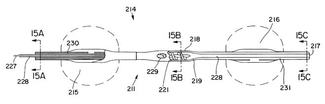
One preferred embodiment is shown at FIGS. 11-15. FIG.
11 shows a catheter 211 embodying features of the invention
disposed within a patient's aorta 210. The catheter 211 has
an elongated shaft 212 having a proximal end 213 and a distal
end 214. The balloon distal end 214 has two occlusion
balloon, proximal balloon 215 and distal balloon 216, disposed
about a portion. Proximal balloon 215 is positioned below the
patient's renal arteries 220, while distal balloon 216 is
positioned above the renal arteries 220. Proximal balloon 215
distal end is about 5 cm to about 15 cm from distal balloon
216 proximal end, preferably about 5 to about 10 cm. Both
proximal balloon 215 and distal balloon 216 are capable of
inflation to about 20 to about 30 millimeters outside
diameter. The catheter distal end 214 also includes a blood
inlet 217. Between balloon 215 and 216 is located an
archimedean screw pump 218. Screw pump 218 includes a housing
219, a rotor 221 and a seal 222. The seal 222 may or may not
let blood pass. The catheter proximal end 213 includes a
drive mechanism 223. The drive mechanism may be a DC, AC or
pneumatic motor capable of maintaining high speed, about 5000
to about 30,000 rpm, and moderate torques for periods lasting
at least 20 minutes. Also connected to the catheter proximal
end 213 are inflation sources 224 and 225 for proximal balloon
215 and distal balloon 216 respectively. The screw pump 218
may be controlled automatically by an autocontroller 226,
which may also be automated to control the inflation of the
proximal balloon 215 and the distal balloon 216.
FIG. 12 better illustrates the inner workings of catheter
211 at the catheter distal end 214. A drive shaft 227 is
located within main lumen 228. The drive shaft 227 is
connected to the drive mechanism 223 on its proximal and, and
the screw pump 218 in its distal end. The drive mechanism 223
CA 02407938 2002-11-O1
WO 01/83016 PCT/USO1/13686
- 24 -
turns the drive shaft 227, which then turns the rotor 221.
Blood enters the catheter 211 through the inlet 217 and
travels to the screw pump housing 219. The rotor 221 turns,
causing a pressure increase within the housing 219. Blood
then exits out the blood outlet 229 at a higher pressure. As
was shown in FIG. 11, the housing is located near the renal
arteries 220. Therefore, blood exits from the housing 219
through blood outlet 229 into the renal arteries 220. More
than one blood outlet may be disposed about the radial face
of the catheter shaft 212. Blood flows in total at a rate of
about 600 ml/min to about 1200 ml/min, preferably about 800
to about 1200 ml/min out the blood outlet 229 into the
abdominal aorta 210. This higher pressure blood then moves
through the renal arteries 220, which are constricted causing
ARF. In an alternative embodiment, the seal 222 may allow
some blood to pass into the main lumen 228. An additional
blood outlet then may be provided proximal to proximal balloon
215, thereby providing blood to the lower extremities. This
may also be accomplished by providing a blood pass through
lumen in addition to the lumen shown in this embodiment (not
shown). As screw pump 218 is causing high pressure blood to
exit the blood outlet 229, the proximal balloon 215 and distal
balloon 216 may be inflated. The balloons 215 and 216 may
also be inflated prior to the screw pump 218 activation. The
inflation source 224 is activated, and an inflation fluid
enters inflation lumen 230, which is in fluid communication
with proximal balloon 215. Either simultaneously or at a
desired time, inflation source 225 is activated, sending an
inflation fluid into inflation lumen 231, which is in fluid
communication with distal balloon 216. The balloons 215 and
216 inflate against the aorta 210 to a final outer diameter
indicated in phantom, thereby isolating the area surrounding
the renal arteries 220. This allows the increased pressure
caused by thepump to be most effective. Higher pressure blood
will be more likely to enter the renal arteries 220, thereby
CA 02407938 2002-11-O1
WO 01/83016 PCT/USO1/13686
- 25 -
effectively perfusing the constricted renal arteries 220. In
embodiments which do not include a blood pass through lumen
(not shown) or all blood to flow past the seal 222, the
balloons 215 and 216 may be deflated periodical 1y to allow
blood flow to the lower extremities, or occlusion may be
controlled such that some blood leaks by the balloons 215 and
216 without compromising pressure in the renal arteries 220.
The drive shaft 227 may be a flexible component to
accomplish torque transmission from the motor to the rotor and
overcome any curvature of the catheter shaft imparted by the
vasculature. The drive shaft 227 may be made of a coiled wire
or a flexible mandrel or a combination of these, possibly of
stainless steel or superelastic nitinol. The drive shaft 227
may be coated with a low friction and high temperature
resistant material such as Teflon. The engagement between the
drive mechanism 223 and the drive shaft 227 may be
accomplished by means of a threaded connection, set screw and
collar connection, or a snap fit engagement. The drive shaft
227 may be press fit, welded, threaded, or adhesive bonded to
the rotor 221.
FIG 13 is a detailed view of the screw pump rotor 221.
The rotor is a single helix rotor having a hub 232 and a blade
233. The rotor generally is about 1 cm to about 5 cm,
preferably about 2 cm to about 4 cm long. The diameter of the
rotor is about .1 inch to about .25 inch, preferably about .15
inch to about .2 inch. The distance on the hub 232 between
blade 233 turns may be uniform. In some embodiments, the
distance between blade 233 turns is not uniform. In certain
embodiments, the rotor is a single helix progressive pitch
rotor, and the kick area 234 has a greater distance on the hub
232 between blade 233 turns. FIG. 14 illustrates an
alternative embodiment of the catheter of the invention,
wherein the screw pump 218 is located distal to the distal
balloon 216. Blood outlet 229 is still located between
proximal balloon 215 and distal balloon 216.
CA 02407938 2002-11-O1
WO 01/83016 PCT/USO1/13686
- 26 -
The rotor 221 may be an overall cylindrical component
consisting of a helical blade 233 around a hub 232 designed
to transfer rotational motion of the rotor to translational
motion of the blood. 1-5 helical blade components, preferably
1- 3 helical blade components may wrap the hub 232 of the
rotor. The hub 232, will be minimized to increase the blood
volume capacity between the blades 233. A progressive pitch,
or variable, pitch blade may be used to gradually accelerate
the blood along the length of the rotor 221. The helix may
progress from a high pitch to low pitch configuration the last
of which is known as the kick of the blade 234. Maximizing
acceleration of the blood while minimizing possible cavitation
or hemolysis within the system is preferred. The rotor 221 may
be machined, injection molded, or cast as one component or
assembled from multiple parts, such as separate blade and core
components. The rotor 221 may be made of metal or plastic. The
rotor 221 will be encased within a housing designed to confine
the travel of the blood to a translational volume exchange.
The housing 219 may be cylindrical and fit closely with the
diameter of the rotor 221. The housing 219 and rotor 221 will
work together to maximize the translational motion of the
blood and control the centrifugal forces imparted on the
fluid. The housing 219 may be constructed of a metal or
plastic. The housing 219 will be a bearing surface for the
rotor blades 233 and will be required to withstand the forces
and temperatures generated by the rotor 221. It may be a
portion of the catheter shaft 212 in which the rotor 221 is
housed but not a separate component requiring connection to
the catheter shaft 212. If the housing 219 is a separate
component it may be secured to the catheter shaft 212 by heat
fusing, adhesive bonding, chemically welding, or barb fitted.
The housing 219 of the screw pump 218 will be at least as long
as the rotor 221 and may taper at either end of the rotor 221
to optimize the intake and outlet volume of the pumping area.
CA 02407938 2002-11-O1
WO 01/83016 PCT/USO1/13686
- 27 -
The centrifugal force imparted on the blood by the rotor
will help the blood progress toward the outlet do to its
placement along the outer diameter of the catheter shaft. A
backpressure will be created within the central lumen of the
catheter to prevent the flow of blood beyond the outlet. This
backpressure will be created either by a o-ring tip seal
between the central lumen ID and drive shaft or by a
pressurized fluid flow within the annular space between the
drive shaft and catheter ID. This fluid will also serve to
reduce temperatures created by the spinning components. The
fluid may be saline or dextrose and may be heparinized.
Another preferred embodiment is disclosed in FIGS. 16-19.
FIG 16 shows a catheter 311 embodying features of the
invention placed within a patient's aorta 310. The catheter
has a shaft 312, a proximal end 313 and a distal end 314.
Shaft 312 may include markers along the length to assist a
user in proper placement . (not shown) . Such markers are
especially helpful along the proximal end 313, to aid in
placement without the use of X-ray fluoroscopy guidance. The
distal end 314 includes a distal tip portion 315. Distal tip
portion 315 is placed above the patient's renal arteries 320.
The distal end 314 additionally includes an inflatable balloon
316. Inflatable balloon 316 is placed below the patient's
renal arteries 320. Inflatable balloon 316 is about 5 cm to
about 20 cm from the distal tip portion 315 , preferably about
10 cm to about 15 cm. The distal tip portion 315 includes
discharge ports 317. Discharge ports may be formed of slits.
In an alternative embodiment illustrated in FIG. 17, the
discharge ports 317 are sideholes 338, placed along a pigtail
shaped distal tip portion 339 with a tapered closed tip 340.
If the distal tip portion 315 of the catheter 311 is closed,
it may be sealed or include a sealing surface which mated with
an obturator or a stiffening mandrel. In such an even, it may
CA 02407938 2002-11-O1
WO 01/83016 _ 2 $ _ PCT/USO1/13686
become necessary to use a duck-billed valve to provide for
guidewire passage without losing the fluid seal.
The proximal end 313 is connected to a system console
318. The system console 318 includes an inflation source 321
and a drug delivery source 322. Inflation source 321 is in
fluid communication with inflation lumen 323. An inflation
fluid travels through inflation lumen 323, which is in fluid
communication with the inflatable balloon 316, and inflates
inflatable balloon 316. Drug delivery source 322 is in fluid
communication with drug delivery lumen 324. A drug may be
introduced into the drug delivery lumen 324 and travels to the
distal tip portion 315. At the distal tip portion 315, the
drug delivery lumen 324 is in fluid communication with the
discharge ports 317, thereby discharging the drug into the
patient's aorta 310. In alternative embodiments, the catheter
311 additionally includes a blood pass through lumen 325. The
blood pass through lumen 325 will have an inlet port on the
distal tip portion 315 (not shown) and an outlet situated on
the catheter proximal to the balloon (not shown) to supply
blood to the lower extremities during balloon inflation.
The systems console 318 additionally includes an
autocontrol device 319. An example of such an autocontrol
device would be a microprocessor-control module with a user
interface. FIG. 20 illustrates the benefit of including an
autocontrol device 319 in the system console 318. The balloon
316 may be inflated periodically to correspond to drug
delivery. Therefore, the drug will be directed into the
patient's renal arteries 320 because the balloon 316 has
isolated the renal arteries 320. This allows for a localized
delivery of a drug to the renal arteries 320 without having
a system-wide effect on the patient's body. A preferred drug
for this apparatus would include a drug which is a short-
acting vasodilator.
CA 02407938 2002-11-O1
WO 01/83016 PCT/USO1/13686
- 29 -
As illustrated in FIG. 20, the delivery of the drug will
by synchronized with the aortic occlusion to divert the blood
flow and infused drug to the renal arteries. The time lag
between the beginning of balloon inflation and the beginning
of drug infusion in a cycle is called T1. The lag time
between the end of drug infusion and balloon deflation is
called T2. The balloon will occlude the aorta in order to
deliver a high amount of drug to the renal arteries, and only
a minor amount to the lower extremities. The therapy will
be automated to keep the drug level in the renal arteries at
a set minimum to ensure increased renal perfusion is
sustained. The drug may be delivered in increasingly small
amounts, as well, as therapy progresses and the reduce the
patient's risk of systemic effects of too much drug.
while preferred illustrative embodiments of the invention
are described above, it will be apparent to one skilled in the
art that various changes and modifications may be made therein
without departing from the invention, and the appended claims
are intended to cover all such changes and modifications that
fall within the true spirit and scope of the invention.
Additionally, although various features of the invention are
disclosed in specific embodiments, one or more of the features
may be used and exchangeable in other embodiments disclosed
herein.