Note: Descriptions are shown in the official language in which they were submitted.
CA 02408919 2002-11-12
WO 01/91634 PCT/USO1/17009
APPARATUS AND METHOD FOR OBTAINING BLOOD FOR DIAGNOSTIC
TESTS
CROSS REFERENCES TO COPENDING APPLICATIONS
This application relates to four patent applications, each entitled METHOD
AND APPARATUS FOR OBTAINING BLOOD FOR DIAGNOSTIC TESTS, U. S.
Serial No. 08/759,698, filed December 6, 1996, U. S. Serial No. 08/982,323,
filed
December 2, 1997, U. S. Serial No. 08/982,324, filed December 2, 1997, and U.
S.
Serial No. 08/982,721, filed December 2, 1997. The specifications, drawings
and
claims of these applications are incorporated herein by reference. All of the
foregoing applications are commonly owned by the.assignee of this invention.
BACKGROUND OF THE INVENTION
1. Field of the Invention
This invention relates to a method and apparatus for obtaining samples of
blood for diagnostic purposes.
2. Discussion of the Art
The prevalence of diabetes has been increasing markedly in the world. At
this time, diagnosed diabetics represent about 3% of the population of the
United
States. It is believed that the total actual number of diabetics in the United
States is
over 16,000,000. Diabetes can lead to numerous complications, such as, for
example, retinopathy, nephropathy, and neuropathy.
The most important factor for reducing diabetes-associated complications is
the maintenance of an appropriate level of glucose in the blood stream. The
maintenance of the appropriate level of glucose in the blood stream may
prevent
and even reverse many of the effects of diabetes.
Glucose monitoring devices of the prior art have operated on the principle of
taking blood from an individual by a variety of methods, such as by needle or
lancet.
An individual then coats a paper strip carrying chemistry with the blood, and
finally
1
CA 02408919 2002-11-12
WO 01/91634 PCT/USO1/17009
inserts the blood-coated strip into a blood glucose meter for measurement of
glucose concentration by determination of change in reflectance.
The medical apparatus of the prior art for monitoring the level of glucose in
the blood stream required that an individual have separately available a
needle or
lancet for collecting blood from the individual, strips carrying blood
chemistry for
creating a chemical reaction with respect to the glucose in the blood stream
and
changing color, and a blood glucose meter for reading the change in color
indicating
the level of glucose in the blood stream. The level of blood glucose, when
measured by a glucose meter, is read from a strip carrying the blood chemistry
through the well-known process of reading reflectometers for glucose
oxidation.
Generally lancets comprise a blade and a pressable end opposed thereto,
with the blade having an acute end capable of being thrust into skin of a
human. By
striking the pressable portion, the acute end of the blade will pierce the
skin, for
example, of the finger. The finger lancet is primarily used to obtain small
volumes of
blood, i. e., less than 1 mL. Diabetics use the finger lancet to obtain
volumes of
blood less than 25 pL for analysis for glucose. A small amount of blood for
the
blood test will ooze out of the skin. There are many small blood vessels in
each
finger so that a finger can be squeezed to cause a larger drop of blood to
ooze. The
finger is one of the most sensitive parts of the body; accordingly, the finger
lancet
leads to even more pain than what would be experienced by collecting blood via
lancet at a different body site. The finger lancet presents another problem
because
of the limited area available on the fingers for lancing. Because it is
recommended
that diabetics monitor their blood glucose levels four to six times per day,
the limited
area on the fingers calls for repeated lancing of areas that are already sore.
Because fingers are sensitive to pain, it is a recent tendency that the arm is
subjected to blood sampling. See, for example, U. S. Patent No. 4,653,513. The
device of U. S. Patent No. 4,653,513 comprises a cylindrical housing and a
lancet
support, which has a gasket or flexible portion slidably accommodated in the
housing. Springs will retract the lancet support to thereby reduce air
pressure in the
housing so that it sucks a blood sample, automatically and immediately after a
lancet pierces the skin. See also U. S. Patent No. 5,320,607, which discloses
a
device comprising a sealed vacuum chamber in a state of preexisting reduced
pressure, a support member for the sealed vacuum chamber, the support member
defining a suction portion adjacent the sealed vacuum chamber, the suction
portion,
in cooperation with the sealed vacuum chamber, exposing an area of the skin of
a
patient to a reduced pressure state when the device is actuated, and means
2
CA 02408919 2002-11-12
WO 01/91634 PCT/USO1/17009
arranged within the suction portion for slightly rupturing a portion of the
area of skin
of the patient exposed to the reduced pressure state.
Because the blood volume requirements for a standard glucose test strip are
typically 3 pL or more, an area of the body that can generate that much blood
from a
lancet wound must be used. It is believed, however, that improvements in
glucose
test strip technology will reduce the volume of blood needed to 1 to 3 pL.
Because
the finger is well supplied with blood and the amount of blood can be
increased by
squeezing the finger after lancing, the finger is the currently preferred body
site for
lancing, even though lancing of the finger is painful.
A less painful technique for obtaining body fluids is described in U. S.
Serial
No. 08/982,721, filed December 2, 1997. This application discloses an
apparatus
for obtaining blood for diagnostic tests. The apparatus comprises a housing
having
a sealable chamber located therein and a sealable opening in fluid
communication
with the sealable chamber, a power source, a vacuum pump operably connected to
the power source, the vacuum pump in communication with the sealable chamber,
a
lancing assembly positioned within the sealable chamber, and a fluid collector
positioned in the sealable chamber, the fluid collector in fluid communication
with
the sealable opening. It would be desirable to improve that apparatus in order
to
ensure that the fluid collector is properly positioned in the apparatus during
the
lancing and fluid collecting steps.
SUMMARY OF THE INVENTION
This invention provides a method and apparatus for collecting a sample of
blood from a patient for subsequent diagnostic tests, e.g., glucose
monitoring.
In one aspect of the invention, an apparatus for collecting a sample of body
fluid, e. g., blood, for analysis in a diagnostic test is provided. In a
preferred
embodiment, the apparatus comprises:
(a) a housing having a sealable chamber located therein and a sealable
opening in fluid communication with the sealable chamber;
(b) a vacuum pump in communication with the sealable chamber;
(c) a device for forming an unobstructed opening in an area of skin from
which a sample is to be collected, preferably a lancing assembly, the device
positioned within the sealable chamber;
3
CA 02408919 2002-11-12
WO 01/91634 PCT/USO1/17009
(d) a movable support for supporting and positioning a port for a fluid
collector in the sealable chamber, the movable support capable of moving the
port
within the sealable chamber between a first position and a second position;
and
(e) a stop for aligning the fluid collector.
In more preferred embodiments, the apparatus further comprises a power source,
and the vacuum pump is operably connected to the power source. The stop aligns
the fluid collector so that the fluid collector is capable of being properly
positioned in
the apparatus during the lancing and fluid collecting steps.
The fluid collector is preferably a test strip that contains at least one
chemical
reagent for conducting a diagnostic test, e. g., a test for determining blood
glucose
level. Typically the test strip has an opening formed therein, which opening
is
capable of being aligned with the sealable opening of the housing. A preferred
device for forming an unobstructed opening in the area of the skin from which
the
sample of blood is to be collected is a lancing assembly, which comprises a
lancet
for forming an opening in the skin. Alternatively, the unobstructed opening in
the
skin can be formed by a laser or by a fluid jet. In the case of a lancing
assembly,
when the lancing assembly is triggered, the lancet of the lancing assembly
passes
through the opening of the test strip and the sealable opening of the housing
to form
~ an opening in the skin of the patient. The sample of body fluid, e. g.,
blood, is
obtained from the opening formed in the skin of the patient. The opening of
the test
strip should be properly aligned with the sealable opening of the housing
before the
lancing assembly is triggered, because misalignment of these openings may
result
in one or more of the following undesirable occurrences: (1 ) an unsuccessful
assay;
(2) a longer period of time required to collect an adequate amount of sample;
(3)
extensive contamination of the apparatus by the sample. The movable support
(d)
and the stop (e) are designed to operate in concert to ensure that the opening
of the
test strip and the sealable opening of the housing are properly aligned. By
designing the movable support to move the fluid collector port from a first
position to
a second position, the opening in the test strip can be properly aligned with
the
sealable opening in the housing, thereby ensuring that the assay can be
conducted
successfully.
The vacuum pump requires a power source. The power source can be
disposed within the housing. Alternatively, the power source can be external
to the
housing. The vacuum pump can serve the dual purposes of (1) stretching the
skin
and (2) enhancing the collection of the sample of blood from the unobstructed
4
CA 02408919 2002-11-12
WO 01/91634 PCT/USO1/17009
opening in the skin. Preferably, the vacuum pump can serve the triple purposes
of
(1 ) stretching the skin, (2) increasing the availability of blood to the area
of the skin
from which the sample is to be collected, and (3) enhancing the collection of
the
sample of blood from the unobstructed opening in the skin.
The housing comprises a body and a cover. The sealable chamber is located
within the cover. The cover is separated from the body so that the tesfi strip
can be
inserted into a test strip port located in the volume that is to be enclosed
by the
cover when the cover is closed against the body of the housing. After the test
strip
is inserted in the test port, the cover is closed against the body to form a
chamber
that will be sealed when the sealable opening of the housing is placed in
contact
with the skin of a patient. Preferably, the body of the housing contains
electronics
having programmed instructions to control the vacuum pump to maintain the
desired
level of vacuum for the method of this invention.
The apparatus preferably contains valves, such as, for example, solenoid
valves, for triggering the lancet of the lancing assembly and releasing the
vacuum at
the conclusion of the blood collection procedure. The apparatus can optionally
contain a heating element to increase the availability of blood to the area of
the skin
from which the sample is to be collected.
In another aspect of the invention, a method for collecting a sample of
body fluid, e. g., blood, for analysis in a diagnostic test is provided. In
general, the
method comprises the steps of:
(a) inserting a fluid collector into the port;
(b) aligning the fluid collector latitudinally and longitudinally so that an
opening in the fluid collector is aligned with the sealable opening of the
housing of
the apparatus;
(c) placing the sealable opening of the housing against the skin of the
patient;
(d) forming an unobstructed opening in the area of the skin from which the
sample of blood is to be collected;
(e) aligning the fluid collector longitudinally so that an opening in the
fluid
collector is aligned with the sealable opening of the housing of the
apparatus; and
(f) collecting the sample of blood from the unobstructed opening in the
skin onto the fluid collector, with the aid of vacuum and stretching of the
skin.
5
CA 02408919 2002-11-12
WO 01/91634 PCT/USO1/17009
In order to insert a fluid collector into the support, the cover is separated
from
the body of the housing; then the fluid collector, e. g., a test strip, is
inserted into the
port for the fluid collector; and then the cover is closed against the body of
the
housing. The movable support is~actuated to cause the port to move from a
first
position to a second position in order to align an opening in the fluid
collector with
the sealable opening of the housing of the apparatus.
In a preferred embodiment of the method, step (d) is preceded by the step of
increasing the availability of blood in the portion of the skin from which the
sample is
to be collected. In this preferred embodiment, the availability of blood in
the portion
of the skin from which the sample is to be collected can be increased by means
of a
vacuum, which is applied to the surface of the skin in the vicinity of the
opening prior
to forming the opening in the skin. The vacuum causes the portion of the skin
in the
vicinity of the blood collection site to become engorged with blood. The
vacuum
also causes the portion of the skin in the vicinity of the blood collection
site to
become stretched.
An opening in this stretched portion of skin can be formed with a cutting or
puncturing device, e.g., a lancet, or other device capable of forming an
opening in
the skin, e. g., a laser or a fluid jet. If a cutting or puncturing device is
used to form
the opening, it must be retracted from the opening prior to the step of
collecting the
sample of blood from the opening. This retraction will allow the unrestricted
flow of
blood through the opening. After the opening is formed, the movable support is
moved from the first position to the second position in order to align the
opening in
the fluid collector with the sealable opening of the housing of the apparatus.
Then a
vacuum can be used to aid in collecting the sample of blood from the opening
in the
skin.
The method and apparatus of this invention provide several advantages over
the methods and apparatus of the prior art. First, a sufficient amount of
blood can
be collected from parts of the body, other than the finger, for conducting
glucose
monitoring tests. Second, by rendering other parts of the body suitable for
collecting
blood, the use of a painful finger lance can be avoided. Third, by increasing
the
availability of blood at the site where the blood is to be collected, the
period of time
required for collecting the sample can be reduced. Fourth, by improving the
registration of the opening in the fluid collector (e. g., test strip) with
the sealable
opening in the housing, bofih the likelihood that the lancet will strike a
solid portion of
the fluid collector during the lancing step and the likelihood that an
insufficient
amount of sample will be collected will be reduced. Because of these
advantages,
6
CA 02408919 2002-11-12
WO 01/91634 PCT/USO1/17009
the diabetic patient is more likely to monitor glucose levels in the blood at
the
intervals prescribed by his doctor.
BRIEF DESCRIPTION OF THE DRAWINGS
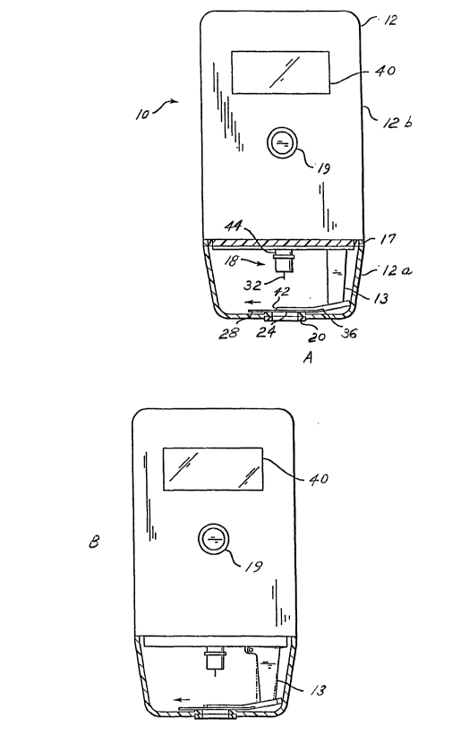
FIGS. 1A and 1 B depict partial cross-sectional views of an embodiment of the
apparatus of this invention. In FIGS. 1A and 1 B, the cover of the housing of
the
apparatus is closed against the body of the housing of the apparatus.
FIG. 2A is a perspective view of the alignment mechanism assembly of this
invention. In this figure, the cover is in its open position.
FlG. 2B is an exploded perspective view showing the components of the
alignment mechanism assembly of this invention.
FIG. 3A is a top view of the cover of the housing of the apparatus of this
invention. FIG. 3B is a side elevation view of the cover of the housing of
this
invention. FIG. 3C is a side elevation view, in cross section, taken along
line C-C of
FIG. 3A, of the alignment mechanism assembly of this invention. In FIG. 3C,
the
cover is in its closed position.
FIG. 4 is a top view of a fluid collector suitable for use in this invention.
FIG. 5A is the same view as FIG. 3C, but indicating the area to be enlarged in
FIGS. 5B, 5C, and 5D. FIGS. 5B, 5C, and 5D are side elevation views, greatly
enlarged, in cross section, of the switch that indicates when the fluid
collector is in
position for an assay. FIG. 5A shows the swifich in the OPEN position, i. e.,
the
assay is not yet ready to be run. FIG. 5B shows the switch in the CLOSED
position,
but the fluid collector is not in proper register. FIG. 5C shows the switch in
the
CLOSED position, and the fluid collector is in proper register.
FIG. 6A is a perspective view of the interior of the cover. FIG. 6B is the
same
view as FIG. 3A. FIG. 6C is a front elevation view, in cross section, takes
along line
C-C of FIG. 6B, of the cover and the alignment mechanism assembly of this
invention. FIG. 6D is a perspective view of the port shroud and the test strip
port of
the alignment mechanism assembly of this invention.
FIGS. 7A, 7B, and 7C are side elevation views, in cross section, of the fluid
collector and alignment mechanism assembly showing the sequence for properly
aligning the fluid collector for an assay. FIG. 7A shows the fluid collector
and the
alignment mechanism assembly prior to the registration procedure. FIG. 7B
shows
the fluid collector and the alignment mechanism assembly during the
registration
CA 02408919 2002-11-12
WO 01/91634 PCT/USO1/17009
procedure. FIG. 7C shows the fluid collector and the alignrrient mechanism
assembly after the registration procedure and prior to the lancing procedure.
The
components of the alignment mechanism assembly in FIGS. 7B and 7C are
identical
to those of the alignment mechanism assembly in FIG. 7A. Therefore, reference
numerals that are not relevant to the registration procedure are not restated
in FIGS.
7B and 7C.
FIGS. 8A, 8B, and 8C are side elevation views, in cross section, of the fluid
collector and alignment mechanism assembly showing the sequence for operating
the lancing assembly for collecting a sample of blood for an assay. FIG. 8A
shows
the lancing assembly prior to the triggering thereof. FIG. 8B shows the
lancing
assembly during the lancing procedure. FIG. 8C shows the lancing assembly
after
the lancet has been retracted from skin of the test subject. The components of
the
alignment mechanism assembly in FIGS. 8B and 8C are identical to those of the
alignment mechanism assembly in FIG. 8A. Therefore, reference numerals that
are
not relevant to the lancing procedure are not restated in FIGS. 8B and 8C.
FIGS. 9A and 9B are side elevation views, in cross section, of the sample
collection procedure. FIG. 9A shows the sample collection procedure prior to
the
movement of the alignment mechanism assembly. but subsequent to the formation
of the opening in the skin of the test subject. FIG. 9B shows the sample
collection
procedure subsequent to the movement of the alignment mechanism assembly
toward the sample. FIG. 9C is a top view of the blood collection area of the
apparatus taken along line C-C of FIG. 9A. FIG. 9D is a top view of the blood
collection area of the apparatus taken along line D-D of FIG. 9B. The
components
of the alignment mechanism assembly in FIG. 9B are identical to those of the
alignment mechanism assembly in FIG. 9A. Therefore, reference numerals that
are
not relevant to the sample collection procedure are not restated in FIG. 9B.
FIG. 10 is a side elevation view, in cross section, of a lancing assembly
installed in an embodiment of an apparatus suitable for use in this invention.
This
figure does not show how the diaphragm of the alignment mechanism assembly is
connected to the pump.
DETAILED DESCRIPTION
As used herein, the terms "register", "registration", and the like refer to a
process resulting in correct alignment or positioning. The term "latitudinal"
refers to
s
CA 02408919 2002-11-12
WO 01/91634 PCT/USO1/17009
the direction running perpendicular to the flow of fluid in the fluid
collector (e. g., test
strip). The term "longitudinal" refers to the direction running parallel to
the flow of
fluid in the fluid collector (e. g., test strip). The term "sample" means a
specimen of
body fluid. In this invention, the body fluid described is blood. However, it
is within
the scope of this invention to obtain samples of other types of specimens of
body
fluid.
The preferred embodiments of the apparatus of this invention utilize the
following components to improve the function of obtaining a sample of blood
for
carrying out a diagnostic test, e. g., determining blood glucose level:
(a) a housing having a sealable chamber located therein and a sealable
opening in fluid communication with the sealable chamber;
(b) a vacuum pump in communication with the sealable chamber;
(c) a device for forming an unobstructed opening in an area of skin from
which a sample is to be collected, preferably a lancing assembly, the device
positioned within the sealable chamber;
(d) a movable support for supporting and positioning a port for a fluid
collector in the sealable chamber, the movable support capable of moving the
port
within the sealable chamber between a first position and a second position;
and
(e) a stop for aligning the fluid collector.
The components of the apparatus other than the movable support (d) and the
stop (e) are described in detail in U. S. Serial No. 08/759,698, filed
December 6,
1996, U. S. Serial No. 08/982,323, filed December 2, 1997, U. S. Serial No.
08/982,324, filed December 2, 1997, and U. S. Serial No. 08/982,721, filed
December 2, 1997, and U. S. Patent No. 6,027,459, all of which are
incorporated
herein by reference. When relevant, portions of the foregoing applications and
patent will be described in detail herein in order to more clearly describe
the
components involved in the alignment of the opening of the fluid collector
with the
sealable opening in the housing.
The apparatus of this invention is designed to perform an assay, such as, for
example, an assay to determine blood glucose level, by means of a simple
procedure involving a minimum of manipulation. The apparatus forms an opening
in
the skin of the patient, collects the sample, such as, for example, a sample
of blood,
and measures and reports the desired information relating to the sample. All
of the
9
CA 02408919 2002-11-12
WO 01/91634 PCT/USO1/17009
foregoing steps are performed by means of a single apparatus, with all the
procedural steps taking place within the apparatus.
The accuracy and reliability of the apparatus and the method require that the
fluid collector, hereinafter alternatively referred to as a "test strip", be
precisely and
correctly aligned within the apparatus. Misalignment of the test strip within
the
apparatus can possibly result in an unsuccessful formation of an opening in
the skin
of the patient because of the lancet's hitting the test strip. Moreover,
misalignment
of the test strip within the apparatus can result in poor sample collection
because
the sample collection area of the test strip is not aligned with the site of
the opening
in the skin of the patient. Either type of misalignment can result in an
unsuccessful
assay (i. e., no results or erroneous results), a longer period of time
required to
collect an adequate amount of sample, or extensive contamination of the
apparatus
by the sample. It is possible to ensure precise and correct alignment of the
test strip
within the apparatus so long as the following procedures are carried out:
(1) manufacture the test strip to proper dimensional specifications;
(2) properly align the properly manufactured test strip in a latitudinal
direction; and
(3) properly align the properly manufactured test strip in a longitudinal
direction.
This invention is concerned with proper alignment of a properly manufactured
test
strip both in a latitudinal direction and in a longitudinal direction.
The dimensional tolerances with respect to the length and width of a test
strip, and the position and diameter of the opening in the test strip, as well
as the
sample collection area, are closely controlled during the fabrication process
in which
the test strips are manufactured. The test strips are designed to work in
concert
with the alignment mechanism assembly to provide an accurate and reliable
assay.
Referring now to FIGS. 1A and 1 B, which schematically depict one
embodiment of the present invention, the apparatus 10 comprises a housing 12
having a cover 12a (shown in the closed position in FIGS. 1A and 1 B). The
cover
12a is attached to the body 12b of the housing 12 by an attachment in the form
of a
hinge (not shown in FIGS. 1A and 1 B but shown in FIGS. 3, 7A, 8A, and 9A as
reference numeral 15). Alternatively, the cover 12a may be attached to the
body
12b by frictional engagement, a detent (not shown), or any combination of a
hinge,
frictional engagement, and a detent. When a hinge is used, it may optionally
be
CA 02408919 2002-11-12
WO 01/91634 PCT/USO1/17009
spring biased to retain the cover 12a in the open or closed position. A detent
(not
shown) may be provided on the cover 12a to engage with a protrusion (not
shown)
on the body 12b, or vice versa, to maintain the cover 12a in the open or
closed
position when desired. Although a hinge (not shown in FIGS. 1A and 1 B) is
utilized
in the embodiment shown in FIGS. 1A and 1 B, any other attachment or
combination
of attachments that allows the cover 12a to attach to the body 12b and
alternate
between an open and closed.position is acceptable. A gasket or other seal
arrangement 17 is provided to seal the housing 12 when the cover 12a is
closed.
Additionally, a latch mechanism may be included to prevent accidental opening
of
the cover 12a when the apparatus 10 is in use. Typically, the latch mechanism
would provide locking engagement of the cover 12a with the body 12b.
Disposed within the housing 12 are a vacuum pump (not shown), a lancet
assembly 18 generally comprising a molded plastic piece 44 to which a lancet
32 is
affixed, a lancing assembly (not shown) into which the lancet assembly 18 is
inserted, a battery (not shown), and electronics (not shown) for purposes
described
hereinafter. A switch 19 is provided to activate the electronics, which may
take the
form as shown in F1G. 3 of U. S. Serial No. 08/982,721, filed December 2,
1997,
incorporated herein by reference. The vacuum pump communicates by an
evacuation tube (not shown) with the volume enclosed by the cover 12a when the
cover 12a is in the closed position. Optionally a check valve (not shown) may
be
placed in the evacuation tube between the vacuum pump and the volume enclosed
by the cover 12a when the cover 12a is in the closed position.
During the process of obtaining the sample, the cover 12a is closed to form a
seal. The seal should be sufficiently tight so that a sufficient vacuum can be
obtained by removing air from the volume enclosed by the cover 12a when the
cover 12a is in the closed position.
The area of the cover 12a of the housing 12 that is to contact the skin is
equipped with a seal 20. The seal 20 surrounds a sealable opening 24 in the
cover
12a as disclosed in U. S. Serial No. 08/982,721, filed December 2, 1997,
incorporated herein by reference. The sealable opening 24 may be round, oval,
rectangular or any other shape. The sealable opening 24 in the cover 12a
allows
communication between the surface of the skin and a blood collection chamber
adjacent to a fluid collector, shown here in the form of a glucose detector
28, which
may take the shape and form of a test strip. Preferably, the glucose detector
28
contains at least one opening (not shown in FIGS. 1A and 1 B) approximately
equidistant from the elongated edges of the middle of glucose detector 28 for
the
11
CA 02408919 2002-11-12
WO 01/91634 PCT/USO1/17009
lancet 32 to pass through, as disclosed in U. S. Serial No. 08/982,721, filed
December 2, 1997, incorporated herein by reference. In this embodiment, the
aforementioned at least one opening in the glucose detector 28 is preferably
in
alignment with sealable opening 24 and lancet 32 during the lancing step. The
opening in the glucose detector 28 may be covered with a mesh. Alternatively,
the
glucose detector 28 used in the embodiment shown in FIGS. 1A and 1 B may
contain a semi-circular notch .(not shown) in the region of the glucose
detector 28
that comes into contact with the blood, as disclosed in U. S. Serial No.
08/982,323,
filed December 2, 1997, incorporated herein by reference. The semi-circular
notch
may be covered with a mesh. Fluid collectors, such as, for example, glucose
detectors, suitable for use in this invention include, but are not limited to,
biosensors and reflectance strips. If a biosensor is used, it is preferred
that the
apparatus 10 include a meter to measure electrical properties, e. g., current,
arising
from the interaction of a sample with the reagents of the biosensor. If a
reflectance
strip is used, it is preferred that the apparatus 10 include a meter, e. g.,
reflectometer, to measure optical properties, e. g., reflectance, arising from
the
interaction'of a sample with the reagents of the reflectance strip.
When in use, the apparatus 10 is positioned so that the lancing assembly is
placed over the region on the surface of the skin from which the fluid sample
is to be
obtained such that the lancing assembly is approximately perpendicular to the
surface of the skin. Prior to actuating the apparatus 10, a fluid collector,
e, g., a
glucose detector in the form of a test strip, is inserted into a slot 36 of a
movable
projection 13 of the body 12b of the housing 12. The glucose detector 28
contains
one or more electrical contacts (not shown) on the end inserted into the slot
36,
which contacts engage one or more electrical contacts (not shown) positioned
within
the slot 36. In order to obtain the sample of blood, the cover 12a of the
housing 12
is placed against the skin, whereby the seal 20 surrounding the sealable
opening 24
allows a satisfactory vacuum to be effected. The switch 19 is actuated,
typically by
being pressed, thereby activating the electronics, described in FIG. 3 of U.
S. Serial
No. 08/982,721, filed December 2, 1997 and discussed above, which starts the
vacuum pump. The action of the vacuum pump withdraws air from the volume
enclosed by the cover .12a when the cover 12a is in the closed position and
causes
the skin circumscribed by the seal 20 to be drawn toward the sealable opening
24.
This results in the skin becoming engorged with blood. Engorgement of the skin
with blood is accompanied by a stretching of and rising up of the skin to the
sealable
opening 24 in the cover 12a.
12
CA 02408919 2002-11-12
WO 01/91634 PCT/USO1/17009
After an appropriate period of time, which is typically pre-set by the
programmed electronics, the lancing assembly is triggered, thereby causing the
lancet 32 to penetrate the skin that has been pulled up into the sealable
opening 24
of the cover 12a. The lancet 32 is preferably triggered automatically by
activation of
a solenoid valve (not shown) that causes a vacuum-actuated piston (not shown)
to
trigger the lancet 32, as disclosed in U. S. Patent No. 6,027,459,
incorporated
herein by reference. .
The description to this point has dealt with apparatus and methods described
previously in U. S. Serial No. 08!982,721, filed December 2, 1997 and shown in
FIGS. 1A and 1 B. The apparatus particular to this invention will now be
described in
greater detail. Referring now to FIGS. 2A, 2B, 3A, 3B, 3C, 5A, 5B, 5C, 5D, 6A,
6B,
6C, and 6D, the alignment mechanism assembly 50 comprises the cover 12a, a
port
shroud 54, a skirt 56, a movable support 58, a vacuum plate 60, a diaphragm
62, a
diaphragm plate 63, and a test strip port 64. The movable support 58 is
analogous
to the movable projection 13 shown in FIGS. 1A and 1 B. The test strip port
64, into
which a test strip 70 for an assay is inserted, is inserted into an opening 65
in the
port shroud 54. The port shroud 54 has a slot 54a formed therein to
accommodate
any excess fluid from the sample, so that excess fluid does not flow past the
slot
54a and consequently contaminate the critical parts of the test strip port 64.
The
test strip 70 is analogous to the glucose detector 28 shown in FIGS. 1A and 1
B.
The skirt 56 functions as a means for positioning the vacuum plate 60, the
movable
support 58, and the diaphragm 62 and as a means for providing a base for the
movable support 58 and the vacuum plate 60. A first resilient biasing element
66, e.
g., a spring, connects the test strip port 64 to the vacuum plate 60. A second
resilient biasing element 67, e. g., a spring, connects the movable support 58
to the
vacuum plate 60. The first resilient biasing element 66 operates to bias one
end
54b of the port shroud 54 upwards, so that the user of the apparatus can
easily
insert a test strip 70 into the test strip port 64, which is carried in the
port shroud 54.
The upper surface of the port shroud 54 and the upper surface of the test
strip port
64, which is held in the port shroud 54, are induced to be maintained in a
position
parallel to both the upper surface of the vacuum plate 60 and the top interior
surface
71 of the cover 12a when the cover 12a is in the closed position. The
operation of
the second resilient biasing element 67 is described below, as it relates to
the steps
involved in moving the movable support 58. The skirt 56 has an opening 72
formed
therein through which the lancet 74 passes during the lancing step of the
method of
this invention. The lancet 74 is analogous to the lancet 32 shown in FIGS. 1A
and
13
CA 02408919 2002-11-12
WO 01/91634 PCT/USO1/17009
1 B. The vacuum plate 60 functions to ensure that a vacuum is maintained in
the
chamber enclosed by the cover 12a and the skirt 56. The diaphragm 62 functions
as the agent that causes the movable support 58 to move sufficiently to move
test
strip port 64 sufficiently to properly align the test strip with the sealable
opening 24
in the cover 12a of the housing 12. The ultimate function of the movable
support 58
is to move the test strip port 64 sufficiently to properly align an opening
70a in the
tesfi strip 70 with the sealable.opening 24 in the cover 12a of the housing
12. In
order to perform this function, the movable support 58 also supports the port
shroud
54, which contains the test strip port 64.
In the embodiment shown in FIGS. 2B, 3C, 5A-5D, 7A-7C, 8A-8C, 9A-9B, the
movable support 58 is an L-shaped structure having a leg 58a and a base 58b. A
pivot 75 is included at the junction of the leg 58a and the base 58b. The
pivot 75
can be of simple construction, such as, for example, at least one shaft
supported in
at least one bearing. The movable support 58 is capable of rotation about this
pivot
75 at an angle of rotafiion sufficient to move the test strip port 64 a
sufficient lateral
distance toward the sealable opening 24 in the cover 12a of the housing 12,
whereby an opening 70a in the test strip 70 is placed in register with the
sealable
opening 24 in the cover 12a of the housing 12. The base 58b is of sufficient
length
that the end thereof located distally of the pivot 75 can be moved vertically
by
~ expansion of the diaphragm 62 when the diaphragm 62 is subjected to a
pressure
gradient. The force needed to actuate the diaphragm 62 is furnished by the
ambient
pressure, which is the pressure within the body 12b of the housing 12. The leg
58a
must of sufficient length to position the port shroud 54 sufficiently close to
the top
interior surface 71 of the cover 12a (when the cover 12a is closed), so that
when a
test strip 70 is inserted in the test strip port 64; the test strip 70 will be
sufficiently
close to the sealable opening 24 in the cover 12a of the housing 12 so that a
sample
of blood or other body fluid for an assay can be collected successfully. The
movable support 58 is connected to the port shroud 54 by means of a pivot 76.
Like
the pivot 75, the pivot 76 can be of simple construction, such as, for
example, at
least one shaft supported in at least one bearing. This type of connection is
used so
that when (1) the diaphragm 62 expands, (2) the base 58b is raised, and (3)
the leg
58a is tilted at a sufficient angle, the pivot 76 will allow the test strip
port 64 of the
port shroud 54 to move from a first position to a second position, at which
second
position the end 70b of the test strip 70 located distally from the end 70c of
the test
strip 70 inserted in the test strip port 64 will abut a test strip stop 76
located on the
cover 12a of the housing 12, thereby placing the opening 70a of the test strip
70 in
14
CA 02408919 2002-11-12
WO 01/91634 PCT/USO1/17009
register with the sealable opening 24 in the cover 12a of the housing 12. The
second biasing element 67, which was disclosed earlier, operates to urge the
leg
58a of the movable support 58 to its upright position when the diaphragm 62 is
not
being subjected to a pressure gradient.
Referring specifically now to FIGS. 5A, 5B, 5C, and 5D, the test strip port 64
comprises a "no touch" switch 80 and a biasing bar 82. When a test strip 70 is
inserted into the test strip port 64, the force of the biasing bar 82 acting
upon the
upper surface of the test strip 70 causes the "no touch" switch 80 to close.
When
the "no touch" switch 80 closes, a signal indicates that the lancing phase of
the
assay can begin.
Referring specifically now to FIGS. 6A, 6B, 6C, and 6D, registration of the
test strip 70 in the apparatus in the latitudinal direction is accomplished as
the cover
12a of the housing 12 is closed. As the cover 12a of the housing 12 is closed,
the
latitudinal registration features 84 disposed on the cover 12a interlock with
latitudinal
registration features 86 disposed on the port shroud 54. This interlocking
action
precisely positions the test strip 70 in the latitudinal direction.
All of the parts of the alignment mechanism are preferably made of molded
plastic, with the exception of the diaphragm 62, which is preferably made of a
flexible elastomeric material, and the biasing elements 66 and 67, which are
preferably helical springs formed from metal. Of course, the electrical
contacts in
the test strip port 64 and the electrical contacts of the "no touch" switch 80
are
preferably made of electrically conductive metal. The biasing bar 82 is
preferably
made of metal.
OPERATION
Operation of the apparatus 10 will now be described. FIG. 4 illustrates a test
strip suitable for use in this invention. FIGS. 5A, 5B, 5C, and 5D illustrate
the
operation of the switch that indicates when a test strip 70 is inserted in the
test strip
port 64. FIGS. 7A, 7B, and 7C illustrate how the apparatus aligns the opening
70a
of the test strip 70 with the sealable opening 24 of the cover 12a of the
housing 12
prior to lancing or prior to collecting a sample of blood. FIGS. 8A, 8B, and
8C
illustrate the key steps of the lancing procedure. FIGS. 9A, 9B, 9C, and 9D
illustrate
alignment of the opening 70a of the test strip 70 with the sealable opening 24
of the
cover 12a of the housing 12 during the blood collection step.
CA 02408919 2002-11-12
WO 01/91634 PCT/USO1/17009
The test strip 70 is specifically designed to fit in to the test strip port
64. The
test strip port 64 includes a "no touch" switch 80. This "no touch" switch 80
'
indicates when a test strip 70 has been inserted into the test strip port 64.
As a test
strip 70 is being inserted into the test strip port 64, the test strip 70
deflects fhe "no
touch" contact 80a until the "no touch" contact 80a makes electrical contact
with a
"pull up" contact 80b, thereby causing the apparatus to be electrically
actuated, so
that an assay can be performed. Downward force provided by the biasing bar 82
aids the test strip 70 in bringing about contact of the "no touch" contact 80a
and the
"pull up" contact 80b. See FIGS. 5A, 5B, 5C, and 5D. However, even though the
"no touch" contact 80a makes electrical contact with the "pull up" contact
80b, the
user cannot be certain that the test strip 70 has been properly positioned in
the test
strip port 64. For example, the end 70c of the test strip 70 that contacts the
"no
touch" switch 80 is designed so that it abuts against the end 64a of the test
strip port
64. In a worst case situation, this end 70c of the test strip 70 could be a
small
distance, e. g., 0.045 inch from the end 64a of the test strip port 64. This
failure of
the end 70c of the test strip 70 to abut the end 64a of the test strip port 64
directly
translates to a misalignment between the opening 70a in the test strip 70 and
the
sealable opening 24 in the cover 12a. If this misalignment is not corrected,
the
lancet 74, when triggered, could possibly strike a solid portion of the test
strip 70
20. and fail to pass through the opening 70a in the test strip 70, and,
consequently (1)
fail to pass through the sealable opening 24 in the cover 12a, (2) fail to
lance the
patient, and (3) fail to allow access to a blood sample. (n order to correct
for any
possibility of misalignment of the test strip 70 in the apparatus, the
apparatus
undergoes the following procedure.
After the test strip 70 is inserted into the test strip port 64, the cover 12a
of
the housing 12 is closed against the body 12b of the housing 12. A pressure
gradient is applied to the diaphragm 62. The pressure gradient causes the
diaphragm 62, which is preferably made of a flexible elastomeric material, to
expand. This expansion of the diaphragm 62 causes the base 58b of the movable
support 58 to move upwardly, thereby causing the movable support 58 to tilt
forward, i. e., toward the sealable opening 24 in the cover 12a, via the
pivofi 75. As
the movable support 58 tilts forward, the pivot 76 allows the test strip port
64 to
move forward, i. e., toward the sealable opening 24 in the cover 12a. By this
movement, the end 70b of the test strip 70 is caused to abut against a test
strip stop
77, which projects from the cover 12a of the housing 12. Because the force
supplied by the diaphragm 62 exceeds the static friction force existing
between the
16
CA 02408919 2002-11-12
WO 01/91634 PCT/USO1/17009
test strip 70 and the test strip port 64, the test strip 70 is pushed backward
into the
test strip port 64 until the end 70c of the test strip 70 abuts the end 64a of
the test
strip port 64. At this point, the test strip 70 is fully seated in the test
strip port 64,
and, consequently, is properly aligned in the apparatus 10. In other words,
the
opening 70a in the test strip 70 is properly aligned with the sealable opening
24 in
the cover 12a of the housing 12. Then the pressure gradient is removed, the
diaphragm 62 deflates, and the movable support 58 is reset. The second biasing
element 67 operates to urge the leg 58a of the movable support 58 to its
upright
position when the diaphragm 62 is not being subjected to a pressure gradient.
The apparatus is now ready to commence the lancing phase of the assay. See
FIGS. 7A, 7B, and 7C.
In order for the diaphragm 62 to be actuated to initiate the operation of the
alignment mechanism assembly 50, the pressure to which the diaphragm 62 is
subjected must be varied at the appropriate times to bring about the pressure
gradient previously mentioned. Referring now to FIGS. 7A through 9D,
inclusive,
during the time the vacuum pump (not shown) of the apparatus 10 is in
operation,
the pressure within the volume enclosed by the vacuum plate 60 and the cover
12a
of the housing 12 is below the ambient pressure. It should be mentioned at
this
point that in order to maintain the pressure within the volume enclosed by the
vacuum plate 60 and the cover 12a of the housing 12 below the ambient pressure
while the vacuum pump is in operation, the sealable opening 24 in the cover
12a of
the housing 12 must be sealed. The seal is formed by a seal 78 placed against
the
surface of the skin, designated herein by the letter "S". Additional details
relating to
the seal 78 will be described below. In order to simplify the explanation of
the steps
involved in varying the pressure for operating the diaphragm 62, it will be
assumed
that when the vacuum pump is operating, the pressure within the volume
enclosed
by the vacuum plate'60 and the cover 12a of the housing 12 is 6.7 psi, while
the
ambient pressure is 14.7 psi. The pressure within the body 12b of the housing
12 is
the ambient pressure. These particular values of pressure are not required,
but in
all cases the pressure within the body 12b of the housing 12 exceeds the
pressure
within the volume enclosed by the vacuum plate 60 and the cover 12a of the
housing 12 when the vacuum pump is operating. As the vacuum pump is operating,
the diaphragm 62 is maintained in an equilibrium condition, wherein the
pressure
upon the diaphragm 62 is below ambient pressure. For example, the pressure of
the equilibrium condition is 6.7 psi. The diaphragm 62 is maintained in this
equilibrium condition by means of the diaphragm plate 63, which separates the
17
CA 02408919 2002-11-12
WO 01/91634 PCT/USO1/17009
diaphragm 62 from the body 12b of the housing 12. In other words, when the
vacuum pump is operating, both the surface of the diaphragm 62 facing the
cover
12a of the housing 12 is under a pressure (e. g., 6.7 psi) below ambient
pressure
(e. g., 14.7 psi) and the surface of the diaphragm facing the body 12b of the
housing
12 is under a pressure (e. g., 6.7 psi) below ambient pressure (e: g., 14.7
psi). The
diaphragm 62 is connected to the ambient environment of the body 12b of the
housing 12 by means of a passageway (not shoviin), which faces the surface of
the
diaphragm 62 facing the body 12b of the housing 12. )n the passageway is a
valve
(not shown), such as, for example, a solenoid valve. The passageway is blocked
when the valve is closed; the passageway is open when the valve is open. When
the valve is closed, the pressure on the diaphragm 62 is the equilibrium
condition
pressure, e. g., 6.7 psi, which is below ambient pressure (e, g., 14. 7 psi).
When it is
the appropriate time for the test strip 70 to be properly aligned,. a signal
provided by
the electronics causes the valve to be opened, thereby allowing ambient air to
impinge upon the surface of the diaphragm 62 facing the body 12b of the
housing
12. This influx of ambient air increases the air pressure impinging upon the
diaphragm 62, thereby causing the diaphragm 62 to expand. The expansion of the
diaphragm 62 causes the base 58b of the support 58 to be raised, thereby
causing
the leg 58a of the support 58 to be tilted toward the opening 24 in the cover
12a of
the housing 12. When it is time for the support 58 to be reset, a signal from
the
electronics causes the valve to be closed, thereby allowing the pressure upon
the
diaphragm 62 to reach its equilibrium condition, e. g., 6.7 psi. At that
pressure, the
diaphragm 62 remains in its normal, unexpanded position.
After the test strip 70 has been correctly positioned and aligned in the
apparatus 10, the lancing assembly can form an opening in the skin of the
patient.
. As indicated previously, the seal 78 of the apparatus 10 has been placed
against
the surface of the skin, designated herein by the letter "S", to maintain the
pressure
within the volume enclosed by the vacuum plate 60 and the cover 12a of the
housing 12 below the ambient pressure while the vacuum pump is in operation.
The
seal 78 is analogous to the seal 20 shown in FIGS. 1A and 1 B. The purpose of
the
seal 78 is to prevent air from leaking into apparatus 10, so that the vacuum
pump
(not shown in FIGS. 1A and 1 B) can provide sufficient suction action for (1)
increasing the availability of blood to the area of the skin from which the
sample is to
be collected, (2) stretching the skin, and (3) collecting the sample of blood
from an
unobstructed opening in the skin. The seal 78 surrounds the sealable opening
24 in
the cover 12a of the housing 12. The sealable opening 24 in the cover 12a
allows
18
CA 02408919 2002-11-12
WO 01/91634 PCT/USO1/17009
communication between the surface of the skin and the test strip 70. The seal
78 is
preferably made of a rubber or an elastomeric material. After an appropriate
period
of time, a pressure gradient is applied to the lancing assembly, thereby
triggering
the lancet 74, which is propelled toward the sealable opening 24 in the cover
12a at
a nominal rate of speed, for example, 3.3 m/sec. The lancet 74 passes through
the
opening 70a in the test strip 70, the sealable opening 24 in the cover 12a,
and
contacts the skin of the patient at a sufficient rate of speed to form an
opening in the
skin of the patient. Then the pressure gradient is removed and the lancing
assembly retracts the lancet 74 to its original unfired position.
Alternatively, if the
test strip 70 is of the type that has a semi-circular notch at the end 70b
thereof, the
lancet 74 passes the end 70b of the test strip 70. See FIGS. 8A, 8B, and 8C.
After an opening has been formed in the skin of the patient, the sample, for
example, a blood sample, can be collected so that an assay can be performed.
The
sample collection sequence is as follows. First, a pressure gradient is
applied to the
diaphragm 62. The pressure gradient causes the diaphragm 62 to expand. This
expansion of the diaphragm 62 causes fihe base 58b of the movable support 58
to
move upwardly, thereby causing the movable support 58 to tilt forward, i. e.,
toward
the sealable opening 24 in the cover 12a, via the pivot 75. As the movable
support
58 tilts forward, the pivot 76 allows the test strip port 64 to move forward,
i. e.,
toward the sealable opening 24 in the cover 12a. By this movement, the end 70b
of
the test strip 70 is caused to abut against a test strip stop 77, which
projects from
the cover 12a of the housing 12. The test strip stop is designed to allow the
alignment mechanism assembly to move the test strip 70 forward a small
distance,
which distance is equal to the distance between the center of the sealable
opening
24 in the cover 12a and the sample collection area 70d located on the test
strip 70.
As an example, this distance is approximately 0.085 inch. The sample, e. g.,
blood,
emerging from the opening in the skin, begins to contact the sample collection
area
70d of the test strip 70. Once the apparatus 10 determines that a sufficient
volume
of sample has been collected, by means of the electronics, the pressure
gradient is
removed, by means of a signal from the electronics, the diaphragm 62 deflates,
and
the movable support 58 is reset. The second biasing element 67 operates to
urge
the leg 58a of the movable support 58 to its upright position when the
diaphragm 62
is not being subjected to a pressure gradient. See FIGS. 9A, 9B, 9C, and 9D.
The
diaphragm 62 is actuated by means of the procedure relating to alignment of
the
test strip for the lancing step described previously. The apparatus is now
ready to
commence the analytical or measurement phase of the assay.
19
CA 02408919 2002-11-12
WO 01/91634 PCT/USO1/17009
When a sufficient amount of blood has been collected, the test strip 70 then
generates a signal, which results in deactivation of the vacuum pump, and the
vacuum is released by, for example, an electronically controlled valve.
Alternatively,
the vacuum pump may be stopped after a pre-set time interval. The apparatus 10
may then be removed from the individual's skin. The apparatus 10 performs the
assay and the results are displayed on a display such as for example a liquid
crystal
display. More particularly, the test strip 70 generates a signal, as described
above,
indicative of a condition, e. g., blood glucose level, which signal is
transmitted via
electrical circuitry to the electronics housed in the apparatus 10. The signal
is
processed by such electronics, in a manner known to those skilled in the art,
and
the results obtained from the test strip 70 can be displayed on a screen 40,
typically
a conventional liquid crystal digital display. Other manners of display may
also be
used.
Upon completion of the measurement, the cover 12a may be opened and the
test strip 70 and the lancet 74 may be replaced. The lancet 74 and the test
strip 70
may be replaced immediately after use, immediately before use, or may be
replaced
at any other time.
In the embodiment described herein, the test strip 70 was moved to effect
proper alignment by means of an alignment mechanism assembly 50 comprising a
~ port shroud 54, a skirt 56, a movable support 58, a vacuum plate 60, a
diaphragm
62, a test strip port 64, and other accessories needed to allow the foregoing
components to operate in the manner desired. Alternatively, the test strip 70
may
be moved incrementally through the action of a solenoid or other
electromechanical
device. For example, the test strip 70 may be moved by a movable support that
is
moved by via a four-bar linkage.
In FIGS. 1A and 1B, an extension 42 extending laterally across the width of
the apparatus 10 is shown. When present, the extension 42 stops the lancet
assembly from extending beyond the extension 42 and prevents the lancet 74
from
extending more than is desired into the skin. The preferred lancing depth
typically
ranges from about 0.5 mm to about 3 mm into the skin.
Useful practices for various steps related to the overall method, but not
specifically related to the method of alignment, will now be described in
detail.
An unobstructed opening in the area of the skin from which the sample of
blood is to be collected is formed by a piercing device or some other type of
device
capable of forming an unobstructed opening in the skin. Piercing devices
suitable
for this invention include, but are not limited to, mechanical lancing
assemblies.
CA 02408919 2002-11-12
WO 01/91634 PCT/USO1/17009
Other types of devices capable of forming an unobstructed opening in the skin
include, but are not limited to, lasers and fluid jets. Other types of devices
capable
of forming an unobstructed opening in the skin can be used, and this
disclosure
should not be construed so as to be limited to the devices listed. Mechanical
lancing assemblies are well-known in the art. These assemblies comprise
standard
steel lancets, serrated devices, and multiple tip devices. The lancets can be
made
from metal or plastic. Multiple tip devices provide redundancy, which can
reduce the
number of failures and increase the volume of blood collected.
Lasers suitable for forming an unobstructed opening in the skin to draw blood
are also well-known in the art. See for example, U. S. Patent Nos. 4,775,361,
5,165,418, 5,374,556, International Publication Number WO 94/09713, and Lane
et
al. (1984) IBM Research Report - "Ultraviolet-Laser Ablation of Skin", all of
which
are incorporated herein by reference. Lasers that are suitable for forming an
unobstructed opening in the skin include Er:YAG, Nd:YAG, and semiconductor
lasers.
Fluid jets suitable for forming an unobstructed opening in the skin employ a
high pressure jet of fluid, preferably a saline solution, to penetrate the
skin.
Regardless of what type of device is utilized to form an unobstructed opening
in fihe skin, the opening formed by the device must be unobstructed. As used
, herein, the term "unobstructed" means free from clogging, hampering,
blocking, or
closing up by an obstacle. More specifically, the expressions "unobstructed
opening
in the area of the skin from which the sample is to be collected",
"unobstructed
opening in the skin", and the like are intended to mean that the portion of
the
opening below the surface of the skin is free from any foreign object that
would clog,
hamper, block, or close up the opening, such as, for example, a needle of any
type.
For example, if a lancet is used to form the opening, it must be retracted
from the
opening prior to the commencement of the collection of blood. Because lasers
and
fluid jets do not require contact with the skin to form openings in the skin,
these
types of devices typically provide unobstructed openings. However, these
expressions are not intended to include foreign objects at the surface of the
skin or
above the surface of the skin, such as, for example, a glucose monitor. By
leaving
the opening unobstructed, blood can be collected much more rapidly from the
opening than it would be collected if the piercing and cutting means were
allowed to
remain in the opening. In addition, the requirement of an unobstructed opening
exposes the body to a foreign object either not at all or for only a very
short period of
time, which is welcomed by the patient.
21
CA 02408919 2002-11-12
WO 01/91634 PCT/USO1/17009
The step of collecting the sample of blood from the opening in the skin is
carried out by a combination of collection enhancing elements. Collection
enhancing elements suitable for use in this invention include, but are not
limited to,
vacuum, skin stretching elements, and heating elements. When these elements
are
used in combination, the volume of blood collected is greatly increased,
particularly
when a vacuum is applied in combination with skin stretching. In this
combination,
the vacuum not only causes the blood to be rapidly removed from the
unobstructed
opening by suction, it also causes a portion of the skin in the vicinity of
the opening
. to be stretched. Stretching of the skin can be effected by other means, such
as
mechanical means or adhesives. Mechanical means include devices for pinching
or
pulling the skin; adhesives bring about stretching of the skin by means of
pulling. It
is preferred to use a vacuum to effect stretching of the skin. Like a vacuum,
a
heating element ,operates more effectively in combination with other
techniques, e.
g., stretching of the skin.
In the preferred embodiment of this invention, step (d), the step of forming
the
unobstructed opening, is preceded by fihe step of increasing the availability
of blood
at the area of the skin from which the sample is to be collected. The
availability of
blood at a given area of the skin can be increased by at least two methods. In
one
method, a vacuum can be used to cause blood flowing through blood vessels to
pool in the area of the skin where the vacuum is applied. In another method,
heat
can be used to cause blood flowing through blood vessels to flow more rapidly
in the
area of the skin where heat is applied, thereby allowing a greater quantity of
blood
to be collected from the blood collection site per unit of time. Although the
step of
increasing the availability of blood in the vicinity of the blood collection
site is not
required, the employment of this step can result in a greater volume of blood
collected. Elements for increasing the availability of blood at a blood
collection site
that are suitable for use in this invention include, but are not limited to,
vacuum,
localized heating element, skin stretching element, and chemicals. As stated
previously, applying a vacuum to the area of the skin from which blood is to
be
collected can increase blood availability under and within the skin at the
application
site. The vacuum can also be used to stretch the skin upwardly into a chamber,
thereby increasing pooling of blood under and within the skin. This
combination of
vacuum and skin stretching can be an extension of the combination used to
collect
blood from the opening in the skin, as previously described. It is well-known
that
heat can increase perfusion on the large scale of a limb or a finger. Chemical
22
CA 02408919 2002-11-12
WO 01/91634 PCT/USO1/17009
means, such as histamine, can be used to cause a physiological response to
increase perfusion under and within the skin.
Specifications for various components related to the overall apparatus 10, but
not specifically related to the alignment will now be described in detail.
Referring
again to FIGS. 1A and 1 B, the apparatus 10 comprises a housing 12, and
disposed
within the housing 12 are a vacuum pump (not shown), a lancing assembly (not
shown), a battery (not shown), and electronics (not shown). A switch 22 is
provided
to activate electronics.
The housing 12 is preferably made from a plastic material. It is preferably of
sufficient size to contain all of the components that are required for forming
an
unobstructed opening in the area of the skin from which the sample of blood is
to be
collected, collecting the sample of blood from the unobstructed opening in the
skin,
preferably with the aid of a vacuum and a stretching of the skin, and
collecting the
collected sample in an amount sufficient to carry out a diagnostic test.
Methods of
preparing the housing 12 are well-known to one of ordinary skill in the art.
The vacuum pump must be capable of providing a vacuum that will provide
sufficient suction to stretch the portion of the skin in the region from which
the
sample of blood is to be collected. Typically, the portion of stretched skin
is raised a
distance of 1 to 10 mm, preferably 3 to 5 mm, from the plane of the body part
of
which it is a portion. As the suction provided by the vacuum pump is
stretching the
appropriate portion of skin, the suction provided by the vacuum pump also
causes
the stretched portion to become engorged with blood. The level of suction
provided
must be sufficient to cause a relatively large volume of blood to become
engorged at
the point that the vacuum is applied. The vacuum pump must also be capable of
providing sufficient suction to collect blood from the opening in the skin at
a rate
sufficient to collect at least 1 pL of blood within a period of five minutes.
A vacuum
pump that is suitable for the device of this invention can be a diaphragm
pump, a
piston pump, a rotary vane pump, or any other pump that will perform the
required
functions set forth previously. Typically, the vacuum pump employs a self-
contained
permanent magnet DC motor. Vacuum pumps that are suitable for this invention
are well-known to those of ordinary skill in the art and are commercially
available. A
vacuum pump suitable for use in the present invention is available from T-
Squared
Manufacturing Company, Nutley, NJ, and has the part number T2-03.08.004.
The vacuum pump is preferably capable of providing a pressure of down to
about -14.7 psig (0 psi), and is more preferably operated at from about -3.0
psig
(11.7 psi) to about -10.0 psig (4.7 psi). The area of the skin subjected to
vacuum
23
CA 02408919 2002-11-12
WO 01/91634 PCT/USO1/17009
preferably ranges up to about 50 cm2, more preferably from about 0.1 to about
5.0
cm2. The period of vacuum application prior to forming the opening in the
skin, i. e.,
for increasing the availability of blood to the application site, preferably
ranges up to
about 5 minutes, preferably from about 1 to about 15 seconds. The period of
vacuum application subsequent to forming the opening in the skin, i. e., for
aiding in
the collection of blood from the unobstructed opening, preferably ranges up to
about
5 minutes, preferably from about 1 to about 60 seconds. The vacuum provided by
the vacuum pump can be continuous or pulsed. A continuous vacuum is preferred
for the reason that it requires fewer components than does a pulsed vacuum. It
is
preferred that the vacuum applied not cause irreversible damage to the skin.
It is
preferred that the vacuum applied not produce bruises and discolorations of
the skin
that persist for several days. It is also preferred that the level of vacuum
applied
and duration of application of vacuum not be so excessive that it causes the
dermis
to separate from the epidermis, which results in the formation of a blister
filled with,
fluid.
The lancing assembly comprises at least one lancet. Standard lancets can
be used in the lancing assembly of this invention. Narrow gauge (28 to 30
gauge)
lancets are preferred. Lancets suitable for this invention can be made from
metal or
plastic. Lancets suitable for this invention can have single points or
multiple points.
The depth of penetration of the lancet preferably ranges from about 0.4 to
about 2.5
mm, more preferably from about 0.4 to about 1.6 mm. The length of the lancet
or
lancets preferably ranges. from about 1 mm to about 5 mm. The lancing assembly
is
preferably located so that the user can easily replace used lancets. The
lancet of
the lancing assembly can be cocked manually or automatically, e. g., by means
of a
vacuum-actuated piston or diaphragm. The lancet of the lancing assembly can be
triggered manually or automatically, e. g., by means of a vacuum-actuated
piston or
diaphragm.
Lancing assemblies are well-known in the art. Representative examples of
lancing assemblies suitable for this invention are described in U. S. Patent
Nos. Re.
32,922, 4,203,446, 4,990,154, and 5,487,748, all of which are incorporated
herein
by reference. A particularly suitable lancing assembly for this invention is
described
in U. S. Patent No. Re. 32,922. However, any lancing. assembly selected should
operate in conjunction with the other features of the apparatus.10 of this
invention.
For example, if a vacuum is employed, the lancing assembly must be designed so
that a vacuum can be formed and drawn through the assembly. The lancing
24
CA 02408919 2002-11-12
WO 01/91634 PCT/USO1/17009
assembly can be designed to allow automatic cocking and automatic triggering
of
the lancet.
While conventional lancing assemblies are suitable for use in this
invention, a lancing assembly that utilizes differential gas pressure to
thrust a
lancet into skin tissue has been developed for use with this invention.
A source of power for the vacuum pump can be disposed wifihin the housing
12. A source of power suitable for the device of this invention is a battery.
Alternatively, an external source of power can be used to operate the vacuum
pump. The power source is actuated by the electronics, which, in turn, is
actuated
by the switch 22. ,
The electronics may incorporate a microprocessor or microcontroller. The
function of the electronics is to switch power on and off to operate the
various
components in the apparatus 10. These components include, but are not limited
to,
the vacuum pump. The electronics can also be use to switch power on and off to
operate components in alternative embodiments, e. ~g., heating elements,
lancets,
indicating devices, and valves. Electronics suitable for this invention is the
"TATTLETALE MODEL 5F" controller/data logger, commercially available from
Onset Computer Corporation, 536 MacArthur Blvd. P. O. Box 3450, Pocasset,
Massachusetts 02559-3450. Auxiliary electronic devices, such as power
transistors,
20, pressure monitors, and OP-Amps (operational amplifiers), may also be
required in
order to provide an interface between the controller and the operational
components. All electronics required for this invention are well-known to one
of
ordinary skill in the art and are commercially available.
FIG. 10 illustrates a preferred installation of the lancing assembly analogous
to that shown in FIG. 25 of U. S. Serial No. 08/982,721, filed December 2,
1997,
inside a prototype of an embodiment of the apparatus 10 of this invention. The
lancing assembly 120, shown in its retracted pre-thrust position, has been
fitted with
a standard lancet assembly 122 and a three-way solenoid valve 124. The cap 126
of the lancing assembly 120 is fitted into the partition 127 of the apparatus
10,
thereby forming an effective seal agairat the partition 127. The apparatus 10
comprises a housing 12, which comprises a cover 12a and a body 12b. The exit
port 128 of the lancing assembly 120 is connected to a vacuum pump 130 by
means
of a passageway 132, such as, for example, a connecting tube. The passageway
132 is also connected to a cavity 133 inside the cover 12a of the apparatus
10. in
this manner, the vacuum pump 130 can deliver an equal level of vacuum pressure
to the cavity 133 and to the exit port 128. The vacuum pressure inside the
cavity
CA 02408919 2002-11-12
WO 01/91634 PCT/USO1/17009
133 can be maintained at a level at which the apparatus 10 operates, because
the
vacuum pump 130 can.draw evacuated air from the cavity 133 at a rate faster
than
the rate at which ambient air leaks into the cavity 133 by way of the door
seal 17,
the seal placed against the skin of a patient 20, and the seal formed between
the
cap 126 and the partition 127. The body 12b of the housing 12 of the apparatus
10
contains air having a pressure level equal to the ambient pressure surrounding
the
apparatus 10. The level of pressure inside the body 12b of the housing 12 does
not
change during operation of the apparatus 10 because the body 12b of the
housing
12 contains a sufficient number of openings (not shown) that communicate with
the
surrounding ambient air. The air inside the body 12b of the housing 12 can
enfier
the lancing assembly 120 through the inlet port 134 when the solenoid valve
124 is
activated to begin the lancing step. The difference in air pressure between
the
ambient air inside the body 12b of the housing 12 and the evacuated air inside
the
cavity 133 in the cover 12a of the housing 12 brings about the difFerential
gas
IS pressure needed to operate the lancing assembly. During the lancing step,
the
thrusting motion of the lancet assembly 122 is halted by a lancet stop 136.
The
lancet stop 136 has an opening (not shown) that allows the lancet 138 to pass
through and penetrate the skin which is placed against the seal 20.
It should be noted that the designs of the various housings shown in FIGS.
1A-10 can be modified without substantially affecting the functioning of the
components disposed within the housing or on the surface of the housing. For
example, the shapes of the housings, the shapes of the covers of the housings,
the
shapes of the cover portions of the housings, and the shapes of the remaining
portions of the housings can be modified without departing from the scope and
spirit
of this invention.
This invention provides numerous advantages over blood collection devices
of the prior art. Among these advantages are the following:
1. Ability to use parts of the body, other than the finger, as a site for the
collection of blood;
2. Reduction of pain by eliminating the need to lance the finger;
3. Increase in speed of collection of blood samples by means of pre-
treatment comprising a combination of stretching of the skin in conjunction
with heat
or vacuum or both heat and vacuum;
4. Incorporation of glucose detector in apparatus for collecting the blood
sample;
26
CA 02408919 2002-11-12
WO 01/91634 PCT/USO1/17009
5. Ability to minimize user errors, which typically arise upon insertion of
the test strip;
6. Ability to provide automatic transfer of samples;
7. Ability to provide reproducible transfer of samples;
8. Ability to properly align the glucose detector in the apparatus so that
the lancet does not strike the glucose detector during the lancing step and
the blood
emerging fror~i the lanced skin contacts the appropriate location of the
glucose
detector.
Various modifications and alterations of this invention will become apparent
to those skilled in the art without departing from the scope and spirit of
this
invention, and it should be understood that this invention is not to be unduly
limited
to the illustrative embodiments set forth herein.
27