Note: Descriptions are shown in the official language in which they were submitted.
CA 02409674 2002-11-21
WO 01/89583 PCT/US01/16592
GLUTAMIC ACID DECARBOXYLASE (GAD) BASED DELIVERY SYSTEMS
Background Of The Invention
The invention is generally in the field of methods and compositions for
treating
neurodegenerative diseases such as Parkinson's disease, using viral and non-
viral
delivery systems that deliver therapeutic agents to specific regions of the
brain. More
specifically, using an adeno-associated viral vector to deliver a nucleotide
sequence
encoding glutamic acid decarboxylase (GAD) to specific regions of the brain
that are
overstimulated or disinhibited in neurodegenerative diseases.
The major inhibitory neurotransmitter in the brain is gamma-aminobutyric acid
(GABA), (Roberts et al, GABA in Nervous System Function, Raven Press: New
York,
1976; McGeer EG, et al, Glutamine, Glutamate, and GABA in the Central Nervous
System; Hertz L, Kvanune E, McGeer E G, Schousbal A, eds., Liss: New York,
1983;3-17). Loss of GABA signaling, by a reduction in release, loss of neurons
which
synthesize GABA, or antagonism of GABA receptors leads to disinhibition,
overexcitation and depending on the specific brain region involved, may result
in
epilepsy, movement disorders or other neurological deficits and symptoms.
Diseases such as Parkinson's disease, Huntington's disease, Amyotrophic
Lateral
Sclerosis (ALS or Lou Gehrig's Disease), Epilepsy and Alzheimer's disease,
have proved
difficult to treat. Few, if any therapies, have proved effective in slowing or
arresting the
degenerative process associated with these diseases. In Parkinson's Disease
(PD), the
primary neurochemical disturbance is believed to be the loss of substantia
nigra (SN)
dopaminergic (DA) neurons. This loss of DA neurons leads to a profound deficit
of DA
in the projection areas of the caudate and putamen and results in a loss of
signaling
through dopamine receptors in the postsynaptic neurons. These neurons, via
efferents
referred to as the direct and indirect pathways, synapse on other cells in the
basal ganglia
circuitry. Of most relevance to PD, the loss of dopamine receptors in the
basal ganglia
circuitry leads to loss of drive in the GABAergic inhibitory input to the
subthalamic
nucleus'.
The loss of inhibitory GABAergic drive to the subthalmic nucleus (STN) results
in increased activity of the STN which sends excitatory (glutamatergic)
afferents to the
ventrial media (VM) thalamus, the substantia nigra pars reticulata (SNPR) and
a lesser
projection to the pars compacta, as well as other cells within the basal
ganglia including
the globus pallidus. When the concentration of GABA diminishes below a
threshold
level in the brain, movement disorders and convulsions may result (See e.g.,
Karlsson et
al, (1974) Biochein. Plaarnzacol23:3053-3061). GABA synthesis is regulated by
CA 02409674 2002-11-21
WO 01/89583 PCT/US01/16592
glutamic acid decarboxylase (GAD). GAD is present in the brain as two
isoforms, GAD-
65 and GAD-67. When the GABA levels rise in the brain the convulsions
terminate (See
e.g., Hayashi (1959) Physiol. 145:570-578). In convulsive disorders, the
reduction in
brain GABA levels is often paralleled by a diminished level of GAD (McGeer, et
al.
GABA in Nervous System Function; Roberts E, Chase T N, Tower D B, eds., Raven
Press: New York 1976:487-495; Butterworth et al. (1983) Neurochem. 41:440-447;
Spokes et al. (1978) Adv. Exp. Med. Biol. 123:461-473).
Levodopa (L-dopa) has historically been the medication of choice to treat
Parkinson's disease. L-dopa is a precursor to dopamine and is able to cross
the
blood-brain barrier to target the brain. In order to reduce the global effects
of L-dopa, it is
often given with carbidopa, a peripheral decarboxylase inhibitor which
decreases the
metabolism of L-dopa in the peripheral tissues. Unfortunately, the response
with L-dopa
is not sustainable. Most patients develop adverse effects after long-term
usage of L-dopa,
and often the benefits of treatment wane as the disease progresses. In
addition, several
common types of central nervous system dysfunction and peripheral side effects
are
associated with administration of L-dopa. Toxic side effects to the central
nervous system
include mental changes, such as confusion, agitation, hallucinations,
delusions,
depression, mania and excessive sleeping. In addition, L-dopa can exacerbate
malignant
melanomas or other skin lesions and can have untoward effects in patients with
cardiovascular or pulmonary disease, asthma, or renal, hepatic or endocrine
disease.
Other methods for treating Parkinson's disease include transplantation of
cells
used to repair regions of the brain damaged by neurodegeneration. These cells
can be
engineered to secrete neuroactive substances such as L-dopa. The procedure
typically
involves cell transplantation into the stratium. Repair of the damaged regions
and
secretion of L-dopa depends on the transplanted cells being able to re-
establish synaptic
connections with several structures situated at a considerable distance from
the area of
neurodegeneration. However, cell transplantation is a complicated procedure
which
requires donor tissue, and there have been reports of mortality associated
with this
procedure.
Alternative forms of treating Parkinson's disease involve implanting devices
for
deep-brain stimulation (DBS) in specific regions of the brain. For example,
DBS of the
STN. These devices are typically electrodes implanted into the STN. The
electrode is
then stimulated at a desired frequency to reduce the effect of Parkinson's
disease. The
significance of the STN overactivity is reflected in the success of ablative
surgery of the
-2-
CA 02409674 2002-11-21
WO 01/89583 PCT/US01/16592
STN in both animal models of Parkinson's disease, as well as in human
Parkinson's
disease itself. In addition to ablation, implantation of nedtronic stimulators
are
commonly employed. The mechanism of the stimulators is believed to be mediated
by
local inhibition (via GABA signaling), and is replicated by the local infusion
of GABA
agonists.
Each of these approaches, surgical ablation, electrical stimulation and
infusion of
pharmacological GABA agonists is effective in disease palliation, but each has
significant adverse effects. For example, extensive invasive surgery, a high
risk of
infection and potential damage to the brain and in the case of drug infusion,
very transient
efficiency.
Thus, the treatments for neurodegenerative disorders are palliative at best,
with
limited and transient efficacy. Therefore, a need exists for a therapeutic
approach which
has advantages in targeting specificity, both short and long-term efficacy, as
well as
neuroprotection, without extensive surgery or side-effects.
Summary Of The Invention
The invention is based, at least in part, on the discovery that localized
delivery of
a vector comprising a therapeutic agent to a specific region of the brain that
is
overstimulated or disinhibited in neurodegenerative diseases, can reduce the
effect of
overstimulation and promote the improvement of the neurodegenerative disease.
In
particular, the invention pertains to methods and compositions used to deliver
a vector,
(e.g., an adeno-associated virus vector (AAV)) comprising a nucleotide
sequence
encoding glutamic acid decarboxylase (GAD) to target cells, e.g., the
subthalmic nucleus
of the basal ganglia.
Particularly preferred methods of delivering the vector, to specific regions
of the
brain are those techniques that are simple, safe, and have a lower risk
associated with
them than lesioning, electrode implantation or cell transplantation. For
example, delivery
of the vector using stereotactic microinjection techniques, or delivery of the
vector using
specialized probes, or percutaneous delivery via disruption of the blood-brain
barrier.
Delivery of the vector using the method of the invention results in minimal
immunological or inflammatory responses within the regions of the brain, thus
eliminating the need for immunosupression. After delivery of the vector to a
specific
region of the brain, regional dispersion and/or diffusion of vector occurs
ensuring local
distribution of gene and stable gene expression.
-3-
CA 02409674 2002-11-21
WO 01/89583 PCT/US01/16592
The methods and compositions are particularly useful for treating
neurodegenerative diseases, such as Parkinson's disease, Huntington's disease,
Amyotrophic Lateral Sclerosis (ALS or Lou Gehrig's Disease), Alzheimer's
Disease as
well as epilepsy.
Accordingly, in one aspect, the invention features a method for treating or
reducing a neurodegenerative disease in a subject comprising:
identifying a target site in the central nervous system that requires
modification;
delivering a vector comprising a nucleotide sequence encoding a glutamic acid
decarboxylase (GAD) to the target site in the central nervous system; and
expressing the GAD in the target site in an amount effective to treat or
reduce the
neurodegenerative disease.
In one embodiment, the vector is a viral vector, and is selected form the
group
consisting of adenovirus vectors, herpes virus vectors, parvovirus vectors,
and lentiviras
vectors. In a preferred embodiment, the viral vector is an adeno-associated
viral vector.
In another embodiment, the vector is a non-viral vector. In a preferred
embodiment, the non-viral vector is a liposome-mediated delivery vector.
In one embodiment, the vector is delivered to a specific target site of the
central
nervous system. In a preferred embodiment, the vector is delivered using
stereotaxic
delivery, or delivery using specialized probes. In a preferred embodiment, the
target site
of the central nervous system is a region of the brain. In another preferred
embodiment,
the region of the brain is selected from the group corisisting of basal
ganglia, subthalmic
nucleus (STN), pedunculopontine nucleus (PPN), substantia nigra (SN),
thalamus,
hippocampus, cortex and conibinations thereof. In a more preferred embodiment,
the
region of brain is the subthalmic nucleus (STN).
In one embodiment, the neurodegenerative disease is selected from the group
consisting of Parkinson's disease and related movement disorders, Alzheimer's
disease,
senile dementia, Amyloid Lateral Schlerosis (ALS), and epilepsy.
In another aspect, the invention features a method for treating or reducing a
Parkinson's disease in a subject comprising:
identifying one or more regions of the brain that require modification;
delivering a vector comprising a nucleotide sequence encoding a glutamic acid
decarboxylase (GAD) to the region of the brain; and
expressing the GAD in the region of the brain an amount effective to treat or
reduce Parkinson's disease.
-4-
CA 02409674 2005-03-07
In yet another aspect, the invention features a vector for expression of GAD
cells
of the central nervous system comprising a tissue specific promoter operably
linked to
a nucleic acid encoding GAD, and a post-transcriptional regulatory element.
In one embodiment, the promoter is specific for central nervous system cells
and
tissues, such as the cells and tissues of the brain. In a preferred
embodiment, the
promoter is the neuron specific enolase (NSE) promoter.
The vector also preferably comprises post-transcriptional regulatory elements
to
enhance expression of the encoded protein. In a preferred embodiment, the post-
transcriptional regulatory element is the woodchuck post-transcriptional
regulatory
io element. In another preferred embodiment, the GAD is selected from the
group
consisting of GAD65 and GAD67.
In another aspect, the present invention provides use of a vector comprising a
nucleotide sequence encoding a glutamic acid decarboxylase (GAD) and a 3' post-
transcriptional regulatory element for the manufacture of a medicament for
treating a
neurodegenerative disease in a subject by expressing GAD in a target site in
the
central nervous system that requires modification.
In another aspect, the present invention provides use of a vector comprising a
nucleotide sequence encoding a glutamic acid decarboxylase (GAD) for treating
or
reducing Parkinson's disease in one or more regions of the brain of a subject.
In another aspect, the present invention provides use of an adeno-associated
viral
(AAV) vector comprising a nucleotide sequence encoding a glutamic acid
decarboxylase (GAD) for treating or reducing Parkinson's disease in one or
more
regions of the brain of a subject.
Brief Description Of Figures
FIGURE 1 shows images of in primary neuronal cultures from the subthalamic
nucleus infected with AAV virus vectors expressing GAD-67 (top two panels), or
virus vectors expressing GAD65 (middle two panels). The bottom two panels show
cells infected with the GAD65 plasmid (left bottom panel) and the GAD67
plasmid
(right bottom panel).
FIGURES IA - IF are microphotographs showing plasmid transfection according
to the invention; FIGURES A and D show plasmid transfection of HEK 293 cells
with
-5-
CA 02409674 2005-03-07
1 g of rAAV DNA and FIGURES B and E show rAAV vector transduction of HEK
293 cells with 50 rAAV vector while FIGURES C and F show non-transfected HEK
293 cells. FIGURE 2 is a graph showing the effect of rAAV transduction on the
GABA release of primary cultured striatal neurons;
FIGURE 3 is a graph showing the effect of rAAV-GAD treatment on
apomorphine-induced rotation in chronic Parkinson's disease rats;
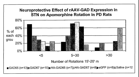
FIGURE 4 is a graph showing the neuroprotective effect of rAAV-GAD
treatment on apomorphine-induced rotation.
FIGURE 5A is a graph showing the potent neuroprotective effect of GAD65 on
to apomorphine rotation.
FIGURE 5B is another graph showing the potent neuroprotective effect of
GAD65 on apomorphine rotation.
FIGURE 6A is a graph showing that there was no significant reduction in head
position bias 2 months after rAAV transduction in chronic Parkinson's disease
rats.
-5a-
CA 02409674 2002-11-21
WO 01/89583 PCT/US01/16592
FIGURE 6B is a further graph showing that there was no significant reduction
in
head position bias 4 months after rAAV transduction in chronic Parkinson's
Disease
Rats.
FIGURE 7A is a graph demonstrating that head position bias was improved in
rats transduced with rAAV-GAD65.
FIGURE 7B is a further graph showing that rAAV-GAD65 transduced rats
showed marked effects on head position bias.
FIGURE 8 is a graph demonstrating a direct correlation between apomorphine
rotation and head position bias.
FIGURE 9 is graph showing that paw touching counts were significantly
improved in all rAAV-GAD and lbotenic acid lesion groups.
FIGURE 10 is a further graph showing that rAAV-GAD-65 had a marked
neuroprotective effect on paw touching counts.
FIGURE 11A is a graph demonstrating that a marked improvement in locomotor
activity was observed in Parkinson's Rats with combined rAAV-GAD65 and 67.
FIGURE 11B is a another graph further demonstrating that a marked
improvement in locomotor activity was observed in Parkinson's Rats with
combined
rAAV-GAD65 and 67.
FIGURE 12A is a graph showing that there was also evidence of neuroprotective
effects on locomoter activity by rAAV-GAD transduction.
FIGURE 12B is a graph further showing a neuroprotective effects on locomoter
activity by rAAV-GAD transduction;
FIGURE 13 is a graph of extracellular GABA Concentration during STN
Stimulation;
FIGURE 14 is a graph of Extracellular Glutamate Concentration during STN
Stimulation;
FIGURE 15 is a histogram showing the response of neurons in the Substantia
Nigra to electrical stimulation in the STN of a normal rat;
FIGURE 16 is a histogram showing the response of neurons in the Substantia
Nigra to electrical stimulation in the STN in rAAV-GAD transduced rat;
FIGURE 17A is a graph of extracellular GABA concentration in the SN during
STN stimulation in naive rats;
FIGURE 17B is a graph of extracellular GABA concentration in the SN during
STN stimulation in rAAV-GAD rats;
-6-
CA 02409674 2008-01-09
FIGURE 18A -18F are micrnphotographs showing rAAV-GAD65 expression
vivo. FIGURES 18A,B,C, and D show GAD65 expression in the STN detected with
GAD65 Ab (Boehringer). FIGURES 18A and C are derived from naive STN, showing
endogenous GAD65 expression. FIGURES 18B and D are based on rAAV-GAD65
transduced STN, such that an increase in cell bodies expressing GAD65 is seen,
while
FIGURES 18 E and Fshow GAD65 expression in the hippocampus. (FIGURE 18E being
naive and FIGURE 18Fbeing rAAV-GAD65 transduced);
FIGURES 19A and 19B are rasterplots showing activity in a monkey before
GAD67 treatment, respectively.
FIGURE 20 is a microphotograph showing GFP immunostaining at a injection
site.
FIGURES 21A and 21B are more detailed images, showing neuronal-like cells
stained with GFP antibody in 21A and glial-like cells stained with GFP
antibody shown
in 21B.
FIGURE 22 is a photograph of GAD immunostaining on rAAV-GAD treated
monkey, showing an increase in immunostaining on the rAAV-GAD treated side on
the
right while the morphology of the region remained unaltered after surgery.
Detailed Description
The practice of the present invention employs, unless otherwise indicated,
conventional methods of virology, microbiology, molecular biology and
recombinant
DNA techniques within the skill of the art
30 So that the invention is more clearly understood, the following terms are
defined: The term "neurodegenerative disorder" as used herein refers to a
disorder which
causes morphological and/or functional abnormality of a neural cell or a
population of
neural cells. The neurodegenerative disorder can result in an impairment or
absence of a
normal neurological function or presence of an abnormal neurological function
in a
-7-
CA 02409674 2002-11-21
WO 01/89583 PCT/US01/16592
subject. For example, neurodegenerative disorders can be the result of
disease, injury,
and/or aging. Non-limiting examples of morphological and functional
abnormalities
include physical deterioration and/or death of neural cells, abnormal growth
patterns of
neural cells, abnormalities in the physical connection between neural cells,
under- or over
production of a substance or substances, e.g., a neurotransmitter, by neural
cells, failure
of neural cells to produce a substance or substances which it normally
produces,
production of substances, e.g., neurotransmitters, and/or transmission of
electrical
impulses in abnormal patterns or at abnormal times. Neurodegeneration can
occur in any
area of the brain of a subject and is seen with many disorders including, for
example,
head trauma, stroke, ALS, multiple sclerosis, Huntington's disease,
Parkinson's disease,
and Alzheimer's disease.
The term "subject" as used herein refers to any living organism in which an
immune response is elicited. The terni subject includes, but is not limited
to, humans,
nonhuman primates such as chimpanzees and other apes and monkey species; farm
animals such as cattle, sheep, pigs, goats and horses; domestic mammals such
as dogs and
cats; laboratory animals including rodents such as mice, rats and guinea pigs,
and the like.
The term does not denote a particular age or sex. Thus, adult and newborn
subjects, as
well as fetuses, whether male or female, are intended to be covered. The term
"central
nervous system" or "CNS" as used herein refers to the art recognized use of
the term. The
CNS pertains to the brain, cranial nerves and spinal cord. The CNS also
comprises the
cerebrospinal fluid, which fills the ventricles of the brain and the central
canal of the
spinal cord.
The term "modifies" or "modified" are used interchangeably herein and refer to
the up-regulation or down-regulation of a target gene or a target protein. The
term
modifies or modified also refers to the increase, decrease, elevation, or
depression of
processes or signal transduction cascades involving a target gene or a target
protein. For
example, a target protein can be a GABA. Modification to the GABA
concentrations
may occur when a therapeutic agent, e.g., GAD, alters GABA concentration. For
example, modifications that result in an increase in GABA concentration by the
expression of GAD in glutaminergic neurons and intrinsic cells of the STN.
Modifications can also result from the addition of a therapeutic agent that
inactivates
GABA aminotransferase. The effect is to block the degradation of GABA and
thereby
increase its concentration. Numerous mechanism-based inactivators of GABA
aminotransferase are known (See e.g., Silverman Mechanism-Based Enzyme
-8-
CA 02409674 2002-11-21
WO 01/89583 PCT/US01/16592
Inactivation: Chemistry and Enzymology, Vol. I and II, CRC: Boca Raton 1988).
The
term modifies also includes increasing, or activating GAD with therapeutic
agents that
activate GAD, such as sodium valporate. The increase in GAD results in an
increase in
GABA, which subsequently reduces overstimulation of basal ganglia circuits.
Non-limiting examples of modifications includes modifications of morphological
and functional processes, under- or over production or expression of a
substance or
substances, e.g., a neurotransmitter, by neural cells, failure of neural cells
to produce a
substance or substances which it normally produces, production of substances,
e.g.,
neurotransmitters, and/or transmission of electrical impulses.
The term "tissue-specific promoter" as used herein refers to a promoter that
is
operable in cells of the central nervous sytem (CNS). Examples of promoters
for the
CNS include but are not limited to, neuron-specific promoters (e.g., the
neurofilament
promoter; Byrne and Ruddle (1989) Proc. Natl. Acad. Sci. USA 86:5473-5477) and
glial
specific promoters (Morii et al. (1991) Biochem. Biophys Res. Commun. 175: 185-
191).
Preferably, the promoter is tissue specific and is essentially not active
outside the central
nervous system, or the activity of the promoter is higher in the central
nervous system
that in other systems. For example, a promoter specific for the spinal cord,
brainstem,
(medulla, pons, and midbrain), cerebellum, diencephalon (thalamus,
hypothalamus),
telencephalon (corpus stratium, cerebral cortex, or within the cortex, the
occipital,
temporal, parietal or frontal lobes), STN, SN,or combinations, thereof. The
promoter
may also be one that can be used in combination with an AAV to result in
higher
expression. For example, a cytomegalovirus enhncer/chicken-Actin (CBA) hybrid
promoter that functions in cenll of the CNS (Xu et al. (2001) Hum Gene Ther.
12:563-
73).
The term "homology" or "identity" as used herein refers to the percentage of
likeness between nucleic acid molecules. To determine the homology or percent
identity
of two amino acid sequences or of two nucleic acid sequences, the sequences
are aligned
for optimal comparison purposes (e.g., gaps can be introduced in one or both
of a first
and a second amino acid or nucleic acid sequence for optimal alignment and non-
homologous sequences can be disregarded for comparison purposes). In a
preferred
embodiment, the length of a reference sequence aligned for comparison purposes
is at
least 30%, preferably at least 40%, more preferably at least 50%, even more
preferably at
least 60%, and even more preferably at least 70%, 80%, or 90% of the length of
the
reference sequence. The amino acid residues or nucleotides at corresponding
amino acid
-9-
CA 02409674 2002-11-21
WO 01/89583 PCT/US01/16592
positions or nucleotide positions are then compared. When a position in the
first
sequence is occupied by the same amino acid residue or nucleotide as the
corresponding
position in the second sequence, then the molecules are identical at that
position (as used
herein amino acid or nucleic acid "identity" is equivalent to amino acid or
nucleic acid
"homology"). The percent identity between the two sequences is a function of
the
number of identical positions shared by the sequences, taking into account the
number of
gaps, and the length of each gap, which need to be introduced for optimal
alignment of
the two sequences.
The comparison of sequences and determination of percent identity between two
sequences can be accomplished using a mathematical algorithm. For example, the
percent identity between two amino acid sequences can be determined using the
Needleman and Wunsch ((1970) J. Mol. Biol. (48):444-453) algorithm which has
been
incorporated into the GAP program in the GCG software package (available at
http://www.geg.com), using either a Blossom 62 matrix or a PAM250 matrix, and
a gap
weight of 16, 14, 12, 10, 8, 6, or 4 and a length weight of 1, 2, 3, 4, 5, or
6. In another
example, the percent identity between two nucleotide sequences is determined
using the
GAP program in the GCG software package (available at http://www.gcg.com),
using a
NWSgapdna.CMP matrix and a gap weight of 40, 50, 60, 70, or 80 and a length
weight of
1, 2, 3, 4, 5, or 6. In yet another example, the percent identity between two
amino acid or
nucleotide sequences is determined using the algorithm of E. Meyers and W.
Miller
(CABIOS, 4:11-17 (1989)) which has been incorporated into the ALIGN program
(version 2.0), using a PAM120 weight residue table, a gap length penalty of 12
and a gap
penalty.
The invention is described in more detail in the following subsections:
I. Neurodegenerative Diseases
(a) Parkinson's Disease
Parkinson's disease is associated with a disturbances of posture, locomotion,
facial expression or speech. The manifestations may be asymmetric, e.g., a
slight tremor
of the fingers on one hand at rest, and then become bilateral. Symptoms of
Parkinson's
disease are caused by loss of nerve cells in the pigmented substantia nigra
pars compacta
(SNPC) and the locus coeruleus in the midbrain. The stratium or corpus
stratium is a
structure in the cerebral hemispheres consisting of two basal ganglia (the
caudate nucleus
and the putnam) and the fibre of the internal capsule that separate them.
Parkinson's
disease in humans primarily effects the subcortical structures, especially the
substantai
-10-
CA 02409674 2002-11-21
WO 01/89583 PCT/US01/16592
nigra and the locus ceruleus. It is characterized by the loss of dopamine
neurons in the
substanta nigra, which have the basal ganglia as their major target organ.
Cell loss also
occurs in the globus pallidus and putamen.
Parkinson's disease is also associated with eosinophilic intraneural inclusion
granules (Lewy bodies) which are present in the basal ganglia, brainstem,
spinal cord,
and sympathetic ganglia. The pars compacta neurons of the substantia nigra
(SN)
provide dopaminergic input into the stratium, which is part of the basal
ganglia. These
dopaminergic neurons modulate a monosynaptic gamma-aminobutyric acid (GABA)
inhibitory output in the globus pallidus interna and pars reticulata of the
substantia nigra.
In Parkinson's disease, loss of dopaminergic cells in the substantia nigra
leads to stratial
dopamine depletion. This loss of dopamine alters the activity of neurons
within the basal
ganglia circuitry, including excessive firing and activity of these cells.
Accordingly, for the treatment of neurodegenarive disease of the substantia
nigra, a
vector comprising a therapeutic agent, e.g., a nucleotide sequence encoding
GAD, can be
delivered to the site of domaminergic cell loss or other regions of the basal
ganglia and
output nuclei. In one embodiment, the vector comprising a therapeutic agent
can be
delivered to the subthalmic nucleus (SN). In another embodiment, the vector
comprising
a therapeutic agent can be delivered to the substantia nigra pars reticulata.
(SNPR).
(b) Alzheimer's Disease
Alzheimer's disease is characterized by the gradual loss of intellectual
capabilities. Post-mortem examination of the brain shows a generalized
atrophy. There
are extensive histologic changes in Alzheimer's disease dominated by the
presence of
intracellular amyloid plaques and neurofibrillary tangles. Plaques and tangles
are rare,
however, in the basal ganglia and substantia nigra. Many specimens from
Alzheimer's
disease patients demonstrate a loss of pigmentation in the area of the locus
ceruleus,
which is a major source of noradrenergic synthesis in the brain.
II. Gamma aminobutyric acid (GABA) and glutamic acid decarboxylase (GAD)
Gamma aminobutyric acid (GABA) and glutamic acid are two major
neurotransmitters involved in the regulation of brain neuronal activity. GABA
is the
major inhibitory neurotransmitter and L-glutamic acid is an excitatory
transmitter
(Roberts et al. GABA in Nervous System Function, Raven Press: New York, 1976;
McGeer et al. Glutamine, Glutamate, and GABA in the Central Nervous System;
Hertz
L, Kvamme E, McGeer E G, Schousbal A, eds., Liss: New York, 1983;3-17). GABA
is
released from dopaminergic cells. An imbalance in the concentration of these
-11-
CA 02409674 2002-11-21
WO 01/89583 PCT/US01/16592
neurotransmitters can lead to convulsive states. When the concentration of
GABA
diminishes below a threshold level in the brain, convulsions result (Karlsson
et al., (1974)
Biochem. Plzarmacol. 23:3053-3061). When the GABA levels rise in the brain the
convulsions terminate (Hayashi (1959) supra). In several convulsive disorders
there is
concomitant with reduced brain GABA levels a diminished level of glutamic acid
decarboxylase (GAD) activity (McGeer et al., GABA in Nervous System Function;
Roberts E, Chase T N, Tower D B, eds., Raven Press: New York 1976:487-495;
Butterworth et al., (1983) NeuYochern.41:440-447). The concentrations of GAD
and
GABA vary in parallel because decreased GAD concentration results in lower
GABA
production.
GABA interacts with a least two receptors, GABA-A and GABA-B. GABA-A
receptors have been well characterized and are coupled to chloride channels
(Bormann
(1988) Trends Neurosci. 11: 112-116). GABA-A receptors are related to ligand
gated ion
channels belonging to the same superfamily as the nicotrinic receptor for
achetylcholine.
In contrast, GABA-B receptors are less well understood, although reports
describe that
the GABA-B receptors are coupled to either calcium or potassium channels
(Bormann
(1988) Trends Neurosci. 11:112-116 supra).
The majority of neurons in the striatum (caudate-putamen, dorsal striatum;
nucleus accumbens, ventral striatum) and in striatal projection regions (the
pallidum, the
entopeduncular nucleus and substantia nigra reticulata) use GABA as
transmitter and
express GAD in the synthesis of GABA. Brain contains at least two molecular
forms of
GAD, the principal synthetic enzyme for GABA. Two forms, termed GAD-65 and GAD-
67, are the products of two genes and differ in sequence, molecular weight,
and level of
expression among brain regions. GAD-65 appears to be localized in nerve
terminals to a
greater degree than GAD-67, which appears to be more uniformly distributed
throughout
the cell. Although GAD-65 and GAD-67 differ significantly in several
characteristics,
they also have substantial similarities and interactions, and the presence of
individual
forms of GAD in certain cell types is consistent with the idea that GAD-65 and
GAD-67
can each synthesize GABA. Thus, GAD-65 and GAD-67 seem to provide a dual
system
for the control of neuronal GABA synthesis. Specific changes in activity in
subpopulations of striatal GABA neurons mediate the dopamine-dependent effects
seen
in Parkinson's disease (Lindefors (1993) Prog Neuropsychopharmacol Biol
Psychiatry
17:887-903).
-12-
CA 02409674 2002-11-21
WO 01/89583 PCT/US01/16592
Human GAD-65 and GAD-67 have been isolated and cloned by Bu et al. (1992)
Proc Natl Acad Sci 89:2115-2119. Human GAD-65 cDNA encodes a Mr 65,000
polypeptide, with 585 amino acid residues (Genbank Accession No.
NM000818;M81882), Human GAD-67 encodes a Mr 67,000 polypeptide, with 594
amino acid residues (Genbank Accession No. NM013445;M81883).
In one embodiment, the invention features a vector comprising a nucleotide
sequence encoding GAD-65. In another embodiment, the invention features a
vector
comprising a nucleotide sequence encoding GAD-67.
Also within the scope of the invention is a polypeptide encoded by nucleotide
sequence that has at least 60% homology to GAD-65 or a fragment thereof. A
polypeptide encoded by nucleotide sequence that about 70% homology, about 75%
homology, about 80% homology, about 85% homology, about 90% homology, about
95% homology, about 99% homology to GAD-65 or a fragment thereof. Also within
the
scope of the invention is a polypeptide encoded by nucleotide sequence that
has at least
60% homology to GAD-67 or a fragment thereof. A polypeptide encoded by
nucleotide
sequence that about 70% homology, about 75% homology, about 80% homology,
about
85% homology, about 90% homology, about 95% homology, about 99% homology to
GAD-67 or a fragment thereof.
The GAD transduction in target cells of the STN will specifically increase the
local inhibitory tone, acting via increasing extracellular GABA and inhibiting
neuronal
activity in the STN by acting on both GABA-A and GABA-B receptors. Gene
expression
using the method of the invention provides completely stable levels of the
transgene
expression for at least 15 months ifa vivo (see Example 3). The release of
GABA from
the transduced cells diffuses locally binds to the GABA receptors thereby
leading to
significant depression of activity. Importantly, unlike either ablation or
DBS, the gene
transfer using AAV is devoid of any cellular infiltration, any microglial cell
activation
and lack of reactive astrocytosis. Each of these compensatory or inflammatory
responses
to the ablative or DBS approaches is likely to reduce the efficacy of these
respective
strategies and potentially have other.
Other inhibitory genes that can be used in the method of the invention
includes,
but are not limited to, genes which encode potassium channels, genes which
encode other
ion channels and genes that act on the neurotransmitter release machinery,
including
endocytosis and exocytosis. Examples of genes include for example, frequenin
and AP
180.
-13-
CA 02409674 2002-11-21
WO 01/89583 PCT/US01/16592
III. Vectors
The vectors of the invention can be delivered to the cells of the central
nervous
system by using viral vectors or by using non-viral vectors. In a preferred
embodiment,
the invention uses adeno-associated viral vectors comprising the a nucleotide
sequence
encoding GAD for gene delivery. AAV vectors can be constructed using known
techniques to provide at least the operatively linked components of control
elements
including a transcriptional initiation region, a exogenous nucleic acid
molecule, a
transcriptional termination region and at least one post-transcriptional
regulatory
sequence. The control elements are selected to be functional in the targeted
cell. The
resulting construct which contains the operatively linked components is
flanked at the 5'
and 3' region with functional AAV ITR sequences.
The nucleotide sequences of AAV ITR regions are known. The ITR sequences for
AAV-2 are described, for example by Kotin et al. (1994) Human Gene Therapy
5:793-801; Berns "Parvoviridae and their Replication" in Fundamental Virology,
2nd
Edition, (B. N. Fields and D. M. Knipe, eds.) The skilled artisan will
appreciate that
AAV ITR's can be modified using standard molecular biology techniques.
Accordingly,
AAV ITRs used in the vectors of the invention need not have a wild-type
nucleotide
sequence, and may be altered, e.g., by the insertion, deletion or substitution
of
nucleotides. Additionally, AAV ITRs may be derived from any of several AAV
serotypes, including but not limited to, AAV-l, AAV-2, AAV-3, AAV-4, AAV-5,
AAVX7, and the like. Furthermore, 5' and 3' ITRs which flank a selected
nucleotide
sequence in an AAV expression vector need not necessarily be identical or
derived from
the same AAV serotype or isolate, so long as the ITR's function as intended,
i.e., to allow
for excision and replication of the bounded nucleotide sequence of interest
when AAV
rep gene products are present in the cell.
The skilled artisan can appreciate that regulatory sequences can often be
provided
from commonly used promoters derived from viruses such as, polyoma, Adenovirus
2,
cytomegalovirus and Simian Virus 40. Use of viral regulatory elements to
direct
expression of the protein can allow for high level constitutive expression of
the protein in
a variety of host cells. Ubiquitously expressing promoters can also be used
include, for
example, the early cytomegalovirus promoter Boshart et al. (1985) Cell 41:521-
530,
herpesviras thymidine kinase (HSV-TK) promoter (McKnight et al. (1984) Cell
37: 253-
262), (3-actin promoters (e.g., the human 0-actin promoter as described by Ng
et al.
-14-
CA 02409674 2002-11-21
WO 01/89583 PCT/US01/16592
(1985) Mol. Cell Biol. 5: 2720-2732) and colony stimulating factor-1 (CSF-1)
promoter
(Ladner et al. (1987) EMBO J. 6: 2693-2698).
Alternatively, the regulatory sequences of the AAV vector can direct
expression
of the gene preferentially in a particular cell type, i.e., tissue-specific
regulatory elements
can be used. Non-limiting examples of tissue-specific promoters which can be
used
include, central nervous system (CNS) specific promoters such as, neuron-
specific
promoters (e.g., the neurofilament promoter; Byrne and Ruddle (1989) Proc.
Natl. Acad.
Sci. USA 86:5473-5477) and glial specific promoters (Morii et al. (1991)
Biochem.
Biophys Res. Commun. 175: 185-191). Preferably, the promoter is tissue
specific and is
essentially not active outside the central nervous system, or the activity of
the promoter is
higher in the central nervous system that in other systems. For example, a
promoter
specific for the spinal cord, brainstem, (medulla, pons, and midbrain),
cerebellum,
diencephalon (thalamus, hypothalamus), telencephalon (corpus stratium,
cerebral cortex,
or within the cortex, the occipital, temporal, parietal or frontal lobes), or
combinations,
thereof. The promoter may be specific for particular cell types, such as
neurons or glial
cells in the CNS. If it is active in glial cells, it may be specific for
astrocytes,
oligodentrocytes, ependymal cells, Schwann cells, or microglia. If it is
active in neurons,
it may be specific for particular types of neurons, e.g., motor neurons,
sensory neurons,
or interneurons. Preferably, the promoter is specific for cells in particular
regions of the
brain, for example, the cortex, stratium, nigra and hippocampus.
Suitable neuronal specific promoters include, but are not limited to, neuron
specific enolase (NSE) (Olivia et al. (1991) Genomics 10: 157-165, GenBank
Accession
No: X51956), and human neurofilament light chain promoter (NEFL) (Rogaev et
al.
(1992) Hum. Mol. Genet. 1: 781, GenBank Accession No: L04147). Glial specific
promoters include, but are not limited to, glial fibrillary acidic protein
(GFAP) promoter
(Morii et al. (1991) Biochem. Biophys Res. Commun. 175: 185-191, GenBank
Accession
No:M65210), S 100 promoter (Morii et al. (1991) Biochem. Biophys Res. Commun.
175:
185-191, GenBank Accession No: M65210) and glutamine synthase promoter (Van
den
et al. (1991) Biochem. Biophys. Acta. 2: 249-25 1, GenBank Accession No:
X59834). In
a preferred embodiment, the gene is flanked upstream (i.e., 5') by the neuron
specific
enolase (NSE) promoter. In another preferred embodiment, the gene of interest
is
flanked upstream (i.e., 5') by the elongation factor 1 alpha (EF) promoter.
The AAV vector harboring the nucleotide sequence encoding a protein of
interest, e.g., GAD, and a post-transcriptional regulatory sequence (PRE)
flanked by
-15-
CA 02409674 2008-01-09
AAV ITRs, can be constructed by directly inserting the nucleotide sequence
encoding the
protein of interest and the PRE into an AAV genome which has had the major AAV
open
reading frames ("ORFs") excised therefrom. Other portions of the AAV genome
can also
be deleted, as long as a sufficient portion of the ITRs remain to allow for
replication and
packaging functions. These constructs can be designed using techniques well
known in
the art.
Alternatively, AAV ITRs can be excised from the viral genome or from an AAV
vector containing the same and fused 5' and 3' of a selected nucleic acid
construct that is
present in another vector using standard ligation techniques, such as those
described in
Sambrook et al. , Supra. Several AAV vectors are available from the American
Type
Culture Collection ("ATCC") under Accession Numbers 53222, 53223, 53224, 53225
and 53226.
In order to produce recombinant AAV particles, an AAV vector can be introduced
into a suitable host cell using known techniques, such as by transfection. A
number of
transfection techniques are generally known in the art. See, e.g., Graham et
al. (1973)
Virology, 52:456, Sambrook et al. (1989) Molecular Cloning, a laboratory
manual, Cold
Spring Harbor Laboratories, N. Y., Davis et al. (1986) Basic Methods in
Molecular
Biology, Elsevier, and Chu et al.. (1981) Gene 13:197. Particularly suitable
transfection
methods include calcium phosphate co-precipitation (Graham et al. (1973)
Virol.
52:456-467), direct micro-injection into cultured cells (Capecchi (1980) Cell
22:479-488), electroporation (Shigekawa et al. (1988) BioTeclmiques 6:742-
751),
liposome mediated gene transfer (Mannino et al. (1988) BioTechniques 6:682-
690),
lipid-mediated transduction (Felgner et al. (1987) Proc. Natl. Acad. Sci. USA
84:7413-7417), and nucleic acid delivery using high-velocity microprojectiles
(Klein et
al. (1987) Nature 327:70-73).
Suitable host cells for producing recombinant AAV particles include, but are
not
limited to, microorganisms, yeast cells, insect cells, and mammalian cells,
that can be, or
have been, used as recipients of a exogenous nucleic acid molecule. Thus, a
"host cell"
as used herein generally refers to a cell which has been transfected with an
exogenous
nucleic acid molecule. The host cell includes any eukaryotic cell or cell line
so long as
-16-
CA 02409674 2002-11-21
WO 01/89583 PCT/US01/16592
the cell or cell line is not incompatible with the protein to be expressed,
the selection
system chosen or the fermentation system employed. Non-limiting examples
include
CHO dhfr- cells (Urlaub and Chasin (1980) Proc. Natl. Acad. Sci. USA 77:4216-
4220),
293 cells (Graham et al. (1977) J. Gen. Virol. 36: 59) or myeloma cells like
SP2 or NSO
(Galfre and Milstein (1981) Meth. Enzymol. 73(B):3-46).
In one embodiment, cells from the stable human cell line, 293 (readily
available
through, e.g., the ATCC under Accession No. ATCC CRL1573) are preferred in the
practice of the present invention. Particularly, the human cell line 293,
which is a human
embryonic kidney cell line that has been transformed with adenovirus type-5
DNA
fragments (Graham et al. (1977) J Gen. Virol. 36:59), and expresses the
adenoviral Ela
and Elb genes (Aiello et al. (1979) Virology 94:460). The 293 cell line is
readily
transfected, and provides a particularly convenient platform in which to
produce rAAV
virions.
Host cells containing the above-described AAV vectors must be rendered capable
of providing AAV helper functions in order to replicate and encapsidate the
expression
cassette flanked by the AAV ITRs to produce recombinant AAV particles. AAV
helper
functions are generally AAV-derived coding sequences which can be expressed to
provide AAV gene products that, in turn, function in trans for productive AAV
replication. AAV helper functions are used herein to complement necessary AAV
functions that are missing from the AAV vectors. Thus, AAV helper functions
include
one, or both of the major AAV open reading frames (ORFs), namely the rep and
cap
coding regions, or functional homologues thereof.
The AAV rep coding region of the AAV genome encodes the replication proteins
Rep 78, Rep 68, Rep 52 and Rep 40. These Rep expression products have been
shown to
possess many functions, including recognition, binding and nicking of the AAV
origin of
DNA replication, DNA helicase activity and modulation of transcription from
AAV (or
other exogenous) promoters. The Rep expression products are collectively
required for
replicating the AAV genome. The AAV cap coding regiori of the AAV genome
encodes
the capsid proteins VP 1, VP2, and VP3, or, functional homologues thereof. AAV
helper
functions can be introduced into the host cell by transfecting the host cell
with an AAV
helper construct either prior to, or concurrently with, the transfection of
the AAV vector
comprising the expression cassette, AAV helper constructs are thus used to
provide at
least transient expression of AAV rep and/or cap genes to complement missing
AAV
functions that are necessary for productive AAV infection. AAV helper
constructs lack
-17-
CA 02409674 2002-11-21
WO 01/89583 PCT/US01/16592
AAV ITRs and can neither replicate nor package themselves. These constructs
can be in
the form of a plasmid, phage, transposon, cosmid, virus, or virion. A number
of AAV
helper constructs have been described, such as the commonly used plasmids
pAAV/Ad
and pIM29+45 which encode both Rep and Cap expression products. (See, e.g.,
Samulski
et al. (1989) J. Virol. 63:3822-3828; and McCarty et al. (1991) J. Virol.
65:2936-2945).
A number of other vectors have been described which encode Rep and/or Cap
expression
products. See, e.g., U.S. Pat. No. 5,139,941.
As a consequence of the infection of the host cell with a helper virus, the
AAV
Rep and/or Cap proteins are produced. The Rep proteins also serve to duplicate
the AAV
genome. The expressed Cap proteins assemble into capsids, and the AAV genome
is
packaged into the capsids. This results the AAV being packaged into
recombinant AAV
particles comprising the expression cassette. Following recombinant AAV
replication,
recombinant AAV particles can be purified from the host cell using a variety
of
conventional purification methods, such as CsCl gradients. The resulting
recombinant
AAV particles are then ready for use for gene delivery to various cell types.
Alternatively, a vector of the invention can be a virus other than the adeno-
associated virus, or portion thereof, which allows for expression of a nucleic
acid
molecule introduced into the viral nucleic acid. For example, replication
defective
retroviruses, adenoviruses and lentivirus can be used. Protocols for producing
recombinant retroviruses and for infecting cells in vitro or in vivo with such
viruses can
be found in Current Protocols in Molecular Biology, Ausubel et al. (eds.)
Greene
Publishing Associates, (1989), Sections 9.10-9.14 and other standard
laboratory manuals.
Examples of suitable retroviruses include pLJ, pZIP, pWE and pEM which are
well
known to those skilled in the art. Examples of suitable packaging virus lines
include
Crip, Cre, 2 and Am. The genome of adenovirus can be manipulated such that it
encodes and expresses the protein of interest but is inactivated in terms of
its ability to
replicate in a normal lytic viral life cycle. See e.g., Berkner et al. (1988)
BioTechniques
6:616; Rosenfeld et al. (1991) Science 252:431-434; and Rosenfeld et al.
(1992) Cell
68:143-155. Suitable adenoviral vectors derived from the adenovirus strain Ad
type 5
d1324 or other strains of adenovirus (e.g., Ad2, Ad3, Ad7 etc.) are well known
to those
skilled in the art.
Alternatively, the vector can be delivered using a non-viral delivery system.
This
includes delivery of the vector to the desired tissues in colloidal dispersion
systems that
include, for example, macromolecule complexes, nanocapsules, microspheres,
beads,
-18-
CA 02409674 2002-11-21
WO 01/89583 PCT/US01/16592
and lipid-based systems including oil-in-water emulsions, micelles, mixed
micelles, and
liposomes.
Liposomes are artificial membrane vesicles which are useful as delivery
vehicles
in vitro and in vivo. In order for a liposome to be an efficient gene transfer
vehicle, the
following characteristics should be present: (1) encapsulation of the genetic
material at
high efficiency while not compromising the biological activity; (2)
preferential and
substantial binding to a target cell in comparison to non-target cells; (3)
delivery of the
aqueous contents of the vesicle to the target cell cytoplasm at high
efficiency; and (4)
accurate and effective expression of genetic information (Mannino, et al.
(1988)
Biotechniques, 6:682). Examples of suitable lipids liposomes production
include
phosphatidyl compounds, such as phosphatidylglycerol, phosphatidylcholine,
phosphatidylserine, phosphatidylethanolamine, sphingolipids, cerebrosides, and
gangliosides. Additional examples of lipids include, but are not limited to,
polylysine,
protamine, sulfate and 3b -[N- (N',N' dimethylaminoethane) carbamoyl]
cholesterol.
Alternatively, the vector can be coupled with a carrier for delivery Exemplary
and
preferred carriers are keyhole limpet hemocyanin (KLH) and human serum
albumin.
Other carriers may include a variety of lymphokines and adjuvants such as INF,
IL-2, IL-
4, IL-8 and others. Means for conjugating a peptide to a carrier protein are
well known in
the art and include glutaraldehyde, m-maleimidobenzoyl- N-hydroxysuccinimide
ester,
carbodiimyde and bis-biazotized benzidine. The vector can be conjugated to a
carrier by
genetic engineering techniques that are well known in the art. (See e.g., U.S.
Pat. Nos.
4,608,251; 4,601,903; 4,599,231; 4,599,230; 4,596,792; and 4,578,770).
In one embodiment, particle-mediated delivery using a gene-gun can be used as
a
method to deliver the vector. Suitable particles for gene gun-based delivery
of include
gold particles. In one embodiment, the vector can be delivered as naked DNA.
Gene gun
based delivery is described, for example by, Braun et al. (1999) Virology
265:46-56;
Drew et al. (1999) Vaccine 18:692-702; Degano et al. (1999) Vaccine 18:623-
632; and
Robinson (1999) Int JMol Med 4:549-555; Lai et al. (1998) Crit Rev hnmunol
18:449-84; See e.g., Accede et al. (1991) Nature 332: 815-818; and Wolff et
al. (1990)
Science 247:1465-1468 Murashatsu et al. , (1998) Int. J. Mol. Med. 1: 55-62;
Agracetus
et al. (1996) J. Biotechnol. 26: 37-42; Johnson et al. (1993) Genet. Eng.15:
225-236).
Also within the scope of the invention is the delivery of the vector in one or
more
combinations of the above delivery methods.
-19-
CA 02409674 2002-11-21
WO 01/89583 PCT/US01/16592
IV. Delivery Systems
Delivery systems include methods of in vitro, in vivo and e.x vivo delivery of
the
vector. For in vivo delivery, the vector can be administered to a subject in a
pharmaceutically acceptable carrier. The term "pharmaceutically acceptable
carrier", as
used herein, refers to any physiologically acceptable carrier for in vivo
administration of
the vectors of the present invention. Such carriers do not induce an immune
response
harmful to the individual receiving the composition, and are discussed in
section V.
In one embodiment, vector can be distributed throughout a wide region of the
CNS, by
injecting the vector into the cerebrospinal fluid, e.g., by lumbar puncture
(See e.g.,
Kapadia et al. (1996) Neurosurg 10: 585-587).
Alternatively, precise delivery of the vector into specific sites of the
brain, can be
conducted using stereotactic microinjection techniques. For example, the
subject being
treated can be placed within a stereotactic frame base (MRI-compatible) and
then imaged
using high resolution MRI to determine the three-dimensional positioning of
the
particular region to be treated. The MRI images can then be transferred to a
computer
having the appropriate stereotactic software, and a number of images are used
to
determine a target site and trajectory for antibody microinjection. The
software translates
the trajectory into three-dimensional coordinates that are precisely
registered for the
stereotactic frame. In the case of intracranial delivery, the skull will be
exposed, burr
holes will be drilled above the entry site, and the stereotactic apparatus
used to position
the needle and ensure implantation at a predetermined depth. The vector can be
delivered
to regions, such as the cells of the spinal cord, brainstem, (medulla, pons,
and midbrain),
cerebellum, diencephalon (thalamus, hypothalamus), telencephalon (corpus
stratium,
cerebral cortex, or within the cortex, the occipital, temporal, parietal or
frontal lobes), or
combinations, thereof. In another preferred embodiment, the vector is
delivered using
other delivery methods suitable for localized delivery, such as localized
permeation of the
blood-brain barrier. Particularly preferred delivery methods are those that
deliver the
vector to regions of the brain that require modification. Modification as used
herein
refers to a change in the cellular activity in the region of the brain
injected with the
vector. The change in cellular activity can result from changing the
expression, or
production of genes responsible for stimulating a cell. For example, delivery
of a vector
comprising a nucleotide sequence encoding GAD, to a region of the brain that
is
overstimulated, such as the basal ganglia. In particular, delivery of the
vector to the STN
-20-
CA 02409674 2002-11-21
WO 01/89583 PCT/US01/16592
which are overactive in diseases such as Parkinson's, will result in
expression of GAD in
this region. While not being required to provide a mechanism of action, the
expression of
GAD in the STN results in production of GABA within the STN cells, the STN
cells
release GABA locally such that the released GABA binds to GABA-A and GABA-B
receptors on the STN cell surface. GABA binding to the GABA receptors results
in a
reduction in cell stimulation, thereby reducing overactivity in the STN cells
and prevent
neuronal destruction.
V. Pharmaceutical Compositions and Pharmaceutical Administration
The vector of the invention can be incorporated into pharmaceutical
compositions
suitable for administration to a subject. Typically, the pharmaceutical
composition
comprises the vector of the invention and a pharmaceutically acceptable
carrier. As used
herein, "pharmaceutically acceptable carrier" includes any and all solvents,
dispersion
media, coatings, antibacterial and antifungal agents, isotonic and absorption
delaying
agents, and the like that are physiologically compatible. Examples of
pharmaceutically
acceptable carriers include one or more of water, saline, phosphate buffered
saline,
dextrose, glycerol, ethanol and the like, as well as combinations thereof. In
many cases,
it will be preferable to include isotonic agents, for example, sugars,
polyalcohols such as
mannitol, sorbitol, or sodium chloride in the composition. Pharmaceutically
acceptable
carriers may further comprise minor amounts of auxiliary substances such as
wetting or
emulsifying agents, preservatives or buffers, which enhance the shelf life or
effectiveness
of the antibody or antibody portion.
The compositions of this invention may be in a variety of forms. These
include,
for example, liquid, semi-solid and solid dosage forms, such as liquid
solutions (e.g.,
injectable and infusible solutions), dispersions or suspensions,
tablets,pills, powders,
liposomes and suppositories. The preferred form depends on the intended mode
of
administration and therapeutic application. Typical preferred compositions are
in the
form of injectable or infusible solutions, such as compositions similar to
those used for
passive immunization of humans. The preferred mode of administration is
parenteral
(e.g., intravenous, subcutaneous, intraperitoneal, intramuscular). In one
embodiment, the
vector is administered by intravenous infusion or injection. In another
embodiment, the
vector is administered by intramuscular or subcutaneous injection. In another
embodiment, the vector is administered perorally. In the most preferred
embodiment, the
vector is delivered to a specific location using stereostatic delivery.
-21-
CA 02409674 2002-11-21
WO 01/89583 PCT/US01/16592
Therapeutic compositions typically must be sterile and stable under the
conditions
of manufacture and storage. The composition can be formulated as a solution,
microemulsion, dispersion, liposome, or other ordered structure suitable to
high drug
concentration. Sterile injectable solutions can be prepared by incorporating
the active
compound (i.e., antigen, antibody or antibody portion) in the required amount
in an
appropriate solvent with one or a combination of ingredients enumerated above,
as
required, followed by filtered sterilization.
Generally, dispersions are prepared by incorporating the active -compound into
a
sterile vehicle that contains a basic dispersion medium and the required other
ingredients
from those enumerated above. In the case of sterile, lyophilized powders for
the
preparation of sterile injectable solutions, the preferred methods of
preparation are
vacuum drying and spray-drying that yields a powder of the active ingredient
plus any
additional desired ingredient from a previously sterile-filtered solution
thereof. The
proper fluidity of a solution can be maintained, for example, by the use of a
coating such
as lecithin, by the maintenance of the required particle size in the case of
dispersion and
by the use of surfactants. Prolonged absorption of injectable compositions can
be
brought about by including in the composition an agent that delays absorption,
for
example, monostearate salts and gelatin.
The vector of the present invention can be administered by a variety of
methods
known in the art. As will be appreciated by the skilled artisan, the route
and/or mode of
administration will vary depending upon the desired results. In certain
embodiments, the
active compound may be prepared with a carrier that will protect the compound
against
rapid release, such as a controlled release formulation, including implants,
transdermal
patches, and microencapsulated delivery systems. Biodegradable, biocompatible
polymers can be used, such as ethylene vinyl acetate, polyanhydrides,
polyglycolic acid,
collagen, polyorthoesters, and polylactic acid. Many methods for the
preparation of such
formulations are patented or generally known to those skilled in the art. See,
e.g.,
Sustained and Controlled Release Drug Delivery Systenzs, J.R. Robinson, ed.,
Marcel
Dekker, Inc., New York, 1978. The pharmaceutical compositions of the invention
may
include a "therapeutically effective amount" or a "prophylactically effective
amount" of
the vectors of the invention. A "therapeutically effective amount" refers to
an amount
effective, at dosages and for periods of time necessary, to achieve the
desired therapeutic
result. A therapeutically effective amount of the vector may vary according to
factors
such as the disease state, age, sex, and weight of the individual, and the
ability of the
-22-
CA 02409674 2005-03-07
vector to elicit a desired response in the individual. A therapeutically
ei3ective amount is
also one in which any toxic or detrimental effects of the vector are
outweighed by the
therapeutically beneficial effects. A"prophylactically effective amount"
refers to an
amount effective, at dosages and for periods of time necessary, to achieve the
desired
prophylactic result. TypicaIly, since a prophylactic dose is used in subjects
prior to or at
an earlier stage of disease, the prophylactically effective amount will be
less than the
therapeutically effective amount.
Dosage regimens may be adjusted to provide the optimum desired response (e.g.,
a therapeutic or prophylactic response). For exaniple, a single bolus may be
administered, several divided doses may be administered over time or the dose
may be
proportionally reduced or increased as indicated by the exigencies of the
therapeutic
situation. It is especially advantageous to formulate parenteral compositions
in dosage
unit form for ease of administration and unifomiity of dosage. Dosage unit
form as used
herein refers to physically discrete units suited as unitary dosages for the
mammalian
subjects to be treated; each unit containing a predetermined quantity of
active compound
calculated to produce the desired tberapeutic effect in association with the
required
pharmaceutical carrier. The specification for the dosage unit forms of the
invention are
dictated by and directly dependent on (a) the unique characteristics of the
active
compound and the particular therapeutic or prophylactic effect to be achieved,
and (b) the
limitations inherent in the art of compounding such an active compound for the
treatment
of sensitivity in individuals.
One skilled in the art will appreciate further features and advantages of the
invention based on the above-described embodiments. Accordingly, the invention
is not
to be limited by what has been particularly shown and descn'bed, except as
indicated by
the appended claims.
Examples
Example 1: Methods and Materials
(i) Vector Construction
This example descrfbes the construction of an adeno-associated virus vector
with
an GAD cDNA. A full length human GAD-65 cDNA was subcloned into an AAV
plasmid under the control of a 1.8 kb rat NSE (neuron specific enolase)
promoter (Foress-
petter et al. (1986) J. Neurosci. Res. 16, 141-156 (1998)) 5' of the GAD cDNA
followed
by the Woodchuck Hepatitis Post-Transcriptional Regulatory Element (WPRE) and
a
-23-
CA 02409674 2002-11-21
WO 01/89583 PCT/US01/16592
bovine growth hormone (BGH) polyadenylation site between the AAV inverted
terminal
repeats, as previously described (During et al. (1998) Nature Med. 4:1131-
1135). The
resulting plasmid is referred to as pAAV-NSE-GAD-WPRE.
The plasmids were packaged to generate high titer rAAV-GAD viral particles
using an optimized protocol based on the original helper-free transient
transfection
method described by Samulski et al. (1989) J. Virol. 63:3822-3828), but
modified by
using an improved 4rd generation helper plasmid, pDG as described by Grimm et
al.
(1999) Hum Gene Ther 10, 2445-2450. The helper plasmid contains both the rep
and
cap open reading frames, as well the minimal set of adenoviral genes necessary
for helper
functions. The vectors were generated using calcium phosphate transfection of
both
plasmids into 293 cells. Vector stocks were purified using ammonium sulfate
followed
by double cesium banding. The bands containing the viral particle were
isolated from the
cesium chloride preparation and dialysis into suitable buffer.
Particle titers were determined using an ELISA assay kit available (Progen,
Inc.)
which uses an A20 monoclonal antibody that recognizes intact particles.
Purification of
the viral particles was performed as described by Clark et al., (1999) Huin.
Gene. Ther.
10: 1031-1039 and Zolutkhin et al. (1999) Gene Therapy 9: 973-985.
(ii) Packaging protocol
To package the recombinant vectors, human embryonic kidney cells, 293 cells
(from American Type Culture Collection (ATCC # CRL-1573)), passage 4-12 were
used. The 293 kidney cells (1.5 x 10⁷ cells) were seeded into forty 15 cm
dishes in
complete DMEM (Gibco) containing 10% fetal bovine serum (Hyclone), 0.1 mM MEM
non-essential amino acids solution (Gibco), 1 mM MEM sodium pyruvate (Gibco),
0.05% Penicillin-Streptomycin (5, 000 units/ml, Gibco), and incubated
overnight at
376C. When the cells were 70% confluent and 2-3 hours prior to transfection,
the cells
were fed fresh Iscove modified Dulbecco medium (IMDM, Gibco) containing 10%
fetal
bovine serum (Hyclone) without antibiotics.
All plasmids were isolated from the cells by the alkaline lysis method
(Sambrook
et al., supra), and were further purified by HPLC (BioCAD, Sprint, PerSeptive
Biosystems), and concentrated with 2 volumes of 100% ethanol (AR grade, BDH).
All
HPLC elute buffers (Buffer A: 250mM TrisHCI, 10mM EDTA, pH 8.0; Buffer B: 25
mM TrisHCl, 1 mM EDTA, 2M NaCl, pH, 8.0; Buffer C: Milli Q water) used for
purification were autoclaved and filter sterilized prior to use. For each 15
cm tissue
culture plate, a total of 60 g of plasmid DNA was dissolved in 3.0 ml of
0.25M CaC12
-24-
CA 02409674 2005-03-07
and then quickly mixed with 3.0 ml of HEPES-buffered saline (50 mM HEPES, 280
mM
NaCI, 1.5 mM NaZHPO4 [pH 7.05-7.10]), incubated for 2 min and then added to
the
cells. 6-8 hours after transfection, the medium was aspirated and cells were
washed with
IMDM supplemented with 10% fetal bovine senun without antibiotics. The washing
medium was then aspirated and replaced with fresh IMDM (Gibco) containing 10%
fetal
bovine serum with trace pen/strep. The cells were harvested at 48 hours after
transfection. After low-speed centrifugation on a tabletop centrifuge, the
cell pellets were
resuspended in 20 ml of Opti-MEM (Gibco) and subjected to sonication using 15-
20%
energy for 50 bursts lasting 1 min. Cell debris was removed with low speed
centrifugation. The clarified supematant was collected into a 50 nil
polypropylene tube,
the cell pellets were resuspended in 20 ml of Opti-MEM for reextraction. The
supematants were combined.
One-third volume of ice-cold saturated (NH4)2SO4 was added to the supematant,
mixed and placed on ice for 10 minutes. The sample was then centrifuged at
8,000 rpm
at 4 C for 10 min, supernatant was transferred to a polypropylene centrifuge
tube, 2/3
volume of the initial lysate of saturated (NH4)2SO4 was added and mixed well,
then
placed on ice for 20 min prior to centrifugation at 12,000 rpm for 20 min at 4
C. The
pellet was redissolved in CsCI-phosphate buffered saline (PBS) (pH 7.4)
solution
(density 1.37 g/1) and centrifuged in an SW41 rotor Beclcman at 80,000 rpm
(for 24 hours
with a 0.5 ml CsCI-PBS cushion (density, 1.5 g/ml).
The band containing recombinant AAV particle (rAAV) was collected and re-
centrifuged as described above for a further 24 hours. Finally, the rAAV band
was
collected following the second CsCI centrifugation and dialyzed against one
liter sterile
dialysis buffer containing 50 mM NaCl, 5 mM Tris-HC1 and 0.5 mM MgC12 (pH 7.4)
for
an initial 4 hours. Dialysis was repeated using one liter of fresh cold
sterile dialysis
buffer for another 4 hours and finally ovennight dialysis using a 50,000
molecular weight
cut off dialysis membrane (Spectrapor) and fresh sterile dialysis buffer. The
AAV virus
particle titer was determined using an ELISA method descnbed by Wistuba et al.
((1997)
J. Yfrol. 71: 1341-1352). Briefly, a monoclonal antibody specific for AAV
assembled
capsids is coated onto microtiter strips and is used to capture AAV particles.
A biotin-
conjugated monoclonal antibody to AAV is bound to the inunune complex,
streptavidin
peroxidase conjugate reacts with the biotin molecules. Addition of substrate
solution
results in a color reaction which is proportional to specifically bound virus
particles, and
allows the quantitative determination of an unknown particle titer.
* Tradc-msrk
-25-
CA 02409674 2005-03-07
Viral particle titre was also determined by the AAV titration ELISA kit is
provided by Progen (Germany). One hundred microliter of ready-to-use wash
buffer,
positive, negative controls, and dilutions of standard and samples were
pipetted into
appropriate wells of the microtiter strips which were sealed with adhesion
foil. After
incubation for 1 hour at 37 C, the solution was removed and each well was
rinsed 3 times
with 200p1 of washing buffer for 30 seconds. The washing buffer was removed
and
100 1 of ready to use biotin conjugate was added. The strips were sealed with
adhesion
foil and incubated for one hour at 37 C. The strips were washed as descn'bed
above. A
volume of l 00 1 of ready-to-use streptavidin conjugate was added, and the
strips were
sealed with adhesion foil and incubated for one hour at 37 C. The washing
steps were
then repeated as descn'bed above. Substrate at a volume of 100}il was pipetted
into each
well and incubated at room temperature for 10 min. The reaction was stopped by
adding
100 1 of stop solution into each well. Absorbance of each well was measured
photometrically at 450 mn wavelength.
(fii) Detern:ination of AAV particle to transducing unit ratio.
To detenmine the transducing unit ratio of the AAV particles, 293 cells were
seeded onto a collagen-coated 24 well plate at a cell number of 5 x
104cells/well. The
cells were grown in Dulbecco's modified Eagle medium (DMEM, GIBCO) containing
10% fetal bovine serum (Hyclone), 0.1 mM MEM non-essential amino acids
solution
(GIBCO), 1 mM MEM sodium pyruvate (GIBCO), 0.05% Penicillin-Streptomycin
(5,000 units/ml, GIBCO), at 5 % C02, 37 C overnight. AAV/gfap-TH virus at a
volume
of 0.5 nil was added to each well and incubated for 48 hours.
For rat primary neurons and glia, E15 rats was used for nigra and cortex
preparation, while E18 rats were used for hippocampal and striatal primary
cell
preparation. The primary cultures were pipetted into poly-l-lysine-treated 24
well plates
at 250,000 cells per well, and incubated in 5% C02, at 37 C for 24-48 hours.
Following
the incubation, medium B containing 15% FCS, 0.6% glucose, 100U/100 g per ml
pen/strep in DMEMYF12 was added to the cultures and the cultures incubated.
After 3
days incubation, 0.5 ml of AAV virus was added onto the cortical culture.
After 4-5 days
incubation, 0.5 ml of AAV/gfpa-TH virus was added onto nigral, hippocainpual
and
striatal cultures. All medium was replaced with fresh culture medium one day
before the
virus addition, cultures were incubated in 5% C02, at 37 C for 3 days. The
cells were
then fixed with 4% paraformaldehyde in 0.1 M phosphate buffer (pH 7.4) for 15
min, and
washed with phosphate buffered saline (PBS) containing Trition x 100~. TH
antibody
* Trade-mark
-26-
CA 02409674 2005-03-07
(dilution 1: 500, Boehringer Mannheim) was used to determine total TH level,
while
haemogglutin (HA) antibody (dilution 1:500, Berkeley Antibody Company) was
used to
confirm exogenous TH inununoreactivity. The numbers of positive cells was
counted.
Exainple 2: Iit vitro transduction of the AA VGAD vectors
The GAD-65 and GAD-67 vectors were transduced into primary neuronal
cultures from the subthalamic nucleus. Fig. 1 shows an image of cells infected
with AAV
vectors expressing GAD-67 (top two panels) with a MOI of 10 (multiplicity of
infection)
in transient transfection experiments. The antibodies were detected using a
commercially
available antibody for Immunocytochemical detection. A similar experiment was
conducted using cells infected with AAV vectors expressing GAD-65 with an MOI
of 10
(middle two panels), and detected using an antibody specific for GAD-65. This
data
demonstrates successful transduction of vectors and successful expression of
the vectors
in-vitro in primary neuronal cultures from the subthalarnic nucleus.
Example 3: Additional Vector Constructs and Materials
Other AAV plasmid constructs that can be used include those containing
different
enhancers and promoters. For example, an AAV plasmid construct for GAD65 with
a
1.1kb Cytomegalovirus Enhancer/Chicken-Actin (CBA) hybrid promoter, 1760 base
pair
(bp) human GAD65 cDNA (Genbank accession number M81882), 647bp Woodchuck
Hepadnavirus Post Transcriptional Regulatory Element (WPRE), 269bp Bovine
Growth
Hormone Polyadenylation sequence (BGH-polyA), flanked by 145bp AAV Inverted
Terminal Repeats (ITRs). This construct is referred to as pAM/CBA-hGAD65-WPRE-
BGHpoIyA.
Another AAV plasmid construct for GAD67 is one with a 1.lkb CBA promoter,
1780bp human GAD67 cDNA (Genbank accession number M81883), 647bp WPRE,
269bp BGH-polyA, flanked by 145bp AAV ITRs. This construct is referred to as
pAM/CBA-hGAD67-WPRE-BGHpolyA.
The advantages using CBA is demonstrated by Xu et al., (2001) Human Gene
Therapy, 12:563-573, have shown that an AAV vector with the CBA promoter
resulted in
9.5-fold higher expression after portal vein injection compared with an AAV
vector with
the EFlalpha promoter, and 137-fold higher expression than an AAV vector with
the CMV
promoter/enhancer (Xu et al. (2001) Human Gene Therapy, 12:563-73).
The constructs also contains a 647bp Woodchuck hepadnavirus postregulatory
element (WPRE), originating from the 3' region of the viral S transcript,
directly
downstream of the human GAD genes. WPRE appears to be important for high-level
-27-
CA 02409674 2005-03-07
expression of native mRNA transcripts, acting to enhance mRNA processing and
transport of intronless genes (Donello et al. (1998) J. Virol. 72: 5085-92).
The bovine growth hormone polyadenylation (BGH-polyA) sequence used in the
constructs, drives higher expression than other polyA sequences such as SV40
early
polyA and human collagen polyA, and was thus incorporated to enhance
expression
(Pfarr et al. (1986) DNA 5:115-22).
(i) Construction of pAM/CB-hGAD65-WPRE-BGH and pAM/CB-hGAD67-
WPRE-BGH
The DNA cassette that was packaged inside each AAV virion contains the AAV
Inverted Terminal Repeats derived from pSub201 (pSub201) is also known as
pSSV9.
pSub201 was first described in the J.Virol. (1987)61:3096-3101 by R. Samulski
and
coworkers. This vector contains all of the Adeno-Associated Virus type 2 (AAV-
2)
wild-type coding regions and cis-acting terminal repeats cloned into a plasmid
backbone. This vector is ideal for cloning, and was engineered in such a way
that
restriction digest with Xba I allowed the removal of the AAV coding region
while
leaving the AAV terminal repeats intact in the plasmid backbone. This is
important
because the terminal repeats are the only cis acting sequences required for
recombinant
virus production.
The unpackaged backbone of the AAV plasmid (pAM) was derived from
plasmid pSV2-gpt (ATCC 37145). The insert containing the ampicillin resistance
gene,
the E coli. ori and the SV40 ori was cloned out of pSV2-gpt using the EcoRl
and
HindIII sites. The pSub201 backbone was swapped for the pSV2-gpt insert
leaving the
AAV ITRs. The SV40 ori was inserted adjacent to the 5' ITR. WPRE-BGH was
inserted into SacI and SaII which created pAM-pL-WPRE-BGH. Next, the rat
Neuron
Specific Enolase (NSE) promoter (Peel et al. (1997) Gene Therapy, 4:16-24),
acting as an
intermediate promoter, was inserted into the rAAV/pL-WPRE-BGH at the 5' ITR
using
Asp718 and HindIII. This NSE polylinker-WPRE-BGH cloning plasmid provided the
basis for cloning CBA-GAD65-WPRE and CBA-GAD67-WPRE.
(ii) PCR to obtain GAD65 and GAD67 PCR Amplification and 5ubcloning of
AAV/CBA-hGAD65-WPRE
pBluescript II SK+ plasmids containing human GAD65 and GAD67 cDNAs
were used. Firstly, the ATG start codon and, 5' and 3' flanking sequences were
removed by PCR amplification, using the following primers;
-28-
CA 02409674 2002-11-21
WO 01/89583 PCT/US01/16592
hGAD65up (5' ATATATCTCGAGATGGCATCTCGGGGCTC 3') (SEQ ID NO: 1)
and
hGAD651o (5' GCGCGCGAATTCTTATAAATCTTGTCCAAGGCG 3') (SEQ ID
NO: 2).
The PCR product was amplified using Expand Polymerase (Roche Molecular
Biochemicals) with the following cycling parameters: Cycle 1: 94 C 5 min;
Cycles 2-4:
94 C 30 sec, 50 C 30 sec, 72 C 2 min, Cycles 5-24: 94 C 30 sec, 72 C
2 min, Cycle
25: 72 C 5 min. The 1.76 kb product was digested with EcoRI and Xhol and
subcloned
into EcoRI and XhoI digested pBSII KS + as an intermediate cloning step.
The 1.76 kb hGAD65 insert was removed from pBSII Sk+ with Xhol (blunt)
and EcoRI and inserted into Not1(blunt) and EcoRI of pAM/NSE pl-WPRE-bGH to
create pAM/NSE-hGAD65-WPRE bGH.
The CMV enhancer/chicken B-actin (CBA) hybrid promoter was removed from
pBACMAM3 (Novagen) with Hgal (blunt/partial) and EcoRI (blunt) and inserted
into
Asp718 (blunt) and EcoRI (blunt) digested pAM/NSE-hGAD65-WPRE to create
pAM/CBA-hGAD65-WPRE.
(iii) PCR Amplification and Subcloning of AAV/CBA-hGAD67-WPRE
The corresponding plasmid containing hGAD67 (1.78kb) was constracted by
PCR amplification of hGAD67 from pBSII SK+/hGAD67 (2.01kb).
The following primers were used:
hGAD67up (5' TATATCTCGAGATGGCGTCTTCGACCCA 3') (SEQ ID NO: 3) and
hGAD671o (5' CAGCTGAATTCTTACAGATCCTGGCCCAG 3') (SEQ ID NO: 4).
The PCR conditions used were identical to those used for hGAD65
amplification (See above). The 1.78 kb PCR product was digested with Xhol and
EcoRI and inserted into XhoI and EcoRI digested pBSII KS + as an intermediate
cloning site. hGAD67 was removed from pBSII KS+/hGAD67 with Xhol (blunt) and
EcoRI and inserted into BamH1(blunt) and EcoRI of pAM/CBA-hGAD65-WPRE to
create pAM/CBA-hGAD67-WPRE.
iv Protocal for Vector Production and Purification Cell Growtlz
The 293 cells were cultured in conditions to optimize transfection.
(Confluency
and Media detailed in SOP.
-29-
CA 02409674 2005-03-07
Transfection
Calcium chloride was used in conjunction with the AAV plasmid (containing
GAD65 or 67) and the packaging/helper plasmids (pRV1 and PFA6) to transfect
293
cells with both plasmids. A Media wash was then performed.
Harvesting Cells
Cells were washed in PBS, then sodium deoxycholate and benzonase was added.
Sodium deoxycholate is used to lyse the cell membranes, and benzonase is an
endonucleaused to breat up cellular DNA and RNA. The mixture was centrifuged
to
pellet cellular components/debris, with the rAAV fraction being left in the
supernatant.
Heparin Column Purification
A heparin column was used to purify rAAV, based on heparin being a ligand for
rAAV. The eluted rAAV was then concentrated by centrifugation and twice
dialyzed.
The dialyzed rAAV was then further purified by filtration, and firlally
aliquoted.
Quality Control
rAAV taken from above was run on a protein gel and stained with coomassie
brilliant blue to assess purity. A Western blot is run with anti VP1, 2 & 3 to
verify the
presence of the viral capsid proteins (identity testing).
(t) Genoneic 7iter Assay for rAA V
*
Genomic titering was performed using the Peridn Elmer 7700 Quantitative PCR
Method. This
method allows quantification of genomic copy number. Two samples of the vector
stock were diluted in
PCR buffer (1:50 dilution, usually produces a genomic titer of 10'/ml), one
was then used as a no DNase
control. Next 350 units of DNase I(Boehringer Mannheim) were added to one
sample and incubated at
37 C for 30 mins. Following DNase treatment 10 g of Proteinase K was added to
both saniples and they
were incubated at 50 C for i hour. Proteinase K was dten inactivated by
heating to 95 C for 20 mins. A
dilution series was then made for both samples. A dilution series of the rAAV
plasniid containing the
GAD isoform was then made with the consideration that linear amplification was
possible in the range of
10'-1012 total copies per ml. Both plasmid and sample dilutions were further
diluted 1:4, and 5 l of each
added to a separate PCR reaction tube. A SYBR green probe was then prepared
with the PCR reaction.
Triplicates of each sample, standard and no template control were prepared,
with a total votume for each
reaction of 25 l. The ABI Prism 7700 was used to detect the PCR reaction and
incorporation of the
SYBR probe in the PCR product at each cycle. A standard curve was produced by
taldng the average for
each point in the linear range of the standard plasniid dilution series and
plotting the log copy number
against the average CT value for each point. An adjustment was made to take
into account the single
stranded genome of the rAAV as compared to the double sttanded plasmid. Every
10-fold difference in
copy number should correspond to approximately 3 cycles of PCR. See paper by
Clark et al., (1999)
* Ti ael::-in:irl.
-30-
CA 02409674 2002-11-21
WO 01/89583 PCT/US01/16592
Flumazz Gene Tfzerapy 10: 1031-39 for further details. A standardized genomic
titer (dose) of 1 x 10¹⁰
genomes per ml is sufficient to use in a patient. Stocks can be diluted to the
final formulation in 1xPBS.
(vi) Infectious Titer Assay for rAA V
The infectious titer is an indicator of the concentration of rAAV particles
that
have the ability to enter a cell and to release their DNA cassette into the
cellular milieu.
This method provides reliable infectious titers for different rAAV's,
independent of the
particular transgene in the rAAV. Replication requires the presence of a
helper virus as
well as wild-type AAV genes involved in virion construction and packaging.
Therefore
a permissive cell line (C12) containing the rep and cap genes from the wild-
type AAV
genome was used in combination with coinfection using Adenovirus 5 enabling
the
replicative production of rAAVs when rAAV was added. Quantitative PCR was used
to
assess the quantity of rAAV genomes after addition to C12 cells. A dilution
gradient
was produced from the rAAV. Aliquots of rAAV at decreasing concentrations were
added to C12 cells previously transfected with Adenovirus 5. After allowing
time for
replication, quantitative PCR was used to assess the number of rAAV genomes
produced at each dilution of rAAV added to cells. Two controls were used in
this
assay. The first set of controls for the PCR amplification of the original
rAAVs that
were added to the cells. Aliquots from the rAAV dilution gradient were added
to C12
cells without Adenovirus. No rAAV replication occurs without the presence of a
helper
virus. The quantitative PCR technique is sensitive enough to amplify a PCR
product
from the rAAV genomes originally added to the cells, but over four PCR
amplification
cycles were necessary to produce the same amount of the rAAV amplicon as would
be
present had replication occured. The lowest dilution at which the threshold
amplification was reached at least 4 cycles earlier in the cells that had both
rAAV.and
Adenovirus added is used to calculate the infectious titer. The second control
is the
negative control using C12 cells without addition=of either rAAV or
Adenovirus. Only
stocks with an infectious titer greater that 1 x 10⁹ infectious particles per
ml will be
used (lot release specification). I
(vii) Derivation of the Packaging/Helper plasmid pRVl
The pRVl plasmid was developed based on two AAV helper plasmids, pCLRl-
1.5k and pCLV1, with an intron inserted into the Rep coding region and VP1
coding
region, respectively (Cao et al. (2000) J Virol. 74:11456-63). The 850bp human
(3-
globin intron 2 was amplified by PCR from human genomic DNA using primers:
-31-
CA 02409674 2002-11-21
WO 01/89583 PCT/US01/16592
INS1 (5' Gtt ttg gga cgt ttc ctg agt cag gtg agt cta tgg gac cct tga tg 3')
(SEQ ID NO:5)
and
INA2 (5' cag ttt ttc gcg aat ctg tgg gag gaa gat aag agg tat g 3') (SEQ ID
NO:6).
An AAV fragment was amplified with primers:
VS 1 (5' ccg tgg ccg aga agc tgc agc gcg act ttc 3') (SEQ ID NO:7) and
INA1 (5' cat caa ggg tcc cat aga ctc acc tga ctc agg aaa cgt ccc aaa ac 3')
(SEQ ID
NO:8).
The intron fragment and AAV fragment were linked together by PCR
amplification using primer VS 1 and INA2. The resulting fragment was digested
with
Sfil and Nrul and cloned into pSub201 at the same sites to obtain piAAV. The
resulting plasmid, piAAV has the (3-globin intron at position 654. The helper
plasmid
pCLR1 was cloned by inserting the Sfil-Nrul of piAAV850 into pAd/AAV
(Samulski,
et al. (1989) J Virol. 63:3822-8). The 1.5kb Lambda DNA fragments
(EcoRI/HindIII
digestion) was cloned into the MfeI site in the globin intron in pCLR1 to
generate
pCLR1-1.5k.
Plasmid pCLV 1 was constructed by a similar method. The human (3 globin
intron 2 was ainplified by using primers:
D2 (5' cca cca cca cca aag ccc gca ggt gag tct atg gga ccc ttg at 3') (SEQ ID
NO: 9)
and
D4 (5' cct gct gtc gtc ctt atg ccg ctc tgt ggg agg aag ata aga ggt 3') (SEQ ID
NO: 10).
An AAV fragment was amplified from pAAV/Ad with primers:
XF (5' agt ctc tag agt cct gta tta gag gtc acg 3') (SEQ ID NO: 11) and
D2 (5' atc aag ggt ccc ata gac tca cct gcg ggc ttt ggt ggt ggt gg 3') (SEQ ID
NO: 12).
Another AAV fragment was amplified with primers:
D3 (5' acc tet tat ctt cct ccc aca gag cgg cat aag gac gac agc agg 3') (SEQ ID
NO: 13)
and
XR (5' cgg gtg acg tag tag tct aga gca tgg aaa 3') (SEQ ID NO: 14).
The intron fragment and 2 AAV fragments were linked together by PCR
amplification using primer XF and XR. The resulting fragment was digested with
Xbal
and cloned into pAAV/Ad at the same site to obtain pCLV1. The resulting
plasmid
-32-
CA 02409674 2002-11-21
WO 01/89583 PCT/US01/16592
pCLVl has the (3-globin intron at position 2309. These insertion sites in
pCLR1-1.5k
and pCLVl correspond to the position in RNA for Rep78/68 and VP1,
respectively. All
these insertions in the helper plasmids maintained the consensus sequences for
the splice
donor sites and acceptor sites. The pRV 1 plasmid was constructed by replacing
the
XhoI fragment in pCLR1-1.5k with the correspondent XhoI fragment containing
globin
intron in VP1 from pCLV1. The pRV1 plasmid has an intron inserted at position
654
and the other at 2309.
viii Derivation of Packaging/Helper plasmid pF6
The pF6 helper plasmid was constructed from the pBHG10 plasmid. The
pBHG10 plasmid was purchased from Microbix (Canada). The plasmid padFl was
constructed by cloning the Asp700/Sal I fragment with a Pme I-Sgf I deletion,
isolated
from pBHG10, into pBluescript. Further deletions of a 2.3kb Nru I fragment and
0.5kb
RsrII/Nrul fragment generated helper plasmid pF6.
viiii Packaging the Virions
Human embryonic 293 kidney cells were used for packaging. 293 cells were
obtained from the American Type Culture Collection (ATCC # CRL-1573), and
express the transforming gene of .adenovirus 5(E1 gene). The 293 cell line is
a
permanent line of primary human embryonal kidney transformed by sheared human
adenovirus type 5 (Ad 5) DNA. The Master Cell Bank was created using cells
supplied
by the American Type Culture Collection (ATCC # CRL-1573). Wild-type AAV is a
dependovirus which means it requires the presence of a helper virus for normal
replication. In addition to the AAV helper plasmids pRV 1 and pF6, the 293
cells supply
the remaining helper virus sequence necessary for AAV capsid production and
genome
packaging.
x Transduction of neurons
Target cells are the intrinsic neurons of the subthalamic nucleus (STN). The
vector was administered at a dose of 3.5x10⁹ virions in a volume of 35
microliters
(based on genomic titer of rAAV stocks of 10"/ml) with an additional 15 l of
USP
25 % mannitol as a flush. Based on the extensive analysis of vector
distribution using
AAV in the rodent brain, it has been shown that if rAAV is delivered at low
infusion
rates (< 1.01tl /min), the best transduction levels were obtained. Moreover
the vector
is delivered with high efficiency to cells immediately surrounding the
injection tract,
with an exponential fall off in gene expression extending from the tip of the
injection
-33-
CA 02409674 2002-11-21
WO 01/89583 PCT/US01/16592
cannula. Using volumes of 3 microliters delivering "5x10y virions, 80% of
transduced
cells lie within 1mm of the injection site with less than 5% of transduced
cells lying
greater than 2 mm from the injection site. In the study using a 351 volume of
vector (12
fold greater volume) but a titer approximately 15-20 fold lower (i.e. roughly
equivalent
number of vector genomes delivered), gene expression was restricted to a
volume of
several millimeters. This would confine the vector to the STN whose dimensions
are
approximately 4.8mm x 5mm x 6mm or " 140mm.
xi. Efficiency of Transduction
Transduction efficiencies can reach 100 % in permissive cell-lines and
permissive target cells in vivo if sufficient MOI are used. Based on rodent
data it is
expected that an injection volume of 35 microliters into a human STN with the
absolute
number of virion genomic particles of "3.5x10⁹ is likely to transduce from 70-
175,000
cells. This represents approximately 25-60% of target cells transduced.
xii. Gene Transfer and Expression
With rodent data, using both GAD-65, GAD-67, combinations, as well as HA-
tagged GAD-65 and GAD-67, and using injection volumes of 2 l of vector stocks
of
approximately 5x10¹⁰ genomic particles per ml, i.e. a total of 108 vector
genomes,
approximately 2000 cells in the rodent STN (50,000 vector genomes for 1 neuron
transduced) were transduced. This number reflects 15 % of the total STN
neurons
("13,600 in the rat (Oorshcot (1996) J Comp Neurol. 366:580-99) and is
sufficient for
both partial behavioral recovery as well as suggestive of neuroprotection as
shown by
the data. The ratio of expression to vector dose administered appears fairly
linear, with
1 neuron transduced for every r50,000 genomic AAV particles. Hence, to obtain
100,000 transduced STN neurons in the human STN we estimate a vector dose of
(100,000 cells x 50,000 virions) or 5x10⁹ vector genomes.
Example 4: In vitro Expression Studies with rAAV-GAD65 and rAA V GAD67
HEK 293 cells were plated out at a density of 1x10⁵ cells/well onto a 24 well
plate, 24 hours prior to addition of 5 l of virus in 100 1 DMEM per well.
Forty eight
hours later, the cells were processed for immunocytochemistry. The media was
aspirated then the cells were washed with 1 x PBS. 1 m14% paraformaldehyde was
added per well and incubated for 15 minutes (min). After aspiration of the 4%
PFA, the
cells were washed with 1 x PBS then briefly incubated in 1 % H202 in methanol.
The
cells were washed in 1 x PBS then incubated overnight at room temperature in
-34-
CA 02409674 2002-11-21
WO 01/89583 PCT/US01/16592
immunobuffer containing the appropriate dilution of the antibody. (GAD65,
Boehringer
Mannheim, 1/1000; GAD67, Chemicon, 1/1000). After two five minute washes in
1xPBS, the cells were incubated in immunobuffer containing the appropriate
secondary
antibody (GAD65, 2 mouse, 1/500. Sigma; GAD67, 2 rabbit, 1/500) for three
hours at
room temperature. After two five minute washes in 1xPBS, the cells were
incubated in
immunobuffer containing ExtrAvidin, 1/500, Sigma for two hours at room
temperature.
After two five minute washes in PBS, the antigen was detected with
diaminobenzidine
for five minutes where a brown color change indicated the presence of positive
cells.
Results
The results showed that GAD65/GAD67 expression was detected after plasmid
transfection and virus transduction of HEK 293 cells. No GAD65 or GAD67 was
detected in untransfected or untransduced cells. Fig. 1A and Fig. 1D show
plasmid
transfection of HEK 293 cells with 1 g of rAAV DNA. Fig. 1B, Fig. 1E show rAAV
vector transduction of HEK 293 cells with 5 1 rAAV vector. Fig. 1C and Fig. 1F
shows non-transfected HEK 293 cells.
Example 5: GABA Release from Primary Cultured Striatal Neurons Tratzsduced
with
rAAV-GAD Vectors
Primary striatal cultures were prepared from day 15 embryos and plated onto
poly-l-lysine coated wells of a 24 well plate at a density of 2.5 x 10⁵ for
striatal culture
and 48 hours later, 21 of the following viruses was added to each well in
triplicate:
AAV/CB-hGAD65-WPRE
AAV/CB-hGAD67-WPRE
AAV/CB-EGFP-WPRE (control virus).
Ten days later the cells were washed five times in PBS then incubated 5 min in
200 1 aCSF. (first wash). This was collected then the cells were incubated in
200 l
aCSF+ 56mM KCl for 10 mins at 37 C (high K+). HPLC was performed to
determine the amount of GABA released.
The results demonstated that both GAD67 and GAD65 expression significantly
increased the basal and K+-induced release of GABA compared to GFP control
(see
Fig. 2).
-35-
CA 02409674 2002-11-21
WO 01/89583 PCT/US01/16592
Example 6: In-vivo rodent studies with Neuroprotective and Chronic Lesioned
Parkinson's Disease Models.
Methods
a) Animals
Male Sprague-Dawley rats (275"325 g) were obtained from Charles River,
hosted in standard conditions with constant temperature (22+1 C) humidity
(relative,
30%), 12 hour light/dark cycles (light period 7 a.m./7 p.m.). Animals were
allowed
free access to food (rodent diet, Labdiet 5001) and water.
b) Surgery
All surgeries were carried out under fresh mixed Ketamine (67mg/kg) /
Xylazine (6.7mg/kg) (i.p.) injection; animals were mounted in a KOPF 900
series
stereotaxic frame. The skull was exposed and a hole drilled above the area of
interest.
Each intracerebral injection was made by stereotaxic infusion through a 26-
gauge
stainless steel needle with 10 1 Hamilton syringe and a microsyringe pump
(World
Precision Instruments).
c) Unilateral lesion of the Medial Forebrain Bundle (MFB):
Hemiparkinsonian rat models were generated by 6-Hydroxydopamoine (6-
OHDA) lesion of the left MFB. Thirty minutes before lesioning the animals were
injected with desipramine (10mg/kg, s. c.) (Sigma) (A noradrenaline uptake
inhibitor to
minimize damage to noradrenergic neurons). Each animal received a unilateral
injection
of 8 g/4 1 sterile 6-hydroxydopamine HCl (Sigma) with 0.1 % ascorbic acid
(Sigma)
into the left MFB at coordinates -2.2mm from Bregma, 1.5mm from the midline,
and
7.8mm below the dura, with the incisor bar placed at +5 mm above horizontal
zero.
The injection was made over a 4-min period (1 1/min). The needle was left in
situ for
an additional 5 minutes before removal.
d) rAAV Vector Transduction into the Subthalamic Nucleus (STN):
High titer vectors were used in the intra-STN transduction. The concentrates
or
vectors were: rAAV CBA-hGAD65-WPRE-BGH (6X10¹⁰ particles/ml), rAAV CBA-
hGAD67-WPRE-BGH (5X10¹⁰ particles/ml), and rAAV CBA-EGFP-WPRE-BGH
(5X10⁵ particles/ml).
To enhance the gene expression, combined injection of rAAV with mannitol
(2 l:l l) were used. A total volume of 3 1 rAAV vectors or control vector
(saline)
were injected into the ipsilateral (left) STN at coordinates -3.8mm from
Bregma,
-36-
CA 02409674 2002-11-21
WO 01/89583 PCT/US01/16592
2.4mm from the midline, and 7.7+-0.1mm below the dura, with the incisor bar
placed at
3.5 0.3mm below the horizontal zero. The intracerebral vector injection was
perfused at
the rate of 0.2 l/min. The needle was left in situ for an additional 5 min
before
removal.
e) Ibotenic Acid-Lesion of the Subthalamic Nucleus (STN):
Since deep brain stimulation (DBS) and direct lesions of STN both have shown
ameliorate the cardinal symptoms in clinical and preclinical studies, ibotenic
acid-lesion
of the STN group were used to compare the therapeutic efficiency of rAAV-GAD
transduction of STN neurons. Ibotenic Acid solution (3 g/1.5 1, dissolved in
10 mM
phosphate-buffered saline, pH adjusted to 7.4 with NaOH) was injected into the
ipsilateral STN, using the following stereotaxic coordinates: -3.8mm from the
bregma,
2.4mm lateral to the midline, and 7.7 0.1mm from the dural surface. The
intracerebral
infusion was administered at the rate of 0.2 l/min, and the needle was left
in situ for
an additional 5 min before removal.
J) Behavioral Tests
i) Apomorphine-induced Rotation
Rats were tested for rotational behavior induced by apomorphine. For each
test,
the rat was injected apomorphine hydrochloride (0.lmg/kg, s. c.) (Sigma)
dissolved in
sterile 0.1 % ascorbate-saline, and 15 min after injection each animal was
placed into
the 60cm-diameter hemispherical bowls and the total number of contralateral
rotations
over 5 mins were counted. The first rotation test began at three weeks after
the 6-
OHDA-lesion of MFB, and the following tests were performed every three weeks.
6-
OHDA-lesioned animals showing apomorphine-induced rotations less than 15 in
the
total 5 min test were removed from the gene therapy of chronic PD group.
ii) Head Position
The position of the head relative to the body axis was measured before the
surgeries, and every three weeks after the lesion and rAAV transduction till
the end of
the experiment. The rats were placed in the standard trays, allowed to
habituate freely,
and the position of the head (> 10 deviation left or right of the midline, or
neutral) was
counted in 60 seconds (sec). The ipsilateral head postion bias of unilateral
parkinsonian
rats were analyzed using the mean percentage the head was oriented in the
ipsilateral,
contralateral or neutral direction at 2 and 4 months after the vector
transdution.
-37-
CA 02409674 2005-03-07
iii) Paw touchiaig
The paw-touching test assesses the independent use of the forepaws for
touching
movements. Rats were placed in plastic cylinders (height 30cm, diameter 25cm).
The
number of times the rat rose up and touched the wall of the cylinder with
either left,
right or both forepaws was counted in a 3 min test. The decreased paw touching
movements and bias of unilateral parkinsionian rats were analyzed at 2 and 4
months
after the vector transduction.
iv) Locomotor Activity
Locomotor activity of each animal was measured at 3 and 6 months after vector
transduction using MED Associate Activity Monitors (ENV-515). On test days
each rat
was placed individually inside a polycarbonate activity monitor chamber
(17x17x12
inches). Activity was monitored by infrared light beam sensors (sixteen beams
per side)
located in the X, Y, and Z planes. Distance traveled was measured at 5 min
intervals
*
for sixty minutes with a Pentium II PC computer and Activity Monitor software
(Version 4). The mean distance traveled in the 60 min period was then
analyzed.
g) In vivo Substantia Nigra Electrophysiology during STiV Stinculatibn
Ten male Sprague-Dawley rats (450-700 g) were used in these experiments.
Animals were initially anesthetized with 3 9o halothane; 1.5 Y halaothane was
maintained during surgery and the experiment to maintain a deep and constant
level of
anesthesia as determined by lack of movement to a strong tail pinch. Animals
were
placed in a stereotaxic instrument (Cartesian Research) with the incisor bar
angled to
establish a flat head between lamba and bregma. Body temperature was
maintained at
37 C with a Thermistor-controlled heating pad (FHC, Inc.).
(h) Subthalcunic Nucleus (STN) Stimulation electrode implantation:
The tissue at the rostral skull margin was reflected and cranial bones were
partially removed. Placement of stimulation electrodes in STN was accomplished
using
streotaxic coordinates (-0.6mm Bregman, 2.6mm lateral to midline, 15 degree
angle,
8.1 mm deep) . Stimulation electrodes consisted of a pair of twisted 150 micro
diameter
stainless steel wires, insulated except for bluntly cut tips. Electrical
stimuli were
unipolar pulses (0.5ms duration) from a square wave stimulator (AMPI, Master
8) and
a constant current stimulus isolation unit (AMPI, Iso-Flex). Logic pulses
synchronized
with STN stimulation were led to a computer for on-line peristhnulus time
histogram
(P5TH) generation.
* Trade-mark
-38-
CA 02409674 2002-11-21
WO 01/89583 PCT/US01/16592
(i) Substania Nigra (SN) Recordings
A 3mm diameter hole was drilled in the skull above the SN (5.3mm caudal to
bregma and 2.2mm lateral to midline), and the dura was reflected.
Extracellular
recordings from individual neurons were obtained with glass micropipettes (2-
4 m tip
diameter, 10 - 20 MOhm impedance) filled with 1% Pontamine sky blue dye in
0.5M
sodium acetate, 0.5M NaC1. Recordings were obtained and processed by standard
electrophysiological methods. Baseline spontaneous discharge was monitored for
1-3
min and collected on-line by computer. Neuronal responses to single-pulse STN
stimulation were examined and threshold for synaptic activation (driving on
approximately half the stimuli) was determined. PSTHs of SN responses to STN
stimulation for at least 30 consecutive stimulus trials presented at 1/s (up
to 5mA).
(j) Data analysis
Spontaneous spike discharge rates were calculated from computer records
averaged over 1 min. To quantify the effects of STN stimulation, individual
PSTHs
were analyzed by computer to determine excitatory and inhibitory epochs. A
baseline
period was defined as the 200 ms epoch preceding stimulation, and the mean and
standard deviation of counts per baseline bin were determined. The onset of
significant
excitation was defined as the first of 5 consecutive bibs (lOms bin width)
whose mean
value exceeded mean baseline activity by two standard deviations.
(k) In vivo Substantia Nigra Microdialysis during STN Stimulation
The experiments were carried out at four to five months after the vector
transduction into the STN. The rats weighed between 550-650g. Animals were
anaesthetized with isoflurane with oxygen and placed in the stereotaxic
apparatus
(Anilam, Cartesian Research, INC.)
(1) STN stimulation
The stiinulator was placed at the coordinates: -0.6mm from bregma and 2.6mm
from the midline, and the stimulator was inserted 8.2 0.1mm from the dura with
an
angle of 15 degrees from dorsal to ventral. Stimuli were delivered by an AMPI
accupulser (Master-8, AMPI) and stimulus isolation units (ISO-Flex, AMPI)
which
gave a rectangular pulse. Low and high frequency stiinulation (LFS, HFS)
parameters
used were: frequency, 10Hz; pulse width, 500 s; intensity 500 A for STN-LFS;
and
frequency, 130Hz; pulse width, 500 s; intensity 500 A for STN-HFS.
-39-
CA 02409674 2005-03-07
(m) Substantia Nigra Microdialysis
CMA microdialysis probes were customized with an active dialyzing membrane
length of 0.5"0.7mm especially for microdialysis in small regions. The probe
membrane (cuprophane) had a molecular weight cut off of 6000 Dalton and the
outer
diameter of the probe was 0.24mm. When inserted, the tip of the microdialysis
probe
was placed into the SN: -5.8mm from bregma, 2.4mm from midline and 8.3 0.2mm
ventral from dura matter.
Probes were inserted 2"3 hours before the microdialysis study, connected using
vitreous silica tubing (1.2 1/100mm) to 1-ml glass syringes mounted on a
CMA/100
Microinjection Pump The dialysis system was perfused at 1.0 1/min with
sterilized,
pyrogen-free artificial extracellular fluid (aECF) (composition in mmol/L:
NaCI, 135;
KC1, 3; MgC12, 1.0; CaC12, 1.2; ascobate, 0.2 and 2 mM sodium mono- and
dibasic
phosphate to pH 7.4). The collection period was 5 min during the STN
stimulation.
At the end of experiments, the microdialysis probes were removed and stored in
distilled water between experiments. The animals were anaesthetized with
Euthasol and
perfused intracardially with 0.O1M phosphate-saline buffer followed by 4%
paraformaldehyde. The brain was removed and cut into 20 m sections using
freezing
cryostat. Cresyl violet staining was performed to check the position of the
microdialysis
probes and the stimulation electrode. All animals presenting misplaced
microdialysis
probes or stimulation electrode were eliminated.
(n) Chromatographic Method for Amino Acid Analysis
The amino acids content of each sample (specifically GABA and Glutamate) was
analyzed by using a binary gradient high performan.ce liquid chromatography
(HPLC)
(Shimazu) with fluorescence detection and pre-column derivatization O-
phthalaldehyde
(OPA) (obtained from Pierce). A sample to reagent ratio of 1:3 (v/v) was used
(5 1
dialysate sample + 150 OPA). After a 60 second reaction, 151 of each sample
was
auto-injected into the column (100x3, 3 m, 120A, Keystone). The mobile phases
used
for separation were A: 0.03M Fr,'ium acetate, 1.0% tetrahydrofuran solution
(pH 6.88)
and B: 0.02M sodium acetate, 80.0% acetonitrfle solution (pH 6.82).
(o) Histology
Approximately 4-5 months after the rAAV transduction, the animal were deeply
*
anaesthetized with Euthasol and perfused intracardially with 0.O1M phosphate-
saline
* Trade-inark
-40-
CA 02409674 2002-11-21
WO 01/89583 PCT/US01/16592
buffer followed by 4% parafonnaldehyde. The brain was removed and placed into
4%
paraformaldehyde solution about 4 hours and then transferred to 20% and 30%
sucrose
solution for 48 hours. Corona120 m tissue sections were cut at -20 C using a
freezing
cryostat (Leica, Germany) at the pallidal, subthalamic and nigra levels.
(p) Real Time Quantitative RT-PCR for gene expression
3-4 months after rAAV transduction, animals were anesthetized with Euthasol
and the brains were removed quicldy. Bilateral STN, Nigra and GPe were
dissected.
Total RNA was isolated from each brain regions using TRIzol reagent (Life
Technologies, Inc) as per the manufacturer's protocol. Before RT-PCR, RNA was
incubated with RQ DNase (RNase free) for 30 min at 37 C followed by heat
denaturation for 5 min at 75 C.
The mRNA for WPRE was measured by real-time quantitative RT-PCR using
PE Applied Biosystem prism mode17700 sequence detection system. The sequences
of
forward and reverse primers were 5'-TGGCGTGGTGTGCACTGT-3' (SEQ ID NO:
15) and 5'-GTTCCGCCGTGGCAATAG-3' (SEQ ID NO: 16) respectively. The
WPRE Taqman fluorogenic probe was 5'-6FAM-TCCGGGACTTTCGCTTTCCCCC-
TAMRA-3' SEQ ID NO: 17).
The mRNA for GAPDH in each sample was used as the endogenous control to
normalize quantitation of hGAD65/67 mRNA for difference in the amount of total
RNA
added to each reaction. Taqman rodent GAPDH control kit from PE Applied
Biosystem
was used. The sequences of primers and probe are company's proprietary. RT-PCR
was done in two-steps as per company's protocol. Targets and endogenous
control were
run in the same tube with different reporter dyes. Delta Ct represents WPRE
threshold
cycle nomalized to GAPDH (ACt=Ct WPRE-Ct GAPDH).
(q) Sta.tistical Analysis
Statistical analysis was performed on the data using the STATVIEW program
for ANOVA and t-test.
(r) Summary of Experimental Design
i) Gene Therapy of Chronic PD Study 1
In this study, rAAV were administrated three to four months after the 6-OHDA
unilateral lesion of MFB. Animals were grouped equally according to the stable
baseline apomorphine-induced rotation data as shown in Table 1.
-41-
CA 02409674 2002-11-21
WO 01/89583 PCT/US01/16592
Table 1
Groups Number AAV Dose Survival
Injection site AAV after AAV
+mannitol injection
NSE-rGAD65 n=10 ipsi STN 2 l + 1 l 10 months
NSE-rGAD67 n= 10 ipsi STN 2 l + 1 l 10 months
NSE-rGAD65& n=10 ipsi STN 2 l + 1 l 10 months
67
NSE-EGFP n=10 ipsi STN 2 l + 1 l 10 months
PBS control n= 5 ipsi STN 2 l + 1 l 10 months
CBA-hGAD65 n=10 ipsi STN 2 l + 1 l 8 months
empty rAAV n= 8 ipsi STN 2 l +1 l 14 months
ii) Gene Therapy on Chronic PD Study 2
In this study, rAAV were administered three months after the 6-OHDA
unilateral lesion of MFB. Animals were grouped equally according to the stable
baseline apomorphine-induced rotation data as shown in Table 2.
Table 2
Groups Number AAV Injection Dose Survival
site AAV+mannitol after AAV inj.
CBA-hGAD65 n= 10 ipsi STN 2 l + 1 gl 5 months
CBA-hGAD67 n=10 ipsi STN 2 l + 1 l 5 months
CBA-hGAD65&67 n=10 ipsi STN 1 l +1 l +1 1 5 months
Ibotenic acid n= 10 ipsi STN 2 l + 1 l 5 months
Chronic PD n=20
iii) rAAV Neuroprotective study
In this study, rAAVGAD65/67 were administered three weeks prior to the 6-
OHDA ipsilateral lesion of MFB. Groups are shown in Table 3.
-42-
CA 02409674 2002-11-21
WO 01/89583 PCT/US01/16592
Table 3
Groups Number AAV Injection Dose Survival
site AAV +mannito after AAV inj.
1
CBA-hGAD65 n=13 ipsi STN 2 l +1 l 7 months
CBA-hGAD67 n=10 ipsi STN 2 1 + 1 l 2 months
CBA-HA-hGAD65 n=7 ipsi STN 2 l + 1 l 6 months
CBA-HA-hGAD67 n=8 ipsi STN 2 l +1 l 6 months
CBA-GFP n=8 ipsi STN 2 l +1 l 6 months
Saline n=12 ipsi STN 2 l +1 l 6 months
HA-GAD65/67 refers to the addition of an HA epitope tag to the N-terminus of
the
protein which allowed immunohistochemical detection of recombinant GAD65/67 to
be
distinguished from the endogenous protein.
(s) Results
i) Behavioral Testing
Apomophine-induced rotational asymmetries
In the chronic Parkinson's Disease study, rAAV-GAD treatment groups showed
reduced rotations under apomorphine compared to the progressive PD group,
which
was similar to the ibotenic acid lesioning of STN. Fig. 3 is a graph showing
the effect
of rAAV-GAD treatment on apomorphine-induced rotation in chronic Parkinson's
Disease Rats.
In neuroprotective study, all rats administered rAAV-GAD65/67 showed
protection against 6-OHDA insult. Fig. 4 is a graph showing the
neuroprotective effect
of rAAV-GAD treatment on apomorphine-induced rotation. Rats with rAAV-GAD65
showed the best protective effect, over 69 % rats showed absolutely no
rotational
asymmetry. Figs. 5a and 5b are graphs showing the neuroprotective effect of
rAAV-
GAD treatment on apomorphine-induced rotation. Collectively, this data shows
that
GAD65 and GAD67 injected animals displayed a decrease in apomorphine induced
rotations over 15-20 mins.
Head Position
The 6-OHDA lesion induced ipsilateral bias. This was used as one the
quantitive
markers of the parkinsonian phenotype. No significant reduction in 6-OHDA
lesion
induced ipsilateral head position bias was observed in a rAAV-GAD65, 67 or 65
and 67
-43-
CA 02409674 2002-11-21
WO 01/89583 PCT/US01/16592
administered chronic hemiparkinsonian rats (Figs. 6a and 6b). However, in rats
with
rAAV-GAD65, this symmetry bias was much improved (Figs. 7a and 7b). The
GAD67 group was not tested at 14 weeks. Fig. 8 is a chart showing there is a
direct
correlation between apomorphine rotation and head position bias.
Paw touching
The 6-OHDA lesion induced a decreased forepaw rising and touching movement
as well as an ipsilateral bias. Forepaw touching movement was significantly
improved
in all rAAV-GAD and lbotenic acid lesion groups of Chronic PD rats. Fig. 9 is
a chart
showing paw touching counts were significantly improved in all rAAV-GAD and
Ibotenic acid lesion groups. The GAD65/67 group was not tested at 14 weeks.
Prior
administration of rAAV-GAD65 effectively protected against the loss of paw
touching
movement induced by MFB 6-OHDA lesioning. Fig. 10 is a chart showing rAAV-
GAD-65 had a marked neuroprotective effect on paw touching counts.
Locomotor Activity
The horizontal locomotor activity decreased progressively in chronic
Parkinson's rats. Combined rAAV-GAD65 and 67 transduced rats showed marked
improvements in their locomotor function. Figs 11a and 1 lb are graphs showing
a
marked improvement in locomotor activity was observed in Parkinson's Rats with
combined rAAV-GAD65 and 67.
Prior administration of rAAVGAD65 also protected effectively against the
reducing horizontal locomotor activity induced by MFB 6-OHDA lesion. Figs. 12a
and
12b are charts showing there was evidence of neuroprotective effects on
locomoter
activity by rAAV-GAD transduction.
ii) In vivo Substantia Nigra Electrophysiology during STN Stimulation
Electrophysiology and microdyalisis was performed in the substantia nigra (SN)
of normal rats and rats treated with the CBA-GAD65 virus containing human
glutamic
acid decarcoxylase (GAD65/67) which converts glutamate to GABA in neurons. In
rats
that received the virus, 6-OHDA lesions of the medial forebrain bundle were
performed
three weeks after the virus was injected into the subthalamic nucleus (STN) to
model
the degeneration of dopamine neurons in PD. Electrophysiology and
microdyalysis was
performed at least 4 months after the transduction of the virus.
Inhibitory GABA containing connections were detected from the STN to the SN
using electrophysiology and microdialysis. In the microdialysis experiments, a
lOX
-44-
CA 02409674 2002-11-21
WO 01/89583 PCT/US01/16592
increase in GABA was detected due to low frequency electrical stimulation of
the STN,
compared to a 3X increase in control rats. Table 4 for GAD rat #304 and for
control
rat # 217 shows the concentration of GABA, glutamate and aspartate in the SN
obtained
before and after low frequency stimulation. The sample labels are Basal #, for
the
samples taken before stimulation, ST1 - #, for successive samples after the
first low
frequency stiinulation for 2 minutes and ST2 - #, for successive samples after
the first
low frequency stimulation for 5 minutes. Figs 13 and 14 are charts showing
extracellular GABA concentration during STN stimulation and correspond to the
GABA
and glutamate data in Table 4.
-45-
CA 02409674 2002-11-21
WO 01/89583 PCT/US01/16592
TABLE 4. SN Microdialysis during STN stimulation.
Substantia Nigra Microdialysis During the Subthalamic Nucleus Stimulation
GAD65 Naive
Sample Flow Rate (1.0 ul/min) GABA Glu GABA Glu
5ul/ 15u1 uM uM uM uM
Basall 0.031 0.351 0.027 0.056
Basa12 0.033 0.328 0.007 0.133
Basa13 0.030 0.357 0.004 0.168
ST1-1 LFS-1: 10Hz, 500uA for 0.006 0.125 0.004 0.178
2'
ST1-2 0.010 0.143 0.031 0.553
ST1-3 0.410 1.008 0.021 0.606
ST1-4 0.026 0.673 0.011 0.501
ST1-5 0.139 1.290 0.032 0.644
ST1-6 0.033 0.624 0.037 0.623
ST1-7 0.034 0.787 0.052 0.904
ST1-8 0.065 1.009 0.027 0.514
ST1-9 0.043 0.976 0.023 0.639
ST2-1 LFS-2: 10Hz, 500uA for 0.032 0.758 0.078 0.938
5'
ST2-2 0.023 0.819 0.108 1.121
ST2-3 0.033 0.580 0.061 1.213
ST2-4 0.016 0.629 0.043 0.661
ST2-5 0.332 1.564 0.036 0.718
ST2-6 0.044 0.809 0.068 1.220
ST2-7 0.049 0.863 0.049 0.796
ST2-8 0.041 0.866 0.164 1.183
ST2-9 0.038 0.951 0.061 0.852
Note: each sample was collected every 5-6 min
Figs. 15 and 16 show the response of neurons in the Substantia Nigra (SN) to
electrical stimulation of the STN. These Figures show a histogram (20 ms bins)
of
spike counts after a electrical stimulation at t = 0. Each trial of the
stimulation used to
create the histogram is included and labeled sweep of the graph. Fig. 15 is a
chart
showing the response of neurons in the Substantia Nigra to electrical
stimulation in the
STN of a normal rat and shows that in normal rats there is a large increase in
impulse
activity due to STN stimulation. Fig. 16 is a chart showing the response of
neurons in
the Substantia Nigra to electrical stimulation in the STN in rAAV-GAD
transduced rat
and shows an inhibition of spontaneous firing of the neuron in the SN due to
STN
stimulation. The stimulation in each of Figs. 15 and 16 occurred at time = 0.
The
histograms and raster plots shows 200ms before and 800ms after the stimulus
for
comparison of the impulse rate immediately after stimulation.
-46-
CA 02409674 2002-11-21
WO 01/89583 PCT/US01/16592
iii) Extracellular GABA and Glu concentrations in Substantia Nigra
Microdialysis
during STN Stimulation
The current data show a significant increase in extracellular GABA in GAD65
transduced compared to naive rats following low frequency stimulation of the
STN.
There was a 4.4x increase in mean GABA concentration during the first 15 min
fractions after the LFS in GAD65 transduced group, compare to a 1.5x increase
in
naive control. An increasing extracellular glutamate was also observed in both
naive
and GAD65 transduced rats. Fig. 17a is a chart showing extracellular GABA
concentration in the SN during STN stimulation in naive rats (N=4). Fig. 17b
is a
chart showing extracellular GABA concentration in the SN during STN
stimulation in
rAAV-GAD rats (N=3) NB. ST1 - 2 min Low Freq Stim ST2-5 min Low Freq Stim.
Fig. 18A-F is a photograph showing AAV-GAD65 expression in vivo in naive
and GAD65 transduced animals. A,B,C, and D; GAD65 expression in the STN
detected with GAD65 Ab (Boehringer). A and C; Naive STN, showing endogenous
GAD65 expression. B and D; rAAV-GAD65 transduced STN, an increase in cell
bodies expressing GAD65 is seen. E and F; GAD65 expression in the hippocampus.
E; naive. F; rAAV-GAD65 transduced.
Example 7: In Vivo Primate Studies
Methods
i) Subjects
Seven Rhesus monkeys were housed at the Biological Research Laboratories at
the University of Illinois. The monkeys were singly housed in quarters with a
12-hour
light/dark cycle. The animals received food and water ad libitum. The study
was
performed in accordance with federal guidelines of proper animal care and with
the
approval of both Rush Presbyterian and University of Illinois Animal Care
Committees.
-47-
CA 02409674 2002-11-21
WO 01/89583 PCT/US01/16592
TABLE 5. Subjects of the present study. D.O.B: date of birth. Weight
corresponds to data obtained on the day of rAAV surgery (see Table 6 for
experimental groups and Table 7 for progression of weight throughout the
study)
Monkey # D.O.B. Age Sex Weight MPTP MRI rAAV Injection Necropsy
10/6/99
6436 Jul-94 6 M 7.lkg 11/18/99 5/24/00 9/29/00. 1/10/01
12/6/99
10/6/99
6442 Jul-94 6 M 8kg 11/18/99 5/24/00 9/29/00 1/10/01
12/6/99
10/7/99
6474 Apr-93 7 M 5.9kg 11/18/99 5/24/00 9/29/00 1/10/01
12/6/99
10/7/99
6485 Nov-94 5 M 6.lkg 11/18/99 5/24/00 9/29/00 1/10/01
12/6/99
10/6/99 planned
6446 Jul-94 6 M 7.5kg 11/18/99 5/24/00 9/29/00 6/29/01
12/6/99
10/6/99 planned
6469 Feb-94 6 M 5.9kg 11/18/99 5/24/00 9/29/00 6/29/01
12/6/99
10/6/99 planned
6482 Feb-94 6 M 6.6kg 11/18/99 5/24/00 9/29/00 6/29/01
12/6/99
ii) Behavioral Testing
Clinical Rating
A clinical rating scale (CR scale) was used monthly before and after MPTP
administration to quantitatively assess the clinical status of the monkeys by
using a
previously validated measure (Kurlan, et al. (1991)) Ann Neurol. 29:677-9:
(Kurlan, et
al. (1991)) Mov Disord. 16:111-8. (Jagust, et al. (1997) Ann N Y Acad Sci.
826:254-
-48-
CA 02409674 2005-03-07
62), (Emborg, et al. (1998) J Conip Neurol. 401:253-65). All the ratings were
obtained from videotape records by a trained observer blind to the treatment
conditions.
The scale consists of ratings of tremor (0-3 for each arm), posture (0-2),
gait (0-5),
bradykinesia (0-5), balance (0-2), gross motor skills (0-4 for each arip),
defense
reaction (0-2) and freezing (0-2). The score was obtained as the sum of the
features out
of a total of 32 points, 0 corresponds to normal scoring and 32 to extreme
severe
disability. Occurrence of dyskinesias, psychological disturbances and vomiting
was also
recorded.
Activity Monitoring
Each monkey was tranquilized with ketamine (10mg/kg, i.m.) and fitted with a
primate vest that contained a PAM2 activity monitor (IM Systems, Baltimore,
MD;
(Emborg, et al. (1998) J Coinp Neurol. 401:253-65) in the inside back pocket.
These
monitors measure acceleration. Every time a monitor senses an acceleration
that
exceeds a threshold of 0.1 G, and electrical pulse is generated and recorded.
Thus, each
pulse represents 234 msec. of acceleration above the 0.1 G threshold. The
number of
pulses is expressed for a preselected time period (1 min.).After one week
period, the
animals were again tranquilized with ketamine (15mg/kg, i.m.), the jacket was
~
removed, the activity monitor interfaced with a Macintosh computer and the
data was
downloaded. The data was expressed as the mean of each 12 hour light/dark
cycle.
iii) MRI Scanning (MRI)
All stereotaxic injections were performed under MRI guidance. The MRI scans
were performed in a 1.5T Sigma Unit. The animals were anesthetized with
telazol (4-
6mg/kg, im) for transportation and scanning. Atropine (0.02-0.04mg/kg, s.c.)
was also
administered. Vital signs were monitored throughout the procedure and until
waking up
response. The animals were placed in a MRI-compatible stereotaxic frame. Head
orientation coordinates were recorded in order to replicate the head position
during
surgery. Ti and T2 weighed images were obtained, as well as a 3D
reconstruction with
1mm thickness slices. The coronal zero was identified by the location of ear
bars that
were filled with vegetable oil.
iii) Surgical Procedures
MPTP Treatment
Intracarotid injections of MPTP were performed according to our previously
published protocols (Kordower, et al. (1994) Proc Natl Acad Sci U S A.
91:10898-
* Trade-naark
-49-
CA 02409674 2005-03-07
902), (Emborg and Colombo (1994) Mol Cheyn Neuropatl:ol. 21:75-82), ((Emborg,
et
al. (1998) J Contp Neurol. 401:253-65)). The monkeys were first tranquilized
with
ketamine (10mg/kg, i.m.) and then anesihesia was induced and maintained with
isoIIuorane (1-2%). Each animal received prophylactic antibiotic treatment
previous to
the incision (cefazolin 25mg/kg i.v.).The animals were positioned in the
supine position
with neck hyperextended and slightly turned left. Under sterile conditions a
number 15
blade was used to cut through the skin along the medial edge of the
esternocleidomastoide muscle. The carotid sheath were opened using fine iris
scissors
and the common carotid artery, internal jugular vein and vagus nerves
identified. The
common carotid were exposed below the carotid bifurcation. Silk (2.0) thread
was
looped around the common carotid artery while the external carotid artery was
identified with the superior thyroid artery seen branching distal to the
bifurcation and
clamped. A 27-G butterfly needle was inserted into the common carotid artery
in a
direction retrograde to the direction of the blood flow, and 20m1 of saline
containing
3mg of MPTP-HCL was infused at a rate of 1.33m1/min. (15 min.). After the
infusion
was completed, a 3ml post-flush of saline was delivered. T'ue needle was
witlidrawn
from the carotid artery, and a small piece of Gelfoarr, was used to apply
focal pressure
to the penetrated vessel. The musculature, SC tissues and skin were then
closed in a
routine fashion. Buprenex (0.01 mg/kg i.m.) was given upon waking up response
and
24 hours post surgery.
rAAV li:jections
At least 6 months post last unilateral intracarotid MPTP administration (see
Table 6) the animals received AAV intracerebral injections. Monkeys were
intubated
and anesthetized with isofluorane (1-2%). The monkeys were placed in the
stereotaxic
frame in the same orientation used during the MRI scans. Under sterile
conditions, a
coronal incision was made over the scalp. Entry point was identified according
to its
distance from the MRI-calculated zero mark, then an entry hole was drilled.
The
exposure of the superior sagittal sinus served as the midline zero. Before
loading the
vector in the syringe, a 20% solution of mannitol was drawn. The vector was
drawn
after vortexing the vial for few seconds before injection. The vector was
combined in a
proportion of 1 part virus + 1/2 part mannito120% (e.g: 101il AAV + 5 1
mannitol).
Measurement of cortical surface was recorded and the Hamilton syringe was
lowered to
the target. The infusion of the vector was performed with an infusion pump
attached to
* Trade-mark
-50-
CA 02409674 2005-03-07
the stereotaxic micromanipulator. The rate of infusion was 1.0 l/m.in. After
the
injection was completed, we waited 3 minutes before retrieving the syringe.
The needle
gauge was: 22S (251i1 and 500 syringes according to final total volume, models
1701
and 1705 Hamilton syringes with removable needles and teflon tip plungers):
The target
was the subthalamic nucleus, ipsilateral to the intracarotid MPTP infusion.
Identical
infusion procedures were employed for experimental and control animals.
Following
the injections, the burr holes were filled with Gelfoam and the skin was
closed in
anatomical layers. Analgesics (buprenex, 0.01mg/kg i.m.) were administered
upon
waking up response and 24 hours post surgery. Prophylactic antibiotic
treatment was
avoided to prevent possible interaction with lentiviral transfection.
TABLE 6. Stereotaxic coordinates (based on MRI measurements) and
experimental groups. Injection site: right subthalamic nucleus. AP 0
corresponds to the
MRI image where the ear bars of the stereotaxic frame were present. ML 0
corresponds
to the sagittal sinus.
Monkey # 6436 6442 6446 6469 6474 6482 6485
rAAV Vector GFP GFP GAD65 GAD65 GAD65 GAD67 GAD67
Vector Volume 10 l 10111 20 l 10 Icl 10 1 10 1 10 1
Mannitol Volume 5gl 5 l 10 l 5 l 5ic1 54 5 Ecl
ANTEROSTERIOR(AP) 12 13 12 9 10 12 12
MEDIOLATERAL (ML) 7 8 9 8 7 5 6
DORSOVENTRAL(DV) 29 28 25 31 28 30 32
MRI- AP 0 S i.l S 0.9 S2.3 S 1.1 S 1.4 10.4 S 1.6
iv) Necropsy, Preparatton of Tlissue
Three months rAAV infusions, 4 monkeys (see Table 5) were anesthetized with
pentobarbital (25mg/kg, iv.) and perfused transcardially (previous
intraventricular
injection of lml of heparin) with normal saline (300m1) followed by 4%
Zamboni's
fixative (400m1). The brains were then immersed in a 4% Zamboni's fixative for
48
hours of post-fixation, cryoprotected by immersion in a graded (10-40%)
sucrose/0.1 M
phosphate buffered saline (PBS, pH 7.2) solution. The brains were cut frozen
(40 m)
on a sliding knife microtome. All the sections were stored in a cryoprotectant
solution
before processing.
Samples of fluids and tissue were obtained for analysis of unspecific side
effects
or propagation of viral particles. Serum samples were obtained previous to
necropsy
procedure. Before Zamboni's fixative was perfused samples of heart, liver,
kidney,
-51-
CA 02409674 2005-03-07
striate muscie and testicle were obtained and immediately frozen for posterior
PCR
analysis of AAV presence. Additional kidney and liver samples were obtained
and
postfixated in Zamboni's for histhopathology.
v) Iinmunohistocheinistry
Sections through midbrain and striatum were used for immunohistochemical
staining of TH and GAD according to our previously published protocol.
Endogenous
peroxidase activity was removed with a 20 minute incubation in 0.1 M sodium
periodate. After 3 x 10 minute washes in PBS plus 0.05 % Triton-X (dilution
media)
background staining were blocked with a 1 hour incubation in a Tris buffered
saline
solution containing 3% normal horse serum, 2% bovine serum albumin, and 0.05
9b
Triton X-100 The sections were then incubated with a monoclonal TH (1:20,000);
Chemicon Inc., CA) primary antibody for 48 hours at room temperature. Sections
were
then incubated for 1 hour in horse antimouse (TH) biotinylated secondary
antibodies
(1:100; Vector Laboratories, Burlingame, CA). After 12 x 10 minute washes in
dilution
media, the sections were placed in the avidin biotin (ABC, "Elite" kit, Vector
Laboratories) substrate (1:1,000) for 75 minutes. sections were then wasned in
a 0.1 M
imidazole/1.0 M acetate buffer, pH 7.4, and then reacted in a chromagen
solution
containing 0.05 % 3,3'-diaminobenzidine, and 0.05% H202.
Controls consisted of processing tissue in an identical manner except for by
using the primary antibody solvent or an irrelevant immunoglobulin G(IgG) in
lieu of
the primary antibody. sections were mounted on gelatin-coated slides,
dehydrated, and
coverslipped with Permount.
Additional sections were mounted and coverslipped with DPX for observation of
GFP fluorescence with ultraviolet light.
Results
i) General Observations
All animals tolerated the MPTP lesion and AAV injections without
complications. The animals increased or maintained their weight throughout the
study
and did not display evidence of nausea, vomiting, diarrhea, signs of weakness,
fever or
infection. Throughout the study, they were cooperative during test sessions
and
responsive to food stimuli (See Table 7 below).
Tratle-mark
-52-
CA 02409674 2002-11-21
WO 01/89583 PCT/US01/16592
TABLE 7. Animal weights throughout the course of the study. The sac/ present
weight column corresponds to the weight at the time of sacrifice for monkeys
6436,
6442, 6474 and 6485. Animals 6446, 6469 and 6482 remain alive.
Monkey # Last MPTP surgery rAAV surgery sac/present
(Kg.) (Kg.) (Kg.)
6436 6 7.1 7.2
6442 7.1 8 8.4
6474 5.2 5.9 6.6
6485 4 6.1 6.1
6446 6.5 7.5 7.4
6469 5.9 5.9 6.9
6482 5.1 6.6 7.2
ii) Clinical Rating
Prior to the administration of MPTP all the animals displayed behavior
indicative of normal= young adult male Rhesus monkeys. They were fast with
steady
movements and did not show any neurological impainnent. As assessed using the
rating
scale, all the animals scored 0 in the pre-MPTP condition. There were no
changes in
clinical rating scores during the two weeks period prior to MPTP treatment.
After the intracarotid MPTP infusion, there was significant variability in the
parkinsonian status of the animals and to further their motor impairments ice.
MPTP
infusions were repeated. After the third MPTP some animals appeared mildly
hemiparkinsonian, while one animal in particular (6474) appeared severely
hemiparkinsonian, presenting tremor, flexed posture and impaired motor skills
in the
hand contralateral to the infusion, as well as balance disturbance, stooped
posture,
bradykinesia and slow spontaneous circling ipsilateral to the lesion side (see
Table 8).
The animals recovered from the rAAV surgery uneventfully. Two monkeys
showed moderate improvement in their clinical score. Interestingly, 6446 that
received
the highest total volume of vector and mannitol improved his score. Another
monkey,
6485 also showed some improvement. The rest of the animals did not show
significant
changes.
-53-
CA 02409674 2002-11-21
WO 01/89583 PCT/US01/16592
TABLE 8. Clinical Rating Score
Treatment Monkey # Pre Post 1 Post 2 Post 3
GFP 6436 7.5 7.5 8 7.5
6442 8 6.5 7 7
GAD65 6446 7.5 7.5 5 5.5
20+10
GAD65 6469 5.5 4.5 5.5 5.5
10+5 6474 11 10 11.5 10
GAD67 6482 5.5 4 5 5
10+5 6485 6.5 4 4 4.5
iii) Activity
Prior to any treatment, spontaneous general activity levels in the home
cage measured with personal activity monitors located in primate jackets were
similar to
what was observed in previous studies. As observed in the clinical rating,
after MPTP
treatment the animals presented variable activity levels during the day. Figs.
19A and
19B are rasterplots showing activity before (A) and after (B) GAD67 treatment
(monkey 6482). Observe the presence of hills and valleys corresponding to the
activity
during the day and night respectively. In all the cases, a circadian rhythm
was
observed and remained unaffected after AAV surgery (Fig. 19).
In general, the animals' activity during the day decreased after MPTP
treatment.
After AAV surgery, the activity of two animals that received GAD67 was
increased
(Table 9).
-54-
CA 02409674 2002-11-21
WO 01/89583 PCT/US01/16592
TABLE 9. Activity recorded with personal monitors.
DAY Pre Post 1 Post 2 Post 3
Treatment Monkey Mean SE Mean SE Mean SE Mean SE
#
GFP 6436 7.17 2.09 7.93 1.01 7.14 1.54 7.54 0.46
6442 29.65 9.9 18.45 5.13 14.61 0.47 13.24 0.78
GAD65 6446 5.94 0.75 4.96 0.51 6.32 1.07 4.13 0.47
20+ 10
GAD65 6469 12.45 2.14 11.5 3.02 10.83 3.43 12.74 1.72
10+5 6474 13.96 2.71 13.16 1.25 10.22 0.99 7.61 1.43
GAD67 6482 8.23 1.67 5.63 0.98 5.78 1.17 25.89 1.71
10+5 6485 21.17 6.13 28.24 0.96 46.72 2.61 32.61 1.49
NIGHT Pre Post 1 Post 2 Post 3
Treatment Monkey Mean SE Mean SE Mean SE Mean SE
GFP 6436 3.91 1.03 2 0.48 2.19 0.25 2.26 0.16
6442 3.85 0.33 4.29 1.47 3.18 0.16 2.5 0.2
GAD65 6446 1.12 0.33 1.3 0.1 1.9 0.36 0.71 0.11
20+10
GAD65 6469 2.7 0.69 3.02 0.82 2.7 0.65 2.2 0.76
10+5 6474 2.5 0.31 3.18 0.76 2.17 0.15 1.43 0.09
GAD67 6482 3.07 0.67 2.06 0.34 1.76 0.19 4.5 0.66
10+5 6485 1.29 0.59 0.68 0.02 0.64 0.11 0.59 0.09
iv) TH Immunostaining
Sections through the midbrain showed varying degrees of degeneration of TH-
immunoreactive neurons within the substantia nigra pars compacta ipsilateral
to the
intracarotid MPTP infusion. Rhesus 6474, displayed a comprehensive loss of TH-
ir
neurons within the central and ventrolateral portions of the A9 region while
A10 ventral
tegmental area was minimally affected. In addition, severe loss of TH-ir
positive fibers
in the caudate and putamen was also observed. Three of the 4 animals (Rh #
6436,
6442, 6482) displayed minimal neuronal degeneration within the substantia
nigra pars
compacta as well as a mild decrease of TH immunostaining in the striatum
ipsilateral to
the side of MPTP intracarotid infusion.
These findings corresponded to the data obtained with the clinical ratings
scale,
e.g. Rh 6474 presented severe parkinsonism (higher score in the rating scale)
and had
the most extensive loss of TH positive cells and fibers in the nigrostriatal
system.
-55-
CA 02409674 2002-11-21
WO 01/89583 PCT/US01/16592
v) GFP Imrnunoflourescence
rAAV-GFP treated monkeys (6436 and 6442) presented GFP positive cells
limited to the subthalamic nucleus ipsilateral to the rAAV injection. The cell
bodies
were easily identified and limited in number to 6-10 positive neuron-like
cells per
animal. In contrast, no monkeys receiving rAAV-GAD presented GFP positive
cells.
Figs. 20A and 20B are photographs of GFP immunostaining at injection site (GFP
antibody from Clontech Palo Alto California). Fig. 21 is a more detailed image
of Fig.
16, showing neuronal-like cells stained with GFP antibody in (A), while glial-
like cells
stained with GFP antibody are shown in (B).
vi) GAD Immunostaining
rAAV-GFP and rAAV-GAD treated animals did not show signs of anatomical
disruption in the area of injection and the neurons presented a normal
morphology. In
the rAAV-GFP treated animals (6442 and 6436) GAD was observed where is
normally
found in areas such as the substantia nigra pars reticulata, striatum,
thalamus and
cerebral cortex. The immunostaining did not show differences between the AAV-
GFP
treated side and the untreated one.
In comparison, rAAV-GAD treated animals showed increase GAD staining in
the subthalamic nucleus ipsilateral to the AAV injection. Rh 6474 (GAD65)
presented
only a mild increase of GAD positive fibers. However, Rh 6485 (GAD67)
displayed
robust expression of GAD distributed throughout the neuropil of the
subthalamic and
immediately adjacent area. Fig. 22 is a photograph of GAD immunostaining on
rAAV-
GAD treated monkey. There is an increase in immunostaining on the rAAV-GAD
treated side on the right. The morphology of the region remained unaltered
after
surgery.
The experimental results appear to demonstrate that the MPTP lesion induced in
most of the animals a mild parkinsonian syndrome. In three of the four animals
that
underwent postmortem evaluation, the dopaminergic marker TH revealed minimal
neuronal degeneration within the substantia nigra pars compacta as well as a
mild
decrease of TH immunostaining in the striatum ipsilateral to the side of MPTP
intracarotid infusion.
The AAV surgery did not further impair the animals. The monkeys maintained
or increased their body weight throughout the study, their circadian rhythm
remained
-56-
CA 02409674 2002-11-21
WO 01/89583 PCT/US01/16592
intact (as measured by the activity monitors) and the animals did not show
signs of
unspecific neurological dysfunction, or infection.
Behaviorally, 6446 (GAD65 20+10) 6485 (GAD67 10+5) showed moderate
improvements in their Parkinsonian signs as measured by the clinical rating
scale. The
activity increased in the two animals that received GAD67.
Histologically, only rAAV-GFP monkeys presented GFP immunofluorescence in 6-10
cells, in the subthalamic nucleus ipsilateral to the rAAV injection. rAAV-GAD
treated
animals displayed mild to strong increase GAD expression in the subthalamic
nucleus
Collectively, these results demonstrate the phenotypic correction of
Parkinsonian
rats following stereotactic injection of rAAV expressing glutamic acid
decarboxylase 65
and 67 into the subthalamic nucleus. Hemiparkinsonian rats were generated by
unilateral 6-hydroxydopamine (6-OHDA) lesioning of the median forebrain
bundle. The
6-OHDA lesion induced ipsilateral bias in head position and rotational
asymmetry, as
well as forepaw touching and locomotor activity decreasing were used as
quantitative
markers of the PD phenotype. In order to inhibit STN activity, high titer
recombinant
AAV vectors expressing human glutamic acid decarboxylase (GAD65/67) were
generated and stereotactically injected into ipsilateral STN. Expression of
the transgenic
human GAD65/67 mRNA and proteins were detected by real time quantitive RT-PCR
and immunocytochemistry. Using in vivo microdialysis, the extracellular GABA
and
glutamate in the SN in response to STN low frequency stimulation (STN-LFS) was
evaluated. In chronically (aged) PD rats administered rAAVGAD65/67 intraSTN,
rotational asymmetry was alleviated and forepaw touching and locomotor
activity were
improved. Of interest, in rats administered rAAVGAD65/67 vectors into the STN
prior
to the MFB 6-OHDA lesion, all asymmetries were markedly improved with the
behavioral phenotype approaching those of nonnal animals. Microdialysis data
also
show a significant increase of extracellular GABA in GAD transduced rats
compared to
normal rats following STN-LFS. These results suggest that transduction of GAD
isoforms into the STN using rAAV vector can inhibit the overactivity of target
neurons
in PD rats and may provide for strong protection against neurotoxic insults to
dopaminergic neurons.
Exanzple 8: GAD65 Transduction of tlze Subtlzalanzic Nucleus Clzanges the
action ofExcitatory Projections to the Substantia Nigra.
-57-
CA 02409674 2002-11-21
WO 01/89583 PCT/US01/16592
This example demsonstrates the change in excitatory projections to the
substantia
nigra (STN). The subthalamic nucleus has a prominent excitatory connection
with the
substantia nigra (SN). In Parkinson's disease, overactivity in the STN leads
to
progressive degeneration of dopamine neurons in the SN, as well as the common
features
of Parkinsonism such as tremor, rigidity and bradykinesia.
The SN of normal rats and rats treated with the recombined associate
adenovirus
(rAAV) containing the gene for human glutamic acid decarboxylase 65 (rAAV CBA-
hGAD65), which converts glutamate to GABA in neurons were used to perform
extracellular electrophysiology and microdialysis. The medial forebrain bundle
was
lesioned after the virus was injected into the STN to model PD.
Results from extracellular recordings of the SN during STN stimulation in
normal
rats (n=4) revealed 78%(n=14/19 neurons) excitatory responses, 5%(n=1/19)
inhibitory,
and 21 %(n=4/19) had no response. In GAD transduced rats (n=5), the results
showed
17%(n=3/18 neurons) with excitatory responses, 78%(n=14/18) with inhibitory
and
5%(n=1/18) had no response. Microdialysis experiments detected a 4.4X increase
in
mean GABA concentration in the SN of GAD transduced rats (n=4) during low
frequency (10Hz, 5') electrical stimulation of the STN, compared to a 1.5X
increase in
control rats (n=3).
These experiments demonstrate that GAD transduction of neurons in the STN
increases inhibition in the SN and decreases the excitatory effect of STN
stimulation on
neurons in the SN which may alleviate the symptoms of PD. This demonstrates
that
changing the excitatory projection from the STN to the SN into an inhibitory
projection,
using a gene therapy approach, alleviates the symptoms of PD.
-58-
CA 02409674 2003-02-06
SEQUENCE LISTING
GENERAL INFORMATION:
APPLICANT: NEUROLOGIX, INC.
TITLE OF THE INVENTION: Glutamic Acid Decarboxylase (GAD)
Delivery System
for Treating Neurodegenerative Diseases
NUMBER OF SEQUENCES: 17
CORRESPONDENCE ADDRESS:
ADDRESSEE: RICHES, McKENZIE & HERBERT LLP
STREET: 2 Bloor Street East, Suite 1800
CITY: Toronto, Ontario, M4W 3J5, Canada
COMPUTER READABLE FORM:
COMPUTER: IBM PC COMPATIBLE
OPERATING SYSTEM: WINDOWS 98
SOFTWARE: ASCII TEXT
CURRENT APPLICATION DATA:
APPLICATION NUMBER: 2,409,674
FILING DATE: 23 MAY 2001
CLASSIFICATION: A61K 48/00
PRIOR APPLICATION DATA:
APPLICATION NUMBER: United States 60/206,281
FILING DATE: 23 May 2000
APPLICATION NUMBER: United States 09/863,179
FILING DATE: 23 May 2001
PATENT AGENT INFORMATION:
NAME: RICHES, McKENZIE & HERBERT LLP
REFERENCE NUMBER: P73502
INFORMATION FOR SEQ ID NO: 1:
SEQUENCE CHARACTERISTICS:
LENGTH: 29
59
CA 02409674 2003-02-06
TYPE: DNA
STRANDEDNESS:
TOPOLOGY:
MOLECULE TYPE:
HYPOTHETICAL:
ANTI-SENSE:
FRAGMENT TYPE:
ORIGINAL SOURCE: Homo sapiens
IMMEDIATE SOURCE:
POSITION IN GENOME:
CHROMOSOME/SEGMENT:
MAP OSITION:
UNITS:
FEATURE:
NAME/KEY:
LOCATION:
IDENTIFICATION METHOD:
OTHER INFORMATION:
PUBLICATION INFORMATION:
AUTHOR:
TITLE:
JOURNAL:
VOLUME:
ISSUE:
PAGES:
DATE:
DOCUMENT NUMBER: WO 01/89583
FILING DATE: 23 May 2001
PUBLICATION DATE: 29 November 2001
RELATED RESIDUES IN SEQ ID NO.:
SEQUENCE DESCRIPTION: SEQ ID NO: 1:
CA 02409674 2003-02-06
atatatctcg agatggcatc tcggggctc
INFORMATION FOR SEQ ID NO: 2:
SEQUENCE CHARACTERISTICS:
LENGTH: 33
TYPE: DNA
STRANDEDNESS:
TOPOLOGY:
MOLECULE TYPE:
HYPOTHETICAL:
ANTI-SENSE:
FRAGMENT TYPE:
ORIGINAL SOURCE: Homo sapiens
IMMEDIATE SOURCE:
POSITION IN GENOME:
CHROMOSOME/SEGMENT:
MAP POSITION:
UNITS:
FEATURE:
NAME/KEY:
LOCATION:
IDENTIFICATION METHOD:
OTHER INFORMATION:
PUBLICATION INFORMATION:
AUTHOR:
TITLE:
JOURNAL:
VOLUME:
ISSUE:
PAGES:
DATE:
DOCUMENT NUMBER: WO 01/89583
FILING DATE: 23 May 2001
61
CA 02409674 2003-02-06
PUBLICATION DATE: 29 November 2001
RELATED RESIDUES IN SEQ ID NO.:
SEQUENCE DESCRIPTION: SEQ ID NO: 2:
gcgcgcgaat tcttataaat cttgtccaag gcg 33
INFORMATION FOR SEQ ID NO: 3:
SEQUENCE CHARACTERISTICS:
LENGTH: 28
TYPE: DNA
STRANDEDNESS:
TOPOLOGY:
MOLECULE TYPE:
HYPOTHETICAL:
ANTI-SENSE:
FRAGMENT TYPE:
ORIGINAL SOURCE: Homo sapiens
IMMEDIATE SOURCE:
POSITION IN GENOME:
CHROMOSOME/SEGMENT:
MAP OSITION:
UNITS:
FEATURE:
NAME/KEY:
LOCATION:
IDENTIFICATION METHOD:
OTHER INFORMATION:
PUBLICATION INFORMATION:
AUTHOR:
TITLE:
JOURNAL:
VOLUME:
62
CA 02409674 2003-02-06
ISSUE:
PAGES:
DATE:
DOCUMENT NUMBER: WO 01/89583
FILING DATE: 23 May 2001
PUBLICATION DATE: 29 November 2001
RELATED RESIDUES IN SEQ ID NO.:
SEQUENCE DESCRIPTION: SEQ ID NO: 3:
tatatctcga gatggcgtct tcgaccca 28
INFORMATION FOR SEQ ID NO: 4:
SEQUENCE CHARACTERISTICS:
LENGTH: 29
TYPE: DNA
STRANDEDNESS:
TOPOLOGY:
MOLECULE TYPE:
HYPOTHETICAL:
ANTI-SENSE:
FRAGMENT TYPE:
ORIGINAL SOURCE: Homo sapiens
IMMEDIATE SOURCE:
POSITION IN GENOME:
CHROMOSOME/SEGMENT:
MAP POSITION:
UNITS:
FEATURE:
NAME/KEY:
LOCATION:
IDENTIFICATION METHOD:
OTHER INFORMATION:
63
CA 02409674 2003-02-06
PUBLICATION INFORMATION:
AUTHOR:
TITLE:
JOURNAL:
VOLUME:
ISSUE:
PAGES:
DATE:
DOCUMENT NUMBER: WO 01/89583
FILING DATE: 23 May 2001
PUBLICATION DATE: 29 November 2001
RELATED RESIDUES IN SEQ ID NO.:
SEQUENCE DESCRIPTION: SEQ ID NO: 4:
cagctgaatt cttacagatc ctggcccag 29
INFORMATION FOR SEQ ID NO: 5:
SEQUENCE CHARACTERISTICS:
LENGTH: 47
TYPE: DNA
STRANDEDNESS:
TOPOLOGY:
MOLECULE TYPE:
HYPOTHETICAL:
ANTI-SENSE:
FRAGMENT TYPE:
ORIGINAL SOURCE: Homo sapiens
IMMEDIATE SOURCE:
POSITION IN GENOME:
CHROMOSOME/SEGMENT:
MAP POSITION:
UNITS:
64
CA 02409674 2003-02-06
FEATURE:
NAME/KEY:
LOCATION:
IDENTIFICATION METHOD:
OTHER INFORMATION:
PUBLICATION INFORMATION:
AUTHOR:
TITLE:
JOURNAL:
VOLUME:
ISSUE:
PAGES:
DATE:
DOCUMENT NUMBER: WO 01/89583
FILING DATE: 23 May 2001
PUBLICATION DATE: 29 November 2001
RELATED RESIDUES IN SEQ ID NO.:
SEQUENCE DESCRIPTION: SEQ ID NO: 5:
gttttgggac gtttcctgag tcaggtgagt ctatgggacc cttgatg
INFORMATION FOR SEQ ID NO: 6:
SEQUENCE CHARACTERISTICS:
LENGTH: 40
TYPE: DNA
STRANDEDNESS:
TOPOLOGY:
MOLECULE TYPE:
HYPOTHETICAL:
ANTI-SENSE:
FRAGMENT TYPE:
ORIGINAL SOURCE: Homo sapiens
CA 02409674 2003-02-06
IMMEDIATE SOURCE:
POSITION IN GENOME:
CHROMOSOME/SEGMENT:
MAP POSITION:
UNITS:
FEATURE:
NAME/KEY:
LOCATION:
IDENTIFICATION METHOD:
OTHER INFORMATION:
PUBLICATION INFORMATION:
AUTHOR:
TITLE:
JOURNAL:
VOLUME:
ISSUE:
PAGES:
DATE:
DOCUMENT NUMBER: WO 01/89583
FILING DATE: 23 May 2001
PUBLICATION DATE: 29 November 2001
RELATED RESIDUES IN SEQ ID NO.:
SEQUENCE DESCRIPTION: SEQ ID NO: 6:
cagtttttcg cgaatctgtg ggaggaagat aagaggtatg
INFORMATION FOR SEQ ID NO: 7:
SEQUENCE CHARACTERISTICS:
LENGTH: 30
TYPE: DNA
STRANDEDNESS:
TOPOLOGY:
66
CA 02409674 2003-02-06
MOLECULE TYPE:
HYPOTHETICAL:
ANTI-SENSE:
FRAGMENT TYPE:
ORIGINAL SOURCE: adeno-associated virus 2
IMMEDIATE SOURCE:
POSITION IN GENOME:
CHROMOSOME/SEGMENT:
MAP POSITION:
UNITS:
FEATURE:
NAME/KEY:
LOCATION:
IDENTIFICATION METHOD:
OTHER INFORMATION:
PUBLICATION INFORMATION:
AUTHOR:
TITLE:
JOURNAL:
VOLUME:
ISSUE:
PAGES:
DATE:
DOCUMENT NUMBER: WO 01/89583
FILING DATE: 23 May 2001
PUBLICATION DATE: 29 November 2001
RELATED RESIDUES IN SEQ ID NO.:
SEQUENCE DESCRIPTION: SEQ ID NO: 7:
ccgtggccga gaagctgcag cgcgactttc 30
INFORMATION FOR SEQ ID NO: 8:
67
CA 02409674 2003-02-06
SEQUENCE CHARACTERISTICS:
LENGTH: 47
TYPE: DNA
STRANDEDNESS:
TOPOLOGY:
MOLECULE TYPE:
HYPOTHETICAL:
ANTI-SENSE:
FRAGMENT TYPE:
ORIGINAL SOURCE: adeno-associated virus 2
IMMEDIATE SOURCE:
POSITION IN GENOME:
CHROMOSOME/SEGMENT:
MAP POSITION:
UNITS:
FEATURE:
NAME/KEY:
LOCATION:
IDENTIFICATION METHOD:
OTHER INFORMATION:
PUBLICATION INFORMATION:
AUTHOR:
TITLE:
JOURNAL:
VOLUME:
ISSUE:
PAGES:
DATE:
DOCUMENT NUMBER: WO 01/89583
FILING DATE: 23 May 2001
PUBLICATION DATE: 29 November 2001
RELATED RESIDUES IN SEQ ID NO.:
68
CA 02409674 2003-02-06
SEQUENCE DESCRIPTION: SEQ ID NO: 8:
catcaagggt cccatagact cacctgactc aggaaacqtc ccaaaac 47
INFORMATION FOR SEQ ID NO: 9:
SEQUENCE CHARACTERISTICS:
LENGTH: 44
TYPE: DNA
STRANDEDNESS:
TOPOLOGY:
MOLECULE TYPE:
HYPOTHETICAL:
ANTI-SENSE:
FRAGMENT TYPE:
ORIGINAL SOURCE: Homo sapiens
IMMEDIATE SOURCE:
POSITION IN GENOME:
CHROMOSOME/SEGMENT:
MAP POSITION:
UNITS:
FEATURE:
NAME/KEY:
LOCATION:
IDENTIFICATION METHOD:
OTHER INFORMATION:
PUBLICATION INFORMATION:
AUTHOR:
TITLE:
JOURNAL:
VOLUME:
ISSUE:
PAGES:
DATE:
69
CA 02409674 2003-02-06
DOCUMENT NUMBER: WO 01/89583
FILING DATE: 23 May 2001
PUBLICATION DATE: 29 November 2001
RELATED RESIDUES IN SEQ ID NO.:
SEQUENCE DESCRIPTION: SEQ ID NO: 9:
ccaccaccac caaagcccgc aggtgagtct atgggaccct tgat
INFORMATION FOR SEQ ID NO: 10:
SEQUENCE CHARACTERISTICS:
LENGTH: 45
TYPE: DNA
STRANDEDNESS:
TOPOLOGY:
MOLECULE TYPE:
HYPOTHETICAL:
ANTI-SENSE:
FRAGMENT TYPE:
ORIGINAL SOURCE: Homo sapiens
IMMEDIATE SOURCE:
POSITION IN GENOME:
CHROMOSOME/SEGMENT:
MAP POSITION:
UNITS:
FEATURE:
NAME/KEY:
LOCATION:
IDENTIFICATION METHOD:
OTHER INFORMATION:
PUBLICATION INFORMATION:
AUTHOR:
TITLE:
CA 02409674 2003-02-06
JOURNAL:
VOLUME:
ISSUE:
PAGES:
DATE:
DOCUMENT NUMBER: WO 01/89583
FILING DATE: 23 May 2001
PUBLICATION DATE: 29 November 2001
RELATED RESIDUES IN SEQ ID NO.:
SEQUENCE DESCRIPTION: SEQ ID NO: 10:
cctgctgtcg tccttatgcc gctctgtggg aggaagataa gagrt
INFORMATION FOR SEQ ID NO: 11:
SEQUENCE CHARACTERISTICS:
LENGTH: 30
TYPE: DNA
STRANDEDNESS:
TOPOLOGY:
MOLECULE TYPE:
HYPOTHETICAL:
ANTI-SENSE:
FRAGMENT TYPE:
ORIGINAL SOURCE: adeno-associated virus 2
IMMEDIATE SOURCE:
POSITION IN GENOME:
CHROMOSOME/SEGMENT:
MAP POSITION:
UNITS:
FEATURE:
NAME/KEY:
LOCATION:
71
CA 02409674 2003-02-06
IDENTIFICATION METHOD:
OTHER INFORMATION:
PUBLICATION INFORMATION:
AUTHOR:
TITLE:
JOURNAL:
VOLUME:
ISSUE:
PAGES:
DATE:
DOCUMENT NUMBER: WO 01/89583
FILING DATE: 23 May 2001
PUBLICATION DATE: 29 November 2001
RELATED RESIDUES IN SEQ ID NO.:
SEQUENCE DESCRIPTION: SEQ ID NO: 11:
agtctctaga gtcctgtatt agaggtcacg 30
INFORMATION FOR SEQ ID NO: 12:
SEQUENCE CHARACTERISTICS:
LENGTH: 44
TYPE: DNA
STRANDEDNESS:
TOPOLOGY:
MOLECULE TYPE:
HYPOTHETICAL:
ANTI-SENSE:
FRAGMENT TYPE:
ORIGINAL SOURCE: adeno-associated virus 2
IMMEDIATE SOURCE:
POSITION IN GENOME:
CHROMOSOME/SEGMENT:
72
CA 02409674 2003-02-06
MAP POSITION:
UNITS:
FEATURE:
NAME/KEY:
LOCATION:
IDENTIFICATION METHOD:
OTHER INFORMATION:
PUBLICATION INFORMATION:
AUTHOR:
TITLE:
JOURNAL:
VOLUME:
ISSUE:
PAGES:
DATE:
DOCUMENT NUMBER: WO 01/89583
FILING DATE: 23 May 2001
PUBLICATION DATE: 29 November 2001
RELATED RESIDUES IN SEQ ID NO.:
SEQUENCE DESCRIPTION: SEQ ID NO: 12:
atcaagggtc ccatagactc acctgcgggc tttggtggtg gtgc: 41
INFORMATION FOR SEQ ID NO: 13:
SEQUENCE CHARACTERISTICS:
LENGTH: 45
TYPE: DNA
STRANDEDNESS:
TOPOLOGY:
MOLECULE TYPE:
HYPOTHETICAL:
ANTI-SENSE:
73
CA 02409674 2003-02-06
FRAGMENT TYPE:
ORIGINAL SOURCE: adeno-associated virus 2
IMMEDIATE SOURCE:
POSITION IN GENOME:
CHROMOSOME/SEGMENT:
MAP POSITION:
UNITS:
FEATURE:
NAME/KEY:
LOCATION:
IDENTIFICATION METHOD:
OTHER INFORMATION:
PUBLICATION INFORMATION:
AUTHOR:
TITLE:
JOURNAL:
VOLUME:
ISSUE:
PAGES:
DATE:
DOCUMENT NUMBER: WO 01/89583
FILING DATE: 23 May 2001
PUBLICATION DATE: 29 November 2001
RELATED RESIDUES IN SEQ ID NO.:
SEQUENCE DESCRIPTION: SEQ ID NO: 13:
acctcttatc ttcctcccac agagcggcat aaggacgaca gcaqg 95
INFORMATION FOR SEQ ID NO: 14:
SEQUENCE CHARACTERISTICS:
LENGTH: 30
TYPE: DNA
74
CA 02409674 2003-02-06
STRANDEDNESS:
TOPOLOGY:
MOLECULE TYPE:
HYPOTHETICAL:
ANTI-SENSE:
FRAGMENT TYPE:
ORIGINAL SOURCE: adeno-associated virus 2
IMMEDIATE SOURCE:
POSITION IN GENOME:
CHROMOSOME/SEGMENT:
MAP POSITION:
UNITS:
FEATURE:
NAME/KEY:
LOCATION:
IDENTIFICATION METHOD:
OTHER INFORMATION:
PUBLICATION INFORMATION:
AUTHOR:
TITLE:
JOURNAL:
VOLUME:
ISSUE:
PAGES:
DATE:
DOCUMENT NUMBER: WO 01/89583
FILING DATE: 23 May 2001
PUBLICATION DATE: 29 November 2001
RELATED RESIDUES IN SEQ ID NO.:
SEQUENCE DESCRIPTION: SEQ ID NO: 14:
cgggtgacgt agtagtctag agcatggaaa
CA 02409674 2003-02-06
INFORMATION FOR SEQ ID NO: 15:
SEQUENCE CHARACTERISTICS:
LENGTH: 18
TYPE: DNA
STRANDEDNESS:
TOPOLOGY:
MOLECULE TYPE:
HYPOTHETICAL:
ANTI-SENSE:
FRAGMENT TYPE:
ORIGINAL SOURCE: Woodchuck hepatitis virus
IMMEDIATE SOURCE:
POSITION IN GENOME:
CHROMOSOME/SEGMENT:
MAP POSITION:
UNITS:
FEATURE:
NAME/KEY:
LOCATION:
IDENTIFICATION METHOD:
OTHER INFORMATION:
PUBLICATION INFORMATION:
AUTHOR:
TITLE:
JOURNAL:
VOLUME:
ISSUE:
PAGES:
DATE:
DOCUMENT NUMBER: WO 01/89583
FILING DATE: 23 May 2001
PUBLICATION DATE: 29 November 2001
76
CA 02409674 2003-02-06
RELATED RESIDUES IN SEQ ID NO.:
SEQUENCE DESCRIPTION: SEQ ID NO: 15:
tggcgtggtg tgcactgt 18
INFORMATION FOR SEQ ID NO: 16:
SEQUENCE CHARACTERISTICS:
LENGTH: 18
TYPE: DNA
STRANDEDNESS:
TOPOLOGY:
MOLECULE TYPE:
HYPOTHETICAL:
ANTI-SENSE:
FRAGMENT TYPE:
ORIGINAL SOURCE: Woodchuck hepatitis virus
IMMEDIATE SOURCE:
POSITION IN GENOME:
CHROMOSOME/SEGMENT:
MAP POSITION:
UNITS:
FEATURE:
NAME/KEY:
LOCATION:
IDENTIFICATION METHOD:
OTHER INFORMATION:
PUBLICATION INFORMATION:
AUTHOR:
TITLE:
JOURNAL:
VOLUME:
ISSUE:
77
CA 02409674 2003-02-06
PAGES:
DATE:
DOCUMENT NUMBER: WO 01/89583
FILING DATE: 23 May 2001
PUBLICATION DATE: 29 November 2001
RELATED RESIDUES IN SEQ ID NO.:
SEQUENCE DESCRIPTION: SEQ ID NO: 16:
gttccgccgt ggcaatag 18
INFORMATION FOR SEQ ID NO: 17:
SEQUENCE CHARACTERISTICS:
LENGTH: 22
TYPE: DNA
STRANDEDNESS:
TOPOLOGY:
MOLECULE TYPE:
HYPOTHETICAL:
ANTI-SENSE:
FRAGMENT TYPE:
ORIGINAL SOURCE: Woodchuck hepatitis virus
IMMEDIATE SOURCE:
POSITION IN GENOME:
CHROMOSOME/SEGMENT:
MAP POSITION:
UNITS:
FEATURE:
NAME/KEY:
LOCATION:
IDENTIFICATION METHOD:
OTHER INFORMATION:
PUBLICATION INFORMATION:
78
CA 02409674 2003-02-06
AUTHOR:
TITLE:
JOURNAL:
VOLUME:
ISSUE:
PAGES:
DATE:
DOCUMENT NUMBER: WO 01/89583
FILING DATE: 23 May 2001
PUBLICATION DATE: 29 November 2001
RELATED RESIDUES IN SEQ ID NO.:
SEQUENCE DESCRIPTION: SEQ ID NO: 17:
tccgggactt tcgctttccc cc 22
79