Note: Descriptions are shown in the official language in which they were submitted.
CA 02410460 2002-11-22
i
i '
i
sPEeIFICATION
I
iPROHES FOR IMAGING PROTEIN PHOSPHORYLATION AND
DEPHbspHORYLATION ANO r3ETHOD FOR DETECTING AND DETERMZ ING
PROTgIN PHOSPHORYLATION AND DEPHOSPHORYLATION USING THE AME
Technical Field
i ~he present inveation relates to a probe for detec ing
and essaying protein phosphorylation and dephosphorylat on.
IO Morelparticularly, the present invention relates to a p obe
for imaging protein phosphorylation, and dephosphoryla ion
comp~i~ing a substrate domain that contains a site that can
be phoephorylated and a phosphorylation recognition domain,
bound together by a linker sequence, interposed between a donor
ehromophore and an acceptor chromophorethateause fluorese nee
resonance energy transfer to occur, as well as a methodlfor
r,
deteCt~,ng and assaying protein phosphozylation land
dephoephorylation using the same.
Haekground Art
protein phosphorylation by intraesllulxr kinasa9 is one
of the most critical reactions in signaling within cells and
..
is kn~a~r~ to play important roles in various processes suc $s
surva,rv~l, proliferation, and differentiation of sells ( e11
100, 213-127 (2000))_ Protein kinasws catalyze transfe of
1
CA 02410460 2002-11-22
l
l
I
the ', -phosphate of ATp and phosphorylation of hydroxyl g ups
of perinea, threonines and/or tyrosines on the subat ate
prote~,ns, and upon such phosphorylation, substrate pros ins
are 9u~bject to conxormational changes due to negative cha ges
of tl~ejphosphates, which subsequently triggers their enz tic
activation and inCeraction with their respective tapCget
prote~;ns. Therefore, it is expected that by acres ing
subs ~nces that enhance or suppress intracellular siqna ing
trig e'~red by protein phosphorylation and dephosphorylat on,
not only maydiagnosis of diseasesbecomepossible, but important
information for the development of new drugs .may be obtained,
as well.
l
IConventioaally, analysis of signaling related to the
l
kina~~ proteins has been preformed using means suc as
elec~to horesis immunoc tochemistry, and kinase sass in
p . Y
vitro. However, these conventional methods are destruc ive
!t
methpc'IsalndcouldnotprovideinformationonspaGialandtemp ral
anal~dis of signals from protein phosphorylation and
dpphosjphorylatior~, is living cells.
~ 'In contrast, unlikQ kinase signaling, second mesas ger
sign;l~ing such as Caa~' (Nature 388, 882-887 (1997) , inos~ttol
1,4,~~triphosphate (Science a4~, 1527-1530 (199 )).
diacjrljglycerol (J. Ce~I 9ioI . I~0, 485-498 (1998) ) , eycli AMP
(Nature 349, 694-697 (1997) : Nat. Cell Hiol. Z, 25-29 (19 9) )
and ~g~c7.ic GMP (Anal. Ch em. 7~, 5918-5924 (20001 ) has een
l
2
CA 02410460 2002-11-22
visualized using fluorescent indicators; it has been rep rted
that i,n such measurement methods, highly accurate spatia and
Ii
tempb$al analysis of second messenger signaling in single 1 ' wing
cell~~is made possible (Curt. Opjnlon Neurobaol. 10, 41 -421
(aoo ) .
In recent years, along with probes for visualizing s cond
I
mease~ger signaling, probes for visualizing kinase sign ling
I
in livfing cells have beers studied and a few have been rep rted
i
(Anal ~ H~ochem_ 195, 148-152 (1991) ; NeuroReport ?, 2695 2700
(199,5 ; F'EHS Lect. 41', 55-60 (Z997) : Nat. 9iotechaol. 18.
I
313-~ 6 (2000) ) . However, these imaging probes are all aced
on c formational changes of the substrate peptides theme lees
upon phosphorylation. Because controlling such
confo~ational changes is impo:sible, such probes were only
applil~able to »pecific kinase signaling and 1 eked
pract~,cality.
Accordingly. the invention of the present p tent
appli ation has been made in view of the above problems and
the ~ j ect of the present invention is to provide a Drac iCal
met~ioø for the detection and measurement of pr tein
I
Dhospl~orylation and dephosphorylation in living cells, a imal
bodile . plant bodies etc . , that enables a non-destructive m thod
fox o itoring andEurther enablesapatialandtemporalana ysis.
thereby solving the problems og conventional technique .
3
CA 02410460 2002-11-22
Disc~,dsure of 2nvention
In order to solve the above-described problems,i the
presi~er~t invention .firstly provides a probe for imaging protein
phos~l~orylation and dephosphorylation, which comprises a
subs!t~ate domain that contains a site that can be phosphoryl~ated
and phosphorylation recognition domain, bound togeth r by
a 1i er sequence, interposed between a donor chromophor and
an a~c eptor chromophore that cause fluorescence reso anee
energ transfer t~a oecur.
The presentinvention provides, secondly, the above robe
i
for 1 aging protein phosphorylation and dephosphoryla ion,
whe a n the donor chromophore and the acceptor chromophor are
flubr~scent proteins each having different fluores ence
wavelengths; thirdly, the above probe for imaging pr tein
phos~p~.orylation and dephosphorylation, vrherein the mutan s of
the ~g~een fluorescent protein are a cyan fluorescent protein
and ellow fluorescentprotein; andfourthly, the above robe
for i aging protein phosphorylation and dephosphoryla iori.
whez~e~in the mutants of the green fluorescent protein are aicyan
flur~r~escent protein and a yellow fluorescent protein.
Furthermore, the present invention fifthly provied s the
abo ~e~described probe for imaging protein phosphorylatio and
i
dep o~sphorylation, wherein the site that can b: phosphory ated
in t, e~ substrate domain conta3na an amino acid rQSidue sel cted
ZS frog tyrosine, serine and threonine.
i
~ _ -
d
CA 02410460 2002-11-22
'he present invention provides, sixthly, the proh~ for
protein phoaphorylation and dephosphorylation, whe~reir~
the ~hbsphorylation recognition domain is an endogenous domain
sele~Ct~ed from SH2 domain, phoaphotyrosine binding domaa~n or
f~lt~1 dbt>fain; further, seventhly, the probe for imaging protein
phosp~orylation and dephosphorylation, wherein ~ the
phos~k~.orylation recognition domain is a single chain ant
obta~i~ed using the phosphorylated substrate domain a~ an
antict~n .
'Further, the presentinvention eighthly provides an one
i
of Gh above probe for imaging protein phosphorylatio and
dep. phorylation, which comprises a localization aequen a ae
the t rminal end.
I
J~lso, the present invention provides, ninthiy, a m thod
~or,s reening substances that enhance or suppret~s pr tein
pho orylation, which comprises making the probe for im ging
pro a n phosphorylation and dophosphorylation of any o a of
the i a t to eighth inventions coexi s t wi th a candidate subs ance,
i
andlm~asuring the change in fluorescence wavelength.
2 0 ~ ~ Further, tenthiy, the present invention provides a m thod
for.s reening substances that enhance or suppress pr rein
pho p orylation, which comprises making the probe for im ging
pro a n phosphorylation and dephosphorylation of any o a of
the fret to eighth inventions, wherein the substrate d main
has been phosphorylated, coexist with a candidate subst nce,
5
CA 02410460 2002-11-22
and tn~asuring the ehange in fluorescence wavelength.
present invention eleventhly provides, the above
method for screening subs tances that enhance or suppress pro ein
phosp?~orylation, wherein the probe and the candidate subst nce
are u~a~ie to coexist by introducing the probe for imaging protein
phos~l'~orylation and dephosphorylation in to cells.
3ti11 further, twelfthly, the present invention provides
a rn~t od for assaying a substance that causes pro ein
phoa~ orylation, which comprises introducing the probe for
imaging protein Dhosphorylation and dephosphorylation o any
of e~ first to eighth inventions into cells, and measu ing
the ~ ange in fluorescence wavelength.
i
And, thirteer~~hly, the present invention also prow des
a m~t od for assaying a substance that causes pro ein
deph~ phorylation, which comprises introducing the prob for
imag~.r~g protein phosphorylation and dephosphorylation o any
one ~~ the first to eighth inventions, wherein the aubst ate
doma~fr~ has been phosphorylated, and measuring the ehang~e in
fluo scence wavelength.
Brie Description of the Drawings
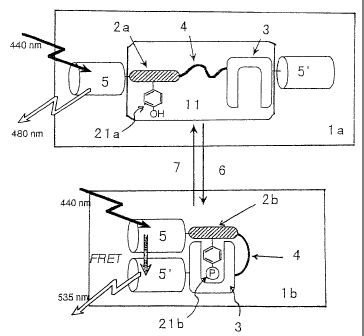
Fig. 1 is a schematic diagram that describes the
cons~l~uction and principle of the probe for imaging pro ein
phos~~orylation and dephosphorylation of the preaentinven ion.
1 to '8 represent the fol lowing : t in : probe for imaging pro ein
I
'I
~ _
6
CA 02410460 2002-11-22
phos~ orylation ore
and
dephosphorylation
(be
phoap orylation), tein
1b:
probe
for
imaging
pro
phosp , 'on),
orylation
and
dephosphorylat~on
(after
phosphorylat
11. andem ore
fusion
unit,
2a:
substrate
domain
(be
phos~ orylation),.2b:substrate
on),
domain(after
phosphorylat'
21a:' hosphorylation 1b:
site
(before
phosphorylation),
phosp orylation 3:
f site
(after
phosphorylation),
phos orylation aor
~ recognition
site,
4:
linker
sequence.
S:
d
chro~ phore, ioa
5':
acceptor
chromophore,
6:
phosphoryla
subs nce,
~:
dephosphorylation
substance)
Pig. fic
2
is
a
schematic
representation
that
shows
spec
stru, urea ion
of
the
probe
for
imaging
protein
phosphoryla
and ~hosphorylation ent
~e constructed
as
an
Example
of
the
pre
inven tion_
' ig. ser
3
is
a
representation
of
she
confocal
1
fluol scence ing
image,
which
shows
the
result
of
immunoblot
using phosphotyroSine ing
' antibody,
when
the
probe
fvr
ims
prot 'n (a)
phosphorylation
and
dephosphorylation
of
Figs.
2
and ) ple
2 were
( introduced
into
cells,
as
described
in
the
Exa
of 1 present invention.
t~
ig ing
.
4A
i
a
a
f
luorescence
image
of
CP'P
of
ter
introdu
the robe and
~ for
imaging
protein
phosphorylatian
deph~s an
horylation
of
Fig.
2
(b)
into
cells,
taken
usin
emisSibn
filter
for
~FP
(480
nm
t
15
nm)_
~ig. ges
4H
is
a
representation
of
the
pseudocolor
im
9
CA 02410460 2002-11-22
thatls~ows time course change of emission ratios between CFP
I.
(480 t1 15 nm) and YFP (5~5 f 12.5 nm) (herein after refe red
to a FP/YFP) excited at 440 t 10 nm, when cells to whic the
prob; fox imaging protein phosphorylation and
dephl phorylation of Fig. 2 (b) were introduced were stimul ted
with) asulin.
I
Fig. 4C is a graph that shows the time course chap a in
CFP/ P in the cytosol and the nucleus when excited at 40 ~
auh;; when the Bells to which the probe for imaging pr tein
10 phosb orylation and dephosphorylation of Fig. 2 (b) ere
intr.Ib aced were stimulated with insulin.
Fig. 9 D is a graph that shows the time course c ange
of CFFI/YPP (excitation at 440 t 10 nm) in the cyiosols o the
cell's jto which the probe for imaging protein phoephoryl tion
and ~e~hosphorylation of Fig. 2 (b) were introduced and treat~d
with ~yrphostin (an inhibitor of insulin receptor) (~).~ and
the i~e~lls to which the probe f'or imaging protein phosphoryla~tion
and ~i~phosphorylation of Fig. 2 (c) were introduced (~) , when
stin(u~ated with insulin.
~ ~ Fig. Spr is a representation of the pseudocolor it~ages
than shows the time Bourse change of CFP/YFP (excitati n at
440 t 10 nm) in the nucleus and the cytosol of the cel a to
which the probe for imaging protein phosphorylation and
dep o phorylation that contains a nuclear-export s gnal
segue ce as shown in Fig. 2 (d) were introduced, fter
a
CA 02410460 2002-11-22
sti ation
with
insulin.
Fig. a
5H of
is
a
graph
that
shows
the
tims
course
chap
CFp/Y P osol
I (excitation
at
40
t
10
am)
in
the
nucleus
and
the
cy
when the tein
cells
to
which
the
probe
for
imaging
pr
phos~ orylation ere
i and
dephosphorylation
of
Fig.
2
(d)
intro uced ious
were
stimulated
with
insulin
of
va
cone tration.
i
Fig. a
5A of
is
a
graph
that
Shows
the
time
course
chap
CFP/~ P sol
(excitation
at
40
t
10
nm)
in
the
nucleus
and
the
cy
when ;the tein
sells
to
which
the
probe
for
imaging
pr
phos~~orylation 3g.
and
dephoSphorylation
of
Fig.
2
(b)
and
2 ~were
(c~ introduced
were
stimulated
with
insulin.
IFig. ser
6B
is
a
representation
of
the
confocal
1
fluo~ scencg the
image
that
confirms
the
co-locali2ation
o
prob ' for and
iae~aging
protein
phoaphorylation
deph phorylation r
of in
Fig.
2
(e)
and
the
insulin
recept
I
the e is tion
d to
which
the
probe
for
imaging
protein
phosphoryl
and phoephorylation hen
I<l of
i Fig.
Z
(e)
were
introduced,
stun atsd
with
insulin.
Best ode
for
Carrying
Out
the
Invention
As by
described
above,
protein
phosphorylation
intrh cellular :
kinases in
is
one
of
the
most
important
ste
l
intrat ;ellular signaling and participates profoundl in
l
psoc~ ses tion
such
as
survival,
proliferation,
and
differentia
9
CA 02410460 2002-11-22
of c 11s. Accordingly, protein phosphorylation and
deph~ phorylation are observed as a phenomenon related t the
cause r certain symptoms of various diseases . In other w ds,
if al ethod for detecting and assaying phosphorylatio of
S spec is proteins is realized, early diagnosis of va ous
dise e8 may become possible. Moreover, the realizati of
a me~ d for screening factors or substances that enhan a or
inhi>~'t protein phosphorylation and dephoephorylation may
cont. buts greatly to the discovery of substances relati g to
suchj~iseases and the development of novel treatment dr gs.
IThe probe for imaging protein phosphorylation and
. . .
deph~~phorylation of the present invention is a probe that m kes
visil~ a and thereby enables the detection and assn of
l
phos orylation of protein by phosphorylatingsubstances. The
atru~ ure and principle of such probes for pro sin
phos~ orylation and dephosphorylation is shown in Pig. 1.
specifically, the probe for imaging pro sin
phos orylation and dephosphorylation tia) of the pre ent
inve ion comprises a tandem fusion uuit (11) that compr ses
a su trate domain (2a), which contains a site that ca be t
pho$' rylated (21a) and a phosphorylation recognition do sin
(3) nd together by a linker sequence (4), interpose or
sand~i hed between a donor chromophore (5) and an acce for
chromo hone t5') chat cause fluorescence resonance en rgy
transfer to oeeur.
CA 02410460 2002-11-22
For and .
the
probe
for
imaging
protein
phosphorylatio
dep o phorylation pee,
~ (1a)
of
the
present
invention,
for
exa
whe he main
phosphorylation
site
(21a)
of
the
substrate
d
(as) a the
phoaphorylated
by
a
phosphorylation
substance
(6)
adja;c ntphosphorylationreeognitiondomain
it,
(3)
recognise
and eeifically rate
~ interacts
with
the
phosphorylazed
subs
doma,i tale) tion
.
In
the
probe
for
imaging
protein
phosphoryl
and hosphorylation(lb)
rred,
a whet~insuch
aninteraction
occ
the nor (5')
d chromophore
(5)
and
the
acceptor
chromophore
come~i to sure
closeproximitytoeachother;
therefore,
uponexp
to ernal (5)
a light,
excitation
of
the
donor
chrvmophor
f ed akes
oil by
an
energy
transf
er
to
the
acceptor
chromophore
t
place resulting nce
in
a
change
in
efficiency
of
fluoresc
reso~ nce uch
energy
transfer
(FRET).
Thus,
by
detecting
chant in sin
FRET,
phosphorylation
(2b)
og
the
substrate
do
(2a) an
be
confirmed.
In ein
a
similar
manner,
when
the
probe
for
imaging
pro
phos orylation g a
and
dephoaphorylation
(1b)
containi
phos orylatsd a
substrate
domain
(21b)
coexists
wit
deph phorylation ite
substance
('1),
and
the
phosphorylated
of substrate hen
t domain
(2b)
is
dephosphorylated
(21a),
Intel coon (3)
b<t~reen
the
phosphorylation
recognition
domai
and substrate the '
~ domain
(2b)
disaDDears,
which
leads
to
sepal tion for
of
the
donor
chromophore
(S)
and
the
acce
chro~a ~phore (S'). By exposing external light, only the
__
I1
CA 02410460 2002-11-22
ex~'t tion of the donor chromophore (S) occur without a ergs
tra a er to the acceptor chromophore. Accordi gly,
den ~o nhorvlation of the substrate domain (2b) can be det cted
fro he change that appears in the FRET efficiency, sirag
fluol~r seance analysis_
Each unit constituting the probe for imaging pr tein
phos~ orylation and dephosphorylation of the presentinve tion
is c~ Cribed more specifically. First, as described a ve,
the ndem fusion unit (11) consists of the substrate do sin
(2a) ~, the phosphorylation recognition domain (3) arid the li~aker
segue ce (~) that bind them together. Here. the aequen or
stru ure of the Substrate domain (2a) is not sestricte as
longs s it contains a site that can be phosphorylated (2 a) . .
i
The ~i a capable of being phosphorylated (21a) usually corn sin
an -4H group, and natural amino acids such as tyrosine (T r) ,
serif (Ser) , threonine (Thr) and the like, as well as peps des
to w ch an OH group is introduced by chemical modifica ioti
may exemplified.
ext, the phosphorylation recognition domain recogn zes j
i
phos orylation of the substrate domain (2a) and inter cts
i
spec fically with the phosphorylatAd substrate domain t b).
The h sphorylation recognition domain may have any struc ure
i
as 1 as it fulfills the above conditions. For exam 1e,
endo a ous domains such as SH2 domain, phosphotyrosine bin ing
doma n, W?~t domain and the like that are known to recog ize
12
CA 02410460 2002-11-22
spedi is phosphorylated substrates are applicable. Fur her,
for tecting and assaying a substrate domain (2b) for hich
the h sphorylaticn recognition domain t3) that interacts ith
w
it is ~uxiknown, a single chain antibody may be prepared sing
the ~'osphorylated substrate domain (2b) as an antigen, and
used, s the phosphorylatien recognition domain (3) . By ing
suchla antibody, a phvsphorylation recognition domain t3) hat
Intel cts specifically with any desired phosphoryl ted
subs ate domain (2b) can be obtained, thereby enhancin the
versa l 1 l ty of such probe f or imaging protein phosphoryla~ion
and jd~phosphorylation. The single chain antibodies that
uCil~. a the substrate domain (Zb) as an immunogeamaybeprep red
by kn wn immunological means.
ext, in the probe for imaging Drotsin phosphvryla ions
and de hosphorylation of the present invention, the sego nce
and l;e$lgth of the linker sequence (4) are not limited as ~.on9 ,
as l sables appropriate flexibility and does not coma n a
site at can bephosphorylated. when the sequence (4) cont ins ;
a sil (21a) that can be phosphorylated, the site ma be
phos ~h rylated by a phosphorylation substance (6) , which m kes
the c orate detection and assay of protein. phoephoryla ion
impo s ble. The linker sequence (~i) may be any polypep ids
or ol~ opeptide, whichpreterablyhas a chain length long en ugh
co end. 1e interaction between the phosphorylation recogni ion
zite~(~) and the substrate domain (2b) upon phosphoryla ion
I I
~ ~ _
~ _
13
CA 02410460 2002-11-22
of substrate domain (2b), thereby approximating the onor
t
chr phore (5) and the acceptor chromophore (5').
I
The probe for imaging protein Dhosphorylation and
deg~ phorylation of the present invention comprises dem
the t
fusio unit (11) of the above-described structure, inter sed
betty n a donor chromophore (5) and an acceptor ehromo ore
(5' t at cause fluorescence resonance energy transfer ur;
) to oc
wheni a substrate domain (2a) is phoaphorylated, a chapa
in
FRET. s induced by the mechanism described previously. uch
10donor chromophore t 5 ) and acceptor chromophore ( 5 be
~ ' ) ma
selet ed from various fluorescent substances; partieula 1y,
fluo~ scent proteins are considered. The chromophores be
m
any stances that show f luorescence at dif f erent the
s~u iravelen
upon'e posure to external light, and cyan fluorescent ein
pro
15(CFP~ nd a yellow fluorescent protein (YFP) , which nts
are mut
of a grQen fluorescent protein (GFP), are preferable. CFP
t
and F axe particularly preferable; their mutated forms may
~
be e ared in accordance with their use and utilized the
p~ as
doao~ ndlor acceptor chromophare.
W
aoI n the above-described probe for imaging pro ein
phosp h tv
rylation
sad
dephosphorylation,
the
domain
adjacen
the or chromophore may be either of the substrate sin
o do
I
(2a) the phosphorylation recognition domain (3) . Since the
~
'
pref r ure
ble
linking
order
differs
depending
on
the
strut
ZSor is hindrance of these domains and linker sequence 4) ,
ste j _
1~
CA 02410460 2002-11-22
the b der away be selected according to the combination o the
subait ate domain (2a) and the phosphorylation recogn lion
domai (3) .
i Further, the probe for imaging protein phosphoryl tiOri
S and ~ phosphorylation of the present invention may cons in a ,
vari~ y of localization sequences at terminal end. ueh .
locals nation seguences can recognize specifie cells, spec' tic
regip s in th~ cells or specific tissues, and therefor can
loca~'ze the probe for imaging protein phosphorylation and
deph~ phorylation_ Specifically, a nucleag-export-si nal
seguer~ce or a plasma membrane biading sequence such as Alecks rin
homo~. Igy (PH) domain may be ligated as a localization segue ce.
he probe for imaging protein phosphorylation and
dephC~ 'phorylation of the present iavention is as described a vve .
i
But Lh method for it3 preparation is not particularly limi ed.
and m y be constgucted by total synthesis; however, i i9
pref. able to ligat~ each domain by known genetic engines ing
tec i es such as polymeraee chain rQaction (PCR). H re, ,
vdri! us restriction sites and the like may also be inser ed.
a
n the invention of the present patent applicatio
method for screening substances that enhance or suppress pro sin
i
phos~h rylation using the above-described probe for ima ing
prot~i phosphorylation and dephosphorylationis also prow ded.
Ia o r words, if the substrate domain (2a) of the probe for .
imag3,n proeein phosphorylation and dephosphorylation (la is ;
Z5
CA 02410460 2002-11-22
phos orylated rorhen the probe for imaging pr tein
phoa~ orylation and dephoaphorylation of the presentinve tion
i ,
and i andidate subseance coexist, protein phosphorylati n is
detec ed by the change is FRBT under the m~chanism previ usly '
desq~r~bed, thereby enabling the screening of subatances~that
phoslp~orylate the substrate domain (2a). These candidate
subset nces may act directly as a protein kinase that
phos~ orylate the protein, or may be substances that a t at
an e~ 1y stage of intracellular signaling, that is, act as a
prot;e a kinase activating substance.
vn the othex hand. in order to confirm enhanceme t or
supgr ssion of dephosphorylation by a candidate substanc and
i
to sit ~n a substance that enhance or suppress dephosphoryl tion.
the ~s~betrste domain (2a) of the probe for imaging protein
phoslp orylation and dephosphorylation (la) is first
phosp orylated (2b), and the change in FRET that occur when
in c~o~xistence with a Candidate sub9tance is m~asured.
Is the above-described method for screening subst nces
the hence orsuppressphosphorylation and dephosphoryi lion,
the probe for imaging protein phosphorylation and
depho phorylation ( la) may be made to coexist wi th the cand' data
subset nce in a solution, for whieh, for example, the pH, salt
con 'e tration or the like is adjusted, or alternatively the
pro a for imaging protein phosphorylation and
depho~phorylation may be introduced into cells by ge etic
16
CA 02410460 2002-11-22
i
I
engij eying techniques thereby made to coexist with the
candi ate substances. Here, the candidate substances m y be
pres!e t outside the cells or may bs incorporated into the c 11s;
fur 'h r, they may be pre-introduced into the cells by ge etic
S eng'~n eying techniques. The candidate substances rnay al o be
en2 s, receptors or the like that exist in the cells.
Further, by using the Drobe for imaging pr lain
pho p orylation and dephosphorylation of the presentinve lion,
the;p osphorylating substances may be assayed, as well In
othdr words, by introducing the above probe for imaging pr lain
pholp orylation and dephosphorylacion into the cells, and
men a ing the changes in fluorescence wavelengths, the a ount
o~ ph sphorylation substances can be determtrred. For exa ple,
for a balance A known to phoaphorylate protein a, by obse wing
the ~'me course changes that occur in FRET in vitro when va ioua
coats trations of the phosphorylation substance A a a in
coed t~nc~ with the proi~e fog imaging protein phosphoryl Lion
aad~d phosphorylation, and the time at which each FRET slue
reach s a plateau is predetermined. sy creating a calibr tion ,
cur a of the FRET value and the concentration of substa cs A
at h t time, preparing the probe for imaging phosphoryl lion
andl ephosphorylaeion that contains protein a, whit is
pho p orylated by substance A, introducing such the prob into
cal s, and measuring the FRET value, substance A in the ells
can a quantitated. Likewise, quantitative analysis of
1 'l
CA 02410460 2002-11-22
depi ophoryiation substances may be pezformed in a si ilar
manne .
As has been described previously, known ge etic
Il
engin eying teehniques are applicable for as a metho for
int oucing the probe for imaging protein phosphorylatioand
dep. ophorylationintothe cells. Specifically, an expre aion
vec oin which the probe for imaging protein phosphoryllion
and! phosphorylation is incorporated may be introducedinto
the lls by known methods such as electroporation, cium
the ca
phoap ate method, the liposome method, the DEAF dextraahod.
me
Thug, by introducing tile probe for imaging pr tein
phosp l orylation and dephosphorylation into the cells king
andm
the obe coexist wieh phosphorylation (or dephosphorylaion)
~a
i
sub races, an in vivo x~ethod for detecting and assayingtein
~t pr
pho~ porylation (or dephosphorylation) that does not ire
re
the dstruction of the cells is enabled.
Th~ probe for imaging protein phosphorylstion and
deptio phorylation of the present invention is advaneag ous,
not 1y for enabling the imaging of kinase signal transdulion
b
in gle live cells at high Spatial and temporal resoluion, '.
s;i
but so for being valuable in multi-cell analysis, aims
; which
i
for a high- rhrvughput screening of substances that late
~ reg
i
pho~ porylation or dephosphorylation (science X79, 4-88
(199 18; Drug Discovery Tvday 4. 363-369 (1999).
. Further, in the probe for imaging protein phosphoryllion
18
CA 02410460 2002-11-22
and. dephosphorylation of the present invention a
pol . cleotide for the expression of the probe may be intro uced
into', c 11s, and used fvr the ontogenesis of non-human totip tent
cell' thereby creating an animal or a progeny animal ich
in
S the robe for imaging protein phosphorylation and
dephb phorylation and the phosphorylation (or
depho phorylation)substance coexistin allofitscells. ese
T
so-c~~ led non-human transgenic animals may be produce in
accoilr ance with known production methods (for example, oc.
I P
Nat3 Acad. Sc~. USA ??, 7348-, (1980)). The non-h man
.
traps epic animals described above possess the probe ing
far ima
prvte n phosphorylation and dephosphorylation in all sir
of t
somas' c cells, and therefore. may be used to measure the
concen tration of phosphorylation (or dephosphorylat on)
suhst nces in their cells or tissues; or by introdu ing
candi aces of phosphorylation (or dephosphorylat on)
subs nce8, phosphorylation (or dephosphorylation) enhaning
subs ces, and phosphorylation (or dephosphorylat on)
inhi i ting substances, suchas drugs an toxins, into cheirboia,
substa pnces that show effect in cells or tissues may ed.
be acres
' ereinafter, the present invention is describe in
l
Curt a detail by the Lxamples wi ch reference to the accompaning
draw n s. of course, it should be needless to mention hat
the ~ sent invention is not limited to the following les
l Exam
and t various modifications may be made for the data 1s.
L'h
19
CA 02410460 2002-11-22
I
Exam ee
Among various phosphorylation substances, nonrec for
tyre ne kiaas~s and seine/threonine kinasee function
throliu bout the entire signal transduction cascades. O the
other and, tyrosine kinase rec~ptors such as insulin rec for
and h rmone receptor function at the beginning of a numb r of
signal transduction eascades.
In the following examples, a probe for imaging pr tein
phos orylation and dephosphorylation uain~ insulin ei nal
tray;' uction protein was evaluated for the detection and ssay
of p tein phosphorylation by insulin receptor. which is lso
W
a prot~e3n kinase.
<Preparation>
i In the following escamplee, samples and reagents arsre ssd
I
as fo lows:
Human insulin was
purchasedfrom Peptide instituee, Iac.
(Osa~ . Japan). .
Ham's F-2 medium, fetal calf serum, Hank's balanced salt
solu't on and. LipofectAMINE 2000 reagent were obtained from ife
i
Tech logies (l~ockville, MD).
Tyrphostin 25 was purchased from sigma Chemical Co. (St.
Louis MO) .
Anti-phosphotyrosine antibodytPY20) and anti-~-su unit
of h~ an insulin receptor antibody were purchased from ants
CA 02410460 2002-11-22
Cruz ioteehnology,
Tnc.
(Santa
Cruz,
CA).
y
Anti-GFP Palo
antibody
were
purchased
from
by
Clontech
(
Altos, CA)
.
Anti-rabbit fined '
IgG
antibody
labeled
with
Cy5
was
obt
from acson ) .
Irn~aunoResearch
Lab.
,
Znc.
(Pennsylvania,
F
Other de.
Chemicals
a~edwere
all
of
analytical
reagent
g
<Exa ~n1e tion
1>
preparation
of
the
imaging
probe
for
the
detec
of tein
pjt phosphorylation
~ by
insulin
receptor
(1) lasmid
~ Construction
; Fig. a of
2
is
a
representation
o
the
specific
atructu
each probe and
for
imaging
protein
phosphorylation
deph~ phorylation
that
were
prepared.
As so
shown
in
Fig.
2,
the
imaging
probe
was
prepare
as omprise ain
tc~ a
tandem
fusion
unit
wherein
a
substrate
do
cont. ning ion
a
site
to
be
phosphorylated
and
a
phosphoryla
reco itian ce,
domain
are
bound
together
by
a
linker
segue
I
which t~ ant
interpoeedbetwesn
two
mutants
of
the
green
fluores
prot~ n.
First, sin
as
a
fluoresCentpsotein,
cyan
fluorescent
pro
(CFP~ and are
yellow
fluorescent
protein
(YFP),
which
diff~ !nt-colored sin .
mutants
of
the
green
fluorosc~nt
pro
(GFp~ riginating er.
fromAeguorea
victoria,
were
used.
Furt
CFP was of
~ subjected
to
additional
mutations
F64L,~ 65T/Y66W/N1961/M153T/V163A/N212K.arid
tad
YFP
wassubje
to itional mutations of S65G/V68L/Q69K/S72A/T203Y.
add
21
CA 02410460 2002-11-22
Next, as a substrate domain, a tyrosine phosphoryl tion
domaii (Y941: 9EQ ID Yd~: 1) derived from insulin rec ptor
subset ate-1 (IRS-1) was used. In this domain. insulin rec for
phosp~orylates the tyrosine residue 941 in an insulin depe dent
manna tMol. Cell Eiol. 13, X418-7428 (1993)7.
Next, as a phosphorylation recognition domain an
i
N-t ! final S;H2 domain (SH2n: pe533o-gag) of p85 regulatory au nit
of Y~o fine phosphatidylinositol 3-kinase, which has een
repo~ ed eo bind to the phosphorylation substrate domain ofI S-1
protein, was chosen. (~. Hfol. Chem. 26~, 25959-25966)
IAs a linker sequence (Ln), the oligopeptide of 5E ID
I
NO: ~ was used.
xestriccion sites shown in Fig. 2 were inserted to the
cDNA ~f CFP, YFP, the substrate domain and the phosphoryla ion
rsco' ~it3on domain, uairbg standard polymerise Chain rear ion
(PCR).I~ All cloning :nzymee were purchased from Ta ara
Biorn$ ical (Tokyo, Japan). PCRfragments weresequenced a ing
AH13i0~ genetic analyzer.
i urth~r, cDNA encoding each probe for imaging pro ein
phos~ rylation and dephosphorylation was subcloned at jnd
III Xba Z sites of a mammalian expression vector, pcDN 3 .1
t + ) ( nvi trogen Co . , Carlsbad, CA) .
(2) timization of the structure of the probe for ima ing
a
protein phosphorylation '
ZS ~ ~n the probe ~or imaging protein phosphorylation and
as
CA 02410460 2002-11-22
o~aphorylation shown in Pig. a (a) to (e) , the order ofd sH2n
and ;~r~41 in the tandem fusion units of probe (a) and probe (b)
are ~Ir~versed.
In the present study, to determine which tandem f~siori
unid, ~ that of prabe (a) or probe (b) , is more efficl~nely
phoslp orylated by insulin receptor, immunobiotting was
perE~o ed using phosphatyrosine antibody after ~timul ting
CHO~.I~t Cells expressing probe (a) and probe .(b) with iC~O nM
insu'l~a.
' First, IR cells were cultured in 6-well plates ere
and
tram acted with 2 ug of each plasmid containing probe cDNA
(a)
and be (b) cDNA. CHO-~ITR Cells overexpressing human lizi
g~r ins
recap#or ith
were
cultured
in
Ham's
F-12
medium
supplemented
G
10 etal eal serum at 37 C in 5 % CO=. The cells ere
%~
~
tram 'acted with LipofectAMINE 2000 reagent. 12 to 24 urs
h
after he transfection, the cells were spread onto glass tom .
bo
dish's , glass coversiipa or plastic culture dishes.
ext. CHO-IR cells expressing probe (a> and probe (b)
were imulated with 100 nM of insulin for 20 minutes 5C
s at 2
j .
he cells were ly8ed with an ice-cold lysis buffer (50
'
mM -HC1, pH 7.4, 100 mM NaCl, imM BDTA, 0. 1 % Triron 00,
T X-
~i
l
10 NF, 2mM sodium orthovanadate, 1 mM PMSP, 10 ug/mL tin,
mi~ pepst
10 /L leupeptin, 10 ~tg/mL aprotinin) _ The Imaging bas
N pr
for rteia phosphorylation were immunoprecipitated from the
whole ell lyeates of the CHO-IR cells with anti-GFP ody
anti
l ~ _
23
CA 02410460 2002-11-22
for hours at 4 °C.
Protein G-8epha=oee 4 FF beads were used to absorb the
immu precipitates and then washed four times with an ice- old
wash g buf fer (50 mM Tri~-HC1, pH 7 . & , 100 mM NaCl , imM E TA,
0.1 ~ riron X-100, 10 mtrl NaF, 2mM sodium ozthovanadate, mM
PMSF, 0 ~tg/mL peps tatin, 10 ~tg/mL leupeptin. 10 ~g/mL aproti in) .
The ampler were separated by SDS-polyacrylamide gel
elec. ophoresis ~tn~ analysed by an immusioblotting method a ing
i
anti' hosphotyrosine antibody tPY20, 1:500 dilution).
'The result ef the icnmunoblotting is shown in Fig. 3.
s shown in Fig. 3, probe (b) was well phosphoryl ted
i
by ins lin receptor, whereas probe (a) was poorlyphosphoryl ted.
This indicates thae in the present experiment, the tandem fu ioa
unit:l~inked in the order shown in Fig. 2 (b) is more effec ive
as a probe for imagiaag protein ~xhosphorylation than the to dem
fusir~l unit linked in the order shown in Fig. 2 ta).
i
i In view of the X-ray crystal structure of i~nauliti rocs for
in cl lex with a substrate peptide derived from IRS-1, the
diff~ ence in the phosphorylation efficiency between prob (a)
and pf be (b) may be ascribed to the difference in steric eff ct.
n the following example, phosphorylation by ins lin
rece~ or was detected using probe (b).
<Exath 1e 2> Detection of phoaphorylation using the probe Lor
image g protein phosphorylation
24
CA 02410460 2002-11-22
i The increase in FRET efficiency after the phosphoryl tion
of p!r be (b) by an insulin receptoz was observed.
CHO-IR cells were transfected using the cDNA enc ding
prop tb) inserted in a mammalian expressionvector, as dese ibed
in E~ mple 1.
I! After serum starvation with a serum-free medium, the
i
cule~ emediumwasreplacedwithaHank'sbalancedsaltsolut'on.
3 to~ days aft~r the transfection, the cells were obse ved
at r m temperature on a Carl Zeies Axiovert 135 micros ope
with.a ooledCCDcarneraMicroMAX (Roper ScientificInc. , Tuc on.
A2), c ntrolied by:MetaFluor (Universal Imaging, west Ches er,
PA) inlaecordanee with known methods (a.na1 . Ghem. Via. 5918= 924
(1999)0 .
a exposure time at 440 1- 10 nm excitation was 1Q0~ ma .
The florescence images ware obtained through 480 ~ 15 nmf and
535 t; 1~2 . 5 nm filters with a 40x oil immersion lens (Carl Ze~.sa,
Jean ~ ~erma:ny) .
ig. 4A shows fluorescence microscope images of
(b) ~x~ressian sells, taken using an emission filter (410 t
15 ruts) I for CFP.
robe (b7 was found to be distributed uniformly in both
the ~y~osolic compartment and in the nucleus.
ext, to evaluate the response of probe (b) for~its
pho:~h~rylation, CHO-IR cells expressing probe tb) here
stimulated with 100 nM of insulin in the same manner as descr' bed
ZS
CA 02410460 2002-11-22
I
in mple
I 1.
Pig ages
.
4B
shows
the
time
course
changes
of
pseudocolor
i
of ssion 535
eI ratio
of
CFP
at
480
t
15
nm
to
that
of
YFP
a
i nm
12~. excited
at
4~0
~
to
nm.
~ Further, atio
4C
shows
the
time
courses
of
she
emission
i
chaz~g s tion
in
the
cytosol
and
in
the
nucleus.
The
adminietr
of ~sulin the
eaused
a
rapid
and
significant
decrease
i
cyt alic reas
emission
ratio
foz
cells
expressing
probe
(b)
,
wh
the iseion ange
'e ratio
in
the
nucleus
showed
no
significant
c
(Fig. 4H
and
4C).
IFurthermore,theinsulin-induced
atio
changein
emission
in cytosol were
~h was
completely
suppressed
when
the
cells
pre sated ultn
with
900
~M
tyrphostin,
an
inhibitor
for
in
rec! tor. robe
As
a
negative
control.CHO-IR
cellsexpresaing
(c). in the
which
tyrosine
was
replaced
with
alaniae
a
phos ~hoacceptoz ated
I site .
of
the
substrate
domain,
were
simu
with ~inaulin: olio
however,
no
significant
change
in
the
cyto
emi ion
ratio
was
observed
(gig.
gD).
The YFP
above ,
results
demoastrate
that
FRET
from
CFP
i seed the
inch upon
phoaphorylation
of
Y941
of
probe
(b)
1
cyto olic the
compartment
and
subsequent
binding
of
phos horylated tion
Y941
with
the
adjacent
phosphoryl
rec. nition ales
domain
SH2n.
Accordingly,
this
result
indi
,
that probe (b? may be effective as a probe for imaging tein
pr
~5 pho~~ horylation 1s.
by
insulin
receptor
in
single
live
ce
26
CA 02410460 2002-11-22
°s3owever, no significant change in FRET efficiency) was '
obse~ ed in the nucleus; a more rigidly packed conform lion
of t~ osine-phosphorylated probe (b), compared to the f oppy
confb maLion of unphosphorylated probQ (b) due to the axis eace
of t linker sequence, may have restricted the traffic o the
phoe~ orylated probe (b) through th~ nuclear pore, which f rced
the; oaphorylated probe fib) to remain in the cyto olic
coma tment .
I
<Exalm 1e 3> Probe for imaging protein phosphoryl tion
conta'ning a nuclear-export-signal sequence
To prevent the probe four imaging protein phosphoryl tion
fro eing transferred to the nucleus, where FRET change did
not'o cur upon insulin stimulation, as described in Ex mple
l
2, ~ ~ probe for imagi3ag protefa phosphorylaCion haul g a
nucXelr-export-signal sequence (d) was developed. A the
nud a r-export-signal sequence, a nuclear-export-s gaal
aeq~e ce (nes; sEQ ID NO: 3 ) derived from human immunodsf is ency
virus protein, Rev (EMHO J. I~, 5573-558i) , was linked t the
I
tar final ead of the probe for imaging protein phosphoryla ion.
I Plasmid construction and transfection was do as
des ibed in Example 1.
' No significant f luorescence was observed from the nu leus
of h probe (d) -expressing cells. It was confirmed that the
pro fox imaging protein phvsphorylation (d) was remove from
27
CA 02410460 2002-11-22
the nucleus (Fig. 5A).
Further, upon stimulating the cells expressing prob (d)
with 00 n>~ insulin, in the same manner as in Example , a
prog naive decrease in the cytosolic emission ratio ways
obs r ed (Fig. 5A). No significant difference was obs rued
I
in t~ time course of the prabe (d) response, even when com ared
withl~ hat of probe (b) (Fig. 4B).
Fig . 5B shows the response of probe ( d) to di f f ring
con a trations of insulin in CHO-IR cells. The accumui tion
xat of phosphorylated probe (d) by insulin recepto was
znc~e sed in para2lel with increasing insulin concentra ion.
when ~he concentration of insulin was 0.1 nM, no accumul tivn
of p;~osphorylated probe (d) was observed.
I The relation between the emission ratio of probe ( ) and
ins,l in concentration was similar to the results report d fot
tyr ins phosphorylation of native IRS-1 protein in the e11,
pre ously measured by autoradiography (EMBO J. 16, 5573 5581
( 199 ) ) .
The above results indicate that probe (d) is suitable
as . probe for t~sulti-cell analysis that utilize fluore cenco
I
mu1 t -well plate reader, wherein the probe protein in the c tosol
an~ he nucleus cannot be discriminated.
Hence, by using probe (d) , high-throughput screen ag of
an Ii diabetic small molecules, such as L-'783, 281 (Netur 318,
18~- 66 (1985); Science 38~, 9'74-977 (1999)), whic were
za
CA 02410460 2002-11-22
repel eel to directly st3mulata the kinase activity of in ulin
rece~ or, from thousands of candidate chemicals, ma be
realllited.
<Exx~m 1e 4> Probe for imaging protein phoephorylatio and
depl~o phorylativn comprising a living cell membrane bi ding
seq~e ce.
Signal transduction proteins, such as kin sea.
pho p stases and their substrates, are often localized the
l
cell ad are organized to form domains of signal transdulion
by ~tracellular stimuli. This mechanism is thought be a
ex to
cri ~ial factor to determine the efficiency and speeif city
l
oL gnal transduction in the cell.
si
It has been known that IRS-Z, which is the endog nous
sub rate protein for insulin receptor, contains a trin
g Aleck
home ogy (pH)., domain and a phosphotyrosine binding main
(PTB) d
to s 1e1.-tera~ina7. end (Diebetologia ~0, S2-S17
~ (1997)
The PH and PTB domain bind with the phosphoinoai ides
of cell membrane and wi th the juxtatnembrane domainu1 is
h of in
recd tor, which is tyrosine-phosphorylated by l ulia
sini ~ ation, respectively (Pros. Natl. Aced. Sci. US 96,
I
837~ 8383). Thus, the concentration of IRS-1 is ins eased
arv d insulin receptor at the plasma membrane upon sulin
l
sti lation, which underlies efficient and sel ctiva
phas horylatian of ZRS-1 by insulin receptor (J. H.fol.chem.
~9
CA 02410460 2002-11-22
2'i0,' 1715-11?18 (1995)).
Then, probe (b) was fused with PH-PTH domain derived from
i
the I S°1 protein to construct the probe of Fig. 2 (e)
CbIO-Ilt cells expressing probe (e) were stimulated with
100 of insulin for ? min at Z5 °C. The cells weze fixedlwith
2 ~ ' pmraformaldehyde and were permeabilized with a
phodp ate-buffered saline containing 0.2 % Triton X-10 for
i
m~i . After 45 rnin of incubation with rabbit anti-~ su unit
of h~an~an insulin receptor antibody (1:100 dilution) , the ells
10 wer ashed with a phosphate-buffered saline containing .2 %
fis kin gelatin and incubated with anti-rabbit IgG sat body
label d with Cy5 (1:500) for 30 min.
The coverslips were mounted onto the slide and oba rued
with confocal laser scanning microscope ( LSM 510, earl se ss) .
1S i ~ Fig. 6A shows a comparieoa of the cytosolic emission atio
changle for probe (e) and that for probe (b) in CHO-IR ells
whe ~ timulated with 100 nM insulin.. Although th~ rate o the
cyt s lit emission ratio ehange for probe (e) was eignifis ntly
fas~e than that for probe (b), both emission ratios r were
not~s'gnificantly different when they plateaued.
iAecordingly, this indicates that by introducin the
end g noun targeting damain within IRS-1, the phosphoryl tion '
rat f probe (e) by the activated in9ulin receptor was enha ced,
Whig demonstrated that the localized kinase signaling i the
livix~g Cells can be visualized effectively using this robe
CA 02410460 2002-11-22
for aging protein phosphorylation.
Insulin-stimulated co-localization of probe (e) an the
inau ~.n receptor at the plasma membrane were confirmed the
' b
fluo sconce images taken by the confocal laser scan ing
micr cope (Fig. 6S) . This membrane localization of (e)
prob
was observed before insulin simulation. on the other nd,
~o h
whenlp obe (b) was used, no significant subcellular loealization,
inc ing the plasma membrane, by insulin stimulation was
h
obser ed (Fig. 4H>.
From these results, it was demonstrated that the -PT8
P
doma~ was ascribed to be responsible for the Insulin-incod
I
targ~ ing of probe (e) to the membrane insulin recepto .
Furthermore, it ie suggested that upon ins 11n
i
stim~ ation, S~2n within the probe for imaging pr tein
l
phos orylation pzeferentiaily binds via intramole lar
~
react on with the adjacent phosphorylated Y941 rather than
bin g via intermolecular reaction with the other localized
phosi oproteine such as endogenous IRS proteins (J. em. .
Hio1 .
aT3,~2 9686-29692 (1998) ; MoI. Endvcranol. 14, 823-836 0) ) .
i20
Indttla rial Applicability
As described above in detail, the present inve Zion
pro d ea a method for imaging signal tr:nsduetion caus d by
i
protls n phosphorylatloaa within living cells. The pr sent
I
invent ion not only enable: the of sualization of kinasegnat
a '
31
CA 02410460 2002-11-22
traps uction within single live cells in high spatial and
tempo al resolution, but also enables the high-throe hput
screie ing of substances that regulate the activity of va ious
pho~p orylation and dephosphorylation substances_ Fez her,
by gl erating tranagenic animals or plants using the prob for
imac,~i g protein phosphorylation of the present invents n, a
none tructive continuous method for monitoring events re ated
s
to a~i pal transduction due to protein phosphorylation w thin
tar a tissues and organs can be realized.
I
3~
CA 02410460 2002-11-22
Seouente Listing
i
0110>iJ pan Science and Technology Corporation
C120>~P obe for inutging protein ohosphorylation / dephosphorylation and ethod
of de a tine and determining protein ohosphorylation / deDhosporyiation
C130> 0 -F-008PCT
<160>i3
C210> ~1 ,
C211> ~1 '
<212> ~P 1
C213>:A tificiai SeQUence
X220>~S nthesized Oligoaeptide
C400> 1
Glu T r Gly Thr Glu Glu Tyr stet Lys I~et Asp Leu Gly
1 ~ 5 10
C210> .2 i
C211>',11
C212> !P T
C213> ~ tificial Seauence
C220> S nthesized 0ligopeptide
C400>
Gly As~ sn Gly Gly Asn Asn Asn Gly Gly Ser
1 s to
<214> ;
<211~
C212> R '
C213~ r ificial Sequence
C220~ y thesized Oligoaevtide
<400> b
Leu Prp ro lec~ Giy Are Leu Thr Leu
1 I 5
1/t