Note: Descriptions are shown in the official language in which they were submitted.
CA 02411056 2002-11-21
1
DESCRIPTION
PHARMACEUTICAL COMPOSITIONS CONTAINING
PROSTACYCLIN SYNTHASE GENE
Technical Field
The present invention relates to pharmaceutical compositions
for inducing apoptosis, which are useful for disease treatment where
the induction of apoptosis produces therapeutic effects;
pharmaceutical compositions. for gene therapy, the pharmaceutical
composition being useful for treating cancer; and screening methods
for agents that induce apoptosis in cells.
Background Art
Prostaglandin (PG) is a family of various eicosanoids derived
from arachidonic acid (Vane, J. R. et al. , Am. J. Cardiol. 75, 3A-10A
(1995)). Arachidonic acid is converted into an endoperoxide
intermediate, prostaglandin H2 (PGH2) , by cycloxygenase. There are
at least two types of isoforms of cyclooxygenase (COX-1 and COX-2).
COX-1 is expressed constitutively in most tissues and cells; in some
cases, the expression level is elevated during cell differentiation.
In contrast, the expression of COX-2 is often up-regulated upon
various types of stimulations (for example, mitogen, cytokine and
endotoxin). Subsequently, the product PGH2 produced by the action
of COX is converted into various types of prostaglandins (e.g. , PGD2
and PGF2o6 and prostacyclin (PGI2)) as end products by specific
synthases (Tanabe, T. et al., J. Lipid. Mediat. Cell Signal. 12,
243-255 (1995)). Prostaglandins produce a variety of effects in
various aspects of regulation in homeostasis and pathogenesis. For
example, PGE2 regulates blood pressure, fertilization efficiency, and
cell protection; prostacyclin not only contributes to the maintenance
of cardiovascular system but also shows proliferation-inhibiting
activity and cell-protecting activity. It has been reported that the
overexpression of COX-2 in cells of epithelial cell line from
gastrointestinal tract is associated with inhibition of apoptosis
(Tsuj ii, M. et al. , Cell 83, 493-501 (1995)) and PGE2 is a major product
CA 02411056 2002-11-21
2
in cells (Tsujii, M. et al. , Cell 93, 705-716 (1998)) . In contrast,
the overexpression of COX-2 in an immortalized endothelial cell line
results in retardation of cell proliferation and increases the
frequency of cell death (Narko, K. et al., J. Biol. Chem. 272,
21455-21460 (1997)). Prostacyclin synthase (PGIS) is expressed
endogenously in endothelial cells at high levels; prostacyclin is
a major derivative of PGH2 in cells (Kara, S. et al. , J. Biol. Chem.
269, 19897-19903 (1994)).
It is known that the above-mentioned prostacyclin is an unstable
lipid mediator whose half life is 5 to 10 minutes (Sinzinger, H. et
al., Arch. Gynecol. Obstet., 243, 187-190(1988)), and that
prostacyclin plays important roles as a strong vasodilating subs tance
and a strong endogenous inhibitor to platelet aggregation (Moncada,
S. et al. , N. Engl. J. Med. 17, 1142-1147 (1979) ) , which are presumed
to be mediated by G protein-coupled receptor which increases the cAMP
level (Smith, E. M. et al. , J. Biol. Chem. 271, 33698-33704 (1996) ) .
So far, prostacyclin is poorly characterized only by the presence
of cell-protecting activity to various cells (for example, vascular
endothelial cell, myocardial cell, gastric cell, hepatocyte, and
renal cell) in addition to the well-known activity (Vane, J. R. et
al., Am. J. Cardiol. 75, 3A-10A (1995)).
Disclosure of the Invention
An objective of the present invention is to provide
pharmaceutical compositions for inducing apoptosis, which are useful
for disease treatment in which the induction of apoptosis produces
therapeutic effects. Another objective of the present invention is
to provide pharmaceutical compositions for gene therapy, the
pharmaceutical compositions inducing apoptosis and thereby leading
cancer cells to cell death. Yet another objective of the present
invention is to provide screening methods for agents that induce
apoptosis in cells, the method enabling conveniently screening for
agents capable of inducing apoptosis.
Specifically, the present invention relates to:
(1) a pharmaceutical composition for inducing apoptosis in a
cell, the pharmaceutical composition comprising a prostacyclin
CA 02411056 2002-11-21
3
synthase gene as an active ingredient;
(2) a pharmaceutical composition for gene therapy of cancer,
the pharmaceutical composition comprising the pharmaceutical
composition according to (1) above as an active ingredient; and
(3) a method of screening for an agent that induces apoptosis
in a cell, the method comprising determining activation of peroxisome
proliferator-activated receptor (PPAR)-S in the presence of a test
substance.
Brief Description of the Drawings
Fig. 1 shows photographic patterns indicating influences of
COX-2 expression on BAEC cells, SVS30 cells, HEK-293 cells, and CV-1
cells. An expression vector forj3-galactosidase was co-transfected
into cells with a mock-transfection vector (a, c, e, h) or COX-2
expression vector (b, d, f, i) , and then the cells were stained with
X-Gal. An expression vector for wild-type PGIS (PGISwt) was
co-transfected into HEK-293 cells (g) and CV-1 cells (j).
Fig. 2 shows photographic patterns indicating influences of the
expression of wild-type PGIS (PGISwt) or inactive PGIS (PGISC441A)
on human fetal kidney epithelial cell line HEK-293. The wild-type
PGIS (PGISwt) enhanced apoptosis in HEK-293. The cells were
transfected with a PGISwt expression plasmid (a, b), expression
plasmid for inactive PGIS (PGISC441A) (c, d), or negative control
mock plasmid (e, f). The cells were fixed with 3.7% formaldehyde,
and then double-stained with anti-PGIS polyclonal antibody P4 (a,
c, e) and fluorescent dye bis-benzimide (Hoechst 33258)(b, d, f).
PGISwt (a) and PGISC441A (c) expressed in the cells were detected
immunologically using a Texas-red-conjugated secondary antibody.
Fig. 3 shows diagrams indicating: (a) the time course of % ratio
of apoptosis in anti-PGIS positive cells and Hoechst 33258 positive
cells, and (b) an influence of increased amount of PGISwt or PGISC441A
expression plasmid on apoptosis. In the upper part of panel (b) , the
expressed PGISs (PGISwt and PGISC441A) were detected by
immunoblotting analysis using an anti-PGIS antibody P1. Percentage
of apoptotic cells was increase being in correlation with the amount
of PGISwt protein expressed in the cells (the bottom part of panel
CA 02411056 2002-11-21
4
(b)) .
Fig. 4 illustrates photographic patterns showing results of
co-transfection of the expression construct for P-galactosidase with
another construct for PGISwt (a) or PGISC441A (b) into cells.
Fig. 5 shows a diagram indicating percentage of apoptotic cells
in total cells to which the indicated expression constructs had been
transfected. The cells were transfected with expression vectors for
P-galactosidase, COX-2, and for PGISwt or PGISC441A or a mock
expression vector in combination as shown in Fig. 5 in the presence
or absence of arachidonic acid, followed by determination of the
percentage ratio of cells stained blue with apoptotic morphology (for
example, membrane vesicle formation and cell body shrinkage) to total
cells stained blue. All results were obtained from three sets of
experiments (n=3) and are presented as mean S.D.
Fig. 6 shows a diagram indicating levels of 6-keto-PGFIU
determined for the respective cells to which the indicated expression
constructs had been transfected. All results were obtained from
three sets of experiments (n=3) and are presented as mean S.D.
Fig. 7 illustrates a diagram showing the percentage ratio of
specific apoptotic cells in serum-free medium containing Iloprost
at the concentrations indicated (0, 1, 10, or 100 .LM) . The cells were
transfected with expression plasmids for PGISwt, PGISC441A, or a mock
control expression plasmid and (3-galactosidase expression plasmid.
After 17 hours, the medium was changed with serum-free medium
containing Iloprost at various concentrations (0, 1, 10, or 100 AM) .
All results were obtained from three sets of experiments (n=3) and
are presented as mean S.D.
Fig. 8 illustrates a diagram showing the intracellular level
of cAMP accumulated in cells in serum-free medium containing Iloprost
at the concentrations indicated (0, 1, 10, or 100 M) . The cells were
transfected with expression plasmids for PGISwt, PGISC441A, or a mock
control expression plasmid and Q-galactosidase expression plasmid.
After 17 hours, the medium was changed with serum-free medium
containing Iloprost at various concentrations (0, 1, 10, or 100 M) .
All results were obtained from three sets of experiments (n=3) and
are presented as mean S.D.
CA 02411056 2002-11-21
Fig. 9 illustrates a diagram showing the percentage ratio of
specific apoptotic cells in serum-free medium containing dbcAMP at
the concentrations indicated (0, 1,.10, or 100 M). The cells.were
transfected with expression plasmids for PGISwt, PGISC441A, or a mock
5 control expression plasmid and 3-galactosidase expression plasmid.
After 17 hours, the medium was changed with serum-free medium
containing dbcAMP at various concentrations (0, 1, 10, or 100 M)
All results were obtained from three sets of experiments (n=3) and
are presented as mean S.D.
Fig. 10 illustrates a diagram showing the percentage ratio of
specific apoptotic cells in serum-free medium containing dbcAMP at
the concentrations indicated (0, 1, 10, or 100 M). The cells were
transfected with expression plasmids for PGISwt, PGISC441A, or a mock
control expression plasmid and P-galactosidase expression plasmid.
After 17 hours, the medium was changed with serum-free medium
containing H-7 at various concentrations (0, 1, 10, or 100 M) . All
results were obtained from three sets of experiments (n=3) and are
presented as mean S.D.
Fig. 11 is a diagram showing the influence of the expression
of PGIS on PPAR activation. The degree of PPRE activation (%) was
determined using PPREx3 luciferase reporter plasmid co-transfected
with an expression plasmid for PGISwt or PGIS441A in various amounts
(0, 0.1, 0.5, or 1 g). The results were obtained from three sets
of experiments (n=3) and are presented as mean S.D.
Fig. 12 is a diagram showing the influence of the expression
of PGIS on the intracellular level of 6-keto-PGF1a. The intracellular
level of 6-keto-PGF1a was determined by EIA after the cells were
co-transfected with an expression plasmid for PGISwt or PGIS441A in
various amounts (0, 0.1, 0.5, or 1 g). The results were obtained
from three sets of experiments (n=3) and are presented as mean S.D.
Fig. 13 is a diagram showing the influence of the expression
of PGIS on PPAR activation. The effect of Iloprost (100 J.1M) on the
degree of PPRE activation (o) was determined by using
PPREx3-luciferase plasmid co-transfected with 1 g of PGISC441A
expression vector into HEK-293 cells or CV-1 cells. The results were
obtained from three sets of experiments (n=3) and are presented as
CA 02411056 2002-11-21
6
mean S.D.
Fig. 14 is a diagram showing the influence of the expression
of PGIS on PPAR activation. The permeability of Iloprost was
evaluated by using 3H-Iloprost. HEK-293 cells were cultured in DMEM
containing 100 M 3H-Iloprost for 24 hours. Cell membrane, cytosol,
and nuclear fractions were prepared from the cells, and then the
distribution of 3H-Iloprost was determined using the fractions. The
results were obtained from three sets of experiments (n=3) and are
presented as mean S.D.
Fig. 15 shows a photographic pattern indicating suppression of
PPAR-S expression by an antisense oligonucleotide. A lysate prepared
from cells transfected with the PPAR-6antisense oligonucleotide (AS)
was immuno-blotted using an anti-PPAR-S monoclonal antibody; a sense
oligonucleotide (S) was used as a control.
Fig. 16 illustrates a diagram showing an influence of PPAR-S
suppression by using the PPAR-S antisense oligonucleotide (AS) on
prostacyclin-mediated apoptosis induced through the expression of
PGIS. A sense oligonucleotide (S) was used as a control.
Fig. 17 is a diagram showing an influence of PPAR-S suppression
by using the PPAR-S antisense oligonucleotide (AS) -on PPAR activation
induced through the expression of PGIS.
Fig. 18 is a diagram showing an influence of PPAR-8 suppression
by using the PPAR-S antisense oligonucleotide (AS) on caspase activity.
All results were obtained from three sets of experiments (n=3) and
are presented as mean S.D.
Fig. 19 shows a model of prostacyclin signaling pathway including
the nuclear receptor PPAR-8, which exerts a reverse effect to the one
given via cAMP signaling pathway in the control of cells fate.
Fig. 20 shows photographic patterns of results obtained by
transfecting an inactive PGIS gene (PGISC441A) or the native PGIS
gene (PGISwt) into cells of human colon cancer cell line Caco2.
Best Mode for Carrying out the Invention
The present invention is based on the remarkable finding by the
present inventors that intracellular prostacyclin produced by
prostacyclin synthase in human kidney epithelial cell line 293
CA 02411056 2002-11-21
7
activates endogenous peroxisome proliferator-activated receptor-5
(PPAR-S), which is a member of the nuclear hormone receptor
superfamily, and the activation results in the enhancement of
programed cell death or apoptosis.
Specifically, the present inventors have found that
intracellular prostacyclin produced by prostacyclin synthase in human
kidney epithelial cell line 293 enhances programed cell death or
apoptosis by activating endogenous peroxisome
proliferator-activated receptor (PPAR), which is a member of the
nuclear hormone receptor superfamily. While the above-mentioned
PPAR plays various roles in a variety of aspects in metabolism and
homeostasis, there was extremely limited information about biological
functions of PPAR-S (Xu, H. E. et al. , Mol. Cell 3, 397-403 (1999) ) .
In this study, the present inventors have found that the
prostacyclin-mediated apoptosis is blocked by transfecting PPAR-S
antisense oligonucleotide according to the HVJ-liposome method. The
present inventors have also found that the stimulation with
extracellular prostacyclin or dbcAMP indeed results in not induction
of apoptosis but suppression of apoptosis. These observations
indicated that: (1) intracellular prostacyclin -is a natural ligand
for endogenous PPAR-S; and (2) there is a second prostacyclin
signaling pathway, which is mediated by PPAR-S, and the pathway is
associated with a reverse effect to the one exerted via cAMP pathway
for cell fate control.
Based on the above-described finding, the present inventors have
further found that, surprisingly, the introduction of the
prostacyclin gene into cancer cells results in apoptosis of the cancer
cells.
Peroxisome proliferator-activated receptor (PPAR) is a member
of the nuclear hormone-receptor family and itself is a
ligand-activated transcription factor (Nagy, L. et al., Cell 93,
229-240 (1998)). These receptors can be activated by lipid-reducing
fibrates (for example, clofibrate) , various types of fatty acids and
some metabolites of arachidonic acid. The identified receptor
includes three subtypes, namely PPAR-a, -S (also known as PPAR-(3 or
NUCI) , and -y (Braissant, 0. et al. , Endocrinology 137, 354-366 (1996)) .
CA 02411056 2002-11-21
8
PPAR is known to activate their target genes through the binding of
a PPAR-RXR heterodimer to a DNA motif called PPAR-responsive element
(PPRE) in the promoters of the target genes.
The biology of the PPAR-S subgroup is most poorly understood
among the PPARs ; Iloprost and Carbacyclin have been reported to serve
as ligands for recombinant PPAR-S overexpressed in CV-1 (Forman. B.
M. et al. , Proc. Natl. Acad. Sci. USA 94, 4312-4317 (1997) ) . On the
other hand, it had been unclear whether an unstable eicosanoid,
prostacyclin, was a native ligand for endogenous PPAR-S because of
its instability. The present invention, however, demonstrated that
for the first time.
1. Pharmaceutical compositions for inducing apoptosis of cells
The pharmaceutical compositions of the present invention for
inducing apoptosis of cells comprise the prostacyclin synthase gene
as an active ingredient. Accordingly, the introduction of a
pharmaceutical composition of the present invention for gene therapy
into target cells can result in intracellular production of
prostacyclin, and thereby induces apoptosis in target cells; thus
the composition show such an excellent effect. In addition, the half
life of prostacyclin is as short as about 5 to 10 minutes, and when
present outside cells, the prostacyclin does not induce apoptosis
and instead shows cytoprotective activity; thus the composition shows
an excellent effect of minimizing influences on cells surrounding
the target cells.
The pharmaceutical composition of the present invention for
inducing apoptosis includes those comprising the prostacyclin
synthase gene as an active ingredient and to be used in combination
with additional agents.
The additional agents include substances potentiating the
ability of the above-mentioned prostacyclin synthase gene to induce
apoptosis, substances allowing target cell-specific introduction,
etc., and specifically include those containing the cycloxygenase-2
(COX-2) gene, those containing the cycloxygenase-1 (COX-1) gene, etc.
Apoptosis is a phenomenon characterized by: fragmentation of
chromosomal DNA into nucleosomal fragments in cells;. chromatin
CA 02411056 2002-11-21
9
condensation; cell shrinkage; blebbing; loss of microvilli; cellar
and nuclear fragmentation; and the formation of apoptotic bodies.
The above-mentioned apoptosis can be assessed by testing for the
above-mentioned characteristics of apoptosis, for example, detection
of DNA ladders due to fragmentation by TUNEL method or
electrophoresis; microscopic observation under a phase-contrast
microscope; histological observation of fixed samples stained with
hematoxylin, eosin or the like; observation of chromatin condensation
to the perinuclear region under a fluorescence, microscope after
staining with aminobenzimide that is a DNA-binding fluorescent dye
(for example, Hoechst 33342, Hoechst 33258, etc.), etc.
Further, caspase has been suggested to participate in the
progress of apoptosis. Thus, caspase activity can be used as an index
to assess the induction of apoptosis in cells.
(1) Prostacyclin synthase gene
There is no limitation on the origin of the prostacyclin synthase
(hereinafter also referred to as PGIS) gene to be used in the present
invention, including, for example, those from human, bovine, rat,
etc.
The nucleotide sequence of the above-mentioned human PGIS gene
(cDNA) and the amino acid sequence encoded by the nucleotide sequence
have been disclosed, for example, in B.B.R.C., Vol. 200. No. 3,
p1728-1734 (1994) and international patent publication WO 95/30013.
The bovine PGIS gene has also been reported in J. Biol. Chem., 269,
19897-19903 (1994) ; the rat PGIS gene has been described in Eur. J.
Cell. Biol., 72, 268-277 (1997).
The PGIS genes to be used in the present invention can be cloned
by carrying out RT-PCR, for example, using mRNA from vascular
endothelial cells, and using appropriate DNA portions as PCR primers,
based on the nucleotide sequence information provided in the
above-mentioned references. Those skilled in the art can readily
achieve the above-mentioned cloning, for example, according to any
one of methods described in fundamental books, such as "Molecular
Cloning; A Laboratory Manual 2nd Ed., Cold Spring Harbor Laboratory
Press (1989)" (hereinafter called simply "Molecular Cloning").
CA 02411056 2002-11-21
In addition, the PGIS gene to be used in the present invention
is not limited to those having the gene organizations described in
the references indicated above, and includes those modified but
retaining the activity.
5 Specifically, the prostacyclin synthase gene to be used in the
present invention includes: (i) DNA hybridizing under stringent
conditions to a cDNA of prostacyclin synthase as described in
references indicated above, or
(ii) DNA that encodes a protein comprising an amino acid
10 sequence containing at least an alteration selected from the group
consisting of substitution, deletion and addition of one or more amino
acids in the amino acid sequence of prostacyclin synthase described
in one of the above references, and is capable of inducing apoptosis
when it is expressed in cells. The DNA described in (i) can be obtained
by the standard hybridization technique; the DNA described in (ii)
can readily be obtained, for example, by site-directed mutagenesis,
PCR, etc. Specifically, such DNAs can be obtained by reference to
the description in fundamental books including the above-mentioned
"Molecular Cloning".
The "'stringent conditions" include the hybridization conditions
described in the above-mentioned "Molecular Cloning: A Laboratory
Manual" (2nd Ed.) ", and specifically includes the conditions in which
hybridization is carried out under conditions comprising formamide
concentration of 45% (V/V) , salt concentration of 5 x SSPE, and
temperature of 42 C and washing is carried out under conditions
comprising salt concentration of 2 x SSPE, temperature of about 42 C.
It can be confirmed that the polypeptide encoded by the DNA
according to the above (i) or (ii) is a desired prostacyclin synthase
according to an assay method for the activity of prostacyclin
synthase; the presence or absence of the enzyme activity is determined
by the assay and the activity is used as an index of the desired enzyme.
Such assay methods for the activity include, for example, enzyme
immunoassay using a 6-keto Prostaglandin Fl a enzyme immunoassay kit
(Cayman; catalogue number: #1515211) or a method of thin layer
chromatography (TLC) for detecting the metabolite produced by
prostacyclin synthase. It can also be confirmed that the polypeptide
CA 02411056 2002-11-21
11
encoded by the DNA according to the above (i) or (ii) can induce
apoptosis by introducing and expressing the DNA in cells and then
subjecting the cells to one of various types of apoptosis assays,
as described below in the Examples.
Further, the prostacyclin synthase gene to be used in the present
invention may be: (iii) a DNA comprising a different nucleotide
sequence from those of the prostacyclin synthase genes described in
the above references due to the codon degeneracy.
(2) Cyclooxygenase-2 gene
Furthermore, the present inventors found that the co-expression
of the above-mentioned PGIS and cycloxygenase-2 (hereinafter
abbreviated as COX-2) resulted in potentiation of the ability to
induce apoptosis relative to the expression of PGIS alone.
There is no limitation on the origin of the COX-2 gene to be
used in the present invention, including, for example, those from
human, bovine, rat, etc.
The human COX-2 gene (cDNA) is described in Proc. Natl. Acad.
Sci. USA, 89 (16), 7384-7388 (1992). The gene can be cloned based
on the nucleotide sequence information described in the reference
by the same procedure as used to clone the above-mentioned the PGIS
gene. Further, the COX-2 gene to be used in the present invention
includes modified genes derived from the COX-2 gene described in the
above reference when the modified ones exhibit the activity of COX-2
enzyme when expressed or exhibit the activity to potentiate the
ability of the PGIS gene to induce apoptosis. .
Specifically, the COX-2 gene to be used in the present invention
includes: (I) DNA hybridizing under stringent conditions to a cDNA
of cyclooxygenase-2 as described in references indicated above, or
(II) DNA that encodes a protein comprising an amino acid sequence
containing at least an alteration selected from the group consisting
of substitution, deletion, and addition of one or more amino acids
in the amino acid sequence of cycloxygenase-2 described in the above
reference, and is capable of enhancing the ability of the PGIS gene
to induce apoptosis, owing to its expression. The DNAs described in
(I) and (II) can be obtained in the same way as described above for
CA 02411056 2002-11-21
12
the PGIS gene.
The "stringent conditions" include the hybridization conditions
described in the above-mentioned "Molecular Cloning: A Laboratory
Manual (2nd Ed.) ", and specifically includes the conditions in which
hybridization is carried out under conditions comprising formamide
concentration of 45% (V/V), salt concentration of 5xSSPE, and
temperature of 42 C and washing is carried out under conditions
comprising salt concentration of 2 x SSPE, temperature of 42 C.
It can be confirmed that the polypeptide encoded by the DNA
according to the above (I) or (II) is a desired COX-2 based on the
production of PGH2 in the reaction using arachidonic acid as the
substrate. The production of PGH2 can be confirmed, for example, by
a method using TLC (J. Biol. Chem., 274, 34141-34147 (1999), Proc.
Natl. Acad. Sci. USA, 89, 7384-7388 (1992)) . It can also be confirmed
that the polypeptide encoded by the DNA according to the above (I)
or (II) can induce desired apoptosis by introducing and expressing
the DNA with PGIS gene in cells and then subjecting the cells to one
of various types of apoptosis assays, as described below in the
Examples.
(3) Method for administrating pharmaceutical compositions for
inducing apoptosis
Administration methods of pharmaceutical compositions of the
present invention for inducing apoptosis is roughly divided into two
classes, (A) method using non-viral vector and (B) method using viral
vector. For such administration methods, preparation methods,
administration methods, and others are described in detail in
experimental manuals (Basic Techniques for Gene Therapy, Experimental
Medicine (Supplemental volume), Yodosha, 1996; Experiments of Gene
Transfer and Expression Analysis, Experimental Medicine
(Supplemental volume), Yodosha, 1997; Handbook for Development and
Research of Gene Therapy (Ed. The Japanese society of Gene Therapy,
NTS, 1999). Such methods are described specifically below.
A. Method using non-viral vector
The PGIS gene can be introduced into cells and tissues by one
CA 02411056 2002-11-21
13
of the following techniques using a recombinant expression vector
where the PGIS gene has been inserted in a conventional gene expression
vector.
Methods for introducing genes into cells include calcium
phosphate precipitation and direct DNA injection with glass
micro-tubes.
Methods for introducing genes into tissues include gene transfer
method using internal type liposome; gene transfer method using
electrostatic type liposome; HVJ-liposome method; modified
HVJ-liposome method (HVJ-AVE liposome method); receptor-mediated
gene transfer method; method where DNA molecules are introduced into
cells along with carrier (metal particles) by particle gun; method
for directly introducing naked-DNA; transfer method using
poly-cations, etc. A recombinant expression vector can be introduced
into cells by one of the above methods.
Expression vectors that can be used in the present invention
include, for example, pCAGGS (Gene 108 , 193-200 (1991)) , and pBK-CMV,
pcDNA3.1 and pZeoSV (Invitrogen and Stratagene).
B. Method using viral vector
The viral vector includes recombinant adenovirus, retrovirus,
etc. More specifically, genes can be introduced into cells by
inserting the PGIS gene of the present invention into a DNA virus
or RNA virus, for example, attenuated retrovirus, adenovirus,
adeno-associated virus, herpes virus, vaccinia virus, pox virus,
poliovirus, Sindbis virus, Sendai virus, SV40, immunodeficiency virus
(HIV), or the like, and then infecting the recombinant virus into
cells.
The infectivity of adenovirus is known to be much higher than
those of the other viral vectors among the above-listed viral vectors.
Because of this, it is preferable to use the adenoviral vector system.
Methods of delivering a pharmaceutical composition of the
present invention for inducing apoptosis into patients includes in
vivo method where the pharmaceutical composition for inducing
apoptosis is directly delivered into the body and ex vivo method where
particular cells are collected from an human individual and the
CA 02411056 2002-11-21
14
pharmaceutical composition for inducing apoptosis is introduced into
the cells in vitro, which are returned into the body (Nikkei Science,
April issue, 1994, pp 20-45; Gekkan Yakuji, 36(1), 23-48,1994;
Experimental Medicine (Special volume), 12(15), 1994; Handbook for
Development and Research of Gene Therapy (Ed. The Japanese society
of Gene Therapy; NTS) , 1999) . Preferred in the present invention is
the in vivo method because apoptosis is induced in cells in which
a pharmaceutical composition of the present invention has been
introduced.
In the in vivo administration method, an administration route
can be selected appropriately depending on the types of target cell,
tissue, and organ where apoptosis is to be induced. The composition
can be administered, for example, intravenously, intra-arterially,
subcutaneously, intracutaneously, or intramuscularly, or directly
to the lesions of local tissues.
There are various dosage forms for the composition depending
on the type of administration route as described above (for example,
solution, etc.) . For example, when the composition is an injection
containing a DNA as the active ingredient, the injection can be
prepared according to the conventional method. For example, the
composition can be prepared by dissolving the DNA in an appropriate
solvent (buffer such as PBS, physiological saline, sterile water or
the like), if required, sterilizing it with a filter, and filling
a sterile vessel with the resulting solution. Such an injection can
contain conventional carrier or the like as required. A liposome
such as HVJ-liposome can be prepared as a liposome preparation such
as suspension, cryogen, and cryogen concentrated by centrifugation.
To increase the level of the gene in lesions, a sustained release
preparation (minipellet preparation, etc.) may be prepared and
implanted near lesions, or alternatively a preparation can be
administered gradually and continuously into lesions with an osmotic
pump or the like.
The amount of DNA in the above-mentioned preparation can be
adjusted appropriately depending on the type of disease to be treated,
patient's age, weight, and others. For example, it is preferred that
the amount of DNA as the active ingredient can be 0.0001 to 100 mg,
CA 02411056 2002-11-21
preferably 0.001 to 10 mg. It is preferred that such a dose is given
every several days or months.
A pharmaceutical composition of the present invention for
inducing apoptosis can be evaluated pharmacologically, for example,
5 by the procedure described below.
Animal experiments
When a disease to be treated is cancer, a pharmaceutical
composition can be evaluated pharmacologically by the following
10 animal experiment. The composition are administered in adequate dose
at appropriate frequency to nude mice as a cancer model in which tumor
formation has been confirmed; an alteration in tumor diameters is
observed at the same time. Control groups are: a group that is
subjected to administration of DNA containing no prostacyclin
15 synthase gene, and a group that is subjected to administration of
DNA containing an inactive prostacyclin synthase gene where a
site-specific mutation(s) has been introduced. Further, to assess
the dose or frequency of administration, one can use several different
groups where the dose or frequency of administration is varied
appropriately. Once tumor formation is confirmed, then tumor size
is measured in both group subjected to the administration of the
pharmaceutical composition of the present invention for inducing
apoptosis (treated group) and control group. When tumor involution
is seen in the treated group, the involution serves as an index of
therapeutic achievement of cancer therapy at the level of animal
experiment. In addition, tissues can be assessed by the
above-described method for assessing characteristics of apoptosis.
When many apoptotic cells are seen in the treated group, the presence
of apoptotic cells serves as an index of apoptosis-inducing activity
of the pharmaceutical composition of the present invention for
inducing apoptosis at the level of animal experiment.
Clinical test in human
The viral vector should be tested for the cytotoxicity,
infectivity to other individuals and integration into the chromosome
of viral vector, when one intends to use the prostacyclin synthase
CA 02411056 2002-11-21
16
gene inserted into a viral vector or the like. These methods and
therapeutic method can be conducted, for example, by reference to
the descriptions in the above-mentioned "Handbook for Development
and Research of Gene Therapy (Ed. The Japanese society of Gene Therapy;
NTS, 1999)" and others. When target cells are cancer cells, clinical
effects on individuals can be evaluated, for example, by the following
methods. Namely, photographic images, CT scan images, MRI images or
the like of tumors are recorded periodically, thereby measuring
diameters of tumor. Tumor volumes are estimated based on the
orthogonal major and minor axes to determine tumor growth rates. The
therapeutic effects are evaluated based on an assessment index
depending tumor type. Additionally, the presence of apoptosis is
assessed by morphological observations on the tissues. Further,
analysis techniques of molecule biology can be utilized; the presence
of a DNA as the active ingredient in target cells can be confirmed
by the detection method using PCR; apoptosis can be detected by TUNEL
staining.
2. Pharmaceutical compositions of the present invention for gene
therapy of cancer
When introduced into cells, the prostacyclin synthase gene
produces prostacyclin in the cells and thus shows an excellent effect
of inducing apoptosis of the cells. Accordingly, when introduced
into cancer cells, the prostacyclin synthase gene induces apoptosis
in the cancer cells and thus can be used to treat cancers. In addition,
the half life of prostacyclin is as short as about 5 to 10 minutes,
and when present outside cells, the prostacyclin does not induce
apoptosis and instead shows cytoprotective activity. Thus, when the
prostacyclin synthase gene is introduced specifically into cancer
cells, harmful influences on cells other than cancer cells can be
reduced; such an excellent effect can be produced by the prostacyclin
synthase gene. The present invention thus provides a pharmaceutical
composition for gene therapy of cancer, the pharmaceutical
composition containing the above-mentioned prostacyclin synthase
gene as an active ingredient. In other words, the present invention
also includes a pharmaceutical composition for gene therapy of cancer,
CA 02411056 2002-11-21
17
the pharmaceutical composition comprising as an active ingredient
the pharmaceutical composition for inducing apoptosis described in
the above Section 1.
The pharmaceutical composition of the present invention for gene
therapy includes those comprising the above-mentioned prostacyclin
synthase gene as an active ingredient and to be used in combination
with additional agents.
The "additional agents" include substances potentiating the
ability of the above-mentioned prostacyclin synthase gene to induce
apoptosis, substances allowing specific delivery of the
above-mentioned prostacyclin synthase gene into cancer cells, etc.,
and specifically include those containing the cycloxygenase-2 (COX-2)
gene, those containing the cancer cell-specific surface antigen,
those containing the cycloxygenase-1 (COX-1) gene, or the like.
The prostacyclin synthase gene and the COX-2 gene that can be
used in the present invention include the same as those indicated
in the above Section 1.
The pharmaceutical composition of the present invention for gene
therapy of cancer can be used to treat every cancer type,- and in
particular, can be used to treat COX-2-highly-expressing cancers more
effectively. Such cancer includes solid cancer.
In general, approximately 70% of cancer cells are
COX-2-highly-expressing type, namely, cells that express COX-2 at
enhanced levels. Caco2 cell used in the Examples described below is
also a cell line derived from COX-2-highly-expressing cancer cells.
Such COX-2-highly-expressing cancer cells may be treated by gene
therapy with the PGIS gene alone, or alternatively the induction of
apoptosis can be further enhanced by using PGIS in combination with
the COX-2 gene.
On the other hand, when cancer cells express endogenous COX-2
at low or undetectable levels, it is preferable to conduct gene therapy
using the PGIS gene and COX-2 gene in combination in order to maximize
the effect of cancer therapy with enhanced induction of apoptosis.
It is preferable to selectively deliver a pharmaceutical
composition of the present invention for gene therapy into cancer
cells to reduce or avoid influences on cells surrounding the cancer
CA 02411056 2002-11-21
18
cells.
Method for achieving selective gene transfer into and gene
expression in cancer cells are described in detail, for example, in
"Handbook for Development and Research of Gene Therapy (Ed. The
Japanese society of Gene Therapy; NTS, 1999) and others. Specific
examples of such methods are described below in [1] to [5].
[1] Retroviral vector-mediated delivery
Transfer of the retroviral vector is restricted to dividing cells.
Based on this property, the pharmaceutical composition of the present
invention for gene therapy can be introduced selectively into cancer
cells proliferating rapidly. In particular, this method is useful
to administer the vector into ventricular cavities in brain tumor
patients. Specifically, this can be achieved by using, as the
pharmaceutical composition for gene therapy of cancer, a recombinant
retroviral vector which has been prepared by inserting the PGIS gene
into the above-mentioned retroviral vector so as to express PGIS.
Further, when the "additional agent" mentioned above is, for example,
the COX-2 gene, it can be achieved by co-expressing both COX-2 gene
and PGIS. For example, it can be achieved by using a pharmaceutical
composition containing both recombinant retroviral vector prepared
by introducing the COX-2 gene into the above-mentioned retroviral
vector and the recombinant retroviral vector containing the
above-mentioned PGIS.
[2] Gene transfer using mutant adenovirus strain
Recently, McCormick et al. of ONYX Co. has developed a mutant
adenovirus strain that kills specifically cancer cells (Nature Med.,
3 (6) , 639-645 (1997)) . This mutant adenovirus strain cannot infect
to cells in which p53 functions, but can replicate in p53-deficient
cells. 50% or more of cancer cells have no p53 activity.
Using this method to deliver a pharmaceutical composition of
the present invention for gene therapy, the PGIS gene can be introduced
selectively into p53-deficient cancer cells. Specifically, this can
be achieved by using, as the pharmaceutical composition for gene
therapy of cancer, a recombinant mutant adenoviral vector prepared
CA 02411056 2002-11-21
19
by inserting the PGIS gene into the above-mentioned mutant adenoviral
vector so as to express PGIS. Further, when the "additional agent"
mentioned above is, for example, the COX-2 gene, it can be achieved
by co-expressing both COX-2 gene and PGIS. For example, it can be
achieved by using a pharmaceutical composition comprising both the
above-mentioned recombinant mutant adenoviral vector containing the
COX-2 gene and the recombinant mutant adenoviral vector containing
PGIS.
Furthermore, genes can be targeted into pRB-deficient cancer
cells using an adenoviral vector. Like p53 activity, pRB activity
is lost in many cell types (Nature Med., 3(10), 1145-1149, (1997))
This method can also be used in the present invention.
(3] Gene transfer using cancer cell-specific surface antigens as
targets
Selective transfer of genes into cancer cells can be achieved
using, as targets, cancer antigens expressed specifically on the
surface of cancer cell and antigens (transferrin receptor, EGF
receptor, etc.) that are also expressed in normal cells but at much
higher levels particularly in cancer cells. Specific examples of such
methods include the following <1> to <3>.
<1> Gene transfer using immunoliposome coupled with a monoclonal
antibody against to an antigen
A previous report describes transfer of specific genes into
glioma cells using immunoliposome where plasmid (DNA) is encapsulated
in liposome coupled with an antibody against the cells (Cancer Res.,
50, 7826-7829 (1990)).
A pharmaceutical composition of the present invention for gene
therapy can be introduced specifically into cancer cells by using
immunoliposome obtained by encapsulating a pharmaceutical
composition of the present invention for gene therapy in liposome
coupled with a monoclonal antibody against a cancer cell-specific
surface antigen.
<2> Transferrin receptor-mediated gene transfer
As described above, trans f errin receptor is expressed abundantly
on the surface of cancer cells, and thus cancer cell-specific
CA 02411056 2002-11-21
targeting can be achieved by using the receptor. Such methods include,
for example, method using DNA complex comprising transferrin and
plasmid linked to each other via biotin-avidin-biotin (Ann. NY Acad.
Sci., 716, 336-337 (1994)) and.method using transferrin-liposome-DNA
5 complex, etc.
Specifically, for example, DNA complex comprising the PGIS gene
expression vector and transferrin linked to each other via
biotin-avidin-biotin or the like can be used as an active ingredient
in a pharmaceutical composition of the present invention for gene
10 therapy.
<3> EGF receptor-mediated gene transfer
Since EGF receptor is also expressed abundantly on the surface
of cancer cells, it can be used to target genes into cancer cells.
An exemplary method is immunogene method using the complex of a
15 monoclonal antibody against EGF receptor and plasmid cross-linked
via polylysine (Cancer Gene Ther., 3, 113-120 (1996)). Another
established method uses the complex of EGF and DNA linked to each
other (Cancer Gene Ther., 3, 4-10 (1996)).
In the present invention, for example, the complex of monoclonal
20 antibody against EGF receptor and the PGIS gene expression vector
cross-linked via polylysine or complex of EGF and the gene expression
vector linked to each other can be used as an active ingredient for
a pharmaceutical composition of the present invention for gene
therapy.
In addition, for example, a report describes a method for
targeting to hepatocytes using the complex between DNA and polylysine
linked to asialylated sugar chain of galactose, which was designed
based on the fact that the expression of asialoglycoprotein receptor
is restricted to hepatocyte (J. Biol. Chem. , 266, 14338-14342 (1991))
This method can be used effectively to treat hepatic cancers.
Specifically, for example, the complex between an expression
vector for the PGIS gene and polylysine linked to asialylated sugar
chain of galactose, or the like, can be used as an active ingredient
of a pharmaceutical composition for gene therapy of the present
invention.
CA 02411056 2002-11-21
21
[4] Targeting using cancer cell-specific promotor
The targeting to cancer cells can be achieved by using a vector
system (promotor/enhancer system) that directs specific expression
in cancer cells.
Specifically, a vector where the above-mentioned prostacyclin
synthase gene is ligated to and controlled by one of various promotors
as listed in Table 1 on page 505 in "Handbook for Development and
Research of Gene Therapy (Ed. The Japanese society of Gene Therapy;
NTS, 1999) ; -i.e. , cancer cell-specific promotor such as AFP promotor
(hepatic cancer) , CEA promotor (stomach cancer, pancreatic cancer) ,
DF3 promotor (breast cancer) , osteocalcin promotor (osteosarcoma),
or the like, can be used as an active ingredient of a pharmaceutical
composition for gene therapy of the present invention.
[5] Method for directly injecting genes into local sites
The simplest method for delivering a gene into specific cells
is an ex vivo method which comprises collecting target cells (cancer
cells) from a patient, introducing genes into the purified cells,
and returning the cells to the patient.
On the other hand, an in vivo method currently used in clinical
tests is the in situ gene transfer method for directly introducing
vectors into local sites. Some examples have be reported, including
percutaneous direct injection of the HLA-B7 gene into melanoma in
skin using cationic liposome as the carrier (Proc. Natl. Acad. Sci.
USA. 90, 11307-11811 (1993)) and injection of a p53 gene-containing
viral vector into lung cancer percutaneously or via a bronchoscope
(Nature Med., 2, 985-991 (1996)).
In the present invention, one can also use an in situ gene
transfer method for directly introducing an expression vector
containing the above-mentioned prostacyclin synthase gene into local
sites. It is preferable to ensure selectivity of transfer to cancer
cells by using the in situ gene transfer method in combination with
the above-mentioned methods of (1] to [4] in order to achieve high
efficiency specific transfer to target tissues and cells.
Prostacyclin is known to exert the cell-protecting effect via
a G protein-coupled receptor on the cell membrane (Am. J. Cardiol.,
CA 02411056 2002-11-21
22
75, 3A-10A (1995)). Accordingly, it can be predicted that
prostacyclin released from cells in which the PGIS gene has been
introduced shows cell-protecting activity toward surrounding normal
tissues. Hence, a pharmaceutical composition of the present
invention comprising the PGIS gene as an active ingredient can be
a therapeutic agent for cancer showing fewer side effects as compared
to other agents.
A pharmaceutical composition of the present invention for gene
therapy of cancer can be evaluated pharmacologically, for example,
by the same procedure as used to evaluate the above-mentioned
pharmaceutical composition for inducing apoptosis.
3. Screening method for agents that induce apoptosis in cells
The present inventors have revealed an entirely new mechanism
in which intracellular prostacyclin produced via the introduction
of the PGIS gene activates peroxisome proliferator-activated receptor
(PPAR)-S and thereby induces apoptosis. Based on this new finding,
the present invention provides a screening method. for agents that
induces apoptosis, the method comprising using activation (including
binding) of PPAR-S as an index.
The screening method of the present invention for agents that
induce apoptosis in cells comprises determining activation of
peroxisome proliferator-activated receptor (PPAR) -Sin the presence
of a test substance. Substances capable of inducing apoptosis based
on the above-mentioned mechanism can be selected conveniently by using
the screening method.
The screening method of the present invention includes any types
of methods as long as they allow contact between PPAR-S and a test
substance and further assay and evaluation for PPAR-S activation.
Specifically, the screening method of the present invention for
agents that induce apoptosis in cells include: (embodiment 1) a method
that comprises contacting a test substance with transformed cells
harboring a plasmid in which a PPAR-responsive element (PPRE) and
a reporter gene has been ligated, and that comprises using an increase
in the expression level of the reporter gene as an index of ability
of the test substance to induce apoptosis; and (embodiment 2) a method
CA 02411056 2002-11-21
23
that comprises contacting a test substance with peroxisome
proliferator-activated receptor (PPAR) -S in vitro. The methods of
embodiments 1 and 2 are described below.
Method of embodiment 1
First, a plasmid is prepared in which a sequence containing the
PPAR-responsive element PPRE is ligated with a reporter gene. Such
a sequence containing PPRE includes the oligonucleotide containing
three copies of PPRE as described in the Example. The reporter gene
includes genes such as luciferase, CAT (chloramphenicol acetyl
transferase), ALP (alkaline phosphatase), or GH (growth hormone).
Various promoter vectors-,which contain any of the above-mentioned
reporter genes and a promotor such as SV40, f3-globin, thymidine kinase,
or the like are commercially available; any of those are usable.
Specifically, such a vector includes pGL3-promoter vector (Promega).
Then, the above-mentioned plasmid is introduced into cells to
prepare transformed cells. There is no limitation on the type of cell
to be used as the host, so long as it expresses PPAR-S endogenously
and allows detection of the reporter gene activity. Specifically,
such cells include HEK-293 cell (ATCC CRL-1573) used in the Example.
Further, methods for delivering the plasmid into cells include calcium
phosphate method, method using LT-1 (Panvera), and method using
LipofectAMINE (Gibco-BRL).
Transformed cells prepared as described above may be cells
transiently expressing the reporter gene in the above-mentioned
plasmid, or cells stably containing the reporter gene (stable
transformants) . In particular, stable transformants are preferred
cells because gene delivery is not always required at every screening
and thus screening can be carried out more simply in a shorter time.
A candidate for the agent induces apoptosis can be selected by
adding a test substance to transformed cells thus prepared, and
measuring and assessing whether the test substance raises the
expression level of the reporter gene. A test substance that
increases the expression level of the reporter gene is a candidate
for the agent that induces apoptosis. Thus, an agent that induces-
apoptosis in cells can be selected by further subjecting such selected
CA 02411056 2002-11-21
24
candidate test substances to apoptosis assay using, as an index,
caspase activity as described in the Example.
Method of embodiment 2
A test substance is contacted with purified PPAR-& in vitro to
measure and assess whether the two bind to each other. The purified
PPAR-& can be obtained by cloning and expressing the PPAR-& gene
(Endocrinology, 137, No. 1, 354-366 (1996) ; Proc. Natl. Acad. Sci.
U. S. A., Vol. 91, 7355-7359 (1994)) according to the conventional
methods.
The binding between the purified PPAR-& and a test substance can
be determined, for example, by the fluorescence polarization assay
using Full-Range BEACON 2000 from PanVera, or the like.
Alternatively, such binding can be detected by examining whether a
test substance competitively inhibits the binding between labeled
prostacyclin or a derivative thereof (Iloprost, etc.) and PPAR-&
purified or immunoprecipitated.
An agent that induces apoptosis in cells can be selected by
subjecting candidate test substances, which have been selected by
such a screening method, to the same apoptosis assay as described
above.
Herein below, the present invention will be specifically
described using Examples, however, it is not to be construed as being
limited thereto.
[Example 1] Experimental procedures
(1) Preparation of antibodies and double-staining using an
anti-PGIS antibody and Hoechst 33258
The synthesis peptides P1 (PGEPPLDLGSIPWLGYALDC; SEQ ID NO: 1)
corresponding to a segment of amino acids at position 27 to 45 in
human PGIS in and P4 (LMQPEHDVPVRYRIRP; SEQ ID NO: 2) corresponding
to a segment of amino acids at position 485 to 500 that is linked
to keyhole lympet hemocyanin were prepared by Peptide Institute Inc.
Japanese albino rabbits were immunized with 1 mg of peptide combined
with Freund' s complete adjuvant. Both antisera against P1 and P4 were
suitable for immunoblotting experiments. In this study, the
CA 02411056 2002-11-21
antiserum against P1 was used in immunoblotting, and the P4 antiserum
was used in immuno-fluorescence staining.
Cultured monolayer cells of human fetal kidney-derived HEK-293
cell line (ATCC CRL-1573) were harvested and plated in Dulbecco's
5 modified Eagle's medium containing 10% bovine fetal serum (FBS) , 100
U/ml penicillin, and 100 mg/ml streptomycin (3 x10⁵ cells/60-mm dish).
24 hours after incubation, the cells were transfected with 3 g of
any one of PGISwt (described below), PGISC441A (described below),
or control vector pCMV7 (generous gift from Texas University;
10 Andersson, S. et al., J. Biol. Chem., 264, 8222-8229 (1989): this
can be replaced with pcDNA3 (Invitrogen; catalogue number:
#A-150228) ) and 0.3 mg of pvA (plasmid encoding adenovirus-associated
RNA1) using LipofectAMINE (Gibco-BRL) . 5 hours after transfection,
the cells were rinsed twice with phosphate-buffered physiological
15 saline (PBS), and the fresh medium was added thereto. After gene
transfer, the cells were cultured for 48 to 60 hours in total. Then,
the cells were rinsed with PBS, and fixed with 3.7% formaldehyde for
10 minutes. The cells fixed were washed three times with PBS, and
then incubated with the anti-PGIS antibody P4 for 2 hours. After being
20 washed three times with PBS containing 2% FBS, the cells were incubated
with anti-rabbit Ig-Texas Red at 37 C for 1 hour and stained with
1 mM Hoechst 33258 at room temperature for 15 minutes.
(2) Oligonucleotides and HVJ-liposome method
25 The following oligonucleotides were prepared by ESPEC oligo
service Co. Ltd.:
dS, which corresponds to human PPAR-S cDNA sense sequence:
5'-CTCGGTGACTTATCCTGTG-3' (SEQ ID NO: 3); dAS, which corresponds
to human PPAR-8 cDNA antisense sequence: 5'-TCCTCTTTCTCCTCCTCTT-3'
(SEQ ID NO: 4); aS, which corresponds to human PPAR-a cDNA sense
sequence: 5'-CTCGGTGACTTATCCTGTG-3' (SEQ ID NO: 5); and aAS, which
corresponds to human PPAR-a cDNA antisense sequence:
5'-CACAGGATAAGTCACCGAG-3' (SEQ ID NO: 6). These oligonucleotides
were transfected into cells by HVJ-liposome method. Each
oligonucleotide (22 g) was combined with a nuclear protein and high
mobility group (HMG)-1. HVJ-liposome was prepared by combining dry
CA 02411056 2002-11-21
26
lipid (phosphatidyl serine/phosphatidyl choline/cholesterol
(1:4.8:2 w/w/w)) with ultraviolet light-inactivated HVJ virus
(viruses). After incubation and centrifugation with a
sucrose-density gradient, the top layer was collected for
transfection. 48 hours after transfection, cells in which the
expression of endogenous PPAR-S had been suppressed were used in
PPREx3-luciferase assay and apoptosis assay.
(3) PPREx3-luciferase assay
The sense oligonucleotide
CGCGTAAAAACTGGGCCAAAGGTCTCAAAAACTGGGCCAAAGGTCTAAAAACTGGGCCAAAGGT
CTC (SEQ ID NO: 7) and antisense oligonucleotide
TCGAGAGACCTTTGGCCCAGTTTTTAGACCTTTGGCCCAGTTTTTAGACCTTTGGCCCAGTTTT
A (which contains three copies of PPRE; SEQ ID NO: 8) were synthesized
and annealed together; the resulting double-stranded DNA was
subcloned into PGL3-promoter vector (Promega) at the MluI-XhoI site.
PPREx3-luciferase reporter vector and (3-galactosidase expression
vector were co-transfected into HEK-293 cells using LipofectAMINE.
The activity of (3-galactosidase was normalized with absorbance at 405
nm.
(4) Apoptosis assay
Cells were transfected with 1. 0 g of (3-galactosidase expression
vector using LipofectAMINE. After a fixed time, the cells were
stained with X-Gal to evaluate the cells for apoptotic morphology.
Apoptosis was monitored with ApoAlert caspase assay kit using
Ac-DEVD-AFC substrate (Clontech) . The activity was determined by
using lysate pretreated with Ac-DEVD-CHO as a control according to
the supplier's instructions. The caspase activity is defined as a
ratio between the caspase activity of a sample and that of lysate
prepared from HEK-293 cells mock-transfected with a control plasmid.
Results
The present inventors found that the overexpression of COX-2
in bovine aortic endothelial cells (BAEC) or murine vascular smooth
muscle SVS30 cells resulted in significant morphological changes
CA 02411056 2002-11-21
27
including membrane vesicle formation and cell body shrinkage (typical
features of apoptosis) (Yang, X. et al., Cell 89, 1067-1076 (1997))
(Figs lb and ld). On the other hand, transfection of the COX-2
expression vector to human fetal kidney epithelial cell line 293
(HEK-293) or monkey kidney CV-1 cells resulted in no alteration (Figs
if and li). While BAEC and SVS30 express endogenous PGIS, neither
HEK-293 nor CV-1 express PGIS. However, the relationship between
apoptosis and PGIS still remained unclear at this stage.
Thus, to test whether apoptosis depends on the expression of
PGIS, an expression plasmid for PGIS and a COX-2 expression vector
were co-transfected into HEK-293 cells or CV-i cells. As seen in Figs
ig and ij , the morphology of both cell lines was drastically changed.
To test the possibility that prostacyclin produced by PGIS is
associated with induction of apoptosis, HEK-293 was transfected with
an expression vector for enzymatically active wild-type human PGIS
(PGISwt) or inactive mutant PGISC441A (a mutant PGIS enzyme in which
a mutation has been introduced at Cys residue in the active site of
PGIS by site-directed mutagenesis) (Hatae, T. et al. , FEES Lett. 389,
268-272 (1996)). Immuno-fluorescence staining using an anti-PGIS
polyclonal antibody was carried out to confirm the expression of PGIS
in the cells. Concurrently, fluorochrome bis-benzimide (Hoechst
33258) dye staining was carried out to detect the presence of a typical
apoptosis-associated morphological change, chromatin condensation.
The expression of PGISwt significantly reduced the cell viability
and normality as compared to control cells. As shown in Figs 2a and
2b, it was found that PGIS-positive cells expressing PGISwt protein
were specifically stained with Hoechst 33258. The genomic DNA
extracted from these cells showed a ladder pattern (data not shown) .
As Fig. 2c shows, the expression level of mutant PGIS was found to
be similar to that of wild-type enzyme by. immuno-fluorescence staining,
but PGIS-positive cells expressing PGISC441A protein was not
stainable with Hoechst 33258 (Figs 2c and 2d) . As shown in Fig. 3a,
the increase in the number of apoptotic Hoechst 33258-positive cells
was time-dependent. Furthermore, as compared to the number of
PGIS-expressing cells, the number of Hoechst-33258 positive cells
was reduced by treating the cells with 100 M U46619 (PGIS inhibitor)
CA 02411056 2002-11-21
28
(Zou, M. et al., Biol. Chem. 378, 707-713 (1997))(32%) or 100 M
aspirin (COX inhibitor) (48%) (data not shown). With increasing the
amount of the PGISwt expression vector used to transfect the cells,
the level of PGISwt protein expressed in cells, which was detected
by immunoblotting, and the number of apoptotic cells was both
increased being in correlation with each other (Fig. 3b) . On the other
hand, when PGISC441A expression vector was transfected into cells
under the same conditions, the number of apoptotic cells was not
increased, but the degree of increase in the expression level of
PGISC441A protein was comparable to that of PGISwt. These results
suggest that the expression of active PGIS is requisite for the
chromatin condensation in HEK-293 cells.
To strictly ascertain the relationship between the expression
of PGIS and apoptosis, HEK-293 cells were co-transfected with two
types of expression constructs for human PGISwt and(3-galactosidase.
Because normal HEK-293 cells have flat morphology, it is easy to assess
the cells for apoptotic changes characterized by membrane vesicle
formation and cell body shrinkage. After incubation for a
predetermined time following transfection, cells were stained with
X-gal for(3-galactosidase activity to identify cells containing the
transgenes and then assessed for the apoptotic morphology. After 60
hours, drastic morphological changes associated with apoptosis were
observed specifically in the cells stained blue at a significant level
(Fig. 4a). In contrast, when the cells were transfected with the
PGISC441A expression vector, no morphological change was observed
(Fig. 4b). These results suggest that the expression of
enzymatically active PGIS is requisite for apoptosis in the cells.
In HEK-293 cells, apoptosis was induced only by the expression of
PGISwt without the presence of co-expression of COX-2 (Figs 4a and
5) The co-expression of COX-2 with PGISwt increased the number of
apoptotic cells (16%) Furthermore, addition of arachidonic acid to
the medium resulted in the induction of apoptosis in almost all the
cells expressing both PGISwt and COX-2. The level of 6-keto-PGF1a
released into culture medium was correlated with the frequency of
apoptosis (Fig. 6) . The co-expression of COX-1 instead of COX-2 gave
a similar result (data not shown). When the PGISC441A expression
CA 02411056 2002-11-21
29
vector was used in transfection, the production of 6-keto-PGF1a was
not detected and the number of cells showing morphological changes
did not increase (Figs 5, 6, and 4b) . Neither co-expression of
PGISC441A and COX-2 nor further addition of arachidonic acid gave
influence on the production 6-keto-PGF1a and morphological changes
(Figs 5 and 6) . These findings clearly indicate that prostacyclin
produced by active PGIS participates in the process of apoptosis.
The next subject is what cascade contributes to apoptosis that
is induced by prostacyclin in this system. The effect of prostacyclin
on cells was tested using Iloprost (a stable prostacyclin analog)
to determine the pathway inducing apoptosis. As seen in Fig. 7, when
cells expressing both (3-galactosidase and PGISwt or PGISC441A were
treated with Iloprost at various concentrations, apoptosis was not
induced even if intracellular cAMP was accumulated (Fig. 8) . Both
prostacyclin and Iloprost raise the intracellular level of cAMP via
membrane-bound G protein-coupled prostacyclin receptor IP and/or
prostaglandin E receptor EP. While, in this study, the expression
of IP mRNA was not detected by RT-PCR (data not shown), it has been
reported that EP is expressed in HEK-293 cells and PGB1 raises the
intracellular level of cAMP (Venable, M. E. et al., J. Biol. Chem.
269, 26040-26044(1994)). Since a high concentration of prostacyclin
can stimulate EP, it can be presumed that the EP-mediated stimulation
by prostacyclin resulted in the accumulation of cAMP. However, it
did not increase the number of apoptotic cells. Indeed, apoptosis
was not enhanced in cells treated with dibutyryl-cAMP (dbcAMP) (Fig.
9). Interestingly, Iloprost and dbcAMP indeed inhibited the
apoptosis in these cells to some extent (5 to 30%) (Figs 7 and 9)
In general, apoptosis is associated with phosphorylation of cellular
proteins and activation of protein kinase. Cells treated with a
protein kinase inhibitor to study the relationship between protein
kinase pathway and prostacyclin-mediated apoptosis. As shown in Fig.
10, H-7 (a serine-threonine protein kinase inhibitor which acts
equivalently to any of cAMP-dependent protein kinase, cGMP-dependent
protein kinase, and lipid-dependent protein kinase C) did not block
but enhanced prostacyclin-mediated apoptosis. These data suggest
that serine-threonine phosphorylation catalyzed by the kinases are
CA 02411056 2002-11-21
not involved in the induction of prostacyclin-induced apoptosis and
that the induction of prostacyclin-mediated apoptosis is not enhanced
by the stimulation of G protein-coupled receptor/second
messenger/protein kinase signaling pathway. The data obtained by the
5 present inventors suggest the presence of novel prostacyclin
signaling pathway which induces apoptosis independently of IP and
BP.
PPAR is a member of the nuclear hormone-receptor family and
itself is a ligand-activated transcription factor (Nagy, L. et al.,
10 Cell 93, 229-240 (1998)). These receptors can be activated. by
lipid-reducing fibrates (for example, clofibrate) , various types of
fatty acids, and some metabolites of arachidonic acid. The
identified receptor includes three subtypes, namely PPAR-a, -S (also
known as PPAR-(3 or NUCI) , and -'Y (Braissant, 0. et al. , Endocrinology
15 137, 354-366 (1996)). PPAR is known to activate their target genes
through the binding of a PPAR-RXR heterodimer to a DNA motif called
PPAR-responsive element (PPRE) in the promoters of the target genes.
The biology of the PPAR-S subgroup is most poorly understood among
the PPARs; Iloprost and Carbacyclin have been reported to be ligands
20 for recombinant PPAR-S overexpressed in CV-1 (Forman. B. M. et al.,
Proc. Natl. Acad. Sci. USA 94, 4312-4317 (1997)) . On the other hand,
it had been unclear whether an unstable eicosanoid, prostacyclin,
was a native ligand for endogenous PPAR-S because of its instability.
A reporter plasmid, in which the expression of luciferase is under
25 control of PPAR-responsive element (PPRE), was constructed to
determine whether native prostacyclin can activate PPAR. In the
presence of an intracellular ligand, PPAR binds to. PPRE and activates
the transcription of the luciferase gene, which increases luciferase
activity. Cells were co-transfected with a (3-galactosidase
30 expression vector as an internal control, a PPRE-luciferase reporter
plasmid, and an expression plasmid for PGISwt or PGIS441A. When the
PGISwt expression plasmid and PPRE-luciferase reporter were
co-transfected into cells, luciferase activity was increased in
parallel with the level of intracellular 6-keto-of PGFza (Figs 11 and
12). Co-transfection of the PGISC441A and reporter into cells did
not result in neither production of 6-keto-of PGFla nor induction of
CA 02411056 2002-11-21
= 31
luciferase activity. As seen in Fig. 13, Iloprost (1 to 100 LM) could
not increase the activity of PPRE-luciferase in this system. Many
types of PG have been believed to be transported via PC transporter
across the cell membrane into cells (Kanai, N. et al., Science 268,
866-869 (1995) ) . On the other hand, it has been reported that Iloprost
is hardly transported into cells by the PG transporter (Chan, B. S.
et al. , J. Biol. Chem. 273, 6689-6697 (1998)) . The present inventors
also confirmed that Iloprost taken up by HEK-293 cells is very little
and most of the Iloprost incorporated (99%<) is localized on cell
membrane (Fig. 14) . Prostacyclin added to medium could not activate
recombinant PPAR overexpressed in CV-1 cells; it has been reported
that addition of Iloprost results in activation of recombinant PPAR
overexpressed in cells (Hertz, R. et al., Bur. J. Biochem. 235,
242-247 (1996)). The data obtained by the present inventors
indicated that Iloprost added to culture medium could not activate
endogenous PPAR expressed in HEK-293 cells. HEK-293 cell can serve
as an excellent model to characterize intracellular and extracellular
signaling discriminately. From these observations, it can be
concluded that intracellular prostacyclin produced by PGISwt
activates PPAR and thus induces apoptosis but extracellular
prostacyclin does not induce apoptosis.
Iloprost serves as a ligand for both PPAR-a and -S. PPAR-a is
expressed in hepatocyte, myocardial cell, intestinal cells, and cels
of kidney proximal tubule at high levels (Tone, Y. et al., Bur. J.
Cell. Biol. 72, 268-277(1997)). PPAR-a can inhibit apoptosis in
hepatocyte (Carcinogenesis, 19, 43-48 (1998)) and enhance apoptosis
in human macrophages activated by TNF-a (Chinetti, G. et al. , J. Biol.
Chem. 273. 25573-25580 (1998)). On the other hand, PPAR-8 is
expressed ubiquitously and often at higher levels than PPAR-a and
PPAR-'y (Endocrinology 139, 2748-2754 (1998)). Further, biological
and physiological functions of PPAR-8 remain to be clarified.
Antisense oligonucleotides for these PPARs were used to identify the
apoptosis-enhancing signaling pathway activated by prostacyclin.
When the antisense oligonucleotide PPAR-a was transfected into
HEK-293 cells using HVJ-liposome method (Todaka, T. et al., Stroke
30, 419-426 (1999) ) , apoptosis was not inhibited and actually the cell
CA 02411056 2002-11-21
32
viability declined (data not shown). In contrast, transfection of
the cells with a PPAR-a sense oligonucleotide, which was carried out
by the same method, resulted in no marked changes in the cells. These
results demonstrate that PPAR-a can play an important role to maintain
survival of HEK-293 cells and prostacyclin-mediated apoptosis is not
enhanced via PPAR-a.
Thus, the present inventors predicted that PPAR-6 was the second
prostacyclin receptor that was a important molecule involved in
prostacyclin-mediated apoptosis. To directly test this hypothesis,
the present inventors carried our transfection of HEK-293 cells with
a PPAR-8 antisense oligonucleotide using HVJ-liposome method. Using
immunoblotting, the production of PPAR-8 protein was confirmed to be
suppressed after 48 hours (Fig. 15) . The cells treated with the PPAR-6
antisense oligonucleotide showed normal morphology; the number of
cells was increased. When the PGISwt expression vector and
(3-galactosidase expression vector were co-transfected with the
antisense oligonucleotide into cells, prostacyclin-mediated
apoptosis was significantly blocked; the blockage was evaluated based
on the decreased number of apoptotic cells containing the antisense
oligonucleotide (Fig. 16). Further, the activity of luciferase,
which had been co-expressed with PGISwt, was significantly decreased
in cells treated with the PPAR-6 antisense oligonucleotide. These
results clearly demonstrate that not only Iloprost and
Carbaprostacyclin but also prostacyclin is also a ligand for PPAR-6.
As seen in Figs 17 and 18, luciferase activity increased in parallel
with the increase in caspase activity. These results also
demonstrate that prostacyclin-mediated apoptosis depends on the
expression of endogenous PPAR-6.
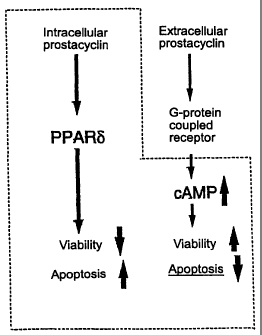
The present inventors demonstrated for the first time that
prostacyclin was authentic, natural ligand for PPAR-6 and activation
of endogenous PPAR-6 by prostacyclin resulted in activation of
apoptotic pathway in HEK-293 cells. Prostacyclin interacts with both
nuclear PPAR-6 and cell-surface receptor and raises the intracellular
level of cAMP; these two types of signaling respectively produce
reverse biological effects on apoptosis and/or cell survival in a
synergistic fashion. As shown in the multi-diagrams in Fig. 19, the
CA 02411056 2002-11-21
33
present inventors focused on the prostacyclin signaling pathway
including nuclear receptor PPAR-S. Why the endogenous agent does not
induce apoptosis in PGIS-expressing endothelial cells and vascular
smooth muscle cells? The reason is that endothelial cells and
vascular smooth muscle cells express also endogenous prostacyclin
receptor (IN (which raises the level of cAMP in the presence of
prostacyclin at a low concentrations) . IP/cAMP/protein kinase
pathway is presumed to protect these cells from prostacyclin-mediated
apoptosis by an autocrine and/or paracrine mechanism. Thus, it can
be assumed that prostacyclin-mediated apoptosis is readily induced
in cells lacking IP expression, such as HEK-293 and CV-1. Further,
there may be a precise mechanism in which cell fate is regulated by
prostacyclin in cooperation with PPAR-a, PPAR-S, and G
protein-coupled PG receptor. The research group of the present
inventors is now characterizing the prostacyclin-signaling cascade
including the novel pathway.
[Example 2] Induction of apoptosis in human colon cancer cells
The wild-type PGIS gene (PGISwt) or inactive PGIS gene
(PGISC441A) prepared by introducing alanine into the active center
of PGIS by conventional site-directed mutagenesis were transfected
into cells of the human colon cancer cell line Caco2. 1.0 gg of
R-galactosidase expression vector was co-transfected with the
above-mentioned gene into the cells using LipofectAMINE. Control
transfection experiments were carried out using HEK-293 cells and
CV-1 cells by the same procedure. After 60 hours, the cells were
stained with X-gal. The results are shown in Fig. 20.
As seen in Fig. 20, apoptosis was recognized only in cells in
which PGISwt had been introduced. Thus, it is demonstrated that the
introduced PGIS gene induces apoptosis in cancer cells and the PGIS
gene can be effective in treating cancers.
[Example 3] Assessment of cancer therapy using introduction of the
PGIS gene
The PGIS gene was inserted into adenoviral vector by the
conventional method to prepare a recombinant adenoviral vector. The
CA 02411056 2002-11-21
34
recombinant vector obtained is used as a pharmaceutical composition
for gene therapy of a cancer.
(1) Animal experiments
The pharmaceutical composition for gene therapy is administered
in adequate dose at appropriate administration frequency (1, 2, or
3 times) to a nude mouse as a cancer model in which tumor formation
has been confirmed; an alteration in tumor diameters is observed at
the same time. A control group is administered adenoviral vector
without the prostacyclin synthase gene. Further, to assess the dose
or frequency of administration, one can use several different groups
where the dose or frequency of administration is varied appropriately.
When tumor formation is confirmed, then tumor size is measured in
both group administered the pharmaceutical composition of the present
invention for inducing apoptosis (treated group) and control group.
When tumor involution is seen in the treated group, the
involution serves as an index of therapeutic achievement of cancer
therapy at the level of animal experiment. In addition, tissues can
be assessed by the about-described method for assessing
characteristics of apoptosis. When many apoptotic cells are seen in
the treated group, the presence of apoptotic cells serves as an index
of apoptosis-inducing activity of the pharmaceutical composition of
the present invention for inducing apoptosis at the level of animal
experiment.
(2) Clinical test in human individuals
The pharmaceutical composition for gene therapy is administered
in adequate dose at appropriate administration frequency. Then, the
recombinant adenoviral vector should be tested for the cytotoxicity,
infectivity to other individuals, and integration into the chromosome
of viral vector according to the descriptions in "Handbook for
Development and Research of Gene Therapy (Ed. The Japanese society
of Gene Therapy; NTS, 1999)".
The clinical effects of cancer therapy on individuals are
evaluated based on an assessment index of tumor by periodically
recording photographic images, CT scan images, MRI images or the like
CA 02411056 2002-11-21
of tumors; by measuring diameters of tumor; and by estimating tumor
volume based on the orthogonal major and minor axes to determine tumor
growth rates. Additionally, the presence of apoptosis is assessed
by morphological observations on the tissues. Further, analysis
5 techniques of molecule biology can be utilized; the presence of a
DNA as the active ingredient in target cells can be confirmed by the
detection method using PCR; apoptosis can be detected by TUNEL
staining.
10 [Example 4] Screening for agents that induce apoptosis in cells
The oligonucleotide, describe in Example 1, containing three
copies of PPRE, which is a PPAR-responsive element, is ligated to
a reporter gene to prepare a plasmid. The luciferase gene is used
as the reporter gene; pGL3-promotor vector (Promega) is used as the
15 plasmid.
Then, the above-mentioned plasmid is introduced into cells to
prepare transformed cells. In this experiment, HEK-293 cell (ATCC
CRL-1573), which expresses endogenous PPAR-S and allows detection of
reporter gene activity, is used as the host. The delivery of the
20 plasmid into cells is carried out by the method using LipofectAMINE
(Gibco-BRL).
A candidate for the agent that induces apoptosis can be selected
by adding a test substance to transformed cells thus prepared, and
measuring and assessing whether the test substance increases the
25 expression level of the reporter gene. A test substance that
increases the expression level of the reporter gene is a candidate
for the agent that induces apoptosis. Thus, an agent that induces
apoptosis in cells can be selected by further subjecting such selected
candidate test substances to apoptosis assay or the like using, as
30 an index, caspase activity as describe in Example 1.
Sequence Listing Free Text
SEQ ID NO: 1 is an amino acid sequence for synthetic peptide
corresponding to amino acid NOs: 27 to 45 of human PGIS.
35 SEQ ID NO: 2 is an amino acid sequence for synthetic peptide
corresponding to amino acid NOs: 485 to 500 of human PGIS.
CA 02411056 2002-11-21
36
SEQ ID NO: 3 is a nucleotide sequence of oligonucleotide for
HVJ-lyposome method.
SEQ ID NO: 4 is a nucleotide sequence of oligonucleotide for
HVJ-lyposome method.
SEQ ID NO: 5 is a nucleotide sequence of oligonucleotide for
HJV-lyposome method.
SEQ ID NO: 6 is a nucleotide sequence of oligonucleotide for
HVJ-lyposome method.
SEQ ID NO: 7 is a nucleotide sequence of oligonucleotide for
PPREx3-luciferase assay.
SEQ ID NO: 8 is a nucleotide sequence of oligonucleotide for
PPREx3-luciferase assay.
Industrial Applicability
The pharmaceutical composition of the present invention for
inducing apoptosis produces an excellent therapeutic effect in which
the diseases can be treated by inducing apoptosis. In addition, the
pharmaceutical composition of the present invention for gene therapy
of cancer can induce apoptosis and thereby cell death of cancer cells
can be induced. Further, the screening method of the present
invention can be used conveniently to screen for agents capable of
inducing apoptosis.
CA 02411056 2003-03-19
37
SEQUENCE LISTING
<110> Tadashi Tanabe
<120> MEDICINAL COMPOSITIONS CONTAINING PROSTACYCLIN SYNTHASE GENE
<130> 12871-53
<140> CA 2,411,056
<141> 2000-11-21
<150> JP 2000-150648
<151> 2000-05-22
<160> 8
<210> 1
<211> 20
<212> PRT
<213> Artificial Sequence
<220>
<223> Description of Artificial Sequence: An amino acid sequence for
synthetic peptide corresponding to amino acid NOs: 27 to 45 of
human PGIS.
<400> 1
Pro Gly Glu Pro Pro Leu Asp Leu Gly Ser Ile Pro Trp Leu Gly Tyr
1 5 10 15
Ala Leu Asp Cys
<210> 2
<211> 16
<212> PRT
<213> Artificial Sequence
<220>
<223> Description of Artificial Sequence: An amino acid sequence for
synthetic peptide corresponding to amino acid NOs: 485 to 500 of
human PGIS.
<400> 2
Leu Met Gln Pro Glu His Asp Val Pro Val Arg Tyr Arg Ile Arg Pro
1 5 10 15
<210> 3
<211> 19
<212> DNA
<213> Artificial Sequence
<220>
<223> Description of Artificial Sequence: A nucleotide sequence of
oligonucleotide for HVJ-lyposome method.
<400> 3
ctcggtgact tatcctgtg 19
<210> 4
<211> 19
<212> DNA
CA 02411056 2003-03-19
38
<213> Artificial Sequence
<220>
<223> Description of Artificial Sequence:A nucleotide sequence of
oligonucleotide for HVJ-lyposome method.
<400> 4
tcctctttct cctcctctt 19
<210> 5
<211> 19
<212> DNA
<213> Artificial Sequence
<220>
<223> Description of Artificial Sequence:A nucleotide sequence of
oligonucleotide for HJV-lyposome method.
<400> 5
ctcggtgact tatcctgtg 19
<210> 6
<211> 19
<212> DNA
<213> Artificial Sequence
<220>
<223> Description of Artificial Sequence:A nucleotide sequence of
oligonucleotide for HVJ-lyposome method.
<400> 6
cacaggataa gtcaccgag 19
<210> 7
<211> 67
<212> DNA
<213> Artificial Sequence
<220>
<223> Description of Artificial Sequence:A nucleotide sequence of
oligonucleotide for PPREx3-luciferase assay.
<400> 7
cgcgtaaaaa ctgggccaaa ggtctcaaaa actgggccaa aggtctaaaa actgggccaa 60
aggtctc 67
<210> 8
<211> 65
<212> DNA
<213> Artificial Sequence
<220>
<223> Description of Artificial Sequence:A nucleotide sequence of
oligonucleotide for PPREx3-luciferase assay.
<400> 8
tcgagagacc tttggcccag tttttagacc tttggcccag tttttagacc tttggcccag 60
tttta 65