Note: Descriptions are shown in the official language in which they were submitted.
CA 02412012 2010-04-21
- 1 -
RESORBABLE EXTRACELLULAR MATRIX CONTAINING COLLAGEN I
AND COLLAGEN II FOR RECONSTRUCTION OF CARTILAGE
FIELD OF THE INVENTION
The present invention relates to the field of
reconstruction of cartilage tissue.
DESCRIPTION OF THE BACKGROUND ART
Joint injuries often result in damage to the cartilage
which lies between the joints. For example, back injuries
often involve damage to one or more vertebral discs.
Similarly, knee injuries often result in meniscus damage.
There remains a need in the art for materials and methods
for promoting regeneration of damaged cartilage.
SUMMARY OF THE INVENTION
In accordance with the present invention, a resorbable
extracellular matrix for reconstruction of cartilage tissue
comprises collagen material including a mixture of collagen I
and collagen II in a respective ratio of from about 1:19 to
about 19:1. The matrix may be utilized as a scaffold implant
for meniscal cartilage regeneration or for vertebral disc
regeneration.
According to one aspect of the present invention,
there is provided a matrix wherein said matrix has a pore
size within a range of 50 - 400 pm.
According to another aspect of the present invention,
there is provided a matrix having a pore size within a
range of 70 - 120 pm.
According to still another aspect of the present
invention, there is provided a scaffold implant for
promoting cartilage regeneration comprising the matrix
described herein, said implant having a thickness of 0.2 -
2 cm.
CA 02412012 2010-04-21
- la -
According to yet another aspect of the present
invention, there is provided the use of the matrix
described herein for reconstruction of cartilage tissue.
According to a further aspect of the present
invention, there is provided the use of the matrix
described herein in manufacture of a medical preparation
for reconstruction of cartilage tissue.
According to yet a further aspect of the present
invention, there is provided the use of the matrix
described herein for the promotion of reconstruction of
cartilage tissue in an area located adjacent to or between
bone material in a patient.
BRIEF DESCRIPTION OF THE DRAWING
Fig. 1 is an elevational view, partly schematic, showing
a meniscal scaffold implant in accordance with the invention.
Fig. 2 is an elevational view, partly schematic, showing
a vertebral disc scaffold implant in accordance with the
present invention.
DETAILED DESCRIPTION OF THE INVENTION
Collagen occurs in a number of forms in the animal body
and different tissues contain different proportions of the
respective types. Thus, whereas bone collagen comprises
predominantly collagen I and III, cartilage comprises
CA 02412012 2002-11-18
2 -
predominantly collagen II together with small quantities of
collagen VI, IX, X, XI and XIII. Such material differs
significantly from collage sponge material used in medicine
and in cosmetics which, being derived from skin and tendons is
mostly made up of collagen I and/or III.
According to one aspect of the present invention,
therefore, there is provided a resorbable extracellular matrix
for reconstruction of cartilage tissue comprising a mixture of
collagen I and collagen II in a respective ratio of from about
1:19 to about 19:1. In preferred embodiments, the collagen I
and collagen II is in a respective ratio of from about 1:9 to
about 9:1. Exemplary mixtures of collagen I and collagen II
are in respective ratios of about 1:9, 25:75, 50:50 and 75:25.
A Collagen I to Collagen II ratio of about 1:9 is particularly
preferred.
In preferred embodiments, the collagen in the.matrix is
subjected to cross-linking by chemical, ultravioloet (UV)
radiation or hydrothermal cross-linking. For example,
chemical cross-linking can be effected with chondroitin 4-
sulphate and/or chondroitin 6-sulphate, alone or together with
hyaluronic acid. Various aldehydes such as hyaluronate
polyaldehyde, formaldehyde or glyoxal may be used. Suitable
chemical cross-linking agents include hyaluronate
polyaldehyde, hexaethylene di-isocyanate, di-ethyl-3-(3-
dimethyl aminopropyl) carbodimide (EDC), and N-hydroxy
succinimide (NHS), mixtures of EDC and NHS, and/or suitable
mixtures of any of the foregoing.
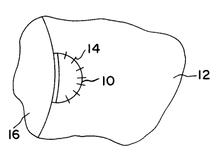
Non-limiting, exemplary uses of the invention are shown
in the drawings. Fig. 1 shows a meniscal scaffold implant 10
inserted into a defect in meniscus 12, fixed in place by
sutures 14 over underlying bone material 16. Fig. 2 shows a
vertebral disc scaffold implant 18 inserted into a defect into
CA 02412012 2002-11-18
3 -
a vertebral disc 20, and fixed in place by sutures 22, or
alternatively glueing with adhesive such as fibrin glue.
A collagen matrix according to the invention may contain
minor quantities of collagen III, VI, IX, X, XI and XIII. The
matrix according to the invention may also contain a hydrogel-
like material, for example comprising glycosaminoglycans such
as chondroitin sulphate, keratan sulphate, dermatan sulphate
and hyaluronic acid, which provides a natural medium in which
chondrocytes can become embedded and grow. The matrix
according to the invention preferably contains 0.1 to 40% by
weight of glycosaminoglycan, for example 1 - 15a, e.g., about
2-3 by weight, most preferably about 2.5o by weight.
The matrix according to the invention may either comprise
natural cartilage material which has been subjected to
defatting and other treatment, leaving the collagen material
together with glycosaminoglycans, or alternatively fibres of
purified collagen I and collagen II may separately or together
be mixed with glycosaminoglycans and/or any other additives.
Such additional additives may, for example, include
chondronectin or anchorin II to assist attachment of the
chondrocytes to the collagen fibres and growth factors such as
cartilage inducing factor (CIF), insulin-like growth factor
(IGF) and transforming growth factor P (TGF~) .
The matrix is capable of acting as a medium for the
ingrowth of native chondrocytes thereby effecting regeneration
of cartilage tissue. However, to further aid in regenerating
cartilage tissue the matrix may be impregnated with
chondrocytes either prior to or following implantation in
vivo. While the matrix may be impregnated with chondrocytes
immediately prior to implantation, e.g. by injection, it is
expected that in general the chondrocytes will be introduced
into the matrix by direct injection of a suspension of
chondrocytes following implantation. In this way,
CA 02412012 2002-11-18
4 -
chondrocytes present in the matrix are able to effect
regeneration of cartilage, and possibly new bone.
Chondrocytes for use in the invention may be obtained
from cell sources which include allogenic or autogenic cells
isolated-from articular cartilage, periosteum and
perichondrium, and mesenchymal (stromal) stem cells from bone
marrow. Since allogenic cells carry the potential for immune
response and infectious complications, it is preferable to
isolate the chondrocytes from autogenic cells, especially from
autogenic articular cartilage. Techniques for harvesting cells
are known and include enzymatic digestion or outgrowth
culture. The harvested cells are then expanded in cell
culture prior to reintroduction to the body. In general, at
least 106, preferably at least 10' cells should be impregnated
into the matrix to provide for optimal regeneration of
cartilage tissue.
In general, it is desirable for the matrix according to
the invention to contain glycosaminoglycans (GAGS) such as
hyaluronic acid, chondroitin 6-sulphate, keratin sulphate,
dermatan sulphate, etc., which serve to provide a natural
medium in which chondrocytes can become embedded and grow.
While it is possible to incorporate into the collagen matrix
glycosaminoglycans from different sources which do not
necessarily have the same composition, molecular weight and
physiological properties as those from cartilage, preferred
glycosaminoglycans are those extracted from cartilage itself.
In native collagen tissues GAGs occur, at least in part,
as a component of proteoglycans (PGs). The use of GAGs in the
form of PGs is undesirable in view of potential immunological
problems which can be caused by the protein content of the
PGs. Preferably, the matrix is thus substantially free from
any proteoglycans. Conveniently, this may be achieved by
CA 02412012 2002-11-18
-
preparing the matrix from a mixture of a purified telopeptide-
free collagen material and glycosaminoglycaris.
Other additives which may also be present in the matrix
include, for example, chondronectin, laminin, fibronectin,
5 calcium alginate or anchorin II to assist attachment of the
chondrocytes to the collagen fibers, bone and cartilage cell
growth-promoting hormones, and growth factors such as
cartilage inducing factor (CIP), insulin-like growth factor
(IGF), transforming growth factor R (TGFI3) present as
10. homodimers or heterodimers, osteogenic protein-1 (OP-1) and
bone morphogenetic factors (BMPs) such as native or
recombinant human BMP-2, BMP-3 (osteogenin), BMP-4, BMP-7,
BMP-8, bFGF, CDMP or other skeletal matrix molecules, as well
as signaling peptides such as transforming growth factor-R
(TGF-13, TGF-11), vascular endothelial growth factor
(EGF/VEGF), insulin-like growth factor (TGF/IGF-1),
parathyroid hormone related protein (PTHrP) and platelet
derived growth factor (PDGF). Nucleic acid sequences coding
for the above, or which are capable of inducing or promoting
in vivo production of the above, may be incorporated into the
collagen matrix material of the present invention.
The product used in the invention also may act as a
carrier for stem cells committed to a particular line of
differentiation such as articular cartilage or bone. Such
stem cells may be grown in vitro to increase their numbers,
and applied to the repair sites in the carrier matrices with
or without growth factors. Examples include mesenchymal stem
cells and bone marrow stromal cells. Nucleic acid sequences
coding for the above, or which are capable of inducing or
promoting in vivo production of the above, may be incorporated
into the collagen matrix material of the present invention.
BMP-2 affects the two pathways of bone formation
independently - the direct formation of bone as well as the
CA 02412012 2002-11-18
6 -
formation of cartilage which is then removed and replaced by
bone. Composites of BMPs and collagen including bone matrix
obtained by extraction from cortical bone from various sources
or demineralized bone matrix comprise about 90% collagen and
about 10% non-collagenous proteins (NCP) for BMP activity or
for BMP/NCP induced chondrogenesis. Bone matrix-insoluble
collagenous matrix and laminin or fibronectin act as carriers
for BMPs. In general, the matrix may contain from about 100 jig
to about 5 mg of growth factors. Nucleic acid sequences
coding for the above, or which are capable of inducing or
promoting in vivo production of the above, may be incorporated
into the collagen matrix material of the present invention.
A matrix material for use in accordance with the present
invention may also be charged with parathyroid hormone (PTH),
a polypeptide involved in regulation of calcium in the body.
Nucleic acid sequences coding for the above, or which are
capable of inducing or promoting in vivo production of the
above, may be incorporated into the collagen matrix material
of the present invention.
As noted above, the present invention may comprise a gene
or nucleic acid-supplemented collagen matrix with cell growth-
promoting genetic material or DNA incorporated therein. The
collagen matrix material may provide for prolonged release of
the. cell growth-promoting genetic material. Upon release from
the matrix into the body, the genetic material may transform
cells in the body so as to promote cell growth and healing.
The present invention may also provide a collagen matrix
material charged with a cell growth-promoting nucleic acid
sequence, preferably an isolated or purified nucleic acid
sequence. The sequence can be a DNA sequence or an RNA
sequence. In particularly preferred embodiments, the collagen
matrix material is charged with an isolated gene sequence,
most preferably of DNA.
CA 02412012 2002-11-18
- 7 -
A nucleic acid sequence for use in accordance with the
present invention may promote cartilage cell growth, bone cell
growth, or both.
Purified therapeutic nucleic acid sequences for use in
accordance with the present invention may be derived from any
suitable source, and may be charged to the collagen matrix
material so as to promote cell growth. In accordance with one
embodiment, a retroviral vector, or any other suitable gene-
carrying and gene-introducing mechanism, is utilized. For
example, a retroviral vector may be utilized for stably
introducing human bone morphogenic protein 7 (BMP-7) cDNA into
mesenchymal stem cells.
Gene therapy in accordance with the present invention
involves the delivery of therapeutic genes or other genetic
material into cells and tissues.
As will be further discussed below, a scaffold implant of
the matrix of the invention may be prepared by forming
separate aqueous collagen I and collagen II slurries, mixing
the slurries, optional partial dehydration of the mixed
collagen I/II slurry, molding the mixed collagen I/II slurry
to the desired shape, drying of the mixed collagen i/II
slurry, partial cross-linking of the collagen I and II fibers
by chemical, ultraviolet (W) radiation or hydrothermal cross-
linking, and sterilizing the collagen i/II implant material.
Alternatively, cross-linking, such as chemical cross-linking,
can be effected after preparation of the individual collagen I
and collagen II slurries, or after forming the mixed collagen
I/II slurry, and prior to molding.
In preferred embodiments, the molded material is dried by
freeze-drying so as to achieve a pore size within the range of
about 0.1 - 500 pm. A preferred pore size for a scaffold
implant in accordance with the invention is within the range
CA 02412012 2002-11-18
- 8 -
of about 50 - 400 pm, most preferably within the range of
about 70 - 120 pm.
The density of the matrix after freeze-drying preferably
is within the range of about 0.1 - 0.3 g/m3, preferably about
0.18 - 0.22 g/m3, most preferably about 0.2 g/m3.
The collagen material may be cross-linked before or after
the freeze-drying step to stabilize the matrix. This also
serves to increase the mechanical stability of the matrix and
to reduce its rate of resorption by the body. Ideally, the
degree of cross-linking should be such that the rate of
degradation of the matrix matches the rate of tissue
regeneration. Physically, cross-linking may be carried out by
heating, but this must be effected carefully to avoid
undesired loss of resorbability. Heating to temperatures of
100-120 C for a period of from about 30 minutes to about 5
hours is preferable. More preferably, cross-linking may be
effected by W irradiation using a W lamp, e.g., for a period
of up to 8 hours.
As noted above, the collagen material advantageously
contains glycosaminoglycans (GAGs). The latter actually reacts
with the collagen to effect some cross-linking and produces an
insoluble product. If necessary, further cross-linking can be
effected by heating the material, by UV irradiation, or by
further chemical cross-linking as discussed above. The
reaction between the glycosaminoglycans and the collagen can
be effected at ambient temperatures at a pH in the range 2.5-
3.5. The material may be subjected to freezing and freeze-
drying immediately after such treatment.
For example, GAGs such as chondroitin sulphate (CS) may be
covalently attached to the collagen matrix using 1-ethyl-3-(3-
dimethyl aminopropyl) carbodiimide (EDC) and N-.
hydroxysuccinimide (NHS) utilizing known methods. EDC/NHS
crosslinking may be utilized for immobilizing GAGs with
CA 02412012 2010-04-21
9 -
collagen matrices, which may include dermatan sulphate,
heparin, heparan sulphate, and hyaluronic acid, as well as CS
as indicated above.
Collagen II slurry formation may be effected by raising
the pH of the collagen mass. In this procedure, the mass is
cooled to about 4 C and the pH value slowly raised by addition
of cold aqueous NaOH at 4 C up to a pH value about 6.5-7.5.
Subsequently, the mass is held at ambient temperature for
about 15-25 hours.
A still further alternative is to neutralize the collagen
II mass to a pH value about 6.8-7.4, subsequent to removal of
air.
The collagen I preferably is of porcine origin. A
purified collagen I material can be provided as disclosed in
U.S. Patent No. 5,837,278..
The collagen I material can be comminuted with distilled water
in a blender to form a suspension, and the water can be
removed to form a collagen I slurry.
The collagen I slurry then can be mixed with a collagen II
slurry as described above, and filled into a mold.
After molding the slurry mixture, the material is frozen.
In order to obtain a reproducible pore size, the freezing must
be carefully controlled and the rate and time of freezing, the
pH value and the particle size must be accurately controlled.
The matrix is then freeze-dried and subsequently heated to
about 110-130 C. In this way, some cross-linking is effected.
Subsequently, the freeze-dried matrix may be adjusted to the
required thickness. The matrix is then sterilized, for
example by gamma-irradiation or with ethyleneoxide.
Sterilization by strong irradiation e.g. with "'Co in doses of
25 kGy may deactivate the BMPs. In such circumstances, the
sterile matrix may be impregnated with BMPs in sterile saline
prior to implantation.
CA 02412012 2002-11-18
-
The thickness of a scaffold implant in accordance with the
present invention may be within the range of about 0.2 - 2 cm,
preferably about 0.3 - 1.5 cm, more preferably about 0.4 - 1
cm, and most preferably about 0.5 - 0.8 cm.
5 There exists a wide range of glycosaminoglycans and
proteoglycans which have different and sometimes undesirable
properties. Thus, although it is possible to incorporate into
the collagen matrix glycosaminoglycans from different sources
which do not have the same composition, molecular weight and
10 physiological properties as glycosaminoglycans from cartilage,
it is particularly preferred to use glycosaminoglycans from
cartilage itself.
As noted above, it is desirable to subject the collagen
matrix to some degree of cross-linking in order to restrict
the extent of swelling when the matrix comes in contact with
aqueous fluids, while retaining the ability of the matrix to
be resorbed. Such swelling leads to loss of strength and
shape. The collagen matrix according to the invention may
advantageously be manufactured by subjecting cartilage tissue
to defatting followed by treatment with a base whereby
proteoglycans and glycosaminoglycans are removed.
The cartilage material will normally be that from readily
available animal sources such as cattle, sheep or pigs. The
preferred source of collagen II material is hyaline cartilage
from pigs. This contains the right type of collagen and
glycosaminoglycan in desirable proportions and is available in
suitably large quantities.
The cartilage is preferably frozen after slaughter and
subjected to size reduction, for example to a particle
diameter of about 8mm. Before size reduction, the cartilage is
preferably soaked in water and mechanically separated from
flesh, bone and other unwanted materials.
CA 02412012 2002-11-18
- 11 -
The particulate cartilage is then preferably subjected to
dewatering by treatment with a water miscible organic solvent
such as acetone, which also serves to remove some fat. The
dewatering shrinks the collagen fibres and separates them from
each other so that the subsequent defatting step is optimised.
The material is then subjected to defatting with a fat-
solvent such as a hydrocarbon e.g., hexane, or a halogenated
hydrocarbon.
After defatting, the material is thoroughly washed and
this is continued until as much water has been taken up as was
present originally. By this procedure, the material is
optimised for the base-treatment which follows.
The base-treatment may be effected with a strong alkali,
for example an alkali metal hydroxide, e.g., sodium hydroxide,
for example at a concentration of 1-8% by weight. The
treatment time, which will vary according to the raw material
and alkali concentration, is generally 10-48 hours. The
treatment temperature will generally be below 20 C. The pH
value is normally in the range 12-14. The above conditions are
those which are optimal for treatment with NaOH. Treatment
with other bases may require slightly modified conditions.
The base-treatment has the following effects:
Small quantities of residual fat are saponified. The non-
collagen, alkali soluble proteins are denatured, destroyed,
dissolved and eliminated.
The amide groups in the collagen are saponified, thereby
changing the electric charge and the isoelectric point of the
collagen.
Bacteria, prions and viruses are inactivated and the
collagen is thus sterilised.
It has been found that by this treatment, proteoglycans
undergo a useful modification which can be characterised as
follows:
CA 02412012 2002-11-18
12 -
the covalent binding of glycosaminoglycans to the core
protein in proteoglycans is cleaved. In this way the
glycosaminoglycans can be liberated from the protein of the
proteoglycans. This is termed (3-elimination.
By the base-treatment, the core protein is split into
small peptides which may be removed from the reaction mixture
by dialysis or ultra filtration.
Due to the strong negative charge, the glycosaminoglycans
form water soluble salts which can partially washed from the
collagen. These are, however, uncleaved or only slightly
cleaved by the base-treatment and can be separated from
peptides by dialysis. A part of the glycosaminoglycan (about
3% by weight of the collagen) is bound to the collagen.
Purified glycosaminoglycans may be obtained by dialysis or
ultrafiltration of the extract arising from the base-treatment
step.
According to the procedure of the present invention,
enzymatic treatment is, in general, not used, in view of the
variety of different substances present. However, further
steps include treating the material with an organic or
inorganic acid, such as hydrochloric acid. This has the
following effect:
Unwanted acid sensitive materials are removed; The fibre
structure is loosened.
Subsequently, the material is washed, generally until the
pH value of the material is between 2.5 and 4Ø The pH value
of the material is preferably controlled accurately. The pH
value of the material should be uniform across the cross-
section of the cartilage.
After the acid treatment, the cartilage is in a water-
swelled condition. The material is then subjected to
mechanical size-reduction, for example using a colloid mill.
The concentration of the collagen in the aqueous medium is
CA 02412012 2002-11-18
- 13 -
then about 2.5-3.5% by weight. The pH value of this mixture
should be somewhat acid, for example 3.5-4.5.
At this point, one or more glycosaminoglycans may be added
to the purified collagen mass, for example in the range 0.1-
40% preferably 1 to 15%, of the weight of collagen.
The glycosaminoglycans added to the collagen preferably
are extracted from the natural cartilage, as indicated above.
The matrix will then contain, besides collagen II, the
glycosaminoglycans hyaluronic acid, chondroitin sulphate and
keratan sulphate. The chondroitin sulphate and keratan
sulphate are covalently bonded to the core protein while
hyaluronic acid is, indeed, bound to the proteoglycan but not
covalently.
By the action of the base, the bonding to the core protein
is cleaved and the glycosaminoglycan is freed from the
protein. Additionally, the core protein is cleaved to small
peptides which are readily removed by dialysis or
ultrafiltration. It is important that the core protein is
removed, since this may be immunologically active. The removal
of the core protein is thus an important part of the process
of the present invention.
The recovery of the glycosaminoglycans from the base
extract may be effected as follows:
The medium is neutralised to a pH value in the range 6-8.
The non-collagen proteins care removed by treatment with
an adsorbent such as kaolin.
Ultrafiltration of the liquid is effected, using a
membrane which permits the passage of molecules of weight less
than 10000 daltons.
Concentration of the liquid is effected to a solids
content of about 2-5 weight percent.
After admixture of the glycosaminoglycan with the collagen
II, the material is homogenised still further in a colloid
CA 02412012 2002-11-18
- 14 -
mill and the solid content is adjusted to 1.5-2.5 weight
percent.
A preferred source of collagen I material is porcine skin,
tendons or peritonea.
After mixing of the collagen I and collagen II slurries,
the resulted mass may be frozen.
The freezing must be precisely controlled, whereby the
freezing time, pH value and particle size are exactly
maintained in order to provide a reproducible pore size. The
frozen product is then freeze-dried. After freeze-drying, the
sponge is warmed to 120-140 C for at least 2 hours. In this
way, the material is stabilised by light cross-linking. After
the freeze-drying the material is cut to a desired thickness,
stamped to the required shape, sterilised and packed.
The matrix according to the invention can be supplemented
with active substances. Thus any physiologically active
substance which is water soluble or water dispersible can be
used. Thus, the matrix may advantageously contain medicinal
substances such as antibacterials, e.g., taurolidine,
taurultam, or antibiotics such as tetracyclines and
gentamycins.
The invention also provides the use of a matrix according
to the invention in guided regeneration and reconstruction of
cartilage tissue, as well as manufacture of a medical
preparation therefor.
A method in accordance with one embodiment of the
invention comprises, removing damaged cartilage. tissue from an
area located adjacent to or between bone material in a
patient, inserting a scaffold implant comprising a matrix
collagen I and II material as described above, which has been
sized to fit the area of damaged cartilage, and fixing the
scaffold implant matrix in the area of damaged cartilage by
CA 02412012 2002-11-18
- 15 -
any suitable means such as adhesive or suturing the scaffold
implant to adjacent cartilage material.
The following examples are given by way of illustration
only.
Example 1
Frozen cartilage from freshly slaughtered pigs was steeped
in cold water, thoroughly washed through and mechanically
purified from flesh residues, bones and hard pieces.
Subsequently, the material was washed for 30 minutes under
flowing water.
Subsequently, the material was ground three times in a
homogenizer. The optical particle size at the end of size
reduction was about 8mm.
The cartilage pieces were dewatered by washing 4 times
with acetone, each time for 8 hours. The cartilage was then
defatted by extraction 4 times with n-hexane. Each treatment
lasted at least 8 hours. The ratio of hexane to cartilage was
1:10.
After defatting, the cartilage was swelled in drinking
water. The ratio of water:material was 10:1. The treatment
time was 24 hours.
The material was then treated with NaOH (5% by weight)
whereby the ratio of cartilage to liquid was 1:4 and the
treatment time was 32 hours. During the treatment, the pieces
of cartilage were well stirred. Subsequently, the alkali was
washed from the cartilage. The original pH of 14 was thereby
reduced to 9-11. The dissolved impurities were washed out and
separated from the cartilage. The liquid resulting from the
alkaline treatment was collected for the recovery of
glycosaminoglycan.
CA 02412012 2002-11-18
- 16 -
The collagen material was then treated with strong HC1
(about 3% by weight) initially at a pH value under 1Ø The
treatment time was 4-6 hours.
Subsequently, the material was washed with cold water long
enough for the pH value to rise to 3-3.5.
All impurities were removed and the product was a salt-
free collagen mass, suitable for-production of a sponge or
other collagen material. For that purpose, the cartilage mass
may be, according to the intended result, degassed, frozen and
freeze-dried.
Example 2
The extract resulting from alkaline treatment in Example 1
contained glycosaminoglycan, alkali, denatured proteins and
salts. The extract was firstly neutralised with HC1, the pH
value after neutralisation being G. The extract was then
treated with a filter aid, namely kieselguhr, which had the
effect of removing the denatured proteins. 0.5 weight percent
of kieselguhr was introduced into the extract and removed by
filtration together with the denatured protein.
The supernatant was then submitted to ultrafiltration
using a membrane having a molecular weight cut off at about
10000 daltons. In this way, salts were removed to leave
purified glycosaminoglycan.
The glycosaminoglycan solution so obtained was admixed
with collagen material from above to provide a collagen II
matrix containing glycosaminoglycan.
Example 3
(1) Determination of hexosamine and amino acid residues in
collagen sponges and fleeces
CA 02412012 2010-04-21
- 17 -
Each sample, exactly weighed (about 10 mg) was hydrolised
in 10 ml of 3M or 6M HC1 at 1.05 C for 15 or 20 hours under
purified nitrogen in a sealed tube. After cooling the tube in
a refrigerator and opening the tube, the contents were
transferred to a 25 ml long neck flask and dried at 40 C in a
vacuum-rotation dryer (Rotavapor RE120, Buchi, Switzerland)
under water jet vacuum. After dissolving the residue in 5m1
water, the residue was again dried under water jet vacuum.
Subsequently, the residue was taken up in 5ml loading buffer
(0.2M relative to Na+) at pH 2.2. For determination of the
glucosamine and galactosamine values, after previous dilution
of an aliquot with loading buffer (1+10) 150 p1 of the sample
hydrolysed in 3M HC1 was injected into the cartouche of an
amino acid analyser (AlphaPlus,t type 4151, Pharmacia-LKB,
Freiburg) and evaluated by comparison with a standard with the
help of a computer (Shimadzu, Duesseldorf). The same procedure
was effected with the sample hydrolised in GM HC1, wherein
50 pl were injected in a further test cartouche. The double
hydrolysis in 3M and 6M HC1 is necessary for optimisation of
the hexosamine and amino acid analysis since the maximal
values for hexosamine and also tyrosine are only obtained
after hydrolysis in 3M HC1 while maximal values are only
obtained for valine, isoleucine and leucine after hydrolysis
in 6M HC1.
2. Determination of native collagen content in collagen
sponges and fleeces
25-30 mg (exactly weighed out) of sample were introduced
into 30 ml 0.1M sodium hydrogen carbonate solution (pA, Merck,
Darmstadt) pH 8.2 to which 1.5 ml of a 6 mg/ml trypsin
solution (lyophilised preparation from bovine pancreas,
Boehringer, Mannheim) and incubated for 8 hours at 23+1 C in a
*Trade-mark
CA 02412012 2010-04-21
- 18 -
shaking water bath (Julabo SWI, Seelbach). After cooling the
sample in a cold room to 4'C, it was centrifuged at 4'C in a
60 Ti-Rotor (Beckman, Munich) at 32000 RpM for 30 minutes. The
residue was filtered in a stirred ultra filtration cell (Mod
8010, Amicon, Witten) through a Diaflow Filter PM 10*(Amicon,
Witten) of diameter 25 mm and 1 ml of the filtrate was
hydrolysed in 6M HC1 for 20 hours at 105'C. The further
working up and analysis of the hydrolysate is identical with
that described under (1) above with the exception that the
further uptake of the sample after twice evaporating to
dryness, was in 150 pl loading buffer, whereby 150 pl was
injected into the test cartouche of the amino acid analyser.
The hydroxyproline value obtained after the amino acid
analysis (in pmol/g starting substance), represents the part
of the degradable collagen in the sample. When the
hydroxyproline value of a parallel hydrolysis (6M HC1 and
analysed sample (see (1) above) which represents the total
collagen content, is compared with the hydroxyproline value,
the percentage proportion of the "native", that is trypsin
non-degradable collagen is indicated.
*Trade-mark
CA 02412012 2002-11-18
19
The results are shown in the following table.
Table
11mol/ mol/1000 mol
Hydroxyproline 795.4 97
Aspartic acid 381.7 47
Threonine 190.1 23
Serine 257.0 31
Glutamic acid 691.3 84
Proline 913.2 112
Glycine 2614.6 320
Alanine 864.9 106
Cysteine/2 11.5 2
Valine 195.7 24
Methionine 62.7 8
Isoleucine 92.8 11
Leucine 229.9 28
Tyrosine 27.0 3
Phenylalanine 119.9 15
Histidine 39.8 5
Hydroxylysine 126.4 15
Lysine 173.5 21
Arginine 395.5 48
Total 8182.9 1000
CA 02412012 2002-11-18
- 20 -
Glucosamine 9.68 1.18
Galactosamine 46.30 5.66
Total Hydroxyproline 795.4 pmol/g
Trypsin-degradable 36.9 imcl/g
hydroxyproline
"Native" collagen content 95.4
Example 4
2.0g of collagen I fibre felt were comminuted with 500g
of distilled water in a blender. This dispersion was
centrifuged in the supernatant water was removed, resulting in
a collagen I fibre slurry.
Example 5
Collagen I and collagen II slurries which are prepared as
described above are mixed and formed into matrices having
collagen I and collagen II in respective weight percent ratios
including 1:9, 25:75, 50:50 and 75:25.
Additionally, collagen I-GAG and collagen II-GAG are
mixed and formed into matrices having respective weight
percent ratios including 1:9, 25:75, 50:50 and 75:25.