Note: Descriptions are shown in the official language in which they were submitted.
CA 02412577 2002-12-18
WO 02/07590 PCT/USO1/23192
PROBE TIP
This invention relates to ultrasonic devices and more particularly to
ultrasound
diagnostic probes tips used in ophthalmology.
Background of the Invention
Many devices use ultrasound energy to construct images of internal organs to
help diagnose and treat diseases and other medical conditions. In
ophthalmology, for
~o example, both A-Scan and B-Scan ultrasound diagnostic devices are used.
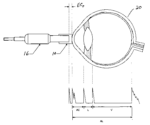
As best seen in FIG. 5, A-Scan biometry uses a relatively small probe and
measures the distances between, and thicknesses of, various structures within
the eye,
such as lens thickness (L), anterior chamber depth (AC), posterior chamber
depth (V)
and overall axial length (AL). These measurements are useful in determining
the
~s required power of the artificial intraocular lens which is implanted during
cataract
surgery.
As best seen in FIG. 6, B-Scan ultrasound uses a larger probe having an
internal
motor that wobbles the ultrasound crystal contained within the handpiece. B-
Scan
ultrasound creates a real time image of the eye and is used to detect the
presence of
io retinal or choroidal detachments, artifacts in the vitreous, tumors or
foreign bodies in
the eye or to view the posterior segment of the eye when visualization is
obscured. B-
Scan ultrasound will also make the same measurements made by A-Scan
ultrasound.
Measurements using either A-Scan or B-Scan probes are usually performed using
the contact method, during which the operative end of the probe is placed in
contact
Zs with the cornea of the eye under anesthesia. A special ultrasound fluid or
gel is used to
couple acoustically the probe with the lid or cornea of the eye. This acoustic
coupling
fluid can make it difficult to align the probe properly, and the gel may
irritate the eye
tissue. To prevent the spread of pathogens, the probe needs to be cleaned
between
uses, a difficult and time-consuming step. Even when the probe is cleaned
adequately
so to minimize the spread of any pathogens, the fluid used to clean the probe
can irritate
eye tissue. In addition, the contact method may not provide precise
measurements due
to corneal depression resulting from probe contact. The contact method is
preferred by
most operators, however, because it is easy to perform compared to the
immersion
method.
CA 02412577 2002-12-18
WO 02/07590 PCT/USO1/23192
-2-
A second technique used to make A-Scan or B-Scan measurements is the
immersion technique. ~Nith the immersion technique, the eye is anesthetized
and
small cup is placed on the eye. The cup is then filled with an acoustic
coupling fluid
and the probe is inserted into the cup. The immersion technique is considered
to be
s more accurate that the contact method because the probe does not contact and
partially
depress the eye. The immersion technique, however, is difficult to perform and
time
consuming and requires that the patient be laying down. In addition, bubbles
in the
coupling fluid can cause inaccuracies in the measurements.
Accordingly, a need continues to exist for a simple, safe, accurate and
reliable
~o ultrasound coupling device.
Brief Summary of the Invention
The present invention improves upon prior art methods by providing a soft,
15 partially solidified, water-based ultrasound conductive cap sided and
shaped to fit over
the operative end of an ultrasound probe. The thickness of the cap may be
varied to
change the focal point during B-Scan biometry.
It is accordingly an objective of the present invention to provide an
ultrasound
conductive cap for ultrasound probes.
zo It is a further objective of the present invention to provide an ultrasound
conductive cap that reduces or eliminates the need for an acoustic gel.
Still another objective of the present invention is to provide an ultrasound
conductive cap that allows for more accurate ultrasound measurements.
Yet another objective of the present invention is to provide an ultrasound
z5 conductive cap that helps prevent the spread of pathogens.
Yet another objective of the present invention is to allow for a biometry
procedure that is simple and faster than the contact method and with the
precision of
the immersion method.
Other objectives, features and advantages of the present invention will become
3o apparent with reference to the drawings, and the following description of
the drawings
and claims.
CA 02412577 2002-12-18
WO 02/07590 PCT/USO1/23192
-3-
Brief Description of the Drawings
FIG. 1 is a top plan view of the probe cap of the present invention useful
with an
A-Scan ultrasound probe.
s FIG. 2 is a cross-section view of the probe cap of the present invention
taken
along line 2-2 in FIG. 1.
FIG. 3 is a top plan view of the probe cap of the present invention useful
with an
B-Scan ultrasound probe.
FIG. 4 is a cross-section view of the probe cap of the present invention taken
~o along line 4-4 in FIG. 3.
FIG. 5 a schematic representation of the cap illustrated in FIGS. 1 and 2
being
used to take a measurement of an eye.
FIG. 6 a schematic representation of the cap illustrated in FIGS. 3 and 4
being
used to take a measurement of an eye.
15'
Detailed Description of the Invention
As best seen in FIGS. 2 and 4, probe tip 10 and 10' of the present invention
are
generally cup-shaped in cross-section, with wall 12 and 12' and end cap 14 and
14',
zo end caps 14 or 14' closing off one end of wall 12 or 12', respectively. Tip
10 illustrated
in FIGS. 1, 2 and 5 is suitable for use with an A-Scan probe and tip 10'
illustrated in
FIGS. 2, 3 and 6 is suitable for use with a B-Scan probe. Tips 10 and 10' are
preferably
made of a water-based gel such as a cross-linked cellulose derivative, but any
suitable
material may be used. Suitable gels are commercially available for sources
such as
zs Pharmaceutical Innovations, Inc., Newark, New Jersey.
As best see in FIGS. 1, 2 and 5 tip 10 suitable for use on an A-Scan probe may
be of any suitable inner and outer diameter, but will generally have an inner
radius R;
of between 0.15 inches and 0.70 inches, with approximately 0.25 inches being
preferred, and an outer radius Ro of between 0.25 inches and 0.85 inches, with
3o approximately 0.45 inches being preferred. Wall 12 may be of any suitable
height Wh
necessary to hold tip 10 on probe 16, but will generally be between 0.10
inches and
0.80 inches high, with approximately 0.40 inches being preferred. To assist in
the
installation of tip 10 on probe 16, vent hole 18 may be provided having a
diameter D
CA 02412577 2002-12-18
WO 02/07590 PCT/USO1/23192
-4-
of between 0.01 inches and 0.10 inches, with 0.05 inches being preferred. End
cap 14
may have any suitable thickness ECt, but preferably is between 0.10 and 0.40
inches
thick, with approximately 0.12 inches being preferred. End cap 14 also
preferably is
formed with a curved shape to fit probe 16 and eye 20 securely with minimal
rocking
s and good acoustic contact,.with an inner radius RZ of between 0.75 inches
and 1.25
inches with approximately 0.90 inches being preferred and an outer radius R3
of
between 0.60 inches and 1.40 inches, with approximately 1.00 inches being
preferred.
Wall 12 and end cap 14 are preferably joined by internal radius R4 which is
preferably
between 0.01 inches and 0.05 inches, with 0.03 inches being preferred and
external
~o radius RS which is preferably between 0.05 inches and 0.15 inches, with
0.10 inches
being preferred.
As best see in FIGS. 3, 4 and 6 tip 10' suitable for use on an B-Scan probe
may
be of any suitable inner and outer diameter, but will generally have an inner
radius R;'
of between 0.50 inches and 1.50 inches, with approximately 0.70 inches being
preferred, and an outer radius Ro' of between 0.85 inches and 1.70 inches,
with
approximately 0.90 inches being preferred. Wall 12' may be of any suitable
height Wh'
necessary to hold tip 10' on probe 22, but will generally be between 0.20
inches and
0.80 inches high, with approximately 0.25 inches being preferred. To assist in
the
installation of tip 10' on probe 22, vent hole 18' may be provided having a
diameter D'
zo of between 0.01 inches and 0.10 inches, with 0.05 inches being preferred,
located a
distance V, from end cap 14' of between 0.01 inches and 0.10 inches, with 0.05
inches
being preferred, from end cap 14'. End cap 14' may have any suitable thickness
ECM,
but preferably is between 0.10 and 0.40 inches thick, with approximately 0.12
inches
being preferred. End cap 14 also preferably is formed with flat internal face
15 and
z5 curved outer face 17 with and an outer radius R3' of between 0.60 inches
and 1.40
inches, with approximately 1.00 inches being preferred. ~Nall 12' and end cap
14' are
preferably joined by internal radius R4' which is preferably between 0.03
inches and
0.07 inches, with 0.05 inches being preferred and external radius RS' which is
preferably between 0.10 inches and 0.30 inches, with 0.20 inches being
preferred.
3o One skilled in the art will recognize that the dimensions discussed above
may
be increased or decreased as repuired for the probe selected.
In use, as best seen in FIGS. 5 and 6, tip 10 or 10' may be place on the end
of A-
Scan probe 16 or B-Scan probe 22, respectively and used to make ultrasound
CA 02412577 2002-12-18
WO 02/07590 PCT/USO1/23192
-5-
measurements of eye 20 or 20'. Tips 10 and 10' may be shipped individually, or
may
be shipped in a multiple egg carton-like container (not shown).
While the probe tips of the present invention has been described with
reference
to ophthalmic ultrasound probes, the present invention may also be useful when
used
s in combination with phased array cardiology probes, linear array, curved
array and
annual array radiology probes
While certain embodiments of the present invention have been described above,
these descriptions are given for purposes of illustration and explanation.
Variations,
changes, modifications and departures from the systems and methods disclosed
above
~o may be adopted without departure from the scope or spirit of the present
invention.