Note: Descriptions are shown in the official language in which they were submitted.
CA 02413907 2002-12-10
AN MRI COMPATIBLE SURGICAL BIOPSY DEVICE
HAVING A TIP WHICH LEAVES AN ARTIFACT
Fie]d of the Inve tion
The present invention relates, in general, to devices for tissue sampling and,
more particularly, to improve biopsy probes for acquiring subcutaneous
biopsies and
for removing lesions.
Bac ground of tie Invention
The diagnosis and treatment of patients with cancerous tumors, pre-malignant
conditions, and other disorders has long been an area of intense
investigation. Non-
invasive methods for examining tissue ate palpation, Thermography, PET, SPECT,
IS Nuclear imaging, X-ray, MRI, CT, and ultrasound imaging. When the physician
suspects that tissue may contain cancerous cells, a biopsy may be done either
in an
open procedure or in a percutaneous procedure. For as open procedure, a
scalpel is
used by the surgeon to create a large incision in the tissue in order to
provide direct
viewing and access to the tissue mass of interest. Removal of the entire mass
(excisional biopsy) or a part of the taass (incisional biopsy) is done. For a
percutaneous biopsy, a needle-like instrument is used through a very small
incision to
access the tissue mass of interest and to obtain a tissue sample for later
examination
and analysis. The advantages of the pcrcutaneous method as compared to the
open
method are significant: less recovery time for the patient, less pain, less
surgical time,
ZS lower cost, less risk of injury to adjacent bodily tissues such as nerves,
and less
disfigurement of the patient's anatomy. Use of the percutaneous method in
combination with artificial imaging devices such as X-ray and ultrasound has
resulted
in highly reliable diagnoses and treatments.
~G! ~3~u~alty th~eret ars two w~ to percutansousIy obtain a portion of tissue
from
within the body, by aspiration or by core sampling. Aspiration of the tissue
through a
fine needle requires the tissue to be fragmented into small enough pieces to
be
withdrawn is a fluid medium. The method is less intrusive than other known
sampling techniques, but one can only examine cells in the liquid (cytology)
and not
1
CA 02413907 2002-12-10
the cells and the structure (pathology). In core sampling, a core or fragment
of tissue
is obtained for histologic examination, genetic tests, which may be done via a
frozen
or paraffin section. The type of biopsy used depends mainly on various factors
present in the patient, and no single procedure is ideal for all cases.
However, core
biopsies seem to be more widely used by physicians.
Recently, core biopsy devices have been combined with imaging technology to
better target the lesion. A number of these devices have been commercialised.
One
such commercially available product is marketed under the trademark name
MAMMOTOMET'", Ethicon Endo-Surgery, Inc. An embodiment of such a device is
described in U.S. Patent No. 5,526,822 issued to Burbank, et al., on Junc 18,
1996,
and is hereby incorporated herein by reference.
As seen from that reference, the instrument is a type of image-guided,
percutaneous, coring, breast biopsy instrument. It is vacuum-assistad, and
some of the
steps for retrieving the tissue samples have been automated. The physician
uses this
device to capture "actively" (using the vacuum) the tissue prior to severing
it from the
body. This allows for sampling tissues of varying hardness. The device can
also be
used to collect multiple samples in numerous positions about its longitudinal
axis, and
without removing the device from the body. These features allow for
substantial
sampling of large lesions and complete removal of small ones.
Co-pending application SIN O8I825,899 filed on April 2, 1997, which is
hereby incorporated herein by reference, described other features and
potential
improvements to the device including a molded tissue cassette housing
permitting the
handling and viewing of multiple tissue samples without physical contact by
the
instrument operator. Another described therein is the interconnection of the
housing
to the piercing needle using a thumbwheel, to permit the needle to rotate
relative to
tho housing, arid preventing the vacuum tube from wrapping about the housing.
During use, the thumbwheel is rotated so that the devico rotates within tha
lesion, and
samples can be taken at different points within the lesion.
In actual clinical use for breast biopsy the instrument (probe and driver
2
CA 02413907 2002-12-10
assembly) is mounted to the three axis-positioning head of an x-ray imaging
machine.
The three axis-positioning head is located in the area between the x-ray
source and the
image plate. The x-ray machines are outfitted with a computerized system which
requires two x-ray images of the breast be taken with the x-ray source at two
different
positions in order for the computer to calculate x, y and z axis location of
the suspect
abnormality. In order to take the stereo x-ray images the x-ray source must be
conveniently movable. The x-ray source therefore is typically mounted to an
arm
which, at the end opposite the x-ray source, is pivotally mounted to the frame
of the
machine in the region of the image plate.
Recently, there has been a need for a hand held core sampling biopsy device.
This need has been fulfilled by Ethicon-Endo-Surgery in US Patent 6,086,544
issued
an 3uly l 1, 2000, which is hereby incorporated herein by reference_ The
aforementioned patent discloses a hand held MAMMOTOMET"". The aforementioned
1 S invention is handpiece in that the handpiece on the 1~~AM'MOTOMET"" may be
held
approximately parallel to the chest wall of the patient for obtaining tissue
portions
closer Lo the chest wall than my be obtained when using an instrument that may
be
obtained when using an instrument that is mounted is manipulated by the
operator's
hand rather than by an electromechanical arm. Thus, the operator may steer the
dp of
the handpiece on the MANlMOTOMET"' with great freedom towards the tissue mass
of interest. The surgeon has tactile feedback while doing so and can thus
ascertain to
a significant, degree the density and hardness of the tissue being
encountered_ In
addition, a hand held MAMMOTOMET"" is desirable because the handpiece on the
MAMMOTOMET"' may be held approximately parallel to the chest wall of the
patient
for obtaining tissue portions closer to the chest wall than may be obtained
when using
an instrument that is mounted to an electromechanical arm-
Recently, there has been a desire to use the above described biopsy devices
with MRI imaging devices instead of x-ray imaging devices. However, existing
medical biopsy sampling devices use small, mufti-lumen probes extensively
fabricated
mostly if not entirely from metal. The metallic nature of these probes has
many
drawbacks. Typically these metal probes are electrically conductive and often
magnetically weak, which interferos with their use under MRI guidance. The
3
CA 02413907 2002-12-10
electrically conductive and magnetically weak nature of metal probes often
work to
create field distortions, called artifacts, on the image. The image of the
lesion will
show the metal probe, and this is problematic because the image of the probe
can
obscure the image of the lesion. Therefore, there has been a desire to have
generally
non-metallic biopsy probe of the type described above. However, elimination of
the
artifact created by the metal probe entirely is also problematic because
physicians rely
extensively on some type of artifact to notify them as to where the tip of the
probe is
relative to the lesion.
St:mmmary of the Inventy~on
In accordance with the present invention there is provided a biopsy device
which is compatible,for use with a magnetic resonance imaging machine. The
device
includes a non-metallic elongated substantially tubular needle having a distal
end, a
proximal end, a longitudinal axis thcrebctween, and a port on the elongated
needle for
receiving a tissue sample: The device further includes a sharpened distal tip
for
insertion within tissue. The sharpened distal tip is attached to the distal
end of the
needle and at least partially comprises a material which will leave an
artifact under
magnetic resonance imaging.
Brief De.~ciiption the Drawings
The novel features of the invention are se forth with particularity in the
appended claims. The invention itself, however, both as to organization and
methods
of operation, together with further objects and advantages thereof, may best
be
understood by reference to the following description, taken in conjunction
with the
accompanying drawings in which:
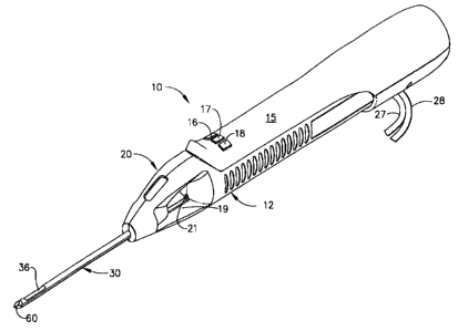
FIQLTRL 1 is atr taometric vlevrr of a hand held vacuum assisted biopsy device
constructed in accordance with a preferred embodiment of this invention.
FIGURE 2 is an isometric view of the elongated needle of the hand held
vacuum assisted biopsy device of figure 1.
4
CA 02413907 2002-12-10
FIGURE 3 is an isometric view of the right body member of the elongated
needle of the hand held vacuum assisted biopsy device of figure 1. A cutter
tube liner
is illustrated in assembly with the elongated needle.
FIGURE 4 is an exploded isometric view of the separated loft body member
and right body member of the elongated needle of the hand held vacuum assisted
biopsy device of figure 1.
FIGURE 5 is an exploded isometric view of the two member needle tip on the
elongated needle of the hand held vacuum assisted biopsy device of figure 1 as
viewed
from the proximal side thereof.
FIGURE 6 is an exploded isometric view of the two member needle tip of the
elongated needle of the hand held vacuum assisted biopsy device of figure 1 as
viewed
from the distal end thereof
Detailed Description o~,the Inventi~
Figure 1 shows a hand-held vacuum assisted biopsy device 10 comprising a
needle assembly 20 and a holster 15. Needle assembly 20 is detachably
connected to
holster 15. Together they constitute a lightweight, ergonomically shaped, hand
manipulatable portion referred to as handpiecc 12. Since handpiece I2 is
manipulated
by the operator's hand rather than by an electromechanical arm, the operator
may steer
the handpiece 12 with great freedom towards the tissue mass of interest. The
surgeon
has tactile feedback while doing so and can thus, ascertain to a sigiificant
degree, the
density and hardness of tissue being encountered. 1n addition, handpiece 12
may be
hold aparoxlmately pat'illd tc~ the Chit wall of a patient for obtaining
tissue portions
closer to the chest wall than may be obtained whop using an instrument mounted
to an
electromechanical arm.
5
CA 02413907 2002-12-10
The device includes a means for obtaining a tissue sample. Holster 1 S
includes a forward button 16 which may be used to move cutter 21 (shown in
Figure
1) distally though cutter lumen 32 and sever tissue collected in port 36.
Holster 15
further includes a reverse button 17 which may be used to move cutter 21
proximally
through cutter lumen 32 and thereby moving the tissue sample in port 36 to a
tissue
collection surface 19. A vacuum button 1 S on holster 1 S is used to open or
close first
and second vacuum lines, 27 and 28, for activating a vacuum lumen 34 so as to
cause
tissue to biome disposed within port 36.
Referring now to Figure 2 there is shown an isometric view of the needle
assembly 20 of the hand held vacuum assisted biopsy device 10 of figure 1.
Needle
assembly 20 includes an elongated needle 30 having a distal end 31, a proximal
end
33 and a longitudinal axis thcrcbetween. Needle assembly 20 has a needle tip
60 at its
distal end for penetrating the soft tissue of a surgical patient. Elongated
needle 30
comprises a cutter lumen 32 and a vacuum ehambeT lumen 34.
At the distal end of the elongated needle 30 is a needle tip 60, which is
sharpened and is preferably made from an MRI compatible resin such as LTltem
or
Vectra. Needle tip 60 is designed to penetrate soft tissue, such as the breast
of a
female surgical patient. In this embodiment, needle tip 60 is a three-sided
pyramidal
shaped point, although the needle tip 60 configuration may also have other
shapes.
Referring now to Figure 3, elongated needle 30 is preferably made from a
thermoplastic material such as Vectra A130 or B130 liquid crystal polymer,
although
ZS other MRI compatible resins may be available from Ticona of Summit, NJ.
Elongated
needle 30 includes a cutter lumen 32 which houses the cutter 21 (shown in
Figure 1).
Adjacent the distal end 31 of the cutter lumen 32 is a port 36 for receiving
the tissue
that is extracted from a surgical patient by the cutter 21. Joined alongside
the cutter
lidrttett 33 ie a va~tutri Chamber lum~rt 34. The vacuum chamber lumen 34
receives
vacuum from the second vacuum lint 28 which is connected the vacuum chamber
lumen 34 on the elongated needle 30 by the vacuum manifold 26 which is located
at
the proximal end 33 of elongated needle 30. Also located at the proximal end
of the
elongated needle 30 is a flange 38, which allows the elongated needle 30 and
needle
6
CA 02413907 2002-12-10
assembly 20 to interlock with the handpiece I2 on the hand-held vacuum
assisted
biopsy device 10. Changing from a stainless steel needle to a polymer may
require a
change in wall thickness, for example from 0.008" to 0.030". The liner 22,
discussed
below, is also made from a MRI compatible material, preferably a polypropylene
such
S as Prolene available firom Ethicon, Inc., Somerville NJ, or a material known
as Radel-
5000, available from British Petroleum, London L1K.
As seen in Figure 4, elongated needle 30 is formed from a left body member
40 and a right body member 50 on either side of the longitudinal axis. The
edges of
the halves 40 and 50 are gated for easy part filling, and the edges are
stepped with
ridges that allow the two halves 40 and 50 to attach together with ease.
Preferably
needle 30 is molded from a very stiff thermoplastic, such as Vectra A130 or
Vectra
B 130 liquid crystal polymer. Other glass fiber reinforced resins known to
those
slatted in the art could also be used. Preferably the probe is made from a
polymer
material having the combination of high stiffness, low viscosity, and low mold
shrink
rate, such as LCP resins.
During assembly of one potential embodiment the elongated needle 30, the left
body member 40 and right body member 50 of the elongated needle 30 are pushed
together. Once the leR body mcmbe~r 40 and the right body member 50 are
pressed
together, a thin-walled sleeve of high strength tubing is slipped over the
elongated
needle and is shrink fitted into place. The shrink tubing holds the left body
member 40
and the right body member 50 together for easier handling prior to adhesive
curing. 1n
addition, the shrink tubing makes the exterior of the elongated needle 30
smoother for
reduced insertion forces.
Referring back to Figure 3, there is shown the right body member 50 of the
elongated needle 30, separated from the left body member 40, which has been
omitted
tlrotrt this figure for clarity. The right body member 50 has upper and lower
ends
comprising alternating male and female portions or members, 42 and 52, which
alternate and are arranged axially along the length of the right body member
SO of the
elongated needle 30. In addition to the male and female members, 42 and 52,
there is
an upper female distal member 54 and a lower male distal member 4S, both of
which
7
CA 02413907 2002-12-10
are located at he distal end of the right body member 50. The upper female
distal
member 54 is located just below the distal end of the cutter lumen 32 and
above the
distal end of the vacuum chamber lumen 34. At the proximal and of the right
body
member 50 are three Female receivers 56 which surround the vacuum manifold 26
at
the proximal end of the right body member S0.
Still referring to figure 3, needle 20 includes a cutter tube liner 22, which
helps
keep adhesive out of the lumen to provide a smooth surface thereon. Liner 22
generally abuts in the inner surface of cutter 20 along lumen 32. The distal
end 31 of
liner 22 is proximal to port 36 but otherwise is disposed along the length of
lumen 32.
The cutter tube liner 22 is formed from a thin-walled extrusion of a low-
friction,
abrasion-resistant plastic, such as polypropylene, polyetherimide or
polyethersulfone.
The cutter tube liner 22 provides a smooth, low-friction, abrasion-resistant
surface for
the cutter 21. The cutter tube liner 22 also acts as an aid for scaling vacuum
and fluid
1 S leakage in that it isolates the cutter lumen 32 from the vacuum chamber
lumen 34 and
ensures that fluid and material from the cutter lumen 32 does not get sucked
into the
vacuum chamber 34 by vacuum suction in the vacuum chamber lumen 34_ Isolating
the cutter lumen 32 from the vacuum chamber lumen 34 may be preferable because
the cutter lumen 32 and vacuum line 27, and the vacuum chamber lumen 34
operates
on the second vacuum line 28.
Still referring to Figure 3, another feature that is included in the preferred
design of the invention to enhance performance is the outside diameter of the
left body
member 40 and right body member 50 could be stepped very slightly, if needed,
to
2S compensate for the thickness of the cutter cube inner 22. This is, the
cutter lumen 32
would be very slightly larger than the inside diameter of the cutter rube
liner 22, which
is a thin walled structure.
Referring again to Figure 4 there is shown an exploded isometric view of the
elongate needle 30 of the and held vacuum assisted biopsy device 10 of figure
1. Both
the left body member 40 and the right body member 50 of the elongated needle
30 are
shown. The female features 52, which are arranged axially on the right body
member
50. Also, the male features 42, which are arranged axially on the left body
momber 40.
CA 02413907 2002-12-10
mate to the female features 52, which are arrange axially on the right body
member
50. Also, the male features 42 are arranged axially on the right body member
SO mate
to the female features 52 which are arranged axially on the left body member
40.
In addition to male and female members, 42 and 52, which are arranged
axially and mate, the left body half 40 and right body member 50 have
additional
features that mate at both the proximal and the distal ends. At the proximal
end of the
right body member 50 are three female receivers 56 which surround the vacuum
manifold 26. At the proximal end of the left body member 40 are three male
bosses
46 which surround the vacuum manifold 36 and correspond to the three female
receivers 56 on the right body member 50. When the left body member 40 and the
right body member SO are pushed together, the three female receivers 56 on the
proximal end of the left body member 40. The proxiatat end of the elongated
needle
30 is thus, retained by the three female receivers 56 and three male bosses
46, which
mate at the proximal end of the elongated needle 30.
The needle tip 60 at the distal end of the elongated needle 30 is retained by
the
upper female distal part 54 and the upper male distal 44 and the Lower female
distal
portion SS on the left body member 40. On the left body member 40 is and upper
male
distal portion 44 and a lower female distal part 55. The upper male distal
portion 44
is located above the cutter lumen 32 at the distal end on the left body member
40, and
the lower female distal part 55 is located below the cutter lumen 32 and above
the
vacuum chamber lumen 34 at the distal end of the left body member 40. On the
right
body 50 is an upper female distal part 54 and a lower male distal portion 45,
which
correspond to the upper male distal portion 44 and the lower female distal
part 55 on
the IeR body member 40. The upper fanale distal part S4 is located above the
cutter
lumen 32 at the distal end of the right body member 50, and the lower male
distal
portion 45 is located below the cutter lumen 32 and above the vacuum chamber
lumen
34 at the distal end of the right body member 50.
Still referring to figure 4, not only do the male and female members, 42 and
52, secure the body of the elongated needle 30, and the proximal and distal
ends of the
elongated needle 30, both female and female members, 42 and 52, also form the
9
CA 02413907 2002-12-10
irterlumen vacuum holes 23, which are located below the port 36 on the distal
end of
the elongated needle 30. The male and female members 42 and 52, on the right
body
member 50, which are located below the port 36 in between the cutter lumen 32
and
vacuum chamber lumen 34, mate with a male and female members, 42 and 52, on
the
left body member 40, which are also located below the port 36 in between the
cutter
lumen 36 and vacuum chamber lumen 34. When these male and ftmale members, 42
and 52, on the left body member 40 and right body member 50 mate, the
incerlumen
vacuum holes 23 on the needle 30 are formed. The interlumen vacuum holes 23
are
six cylindrically shaped holes which are open to port 36, so that the tissue
can be
severed by the cutter 21, which rotates and advances. The cutter 21 deposits
the tissue
into the tissue collection surface 19 by retracting proximally.
StiII refeaing to figure 4, during assembly of the elongated needle 30,
sufficient adhesive is applied to the left body member 40 and right body
member 40
and right body member 50, to fill the narrow axial spaces between the male and
female members, 42 and 52, which mate. After this, the left body member 40 and
right body member 50 are pressed together. 'The adhesive that is used should
be cured
using light, heat, or other appropriate means for the particular types of
adhesive that is
being used. For a light cured adhesive, light could be dircctad inside of the
cutter
lumen 32 and the vaccum lumen 34 using tight stick optics if necessary.
Still referring to figure 4, the male and female members, 42 and 52, which
mate and are located on the left body member 40 arid the right body member 50
have a
number of distinct advantages. The male and female members, 42 and 52, on the
left
ZS body member 40 and right body member 50 orient the lef3 body member 40 and
right
body member 50 during assembly of the elongated needle 30.
The male and female members, 42 and 52, which mate, are also key factors in
increasing both the strength and lateral bending stiffness of the elongated
needle 30.
When the needle 30 is subjected to a lateral bending moment, nearly all of the
material being loaded axially is the high-strength, high stiffiness body
material. Only
the small amount of adhesive that is used to fill the axial clearances between
the male
and female members, 42 and 52, which mate, is of a lower stiffness. A
conventional
lo
CA 02413907 2002-12-10
bonded joint would result in the bond line being loaded in a manner similar to
that
used for adhesive pool strength testing, which is the most severe type of
loading for an
adhesive joint. In contrast to this, the male female members, 42 and 52, which
mate,
would create lateral bond surfaces along the elongated needle 30. This
substantially
increases the bond line length of the elongated needle 30, Because of
significant
portions of the bond line being loaded in shear, the strength and lateral
stiffness of the
elongated needle 30 is increased_ This is improved over a single piece molded
cylinder in that with the bond line loaded in shear, the elongated needle 30
will be
able to sustain bending moments of its joints rather than at its base, which
decreases
the possibility of breakage.
Figure 5 shows and exploded isometric view of the needle tip 60 of the
elongated needle 30 of the hand held vacuum assisted biopsy device 10 of
figure 1 as
viewed fiom the proximal side thereof. The needle tip 60 has two halves; a
composite
tip member ~0, and a composite hub member 80. Both the composite tip member 70
and the composite hub member 80 are preferably molded from a magnetic
Resonance
Imaging (MRI) compatible resin such as Ultem or Vectra ceramic or other MRI
compatible materials known to those skilled in the art is sharp. The composite
tip
member 70 has a three-sided pyramidal shaped point, but may also have other
shapes.
The composite tip member 70 has a hollow cavity 74 and protruding connectors
76.
The two protruding connectors 76 are inserted into the two receiving holes 82
on the
composite hub member 80 when the composite hub member 80 is pushed into the
composite tip member 70 during assembly. Cavity preferably contains a capsule
90
made from a material which will leave and MR,I artifact. Having a capsule 90
made
from and MRI artifact leaving material is necessary because since the
elongated
needle 30 is made of an MRI compatible resin, the elongated needle 30 does not
show
up on an MRI scan. Therefore, it is difficult for a physician to discera the
orientation
of the elongated needle 30 during and MRI scan MRI artifact leaving material
90
tolvrs the aforementioned problems in that a needle tip 60 leaves a small, but
not
troublesome artifact on an MRI scan. This small, artifact indicates the
orientation of
the elongated needle 30 relative to the sight of biopsy, and where the tissue
receiving
bowl begins during and MRI scan. The MRI artifact leaving material 90 that is
preferred is a capsule of Gadolinium. However, there are other materials that
could be
11
CA 02413907 2002-12-10
pat into the hollow cavity74 of the composite tip member 70 that would leave
and
acceptable MRI artifact. These include, but not limited to: liquid Gadolinium,
Titanium Wire, Aluminum, Copper, Brass Iron, and Bronze.
Figure 6 shows an exploded isometric view of the needle tip 60 of the
elongated needle 30 of the hand held vacuum assisted biopsy device 10 of
figure 1 as
viewed from the distal end thereof. This figure clearly illustrated the
components on
the composite hub member 80. On the distal end of the composite hub member 80
is
a male part 84, which pushes the MRI artifact leaving material $0 down into
the
hollow cavity 74 on the composite tip member 70. Also located on the distal
end of
the composite hub member 80 is a knock out boss 86, which pushes a collected
breast
tissue sample into the end of the cutter tube 21 the hand held vacuum assisted
biopsy
device 10 during a breast biopsy. The two receiving holes 82 on the composite
hub
member 80 receive the two protruding connectors 76 on the composite tip member
70
when the composite tip member 70 and composite hub member 80 are pushed
together. The reception of the two protruding connectors 76 on the composite
tip
member 70 by the two receiving holes 82 on the composite hub member 80 locks
the
composite tip member 70 and the composite hub member 80 together, and seals
the
MRI artifact leaving material 90 in the hollow cavity 7~4 in between the
composite tip
member 70 and composite hub member 80.
In using the hand member vacuum assisted biopsy device 10, as shown in
figure 1, for a breast biopsy in an MRI environment, physician will first
positioned
outside of the MRI magnet, the patient is moved into the MRI magnet and
imaging of
the breast is performed. During imaging of the breast, serial slices of the
breast are
examined, and a contrast agent is administered to highlight suspicious areas
of breast
tissue. At this time, the location of the suspicious breast tissue is
detetinined relative
to the compression grid.
After the location of the suspicious breast tissue is determined, the patient
is
moved outside the magnet. Local anesthesia is administered to the patient and
the
probe 20 is inserted into the area of suspicious breast tissue.
iz
CA 02413907 2002-12-10
After the probe is inserted into the suspicious area of breast tissue, the
patient
is moved back into the MRI magnet and a set of images of the breast are taken.
The
sets of images confirm that the probe 20 is adjacent to the suspicious breast
tissue, the
patient is moved outside of the MRI magnet and the hand held vacuum assisted
biopsy
device I O of figure 1 is then inserted into the sleeve, replacing the
obturator.
ARer the hand held vacuum assisted biopsy device 10 of figure 1 is inserted
through the sleeve; multiple tissue samples are taken. In taking multiple
tissue
samples, the needle tip 60 as the distal end of the elongated needle 30 on the
hand
held vacuum assisted biopsy 10, of figure 1, penetrates the breast in the area
that is
adjacent of the suspicious breast tissue. Prior to, and during penetration by
the needle
tip 60, the cutter 21 is fully forward, and is advanced forward through the
cutter lumen
32 by pressing the forward button I6 on the holster 15 of the vacuttm assisted
biopsy
device 10 of figure 1.
Once the elongated needle 30 is positioned in the area adjacent to the
suspicious breast tissue, vacuum suction is applied to he vacuum chamber lumen
34.
?he vacuum suction is applied by pressing the vacuum button 18 on the holster
1 S of
the hand held vacuum assisted biopsy device 10 of figure 1. Pressing the
vacuum
button 18 on the holster 15 opens the second vacuum tine 28, which transports
vacuum suction through the handpiece 12 of the hand held vacuum assisted
biopsy
device 10 and into the vacuum chamber lumen 34 on the elongated needle 30. The
second vacuum line 28 runs thmugh the handpiece 12 of the hand held vacuum
assisted biopsy device 10 and into the elongated needle 30 through the vacuum
manifold 24 at he proximal end of the elongated needle 30. The vacuum suction
that
is applied to the vacuum chamber lumen travels from the proximal, of the
distal end
of the vacuum chamber lumen 34, below the interlumen vacuum holes 23. The
interlumen vacuum holes 23 receive suction from the vacuum chamber lumen 34.
The suction from the imerlumen vacuum holes 23 actively pulls breast tissue
through the port 36 and into the cutter lumen 32 on the elongated needle 30.
After the
breast the tissue is pulled into the elongated needle 30 through the port 36,
the cutter
Z 1 begins to rotate and advances through the breast tissue until a sample has
been
13
CA 02413907 2002-12-10
obtained. ARer the breast tissue sample has been obtained, the elongated
needle 30 is
rotated to position the port 36 toward a different clockwise position in
preparation for
obtaining the next tissue sample. After the elongated 30 is rotated, the
cutter 21 is
withdrawn backwards within the cutter lumen 32 on the elongated needle 30 and
the
breast tissue sample is carried back to a knock-out boss 86, which pushed the
collected breast tissue sample out into a tissue collection surface 19 on the
handheld
vacuum assisted biopsy device 10. Vacuum suction is thrn reapplied to the
vacuum
chamber lumen 34 from the second vacuum line 28, and the aforementioned
process is
repeated continuously until the elongated needle 30 has been rotated clockwise
once
around the entire clock.
After multiple breast tissue samples have been obtained from the patient, the
patient is moved back into the MRI magnet. Once in the MRI magnet, a set of
images
of the breast are taken in order to confirm that the suspicious breast tissue
has been
removed. The artifact in the probe tip is a useful point of reference to
confirm after
tire biopsy site is marked, the breast biopsy in an MRI environment is
complete.
While preferred embodiments of the present invention have been shown and
described herein, it will be obvious to those skilled in the art that such
embodiments
are provided by way of example only. Numerous variations, changes, and
substitutions will now occur to those skilled in the art without departing
from the
present invention. Accordingly, it is intended that the invention be limited
only by the
spirit and scope of the appended claims.
14