Note: Descriptions are shown in the official language in which they were submitted.
CA 02414331 2008-07-07
PD-L2 MOLECULES: NOVEL PD-1 LIGANDS AND USES THEREFOR
15
Background of the Invention
In order for T'cells to respond to foreign.polypeptides, two signals must be
provided by antigen-presenting cells (APCs) to resting T lymphocytes (Jenkins,
M. and
Schwartz, R. (1987) J Exp. Med. 165:302-319; Mueller, D.L. et al. (1990) J
Imvnunol.
144:3701-3709). The first signal, which confers specificity to the immune
response, is
transduced via the T cell receptor (TCR) following recognition of foreign
antigenic
peptide presented in the context of the major histocompatibility complex
(MHC). The
second signal, termed costimulation, induces T cells to proliferate and become
functional (Lenschow et al. (1996) Annu. Rev. Immunol. 14:233). Costimulation
is
neither antigen-specific, nor MHC-restricted, and is thought to be provided by
one or
more distinct cell surface molecules expressed by APCs (Jenkins, M.K. et al.
(1988) J
Immunol. 140:3324-3330; Linsley, P.S. et al. (1991)J Exp. Med. 173:721-730;
Gimmi,
.C.D. et al. (1991) Proc. Natl. Acad. Sci. USA 88:6575-6579; Young, J.W. et
al. (1992)
J. Clin. Invest. 90:229-237; Koulova, L. et al. (1991) J Exp. Med. 173:759-
762; Reiser,
H. et al. (1992) Proc. Natl. Acad. Sc!. USA 89:271-275; van-Seventer, G.A. et
al. (1990)
J Immunol. 144:4579-4586; LaSalle, J.M. et al. (1991) J. Immunol. 147:774-80;
Dustin,
M.I. et al. (1989) J Exp. MecL 169:503; Armitage, R.J. et al. (1992) Nature
357:80-82;
Liu, Y. et al. (1992) J Exp. Med. 175:437-445).
CA 02414331 2002-12-27
WO 02/00730 PCT/US01/20964
2
The CD80 (B7-1) and CD86 (B7-2) proteins, expressed on APCs, are critical
costimulatory molecules (Freeman et al. (1991) J Exp. Med. 174:625; Freeman et
al.
(1989) J. bnmunol. 143:2714; Azuma et al. (1993) Nature 366:76; Freeman et al.
(1993)
Science 262:909). B7-2 appears to play a predominant role during primary
immune
responses, while B7-1, which is upregulated later in the course of an immune
response,
may be important in prolonging primary T cell responses or costimulating
secondary T
cell responses (Bluestone (1995) Immunity 2:555).
One ligand to which B7-1 and B7-2 bind, CD28, is constitutively expressed on
resting T cells and increases in expression after activation. After signaling
through the T
cell receptor, ligation of CD28 and transduction of a costimulatory signal
induces T cells
to proliferate and secrete IL-2 (Linsley, P.S. et al. (1991) J Exp. Med.
173:721-730;
Gimmi, C.D. et al. (1991) Proc. Natl. Acad. Sci. USA 88:6575-6579; June, C.H.
et al.
(1990) Immunol. Today 11:211-6; Harding, F.A. et al. (1992) Nature 356:607-
609). A
second ligand, termed CTLA4 (CD 152) is homologous to CD28 but is not
expressed on
resting T cells and appears following T cell activation (Brunet, J.F. et al.
(1987) Nature
328:267-270). CTLA4 appears to be critical in negative regulation of T cell
responses
(Waterhouse et al. (1995) Science 270:985). Blockade of CTLA4 has been found
to
remove inhibitory signals, while aggregation of CTLA4 has been found to
provide
inhibitory signals that downregulate T cell responses (Allison and Krummel
(1995)
Science 270:932). The B7 molecules have a higher affinity for CTLA4 than for
CD28
(Linsley, P.S. et al. (1991) J. Exp. Med. 174:561-569) and B7-1 and B7-2 have
been
found to bind to distinct regions of the CTLA4 molecule and have different
kinetics of
binding to CTLA4 (Linsley et al. (1994) Immunity 1:793). A new molecule
related to
CD28 and CTLA4, ICOS, has been identified (Hutloff et al. (1999) Nature 3
97:263;
WO 98/38216), as has its ligand, which is a new B7 family member (Aicher A. et
al.
(2000) 1 Immunol. 164:4689-96; Mages H.W. et al. (2000) Eur. J. Immunol.
30:1040-7;
Brodie D. et al. (2000) Curr. Biol. 10:333-6; Ling V. et al. (2000) J.
Immunol.
164:1653-7; Yoshinaga S.K. et al. (1999) Nature 402:827-32). If T cells are
only
stimulated through the T cell receptor, without receiving an additional
costimulatory
signal, they become nonresponsive, anergic, or die, resulting in
downmodulation of the
immune response.
Immune cells have receptors that transmit activating signals. For example, T
cells have T cell receptors and the CD3 complex, B cells have B cell
receptors, and
myeloid cells have Fe receptors. In addition, immune cells bear receptors that
transmit
CA 02414331 2002-12-27
WO 02/00730 PCT/US01/20964
3
signals that provide costimulatory signals or receptors that transmit signals
that inhibit
receptor-mediated signaling. For example, CD28 transmits a costimulatory
signal to T
cells. After ligation of the T cell receptor, ligation of CD28 results in a
costimulatory
signal characterized by, e.g., upregulation of IL-2ra, IL-2r(3, and IL-2ry
receptor,
increased transcription of IL-2 messenger RNA, and increased expression of
cytokine
genes (including IL-2, IFN-y, GM-CSF, and TNF-a). Transmission of a
costimulatory
signal allows the cell to progress through the cell cycle and, thus, increases
T cell
proliferation (Greenfield et al. (1998) Crit. Rev. Immunol. 18:389). Binding
of a
receptor on a T cell which transmits a costimulatory signal to the cell (e.g.,
ligation of a
costimulatory receptor that leads to cytokine secretion and/or proliferation
of the T cell)
by a costimulatory ligand results in costimulation. Thus, inhibition of an
interaction
between a costimulatory ligand and a receptor that transmits a costimulatory
signal on
immune cells results in a downmodulation of the immune response and/or
specific
unresponsiveness, termed immune cell anergy. Inhibition of this interaction
can be
accomplished using, e.g., anti-CD28 Fab fragments, antibodies to B7 family
molecules,
or by using a soluble form of a receptor to which a B7 family member molecule
can
bind as a competitive inhibitor (e.g., CTLA41g).
Inhibitory receptors that bind to costimulatory molecules have also been
identified on immune cells. Activation of CTLA4, for example, transmits a
negative
signal to a T cell. Engagement of CTLA4 inhibits IL-2 production and can
induce cell
cycle arrest (Krummel and Allison (1996) J Exp. Med. 183:2533). In addition,
mice
that lack CTLA4 develop lymphoproliferative disease (Tivol et al. (1995)
Immunity
3:541; Waterhouse et al. (1995) Science 270:985). The blockade of CTLA4 with
antibodies may remove an inhibitory signal, whereas aggregation of CTLA4 with
antibody transmits an inhibitory signal. Therefore, depending upon the
receptor to
which a costimulatory molecule binds (i.e., a costimulatory receptor such as
CD28 or an
inhibitory receptor such as CTLA4), B7 molecules including B7-4 can promote T
cell
costimulation or inhibition.
PD-1 is a member of the immunoglobulin family of molecules (Ishida et al.
(1992) EMBO J 11:3887; Shinohara et al. (1994) Genomics 23:704). PD-1 was
previously identified using a subtraction cloning based approach designed to
identify
modulators of programmed cell death (Ishida et al. (1992) EMBO J. 11:3887-95;
Woronicz et al. (1995) Curr. Top. Microbiol. Immunol. 200:137). PD-1 is
believed to
play a role in regulating lymphocyte survival, e.g., during clonal selection
(Honjo (1992)
CA 02414331 2002-12-27
WO 02/00730 PCT/US01/20964
4
Science 258:591; Agata et al. (1996) Int. Immunology 8:765; Nishimura et al.
(1996) Int.
Immunology 8:773). PD-1 has an extracellular region containing an
immunoglobulin
superfamily domain, a transmembrane domain, and an intracellular region which
includes an immunoreceptor tyrosine kinase-based inhibitory motif (ITIM)
(Ishida et al.
(1992) supra; Shinohara et al. (1994) supra; US Patent 5,698,520). This
features also
define a larger family of molecules, called the immunoinhibitory receptors,
which also
includes gp49B, PIR-B, and the killer inhibitory receptors (KIRs) (Vivier and
Daeron
(1997) Immunology Today 18:286). It is often assumed that the tyrosyl
phosphorylated
ITIM motif of these receptors interacts with the SH2-domain-containing
phosphatases,
which leads to inhibitory signals. A subset of these immunoinhibitory
receptors binds to
MHC molecules, for example the KIRs, and CTLA4 binds to B7-1 and B7-2. It has
been proposed that there is a phylogenetic relationship between the MHC and B7
genes
(Henry et al. (1999) Immunology Today 20:285-288).
PD-I was also implicated as a regulator of B cell responses (Nishimura (1998)
Int. Immunology 10:1563). Unlike CTLA4, which is found only on T cells, PD-I
is also
found on B cells (in response anti-IgM) and on a subset of thymocytes and
myeloid cells
(Agata et al. (1996) supra; Nishimura et al. (1996) Int. Immunology 8:773).
The importance of the B7:CD28/CTLA4 costimulatory pathway has been
demonstrated in vitro and in several in vivo model systems. Blockade of this
costimulatory pathway results in the development of antigen-specific tolerance
in
murine and human systems (Harding, F.A. et al. (1992) Nature 356:607-609;
Lenschow,
D.J. et al. (1992) Science 257:789-792; Turka, L.A. et al. (1992) Proc. Natl.
Acad. Sci.
USA 89:11102-11105; Gimmi, C.D. et al. (1993) Proc. Natl. Acad. Sci. USA
90:6586-
6590; Boussiotis, V. et al. (1993) J.. Exp. Med. 178:1753-1763). Conversely,
expression
of B7 by B7-negative murine tumor cells induces T-cell mediated specific
immunity
accompanied by tumor rejection and long lasting protection to tumor challenge
(Chen,
L. et al. (1992) Cell 71:1093-1102; Townsend, S.E. and Allison, J.P. (1993)
Science
259:368-370; Baskar, S. et al. (1993) Proc. Natl. Acad. Sci 90:5687-5690.).
Therefore,
manipulation of the costimulatory pathways offers great potential to stimulate
or
suppress immune responses in humans.
Summary of the Invention
The present invention is based, at least in part, on the discovery of novel
nucleic
acid molecules and polypeptides encoded by such nucleic acid molecules,
referred to
CA 02414331 2002-12-27
WO 02/00730 PCT/US01/20964
herein as PD-L2 nucleic acid and polypeptide molecules, which are members of
the B7
family and are ligands for PD-1. Interaction of PD-L2 with PD-i transmits a
negative
signal to immune cells, downregulating immune responses. Preferred PD-L2
molecules
are expressed on the surface of professional antigen presenting cells (e.g., B
5 lymphocytes, monocytes, dendritic cells, and Langerhans cells) and other
antigen
presenting cells (e.g., keratinocytes, endothelial cells, astrocytes,
fibroblasts, and
oligodendrocytes), down-regulate lymphocyte activation, and/or are bound by
antibodies
which recognize PD-L2 molecules. The PD-L2 nucleic acid and polypeptide
molecules
of the present invention are useful, e.g., in modulating the immune response.
Accordingly, in one aspect, this invention provides isolated nucleic acid
molecules
encoding PD-L2 polypeptides, as well as nucleic acid fragments suitable as
primers or
hybridization probes for the detection of PD-L2-encoding nucleic acids.
In one embodiment, a PD-L2 nucleic acid molecule of the invention is at least
about 70%,75%,80%,85%,90%,91%,92%,93%,94%,95%,96%,97%,98%,99%
or more identical to the nucleotide sequence (e.g., to the entire length of
the nucleotide
sequence) shown in SEQ ID NO:1 or 3, or a complement thereof.
In a preferred embodiment, the isolated nucleic acid molecule includes the
nucleotide sequence shown in SEQ ID NO:1 or 3, or a complement thereof. In
another
embodiment, the nucleic acid molecule includes the nucleic acid sequence shown
in
SEQ ID NO:3 and nucleotides 1-273 of SEQ ID NO:1. In a further embodiment, the
nucleic acid molecule includes the nucleic acid sequence shown in SEQ ID NO:3
and
nucleotides 1096-1223 of SEQ ID NO:1. In another preferred embodiment, the
nucleic
acid molecule consists of the nucleotide sequence shown in SEQ ID NO:1 or 3.
In another embodiment, a PD-L2 nucleic acid molecule includes a nucleotide
sequence encoding a polypeptide having an amino acid sequence sufficiently
identical to
the amino acid sequence of SEQ ID NO:2. In a preferred embodiment, a PD-L2
nucleic
acid molecule includes a nucleotide sequence encoding a polypeptide having an
amino
acid sequence at least about 71%, 75%, 80%, 85%, 90%, 95%, 96%, 97%, 98%, 99%
or
more identical to the entire length of the amino acid sequence of SEQ ID NO:2.
In another preferred embodiment, an isolated nucleic acid molecule encodes the
amino acid sequence of human PD-L2. In yet another preferred embodiment, the
nucleic acid molecule includes a nucleotide sequence encoding a polypeptide
having the
amino acid sequence of SEQ ID NO:2. In yet another preferred embodiment, the
nucleic acid molecule is at least about 50, 100, 150, 200, 250, 300, 350, 400,
450, 500,
CA 02414331 2002-12-27
WO 02/00730 PCT/US01/20964
6
550, 600, 650, 700, 750, 800, 850, 900, 950, 1000, 1050, 1100, 1150 or more
nucleotides in length. In a further preferred embodiment, the nucleic acid
molecule is at
least about 50, 100, 150, 200, 250, 300, 350, 400, 450, 500, 550, 600, 650,
700, 750,
800, 850, 900, 950, 1000, 1050, 1100, 1150 or more nucleotides in length and
encodes a
polypeptide having a PD-L2 activity (as described herein).
Another embodiment of the invention features nucleic acid molecules,
preferably
PD-L2 nucleic acid molecules, which specifically detect PD-L2 nucleic acid
molecules
relative to nucleic acid molecules encoding non-PD-L2 polypeptides. For
example, in
one embodiment, such a nucleic acid molecule is at least about 880, 900, 950,
1000,
1050, 1100, 1150 or more nucleotides in length and hybridizes under stringent
conditions to a nucleic acid molecule comprising the nucleotide sequence shown
in SEQ
ID NO: 1, or a complement thereof. In another embodiment, such a nucleic acid
molecule is at least 20, 30, 40, 50, 100, 150, 200, 250, 300 or more
nucleotides in length
and hybridizes under stringent conditions to a nucleic acid molecule
comprising
nucleotides 1-358 of SEQ ID NO:1, or a complement thereof. In a further
embodiment,
such a nucleic acid molecule is at least 20, 30, 40, 50, 100, 150, 200, 250,
300, 350, 400,
450, 500, 550, 600, 650, 700, 750, 800, 850, 900, 950, 1000, 1050, 1100, 1150
or more
nucleotides in length, includes at least 15 (i.e., 15 contiguous) nucleotides
of the
sequence comprising nucleotides 1-358 of SEQ ID NO:l, or a complement thereof,
and
hybridizes under stringent conditions to a nucleic acid molecule comprising
the
nucleotide sequence shown in SEQ ID NO:1, or a complement thereof.
In preferred embodiments, the nucleic acid molecules are at least about 880
nucleotides in length and hybridize under stringent conditions to the
nucleotide molecule
set forth in SEQ ID NO:1 {i.e., to 880 contiguous nucleotides of SEQ ID NO:1),
or a
complement thereof. In other preferred embodiments, the nucleic acid molecules
are at
least about 15 nucleotides in length and hybridize under stringent conditions
to
nucleotides 1-358 of the nucleotide molecule set forth in SEQ ID NO:1 (i.e.,
to 15
contiguous nucleotides of nucleotides 1-358 of SEQ ID NO:1), or a complement
thereof.
In further preferred embodiments, the nucleic acid molecules are at least 15
nucleotides
in length, include at least 15 (i.e., 15 contiguous) nucleotides of the
sequence comprising
nucleotides 1-358 of SEQ ID NO: 1, or a complement thereof, and hybridize
under
stringent conditions to a nucleic acid molecule comprising the nucleotide
sequence
shown in SEQ ID NO: 1, or a complement thereof.
CA 02414331 2002-12-27
WO 02/00730 PCT/US01/20964
7
In still other preferred embodiments, the nucleic acid molecule encodes a
naturally occurring allelic variant of a polypeptide comprising the amino acid
sequence
of SEQ ID NO:2, wherein the nucleic acid molecule hybridizes to a complement
of a
nucleic acid molecule comprising SEQ ID NO:1 or 3, or a complement thereof,
under
stringent conditions.
Another embodiment of the invention provides an isolated nucleic acid molecule
which is antisense to a PD-L2 nucleic acid molecule, e.g., is antisense to the
coding
strand of a PD-L2 nucleic acid molecule as shown in SEQ ID NO:1 or 3.
Another aspect of the invention provides a vector comprising a PD-L2 nucleic
acid molecule. In certain embodiments, the vector is a recombinant expression
vector.
In another embodiment, the invention provides a host cell containing a vector
of the
invention. In yet another embodiment, the invention provides a host cell
containing a
nucleic acid molecule of the invention. The invention also provides a method
for
producing a polypeptide, preferably a PD-L2 polypeptide, by culturing in a
suitable
medium, a host cell, e.g., a mammalian host cell such as a non-human mammalian
cell,
of the invention containing a recombinant expression vector, such that the
polypeptide is
produced.
Another aspect of this invention features isolated or recombinant PD-L2
polypeptides (e.g., proteins, polypeptides, peptides, or fragments or portions
thereof). In
one embodiment, an isolated PD-L2 polypeptide includes at least one or more of
the
following domains: a signal peptide domain, an IgV domain, an IgC domain, an
extracellular domain, a transmembrane domain, and a cytoplasmic domain.
In a preferred embodiment, a PD-L2 polypeptide includes at least one or more
of
the following domains: a signal peptide domain, an IgV domain, an IgC domain,
an
extracellular domain, a transmembrane domain, and a cytoplasmic domain, and
has an
amino acid sequence at least about 71%, 75%, 80%, 85%, 90%, 91%, 92%, 93%,
94%,
95%, 96%, 97%, 98%, 99% or more identical to the amino acid sequence of SEQ ID
NO:2. In another preferred embodiment, a PD-L2 polypeptide includes at least
one or
more of the following domains: a signal peptide domain, an IgV domain, an IgC
domain, an extracellular domain, a transmembrane domain, and a cytoplasmic
domain,
and has a PD-L2 activity (as described herein).
In yet another preferred embodiment, a PD-L2 polypeptide includes at least one
or more of the following domains: a signal peptide domain, an IgV domain, an
IgC
domain, an extracellular domain, a transmembrane domain, and a cytoplasmic
domain,
CA 02414331 2002-12-27
WO 02/00730 PCT/US01/20964
8
and is encoded by a nucleic acid molecule having a nucleotide sequence which
hybridizes under stringent hybridization conditions to a complement of a
nucleic acid
molecule comprising the nucleotide sequence of SEQ ID NO:1 or 3.
In another embodiment, the invention features fragments or portions of the
polypeptide having the amino acid sequence of SEQ ID NO:2, wherein the
fragment
comprises at least 15 amino acids (i.e., contiguous amino acids) of the amino
acid
sequence of SEQ ID NO:2. In another embodiment, a PD-L2 polypeptide comprises
or
consists of the amino acid sequence of SEQ ID NO:2.
In another embodiment, the invention features a PD-L2 polypeptide which is
encoded by a nucleic acid molecule consisting of a nucleotide sequence at
least about
70%, 75%, 80%, 85%, 90%, 91%, 92%, 93%, 94%, 95%, 96%, 97%, 98%, 99% or more
identical to a nucleotide sequence of SEQ ID NO:1 or 3, or a complement
thereof. This
invention further features a PD-L2 polypeptide which is encoded by a nucleic
acid
molecule consisting of a nucleotide sequence which hybridizes under stringent
hybridization conditions to a complement of a nucleic acid molecule comprising
the
nucleotide sequence of SEQ ID NO:1 or 3.
The polypeptides of the present invention or portions thereof, e.g.,
biologically
active portions thereof, can be operatively linked to a non-PD-L2 polypeptide
(e.g.,
heterologous amino acid sequences) to form fusion polypeptides. The invention
further
features antibodies, such as monoclonal or polyclonal antibodies, that
specifically bind
polypeptides of the invention, preferably PD-L2 polypeptides. In addition, the
PD-L2
polypeptides (or biologically active portions thereof) or modulators of the PD-
L2
molecules can be incorporated into pharmaceutical compositions, which
optionally
include pharmaceutically acceptable carriers.
In another aspect, the present invention provides a method for detecting the
presence of a PD-L2 nucleic acid molecule, protein, or polypeptide in a
biological
sample by contacting the biological sample with an agent capable of detecting
a PD-L2
nucleic acid molecule, protein, or polypeptide, such that the presence of a PD-
L2 nucleic
acid molecule, protein or polypeptide is detected in the biological sample.
In another aspect, the present invention provides a method for detecting the
presence of PD-L2 activity in a biological sample by contacting the biological
sample
with an agent capable of detecting an indicator of PD-L2 activity, such that
the presence
of PD-L2 activity is detected in the biological sample.
CA 02414331 2002-12-27
WO 02/00730 PCT/US01/20964
9
In another aspect, the invention provides a method for modulating PD-L2
activity, comprising contacting a cell capable of expressing PD-L2 with an
agent that
modulates PD-L2 activity, such that PD-L2 activity in the cell is modulated.
In one
embodiment, the agent inhibits PD-L2 activity. In another embodiment, the
agent
stimulates PD-L2 activity. In a further embodiment, the agent interferes with
or
enhances the interaction between a PD-L2 polypeptide and its natural binding
partner(s),
e.g., PD-1. In a preferred embodiment, the binding partner is PD-1. In one
embodiment, the agent is an antibody that specifically binds to a PD-L2
polypeptide. In
a further embodiment, the agent is a combination of an antibody that
specifically binds
to a PD-L2 polypeptide and an antibody that specifically binds to a PD-LI
polyepeptide.
In another embodiment, the agent is a peptide, peptidomimetic, or other small
molecule
that binds to a PD-L2 polypeptide. In yet another embodiment, the agent is
another PD-
1 ligand which can modulate the interaction between PD-L2 and PD-1. In still
another
embodiment, the agent modulates expression of PD-L2 by modulating
transcription of a
PD-L2 gene, translation of a PD-L2 mRNA, or post-translational modification of
a PD-
L2 polypeptide. In another embodiment, the agent is a nucleic acid molecule
having a
nucleotide sequence that is antisense to the coding strand of a PD-L2 mRNA or
a PD-L2
gene.
In one embodiment, the methods of the present invention are used to treat a
subject having a disorder or condition characterized by aberrant,
insufficient, or
unwanted PD-L2 polypeptide or nucleic acid expression or activity by
administering an
agent which is a PD-L2 modulator to the subject. In one embodiment, the PD-L2
modulator is a PD-L2 polypeptide. In another embodiment the PD-L2 modulator is
a
PD-L2 nucleic acid molecule. In a further embodiment, the PD-L2 modulator is
an
antibody that specifically binds to a PD-L2 polypeptide. In another
embodiment, the
PD-L2 modulator is a combination of an antibody that specifically binds to a
PD-L2
polypeptide and an antibody that specifically binds to a PD-L1 polypeptide. In
yet
another embodiment, the PD-L2 modulator is a peptide, peptidomimetic, or other
small
molecule. In a preferred embodiment, the disorder or condition characterized
by
3o aberrant, insufficient, or unwanted PD-L2 polypeptide or nucleic acid
expression or
activity is an immune response disorder or condition that would benefit from
modulation
of PD-L2 activity. In another embodiment, the invention further provides
treating the
subject with an additional agent that modulates an immune response.
CA 02414331 2002-12-27
WO 02/00730 PCT/US01/20964
In still another embodiment, the invention provides a vaccine comprising an
antigen and an agent that reduces or inhibits PD-L2 activity. In a preferred
embodiment,
the vaccine inhibits the interaction between PD-L2 and its natural binding
partner(s). In
a more preferred embodiment, the binding partner is PD-1.
5 The present invention also provides diagnostic assays for identifying the
presence or absence of a genetic alteration characterized by at least one of
(i) aberrant
modification or mutation of a gene encoding a PD-L2 polypeptide; (ii) mis-
regulation of
the gene; and (iii) aberrant post-translational modification of a PD-L2
polypeptide,
wherein a wild-type form of the gene encodes a polypeptide with a PD-L2
activity.
10 In another aspect the invention provides methods for identifying a compound
that binds to or modulates the activity of a PD-L2 polypeptide, by providing
an indicator
composition comprising a PD-L2 polypeptide having PD-L2 activity, contacting
the
indicator composition with a test compound, and determining the effect of the
test
compound on PD-L2 activity in the indicator composition to identify a compound
that
modulates the activity of a PD-L2 polypeptide.
In another aspect, this invention provides a method for modulating an immune
response by modulating the interaction between PD-1 and PD-L2.
In one aspect, the invention features a method for modulating the interaction
of
PD-L2 with its natural binding partner(s) on an immune cell comprising
contacting an
antigen presenting cell which expresses PD-L2 with an agent selected from the
group
consisting of. a form of PD-L2, a form of PD-1, or an agent that modulates the
interaction of PD-L2 and its natural binding partner(s) such that the
interaction of PD-L2
with it natural binding partner(s) on an immune cell is modulated. In a
preferred
embodiment, an agent that modulates the interaction of PD-L2 and its natural
binding
partner(s) (e.g., PD-1) is an antibody that specifically binds to PD-L2. In
another
preferred embodiment, the agent is a combination of an antibody that
specifically binds
to PD-L2 and an antibody that specifically binds to PD-L1.
In one embodiment, the interaction of PD-L2 with its natural binding
partner(s)
is upregulated. In another embodiment, the interaction of PD-L2 with its
natural binding
partner(s) is downregulated.
In one embodiment, the method further comprises contacting the immune cell or
the antigen presenting cell with an additional agent that modulates an immune
response.
In one embodiment, the step of contacting is performed in vitro. In another
embodiment, the step of contacting is performed in vivo.
CA 02414331 2002-12-27
WO 02/00730 PCT/US01/20964
11
In one embodiment, the immune cell is selected from the group consisting of. a
T cell, a B cell, and a myeloid cell.
In one embodiment, the PD-L2 binding partner is PD-1.
In another aspect, the invention pertains to a method for inhibiting
activation in
an immune cell via a non-apoptotic mechanism comprising increasing the
activity or
expression of PD-L2 in a cell such that immune cell activation is inhibited.
In yet another aspect, the invention pertains to a vaccine comprising an
antigen
and an agent that inhibits the interaction between PD-L2 and its natural
binding
partner(s).
In still another aspect, the invention pertains to a vaccine comprising an
antigen
and an agent that promotes the interaction between PD-L2 and its natural
binding
partner(s).
In one embodiment, the PD-L2 binding partner is PD-1.
In another aspect, the invention pertains to a method for treating a subject
having
a condition that would benefit from upregulation of an immune response
comprising
administering an agent that inhibits the interaction between PD-L2 and its
natural
binding partner(s) on cells of the subject such that a condition that would
benefit from
upregulation of an immune response is treated.
In one embodiment, the agent comprises a blocking antibody or a small molecule
that binds to PD-L2 and inhibits the interaction between PD-L2 and its natural
binding
partner(s). In another embodiment, the agent comprises a combination of an
antibody
that specifically binds to PD-L2 and an antibody that specifically binds to PD-
L 1.
In another embodiment, the method further comprises administering a second
agent that upregulates an immune response to the subject.
In one embodiment, the condition is selected from the group consisting of: a
tumor, a pathogenic infection, or an immunosuppressive disease.
In another embodiment, the PD-L2 binding partner is PD-1.
In one aspect, the invention pertains to a method for treating a subject
having a
condition that would benefit from downregulation of an immune response
comprising
3o administering an agent that stimulates the interaction between PD-L2 and
its natural
binding partner(s) on cells of the subject such that a condition that would
benefit from
downregulation of an immune response is treated.
In one embodiment agent comprises an antibody or a small molecule that
stimulates the interaction between PD-L2 and its natural binding partner(s).
CA 02414331 2002-12-27
WO 02/00730 PCT/US01/20964
12
In another embodiment, the method further comprises administering a second
agent that downregulates an immune response to the subject.
In another embodiment, the condition is selected from the group consisting of.
a
transplant, an allergy, and an autoimmune disorder.
In one embodiment, the PD-L2 binding partner is PD-1.
In another aspect, the invention pertains to a cell-based assay for screening
for
compounds which modulate the activity of PD-L2 comprising contacting a cell
expressing a PD-L2 target molecule with a test compound and determining the
ability of
the test compound to modulate the activity of the PD-L2 target molecule
In still another aspect, the invention pertains to a cell-free assay for
screening for
compounds which modulate the binding of PD-L2 to a target molecule comprising
contacting a PD-L2 polypeptide or biologically active portion thereof with a
test
compound and determining the ability of the test compound to bind to the PD-L2
polypeptide or biologically active portion thereof.
In another embodiment, the invention pertains to a method of identifying a
compound which modulates T cell activation at a first and second antigen
concentration
comprising contacting a T cell expressing a PD-L2 target molecule with a test
compound at a first antigen concentration, determining the ability of the test
compound
to modulate T cell proliferation or cytokine production at the first antigen
concentration,
contacting a T cell expressing a PD-L2 target molecule with the test compound
at a
second antigen concentration, and determining the ability of the test compound
to
modulate T cell proliferation or cytokine production at the second antigen
concentration,
thereby identifying a compound which modulates T cell activation at a first
and second
antigen concentration.
In one embodiment, the PD-L2 target molecule is PD-1.
Other features and advantages of the invention will be apparent from the
following detailed description and claims.
Brief Description of the Drawings
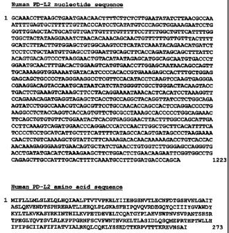
Figure 1 depicts the cDNA sequence and predicted amino acid sequence of
human PD-L2. The nucleotide sequence corresponds to nucleic acids 1-1223 of
SEQ ID
NO:1. The amino acid sequence corresponds to amino acids 1-273 of SEQ ID NO:2.
The coding region without the 5' or 3' untranslated regions of the human PD-L2
gene is
shown in SEQ ID NO:3.
CA 02414331 2002-12-27
WO 02/00730 PCT/US01/20964
13
Figure 2 depicts the cDNA sequence and amino acid sequence of mouse PD-L2.
The nucleotide sequence corresponds to nucleic acids 1-1655 of SEQ ID NO:4.
The
amino acid sequence corresponds to amino acids 1-247 of SEQ ID NO:5. The
coding
region without the 5' or 3' untranslated region of the mouse PD-L2 gene is
shown in
SEQ ID NO:6.
Figure 3 depicts the amino acid sequences of the human and mouse PD-L2
polypeptides (SEQ ID NO:2 and SEQ ID NO:5, respectively) and illustrates the
signal
peptide, IgV, IgC, extracellular, transmembrane, and cytoplasmic domains.
Figure 4 illustrates the results of FAGS analysis of the binding of IgG2a
(control
Ig), ICOS-IgG, and control PD-1-Ig to COS cells transfected with mouse PD-L2
or with
a control mouse PD-1 ligand.
Figure 5 depicts an alignment of the amino acid sequences of the mouse and
human PD-L2 polypeptides (SEQ ID NO:5 and SEQ ID NO:2, respectively).
Identical
amino acids are illustrated between the two sequences.
Figure 6 depicts an alignment of the amino acid sequences of the mouse PD-L2
(SEQ ID NO:5), human PD-L2 (SEQ ID NO:2), mouse PD-L1 (SEQ ID NO: 11), and
human PD-L1 (SEQ ID NO:12).
Figure 7 depicts the binding of PD-1 to PD-L2. CHO cells expressing I-Ad
alone or I-Ad and B7-2 were either not transfected or stably transfected with
mouse PD-
L2. Cells were stained with hPD-1-Ig and stained with PE-goat anti-mouse IgG2a
(thick
line). CHO cells were stained separately using PE-anti-I-Ad or PE-anti-B7-2
(thick
line). The thin lines indicate staining with isotype control monoclonal
antibody. Ten-
thousand events were analyzed.
Figure 8 depicts inhibition of TCR-mediated responses by PD-L2-PD-1
interaction. Purified T cells from BALB/c lymph nodes were stimulated at a 2:1
bead:cell ratio with tosyl beads coated with anti-CD3 + control Ig or anti-CD3
+ mPD-
L2-Ig. Proliferation was measured after 72 hours. These data are
representative of more
than eight independent experiments.
Figure 9 depicts inhibition of TCR-mediated responses by PD-L2-PD-1
interaction. Splenocytes from DO11.10 transgenic mice were activated with OVA
peptide (1 g/ml). CD4} T cells were isolated and rested overnight. Previously
activated T cells (105) were restimulated with peptide (1 or 0.1 g/ml)
presented by
CHO-I-Ad or CHO-I-Ad-PD-L2 for 48 hours. [3H]thymidine incorporation was
measured in triplicate. Aliquots of supernatents were collected at 36 hours
after
CA 02414331 2002-12-27
WO 02/00730 PCT/USO1/20964
14
initiation of cultures and cytokines measured by ELISA. These data are
representative
of four independent experiments.
Figure 10 depicts inhibition of TCR and CD28 mediated responses by PD-L2-
PD-1 interaction. Splenocytes from DO 11.10 transgenic mice were activated
with OVA
peptide (1 g/ml) for 4 days. CD4+ T cells were isolated and rested overnight.
Previously activated T cells (105) were restimulated, with varying peptide
concentrations, by the indicated CHO transfectants for 48 hours. [3H]thymidine
incorporation was measured in triplicate. These data are representative of six
independent experiments.
Figure 11 depicts inhibition of TCR and CD28 mediated responses by PD-L2-
PD-1 interaction. Previously activated T cells (105) were cultured with 0.01
.xg/ml OVA
peptide presented by the indicated CHO transfectants. Aliquots of supernatents
were
collected at 36 hours after initiation of cultures and cytokines measured by
ELISA. The
broken line indicates the sensitivity of the ELISA.
Figure 12 depicts inhibition of TCR and CD28 mediated responses by PD-L2-
PD-1 interaction. Previously activated T cells (105) were cultured with 0.1
g/ml OVA
peptide presented by the indicated CHO transfectants. Aliquots of supernatents
were
collected at 36 hours after initiation of cultures and cytokines measured by
ELISA. The
broken line indicates the sensitivity of the ELISA.
Figure 13 depicts cell cycle arrest and apoptosis as a result of the
engagement of
the PD-L2-PD-1 pathway. Previously activated T cells were restimulated with
OVA
peptide (1 g/ml) and the indicated CHO transfectants. Cells were collected
after 36
hours of culture, stained with anti-CD4 and fixed in 70% ethanol. Cells were
resuspended in propidium iodide solution. FACS profiles are propidium iodide
staining
of the CD4+ population. Subdiploid, diploid, and supradiploid populations are
indicated. Ten-thousand events were collected and analyzed at a constant flow
rate.
These data are representative of three independent experiments.
Figure 14 depicts cell cycle arrest and apoptosis as a result of the
engagement of
the PD-L2-PD-1 pathway. Previously activated T cells were restimulated with
OVA
peptide (0.01 g/ml) and the indicated CHO transfectants. Cells were collected
after 36
hours of culture, stained with anti-CD4 and fixed in 70% ethanol. Cells were
resuspended in propidium iodide solution. FACS profiles are propidium iodide
staining
of the CD4+ population. Subdiploid, diploid, and supradiploid populations are
CA 02414331 2008-07-07
indicated. Ten-thousand events were collected and analyzed at a constant flow
rate.
These data are representative of three independent experiments.
Figure 15 depicts the enhancement of T cell proliferation in the presence of
anti-
PD-L1 or anti PD-L2 antibodies. Allogeneic CD4+ T cells were stimulated in a
mixed
5 lymphocyte reaction by IL-10 treated dendritic cells.
Figure 16 depicts the enhancement of T cell proliferation in the presence of
anti-
PD-L1, anti PD-L2 antibodies, or a combination of anti-PD-L1 and anti-PD-L2
antibodies. Allogeneic CD4+ T cells were stimulated in a mixed lymphocyte
reaction
by IL-10 treated dendritic cells.
Detailed Description of the Invention
In addition to the previously characterized B lymphocyte activation antigens,
e.g., B7-1 and B7-2, there are other antigens on the surface of antigen-
presenting cells
(e.g., B cells, monocytes, dendritic cell, Langerhans cells, keratinocytes,
endothelial
cells, astrocytes, fibroblasts, and oligodendrocytes) which modulate the
activation of B
cells, T cells, and other immune cells. The present invention is based, at
least in part, on
the discovery of novel molecules, referred to herein as PD-L2 polypeptides,
which bind
to the PD-1 receptor and down-regulate the activation of these immune cells
and/or to
downregulate immune responses. These novel molecules play a role in the
modulating
the immune response.
The instant discovery that PD-L2 binds to PD-I places PD-L2 in a family of
inhibitory ligands, and sequence analysis places PD-L2 in the B7 family. While
engagement of a costimulatory receptor results in a costimulatory signal in an
immune
cell, engagement of an inhibitory receptor, e.g., CTLA4 or PD-1 (for example
by
crosslinking or by aggregation), leads to the transmission of an inhibitory
signal in an
immune cell resulting in downmodulation of immune cell responses and/or in
immune
cell anergy. While transmission of an inhibitory signal leads to
downmodulation in
immune cell responses (and a resulting downmodulation in the overall immune
response), the prevention of an inhibitory signal (e.g., by using a non-
activating
antibody against PD-1) in immune cells leads to upmodulation of immune cell
responses
(and a resulting upmodulation of an immune response).
PD-L2 has homology to PD-L1, a previously described ligand for PD-1 (see
U.S. Patent No. 6,936,704; International Publication WO
01/14557; Dong, H. et al. (1999) Nat. Med 5:1365-1369; and Freeman, G.J. et
al.
CA 02414331 2008-07-07
16
(2000) J Exp. Med. 192:1027-1034).
The instant invention makes available agents useful for modulating the
activity
and/or expression of PD-L2; agents for modulating the interaction between PD-
L2 and
its natural binding partner(s) (e. g. , PD-1), and agents for modulating the
immune
response via modulation of the interaction between PD-L2 and its natural
binding
partner(s) (e.g., PD-1). Exemplary modulatory agents for use in these methods
are
described further as follows.
1 o Definitions
As used herein, the term "immune cell" includes cells that are of
hematopoietic
origin and that play a role in the immune response. Immune cells include
lymphocytes,
such as B cells and T cells; natural killer cells; and myeloid cells, such as
monocytes,
macrophages, eosinophils, mast cells, basophils, and granulocytes.
As used herein, the term "T cell" includes CD4+ T cells and CD8+ T cells. The
term T cell also includes both T helper I type T cells and T helper 2 type T
cells. The
term "antigen presenting cell" includes professional antigen presenting cells
(e.g., B
lymphocytes, monocytes, dendritic cells, and Langerhans cells) as well as
other antigen
presenting cells (e.g., keratinocytes, endothelial cells, astrocytes,
fibroblasts, and
oligodendrocytes).
As used herein, the term "immune response" includes T cell-mediated and/or B
cell-mediated immune responses that are influenced by modulation of T cell
costimulation. Exemplary immune responses include B cell responses (e.g.,
antibody
production) T cell responses (e.g., cytokine production, and cellular
cytotoxicity) and
activation of cytokine responsive cells, e.g., macrophages. As used herein,
the term
"downmodulation" with reference to the immune response includes a diminution
in any
one or more immune responses, while the term "upmodulation" with reference to
the
immune response includes an increase in any one or more immune responses. It
will be
understood that upmodulation of one type of immune response may lead to a
corresponding downmodulation in another type of immune response. For example,
upmodulation of the production of certain cytokines (e.g., IL-10) can lead to
downmodulation of cellular immune responses.
CA 02414331 2002-12-27
WO 02/00730 PCT/US01/20964
17
As used herein, the term "costimulatory receptor" includes receptors which
transmit a costimulatory signal to an immune cell, e.g., CD28 or ICOS. As used
herein,
the term "inhibitory receptors" includes receptors which transmit a negative
signal to an
immune cell (e.g., CTLA4 or PD-1).
As used herein, the term "costimulate", with reference to activated immune
cells,
includes the ability of a costimulatory molecule to provide a second, non-
activating,
receptor-mediated signal (a "costimulatory signal") that induces proliferation
or effector
function. For example, a costimulatory signal can result in cytokine
secretion, e.g., in a
T cell that has received a T cell-receptor-mediated signal. Immune cells that
have
received a cell receptor-mediated signal, e.g., via an activating receptor,
are referred to
herein as "activated immune cells."
An inhibitory signal as transduced by an inhibitory receptor can occur even if
a
costimulatory receptor (such as CD28 or ICOS) in not present on the immune
cell and,
thus, is not simply a function of competition between inhibitory receptors and
costimulatory receptors for binding of costimulatory molecules (Fallarino et
al. (1998)
J.. Exp. Med. 188:205). Transmission of an inhibitory signal to an immune cell
can
result in unresponsiveness, anergy or programmed cell death in the immune
cell.
Preferably, transmission of an inhibitory signal operates through a mechanism
that does
not involve apoptosis. As used herein the term "apoptosis" includes programmed
cell
death which can be characterized using techniques which are known in the art.
Apoptotic cell death can be characterized, e.g., by cell shrinkage, membrane
blebbing,
and chromatin condensation culminating in cell fragmentation. Cells undergoing
apoptosis also display a characteristic pattern of internucleosomal DNA
cleavage.
Depending upon the form of the PD-L2 molecule that binds to a receptor, a
signal can be either transmitted (e.g., by a multivalent form of a PD-L2
molecule that
results in crosslinking of the receptor or by a soluble form of PD-L2 that
binds to Fe
receptors on antigen presenting cells) or inhibited (e.g., by a soluble,
monovalent form
of a PD-L2 molecule or a soluble form of PD-L2 that is altered using methods
known in
the art such that it does not bind to Fc receptors on antigen presenting
cells), e.g., by
competing with activating forms of PD-L2 molecules for binding to the
receptor.
However, there are instances in which a soluble molecule can be stimulatory.
The
effects of the various modulatory agents can be easily demonstrated using
routine
screening assays as described herein.
CA 02414331 2002-12-27
WO 02/00730 PCT/US01/20964
18
As used herein, the term "activating receptor" includes immune cell receptors
that bind antigen, complexed antigen (e.g., in the context of MHC molecules),
or
antibodies. Such activating receptors include T cell receptors (TCRs), B cell
receptors
(BCRs), cytokine receptors, LPS receptors, complement receptors, and Fc
receptors.
For example, T cell receptors are present on T cells and are associated with
CD3
molecules. T cell receptors are stimulated by antigen in the context of MHC
molecules
(as well as by polyclonal T cell activating reagents). T cell activation via
the TCR
results in numerous changes, e.g., protein phosphorylation, membrane lipid
changes, ion
fluxes, cyclic nucleotide alterations, RNA transcription changes, protein
synthesis
changes, and cell volume changes.
The term "B cell receptor" (BCR) as used herein includes the complex between
membrane Ig (mIg) and other transmembrane polypeptides (e.g., Iga and 1g3)
found on
B cells. The signal transduction function of mIg is triggered by crosslinking
of receptor
molecules by oligomeric or multimeric antigens. B cells can also be activated
by anti-
immunoglobulin antibodies. Upon BCR activation, numerous changes occur in B
cells,
including tyrosine phosphorylation.
The term "Fc receptor" (FcRs) include cell surface receptors for the Fe
portion of
immunoglobulin molecules (Igs). Fc receptors are found on many cells which
participate in immune responses. Among the human FcRs that have been
identified so
far are those which recognize IgG (designated Fey R), IgE (Fcs RI), IgA (Fca
R), and
polymerized IgM/A (Fc a R). FcRs are found in the following cell types: Fes R
I
(mast cells), Fcs R.II (many leukocytes), Fca R (neutrophils), and Fc a R
(glandular
epithelium, hepatocytes) (Hogg, N. (1988) Immunol. Today 9:185-86). The widely
studied FcyRs are central in cellular immune defenses, and are responsible for
stimulating the release of mediators of inflammation and hydrolytic enzymes
involved in
the pathogenesis of autoimmune disease (Unkeless, J.C. (1988) Annu. Rev.
Immunol.
6:251-87). The FcyRs provide a crucial link between effector cells and the
lymphocytes
that secrete Ig, since the macrophage/monocyte, polymorphonuclear leukocyte,
and
natural killer (NK) cell FcyRs confer an element of specific recognition
mediated by
IgG. Human leukocytes have at least three different receptors for IgG: h Fey
RI (found
on monocytes/macrophages), hFcy RII (on monocytes, neutrophils, eosinophils,
platelets, possibly B cells, and the K562 cell line), and Fey III (on NK
cells, neutrophils,
eosinophils, and macrophages).
CA 02414331 2002-12-27
WO 02/00730 PCT/US01/20964
19
With respect to T cells, transmission of a costimulatory signal to a T cell
involves a signaling pathway that is not inhibited by cyclosporin A. In
addition, a
costimulatory signal can induce cytokine secretion (e.g., IL-2 and/or IL- 10)
in a T cell
and/or can prevent the induction of unresponsiveness to antigen, the induction
of anergy,
or the induction of cell death in the T cell.
As used herein, the term "inhibitory signal" refers to a signal transmitted
via an
inhibitory receptor (e.g., CTLA4 or PD-1) molecule on an immune cell. Such a
signal
antagonizes a signal via an activating receptor (e.g., via a TCR, CD3, BCR, or
Fc
molecule) and can result, e.g., in inhibition of. second messenger generation;
proliferation; or effector function in the immune cell, e.g., reduced
phagocytosis,
antibody production, or cellular cytotoxicity, or the failure of the immune
cell to
produce mediators (such as cytokines (e.g., IL-2) and/or mediators of allergic
responses); or the development of anergy.
As used herein, the term "unresponsiveness" includes refractivity of immune
cells to stimulation, e.g., stimulation via an activating receptor or a
cytokine.
Unresponsiveness can occur, e.g., because of exposure to immunosuppressants or
high
doses of antigen. As used herein, the term "anergy" or "tolerance" includes
refractivity
to activating receptor-mediated stimulation. Such refractivity is generally
antigen-
specific and persists after exposure to the tolerizing antigen has ceased. For
example,
anergy in T cells (as opposed to unresponsiveness) is characterized by lack of
cytokine
production, e.g., IL-2. T cell anergy occurs when T cells are exposed to
antigen and
receive a first signal (a T cell receptor or CD-3 mediated signal) in the
absence of a
second signal (a costimulatory signal). Under these conditions, reexposure of
the cells
to the same antigen (even if reexposure occurs in the presence of a
costimulatory
molecule) results in failure to produce cytokines and, thus, failure to
proliferate.
Anergic T cells can, however, mount responses to unrelated antigens and can
proliferate
if cultured with cytokines (e.g., IL-2). For example, T cell anergy can also
be observed
by the lack of IL-2 production by T lymphocytes as measured by ELISA or by a
proliferation assay using an indicator cell line. Alternatively, a reporter
gene construct
can be used. For example, anergic T cells fail to initiate IL-2 gene
transcription induced
by a heterologous promoter under the control of the 5' IL-2 gene enhancer or
by a
multimer of the AP1 sequence that can be found within the enhancer (Kang et
al. (1992)
Science 257:1134).
CA 02414331 2002-12-27
WO 02/00730 PCT/USO1/20964
Modulation of a costimulatory signal results in modulation of effector
function
of an immune cell. Thus, the term "PD-L2 activity" includes the ability of a
PD-L2
polypeptide to bind its natural binding partner(s), e.g., PD-1, the ability to
modulate
immune cell costimulatory or inhibitory signals, and the ability to modulate
the immune
5 response.
With respect to PD-1, the term "activity" includes the ability of a PD-1
polypeptide to modulate an inhibitory signal in an activated immune cell,
e.g., by
engaging a natural ligand on an antigen presenting cell. PD-1 transmits an
inhibitory
signal to an immune cell in a manner similar to CTLA4. Modulation of an
inhibitory
10 signal in an immune cell results in modulation of proliferation of and/or
cytokine
secretion by an immune cell. PD-1 can also modulate a costimulatory signal by
competing with a costimulatory receptor for binding of its natural ligand(s).
Thus, the
term "PD-1 activity" includes the ability of a PD-1 polypeptide to bind its
natural
ligand(s), the ability to modulate immune cell costimulatory or inhibitory
signals, and
15 the ability to modulate the immune response.
As used herein, a "naturally-occurring" nucleic acid molecule refers to an RNA
or DNA molecule having a nucleotide sequence that occurs in nature (e.g.,
encodes a
natural protein).
As used herein, an "antisense" nucleic acid molecule comprises a nucleotide
20 sequence which is complementary to a "sense" nucleic acid encoding a
protein, e.g.,
complementary to the coding strand of a double-stranded cDNA molecule,
complementary to an mRNA sequence or complementary to the coding strand of a
gene.
Accordingly, an antisense nucleic acid molecule can hydrogen bond to a sense
nucleic
acid molecule.
As used herein, the term "coding region" refers to regions of a nucleotide
sequence comprising codons which are translated into amino acid residues,
whereas the
term "noncoding region" refers to regions of a nucleotide sequence that are
not
translated into amino acids (e.g., 5' and 3' untranslated regions).
As used herein, the term "vector" refers to a nucleic acid molecule capable of
transporting another nucleic acid molecule to which it has been linked. One
type of
vector is a "plasmid", which refers to a circular double stranded DNA loop
into which
additional DNA segments may be ligated. Another type of vector is a viral
vector,
wherein additional DNA segments may be ligated into the viral genome. Certain
vectors are capable of autonomous replication in a host cell into which they
are
CA 02414331 2002-12-27
WO 02/00730 PCT/US01/20964
21
introduced (e.g., bacterial vectors having a bacterial origin of replication
and episomal
mammalian vectors). Other vectors (e.g., non-episomal mammalian vectors) are
integrated into the genome of a host cell upon introduction into the host
cell, and thereby
are replicated along with the host genome. Moreover, certain vectors are
capable of
directing the expression of genes to which they are operatively linked. Such
vectors are
referred to herein as "recombinant expression vectors" or simply "expression
vectors".
In general, expression vectors of utility in recombinant DNA techniques are
often in the
form of plasmids. In the present specification, "plasmid" and "vector" may be
used
interchangeably as the plasmid is the most commonly used form of vector.
However,
the invention is intended to include such other forms of expression vectors,
such as viral
vectors (e.g., replication defective retroviruses, adenoviruses and adeno-
associated
viruses), which serve equivalent functions.
As used herein, the term "host cell" is intended to refer to a cell into which
a
nucleic acid molecule of the invention, such as a recombinant expression
vector of the
invention, has been introduced. The terms "host cell" and "recombinant host
cell" are
used interchangeably herein. It should be understood that such terms refer not
only to
the particular subject cell but to the progeny or potential progeny of such a
cell.
Because certain modifications may occur in succeeding generations due to
either
mutation or environmental influences, such progeny may not, in fact, be
identical to the
parent cell, but are still included within the scope of the term as used
herein.
As used herein, a "transgenic animal" refers to a non-human animal, preferably
a
mammal, more preferably a mouse, in which one or more of the cells of the
animal
includes a "transgene". The term "transgene" refers to exogenous DNA which is
integrated into the genome of a cell from which a transgenic animal develops
and which
remains in the genome of the mature animal, for example directing the
expression of an
encoded gene product in one or more cell types or tissues of the transgenic
animal.
As used herein, a "homologous recombinant animal" refers to a type of
transgenic non-human animal, preferably a mammal, more preferably a mouse, in
which
an endogenous gene has been altered by homologous recombination between the
endogenous gene and an exogenous DNA molecule introduced into a cell of the
animal,
e.g., an embryonic cell of the animal, prior to development of the animal.
CA 02414331 2002-12-27
WO 02/00730 PCT/US01/20964
22
As used herein, an "isolated protein" refers to a protein that is
substantially free
of other proteins, cellular material and culture medium when isolated from
cells or
produced by recombinant DNA techniques, or chemical precursors or other
chemicals
when chemically synthesized.
An "isolated" or "purified" protein or biologically active portion thereof is
substantially free of cellular material or other contaminating proteins from
the cell or
tissue source from which the PD-L2 protein is derived, or substantially free
from
chemical precursors or other chemicals when chemically synthesized. The
language
"substantially free of cellular material" includes preparations of PD-L2
protein in which
the protein is separated from cellular components of the cells from which it
is isolated or
recombinantly produced. In one embodiment, the language "substantially free of
cellular material" includes preparations of PD-L2 protein having less than
about 30%
(by dry weight) of non- PD-L2 protein (also referred to herein as a
"contaminating
protein"), more preferably less than about 20% of non- PD-L2 protein, still
more
preferably less than about 10% of non- PD-L2 protein, and most preferably less
than
about 5% non- PD-L2 protein. When the PD-L2 protein or biologically active
portion
thereof is recombinantly produced, it is also preferably substantially free of
culture
medium, i.e., culture medium represents less than about 20%, more preferably
less than
about 10%, and most preferably less than about 5% of the volume of the protein
preparation.
The language "substantially free of chemical precursors or other chemicals"
includes preparations of PD-L2 protein in which the protein is separated from
chemical
precursors or other chemicals which are involved in the synthesis of the
protein. In one
embodiment, the language "substantially free of chemical precursors or other
chemicals"
includes preparations of PD-L2 protein having less than about 30% (by dry
weight) of
chemical precursors or non- PD-L2 chemicals, more preferably less than about
20%
chemical precursors or non- PD-L2 chemicals, still more preferably less than
about 10%
chemical precursors or non- PD-L2 chemicals, and most preferably less than
about 5%
chemical precursors or non- PD-L2 chemicals.
The term "antibody", as used herein, includes an "antigen-binding portion" of
an
antibody (or simply "antibody portion"), as well as whole antibody molecules.
The term
"antigen-binding portion"; as used herein, refers to one or more fragments of
an
antibody that retain the ability to specifically bind to an antigen (e.g., PD-
L2). It has
been shown that the antigen-binding function of an antibody can be performed
by
CA 02414331 2002-12-27
WO 02/00730 PCT/USO1/20964
23
fragments of a full-length antibody. Examples of binding fragments encompassed
within the term "antigen-binding portion" of an antibody include (i) a Fab
fragment, a
monovalent fragment consisting of the VL, VH, CL and CHI domains; (ii) a
F(ab')2
fragment, a bivalent fragment comprising two Fab fragments linked by a
disulfide
bridge at the hinge region; (iii) a Fd fragment consisting of the VH and CHI
domains;
(iv) a Fv fragment consisting of the VL and VH domains of a single arm of an
antibody;
(v) a dAb fragment (Ward et al. (1989) Nature 341:544-546), which consists of
a VH
domain; and (vi) an isolated complementarity determining region (CDR).
Furthermore,
although the two domains of the Fv fragment, VL and VH, are coded for by
separate
genes, they can be joined, using recombinant methods, by a synthetic linker
that enables
them to be made as a single protein chain in which the VL and VH regions pair
to form
monovalent molecules (known as single chain Fv (scFv); see e.g., Bird et al.
(1988)
Science 242:423-426; and Huston et al. (1988) Proc. Natl. Acad. Sci. USA
85:5879-
5883; and Osbourn et al. 1998 Nat. Biotechnol. 16:778). Such single chain
antibodies
are also intended to be encompassed within the term "antigen-binding portion"
of an
antibody. Any VH and VL sequences of specific scFv can be linked to human
immunoglobulin constant region cDNA or genomic sequences, in order to generate
expression vectors encoding complete IgG molecules or other isotypes. VH and
Vl can
also be used in the generation of Fab, Fv, or other fragments of
immunoglobulins using
either protein chemistry or recombinant DNA technology. Other forms of single
chain
antibodies, such as diabodies are also encompassed. Diabodies are bivalent,
bispecific
antibodies in which VH and VL domains are expressed on a single polypeptide
chain,
but using a linker that is too short to allow for pairing between the two
domains on the
same chain, thereby forcing the domains to pair with complementary domains of
another
chain and creating two antigen binding sites (see e.g., Holliger, P. et al.
(1993) Proc.
Natl. Acad. Sci. USA 90:6444-6448; Poljak, R.J. et al. (1994) Structure 2:1121-
1123).
Still further, an antibody or antigen-binding portion thereof may be part of a
larger immunoadhesion molecules, formed by covalent or noncovalent association
of the
antibody or antibody portion with one or more other proteins or peptides.
Examples of
such immunoadhesion molecules include use of the streptavidin core region to
make a
tetrameric scFv molecule (Kipriyanov, S.M. et al. (1995) Hum. Antibodies
Hybridomas
6:93-101) and use of a cysteine residue, a marker peptide and a C-terminal
polyhistidine
tag to make bivalent and biotinylated scFv molecules (Kipriyanov, S.M. et al.
(1994)
Mol. Immunol. 31:1047-1058). Antibody portions, such as Fab and F(ab')2
fragments,
CA 02414331 2002-12-27
WO 02/00730 PCT/US01/20964
24
can be prepared from whole antibodies using conventional techniques, such as
papain or
pepsin digestion, respectively, of whole antibodies. Moreover, antibodies,
antibody
portions and immunoadhesion molecules can be obtained using standard
recombinant
DNA techniques, as described herein.
Antibodies may be polyclonal or monoclonal; xenogeneic, allogeneic, or
syngeneic; or modified forms thereof, e.g., humanized, chimeric, etc.
Preferably,
antibodies of the invention bind specifically or substantially specifically to
PD-L2
molecules. The terms "monoclonal antibodies" and "monoclonal antibody
composition", as used herein, refer to a population of antibody molecules that
contain
only one species of an antigen binding site capable of immunoreacting with a
particular
epitope of an antigen, whereas the term "polyclonal antibodies" and
"polyclonal
antibody composition" refer to a population of antibody molecules that contain
multiple
species of antigen binding sites capable of interacting with a particular
antigen. A
monoclonal antibody composition, typically displays a single binding affinity
for a
particular antigen with which it immunoreacts.
The term "humanized antibody", as used herein, is intended to include
antibodies
made by a non-human cell having variable and constant regions which have been
altered
to more closely resemble antibodies that would be made by a human cell. For
example,
by altering the non-human antibody amino acid sequence to incorporate amino
acids
found in human germline immunoglobulin sequences. The humanized antibodies of
the
invention may include amino acid residues not encoded by human germline
immunoglobulin sequences (e.g., mutations introduced by random or site-
specific
mutagenesis in vitro or by somatic mutation in vivo), for example in the CDRs.
The
term "humanized antibody", as used herein, also includes antibodies in which
CDR
sequences derived from the germline of another mammalian species, such as a
mouse,
have been grafted onto human framework sequences.
An "isolated antibody", as used herein, is intended to refer to an antibody
that is
substantially free of other antibodies having different antigenic
specificities (e.g., an
isolated antibody that specifically binds PD-L2 is substantially free of
antibodies that
specifically bind antigens other than PD-L2). Moreover, an isolated antibody
may be
substantially free of other cellular material and/or chemicals.
CA 02414331 2002-12-27
WO 02/00730 PCT/US01/20964
PD-L2 Nucleic Acid and Polypeptide Molecules
The term "family" when referring to the polypeptide and nucleic acid molecules
of the invention is intended to mean two or more polypeptide or nucleic acid
molecules
having a common structural domain or motif and having sufficient amino acid or
5 nucleotide sequence homology as defined herein. Such family members can be
naturally or non-naturally occurring and can be from either the same or
different species.
For example, a family can contain a first polypeptide of human origin, as well
as other,
distinct polypeptides of human origin or alternatively, can contain homologues
of non-
human origin, e.g., monkey polypeptides. Members of a family may also have
common
10 functional characteristics.
For example, the family of PD-L2 polypeptides of the present invention
preferably comprises least one "signal peptide domain". As used herein, a
"signal
sequence" or "signal peptide" includes a peptide containing about 15 or more
amino
acids which occurs at the N-terminus of secretory and membrane bound
polypeptides
15 and which contains a large number of hydrophobic amino acid residues. For
example, a
signal sequence contains at least about 10-30 amino acid residues, preferably
about 15-
25 amino acid residues, more preferably about 18-20 amino acid residues, and
even
more preferably about 19 amino acid residues, and has at least about 35-65%,
preferably
about 38-50%, and more preferably about 40-45% hydrophobic amino acid residues
20 (e.g., Valine, Leucine, Isoleucine or Phenylalanine). Such a "signal
sequence", also
referred to in the art as a "signal peptide", serves to direct a polypeptide
containing such
a sequence to a lipid bilayer, and is cleaved in secreted and membrane bound
polypeptides. A signal sequence was identified in the amino acid sequence of
native
human PD-L2 at about amino acids 1-19 of SEQ ID NO:2 (Figure 3). A signal
25 sequence was also identified in the amino acid sequence of native mouse PD-
L2 at
about amino acids 1-19 of SEQ ID NO:5 (Figure 3).
In another embodiment of the invention, a PD-L2 polypeptide of the present
invention is identified based on the presence of a "transmembrane domain". As
used
herein, the term "transmembrane domain" includes an amino acid sequence of
about 15
amino acid residues in length which spans the plasma membrane. More
preferably, a
transmembrane domain includes about at least 20, 25, 30, 35, 40, or 45 amino
acid
residues and spans the plasma membrane. Transmembrane domains are rich in
hydrophobic residues, and typically have an alpha-helical structure. In a
preferred
embodiment, at least 50%, 60%, 70%, 80%, 90%, 95% or more of the amino acids
of a
CA 02414331 2008-07-07
26
transmembrane domain are hydrophobic, e.g., leucines, isoleucines, tyrosines,
or
tryptophans. Transmembrane domains are described in, for example, Zagotta,
W.N. et
al. (1996) Annu. Rev. Neurosci. 19:235-263.
Amino acid residues 220-243 of the native human PD-L2
polypeptide, and amino acid residues 201-243 of the predicted mature
polypeptide, are
predicted to comprise transmembrane domains (see Figure 3). Amino acid
residues 220-
242 of the native mouse PD-L2 polypeptide, and amino acid residues 201-223 of
the
predicted mature polypeptide, are predicted to comprise transmembrane domains
(see
Figure 3). Accordingly, PD-L2 polypeptides having at least 71-80%, or more
preferably
about 80-90% homology with a transmembrane domain of human PD-L2 are within
the
scope of the invention.
In another embodiment, a PD-L2 molecule of the present invention is identified
based on the presence of an "IgC domain" or an "IgV domain" in the polypeptide
or
corresponding nucleic acid molecule. As used herein, IgV and IgC domains are
recognized in the art as Ig superfamily member domains. These domains
correspond to
structural units that have distinct folding patterns called Ig folds. Ig folds
are comprised
of a sandwich of two R sheets, each consisting of antiparallel 0 strands of 5-
10 amino
acids with a conserved disulfide bond between the two sheets in most, but not
all,
domains. IgC domains of Ig, TCR, and MHC molecules share the same types of
sequence patterns and are called the Cl set within the Ig superfamily. Other
IgC
domains fall within other sets. IgV domains also share sequence patterns and
are called
V set domains. IgV domains are longer than C-domains and form an additional
pair of f
strands. Amino acid residues 20-120 of the native human PD-L2 polypeptide, and
amino acid residues 1-101 of the predicted mature polypeptide, are predicted
to
comprise IgV domains (see Figure 3). Amino acid residues 20-120 of the native
mouse
PD-L2 polypeptide, and amino acid residues 1-101 of the predicted mature
polypeptide,
are also predicted to comprise IgV domains (see Figure 3). Amino acid residues
121-
219 of the native human PD-L2 polypeptide, and amino acid residues 102-200 of
the
predicted mature polypeptide, are predicted to comprise IgC domains (see
Figure 3).
Amino acid residues 121-219 of the native mouse PD-L2 polypeptide, and amino
acid
residues 102-200 of the predicted mature polypeptide, are also predicted to
comprise
IgC domains (see Figure 3). In a preferred embodiment, the presence of an IgV
domain
is required for binding of PD-L2 to its natural binding partner, e.g., PD-1.
CA 02414331 2002-12-27
WO 02/00730 PCT/US01/20964
27
In another embodiment, a PD-L2 molecule of the present invention is identified
based on the presence of a "extracellular domain" in the polypeptide or
corresponding
nucleic acid molecule. As used herein, the term "extracellular domain"
represents the
N-terminal amino acids which extend as a tail from the surface of a cell. An
extracellular domain of the present invention includes an IgV domain and an
IgC
domain, and may include a signal peptide domain. Amino acid residues 1-219 of
the
native human PD-L2 polypeptide, and amino acid residues 1-200 of the predicted
mature polypeptide, are predicted to comprise extracellular domains (see
Figure 3).
Amino acid residues 1-219 of the native mouse PD-L2 polypeptide, and amino
acid
residues 1-200 of the predicted mature polypeptide, are also predicted to
comprise
extracellular domains (see Figure 3).
In still another embodiment, a PD-L2 molecule of the present invention is
identified based on the presence of a "cytoplasmic domain" in the polypeptide
or
corresponding nucleic acid molecule. As used herein, the term "cytoplasmic
domain"
represents the C-terminal amino acids which extend as a tail into the
cytoplasm of a cell.
Amino acid residues 244-273 of the native human PD-L2 polypeptide, and amino
acid
residues 225-273 of the predicted mature polypeptide, are predicted to
comprise
cytoplasmic domains (see Figure 3). Amino acid residues 243-247 of the native
mouse
PD-L2 polypeptide, and amino acid residues 224-228 of the predicted mature
polypeptide, are also predicted to comprise cytoplasmic domains.
In a preferred embodiment, the PD-L2 molecules of the invention include at
least
one or more of the following domains: a signal peptide domain, an IgV domain,
an IgC
domain, an extracellular domain, a transmembrane domain, and a cytoplasmic
domain.
Isolated polypeptides of the present invention, preferably PD-L2 polypeptides,
have an amino acid sequence sufficiently identical to the amino acid sequence
of SEQ
ID NO:2, or are encoded by a nucleotide sequence sufficiently identical to SEQ
ID
NO:1 or 3. As used herein, the term "sufficiently identical" refers to a first
amino acid
or nucleotide sequence which contains a sufficient or minimum number of
identical or
equivalent (e.g., an amino acid residue which has a similar side chain) amino
acid
residues or nucleotides to a second amino acid or nucleotide sequence such
that the first
and second amino acid or nucleotide sequences share common structural domains
or
motifs and/or a common functional activity. For example, amino acid or
nucleotide
sequences which share common structural domains have at least 30%, 40%, or 50%
homology, preferably 60% homology, more preferably 70%-80%, and even more
CA 02414331 2002-12-27
WO 02/00730 PCT/US01/20964
28
preferably 90-95% homology across the amino acid sequences of the domains and
contain at least one and preferably two structural domains or motifs, are
defined herein
as sufficiently identical. Furthermore, amino acid or nucleotide sequences
which share
at least 30%, 40%, or 50%, preferably 60%, more preferably 70-80%, or 90-95%
homology and share a common functional activity are defined herein as
sufficiently
identical.
As used interchangeably herein, "PD-L2 activity", "biological activity of PD-
L2" or "functional activity of PD-L2", refers to an activity exerted by a PD-
L2 protein,
polypeptide or nucleic acid molecule on a PD-L2-responsive cell or tissue, or
on a PD-
L2 polypeptide binding partner, as determined in vivo, or in vitro, according
to standard
techniques. In one embodiment, a PD-L2 activity is a direct activity, such as
an
association with a PD-L2 binding partner. As used herein, a "target molecule"
or
"binding partner" is a molecule with which a PD-L2 polypeptide binds or
interacts in
nature, such that PD-L2-mediated function is achieved. In an exemplary
embodiment, a
PD-L2 target molecule is the receptor PD-1. Alternatively, a PD-L2 activity is
an
indirect activity, such as a cellular signaling activity mediated by
interaction of the PD-
L2 polypeptide with its natural binding partner, e.g., PD-1. The biological
activities of
PD-L2 are described herein. For example, the PD-L2 polypeptides of the present
invention can have one or more of the following activities: 1) bind.to and/or
modulate
the activity of the receptor PD-1 or other PD-L2 natural binding partners, 2)
modulate
intra- or intercellular signaling, 3) modulate activation of immune cells,
e.g., T
lymphocytes, and 4) modulate the immune response of an organism, e.g., a mouse
or
human organism.
Accordingly, another embodiment of the invention features isolated PD-L2
proteins and polypeptides having a PD-L2 activity. Other preferred
polypeptides are
PD-L2 polypeptides having one or more of the following domains: a signal
peptide
domain, an IgV domain, an IgC domain, an extracellular domain, a transmembrane
domain, and a cytoplasmic domain, and, preferably, a PD-L2 activity.
Additional preferred PD-L2 polypeptides have at least one extracellular
domain,
3o and one or more of a signal peptide domain, an IgV domain, an IgC domain,
an
transmembrane domain, and a cytoplasmic domain, and are, preferably, encoded
by a
nucleic acid molecule having a nucleotide sequence which hybridizes under
stringent
hybridization conditions to a nucleic acid molecule comprising a complement of
the
nucleotide sequence of SEQ ID NO: 1, 3, 4, or 6.
CA 02414331 2002-12-27
WO 02/00730 PCT/US01/20964
29
The nucleotide sequence of the isolated human PD-L2 cDNA and the predicted
amino acid sequence of the human PD-L2 polypeptide are shown in Figure 1 and
in
SEQ ID NO:1 and 2, respectively. The nucleotide sequence of the isolated mouse
PD-
L2 cDNA and the amino acid sequence of the mouse PD-L2 polypeptide are shown
in
Figure 2 and in SEQ ID NO:4 and 5, respectively.
The human PD-L2 gene, which is approximately 1223 nucleotides in length,
encodes a polypeptide having a molecular weight of approximately 30.0 kD and
which
is approximately 273 amino acid residues in length. The mouse PD-L2 gene,
which is
approximately 1655 nucleotides in length, encodes a polypeptide having a
molecular
weight of approximately 27.2 kD and which is approximately 247 amino acid
residues
in length.
Various aspects of the invention are described in further detail in the
following
subsections:
1. Isolated Nucleic Acid Molecules
One aspect of the invention pertains to isolated nucleic acid molecules that
encode PD-L2 polypeptides or biologically active portions thereof, as well as
nucleic
acid fragments sufficient for use as hybridization probes to identify PD-L2-
encoding
nucleic acid molecules (e.g., PD-L2 mRNA) and fragments for use as PCR primers
for
the amplification or mutation of PD-L2 nucleic acid molecules. As used herein,
the
term "nucleic acid molecule" is intended to include DNA molecules (e.g., cDNA
or
genomic DNA) and RNA molecules (e.g., mRNA) and analogs of the DNA or RNA
generated using nucleotide analogs. The nucleic acid molecule can be single-
stranded or
double-stranded, but preferably is double-stranded DNA.
The term "isolated nucleic acid molecule" includes nucleic acid molecules
which
are separated from other nucleic acid molecules which are present in the
natural source
of the nucleic acid. For example, with regards to genomic DNA, the term
"isolated"
includes nucleic acid molecules which are separated from the chromosome with
which
the genomic DNA is naturally associated. Preferably, an "isolated" nucleic
acid
molecule is free of sequences which naturally flank the nucleic acid (i. e.,
sequences
located at the 5' and 3' ends of the nucleic acid molecule) in the genomic DNA
of the
organism from which the nucleic acid is derived. For example, in various
embodiments,
the isolated PD-L2 nucleic acid molecule can contain less than about 5 kb,
4kb, 3kb,
CA 02414331 2002-12-27
WO 02/00730 PCT/USO1/20964
2kb, 1 kb, 0.5 kb or 0.1 kb of nucleotide sequences which naturally flank the
nucleic
acid molecule in genomic DNA of the cell from which the nucleic acid molecule
is
derived. Moreover, an "isolated" nucleic acid molecule, such as a cDNA
molecule, can
be substantially free of other cellular material, or culture medium, when
produced by
5 recombinant techniques, or substantially free of chemical precursors or
other chemicals
when chemically synthesized.
A nucleic acid molecule of the present invention, e.g., a nucleic acid
molecule
having the nucleotide sequence of SEQ ID NO: 1,3,4, or 6, or a portion
thereof, can be
isolated using standard molecular biology techniques and the sequence
information
10 provided herein. Using all or portion of the nucleic acid sequence of SEQ
ID NO:1, 3,
4, or 6 as a hybridization probe, PD-L2 nucleic acid molecules can be isolated
using
standard hybridization and cloning techniques (e.g., as described in Sambrook,
J. et al.
Molecular Cloning: A Laboratory Manual. 2nd, ed., Cold Spring Harbor
Laboratory,
Cold Spring Harbor Laboratory Press, Cold Spring Harbor, NY, 1989).
15 Moreover, a nucleic acid molecule encompassing all or a portion of SEQ ID
NO:1, 3, 4, or 6 can be isolated by the polyinerase chain reaction (PCR) using
synthetic
oligonucleotide primers designed based upon the sequence of SEQ ID NO:1, 3, 4,
or 6.
A nucleic acid molecule of the invention can be amplified using cDNA, mRNA
or, alternatively, genomic DNA as a template and appropriate oligonucleotide
primers
20 according to standard PCR amplification techniques. The nucleic acid
molecule so
amplified can be cloned into an appropriate vector and characterized by DNA
sequence
analysis. Furthermore, oligonucleotides corresponding to PD-L2 nucleotide
sequences
can be prepared by standard synthetic techniques, e.g., using an automated DNA
synthesizer.
25 In a preferred embodiment, an isolated nucleic acid molecule of the
invention
comprises the nucleotide sequence shown in SEQ ID NO:1, 3, 4, or 6. This cDNA
may
comprise sequences encoding the human PD-L2 polypeptide (i.e., "the coding
region",
from nucleotides 274-1092), as well as 5' untranslated sequences (nucleotides
1-273)
and 3' untranslated sequences (nucleotides 1093-1223) of SEQ ID NO: 1.
Alternatively,
30 the nucleic acid molecule can comprise only the coding region of SEQ ID
NO:1 (i.e.,
nucleotides 274-1092, corresponding to SEQ ID NO:3). Accordingly, in another
embodiment, an isolated nucleic acid molecule of the invention comprises SEQ
ID NO:3
and nucleotides 1-274 of SEQ ID NO: 1. In yet another embodiment, the isolated
nucleic acid molecule comprises SEQ ID NO:3 and nucleotides 1093-1223 of SEQ
ID
CA 02414331 2002-12-27
WO 02/00730 PCT/US01/20964
31
NO: 1. In yet another embodiment, the nucleic acid molecule consists of the
nucleotide
sequence set forth as SEQ ID NO:1 or SEQ ID NO:3. In still another embodiment,
the
nucleic acid molecule can comprise the coding region of SEQ ID NO:1 (e.g.,
nucleotides 274-1092, corresponding to SEQ ID NO:3), as well as a stop codon
(e.g.,
nucleotides 1093-1095 of SEQ ID NO: 1).
This cDNA may also comprise sequences encoding the mouse PD-L2
polypeptide (i.e., the coding region, from nucleotides 210-950), as well as 5'
untranslated sequences (nucleotides 1-209) and 3' untranslated sequences
(nucleotides
951-1655) of SEQ ID NO:4. Alternatively, the nucleic acid molecule can
comprise only
the coding region of SEQ ID NO:4 (i.e., nucleotides 210-950, corresponding to
SEQ ID
NO:6). Accordingly, in another embodiment, an isolated nucleic acid molecule
of the
invention comprises SEQ ID NO:6 and nucleotides 1-210 of SEQ ID NO:4. In yet
another embodiment, the isolated nucleic acid molecule comprises SEQ ID NO:6
and
nucleotides 951-1655 of SEQ ID NO:4. In yet another embodiment, the nucleic
acid
molecule consists of the nucleotide sequence set forth as SEQ ID NO:4 or SEQ
ID
NO:6. In still another embodiment, the nucleic acid molecule can comprise the
coding
region of SEQ ID NO:4 (e.g., nucleotides 210-950, corresponding to SEQ ID
NO:4), as
well as a stop codon (e.g., nucleotides 951-953 of SEQ ID NO:4).
In another preferred embodiment, an isolated nucleic acid molecule of the
invention comprises a nucleic acid molecule which is a complement of the
nucleotide
sequence shown in SEQ ID NO:1, 3, 4, or 6, or a portion of any of these
nucleotide
sequences. A nucleic acid molecule which is complementary to the nucleotide
sequence
shown in SEQ ID NO: 1, 3, 4, or 6, is one which is sufficiently complementary
to the
nucleotide sequence shown in SEQ ID NO: 1, 3, 4, or 6 such that it can
hybridize to the
nucleotide sequence shown in SEQ ID NO: 1, 3, 4, or 6, respectively, thereby
forming a
stable duplex.
In still another preferred embodiment, an isolated nucleic acid molecule of
the
present invention comprises a nucleotide sequence which is at least about 70%,
75%,
80%, 85%, 90%, 91%, 92%, 93%, 94%, 95%, 96%, 97%, 98%, 99% or more identical
to the entire length of the nucleotide sequence shown in SEQ ID NO:1, 3, 4, or
6, or a
portion of any of these nucleotide sequences.
Moreover, the nucleic acid molecule of the invention can comprise only a
portion of the nucleic acid sequence of SEQ ID NO: 1, 3, 4, or 6, for example,
a
fragment which can be used as a probe or primer or a fragment which encodes a
portion
CA 02414331 2002-12-27
WO 02/00730 PCT/US01/20964
32
of a PD-L2 polypeptide, e.g., a biologically active portion of a PD-L2
polypeptide. The
nucleotide sequences determined from the cloning of the human PD-L2 gene allow
for
the generation of probes and primers designed for use in identifying and/or
cloning other
PD-L2 family members, as well as PD-L2 homologues from other species. The
probe/primer typically comprises substantially purified oligonucleotide. The
oligonucleotide typically comprises a region of nucleotide sequence that
hybridizes
under stringent conditions to at least about 12 or 15, preferably about 20 or
25, more
preferably about 30, 35, 40, 45, 50, 55, 60, 65, or 75 consecutive nucleotides
of a sense
sequence of SEQ ID NO: 1, 3, 4, or 6; of an anti-sense sequence of SEQ ID NO:
1, 3, 4,
or 6; or of a naturally occurring allelic variant or mutant of SEQ ID NO:1, 3,
4, or 6.
In one embodiment, a nucleic acid molecule of the present invention comprises
a
nucleotide sequence which is greater than about 50-100, 100-150, 150-200, 200-
250,
250-300, 300-350, 350-400, 400-450, 450-500, 500-550, 550-600, 600-650, 650-
700,
700-750, 750-800, 800-850, 850-900, 900-950, 950-1000, 1000-1050, 1050-1100,
1100-
1150 or more nucleotides in length and hybridizes under stringent
hybridization
conditions to a nucleic acid molecule of SEQ ID NO:1, 3, 4, or 6, or the
complement
thereof. In a further embodiment, a nucleic acid molecule of the present
invention
comprises a nucleotide sequence which is greater than about 880-900, 900-950,
950-
1000, 1000-1050, 1050-1100, 1100-1150 or more nucleotides in length and
hybridizes
under stringent hybridization conditions to a nucleic acid molecule of SEQ ID
NO:1 or
3, or the complement thereof In yet another embodiment, a nucleic acid
molecule of
the present invention comprises a nucleotide sequence which is greater than 50-
100,
100-150, 150-200, 200-250, 250-300 or more nucleotides in length and
hybridizes under
stringent hybridization conditions to a nucleic acid molecule comprising
nucleotides 1-
358 of SEQ ID NO:1, or a complement thereof. In yet a further embodiment, a
nucleic
acid molecule of the present invention comprises a nucleotide sequence which
is greater
than about 50-100, 100-150, 150-200, 200-250, 250-300, 300-350, 350-400, 400-
450,
450-500, 500-550, 550-600, 600-650, 650-700, 700-750, 750-800, 850-900, 900-
950,
950-1000-1050-1100, 1100-1150 or more nucleotides in length, includes at least
about
.30 15 (i.e., 15 contiguous) nucleotides of the sequence comprising
nucleotides 1-358 of
SEQ ID NO: 1, or a complement thereof, and hybridizes under stringent
conditions to a
nucleic acid molecule comprising the nucleotide sequence shown in SEQ ID NO:
1, or a
complement thereof
CA 02414331 2002-12-27
WO 02/00730 PCT/US01/20964
33
Probes based on the PD-L2 nucleotide sequences can be used to detect
transcripts or genomic sequences encoding the same or homologous polypeptides.
In
preferred embodiments, the probe further comprises a label group attached
thereto, e.g.,
the label group can be a radioisotope, a fluorescent compound, an enzyme, or
an enzyme
co-factor. Such probes can be used as a part of a diagnostic test kit for
identifying cells
or tissue which misexpress a PD-L2 polypeptide, such as by measuring a level
of a PD-
L2-encoding nucleic acid in-a sample of cells from a subject e.g., detecting
PD-L2
mRNA levels or determining whether a genomic PD-L2 gene has been mutated or
deleted.
A nucleic acid fragment encoding a "biologically active portion of a PD-L2
polypeptide" can be prepared by isolating a portion of the nucleotide sequence
of SEQ
ID NO:1, 3, 4, or 6 which encodes a polypeptide having a PD-L2 biological
activity
(e.g., the ability to bind to its natural binding partner(s) (e.g., PD-1),
and/or modulate
immune cell activity), expressing the encoded portion of the PD-L2 polypeptide
(e.g., by
recombinant expression in vitro) and assessing the activity of the encoded
portion of the
PD-L2 polypeptide.
The invention further encompasses nucleic acid molecules that differ from the
nucleotide sequence shown in SEQ ID NO:1, 3, 4, or 6 due to degeneracy of the
genetic
code and thus encode the same PD-L2 polypeptides as those encoded by the
nucleotide
sequence shown in SEQ ID NO:1, 3, 4, or 6. In another embodiment, an isolated
nucleic
acid molecule of the invention has a nucleotide sequence encoding a
polypeptide having
an amino acid sequence shown in SEQ ID NO:2 or 5.
In addition to the PD-L2 nucleotide sequences shown in SEQ ID NO:1, 3, 4, or
6, it will be appreciated by those skilled in the art that DNA sequence
polymorphisms
that lead to changes in the amino acid sequences of the PD-L2 polypeptides may
exist
within a population (e.g., the human population). Such genetic polymorphism in
the
PD-L2 genes may exist among individuals within a population due to natural
allelic
variation. As used herein, the terms "gene" and "recombinant gene" refer to
nucleic
acid molecules which include an open reading frame encoding a PD-L2
polypeptide,
preferably a mammalian PD-L2 polypeptide, and can further include non-coding
regulatory sequences, and introns.
Allelic variants of human or mouse PD-L2 include both functional and non-
functional PD-L2 polypeptides. Functional allelic variants are naturally
occurring
amino acid sequence variants of the human or mouse PD-L2 polypeptide that
maintain
CA 02414331 2002-12-27
WO 02/00730 PCT/US01/20964
34
the ability to bind natural PD-L2 binding partner(s), e.g., PD-l, and/or
modulate
lymphocyte activation. Functional allelic variants will typically contain only
conservative substitution of one or more amino acids of SEQ ID NO:2 or 5, or
substitution, deletion or insertion of non-critical residues in non-critical
regions of the
polypeptide.
Non-functional allelic variants are naturally occurring amino acid sequence
variants of the human or mouse PD-L2 polypeptide that do not have the ability
to either
bind natural PD-L2 binding partners, e.g., PD-l, and/or modulate any of the PD-
L2
activities described herein. Non-functional allelic variants will typically
contain a non-
conservative substitution, deletion, or insertion or premature truncation of
the amino
acid sequence of SEQ ID NO:2 or 5, or a substitution, insertion or deletion in
critical
residues or critical regions of the polypeptide, e.g., in an IgV domain.
The present invention further provides non-human, non-mouse orthologues of
the human or mouse PD-L2 polypeptide. Orthologues of the human or mouse PD-L2
polypeptide are polypeptides that are isolated from non-human, non-mouse
organisms
and possess the same PD-1-binding activity and/or lymphocyte activation-
modulating
activity of the PD-L2 polypeptide. Orthologues of the human or mouse PD-L2
polypeptide can readily be identified as comprising an amino acid sequence
that is
substantially identical to SEQ ID NO:2 or 5.
Moreover, nucleic acid molecules encoding other PD-L2 family members and,
thus, which have a nucleotide sequence which differs from the PD-L2 sequences
of SEQ
ID NO:1, 3, 4, or 6 are intended to be within the scope of the invention. For
example,
another PD-L2 cDNA can be identified based on the nucleotide sequence of mouse
or
human PD-L2. Moreover, nucleic acid molecules encoding PD-L2 polypeptides from
different species, and which, thus, have a nucleotide sequence which differs
from the
PD-L2 sequences of SEQ ID NO:1, 3, 4, or 6 are intended to be within the scope
of the
invention. For example, a monkey PD-L2 cDNA can be identified based on the
nucleotide sequence of the mouse or human PD-L2.
Nucleic acid molecules corresponding to natural allelic variants and
homologues
of the PD-L2 cDNAs of the invention can be isolated based on their homology to
the
PD-L2 nucleic acids disclosed herein using the cDNAs disclosed herein, or a
portion
thereof, as a hybridization probe according to standard hybridization
techniques under
stringent hybridization conditions. Nucleic acid molecules corresponding to
natural
CA 02414331 2002-12-27
WO 02/00730 PCT/US01/20964
allelic variants and homologues of the PD-L2 cDNAs of the invention can
further be
isolated by mapping to the same chromosome or locus as the PD-L2 gene.
Accordingly, in another embodiment, an isolated nucleic acid molecule of the
invention is at least 15, 20, 25, 30 or more nucleotides in length and
hybridizes under
5 stringent conditions to the nucleic acid molecule comprising nucleotides 1-
358 of the
nucleotide sequence of SEQ ID NO: I, or nucleotides 1-85 of SEQ ID NO:3. In
other
embodiment, the nucleic acid is at least 880-900, 900-950, 950-1000, 1000-
1050, 1050-
1100, 1100-1150 or more nucleotides in length.
As used herein, the term "hybridizes under stringent conditions" is intended
to
10 describe conditions for hybridization and washing under which nucleotide
sequences
that are significantly identical or homologous to each other remain hybridized
to each
other. Preferably, the conditions are such that sequences at least about 70%,
more
preferably at least about 80%, even more preferably at least about 85% or 90%
identical
to each other remain hybridized to each other. Such stringent conditions are
known to
15 those skilled in the art and can be found in Current Protocols in Molecular
Biology,
Ausubel et al., eds., John Wiley & Sons, Inc. (1995), sections 2, 4 and 6.
Additional
stringent conditions can be found in Molecular Cloning: A Laboratory Manual,
Sambrook et al., Cold Spring Harbor Press, Cold Spring Harbor, NY (1989),
chapters 7,
9 and 11. A preferred, non-limiting example of stringent hybridization
conditions
20 includes hybridization in 4X or 6X sodium chloride/sodium citrate (SSC), at
about 65-
70 C (or hybridization in 4X SSC plus 50% formamide at about 42-50 C) followed
by
one or more washes in 1X SSC, at about 65-70 C. A further preferred, non-
limiting
example of stringent hybridization conditions includes hybridization at 6X SSC
at 45 C,
followed by one or more washes in 0.2X SSC, 0.1% SDS at 65 C. A preferred, non-
25 limiting example of highly stringent hybridization conditions includes
hybridization in
1X SSC, at about 65-70 C (or hybridization in 1X SSC plus 50% formamide at
about
42-50 C) followed by one or more washes in 0.3X SSC, at about 65-70 C. A
preferred,
non-limiting example of reduced stringency hybridization conditions includes
hybridization in 4X or 6X SSC, at about 50-60 C (or alternatively
hybridization in 6X
30 SSC plus 50% formamide at about 40-45 C) followed by one or more washes in
2X
SSC, at about 50-60 C. Ranges intermediate to the above-recited values, e.g.,
at 65-70
C or at 42-50 C are also intended to be encompassed by the present invention.
SSPE
(1xSSPE is 0.15M NaCl, I OmM NaH2PO4, and 1.25mM EDTA, pH 7.4) can be
substituted for SSC (1xSSC is 0.15M NaCl and 15mM sodium citrate) in the
CA 02414331 2002-12-27
WO 02/00730 PCT/US01/20964
36
hybridization and wash buffers; washes are performed for 15 minutes each after
hybridization is complete. The hybridization temperature for hybrids
anticipated to be
less than 50 base pairs in length should be 5-10 C less than the melting
temperature (T,,,)
of the hybrid, where T,,, is determined according to the following equations.
For hybrids
less than 18 base pairs in length, Tm( C) = 2(# of A + T bases) + 4(# of G + C
bases).
For hybrids between 18 and 49 base pairs in length, Tm( C) = 81.5 +
16.6(loglo[Na+]) +
0.41(%G+C) - (600/N), where N is the number of bases in the hybrid, and [Na+]
is the
concentration of sodium ions in the hybridization buffer ([Na ] for 1xSSC =
0.165 M).
It will also be recognized by the skilled practitioner that additional
reagents may be
added to hybridization and/or wash buffers to decrease non-specific
hybridization of
nucleic acid molecules to membranes, for example, nitrocellulose or nylon
membranes,
including but not limited to blocking agents (e.g., BSA or salmon or herring
sperm
carrier DNA), detergents (e.g., SDS), chelating agents (e.g., EDTA), Ficoll,
PVP and the
like. When using nylon membranes, in particular, an additional preferred, non-
limiting
example of stringent hybridization conditions is hybridization in 0.25-0.5M
NaH2PO4,
7% SDS at about 65 C, followed by one or more washes at 0.02M NaH2PO4, I% SDS
at
65 C, see e.g., Church and Gilbert (1984) Proc. Natl. Acad. Sci. USA 81:1991-
1995 (or
alternatively 0.2X SSC, 1% SDS).
Preferably, an isolated nucleic acid molecule of the invention that hybridizes
under stringent conditions to the sequence of SEQ ID NO: 1, 3, 4, or 6
corresponds to a
naturally-occurring nucleic acid molecule. As used herein, a "naturally-
occurring"
nucleic acid molecule refers to an RNA or DNA molecule having a nucleotide
sequence
that occurs in nature (i.e., encodes a natural polypeptide).
In addition to naturally-occurring allelic variants of the PD-L2 sequences
that
may exist in the population, the skilled artisan will further appreciate that
changes can be
introduced by mutation into the nucleotide sequences of SEQ ID NO:1, 3, 4, or
6, thereby
leading to changes in the amino acid sequence of the encoded PD-L2
polypeptides,
without altering the functional ability of the PD-L2 polypeptides. For
example,
nucleotide substitutions leading to amino acid substitutions at "non-
essential" amino acid
residues can be made in the sequence of SEQ ID NO:1, 3, 4, or 6. A "non-
essential"
amino acid residue is a residue that can be altered from the wild-type
sequence of PD-L2
(e.g., the sequence of SEQ ID NO:2 or 5) without altering the biological
activity, whereas
an "essential" amino acid residue is required for biological activity. For
example, amino
acid residues that are conserved among the PD-L2 polypeptides of the present
invention,
CA 02414331 2002-12-27
WO 02/00730 PCT/USO1/20964
37
e.g., those present in an extracellular domain, are predicted to be
particularly unamenable
to alteration. Furthermore, additional amino acid residues that are conserved
between the
PD-L2 polypeptides of the present invention and other members of the PD-L2
family are
not likely to be amenable to alteration.
Accordingly, another aspect of the invention pertains to nucleic acid
molecules
encoding PD-L2 polypeptides that contain changes in amino acid residues that
are not
essential for activity. Such PD-L2 polypeptides differ in amino acid sequence
from SEQ
ID NO:2 or 5, yet retain biological activity. In one embodiment, the isolated
nucleic acid
molecule comprises a nucleotide sequence encoding a polypeptide, wherein the
polypeptide comprises an amino acid sequence at least about 71%, 75%, 80%,
85%,
90%, 91%, 92%, 93%, 94%, 95%, 96%, 97%, 98%, 99% or more identical to SEQ ID
NO:2 or 5.
An isolated nucleic acid molecule encoding a PD-L2 polypeptide identical to
the
polypeptide of SEQ ID NO:2 or 5 can be created by introducing one or more
nucleotide
substitutions, additions or deletions into the nucleotide sequence of SEQ ID
NO:1, 3, 4,
or 6 such that one or more amino acid substitutions, additions or deletions
are introduced
into the encoded polypeptide. Mutations can be introduced into SEQ ID NO:1, 3,
4, or 6
by standard techniques, such as site-directed mutagenesis and PCR-mediated
mutagenesis. Preferably, conservative amino acid substitutions are made at one
or more
predicted non-essential amino acid residues. A "conservative amino acid
substitution" is
one in which the amino acid residue is replaced with an amino acid residue
having a
similar side chain. Families of amino acid residues having similar side chains
have been
defined in the art. These families include amino acids with basic side chains
(e.g., lysine,
arginine, histidine), acidic side chains (e.g., aspartic acid, glutamic acid),
uncharged polar
side chains (e.g., asparagine, glutamine, serine, threonine, tyrosine,
cysteine), nonpolar
side chains (e.g., glycine, alanine, valine, leucine, isoleucine, proline,
phenylalanine,
methionine, tryptophan), beta-branched side chains (e.g., threonine, valine,
isoleucine)
and aromatic side chains (e.g., tyrosine, phenylalanine, tryptophan,
histidine). Thus, a
predicted nonessential amino acid residue in a PD-L2 polypeptide is preferably
replaced
with another amino acid residue from the same side chain family.
Alternatively, in
another embodiment, mutations can be introduced randomly along all or part of
a PD-L2
coding sequence, such as by saturation mutagenesis, and the resultant mutants
can be
screened for PD-L2 biological activity to identify mutants that retain
activity. Following
CA 02414331 2002-12-27
WO 02/00730 PCT/US01/20964
38
mutagenesis of SEQ ID NO:1, 3, 4, or 6, the encoded polypeptide can be
expressed
recombinantly and the activity of the polypeptide can be determined.
In a preferred embodiment, a mutant PD-L2 polypeptide can be assayed for the
ability to bind to and/or modulate the activity of a natural PD-L2 binding
partner, e.g.,
PD-1, modulate intra- or intercellular signaling, modulate activation of T
lymphocytes,
and/or modulate the immune response of an organism.
Yet another aspect of the invention pertains to isolated nucleic acid
molecules
encoding a PD-L2 fusion proteins. Such nucleic acid molecules, comprising at
least a
first nucleotide sequence encoding a PD-L2 protein, polypeptide or peptide
operatively
linked to a second nucleotide sequence encoding a non- a PD-L2 protein,
polypeptide or
peptide, can be prepared by standard recombinant DNA techniques.
In addition to the nucleic acid molecules encoding PD-L2 polypeptides
described
above, another aspect of the invention pertains to isolated nucleic acid
molecules which
are antisense thereto. An "antisense" nucleic acid comprises a nucleotide
sequence
which is complementary to a "sense" nucleic acid encoding a polypeptide, e.g.,
complementary to the coding strand of a double-stranded cDNA molecule or
complementary to an mRNA sequence. Accordingly, an antisense nucleic acid can
hydrogen bond to a sense nucleic acid. The antisense nucleic acid can be
complementary
to an entire PD-L2 coding strand, or to only a portion thereof. In one
embodiment, an
antisense nucleic acid molecule is antisense to a "coding region" of the
coding strand of a
nucleotide sequence encoding a PD-L2. The term "coding region" refers to the
region of
the nucleotide sequence comprising codons which are translated into amino acid
residues
(e.g., the coding region of human PD-L2 corresponds to SEQ ID NO:3). In
another
embodiment, the antisense nucleic acid molecule is antisense to a "noncoding
region" of
the coding strand of a nucleotide sequence encoding PD-L2. The term "noncoding
region" refers to 5' and 3' sequences which flank the coding region that are
not translated
into amino acids (also referred to as 5' and 3' untranslated regions).
Given the coding strand sequences encoding human or mouse PD-L2 disclosed
herein (e.g., SEQ ID NO:3 or 6, respectively), antisense nucleic acids of the
invention
can be designed according to the rules of Watson and Crick base pairing. The
antisense
nucleic acid molecule can be complementary to the entire coding region of PD-
L2
mRNA, but more preferably is an oligonucleotide which is antisense to only a
portion of
the coding or noncoding region of PD-L2 mRNA. For example, the antisense
oligonucleotide can be complementary to the region surrounding the translation
start site
CA 02414331 2002-12-27
WO 02/00730 PCT/US01/20964
39
of PD-L2 mRNA. An antisense oligonucleotide can be, for example, about 5, 10,
15, 20,
25, 30, 35, 40, 45 or 50 nucleotides in length. An antisense nucleic acid
molecule of the
invention can be constructed using chemical synthesis and enzymatic ligation
reactions
using procedures known in the art. For example, an antisense nucleic acid
molecule
(e.g., an antisense oligonucleotide) can be chemically synthesized using
naturally
occurring nucleotides or variously modified nucleotides designed to increase
the
biological stability of the molecules or to increase the physical stability of
the duplex
formed between the antisense and sense nucleic acids, e.g., phosphorothioate
derivatives
and acridine substituted nucleotides can be used. Examples of modified
nucleotides
which can be used to generate the antisense nucleic acid include 5-
fluorouracil, 5-
bromouracil, 5-chlorouracil, 5-iodouracil, hypoxanthine, xantine, 4-
acetylcytosine, 5-
(carboxyhydroxylmethyl) uracil, 5-carboxymethylaminomethyl-2-thiouridine, 5-
carboxymethylaminomethyluracil, dihydrouracil, beta-D-galactosylqueosine,
inosine,
N6-isopentenyladenine, 1-methylguanine, 1-methylinosine, 2,2-dimethylguanine,
2-
methyladenine, 2-methylguanine, 3-methylcytosine, 5-methylcytosine, N6-
adenine, 7-
methylguanine, 5-methylaminomethyluracil, 5-methoxyaminomethyl-2-thiouracil,
beta-
D-mannosylqueosine, 5'-methoxycarboxymethyluracil, 5-methoxyuracil, 2-
methylthio-
N6-isopentenyladenine, uracil-5-oxyacetic acid (v), wybutoxosine,
pseudouracil,
queosine, 2-thiocytosine, 5-methyl-2-thiouracil, 2-thiouracil, 4-thiouracil, 5-
methyluracil,
uracil-5- oxyacetic acid methylester, uracil-5-oxyacetic acid (v), 5-methyl-2-
thiouracil, 3-
(3-amino-3-N-2-carboxypropyl) uracil, (acp3)w, and 2,6-diaminopurine.
Alternatively,
the antisense nucleic acid can be produced biologically using an expression
vector into
which a nucleic acid has been subcloned in an antisense orientation (i.e., RNA
transcribed from the inserted nucleic acid will be of an antisense orientation
to a target
nucleic acid of interest, described further in the following subsection).
The antisense nucleic acid molecules of the invention are typically
administered
to a subject or generated in situ such that they hybridize with or bind to
cellular mRNA
and/or genomic DNA encoding a PD-L2 polypeptide to thereby inhibit expression
of the
polypeptide, e.g., by inhibiting transcription and/or translation. The
hybridization can
be by conventional nucleotide complementarity to form a stable duplex, or, for
example,
in the case of an antisense nucleic acid molecule which binds to DNA duplexes,
through
specific interactions in the major groove of the double helix. An example of a
route of
administration of antisense nucleic acid molecules of the invention include
direct
injection at a tissue site. Alternatively, antisense nucleic acid molecules
can be modified
CA 02414331 2002-12-27
WO 02/00730 PCT/USO1/20964
to target selected cells and then administered systemically. For example, for
systemic
administration, antisense molecules can be modified such that they
specifically bind to
receptors or antigens expressed on a selected cell surface, e.g., by linking
the antisense
nucleic acid molecules to peptides or antibodies which bind to cell surface
receptors or
5 antigens. The antisense nucleic acid molecules can also be delivered to
cells using the
vectors described herein. To achieve sufficient intracellular concentrations
of the
antisense molecules, vector constructs in which the antisense nucleic acid
molecule is
placed under the control of a strong pol II or pol III promoter are preferred.
In yet another embodiment, the antisense nucleic acid molecule of the
invention
10 is an a-anomeric nucleic acid molecule. An a-anomeric nucleic acid molecule
forms
specific double-stranded hybrids with complementary RNA in which, contrary to
the
usual a-units, the strands run parallel to each other (Gaultier et al. (1987)
Nucleic Acids
Res. 15:6625-6641). The antisense nucleic acid molecule can also comprise a 2'-
o-
methylribonucleotide (Inoue et al. (1987) Nucleic Acids Res. 15:6131-6148) or
a
15 chimeric RNA-DNA analogue (Inoue et al. (1987) FEBS Lett. 215:327-330).
In still another embodiment, an antisense nucleic acid of the invention is a
ribozyme. Ribozymes are catalytic RNA molecules with ribonuclease activity
which are
capable of cleaving a single-stranded nucleic acid, such as an mRNA, to which
they
have a complementary region. Thus, ribozymes (e.g., hammerhead ribozymes
20 (described in Haseloff and Gerlach (1988) Nature 334:585-591)) can be used
to
catalytically cleave PD-L2 mRNA transcripts to thereby inhibit translation of
PD-L2
mRNA. A ribozyme having specificity for a PD-L2-encoding nucleic acid can be
designed based upon the nucleotide sequence of a PD-L2 cDNA disclosed herein
(i.e.,
SEQ ID NO:1, 3, 4, or 6). For example, a derivative of a Tetrahymena L-19 IVS
RNA
25 can be constructed in which the nucleotide sequence of the active site is
complementary
to the nucleotide sequence to be cleaved in a PD-L2-encoding mRNA. See, e.g.,
Cech
et al., US Patent No. 4,987,071 and Cech et al., U.S. Patent No. 5,116,742.
Alternatively, PD-L2 mRNA can be used to select a catalytic RNA having a
specific
ribonuclease activity from a pool of RNA molecules. See, e.g., Bartel, D. and
Szostak,
30 J.W. (1993) Science 261:1411-1418.
Alternatively, PD-L2 gene expression can be inhibited by targeting nucleotide
sequences complementary to the regulatory region of the PD-L2 (e.g., the PD-L2
promoter and/or enhancers; e.g., nucleotides 1-273 of SEQ ID NO:1 or
nucleotides 1-
209 of SEQ ID NO:4) to form triple helical structures that prevent
transcription of the
CA 02414331 2002-12-27
WO 02/00730 PCT/US01/20964
41
PD-L2 gene in target cells. See generally, Helene, C. (1991) Anticancer Drug
Des.
6(6):569-84; Helene, C. et al. (1992) Ann. N.Y Acad. Sci. 660:27-36; and
Maher, L.J.
(1992) Bioessays 14(12):807-15.
In yet another embodiment, the PD-L2 nucleic acid molecules of the present
invention can be modified at the base moiety, sugar moiety or phosphate
backbone to
improve, e.g., the stability, hybridization, or solubility of the molecule.
For example,
the deoxyribose phosphate backbone of the nucleic acid molecules can be
modified to
generate peptide nucleic acids (see Hyrup, B. and Nielsen, P. E. (1996)
Bioorg. Med.
Chem. 4(1):5-23). As used herein, the terms "peptide nucleic acids" or "PNAs"
refer to
nucleic acid mimics, e.g., DNA mimics, in which the deoxyribose phosphate
backbone
is replaced by a pseudopeptide backbone and only the four natural nucleobases
are
retained. The neutral backbone of PNAs has been shown to allow for specific
hybridization to DNA and RNA under conditions of low ionic strength. The
synthesis
of PNA oligomers can be performed using standard solid phase peptide synthesis
protocols as described in Hyrup and Nielsen (1996) supra and Perry-O'Keefe et
al.
(1996) Proc. Natl. Acad. Sci. USA 93:14670-675.
PNAs of PD-L2 nucleic acid molecules can be used in therapeutic and diagnostic
applications. For example, PNAs can be used as antisense or antigene agents
for
sequence-specific modulation of gene expression by, for example, inducing
transcription
or translation arrest or inhibiting replication. PNAs of PD-L2 nucleic acid
molecules
can also be used in the analysis of single base pair mutations in a gene
(e.g., by PNA-
directed PCR clamping); as `artificial restriction enzymes' when used in
combination
with other enzymes (e.g., Si nucleases (Hyrup and Nielsen (1996) supra)); or
as probes
or primers for DNA sequencing or hybridization (Hyrup and Nielsen (1996)
supra;
Perry-O'Keefe et al. (1996) supra).
In another embodiment, PNAs of PD-L2 can be modified (e.g., to enhance their
stability or cellular uptake), by attaching lipophilic or other helper groups
to PNA, by
the formation of PNA-DNA chimeras, or by the use of liposomes or other
techniques of
drug delivery known in the art. For example, PNA-DNA chimeras of PD-L2 nucleic
acid molecules can be generated which may combine the advantageous properties
of
PNA and DNA. Such chimeras allow DNA recognition enzymes (e.g., RNAse H and
DNA polymerases), to interact with the DNA portion while the PNA portion would
provide high binding affinity and specificity. PNA-DNA chimeras.can be linked
using
linkers of appropriate lengths selected in terms of base stacking, number of
bonds
CA 02414331 2002-12-27
WO 02/00730 PCT/US01/20964
42
between the nucleobases, and orientation (Hyrup and Nielsen (1996) supra). The
synthesis of PNA-DNA chimeras can be performed as described in Hyrup and
Nielsen
(1996) supra and Finn P.J. et al. (1996) Nucleic Acids Res. 24 (17):3357-63.
For
example, a DNA chain can be synthesized on a solid support using standard
phosphoramidite coupling chemistry and modified nucleoside analogs, e.g., 5'-
(4-
methoxytrityl)amino-5'-deoxy-thymidine phosphoramidite, can be used as a
bridge
between the PNA and the 5' end of DNA (Mag, M. et al. (1989) Nucleic Acids
Res.
17:5973-88). PNA monomers are then coupled in a stepwise manner to produce a
chimeric molecule with a 5' PNA segment and a 3' DNA segment (Finn P.J. et al.
(1996) supra). Alternatively, chimeric molecules can be synthesized with a 5'
DNA
segment and a 3' PNA segment (Peterser, K.H. et al. (1975) Bioorganic Med.
Chem.
Lett. 5:1119-11124).
In other embodiments, the oligonucleotide may include other appended groups
such as peptides (e.g., for targeting host cell receptors in vivo), or agents
facilitating
transport across the cell membrane (see, e.g., Letsinger et al. (1989) Proc.
Natl. Acad.
Sci. USA 86:6553-6556; Lemaitre et al. (1987) Proc. Natl. Acad. Sci. USA
84:648-652;
PCT Publication No. WO 88/09810) or the blood-brain barrier (see, e.g., PCT
Publication No. WO 89/10134). In addition, oligonucleotides can be modified
with
hybridization-triggered cleavage agents (See, e.g., Krol et al. (1988)
Biotechniques
6:958-976) or intercalating agents (See, e.g., Zon (1988) Pharm. Res. 5:539-
549). To
this end, the oligonucleotide may be conjugated to another molecule (e.g., a
peptide,
hybridization triggered cross-linking agent, transport agent, or hybridization-
triggered
cleavage agent).
Alternatively, the expression characteristics of an endogenous PD-L2 gene
within a cell line or microorganism may be modified by inserting a
heterologous DNA
regulatory element into the genome of a stable cell line or cloned
microorganism such
that the inserted regulatory element is operatively linked with the endogenous
PD-L2
gene. For example, an endogenous PD-L2 gene which is normally
"transcriptionally
silent", i.e., a PD-L2 gene which is normally not expressed, or is expressed
only at very
low levels in a cell line or microorganism, may be activated by inserting a
regulatory
element which is capable of promoting the expression of a normally expressed
gene
product in that cell line or microorganism. Alternatively, a transcriptionally
silent,
endogenous PD-L2 gene may be activated by insertion of a promiscuous
regulatory
element that works across cell types.
CA 02414331 2002-12-27
WO 02/00730 PCT/USO1/20964
43
A heterologous regulatory element may be inserted into a stable cell line or
cloned microorganism, such that it is operatively linked with an endogenous PD-
L2
gene, using techniques, such as targeted homologous recombination, which are
well
known to those of skill in the art, and described, e.g., in Chappel, U.S.
Patent No.
5,272,071; PCT publication No. WO 91/06667, published May 16, 1991.
II. Isolated PD-L2 Polypeptides and Anti-PD-L2 Antibodies
One aspect of the invention pertains to isolated PD-L2 polypeptides, and
biologically active portions thereof, as well as polypeptide fragments
suitable for use as
immunogens to raise anti-PD-L2 antibodies. In one embodiment, native PD-L2
polypeptides can be isolated from cells or tissue sources by an appropriate
purification
scheme using standard protein purification techniques. In another embodiment,
PD-L2
polypeptides are produced by recombinant DNA techniques. Alternative to
recombinant
expression, a PD-L2 protein or polypeptide can be synthesized chemically using
standard peptide synthesis techniques.
An "isolated" or "purified" polypeptide or biologically active portion thereof
is
substantially free of cellular material or other contaminating proteins from
the cell or
tissue source from which the PD-L2 polypeptide is derived, or substantially
free from
chemical precursors or other chemicals when chemically synthesized. The
language
"substantially free of cellular material" includes preparations of PD-L2
polypeptide in
which the polypeptide is separated from cellular components of the cells from
which it
is isolated or recombinantly produced. In one embodiment, the language
"substantially
free of cellular material" includes preparations of PD-L2 polypeptide having
less than
about 30% (by dry weight) of non-PD-L2 protein (also referred to herein as a
"contaminating protein"), more preferably less than about 20% of non-PD-L2
protein,
still more preferably less than about 10% of non-PD-L2 protein, and most
preferably
less than about 5% non-PD-L2 protein. When the PD-L2 polypeptide or
biologically
active portion thereof is recombinantly produced, it is also preferably
substantially free
of culture medium, i.e., culture medium represents less than about 20%, more
preferably
less than about 10%, and most preferably less than about 5% of the volume of
the
protein preparation.
The language "substantially free of chemical precursors or other chemicals"
includes preparations of PD-L2 polypeptide in which the polypeptide is
separated from
chemical precursors or other chemicals which are involved in the synthesis of
the
CA 02414331 2002-12-27
WO 02/00730 PCT/US01/20964
44
polypeptide. In one embodiment, the language "substantially free of chemical
precursors or other chemicals" includes preparations of PD-L2 polypeptide
having less
than about 30% (by dry weight) of chemical precursors or non-PD-L2 chemicals,
more
preferably less than about 20% chemical precursors or non-PD-L2 chemicals,
still more
preferably less than about 10% chemical precursors or non-PD-L2 chemicals, and
most
preferably less than about 5% chemical precursors or non-PD-L2 chemicals.
As used herein, a "biologically active portion" of a PD-L2 polypeptide
includes a
fragment of a PD-L2 polypeptide which participates in an interaction between a
PD-L2
molecule and a non-PD-L2 molecule, e.g., a natural ligand of PD-L2, e.g., PD-
1.
Biologically active portions of a PD-L2 polypeptide include peptides
comprising amino
acid sequences sufficiently identical to or derived from the amino acid
sequence of the
PD-L2 polypeptide, e.g., the amino acid sequence shown in SEQ ID NO:2 or 5,
which
include fewer amino acids than the full length PD-L2 polypeptides, and exhibit
at least
one activity of a PD-L2 polypeptide. Typically, biologically active portions
comprise a
domain or motif with at least one activity of the PD-L2 polypeptide, e.g.,
modulating
PD-1 activity. A biologically active portion of a PD-L2 polypeptide can be a
polypeptide which is, for example, 25, 50, 75, 100, 125, 150, 175, 200, 225 or
more
amino acids in length. Biologically active portions of a PD-L2 polypeptide can
be used
as targets for developing agents which modulate a PD-L2-mediated activity,
e.g.,
immune cell activation.
In one embodiment, a biologically active portion of a PD-L2 polypeptide
comprises at least a portion of an extracellular domain. It is to be
understood that a
preferred biologically active portion of a PD-L2 polypeptide of the present
invention
may contain at least a portion of an extracellular domain (e.g., comprising an
IgV and/or
an IgC domain), and one or more of the following domains: a signal peptide
domain, a
transmembrane domain, and a cytoplasmic domain. Moreover, other biologically
active
portions, in which other regions of the polypeptide are deleted, can be
prepared by
recombinant techniques and evaluated for one or more of the functional
activities of a
native PD-L2 polypeptide.
In a preferred embodiment, the PD-L2 polypeptide has an amino acid sequence
shown in SEQ ID NO:2 or 5. In other embodiments, the PD-L2 polypeptide is
substantially identical to SEQ ID NO:2 or 5, and retains the functional
activity of the
polypeptide of SEQ ID NO:2 or 5, yet differs in amino acid sequence due to
natural
allelic variation or mutagenesis, as described in detail in subsection I
above.
CA 02414331 2002-12-27
WO 02/00730 PCT/US01/20964
Accordingly, in another embodiment, the PD-L2 polypeptide is a polypeptide
which
comprises an amino acid sequence at least about 71%, 75%, 80%, 85%, 90%, 91%,
92%, 93%, 94%, 95%, 96%, 97%, 98%, 99% or more identical to SEQ ID NO:2 or 5.
To determine the percent identity of two amino acid sequences or of two
nucleic
5 acid sequences, the sequences are aligned for optimal comparison purposes
(e.g., gaps
can be introduced in one or both of a first and a second amino acid or nucleic
acid
sequence for optimal alignment and non-identical sequences can be disregarded
for
comparison purposes). In a preferred embodiment, the length of a reference
sequence
aligned for comparison purposes is at least 30%, preferably at least 40%, more
10 preferably at least 50%, even more preferably at least 60%, and even more
preferably at
least 70%, 80%, or 90% of the length of the reference sequence (e.g., when
aligning a
second sequence to the human PD-L2 amino acid sequence of SEQ ID NO:2 having
273
amino acid residues, at least 82, preferably at least 109, more preferably at
least 137,
even more preferably at least 164, and even more preferably at least 191, 218,
246 or
15 more amino acid residues are aligned; when aligning a second amino acid
sequence to
the mouse PD-L2 amino acid sequence of SEQ ID NO:5 having 247 amino acid
residues, at least 74, preferably at least 99, more preferably at least 124,
even more
preferably at least 148, and even more preferably at least 173, 198, 222 or
more amino
acid residues are aligned). The amino acid residues or nucleotides at
corresponding
20 amino acid positions or nucleotide positions are then compared. When a
position in the
first sequence is occupied by the same amino acid residue or nucleotide as the
corresponding position in the second sequence, then the molecules are
identical at that
position (as used herein amino acid or nucleic acid "identity" is equivalent
to amino acid
or nucleic acid "homology"). The percent identity between the two sequences is
a
25 function of the number of identical positions shared by the sequences,
taking into
account the number of gaps, and the length of each gap, which need to be
introduced for
optimal alignment of the two sequences.
The comparison of sequences and determination of percent identity between two
sequences can be accomplished using a mathematical algorithm. In a preferred
30 embodiment, the percent identity between two amino acid sequences is
determined
using the Needleman and Wunsch (J. Mol. Biol. 48:444-453 (1970)) algorithm
which
has been incorporated into the GAP program in the GCG software package
(available
online through the Genetics Computer Group), using either a Blosum 62 matrix
or a
PAM250 matrix, and a gap weight of 16, 14, 12, 10, 8, 6, or 4 and a length
weight of 1,
CA 02414331 2002-12-27
WO 02/00730 PCT/USO1/20964
46
2, 3, 4, 5, or 6. In yet another preferred embodiment, the percent identity
between two
nucleotide sequences is determined using the GAP program in the GCG software
package (available online through the Genetics Computer Group), using a
NWSgapdna.CMP matrix and a gap weight of 40, 50, 60, 70, or 80 and a length
weight
of 1, 2, 3, 4, 5, or 6. In another embodiment, the percent identity between
two amino
acid or nucleotide sequences is determined using the algorithm of Meyers, E.
and Miller,
W. (Comput. Appl. Biosci. 4:11-17 (1988)) which has been incorporated into the
ALIGN
program (version 2.0 or 2.OU), using a PAM120 weight residue table, a gap
length
penalty of 12 and a gap penalty of 4.
The nucleic acid and polypeptide sequences of the present invention can
further
be used as a "query sequence" to perform a search against public databases to,
for
example, identify other family members or related sequences. Such searches can
be
performed using the NBLAST and XBLAST programs (version 2.0) of Altschul et
al.
(1990) J. Mol. Biol. 215:403-10. BLAST nucleotide searches can be performed
with the
NBLAST program, score = 100, wordlength = 12 to obtain nucleotide sequences
homologous to PD-L2 nucleic acid molecules of the invention. BLAST protein
searches
can be performed with the XBLAST program, score = 100, wordlength 3 to obtain
amino acid sequences homologous to PD-L2 polypeptide molecules of the
invention.
To obtain gapped alignments for comparison purposes, Gapped BLAST can be
utilized
as described in Altschul et al. (1997) Nucleic Acids Res. 25(17):3389-3402.
When
utilizing BLAST and Gapped BLAST programs, the default parameters of the
respective
programs (e.g., XBLAST and NBLAST) can be used. See the internet website for
the
National Center for Biotechnology Information.
The invention also provides PD-L2 chimeric or fusion proteins. As used herein,
a PD-L2 "chimeric protein" or "fusion protein" comprises a PD-L2 polypeptide
operatively linked to a non-PD-L2 polypeptide. A "PD-L2 polypeptide" refers to
a
polypeptide having an amino acid sequence corresponding to a PD-L2 molecule,
whereas a "non-PD-L2 polypeptide" refers to a polypeptide having an amino acid
sequence corresponding to a polypeptide which is not substantially homologous
to the
PD-L2 polypeptide, e.g., a polypeptide which is different from the PD-L2
polypeptide
and which is derived from the same or a different organism. Within a PD-L2
fusion
protein, the PD-L2 polypeptide can correspond to all or a portion of a PD-L2
polypeptide. In a preferred embodiment, a PD-L2 fusion protein comprises at
least one
biologically active portion of a PD-L2 polypeptide. In another preferred
embodiment, a
CA 02414331 2008-07-07
47
PD-L2 fusion protein comprises at least two domains of a PD-L2 polypeptide.
Within
the fusion protein, the term "operatively linked" is intended to indicate that
the PD-L2
polypeptide and the non-PD-L2 polypeptide are fused in-frame to each other.
The non-
PD-L2 polypeptide can be fused to the N-terminus or C-terminus of the PD-L2
polypeptide and corresponds to a moiety that alters the solubility, binding
affinity,
stability, or valency of the PD-L2 polypeptide.
For example, in one embodiment, the fusion protein is a GST-PD-L2 fusion
protein in which the PD-L2 sequences are fused to the C-terminus of the GST
sequences. Such fusion proteins can facilitate the purification of recombinant
PD-L2.
In another embodiment, the fusion protein is a PD-L2 polypeptide containing a
heterologous signal sequence at its N-terminus. In certain host cells (e.g.,
mammalian
host cells), expression and/or secretion of PD-L2 can be increased through use
of a
heterologous signal sequence.
In a preferred embodiment, the fusion protein is an Ig-PD-L2 fusion protein in
which the PD-L2 sequences are fused to a portion of an Ig molecule. The Ig
portion of
the fusion protein can include and immunoglobulin constant region, e.g., a
human Cyl
domain or a Cy4 domain (e.g., the hinge, CH2, and CH3 regions of human IgCy 1
or
human IgCy4 (see, e.g., Capon et al., U.S. Patent No. 5,116,964; 5,580,756;
5,844,095,
and the like). A resulting fusion protein may have
altered PD-L2 solubility, binding affinity, stability and/or valency (i.e.,
the number of
binding sites per molecule) and may increase the efficiency of protein
purification.
Particularly preferred PD-L2 Ig fusion proteins include an extracellular
domain
portion of PD-L2 coupled to an inununoglobulin constant region (e.g., the Fc
region).
The immunoglobulin constant region may contain genetic modifications which
reduce
or eliminate effector activity inherent in the immunoglobulin structure. For
example,
DNA encoding an extracellular portion of a PD-L2 polypeptide can be joined to
DNA
encoding the hinge, CH2, and CH3 regions of human IgGyl and/or IgGy4 modified
by
site-directed mutagenesis, e.g., as taught in WO 97/28267.
The PD-L2 fusion proteins of the invention can be incorporated into
pharmaceutical compositions and administered to a subject in vivo. The PD-L2
fusion
proteins can be used to affect the bioavailability of a PD-L2 binding partner,
e.g., PD-1.
Use of PD-L2 fusion proteins may be useful therapeutically for the treatment
of
conditions or disorders that would benefit from modulation of the immune
response.
CA 02414331 2002-12-27
WO 02/00730 PCT/US01/20964
48
Moreover, the PD-L2-fusion proteins of the invention can be used as
immunogens to produce anti-PD-L2 antibodies in a subject, to purify PD-L2-
binding
proteins, and in screening assays to identify molecules which inhibit the
interaction of
PD-L2 with its natural binding partner, e.g., PD-1.
Preferably, a PD-L2 chimeric or fusion protein of the invention is produced by
standard recombinant DNA techniques. For example, DNA fragments coding for the
different polypeptide sequences are ligated together in-frame in accordance
with
conventional techniques, for example by employing blunt-ended or stagger-ended
termini for ligation, restriction enzyme digestion to provide for appropriate
termini,
filling-in of cohesive ends as appropriate, alkaline phosphatase treatment to
avoid
undesirable joining, and enzymatic ligation. In another embodiment, the fusion
gene
can be synthesized by conventional techniques including automated DNA
synthesizers.
Alternatively, PCR amplification of gene fragments can be carried out using
anchor
primers which give rise to complementary overhangs between two consecutive
gene
fragments which can subsequently be annealed and reamplified to generate a
chimeric
gene sequence (see, for example, Current Protocols in Molecular Biology,
Ausubel et
al., eds., John Wiley & Sons: 1992). Moreover, many expression vectors are
commercially available that already encode a fusion moiety (e.g., a GST
polypeptide).
A PD-L2-encoding nucleic acid can be cloned into such an expression vector
such that
the fusion moiety is linked in-frame to the PD-L2 polypeptide.
The present invention also pertains to variants of the PD-L2 polypeptides
which
function as either PD-L2 agonists (mimetics) or as PD-L2 antagonists. Variants
of the
PD-L2 polypeptides can be generated by mutagenesis, e.g., discrete point
mutation or
truncation of a PD-L2 polypeptide. An agonist of the PD-L2 polypeptides can
retain
substantially the same, or a subset, of the biological activities of the
naturally occurring
form of a PD-L2 polypeptide. An antagonist of a PD-L2 polypeptide can inhibit
one or
more of the activities of the naturally occurring form of the PD-L2
polypeptide by, for
example, competitively modulating a PD-L2-mediated activity of a PD-L2
polypeptide.
Thus, specific biological effects can be elicited by treatment with a variant
of limited
function. In one embodiment, treatment of a subject with a variant having a
subset of
the biological activities of the naturally occurring form of the polypeptide
has fewer side
effects in a subject relative to treatment with the naturally occurring form
of the PD-L2
polypeptide.
CA 02414331 2002-12-27
WO 02/00730 PCT/USO1/20964
49
In one embodiment, variants of a PD-L2 polypeptide which function as either
PD-L2 agonists (mimetics) or as PD-L2 antagonists can be identified by
screening
combinatorial libraries of mutants, e.g., truncation mutants, of a PD-L2
polypeptide for
PD-L2 polypeptide agonist or antagonist activity. In one embodiment, a
variegated
library of PD-L2 variants is generated by combinatorial mutagenesis at the
nucleic acid
level and is encoded by a variegated gene library. A variegated library of PD-
L2
variants can be produced by, for example, enzymatically ligating a mixture of
synthetic
oligonucleotides into gene sequences such that a degenerate set of potential
PD-L2
sequences is expressible as individual polypeptides, or alternatively, as a
set of larger
fusion proteins (e.g., for phage display) containing the set of PD-L2
sequences therein.
There are a variety of methods which can be used to produce libraries of
potential PD-
L2 variants from a degenerate oligonucleotide sequence. Chemical synthesis of
a
degenerate gene sequence can be performed in an automatic DNA synthesizer, and
the
synthetic gene then ligated into an appropriate expression vector. Use of a
degenerate
set of genes allows for the provision, in one mixture, of all of the sequences
encoding
the desired set of potential PD-L2 sequences. Methods for synthesizing
degenerate
oligonucleotides are known in the art (see, e.g., Narang, S.A. (1983)
Tetrahedron 39:3;
Itakura et al. (1984) Annu. Rev. Biochein. 53:323; Itakura et al. (1984)
Science
198:1056; Ike et al. (1983) Nucleic Acids Res. 11:477).
In addition, libraries of fragments of a PD-L2 polypeptide coding sequence can
be used to generate a variegated population of PD-L2 fragments for screening
and
subsequent selection of variants of a PD-L2 polypeptide. In one embodiment, a
library
of coding sequence fragments can be generated by treating a double stranded
PCR
fragment of a PD-L2 coding sequence with a nuclease under conditions wherein
nicking
occurs only about once per molecule, denaturing the double stranded DNA,
renaturing
the DNA to form double stranded DNA which can include sense/antisense pairs
from
different nicked products, removing single stranded portions from reformed
duplexes by
treatment with S I nuclease, and ligating the resulting fragment library into
an expression
vector. By this method, an expression library can be derived which encodes N-
terminal,
C-terminal and internal fragments of various sizes of the PD-L2 polypeptide.
Several techniques are known in the art for screening gene products of
combinatorial libraries made by point mutations or truncation, and for
screening cDNA
libraries for gene products having a selected property. Such techniques are
adaptable for
rapid screening of the gene libraries generated by the combinatorial
mutagenesis of PD-
CA 02414331 2008-07-07
L2 polypeptides. The most widely used techniques, which are amenable to high
through-put analysis, for screening large gene libraries typically include
cloning the
gene library into replicable expression vectors, transforming appropriate
cells with the
resulting library of vectors, and expressing the combinatorial genes under
conditions in
5 which detection of a desired activity facilitates isolation of the vector
encoding the gene
whose product was detected. Recursive ensemble mutagenesis (REM), a new
technique
which enhances the frequency of functional mutants in the libraries, can be
used in
combination with the screening assays to identify PD-L2 variants (Arkin and
Youvan
(1992) Proc. Nat! Acad. Sci. USA 89:7811-7815; Delagrave et al. (1993) Protein
Eng,
l0 6(3):327-331).
In one embodiment, cell-based assays can be exploited to analyze a variegated
PD-L2 library. For example, a library of expression vectors can be transfected
into a
cell line. The transfected cells are then contacted with PD-1-expressing cells
and the
effect of expression of the mutant on interaction of wild-type PD-L2 with its
natural
15 ligand(s), e.g., PD-1, can be detected. Plasmid DNA can then be recovered
from the
cells which score for inhibition of signaling via PD-1 (leading to T cell
upregulation), or
alternatively, potentiation of signaling by PD-1 (leading to T cell
downregulation), and
the individual clones further characterized.
In addition to PD-L2 polypeptides consisting only of naturally-occurring amino
20 acids, PD-L2 peptidomimetics are also provided. Peptide analogs are
commonly used in
the pharmaceutical industry as non-peptide drugs with properties analogous to
those of
the template peptide. These types of non-peptide compounds are termed "peptide
mimetics" or "peptidomimetics" (Fauchere, J. (1986) Adv. Drug Res. 15:29;
Veber and
Freidinger (1985) TINS p.392; and Evans et al. (1987) J Med. Chem 30:1229),
25 and are usually developed with the aid of
computerized molecular modeling. Peptide mimetics that are structurally
similar to
therapeutically useful peptides can be used to produce an equivalent
therapeutic or
prophylactic effect. Generally, peptidomimetics are structurally similar to a
paradigm
polypeptide (i.e., a polypeptide that has a biological or pharmacological
activity), such
3o as human or mouse PD-L2, but have one or more peptide linkages optionally
replaced
by a linkage selected from the group consisting of, -CH2NH-, -CH2S-, -CH2-CH2-
, -
CH=CH- (cis and trans), -COCH2-, -CH(OH)CH2-, and -CH2SO-, by methods known
in the art and further described in the following references: Spatola, A.F. in
Chemistry
and Biochemistry ofAmino Acids, Peptides, and Proteins Weinstein, B., ed.,
Marcel
CA 02414331 2008-07-07
51
Dekker, New York, p. 267 (1983); Spatola, A.F., Vega Data (March 1983), Vol.
1, Issue
3, "Peptide Backbone Modifications"; Morley, J.S. (1980) Trends. Pharm. Sci.
pp.463-
468; Hudson, D. et al. (1979) Int. J. Pept. Prot. Res. 14:177-185 (-CH2NH-,
CH2CH2-);
Spatola, A.F. et al. (1986) Life. Sci. 38:1243-1249 (-CH2-S); Hann, M.M.
(1982) 1
Chem. Soc. Perkin. Trans. 1307-314 (-CH-CH-, cis and trans); Almquist, R.G. et
al.
(1980) J. Med. Chem. 23:1392-1398 (-COCH2-); Jennings-White, C. et al. (1982)
Tetrahedron Lett. 23:2533 (-COCH2-); Szelke, M. et al., European Patent
Application
No. EP 45665 (1982) CA: 97:39405 (-CH(OH)CH2-); Holladay, M.W. et al. (1983)
Tetrahedron. Lett. 24:4401-4404 (-C(OH)CH2-); and Hruby, V.J. (1982) Life Sci.
31:189-199 (-CH2-S-). A particularly preferred non-peptide linkage is
-CH2NH-. Such peptide mimetics may
have significant advantages over polypeptide embodiments, including, for
example:
more econbmical production, greater chemical stability, enhanced
pharmacological -
properties (half-life, absorption, potency, efficacy, etc.), altered
specificity (e.g., a
broad-spectrum of biological activities), reduced antigenicity, and others.
Labeling of
peptidomimetics usually involves covalent attachment of one or more labels,
directly or
through a spacer (e.g., an amide group), to non-interfering position(s) on the
peptidomimetic that are predicted by quantitative structure-activity data
and/or
molecular modeling. Such non-interfering positions generally are positions
that do not
form direct contacts with the macromolecules(s) to which the peptidomimetic
binds to
produce the therapeutic effect. Derivitization (e.g., labeling) of
peptidomimetics should
not substantially interfere with the desired biological or pharmacological
activity of the
peptidomimetic.
Systematic substitution of one or more amino acids of a PD-L2 amino acid
sequence with a D-amino acid of the same type (e.g., D-lysine in-place of L-
lysine) can
be used to generate more stable peptides. In addition, constrained peptides
comprising a
PD-L2 amino acid sequence or a substantially identical sequence variation can
be
generated by methods known in the. art (Rizo andGierasch (1992) Annu. Rev.
Biochem.
61:387); for example, by adding internal cysteine
residues capable of forming intramolecular disulfide bridges which cyclize the
peptide.
The amino acid sequences of the PD-L2 polypeptides identified herein will
enable those of skill in the art to produce polypeptides corresponding to PD-
L2 peptide
sequences and sequence variants thereof. Such polypeptides can be produced in
prokaryotic or eukaryotic host cells by expression of polynucleotides encoding
a PD-L2
CA 02414331 2008-07-07
52
peptide sequence, frequently as part of a larger polypeptide. Alternatively,
such
peptides can be synthesized by chemical methods. Methods for expression of
heterologous polypeptides in recombinant hosts, chemical synthesis of
polypeptides, and
in vitro translation are well known in the art and are described further in
Maniatis et al.,
Molecular Cloning: A Laboratory Manual (1989), 2nd Ed., Cold Spring Harbor,
N.Y.;
Berger and Kimmel, Methods in Enzymology, Volume 152, Guide to Molecular
Cloning
Techniques (1987), Academic Press, Inc., San Diego, Calif.; Merrifield, J.
(1969) J. Am.
Chem. Soc. 91:501; Chaiken I.M. (1981) CRC Crit. Rev. Biochein. 11:255; Kaiser
et al.
(1989) Science 243:187; Merrifield, B. (1986) Science 232:342; Kent, S.B.H.
(1988)
Annu. Rev. Biochem. 57:957; and Offord, R.E. (1980) Semisynthetic Proteins,
Wiley
Publishing).
Peptides can be produced, typically by direct chemical synthesis, and used
e.g.,
as agonists or antagonists of a PD-L2/PD-1 interaction. Peptides can be
produced as
modified peptides, with nonpeptide moieties attached by covalent linkage to
the N-
terminus and/or C-terminus. In certain preferred embodiments, either the
carboxy-
terminus or the amino terminus, or both, are chemically modified. The most
common
modifications of the terminal amino and carboxyl groups are acetylation and
amidation,
respectively. Amino-terminal modifications such as acylation (e.g.,
acetylation) or
alkylation (e.g., methylation) and carboxy-terminal modifications such as
amidation, as
well as other terminal modifications, including cyclization, can be
incorporated into
various embodiments of the invention. Certain amino-terminal and/or carboxy-
terminal
modifications and/or peptide extensions to the core sequence can provide
advantageous
physical, chemical, biochemical, and pharmacological properties, such as:
enhanced
stability, increased potency and/or efficacy, resistance to serum proteases,
desirable
pharmacokinetic properties, and others. Peptides can be used therapeutically
to treat
disease, e.g., by altering costimulation in a patient.
An isolated PD-L2 polypeptide, or a portion or fragment thereof, can be used
as
an immunogen to generate antibodies that bind PD-L2 using standard techniques
for
polyclonal and monoclonal antibody preparation. A full-length PD-L2
polypeptide can
be used or, alternatively, the invention provides antigenic peptide fragments
of PD-L2
for use as immunogens. In one embodiment, an antigenic peptide of PD-L2
comprises
at least 8 amino acid residues of the amino acid sequence shown in SEQ ID NO:2
or 5
and encompasses an epitope of PD-L2 such that an antibody raised against the
peptide
forms a specific immune complex with the PD-L2 polypeptide. Preferably, the
CA 02414331 2002-12-27
WO 02/00730 PCT/US01/20964
53
antigenic peptide comprises at least 10 amino acid residues, more preferably
at least 15
amino acid residues, even more preferably at least 20 amino acid residues, and
most
preferably at least 30 amino acid residues.
Preferred epitopes encompassed by the antigenic peptide are regions of PD-L2
that are located in the extracellular domain of the polypeptide, e.g.,
hydrophilic regions,
as well as regions with high antigenicity.
A PD-L2 immunogen typically is used to prepare antibodies by immunizing a
suitable subject (e.g., rabbit, goat, mouse, or other mammal) with the
immunogen. An
appropriate immunogenic preparation can contain, for example, recombinantly
to expressed PD-L2 polypeptide or a chemically synthesized PD-L2 polypeptide.
The
preparation can further include an adjuvant, such as Freund's complete or
incomplete
adjuvant, or similar immunostimulatory agent. Immunization of a suitable
subject with
an immunogenic PD-L2 preparation induces a polyclonal anti-PD-L2 antibody
response.
Accordingly, another aspect of the invention pertains to anti-PD-L2
antibodies.
The term "antibody" as used herein refers to immunoglobulin molecules and
immunologically active portions of immunoglobulin molecules, i.e., molecules
that
contain an antigen binding site which specifically binds (immunoreacts with)
an antigen,
such as a PD-L2. Examples of immunologically active portions of immunoglobulin
molecules include F(ab) and F(ab')2 fragments which can be generated by
treating the
antibody with an enzyme such as pepsin. The invention provides polyclonal and
monoclonal antibodies that bind PD-L2 molecules. The term "monoclonal
antibody" or
"monoclonal antibody composition", as used herein, refers to a population of
antibody
molecules that contain only one species of an antigen binding site capable of
immunoreacting with a particular epitope of PD-L2. A monoclonal antibody
composition thus typically displays a single binding affinity for a particular
PD-L2
polypeptide with which it immunoreacts.
Polyclonal anti-PD-L2 antibodies can be prepared as described above by
immunizing a suitable subject with a PD-L2 immunogen. The anti-PD-L2 antibody
titer
in the immunized subject can be monitored over time by standard techniques,
such as
with an enzyme linked immunosorbent assay (ELISA) using immobilized PD-L2. If
desired, the antibody molecules directed against PD-L2 can be isolated from
the
mammal (e.g., from the blood) and further purified by well known techniques,
such as
protein A chromatography to obtain the IgG fraction. At an appropriate time
after
immunization, e.g., when the anti-PD-L2 antibody titers are highest, antibody-
producing
CA 02414331 2002-12-27
WO 02/00730 PCT/US01/20964
54
cells can be obtained from the subject and used to prepare monoclonal
antibodies by
standard techniques, such as the hybridoma technique originally described by
Kohler
and Milstein (1975) Nature 256:495-497 (see also Brown et al. (1981) J
Immunol.
127:539-46; Brown et al. (1980) J Biol. Chem. 255:4980-83; Yeh et al. (1976)
Proc.
Natl. Acad. Sci. USA 76:2927-31; and Yeh et al. (1982) Int. J Cancer 29:269-
75), the
more recent human B cell hybridoma technique (Kozbor et al. (1983) Immunol.
Today
4:72), the EBV-hybridoma technique (Cole et al. (1985) Monoclonal Antibodies
and
Cancer Therapy, Alan R. Liss, Inc., pp. 77-96) or trioma techniques. The
technology
for producing monoclonal antibody hybridomas is well known (see generally
Kenneth,
R.H. in Monoclonal Antibodies: A New Dimension In Biological Analyses, Plenum
Publishing Corp., New York, New York (1980); Lerner, E.A. (1981) Yale J Biol.
Med.
54:387-402; Gefter, M.L. et al. (1977) Somatic Cell Genet. 3:231-36). Briefly,
an
immortal cell line (typically a myeloma) is fused to lymphocytes (typically
splenocytes)
from a mammal immunized with a PD-L2 immunogen as described above, and the
culture supernatants of the resulting hybridoma cells are screened to identify
a
hybridoma producing a monoclonal antibody that binds PD-L2.
Any of the many well known protocols used for fusing lymphocytes and
immortalized cell lines can be applied for the purpose of generating an anti-
PD-L2
monoclonal antibody (see, e.g., Galfre, G. et al. (1977) Nature 266:55052;
Gefter et al.
(1977) supra; Lerner (1981) supra; and Kenneth (1980) supra). Moreover, the
ordinarily skilled worker will appreciate that there are many variations of
such methods
which also would be useful. Typically, the immortal cell line (e.g., a myeloma
cell line)
is derived from the same mammalian species as the lymphocytes. For example,
murine
hybridomas can be made by fusing lymphocytes from a mouse immunized with an
immunogenic preparation of the present invention with an immortalized mouse
cell line.
Preferred immortal cell lines are mouse myeloma cell lines that are sensitive
to culture
medium containing hypoxanthine, aminopterin and thymidine ("HAT medium"). Any
of a number of myeloma cell lines can be used as a fusion partner according to
standard
techniques, e.g., the P3-NS1/1-Ag4-1, P3-x63-Ag8.653 or Sp2/O-Ag14 myeloma
lines.
3o These myeloma lines are available from ATCC. Typically, HAT-sensitive mouse
myeloma cells are fused to mouse splenocytes using polyethylene glycol
("PEG").
Hybridoma cells resulting from the fusion are then selected using HAT medium,
which
kills unfused and unproductively fused myeloma cells (unfused splenocytes die
after
several days because they are not transformed). Hybridoma cells producing a
CA 02414331 2002-12-27
WO 02/00730 PCT/US01/20964
monoclonal antibody of the invention are detected by screening the hybridoma
culture
supernatants for antibodies that bind PD-L2, e.g., using a standard ELISA
assay.
Further methods for producing antibodies that bind PD-L2 are described in
Examples 4 and 5.
5 Alternative to preparing monoclonal antibody-secreting hybridomas, a
monoclonal anti-PD-L2 antibody can be identified and isolated by screening a
recombinant combinatorial immunoglobulin library (e.g., an antibody phage
display
library) with PD-L2 to thereby isolate immunoglobulin library members that
bind PD-
L2. Kits for generating and screening phage display libraries are commercially
available
10 (e.g., the Pharmacia Recombinant Phage Antibody System, Catalog No. 27-9400-
01; and
the Stratagene Sur)ZAPTM Phage Display Kit, Catalog No. 240612). Additionally,
examples of methods and reagents particularly amenable for use in generating
and
screening antibody display library can be found in, for example, Ladner et
al., U.S.
Patent No. 5,223,409; Kang et al., PCT International Publication No. WO
92/18619;
15 Dower et al., PCT International Publication No. WO 91/17271; Winter et al.,
PCT
International Publication WO 92/20791; Markland et al., PCT International
Publication
No. WO 92/15679; Breitling et al., PCT International Publication WO 93/01288;
McCafferty et al., PCT International Publication No. WO 92/01047; Garrard et
al., PCT
International Publication No. WO 92/09690; Ladner et al., PCT International
20 Publication No. WO 90/02809; Fuchs et al. (1991) Biotechnology (NY) 9:1369-
1372;
Hay et al. (1992) Hum. Antibod. Hybridomas 3:81-85; Huse et al. (1989) Science
246:1275-1281; Griffiths et al. (1993) EMBO J. 12:725-734; Hawkins et al.
(1992) J
Mol. Biol. 226:889-896; Clackson et al. (1991) Nature 3 52:624-628; Gram et
al. (1992)
Proc. Natl. Acad. Sci. USA 89:3576-3580; Garrard et al. (1991) Biotechnology
(NY)
25 9:1373-1377; Hoogenboom et al. (1991) Nucleic Acids Res. 19:4133-4137;
Barbas et al.
(1991) Proc. Natl. Acad. Sci. USA 88:7978-7982; and McCafferty et al. (1990)
Nature
348:552-554.
Additionally, recombinant anti-PD-L2 antibodies, such as chimeric and
humanized monoclonal antibodies, comprising both human and non-human portions,
30 which can be made using standard recombinant DNA techniques, are within the
scope of
the invention. Such chimeric and humanized monoclonal antibodies can be
produced by
recombinant DNA techniques known in the art, for example using methods
described in
Robinson et al., International Application No. PCT/US86/02269; Akira et al.,
European
Patent Application 184,187; Taniguchi, M., European Patent Application
171,496;
CA 02414331 2002-12-27
WO 02/00730 PCT/US01/20964
56
Morrison et al., European Patent Application 173,494; Neuberger et al., PCT
International Publication No. WO 86/01533; Cabilly et al., U.S. Patent No.
4,816,567;
Cabilly et al., European Patent Application 125,023; Better et al. (1988)
Science
240:1041-1043; Liu et al. (1987) Proc. Natl. Acad. Sci. USA 84:3439-3443; Liu
et al.
(1987) J. Immunol. 139:3521-3526; Sun et al. (1987) Proc. Natl. Acad. Sci. USA
84:214-218; Nishimura et al. (1987) Cancer Res. 47:999-1005; Wood et al.
(1985)
Nature 314:446-449; Shaw et al. (1988) J Natl. Cancer Inst. 80:1553-1559;
Morrison,
S.L. (1985) Science 229:1202-1207; Oi et al. (1986) Biotechniques 4:214;
Winter, U.S.
Patent 5,225,539; Jones et al. (1986) Nature 321:552-525; Verhoeyen et al.
(1988)
Science 239:1534; and Beidler et al. (1988) J. Immunol. 141:4053-4060.
An anti-PD-L2 antibody (e.g., monoclonal antibody) can be used to isolate PD-
L2 by standard techniques, such as affinity chromatography or
immunoprecipitation.
An anti-PD-L2 antibody can facilitate the purification of natural PD-L2 from
cells and
of recombinantly produced PD-L2 expressed in host cells. Moreover, an anti-PD-
L2
antibody can be used to detect PD-L2 polypeptide (e.g., in a cellular lysate
or cell
supernatant) in order to evaluate the abundance and pattern of expression of
the PD-L2
polypeptide. Anti-PD-L2 antibodies can be used diagnostically to monitor
polypeptide
levels in tissue as part of a clinical testing procedure, e.g., to, for
example, determine the
efficacy of a given treatment regimen. Detection can be facilitated by
coupling (i.e.,
physically linking) the antibody to a detectable substance. Examples of
detectable
substances include various enzymes, prosthetic groups, fluorescent materials,
luminescent materials, bioluminescent materials, and radioactive materials.
Examples
of suitable enzymes include horseradish peroxidase, alkaline phosphatase, (3-
galactosidase, or acetylcholinesterase; examples of suitable prosthetic group
complexes
include streptavidin/biotin and avidin/biotin; examples of suitable
fluorescent materials
include umbelliferone, fluorescein, fluorescein isothiocyanate, rhodamine,
dichlorotriazinylamine fluorescein, dansyl chloride or phycoerythrin; an
example of a
luminescent material includes luminol; examples of bioluminescent materials
include
luciferase, luciferin, and aequorin, and examples of suitable radioactive
material include
1251, 1311, 35S or 3H.
III. Recombinant Expression Vectors and Host Cells
Another aspect of the invention pertains to vectors, preferably expression
vectors, containing a nucleic acid molecule encoding a PD-L2 polypeptide (or a
portion
CA 02414331 2002-12-27
WO 02/00730 PCT/US01/20964
57
thereof). As used herein, the term "vector" refers to a nucleic acid molecule
capable of
transporting another nucleic acid to which it has been linked. One type of
vector is a
"plasmid", which refers to a circular double stranded DNA loop into which
additional
DNA segments can be ligated. Another type of vector is a viral vector, wherein
additional DNA segments can be ligated into the viral genome. Certain vectors
are
capable of autonomous replication in a host cell into which they are
introduced (e.g.,
bacterial vectors having a bacterial origin of replication and episomal
mammalian
vectors). Other vectors (e.g., non-episomal mammalian vectors) are integrated
into the
genome of a host cell upon introduction into the host cell, and thereby are
replicated
along with the host genome. Moreover, certain vectors are capable of directing
the
expression of genes to which they are operatively linked. Such vectors are
referred to
herein as "expression vectors". In general, expression vectors of utility in
recombinant
DNA techniques are often in the form of plasmids. In the present
specification,
"plasmid" and "vector" can be used interchangeably as the plasmid is the most
commonly used form of vector. However, the invention is intended to include
such
other forms of expression vectors, such as viral vectors (e.g., replication
defective
retroviruses, adenoviruses and adeno-associated viruses), which serve
equivalent
functions.
The recombinant expression vectors of the invention comprise a nucleic acid of
the invention in a form suitable for expression of the nucleic acid in a host
cell, which
means that the recombinant expression vectors include one or more regulatory
sequences, selected on the basis of the host cells to be used for expression,
which is
operatively linked to the nucleic acid sequence to be expressed. Within a
recombinant
expression vector, "operably linked" is intended to mean that the nucleotide
sequence of
interest is linked to the regulatory sequence(s) in a manner which allows for
expression
of the nucleotide sequence (e.g., in an in vitro transcription/translation
system or in a
host cell when the vector is introduced into the host cell). The term
"regulatory
sequence" is intended to include promoters, enhancers and other expression
control
elements (e.g., polyadenylation signals). Such regulatory sequences are
described, for
example, in Goeddel (1990) Methods Enzymol. 185:3-7. Regulatory sequences
include
those which direct constitutive expression of a nucleotide sequence in many
types of
host cells and those which direct expression of the nucleotide sequence only
in certain
host cells (e.g., tissue-specific regulatory sequences). It will be
appreciated by those
skilled in the art that the design of the expression vector can depend on such
factors as
CA 02414331 2002-12-27
WO 02/00730 PCT/US01/20964
58
the choice of the host cell to be transformed, the level of expression of
protein desired,
and the like. The expression vectors of the invention can be introduced into
host cells to
thereby produce proteins or peptides, including fusion proteins or peptides,
encoded by
nucleic acids as described herein (e.g., PD-L2 polypeptides, mutant forms of
PD-L2
polypeptides, fusion proteins, and the like).
The recombinant expression vectors of the invention can be designed for
expression of PD-L2 polypeptides in prokaryotic or eukaryotic cells. For
example, PD-
L2 polypeptides can be expressed in bacterial cells such as E. coli, insect
cells (using
baculovirus expression vectors), yeast cells, or mammalian cells. Suitable
host cells are
discussed further in Goeddel (1990) supra. Alternatively, the recombinant
expression
vector can be transcribed and translated in vitro, for example using T7
promoter
regulatory sequences and T7 polymerase.
Expression of polypeptides in prokaryotes is most often carried out in E. coli
with vectors containing constitutive or inducible promoters directing the
expression of
13 either fusion or non-fusion proteins. Fusion vectors add a number of amino
acids to a
polypeptide encoded therein, usually to the amino terminus of the recombinant
polypeptide. Such fusion vectors typically serve three purposes: 1) to
increase
expression of recombinant polypeptide; 2) to increase the solubility of the
recombinant
polypeptide; and 3) to aid in the purification of the recombinant polypeptide
by acting as
a ligand in affinity purification. Often, in fusion expression vectors, a
proteolytic
cleavage site is introduced at the junction of the fusion moiety and the
recombinant
polypeptide to enable separation of the recombinant polypeptide from the
fusion moiety
subsequent to purification of the fusion protein. Such enzymes, and their
cognate
recognition sequences, include Factor Xa, thrombin and enterokinase. Typical
fusion
expression vectors include pGEX (Pharmacia Biotech Inc; Smith, D.B. and
Johnson,
K.S. (1988) Gene 67:31-40), pMAL (New England Biolabs, Beverly, MA) and pRIT5
(Pharmacia, Piscataway, NJ) which fuse glutathione S-transferase (GST),
maltose E
binding protein, or protein A, respectively, to the target recombinant
polypeptide.
Purified fusion proteins can be utilized in PD-L2 activity assays (e.g.,
direct
assays or competitive assays described in detail below), or to generate
antibodies
specific for PD-L2 polypeptides, for example. In a preferred embodiment, a PD-
L2
fusion protein expressed in a retroviral expression vector of the present
invention can be
utilized to infect bone marrow cells which are subsequently transplanted into
irradiated
CA 02414331 2002-12-27
WO 02/00730 PCT/US01/20964
59
recipients. The pathology of the subject recipient is then examined after
sufficient time
has passed (e.g., six weeks).
Examples of suitable inducible non-fusion E. coli expression vectors include
pTrc (Amann et al. (1988) Gene 69:301-315) and pET l Id (Studier et al. (1990)
Methods Enzymol. 185:60-89). Target gene expression from the pTrc vector
relies on
host RNA polymerase transcription from a hybrid trp-lac fusion promoter.
Target gene
expression from the pET 1 Id vector relies on transcription from a T7 gn10-lac
fusion
promoter mediated by a coexpressed viral RNA polymerase (T7 gnl). This viral
polymerase is supplied by host strains BL21(DE3) or HMS 174(DE3) from a
resident
prophage harboring a T7 gnl gene under the transcriptional control of the
lacUV 5
promoter.
One strategy to maximize recombinant polypeptide expression in E. coli is to
express the polypeptide in a host bacteria with an impaired capacity to
proteolytically
cleave the recombinant polypeptide (Gottesman, S. (1990) Methods Enzymol.
185:119-
128). Another strategy is to alter the nucleic acid sequence of the nucleic
acid to be
inserted into an expression vector so that the individual codons for each
amino acid are
those preferentially utilized in E. coli (Wada et al. (1992) Nucleic Acids
Res. 20:2111-
2118). Such alteration of nucleic acid sequences of the invention can be
carried out by
standard DNA synthesis techniques.
In another embodiment, the PD-L2 expression vector is a yeast expression
vector. Examples of vectors for expression in yeast S. cerevisiae include
pYepSec1
(Baldari et al. (1987) EMBG J. 6:229-234), pMFa (Kurjan and Herskowitz (1982)
Cell
30:933-943), pJRY88 (Schultz et al. (1987) Gene 54:113-123), pYES2 (Invitrogen
Corporation, San Diego, CA), and picZ (Invitrogen Corp, San Diego, CA).
Alternatively, PD-L2 polypeptides can be expressed in insect cells using
baculovirus expression vectors. Baculovirus vectors available for expression
of
polypeptides in cultured insect cells (e.g., Sf 9 cells) include the pAc
series (Smith et al.
(1983) Mol. Cell Biol. 3:2156-2165) and the pVL series (Lucklow and Summers
(1989)
Virology 170:31-39).
In yet another embodiment, a nucleic acid of the invention is expressed in
mammalian cells using a mammalian expression vector. Examples of mammalian
expression vectors include pCDM8 (Seed, B: (1987) Nature 329:840) and pMT2PC
(Kaufman et al. (1987) EMBO J. 6:187-195). When used in mammalian cells, the
expression vector's control functions are often provided by viral regulatory
elements.
CA 02414331 2002-12-27
WO 02/00730 PCT/USO1/20964
For example, commonly used promoters are derived from polyoma, Adenovirus 2,
cytomegalovirus and Simian Virus 40. For other suitable expression systems for
both
prokaryotic and eukaryotic cells see chapters 16 and 17 of Sambrook, J. et
al.,
Molecular Cloning: A Laboratory Manual. 2nd ed., Cold Spring Harbor
Laboratory,
5 Cold Spring Harbor Laboratory Press, Cold Spring Harbor, NY, 1989.
In another embodiment, the recombinant mammalian expression vector is
capable of directing expression of the nucleic acid preferentially in a
particular cell type
(e.g., tissue-specific regulatory elements are used to express the nucleic
acid). Tissue-
specific regulatory elements are known in the art. Non-limiting examples of
suitable
10 tissue-specific promoters include the albumin promoter (liver-specific;
Pinkert et al.
(1987) Genes Dev. 1:268-277), lymphoid-specific promoters (Calame and Eaton
(1988)
Adv. Immunol. 43:235-275), particular promoters of T cell receptors (Winoto
and
Baltimore (1989) EMBO J. 8:729-733) and immunoglobulins (Banerji et al. (1983)
Cell
33:729-740; Queen and Baltimore (1983) Cell 33:741-748), neuron-specific
promoters
15 (e.g., the neurofilament promoter; Byrne and Ruddle (1989) Proc. Natl.
Acad. Sci. USA
86:5473-5477), pancreas-specific promoters (Edlund et al. (1985) Science
230:912-916),
and mammary gland-specific promoters (e.g., milk whey promoter; U.S. Patent
No.
4,873,316 and European Application Publication No. 264,166). Developmentally-
regulated promoters are also encompassed, for example by the murine hox
promoters
20 (Kessel and Gruss (1990) Science 249:374-379) and the a-fetoprotein
promoter
(Campes and Tilghman (1989) Genes Dev. 3:537-546).
The invention further provides a recombinant expression vector comprising a
DNA molecule of the invention cloned into the expression vector in an
antisense
orientation. That is, the DNA molecule is operatively linked to a regulatory
sequence in
25 a manner which allows for expression (by transcription of the DNA molecule)
of an
RNA molecule which is antisense to PD-L2 mRNA. Regulatory sequences
operatively
linked to a nucleic acid molecule cloned in the antisense orientation can be
chosen
which direct the continuous expression of the antisense RNA molecule in a
variety of
cell types, for instance viral promoters and/or enhancers, or regulatory
sequences can be
30 chosen which direct constitutive, tissue specific, or cell type specific
expression of
antisense RNA. The antisense expression vector can be in the form of a
recombinant
plasmid, phagemid, or attenuated virus in which antisense nucleic acids are
produced
under the control of a high efficiency regulatory region, the activity of
which can be
determined by the cell type into which the vector is introduced. For a
discussion of the
CA 02414331 2002-12-27
WO 02/00730 PCT/USO1/20964
61
regulation of gene expression using antisense genes, see Weintraub, H. et al.,
Antisense
RNA as a molecular tool for genetic analysis, Reviews - Trends in Genetics,
Vol. 1(1)
1986.
Another aspect of the invention pertains to host cells into which a PD-L2
nucleic
acid molecule of the invention is introduced, e.g., a PD-L2 nucleic acid
molecule within
a recombinant expression vector or a PD-L2 nucleic acid molecule containing
sequences
which allow it to homologously recombine into a specific site of the host
cell's genome.
The terms "host cell" and "recombinant host cell" are used interchangeably
herein. It is
understood that such terms refer not only to the particular subject cell but
to the progeny
or potential progeny of such a cell. Because certain modifications may occur
in
succeeding generations due to either mutation or environmental influences,
such
progeny may not, in fact, be identical to the parent cell, but are still
included within the
scope of the term as used herein.
A host cell can be any prokaryotic or eukaryotic cell. For example, a PD-L2
polypeptide can be expressed in bacterial cells such as E. coli, insect cells,
yeast or
mammalian cells (such as Chinese hamster ovary cells (CHO) or COS cells).
Other
suitable host cells are known to those skilled in the art.
Vector DNA can be introduced into prokaryotic or eukaryotic cells via
conventional transformation or transfection techniques. As used herein, the
terms
"transformation" and "transfection" are intended to refer to a variety of art-
recognized
techniques for introducing foreign nucleic acid (e.g., DNA) into a host cell,
including
calcium phosphate or calcium chloride co-precipitation, DEAE-dextran-mediated
transfection, lipofection, or electroporation. Suitable methods for
transforming or
transfecting host cells can be found in Sambrook et al. (Molecular Cloning: A
Laboratory Manual. 2nd, ed., Cold Spring Harbor Laboratory, Cold Spring Harbor
Laboratory Press, Cold Spring Harbor, NY, 1989), and other laboratory manuals.
For stable transfection of mammalian cells, it is known that, depending upon
the
expression vector and transfection technique used, only a small fraction of
cells may
integrate the foreign DNA into their genome. In order to identify and select
these
integrants, a gene that encodes a selectable marker (e.g., resistance to
antibiotics) is
generally introduced into the host cells along with the gene of interest.
Preferred
selectable markers include those which confer resistance to drugs, such as
G418,
hygromycin and methotrexate. Nucleic acid encoding a selectable marker can be
introduced into a host cell on the same vector as that encoding a PD-L2
polypeptide or
CA 02414331 2002-12-27
WO 02/00730 PCT/US01/20964
62
can be introduced on a separate vector. Cells stably transfected with the
introduced
nucleic acid can be identified by drug selection (e.g., cells that have
incorporated the
selectable marker gene will survive, while the other cells die).
A host cell of the invention, such as a prokaryotic or eukaryotic host cell in
culture, can be used to produce (i.e., express) a PD-L2 polypeptide.
Accordingly, the
invention further provides methods for producing a PD-L2 polypeptide using the
host
cells of the invention. In one embodiment, the method comprises culturing the
host cell
of the invention (into which a recombinant expression vector encoding a PD-L2
polypeptide has been introduced) in a suitable medium such that a PD-L2
polypeptide is
produced. In another embodiment, the method further comprises isolating a PD-
L2
polypeptide from the medium or the host cell.
The host cells of the invention can also be used to produce non-human
transgenic
animals. For example, in one embodiment, a host cell of the invention is a
fertilized
oocyte or an embryonic stem cell into which PD-L2-coding sequences have been
introduced. Such host cells can then be used to create non-human transgenic
animals in
which exogenous PD-L2 sequences have been introduced into their genome or
homologous recombinant animals in which endogenous PD-L2 sequences have been
altered. Such animals are useful for studying the function and/or activity of
a PD-L2
and for identifying and/or evaluating modulators of PD-L2 activity. As used
herein, a
"transgenic animal" is a non-human animal, preferably a mammal, more
preferably a
rodent such as a rat or mouse, in which one or more of the cells of the animal
includes a
transgene. Other examples of transgenic animals include non-human primates,
sheep,
dogs, cows, goats, chickens, amphibians, and the like. A transgene is
exogenous DNA
which is integrated into the genome of a cell from which a transgenic animal
develops
and which remains in the genome of the mature animal, thereby directing the
expression
of an encoded gene product in one or more cell types or tissues of the
transgenic animal.
As used herein, a "homologous recombinant animal" is a non-human animal,
preferably
a mammal, more preferably a mouse, in which an endogenous PD-L2 gene has been
altered by homologous recombination between the endogenous gene and an
exogenous
DNA molecule introduced into a cell of the animal, e.g., an embryonic cell of
the
animal, prior to development of the animal.
A transgenic animal of the invention can be created by introducing a PD-L2-
encoding nucleic acid into the male pronuclei of a fertilized oocyte, e.g., by
microinjection, retroviral infection, and allowing the oocyte to develop in a
CA 02414331 2002-12-27
WO 02/00730 PCT/US01/20964
63
pseudopregnant female foster animal. The PD-L2 cDNA sequence of SEQ ID NO:1 or
4 can be introduced as a transgene into the genome of a non-human animal.
Alternatively, a nonhuman homologue of a human PD-L2 gene, such as a monkey or
rat
PD-L2 gene, can be used as a transgene. Alternatively, a PD-L2 gene homologue,
such
as another PD-L2 family member, can be isolated based on hybridization to the
PD-L2
cDNA sequences of SEQ ID NO:1, 3, 4, or 6 (described further in subsection I
above)
and used as a transgene. Intronic sequences and polyadenylation signals can
also be
included in the transgene to increase the efficiency of expression of the
transgene. A
tissue-specific regulatory sequence(s) can be operably linked to a PD-L2
transgene to
direct expression of a PD-L2 polypeptide to particular cells. Methods for
generating
transgenic animals via embryo manipulation and microinjection, particularly
animals
such as mice, have become conventional in the art and are described, for
example, in
U.S. Patent Nos. 4,736,866 and 4,870,009, both by Leder et al., U.S. Patent
No.
4,873,191 by Wagner et al. and in Hogan, B., Manipulating the Mouse Embryo,
(Cold
Spring Harbor Laboratory Press, Cold Spring Harbor, N.Y., 1986). Similar
methods are
used for production of other transgenic animals. A transgenic founder animal
can be
identified based upon the presence of a PD-L2 transgene in its genome and/or
expression of PD-L2 mRNA in tissues or cells of the animals. A transgenic
founder
animal can then be used to breed additional animals carrying the transgene.
Moreover,
transgenic animals carrying a transgene encoding a PD-L2 polypeptide can
further be
bred to other transgenic animals carrying other transgenes.
To create a homologous recombinant animal, a vector is prepared which contains
at least a portion of a PD-L2 gene into which a deletion, addition or
substitution has
been introduced to thereby alter, e.g., functionally disrupt, the PD-L2 gene.
The PD-L2
gene can be a human gene (e.g., the eDNA of SEQ ID NO:1 or 3), but more
preferably,
is a non-human homologue of a human PD-L2 gene (e.g., a cDNA isolated by
stringent
hybridization with the nucleotide sequence of SEQ ID NO: 1, 3; 4, or 6). For
example, a
mouse PD-L2 gene (e.g., the cDNA of SEQ ID NO:3 or 6) can be used to construct
a
homologous recombination nucleic acid molecule, e.g., a vector, suitable for
altering an
endogenous PD-L2 gene in the mouse genome. In a preferred embodiment, the
homologous recombination nucleic acid molecule is designed such that, upon
homologous recombination, the endogenous PD-L2 gene is functionally disrupted
(i.e.,
no longer encodes a functional polypeptide; also referred to as a "knock out"
vector).
Alternatively, the homologous recombination nucleic acid molecule can be
designed
CA 02414331 2002-12-27
WO 02/00730 PCT/US01/20964
64
such that, upon homologous recombination, the endogenous PD-L2 gene is mutated
or
otherwise altered but still encodes functional polypeptide (e.g., the upstream
regulatory
region can be altered to thereby alter the expression of the endogenous PD-L2
polypeptide). In the homologous recombination nucleic acid molecule, the
altered
portion of the PD-L2 gene is flanked at its 5' and 3' ends by additional
nucleic acid
sequence of the PD-L2 gene to allow for homologous recombination to occur
between
the exogenous PD-L2 gene carried by the homologous recombination nucleic acid
molecule and an endogenous PD-L2 gene in a cell, e.g., an embryonic stem cell.
The
additional flanking PD-L2 nucleic acid sequence is of sufficient length for
successful
homologous recombination with the endogenous gene. Typically, several
kilobases of
flanking DNA (both at the 5' and 3' ends) are included in the homologous
recombination nucleic acid molecule (see, e.g., Thomas, K.R. and Capecchi,
M.R.
(1987) Cell 51:503 for a description of homologous recombination vectors). The
homologous recombination nucleic acid molecule is introduced into a cell,
e.g., an
embryonic stem cell line (e.g., by electroporation) and cells in which the
introduced PD-
L2 gene has homologously recombined with the endogenous PD-L2 gene are
selected
(see e.g., Li, E. et al. (1992) Cell 69:915). The selected cells can then be
injected into a
blastocyst of an animal (e.g., a mouse) to form aggregation chimeras (see
e.g., Bradley,
A. in Teratocarcinomas and Embryonic Stem Cells: A Practical Approach,
Robertson,
E.J., ed. (IRL, Oxford, 1987) pp. 113-152). A chimeric embryo can then be
implanted
into a suitable pseudopregnant female foster animal and the embryo brought to
term.
Progeny harboring the homologously recombined DNA in their germ cells can be
used
to breed animals in which all cells of the animal contain the homologously
recombined
DNA by germline transmission of the transgene. Methods for constructing
homologous
recombination nucleic acid molecules, e.g., vectors, or homologous recombinant
animals are described further in Bradley, A. (1991) Current Opinion in
Biotechnology
2:823-829 and in PCT International Publication Nos. WO 90/11354 by Le Mouellec
et
al.; WO 91/01140 by Smithies et al.; WO 92/0968 by Zijlstra et al.; and WO
93/04169
by Berns et al.
In another embodiment, transgenic non-human animals can be produced which
contain selected systems which allow for regulated expression of the
transgene. One
example of such a system is the cre/loxP recombinase system of bacteriophage
P1. For
a description of the cre/loxP recombinase system, see, e.g., Lakso et al.
(1992) Proc.
Natl. Acad. Sci. USA 89:6232-6236. Another example of a recombinase system is
the
CA 02414331 2002-12-27
WO 02/00730 PCT/US01/20964
FLP recombinase system of S. cerevisiae (O'Gorman et al. (1991) Science
251:1351-
1355. If a cre/loxP recombinase system is used to regulate expression of the
transgene,
animals containing transgenes encoding both the Cre recombinase and a selected
polypeptide are required. Such animals can be provided through the
construction of
5 "double" transgenic animals, e.g., by mating two transgenic animals, one
containing a
transgene encoding a selected polypeptide and the other containing a transgene
encoding
a recombinase.
Clones of the non-human transgenic animals described herein can also be
produced according to the methods described in Wilmut, I. et al. (1997) Nature
10 385:810-813 and PCT International Publication Nos. WO 97/07668 and WO
97/07669.
In brief, a cell, e.g., a somatic cell, from the transgenic animal can be
isolated and
induced to exit the growth cycle and enter Go phase. The quiescent cell can
then be
fused, e.g., through the use of electrical pulses, to an enucleated oocyte
from an animal
of the same species from which the quiescent cell is isolated. The
reconstructed oocyte
15 is then cultured such that it develops to the morula or blastocyte stage
and then
transferred to pseudopregnant female foster animal. The offspring borne of
this female
foster animal will be a clone of the animal from which the cell, e.g., the
somatic cell, is
isolated.
20 IV. Pharmaceutical Compositions
The PD-L2 molecules, e.g., the PD-L2 nucleic acid molecules, fragments of PD-
L2 polypeptides, and anti-PD-L2 antibodies (also referred to herein as "active
compounds" or "modulating agents") of the invention can be incorporated into
pharmaceutical compositions suitable for administration. Such compositions
typically
25 comprise the nucleic acid molecule, polypeptide, or antibody and a carrier,
e.g., a
pharmaceutically acceptable carrier. As used herein the language
"pharmaceutically
acceptable carrier" is intended to include any and all solvents, dispersion
media,
coatings, antibacterial and antifungal agents, isotonic and absorption
delaying agents,
and the like, compatible with pharmaceutical administration. The use of such
media and
30 agents for pharmaceutically active substances is well known in the art.
Except insofar as
any conventional media or agent is incompatible with the active compound, use
thereof
in the compositions is contemplated. Supplementary active compounds can also
be
incorporated into the compositions.
CA 02414331 2002-12-27
WO 02/00730 PCT/US01/20964
66
A pharmaceutical composition of the invention is formulated to be compatible
with its intended route of administration. Examples of routes of
administration include
parenteral, e.g., intravenous, intradermal, subcutaneous, oral (e.g.,
inhalation),
transdermal (topical), transmucosal, and rectal administration. Solutions or
suspensions
used for parenteral, intradermal, or subcutaneous application can include the
following
components: a sterile diluent such as water for injection, saline solution,
fixed oils,
polyethylene glycols, glycerine, propylene glycol or other synthetic solvents;
antibacterial agents such as benzyl alcohol or methyl parabens; antioxidants
such as
ascorbic acid or sodium bisulfite; chelating agents such as
ethylenediaminetetraacetic
acid; buffers such as acetates, citrates or phosphates and agents for the
adjustment of
tonicity such as sodium chloride or dextrose. pH can be adjusted with acids or
bases,
such as hydrochloric acid or sodium hydroxide. The parenteral preparation can
be
enclosed in ampoules, disposable syringes or multiple dose vials made of glass
or
plastic.
Pharmaceutical compositions suitable for injectable use include sterile
aqueous
solutions (where water soluble) or dispersions and sterile powders for the
extemporaneous preparation of sterile injectable solutions or dispersion. For
intravenous administration, suitable carriers include physiological saline,
bacteriostatic
water, Cremophor ELTM (BASF, Parsippany, NJ) or phosphate buffered saline
(PBS).
In all cases, the composition must be sterile and should be fluid to the
extent that easy
syringeability exists. It must be stable under the conditions of manufacture
and storage
and must be preserved against the contaminating action of microorganisms such
as
bacteria and fungi. The carrier can be a solvent or dispersion medium
containing, for
example, water, ethanol, polyol (for example, glycerol, propylene glycol, and
liquid
polyetheylene glycol, and the like), and suitable mixtures thereof. The proper
fluidity
can be maintained, for example, by the use of a coating such as lecithin, by
the
maintenance of the required particle size in the case of dispersion and by the
use of
surfactants. Prevention of the action of microorganisms can be achieved by
various
antibacterial and antifungal agents, for example, parabens, chlorobutanol,
phenol,
ascorbic acid, thimerosal, and the like. In many cases, it will be preferable
to include
isotonic agents, for example, sugars, polyalcohols such as manitol, sorbitol,
and sodium
chloride in the composition. Prolonged absorption of the injectable
compositions can be
brought about by including in the composition an agent which delays
absorption, for
example, aluminum monostearate and gelatin.
CA 02414331 2002-12-27
WO 02/00730 PCT/US01/20964
67
Sterile injectable solutions can be prepared by incorporating the active
compound (e.g., modulating agents such as a PD-L2 nucleic acid molecule, a
fragment
of a PD-L2 polypeptide, an anti-PD-L2 antibody, or a combination of an anti-PD-
L2
antibody and an anti-PD-L 1 antibody) in the required amount in an appropriate
solvent
with one or a combination of ingredients enumerated above, as required,
followed by
filtered sterilization. Generally, dispersions are prepared by incorporating
the active
compound into a sterile vehicle which contains a basic dispersion medium and
the
required other ingredients from those enumerated above. In the case of sterile
powders
for the preparation of sterile injectable solutions, the preferred methods of
preparation
are vacuum drying and freeze-drying which yields a powder of the active
ingredient plus
any additional desired ingredient from a previously sterile-filtered solution
thereof.
Oral compositions generally include an inert diluent or an edible carrier.
They
can be enclosed in gelatin capsules or compressed into tablets. For the
purpose of oral
therapeutic administration, the active compound can be incorporated with
excipients and
used in the form of tablets, troches, or capsules. Oral compositions can also
be prepared
using a fluid carrier for use as a mouthwash, wherein the compound in the
fluid carrier is
applied orally and swished and expectorated or swallowed. Pharmaceutically
compatible binding agents, and/or adjuvant materials can be included as part
of the
composition. The tablets, pills, capsules, troches and the like can contain
any of the
following ingredients, or compounds of a similar nature: a binder such as
microcrystalline cellulose, gum tragacanth or gelatin; an excipient such as
starch or
lactose, a disintegrating agent such as alginic acid, Primogel, or corn
starch; a lubricant
such as magnesium stearate or Sterotes; a glidant such as colloidal silicon
dioxide; a
sweetening agent such as sucrose or saccharin; or a flavoring agent such as
peppermint,
methyl salicylate, or orange flavoring.
For administration by inhalation, the compounds are delivered in the form of
an
aerosol spray from pressured container or dispenser which contains a suitable
propellant,
e.g., a gas such as carbon dioxide, or a nebulizer.
Systemic administration can also be by transmucosal or transdermal means. For
transmucosal or transdermal administration, penetrants appropriate to the
barrier to be
permeated are used in the formulation. Such penetrants are generally known in
the art,
and include, for example, for transmucosal administration, detergents, bile
salts, and
fusidic acid derivatives. Transmucosal administration can be accomplished
through the
use of nasal sprays or suppositories. For transdermal administration, the
active
CA 02414331 2002-12-27
WO 02/00730 PCT/USO1/20964
68
compounds are formulated into ointments, salves, gels, or creams as generally
known in
the art.
The compounds can also be prepared in the form of suppositories (e.g., with
conventional suppository bases such as cocoa butter and other glycerides) or
retention
enemas for rectal delivery.
In one embodiment, the active compounds are prepared with carriers that will
protect the compound against rapid elimination from the body, such as a
controlled
release formulation, including implants and microencapsulated delivery
systems.
Biodegradable, biocompatible polymers can be used, such as ethylene vinyl
acetate,
polyanhydrides, polyglycolic acid, collagen, polyorthoesters, and polylactic
acid.
Methods for preparation of such formulations will be apparent to those skilled
in the art.
The materials can also be obtained commercially from Alza Corporation and Nova
Pharmaceuticals, Inc. Liposomal suspensions (including liposomes targeted to
infected
cells with monoclonal antibodies to viral antigens) can also be used as
pharmaceutically
acceptable carriers. These can be prepared according to methods known to those
skilled
in the art, for example, as described in US Patent No. 4,522,811.
It is especially advantageous to formulate oral or parenteral compositions in
dosage unit form for ease of administration and uniformity of dosage. Dosage
unit form
as used herein refers to physically discrete units suited as unitary dosages
for the subject
to be treated; each unit containing a predetermined quantity of active
compound
calculated to produce the desired therapeutic effect in association with the
required
pharmaceutical carrier. The specification for the dosage unit forms of the
invention are
dictated by and directly dependent on the unique characteristics of the active
compound
and the particular therapeutic effect to be achieved, and the limitations
inherent in the art
of compounding such an active compound for the treatment of individuals.
Toxicity and therapeutic efficacy of such compounds can be determined by
standard pharmaceutical procedures in cell cultures or experimental animals,
e.g., for
determining the LD50 (the dose lethal to 50% of the population) and the ED50
(the dose
therapeutically effective in 50% of the population). The dose ratio between
toxic and
therapeutic effects is the therapeutic index and can be expressed as the ratio
LD50/ED50. Compounds which exhibit large therapeutic indices are preferred.
While
compounds that exhibit toxic side effects may be used, care should be taken to
design a
delivery system that targets such compounds to the site of affected tissue in
order to
minimize potential damage to uninfected cells and, thereby, reduce side
effects.
CA 02414331 2002-12-27
WO 02/00730 PCT/US01/20964
69
The data obtained from the cell culture assays and animal studies can be used
in
formulating a range of dosage for use in humans. The dosage of such compounds
lies
preferably within a range of circulating concentrations that include the ED50
with little
or no toxicity. The dosage may vary within this range depending upon the
dosage form
employed and the route of administration utilized. For any compound used in
the
method of the invention, the therapeutically effective dose can be estimated
initially
from cell culture assays. A dose may be formulated in animal models to achieve
a
circulating plasma concentration range that includes the IC50 (i.e., the
concentration of
the test compound which achieves a half-maximal inhibition of symptoms) as
determined in cell culture. Such information can be used to more accurately
determine
useful doses in humans. Levels in plasma may be measured, for example, by high
performance liquid chromatography.
As defined herein, a therapeutically effective amount of protein or
polypeptide
(i.e., an effective dosage) ranges from about 0.001 to 30 mg/kg body weight,
preferably
about 0.01 to 25 mg/kg body weight, more preferably about 0.1 to 20 mg/kg body
weight, and even more preferably about 1 to 10 mg/kg, 2 to 9 mg/kg, 3 to 8
mg/kg, 4 to
7 mg/kg, or 5 to 6 mg/kg body weight. The skilled artisan will appreciate that
certain
factors may influence the dosage required to effectively treat a subject,
including but not
limited to the severity of the disease or disorder, previous treatments, the
general health
and/or age of the subject, and other diseases present. Moreover, treatment of
a subject
with a therapeutically effective amount of a protein, polypeptide, or antibody
can
include a single treatment or, preferably, can include a series of treatments.
In a preferred example, a subject is treated with antibody, protein, or
polypeptide
in the range of between about 0.1 to 20 mg/kg body weight, one time per week
for
between about 1 to 10 weeks, preferably between 2 to 8 weeks, more preferably
between
about 3 to 7 weeks, and even more preferably for about 4, 5, or 6 weeks. It
will also be
appreciated that the effective dosage of antibody, protein, or polypeptide
used for
treatment may increase or decrease over the course of a particular treatment.
Changes in
dosage may result and become apparent from the results of diagnostic assays as
3o described herein.
The present invention encompasses agents which modulate expression or activity
of PD-L2. An agent may, for example, be a small molecule. For example, such
small
molecules include, but are not limited to, peptides, peptidomimetics, amino
acids, amino
acid analogs, polynucleotides, polynucleotide analogs, nucleotides, nucleotide
analogs,
CA 02414331 2002-12-27
WO 02/00730 PCT/US01/20964
organic or inorganic compounds (i.e., including heteroorganic and
organometallic
compounds) having a molecular weight less than about 10,000 grams per mole,
organic
or inorganic compounds having a molecular weight less than about 5,000 grams
per
mole, organic or inorganic compounds having a molecular weight less than about
1,000
5 grams per mole, organic or inorganic compounds having a molecular weight
less than
about 500 grams per mole, and salts, esters, and other pharmaceutically
acceptable
forms of such compounds. It is understood that appropriate doses of small
molecule
agents depends upon a number of factors within the scope of knowledge of the
ordinarily skilled physician, veterinarian, or researcher. The dose(s) of the
small
10 molecule will vary, for example, depending upon the identity, size, and
condition of the
subject or sample being treated, further depending upon the route by which the
composition is to be administered, if applicable, and the effect which the
practitioner
desires the small molecule to have upon the nucleic acid or polypeptide of the
invention.
Exemplary doses include milligram or microgram amounts of the small molecule
15 per kilogram of subject or sample weight (e.g., about 1 microgram per
kilogram to about
500 milligrams per kilogram, about 100 micrograms per kilogram to about 5
milligrams
per kilogram, or about 1 microgram per kilogram to about 50 micrograms per
kilogram).
It is
furthermore understood that appropriate doses of a small molecule depend upon
the
20 potency of the small molecule with respect to the expression or activity to
be modulated.
Such appropriate doses may be determined using the assays described herein.
When one
or more of these small molecules is to be administered to an animal (e.g., a
human) in
order to modulate expression or activity of a polypeptide or nucleic acid of
the
invention, a physician, veterinarian, or researcher may, for example,
prescribe a
25 relatively low dose at first, subsequently increasing the dose until an
appropriate
response is obtained. In addition, it is understood that the specific dose
level for any
particular animal subject will depend upon a variety of factors including the
activity of
the specific compound employed, the age, body weight, general health, gender,
and diet
of the subject, the time of administration, the route of administration, the
rate of
3o excretion, any drug combination, and the degree of expression or activity
to be
modulated.
Further, an antibody (or fragment thereof) may be conjugated to a therapeutic
moiety such as a cytotoxin, a therapeutic agent or a radioactive metal ion. A
cytotoxin
or cytotoxic agent includes any agent that is detrimental to cells. Examples
include
CA 02414331 2002-12-27
WO 02/00730 PCT/US01/20964
71
taxol, cytochalasin B, gramicidin D, ethidium bromide, emetine, mitomycin,
etoposide,
tenoposide, vincristine, vinblastine, colchicin, doxorubicin, daunorubicin,
dihydroxy
anthracin dione, mitoxantrone, mithramycin, actinomycin D, 1-
dehydrotestosterone,
glucocorticoids, procaine, tetracaine, lidocaine, propranolol, and puromycin
and analogs
or homologs thereof. Therapeutic agents include, but are not limited to,
antimetabolites
(e.g., methotrexate, 6-mercaptopurine, 6-thioguanine, cytarabine, 5-
fluorouracil
decarbazine), alkylating agents (e.g., mechlorethamine, thioepa chlorambucil,
melphalan, carmustine (BSNU) and lomustine (CCNU), cyclothosphamide, busulfan,
dibromomannitol, streptozotocin, mitomycin C, and cis-dichlorodiamine platinum
(II)
(DDP) cisplatin), anthracyclines (e.g., daunorubicin (formerly daunomycin) and
doxorubicin), antibiotics (e.g., dactinomycin (formerly actinomycin),
bleomycin,
mithramycin, and anthramycin (AMC)), and anti-mitotic agents (e.g.,
vincristine and
vinblastine).
The conjugates of the invention can be used for modifying a given biological
response, the drug moiety is not to be construed as limited to classical
chemical
therapeutic agents. For example, the drug moiety may be a protein or
polypeptide
possessing a desired biological activity. Such polypeptides may include, for
example, a
toxin such as abrin, ricin A, pseudomonas exotoxin, or diphtheria toxin; a
protein such
as tumor necrosis factor, alpha-interferon, beta-interferon, nerve growth
factor, platelet
derived growth factor, tissue plasminogen activator; or biological response
modifiers
such as, for example, lymphokines, interleukin-1 ("IL-l"), interleukin-2 ("IL-
2"),
interleukin-6 ("IL-6"), granulocyte macrophage colony stimulating factor ("GM-
CSF"),
granulocyte colony stimulating factor ("G-CSF"), or other growth factors.
Techniques for conjugating such therapeutic moiety to antibodies are well
known, see, e.g., Arnon et al., "Monoclonal Antibodies For Immunotargeting Of
Drugs
In Cancer Therapy", in Monoclonal Antibodies And Cancer Therapy, Reisfeld et
al.
(eds.), pp. 243-56 (Alan R. Liss, Inc. 1985); Hellstrom et al., "Antibodies
For Drug
Delivery", in Controlled Drug Delivery (2nd Ed.), Robinson et al. (eds.), pp.
623-53
(Marcel Dekker, Inc. 1987); Thorpe, "Antibody Carriers Of Cytotoxic Agents In
Cancer
3o Therapy: A Review", in Monoclonal Antibodies `84: Biological And Clinical
Applications, Pinchera et al. (eds.), pp. 475-506 (1985); "Analysis, Results,
And Future
Prospective Of The Therapeutic Use Of Radiolabeled Antibody In Cancer
Therapy", in
Monoclonal Antibodies For Cancer Detection And Therapy, Baldwin et al. (eds.),
pp.
303-16 (Academic Press 1985); and Thorpe et al. "The Preparation And Cytotoxic
CA 02414331 2002-12-27
WO 02/00730 PCT/USO1/20964
72
Properties Of Antibody-Toxin Conjugates", Immunol. Rev. 62:119-58 (1982).
Alternatively, an antibody can be conjugated to a second antibody to form an
antibody
heteroconjugate as described by Segal in US Patent No. 4,676,980.
The nucleic acid molecules of the invention can be inserted into vectors and
used
as gene therapy vectors. Gene therapy vectors can be delivered to a subject
by, for
example, intravenous injection, local administration (see U.S. Patent
5,328,470) or by
stereotactic injection (see, e.g., Chen et al. (1994) Proc. Natl. Acad. Sci.
USA 91:3054-
3057). The pharmaceutical preparation of the gene therapy vector can include
the gene
therapy vector in an acceptable diluent, or can comprise a slow release matrix
in which
the gene delivery vehicle is imbedded. Alternatively, where the complete gene
delivery
vector can be produced intact from recombinant cells, e.g., retroviral
vectors, the
pharmaceutical preparation can include one or more cells which produce the
gene
delivery system.
The pharmaceutical compositions can be included in a container, pack, or
dispenser together with instructions for administration.
V. Uses and Methods of the Invention
The PD-L2 molecules, e.g., the PD-L2 nucleic acid molecules, polypeptides,
polypeptide homologues, and antibodies described herein can be used in one or
more of
the following methods: a) screening assays; b) predictive medicine (e.g.,
diagnostic
assays, prognostic assays, and monitoring clinical trials); and c) methods of
treatment
(e.g., therapeutic and prophylactic, e.g., by up- or down-modulating the
immune
response). As described herein, a PD-L2 polypeptide of the invention has one
or more
of the following activities: 1) binds to and/or modulates the activity of its
natural
binding partner(s), e.g., PD-1, 2) modulates intra- or intercellular
signaling, 3)
modulates activation of T lymphocytes, 4) modulates the immune response of an
organism, e.g., a mammalian organism, such as a mouse or human.
The isolated nucleic acid molecules of the invention can be used, for example,
to
express PD-L2 polypeptide (e.g., via a recombinant expression vector in a host
cell in
gene therapy applications), to detect PD-L2 mRNA (e.g., in a biological
sample) or a
genetic alteration in a PD-L2 gene, and to modulate PD-L2 activity, as
described further
below. The PD-L2 polypeptides can be used to treat conditions or disorders
characterized by insufficient or excessive production of a PD-L2 polypeptide
or
production of PD-L2 inhibitors. In addition, the PD-L2 polypeptides can be
used to
CA 02414331 2002-12-27
WO 02/00730 PCT/US01/20964
73
screen for naturally occurring PD-L2 binding partner(s) (in addition to PD-1),
to screen
for drugs or compounds which modulate PD-L2 activity, as well as to treat
conditions or
disorders characterized by insufficient or excessive production of PD-L2
polypeptide or
production of PD-L2 polypeptide forms which have decreased, aberrant or
unwanted
activity compared to PD-L2 wild-type polypeptide (e.g., immune system
disorders such
as severe combined immunodeficiency, multiple sclerosis, systemic lupus
erythematosus, type I diabetes mellitus, lymphoproliferative syndrome,
inflammatory
bowel disease, allergies, asthma, graft-versus-host disease, and transplant
rejection;
immune responses to infectious pathogens such as bacteria and viruses; and
immune
system cancers such as lymphomas and leukemias). Moreover, the anti-PD-L2
antibodies of the invention can be used to detect and isolate PD-L2
polypeptides,
regulate the bioavailability of PD-L2 polypeptides, and modulate PD-L2
activity, e.g.,
by modulating the interaction between PD-L2 and its natural binding partner(s)
(e.g.,
PD-1).
A. Screening Assays:
The invention provides a method (also referred to herein as a "screening
assay")
for identifying modulators, i.e., candidate or test compounds or agents (e.g.,
peptides,
peptidomimetics, small molecules or other drugs) which bind to PD-L2
polypeptides,
20. have a stimulatory or inhibitory effect on, for example, PD-L2 expression
or PD-L2
activity, or have a stimulatory or inhibitory effect on the interaction
between PD-L2 and
its natural binding partner(s), e.g., PD-1.
In one embodiment, the invention provides assays for screening candidate or
test
compounds which bind to the PD-L2 protein or polypeptide or biologically
active
portion thereof, e.g., modulate the ability of the PD-L2 polypeptide to
interact with its
natural binding partner(s). In a preferred embodiment, the binding partner is
PD-1. In
another embodiment, the invention provides assays for screening candidate or
test
compounds which bind to or modulate the activity of a PD-L2 protein or
polypeptide or
biologically active portion thereof (e.g., cofactor or coenzyme analogs, or
inhibitory
molecules).
In a preferred embodiment, the invention provides assays for screening
candidate
or test compounds which have a stimulatory or inhibitory effect on the
interaction
between PD-L2 and its natural binding partner(s). In an exemplary embodiment,
the
binding partner is PD-1. The test compounds of the present invention can be
obtained
CA 02414331 2002-12-27
WO 02/00730 PCT/US01/20964
74
using any of the numerous approaches in combinatorial library methods known in
the
art, including: biological libraries; spatially addressable parallel solid
phase or solution
phase libraries; synthetic library methods requiring deconvolution; the `one-
bead one-
compound' library method; and synthetic library methods using affinity
chromatography
selection. The biological library approach is limited to peptide libraries,
while the other
four approaches are applicable to peptide, non-peptide oligomer or small
molecule
libraries of compounds (Lam, K. S. (1997) Anticancer Drug Des. 12:145).
Examples of methods for the synthesis of molecular libraries can be found in
the
art, for example in: DeWitt et at. (1993) Proc. Natl. Acad. Sci. USA 90:6909;
Erb et al.
(1994) Proc. Natl. Acad. Sci. USA 91:11422; Zuckermann et at. (1994) J Med.
Chem.
37:2678; Cho et at. (1993) Science 261:1303; Carrell et at. (1994) Angew.
Chem. Int.
Ed. Engl. 33:2059; Carell et al. (1994) Angew. Chem. Int. Ed. Engl. 33:2061;
and
Gallop et al. (1994) J Med. Chem. 37:1233.
Libraries of compounds may be presented in solution (e.g., Houghten (1992)
Biotechniques 13:412-421), or on beads (Lam (1991) Nature 354:82-84), chips
(Fodor
(1993) Nature 364:555-556), bacteria (Ladner USP 5,223,409), spores (Ladner
USP
`409), plasmids (Cull et al. (1992) Proc. Natl. Acad. Sci. USA 89:1865-1869)
or phage
(Scott and Smith (1990) Science 249:386-390; Devlin (1990) Science 249:404-
406;
Cwirla et at. (1990) Proc. Natl. Acad. Sci. USA 87:6378-6382; Felici (1991) J
Mol.
Biol. 222:301-310; Ladner supra.).
In one embodiment, an assay is a cell-based assay in which a cell which
expresses a PD-L2 polypeptide or biologically active portion thereof is
contacted with a
test compound, and the ability of the test compound to modulate PD-L2 activity
is
determined. Determining the ability of the test compound to modulate PD-L2
activity
can be accomplished by monitoring, for example, the ability of PD-L2 to bind
to its
natural binding partner(s), e.g., PD-1, and modulate immune cell activity. The
immune
cell can be, e.g., a T cell, a B cell, or a myeloid cell. Determining the
ability of the test
compound to modulate PD-L2 binding to PD-1 can be accomplished, for example,
by
coupling PD-1 with a radioisotope or enzymatic label such that binding of the
PD-1 to
PD-L2 can be determined by detecting the labeled PD-1 in a complex.
Alternatively,
PD-L2 could be coupled with a radioisotope or enzymatic label to monitor the
ability of
a test compound to modulate PD-L2 binding to PD-1 in a complex. Determining
the
ability of the test compound to bind PD-L2 can be accomplished, for example,
by
coupling the compound with a radioisotope or enzymatic label such that binding
of the
CA 02414331 2002-12-27
WO 02/00730 PCT/USO1/20964
compound to PD-L2 can be determined by detecting the labeled PD-L2 compound in
a
complex. For example, compounds (e.g., PD-1) can be labeled with 125j, 35S,
14C, or
3H, either directly or indirectly, and the radioisotope detected by direct
counting of
radioemission or by scintillation counting. Alternatively, compounds can be
5 enzymatically labeled with, for example, horseradish peroxidase, alkaline
phosphatase,
or luciferase, and the enzymatic label detected by determination of conversion
of an
appropriate substrate to product.
It is also within the scope of this invention to determine the ability of a
compound (e.g., PD-1) to interact with PD-L2 without the labeling of any of
the
10 interactants. For example, a microphysiometer can be used to detect the
interaction of a
compound with PD-L2 without the labeling of either the compound or the PD-L2
(McConnell, H.M. et al. (1992) Science 257:1906-1912). As used herein, a
"microphysiometer" (e.g., Cytosensor) is an analytical instrument that
measures the rate
at which a cell acidifies its environment using a light-addressable
potentiometric sensor
15 (LAPS). Changes in this acidification rate can be used as an indicator of
the interaction
between a compound and PD-L2.
In another embodiment, an assay is a cell-based assay comprising contacting a
cell expressing a PD-L2 binding partner with a test compound and determining
the
ability of the test compound to modulate (e.g., stimulate or inhibit) the
activity of the
20 PD-L2 binding partner. In a preferred embodiment, the binding partner is PD-
1.
Determining the ability of the test compound to modulate the activity of a PD-
L2
binding partner can be accomplished, for example, by determining the ability
of the PD-
L2 polypeptide to bind to or interact with the PD-L2 binding partner.
Determining the ability of the PD-L2 polypeptide, or a biologically active
25 fragment thereof, to bind to or interact with a PD-L2 binding partner,
e.g., PD-1, can be
accomplished by one of the methods described above for determining direct
binding. In
a preferred embodiment, determining the ability of the PD-L2 polypeptide to
bind to or
interact with a PD-L2 binding partner can be accomplished by determining the
activity
of the binding partner. For example, the activity of the binding partner can
be
30 determined by detecting induction of a cellular second messenger (e.g.,
tyrosine kinase
or phosphatase activity), detecting catalytic/enzymatic activity of an
appropriate
substrate, detecting the induction of a reporter gene (comprising a target-
responsive
regulatory element operatively linked to a nucleic acid encoding a detectable
marker,
e.g., luciferase), or detecting a target-regulated cellular response. For
example,
CA 02414331 2002-12-27
WO 02/00730 PCT/US01/20964
76
determining the ability of the PD-L2 polypeptide to bind to or interact with a
natural
PD-L2 binding partner, e.g., PD-1, can be accomplished by measuring the
ability of a
compound to modulate immune cell costimulation or inhibition in a
proliferation assay,
or by interfering with the ability of a PD-L2 polypeptide to bind to
antibodies that
recognize a portion of the PD-L2 polypeptide. In one embodiment, compounds
that
modulate T cell activation can be identified by determining the ability of a
compound to
modulate T cell proliferation or cytokine production. In a preferred
embodiment,
compounds that modulate T cell activation can be identified by determining the
ability
of a compound to modulate T cell proliferation or cytokine production at more
than one
antigen concentration.
In yet another embodiment, an assay of the present invention is a cell-free
assay
in which a PD-L2 polypeptide or biologically active portion thereof is
contacted with a
test compound and the ability of the test compound to bind to the PD-L2
polypeptide or
biologically active portion thereof is determined. Preferred biologically
active portions
of the PD-L2 polypeptides to be used in assays of the present invention
include
fragments which participate in interactions with non-PD-L2 molecules, e.g., at
least a
portion of an extracellular domain which binds to a PD-L2 binding partner. In
a further
preferred embodiment, the binding partner is PD-1. Binding of the test
compound to the
PD-L2 polypeptide can be determined either directly or indirectly as described
above.
In a preferred embodiment, the assay includes contacting the PD-L2 polypeptide
or
biologically active portion thereof with a known compound which binds PD-L2 to
form
an assay mixture, contacting the assay mixture with a test compound, and
determining
the ability of the test compound to interact with a PD-L2 polypeptide, wherein
determining the ability of the test compound to interact with a PD-L2
polypeptide
comprises determining the ability of the test compound to preferentially bind
to PD-L2
or biologically active portion thereof as compared to the known compound.
In another embodiment, the assay is a cell-free assay in which a PD-L2
polypeptide or biologically active portion thereof is contacted with a test
compound and
the ability of the test compound to modulate (e.g., stimulate or inhibit) the
activity of the
PD-L2 polypeptide or biologically active portion thereof is determined.
Determining
the ability of the test compound to modulate the activity of a PD-L2
polypeptide can be
accomplished, for example, by determining the ability of the PD-L2 polypeptide
to bind
to a PD-L2 binding partner by one of the methods described above for
determining
direct binding. Ina preferred embodiment, the binding partner is PD-1.
Determining
CA 02414331 2002-12-27
WO 02/00730 PCT/US01/20964
77
the ability of the PD-L2 polypeptide to bind to a PD-L2 binding partner can
also be
accomplished using a technology such as real-time Biomolecular Interaction
Analysis
(BIA) (Sjolander, S. and Urbaniczky, C. (1991) Anal. Chem. 63:2338-2345; Szabo
et al.
(1995) Cure. Opin. Struct. Biol. 5:699-705). As used herein, "BIA" is a
technology for
studying biospecific interactions in real time, without labeling any of the
interactants
(e.g., BlAcore). Changes in the optical phenomenon of surface plasmon
resonance
(SPR) can be used as an indication of real-time reactions between biological
molecules.
In an alternative embodiment, determining the ability of the test compound to
modulate the activity of a PD-L2 polypeptide can be accomplished by
determining the
ability of the PD-L2 polypeptide to further modulate the activity of a
downstream
effector of a PD-L2 binding partner (e.g., a downstream effector of PD-1). For
example,
the activity of the effector molecule on an appropriate target can be
determined or the
binding of the effector to an appropriate target can be determined as
previously
described.
The cell-free assays of the present invention are amenable to use of both
soluble
and/or membrane-bound forms of polypeptides (e.g., PD-L2 polypeptides or
biologically active portions thereof, or binding partners to which PD-L2
binds, e.g., PD-
1). In the case of cell-free assays in which a membrane-bound form a
polypeptide is
used (e.g., a cell-surface PD-L2), it may be desirable to utilize a
solubilizing agent such
that the membrane-bound form of the polypeptide is maintained in solution.
Examples
of such solubilizing agents include non-ionic detergents such as n-
octylglucoside, n-
dodecylglucoside, n-dodecylmaltoside, octanoyl-N-methylglucamide, decanoyl-N-
methylglucamide, Triton X- 100, Triton X- 114, Thesit ,
Isotridecypoly(ethylene
glycol ether)n, 3-[(3-cholamidopropyl)dimethylamminio]- 1 -propane sulfonate
(CHAPS), 3-[(3-cholamidopropyl)dimethylamminio]-2-hydroxy-l-propane sulfonate
(CHAPSO), or N-dodecyl=N,N-dimethyl-3-ammonio-l-propane sulfonate.
In more than one embodiment of the above assay methods of the present
invention, it may be desirable to immobilize either PD-L2 or its binding
partner to
facilitate separation of complexed from uncomplexed forms of one or both of
the
polypeptides, as well as to accommodate automation of the assay. In a
preferred
embodiment, the binding partner is PD-1. Binding of a test compound to a PD-L2
polypeptide, or interaction of a PD-L2 polypeptide with its binding partner in
the
presence and absence of a candidate compound, can be accomplished in any
vessel
suitable for containing the reactants. Examples of such vessels include
microtitre plates,
CA 02414331 2002-12-27
WO 02/00730 PCT/US01/20964
78
test tubes, and micro-centrifuge tubes. In one embodiment, a fusion protein
can be
provided which adds a domain that allows one or both of the polypeptides to be
bound
to a matrix. For example, glutathione-S-transferase/PD-L2 fusion proteins or
glutathione-S-transferase/binding partner fusion proteins can be adsorbed onto
glutathione sepharose beads (Sigma Chemical, St. Louis, MO) or glutathione
derivatized
microtitre plates, which are then combined with the test compound or the test
compound
and either the non-adsorbed binding partner polypeptide or PD-L2 polypeptide,
and the
mixture incubated under conditions conducive to complex formation (e.g., at
physiological conditions for salt and pH). Following incubation, the beads or
microtitre
plate wells are washed to remove any unbound components, the matrix is
immobilized
in the case of beads, and complex formation is determined either directly or
indirectly,
for example, as described above. Alternatively, the complexes can be
dissociated from
the matrix, and the level of PD-L2 binding or activity determined using
standard
techniques.
Other techniques for immobilizing polypeptides on matrices can also be used in
the screening assays of the invention. For example, either a PD-L2 polypeptide
or a PD-
L2 binding partner can be immobilized utilizing conjugation of biotin and
streptavidin.
In a preferred embodiment, the binding partner is PD- 1. Biotinylated PD-L2
polypeptide or binding partners can be prepared from biotin-NHS (N-hydroxy-
succinimide) using techniques known in the art (e.g., biotinylation kit,
Pierce Chemicals,
Rockford, IL), and immobilized in the wells of streptavidin-coated 96 well
plates (Pierce
Chemical). Alternatively, antibodies which are reactive with PD-L2 polypeptide
or
binding partners but which do not interfere with binding of the PD-L2
polypeptide to its
binding partner can be derivatized to the wells of the plate, and unbound
binding partner
or PD-L2 polypeptide is trapped in the wells by antibody conjugation. Methods
for
detecting such complexes, in addition to those described above for the GST-
immobilized complexes, include immunodetection of complexes using antibodies
reactive with the PD-L2 polypeptide or binding partner, as well as enzyme-
linked assays
which rely on detecting an enzymatic activity associated with the PD-L2
polypeptide or
3o binding partner.
In an alternative embodiment, determining the ability of the test compound to
modulate the activity of a PD-L2 polypeptide can be accomplished by
determining the
ability of the test compound to modulate the activity of a molecule that
functions
downstream of PD-L2, e.g., by interacting with the cytoplasmic domain of a PD-
L2
CA 02414331 2002-12-27
WO 02/00730 PCT/US01/20964
79
binding partner, e.g., PD-1. For example, levels of second messengers, the
activity of
the interacting molecule on an appropriate target, or the binding of the
interactor to an
appropriate target can be determined as previously described.
In another embodiment, modulators of PD-L2 expression are identified in a
method wherein a cell is contacted with a candidate compound and the
expression of
PD-L2 mRNA or polypeptide in the cell is determined. The level of expression
of PD-
L2 mRNA or polypeptide in the presence of the candidate compound is compared
to the
level of expression of PD-L2 mRNA or polypeptide in the absence of the
candidate
compound. The candidate compound can then be identified as a modulator of PD-
L2
expression based on this comparison. For example, when expression of PD-L2
mRNA
or polypeptide is greater (statistically significantly greater) in the
presence of the
candidate compound than in its absence, the candidate compound is identified
as a
stimulator of PD-L2 mRNA or polypeptide expression. Alternatively, when
expression
of PD-L2 mRNA or polypeptide is less (statistically significantly less) in the
presence of
the candidate compound than in its absence, the candidate compound is
identified as an
inhibitor of PD-L2 mRNA or polypeptide expression. The level of PD-L2 mRNA or
polypeptide expression in the cells can be determined by methods described
herein for
detecting PD-L2 mRNA or polypeptide.
In yet another aspect of the invention, the PD-L2 polypeptides can be used as
"bait proteins" in a two-hybrid assay or three-hybrid assay (see, e.g., U.S.
Patent No.
5,283,317; Zervos et al. (1993) Cell 72:223-232; Madura et al. (1993) J Biol.
Chem.
268:12046-12054; Bartel et al. (1993) Biotechniques 14:920-924; Iwabuchi et
al. (1993)
Oncogene 8:1693-1696; and Brent W094/10300), to identify other polypeptides
which
bind to or interact with PD-L2 ("PD-L2-binding proteins", "PD-L2 binding
partners", or
"PD-L2-bp") and are involved in PD-L2 activity. An example of such a binding
protein
is PD-1. Such PD-L2-binding proteins are also likely to be involved in the
propagation
of signals by the PD-L2 polypeptides or PD-L2 targets as, for example,
downstream
elements of a PD-L2-mediated signaling pathway. Alternatively, such PD-L2-
binding
polypeptides may be PD-L2 inhibitors.
The two-hybrid system is based on the modular nature of most transcription
factors, which consist of separable DNA-binding and activation domains.
Briefly, the
assay utilizes two different DNA constructs. In one construct, the gene that
codes for a
PD-L2 polypeptide is fused to a gene encoding the DNA binding domain of a
known
transcription factor (e.g. In the other construct, a DNA sequence, from a
CA 02414331 2002-12-27
WO 02/00730 PCT/US01/20964
library of DNA sequences, that encodes an unidentified polypeptide ("prey" or
"sample") is fused to a gene that codes for the activation domain of the known
transcription factor. If the "bait" and the "prey" polypeptides are able to
interact, in
vivo, forming a PD-L2-dependent complex, the DNA-binding and activation
domains of
5 the transcription factor are brought into close proximity. This proximity
allows
transcription of a reporter gene (e.g., LacZ) which is operably linked to a
transcriptional
regulatory site responsive to the transcription factor. Expression of the
reporter gene
can be detected and cell colonies containing the functional transcription
factor can be
isolated and used to obtain the cloned gene which encodes the polypeptide
which
10 interacts with the PD-L2 polypeptide.
In another aspect, the invention pertains to a combination of two or more of
the
assays described herein. For example, a modulating agent can be identified
using a cell-
based or a cell-free assay, and the ability of the agent to modulate the
activity of a PD-
L2 polypeptide can be confirmed in vivo, e.g., in an animal such as an animal
model for
15 cellular transformation and/or tumorigenesis.
This invention further pertains to novel agents identified by the above-
described
screening assays. Accordingly, it is within the scope of this invention to
further use an
agent identified as described herein in an appropriate animal model. For
example, an
agent identified as described herein (e.g., a PD-L2 modulating agent, an
antisense PD-
20 L2 nucleic acid molecule, a PD-L2-specific antibody, or a PD-L2 binding
partner) can
be used in an animal model to determine the efficacy, toxicity, or side
effects of
treatment with such an agent. Alternatively, an agent identified as described
herein can
be used in an animal model to determine the mechanism of action of such an
agent.
Furthermore, this invention pertains to uses of novel agents identified by the
above-
25 described screening assays for treatments as described herein.
B. Detection Assays
Portions or fragments of the eDNA sequences identified herein (and the
corresponding complete gene sequences) can be used in numerous ways as
30 polynucleotide reagents. For example, these sequences can be used to: (i)
map their
respective genes on a chromosome; and, thus, locate gene regions associated
with
genetic disease; (ii) identify an individual from a minute biological sample
(tissue
typing); and (iii) aid in forensic identification of a biological sample.
These applications
are described in the subsections below.
CA 02414331 2002-12-27
WO 02/00730 PCT/US01/20964
81
1. Chromosome Mapping
Once the sequence (or a portion of the sequence) of a gene has been isolated,
this
sequence can be used to map the location of the gene on a chromosome. This
process is
called chromosome mapping. Accordingly, portions or fragments of the PD-L2
nucleotide sequences, described herein, can be used to map the location of the
PD-L2
genes on a chromosome. The mapping of the PD-L2 sequences to chromosomes is an
important first step in correlating these sequences with genes associated with
disease.
Briefly, PD-L2 genes can be mapped to chromosomes by preparing PCR primers
(preferably 15-25 bp in length) from the PD-L2 nucleotide sequences. Computer
analysis of the PD-L2 sequences can be used to predict primers that do not
span more
than one exon in the genomic DNA, thus complicating the amplification process.
These
primers can then be used for PCR screening of somatic cell hybrids containing
individual human chromosomes. Only those hybrids containing the human gene
corresponding to the PD-L2 sequences will yield an amplified fragment.
Somatic cell hybrids are prepared by fusing somatic cells from different
mammals (e.g., human and mouse cells). As hybrids of human and mouse cells
grow
and divide, they gradually lose human chromosomes in random order, but retain
the
mouse chromosomes. By using media in which mouse cells cannot grow, because
they
lack a particular enzyme, but human cells can, the one human chromosome that
contains
the gene encoding the needed enzyme will be retained. By using various media,
panels
of hybrid cell lines can be established. Each cell line in a panel contains
either a single
human chromosome or a small number of human chromosomes, and a full set of
mouse
chromosomes, allowing easy mapping of individual genes to specific human
chromosomes (D'Eustachio, P. et al. (1983) Science 220:919-924). Somatic cell
hybrids
containing only fragments of human chromosomes can also be produced by using
human chromosomes with translocations and deletions.
PCR mapping of somatic cell hybrids is a rapid procedure for assigning a
particular sequence to a particular chromosome. Three or more sequences can be
assigned per day using a single thermal cycler. Using the PD-L2 nucleotide
sequences
to design oligonucleotide primers, sublocalization can be achieved with panels
of
fragments from specific chromosomes. Other mapping strategies which can
similarly be
used to map a PD-L2 sequence to its chromosome include in situ hybridization
(described in Fan, Y. et al. (1990) Proc. Natl. Acad. Sci. USA 87:6223-27),
pre-
CA 02414331 2002-12-27
WO 02/00730 PCT/US01/20964
82
screening with labeled flow-sorted chromosomes, and pre-selection by
hybridization to
chromosome specific cDNA libraries.
Fluorescence in situ hybridization (FISH) of a DNA sequence to a metaphase
chromosomal spread can further be used to provide a precise chromosomal
location in
one step. Chromosome spreads can be made using cells whose division has been
blocked in metaphase by a chemical such as colcemid that disrupts the mitotic
spindle.
The chromosomes can be treated briefly with trypsin, and then stained with
Giemsa. A
pattern of light and dark bands develops on each chromosome, so that the
chromosomes
can be identified individually. The FISH technique can be used with a DNA
sequence
as short as 500 or 600 bases. However, clones larger than 1,000 bases have a
higher
likelihood of binding to a unique chromosomal location with sufficient signal
intensity
for simple detection. Preferably 1,000 bases, and more preferably 2,000 bases
will
suffice to get good results in a reasonable amount of time. For a review of
this
technique, see Verma et al., Human Chromosomes: A Manual of Basic Techniques
(Pergamon Press, New York 1988).
Reagents for chromosome mapping can be used individually to mark a single
chromosome or a single site on that chromosome, or panels of reagents can be
used for
marking multiple sites and/or multiple chromosomes. Reagents corresponding to
noncoding regions of the genes actually are preferred for mapping purposes.
Coding
sequences are more likely to be conserved within gene families, thus
increasing the
chance of cross hybridization during chromosomal mapping.
Once a sequence has been mapped to a precise chromosomal location, the
physical position of the sequence on the chromosome can be correlated with
genetic
map data (such data are found, for example, in McKusick, V., Mendelian
Inheritance in
Man, available on-line through Johns Hopkins University Welch Medical
Library). The
relationship between a gene and a disease, mapped to the same chromosomal
region, can
then be identified through linkage analysis (co-inheritance of physically
adjacent genes),
described in, for example, Egeland, J. et al. (1987) Nature 325:783-787.
Moreover, differences in the DNA sequences between individuals affected and
unaffected with a disease associated with the PD-L2 gene can be determined. If
a
mutation is observed in some or all of the affected individuals but not in any
unaffected
individuals, then the mutation is likely to be the causative agent of the
particular disease.
Comparison of affected and unaffected individuals generally involves first
looking for
structural alterations in the chromosomes, such as deletions or translocations
that are
CA 02414331 2002-12-27
WO 02/00730 PCT/US01/20964
83
visible from chromosome spreads or detectable using PCR based on that DNA
sequence.
Ultimately, complete sequencing of genes from several individuals can be
performed to
confirm the presence of a mutation and to distinguish mutations from
polymorphisms.
2. Tissue Typing
The PD-L2 sequences of the present invention can also be used to identify
individuals from minute biological samples. The United States military, for
example, is
considering the use of restriction fragment length polymorphism (RFLP) for
identification of its personnel. In this technique, an individual's genomic
DNA is
digested with one or more restriction enzymes, and probed on a Southern blot
to yield
unique bands for identification. This method does not suffer from the current
limitations
of "Dog Tags" which can be lost, switched, or stolen, making positive
identification
difficult. The sequences of the present invention are useful as additional DNA
markers
for RFLP (described in US Patent 5,272,057).
Furthermore, the sequences of the present invention can be used to provide an
alternative technique which determines the actual base-by-base DNA sequence of
selected portions of an individual's genome. Thus, the PD-L2 nucleotide
sequences
described herein can be used to prepare two PCR primers from the 5' and 3'
ends of the
sequences. These primers can then be used to amplify an individual's DNA and
subsequently sequence it.
Panels of corresponding DNA sequences from individuals, prepared in this
manner, can provide unique individual identifications, as each individual will
have a
unique set of such DNA sequences due to allelic differences. The sequences of
the
present invention can be used to obtain such identification sequences from
individuals
and from tissue. The PD-L2 nucleotide sequences of the invention uniquely
represent
portions of the human genome. Allelic variation occurs to some degree in the
coding
regions of these sequences, and to a greater degree in the noncoding regions.
It is
estimated that allelic variation between individual humans occurs with a
frequency of
about once per each 500 bases. Each of the sequences described herein can, to
some
3o degree, be used as a standard against which DNA from an individual can be
compared
for identification purposes. Because greater numbers of polymorphisms occur in
the
noncoding regions, fewer sequences are necessary to differentiate individuals.
The
noncoding sequences of SEQ ID NO:1 or 4 can comfortably provide positive
individual
identification with a panel of perhaps 10 to 1,000 primers which each yield a
noncoding
CA 02414331 2002-12-27
WO 02/00730 PCT/US01/20964
84
amplified sequence of 100 bases. If predicted coding sequences, such as those
in SEQ
ID NO:3 or 6 are used, a more appropriate number of primers for positive
individual
identification would be 500-2000.
If a panel of reagents from PD-L2 nucleotide sequences described herein is
used
to generate a unique identification database for an individual, those same
reagents can
later be used to identify tissue from that individual. Using the unique
identification
database, positive identification of the individual, living or dead, can be
made from
extremely small tissue samples.
3. Use of PD-L2 Sequences in Forensic Biology
DNA-based identification techniques can also be used in forensic biology.
Forensic biology is a scientific field employing genetic typing of biological
evidence
found at a crime scene as a means for positively identifying, for example, a
perpetrator
of a crime. To make such an identification, PCR technology can be used to
amplify
DNA sequences taken from very small biological samples such as tissues, e.g.,
hair or
skin, or body fluids, e.g., blood, saliva, or semen found at a crime scene.
The amplified
sequence can then be compared to a standard, thereby allowing identification
of the
origin of the biological sample.
The sequences of the present invention can be used to provide polynucleotide
reagents, e.g., PCR primers, targeted to specific loci in the human genome,
which can
enhance the reliability of DNA-based forensic identifications by, for example,
providing
another "identification marker" (i.e., another DNA sequence that is unique to
a
particular individual). As mentioned above, actual base sequence information
can be
used for identification as an accurate alternative to patterns formed by
restriction
enzyme generated fragments. Sequences targeted to noncoding regions of SEQ ID
NO:1 or 4 are particularly appropriate for this use as greater numbers of
polymorphisms
occur in the noncoding regions, making it easier to differentiate individuals
using this
technique. Examples of polynucleotide reagents include the PD-L2 nucleotide
sequences or portions thereof, e.g., fragments derived from the noncoding
regions of
SEQ ID NO:1 or 4 having a length of at least 20 bases, preferably at least 30
bases.
The PD-L2 nucleotide sequences described herein can further be used to provide
polynucleotide reagents, e.g., labeled or labelable probes which can be used
in, for
example, an in situ hybridization technique, to identify a specific tissue,
e.g.,
lymphocytes. This can be very useful in cases where a forensic pathologist is
presented
CA 02414331 2002-12-27
WO 02/00730 PCT/US01/20964
with a tissue of unknown origin. Panels of such PD-L2 probes can be used to
identify
tissue by species and/or by organ type.
In a similar fashion, these reagents, e.g., PD-L2 primers or probes can be
used to
screen tissue culture for contamination (i.e., screen for the presence of a
mixture of
5 different types of cells in a culture).
C. Predictive Medicine:
The present invention also pertains to the field of predictive medicine in
which
diagnostic assays, prognostic assays, and monitoring clinical trials are used
for
10 prognostic (predictive) purposes to thereby treat an individual
prophylactically.
Accordingly, one aspect of the present invention relates to diagnostic assays
for
determining PD-L2 polypeptide and/or nucleic acid expression as well as PD-L2
activity, in the context of a biological sample (e.g., blood, serum, cells, or
tissue) to
thereby determine whether an individual is afflicted with a disease or
disorder, or is at
15 risk of developing a disorder, associated with aberrant or unwanted PD-L2
expression or
activity. The invention also provides for prognostic (or predictive) assays
for
determining whether an individual is at risk of developing a disorder
associated with
PD-L2 polypeptide, nucleic acid expression or activity. For example, mutations
in a
PD-L2 gene can be assayed in a biological sample. Such assays can be used for
20 prognostic or predictive purpose to thereby prophylactically treat an
individual prior to
the onset of a disorder characterized by or associated with PD-L2 polypeptide,
nucleic
acid expression or activity.
Another aspect of the invention pertains to monitoring the influence of agents
(e.g., drugs, compounds) on the expression or activity of PD-L2 in clinical
trials.
25 These and other agents are described in further detail in the following
sections.
CA 02414331 2002-12-27
WO 02/00730 PCT/US01/20964
86
1. Diagnostic Assays
An exemplary method for detecting the presence or absence of PD-L2
polypeptide or nucleic acid in a biological sample involves obtaining a
biological
sample from a test subject and contacting the biological sample with a
compound or an
agent capable of detecting PD-L2 polypeptide or nucleic acid (e.g., mRNA or
genomic
DNA) that encodes PD-L2 polypeptide such that the presence of PD-L2
polypeptide or
nucleic acid is detected in the biological sample. A preferred agent for
detecting PD-L2
mRNA or genomic DNA is a labeled nucleic acid probe capable of hybridizing to
PD-
L2 mRNA or genomic DNA. The nucleic acid probe can be, for example, the PD-L2
nucleic acid set forth in SEQ ID NO: 1, 3, 4, or 6, or a portion thereof, such
as an
oligonucleotide of at least 15, 30, 50, 100, 250 or 500 nucleotides in length
and
sufficient to specifically hybridize under stringent conditions to PD-L2 mRNA
or
genomic DNA. Other suitable probes for use in the diagnostic assays of the
invention
are described herein.
A preferred agent for detecting PD-L2 polypeptide is an antibody capable of
binding to PD-L2 polypeptide, preferably an antibody with a detectable label.
Antibodies can be polyclonal, or more preferably, monoclonal. An intact
antibody, or a
fragment thereof (e.g., Fab or F(ab')2) can be used. The term "labeled", with
regard to
the probe or antibody, is intended to encompass direct labeling of the probe
or antibody
by coupling (i.e., physically linking) a detectable substance to the probe or
antibody, as
well as indirect labeling of the probe or antibody by reactivity with another
reagent that
is directly labeled. Examples of indirect labeling include detection of a
primary
antibody using a fluorescently labeled secondary antibody and end-labeling of
a DNA
probe with biotin such that it can be detected with fluorescently labeled
streptavidin.
The term "biological sample" is intended to include tissues, cells, and
biological fluids
isolated from a subject, as well as tissues, cells, and fluids present within
a subject. That
is, the detection method of the invention can be used to detect PD-L2 mRNA,
polypeptide, or genomic DNA in a biological sample in vitro as well as in
vivo. For
example, in vitro techniques for detection of PD-L2 mRNA include Northern
3o hybridizations and in situ hybridizations. In vitro techniques for
detection of PD-L2
polypeptide include enzyme linked immunosorbent assays (ELISAs), Western
blots,
immunoprecipitations and immunofluorescence. In vitro techniques for detection
of
PD-L2 genomic DNA include Southern hybridizations. Furthermore, in vivo
techniques
for detection of PD-L2 polypeptide include introducing into a subject a
labeled anti-PD-
CA 02414331 2002-12-27
WO 02/00730 PCT/US01/20964
87
L2 antibody. For example, the antibody can be labeled with a radioactive
marker whose
presence and location in a subject can be detected by standard imaging
techniques.
In one embodiment, the biological sample contains polypeptide molecules from
the test subject. Alternatively, the biological sample can contain mRNA
molecules from
the test subject or genomic DNA molecules from the test subject. A preferred
biological
sample is a serum sample isolated by conventional means from a subject.
In another embodiment, the methods further involve obtaining a control
biological sample from a control subject, contacting the control sample with a
compound or agent capable of detecting PD-L2 polypeptide, mRNA, or genomic
DNA,
such that the presence of PD-L2 polypeptide, mRNA or genomic DNA is detected
in the
biological sample, and comparing the presence of PD-L2 polypeptide, mRNA or
genomic DNA in the control sample with the presence of PD-L2 polypeptide, mRNA
or
genomic DNA in the test sample.
The invention also encompasses kits for detecting the presence of PD-L2 in a
biological sample. For example, the kit can comprise a labeled compound or
agent
capable of detecting PD-L2 polypeptide or mRNA in a biological sample; means
for
determining the amount of PD-L2 in the sample; and means for comparing the
amount
of PD-L2 in the sample with a standard. The compound or agent can be packaged
in a
suitable container. The kit can further comprise instructions for using the
kit to detect
PD-L2 polypeptide or nucleic acid.
2. Prognostic Assays
The diagnostic methods described herein can furthermore be utilized to
identify
subjects having or at risk of developing a disease or disorder associated with
aberrant or
unwanted PD-L2 expression or activity. As used herein, the term "aberrant"
includes a
PD-L2 expression or activity which deviates from the wild type PD-L2
expression or
activity. Aberrant expression or activity includes increased or decreased
expression or
activity, as well as expression or activity which does not follow the wild
type
developmental pattern of expression or the subcellular pattern of expression.
For
3o example, aberrant PD-L2 expression or activity is intended to include the
cases in which
a mutation in the PD-L2 gene causes the PD-L2 gene to be under-expressed or
over-
expressed and situations in which such mutations result in a non-functional PD-
L2
polypeptide or a polypeptide which does not function in a wild-type fashion,
e.g., a
polypeptide which does not interact with a PD-L2 binding partner (e.g., PD-1),
or one
CA 02414331 2002-12-27
WO 02/00730 PCT/US01/20964
88
which interacts with a non-PD-L2 binding partner. As used herein, the term
"unwanted"
includes an unwanted phenomenon involved in a biological response such as
immune
cell activation. For example, the term unwanted includes a PD-L2 expression or
activity
which is undesirable in a subject.
The assays described herein, such as the preceding diagnostic assays or the
following assays, can be utilized to identify a subject having or at risk.of
developing a
disorder associated with a misregulation in PD-L2 polypeptide activity or
nucleic acid
expression, such as an autoimmune disorder, an immunodeficiency disorder, an
immune
system cancer, or a tendency to have spontaneous abortions. Alternatively, the
prognostic assays can be utilized to identify a subject having or at risk for
developing a
disorder associated with a misregulation of PD-L2 polypeptide activity or
nucleic acid
expression, such as an autoimmune disorder, and immunodeficiency disorder, an
immune system cancer, or a tendency to have spontaneous abortions. Thus, the
present
invention provides a method for identifying a disease or disorder associated
with
aberrant or unwanted PD-L2 expression or activity in which a test sample is
obtained
from a subject and PD-L2 polypeptide or nucleic acid (e.g., mRNA or genomic
DNA) is
detected, wherein the presence of PD-L2 polypeptide or nucleic acid is
diagnostic for a
subject having or at risk of developing a disease or disorder associated with
aberrant or
unwanted PD-L2 expression or activity. As used herein, a "test sample" refers
to a
biological sample obtained from a subject of interest. For example, a test
sample can be
a biological fluid (e.g., cerebrospinal fluid or serum), cell sample, or
tissue.
Furthermore, the prognostic assays described herein can be used to determine
whether a subject can be administered an agent (e.g., an agonist, antagonist,
peptidomimetic, polypeptide, peptide, nucleic acid, small molecule, or other
drug
candidate) to treat a disease or disorder associated with aberrant or unwanted
PD-L2
expression or activity. For example, such methods can be used to determine
whether a
subject can be effectively treated with an agent for an autoimmune disorder,
immunodeficiency disorder, immune system cancer, or tendency to have
spontaneous
abortions. Thus, the present invention provides methods for determining
whether a
subject can be effectively treated with an agent for a disorder associated
with aberrant or
unwanted PD-L2 expression or activity in which a test sample is obtained and
PD-L2
polypeptide or nucleic acid expression or activity is detected (e.g., wherein
the
abundance of PD-L2 polypeptide or nucleic acid expression or activity is
diagnostic for
CA 02414331 2002-12-27
WO 02/00730 PCT/US01/20964
89
a subject that can be administered the agent to treat a disorder associated
with aberrant
or unwanted PD-L2 expression or activity).
The methods of the invention can also be used to detect genetic alterations in
a
PD-L2 gene, thereby determining if a subject with the altered gene is at risk
for a
disorder characterized by misregulation in PD-L2 polypeptide activity or
nucleic acid
expression, such as an autoimmune disorder, an immunodeficiency disorder, an
immune
system cancer, or a tendency to have spontaneous abortions. In preferred
embodiments,
the methods include detecting, in a sample of cells from the subject, the
presence or
absence of a genetic alteration characterized by at least one alteration
affecting the
integrity of a gene encoding a PD-L2 polypeptide, or the mis-expression of the
PD-L2
gene. For example, such genetic alterations can be detected by ascertaining
the
existence of at least one of 1) a deletion of one or more nucleotides from a
PD-L2 gene,
2) an addition of one or more nucleotides to a PD-L2 gene, 3) a substitution
of one or
more nucleotides of a PD-L2 gene, 4) a chromosomal rearrangement of a PD-L2
gene,
5) an alteration in the level of a messenger RNA transcript of a PD-L2 gene,
6) aberrant
modification of a PD-L2 gene, such as of the methylation pattern of the
genomic DNA,
7) the presence of a non-wild type splicing pattern of a messenger RNA
transcript of a
PD-L2 gene, 8) a non-wild type level of a PD-L2 polypeptide, 9) allelic loss
of a PD-L2
gene, and 10) inappropriate post-translational modification of a PD-L2
polypeptide. As
described herein, there are a large number of assays known in the art which
can be used
for detecting alterations in a PD-L2 gene. A preferred biological sample is a
tissue or
serum sample isolated by conventional means from a subject.
In certain embodiments, detection of the alteration involves the use of a
probe/primer in a polymerase chain reaction (PCR) (see, e.g., US Patent Nos.
4,683,195
and 4,683,202), such as anchor PCR or RACE PCR, or, alternatively, in a
ligation chain
reaction (LCR) (see, e.g., Landegran et al. (1988) Science 241:1077-1080; and
Nakazawa et at. (1994) Proc. Natl. Acad. Sci. USA 91:360-364), the latter of
which can
be particularly useful for detecting point mutations in a PD-L2 gene (see
Abravaya et at.
(1995) Nucleic Acids Res. 23:675-682). This method can include the steps of
collecting
a sample of cells from a subject, isolating nucleic acid (e.g., genomic, mRNA
or both)
from the cells of the sample, contacting the nucleic acid sample with one or
more
primers which specifically hybridize to a PD-L2 gene under conditions such
that
hybridization and amplification of the PD-L2 gene (if present) occurs, and
detecting the
presence or absence of an amplification product, or detecting the size of the
CA 02414331 2002-12-27
WO 02/00730 PCT/US01/20964
amplification product and comparing the length to a control sample. It is
anticipated
that PCR and/or LCR may be desirable to use as a preliminary amplification
step in
conjunction with any of the techniques used for detecting mutations described
herein.
Alternative amplification methods include: self sustained sequence replication
5 (Guatelli, J.C. et al. (1990) Proc. Natl. Acad. Sci. USA 87:1874-1878),
transcriptional
amplification system (Kwoh, D.Y. et al. (1989) Proc. Natl. Acad. Sci. USA
86:1173-
1177), Q-Beta Replicase (Lizardi, P.M. et al. (1988) Bio-Technology 6:1197),
or any
other nucleic acid amplification method, followed by the detection of the
amplified
molecules using techniques well known to those of skill in the art. These
detection
10 schemes are especially useful for the detection of nucleic acid molecules
if such
molecules are present in very low numbers.
In an alternative embodiment, mutations in a PD-L2 gene from a sample cell can
be identified by alterations in restriction enzyme cleavage patterns. For
example,
sample and control DNA is isolated, amplified (optionally), digested with one
or more
15 restriction endonucleases, and fragment length sizes are determined by gel
electrophoresis and compared. Differences in fragment length sizes between
sample and
control DNA indicates mutations in the sample DNA. Moreover, the use of
sequence
specific ribozymes (see, for example, US Patent No. 5,498,531) can be used to
score for
the presence of specific mutations by development or loss of a ribozyme
cleavage site.
20 In other embodiments, genetic mutations in PD-L2 can be identified by
hybridizing a sample and control nucleic acids, e.g., DNA or RNA, to high
density
arrays containing hundreds or thousands of oligonucleotide probes (Cronin,
M.T. et al.
(1996) Hum. Mutat. 7:244-255; Kozal, M.J. et al. (1996) Nat. Med. 2:753-759).
For
example, genetic mutations in PD-L2 can be identified in two dimensional
arrays
25 containing light-generated DNA probes as described in Cronin et al. (1996)
supra.
Briefly, a first hybridization array of probes can be used to scan through
long stretches
of DNA in a sample and control to identify base changes between the sequences
by
making linear arrays of sequential, overlapping probes. This step allows the
identification of point mutations. This step is followed by a second
hybridization array
30 that allows the characterization of specific mutations by using smaller,
specialized probe
arrays complementary to all variants or mutations detected. Each mutation
array is
composed of parallel probe sets, one complementary to the wild-type gene and
the other
complementary to the mutant gene.
CA 02414331 2002-12-27
WO 02/00730 PCT/USO1/20964
91
In yet another embodiment, any of a variety of sequencing reactions known in
the art can be used to directly sequence the PD-L2 gene and detect mutations
by
comparing the sequence of the sample PD-L2 with the corresponding wild-type
(control)
sequence. Examples of sequencing reactions include those based on techniques
developed by Maxam and Gilbert (1977) Proc. Natl. Acad. Sci. USA 74:560 or
Sanger
(1977) Proc. Natl. Acad. Sci. USA 74:5463. It is also contemplated that any of
a variety
of automated sequencing procedures can be utilized when performing the
diagnostic
assays (Naeve, C.W. (1995) Biotechniques 19:448-53), including sequencing by
mass
spectrometry (see, e.g., PCT International Publication No. WO 94/16101; Cohen
et al.
(1996) Adv. Chromatogr. 36:127-162; and Griffin et al. (1993) Appl. Biochem.
Biotechnol. 38:147-159).
Other methods for detecting mutations in the PD-L2 gene include methods in
which protection from cleavage agents is used to detect mismatched bases in
RNA/DNA
or RNA/DNA heteroduplexes (Myers et al. (1985) Science 230:1242). In general,
the
art technique of "mismatch cleavage" starts by providing heteroduplexes formed
by
hybridizing (labeled) RNA or DNA containing the wild-type PD-L2 sequence with
potentially mutant RNA or DNA obtained from a tissue sample. The double-
stranded
duplexes are treated with an agent which cleaves single-stranded regions of
the duplex
such as which will exist due to basepair mismatches between the control and
sample
strands. For instance, RNA/DNA duplexes can be treated with RNase and DNA/DNA
hybrids treated with S1 nuclease to enzymatically digest the mismatched
regions. In
other embodiments, either DNA/DNA or RNA/DNA duplexes can be treated with
hydroxylamine or osmium tetroxide and with piperidine in order to digest
mismatched
regions. After digestion of the mismatched regions, the resulting material is
then
separated by size on denaturing polyacrylamide gels to determine the site of
mutation.
See, for example, Cotton et al. (1988) Proc. Natl. Acad. Sci. USA 85:43 97 and
Saleeba
et al. (1992) Methods Enymol. 217:286-295. In a preferred embodiment, the
control
DNA or RNA can be labeled for detection.
In still another embodiment, the mismatch cleavage reaction employs one or
more proteins that recognize mismatched base pairs in double-stranded DNA (so
called
"DNA mismatch repair" enzymes) in defined systems for detecting and mapping
point
mutations in PD-L2 cDNAs obtained from samples of cells. For example, the mutY
enzyme of E. coli cleaves A at G/A mismatches and the thymidine DNA
glycosylase
from HeLa cells cleaves T at G/T mismatches (Hsu et al. (1994) Carcinogenesis
CA 02414331 2002-12-27
WO 02/00730 PCT/US01/20964
92
15:1657-1662). According to an exemplary embodiment, a probe based on a PD-L2
sequence, e.g., a wild-type PD-L2 sequence, is hybridized to a CDNA or other
DNA
product from a test cell(s). The duplex is treated with a DNA mismatch repair
enzyme,
and the cleavage products, if any, can be detected from electrophoresis
protocols or the
like. See, for example, US Patent No. 5,459,039.
In other embodiments, alterations in electrophoretic mobility will be used to
identify mutations in PD-L2 genes. For example, single strand conformation
polymorphism (SSCP) may be used to detect differences in electrophoretic
mobility
between mutant and wild type nucleic acids (Orita et al. (1989) Proc Natl.
Acad. Sci
USA 86:2766; see also Cotton (1993) Mutat. Res. 285:125-144 and Hayashi (1992)
Genet. Anal. Tech. Appl. 9:73-79). Single-stranded DNA fragments of sample and
control PD-L2 nucleic acids will be denatured and allowed to renature. The
secondary
structure of single-stranded nucleic acids varies according to sequence, the
resulting
alteration in electrophoretic mobility enables the detection of even a single
base change.
The DNA fragments may be labeled or detected with labeled probes. The
sensitivity of
the assay may be enhanced by using RNA (rather than DNA), in which the
secondary
structure is more sensitive to a change in sequence. In a preferred
embodiment, the
subject method utilizes heteroduplex analysis to separate double stranded
heteroduplex
molecules on the basis of changes in electrophoretic mobility (Keen et al.
(1991) Trends
Genet. 7:5).
In yet another embodiment the movement of mutant or wild-type fragments in
polyacrylamide gels containing a gradient of denaturant is assayed using
denaturing
gradient gel electrophoresis (DGGE) (Myers et al. (1985) Nature 313:495). When
DGGE is used as the method of analysis, DNA will be modified to ensure that it
does
not completely denature, for example by adding a GC clamp of approximately 40
bp of
high-melting GC-rich DNA by PCR. In a further embodiment, a temperature
gradient is
used in place of a denaturing gradient to identify differences in the mobility
of control
and sample DNA (Rosenbaum and Reissner (1987) Biophys. Chem. 265:12753).
Examples of other techniques for detecting point mutations include, but are
not
limited to, selective oligonucleotide hybridization, selective amplification,
or selective
primer extension. For example, oligonucleotide primers may be prepared in
which the
known mutation is placed centrally and then hybridized to target DNA under
conditions
which permit hybridization only if a perfect match is found (Saiki et al.
(1986) Nature
324:163; Saiki et al. (1989) Proc. Natl. Acad. Sci. USA 86:6230). Such allele
specific
CA 02414331 2002-12-27
WO 02/00730 PCT/US01/20964
93
oligonucleotides are hybridized to PCR amplified target DNA or a number of
different
mutations when the oligonucleotides are attached to the hybridizing membrane
and
hybridized with labeled target DNA.
Alternatively, allele specific amplification technology which depends on
selective
PCR amplification may be used in conjunction with the instant invention.
Oligonucleotides used as primers for specific amplification may carry the
mutation of
interest in the center of the molecule (so that amplification depends on
differential
hybridization) (Gibbs et al. (1989) Nucleic Acids Res. 17:2437-2448) or at the
extreme 3'
end of one primer where, under appropriate conditions, mismatch can prevent,
or reduce
polymerase extension (Prossner (1993) Tibtech 11:238). In addition it may be
desirable
to introduce a novel restriction site in the region of the mutation to create
cleavage-based
detection (Gasparini et al. (1992) Mol. Cell Probes 6:1). It is anticipated
that in certain
embodiments amplification may also be performed using Taq ligase for
amplification
(Barany (1991) Proc. Natl. Acad. Sci USA 88:189). In such cases, ligation will
occur
only if there is a perfect match at the 3' end of the 5' sequence making it
possible to
detect the presence of a known mutation at a specific site by looking for the
presence or
absence of amplification.
The methods described herein may be performed, for example, by utilizing pre-
packaged diagnostic kits comprising at least one probe nucleic acid or
antibody reagent
described herein, which may be conveniently used, e.g., in clinical settings
to diagnose
patients exhibiting symptoms or family history of a disease or illness
involving a PD-L2
gene.
Furthermore, any cell type or tissue in which PD-L2 is expressed may be
utilized
in the prognostic assays described herein.
3. Monitoring of Effects During Clinical Trials
Monitoring the influence of agents (e.g., drugs) on the expression or activity
of a
PD-L2 polypeptide (e.g., the modulation of cell proliferation and/or
migration) can be
applied not only in basic drug screening, but also in clinical trials. For
example, the
effectiveness of an agent determined by a screening assay as described herein
to increase
PD-L2 gene expression, polypeptide levels, or upregulate PD-L2 activity, can
be
monitored in clinical trials of subjects exhibiting decreased PD-L2 gene
expression,
polypeptide levels, or downregulated PD-L2 activity. Alternatively, the
effectiveness of
an agent determined by a screening assay to decrease PD-L2 gene expression,
CA 02414331 2002-12-27
WO 02/00730 PCT/US01/20964
94
polypeptide levels, or downregulate PD-L2 activity, can be monitored in
clinical trials of
subjects exhibiting increased PD-L2 gene expression, polypeptide levels, or PD-
L2
activity. In such clinical trials, the expression or activity of a PD-L2 gene,
and
preferably, other genes that have been implicated in, for example, a PD-L2-
associated
disorder can be used as a "read out" or marker of the phenotype of a
particular cell.
For example, and not by way of limitation, genes, including PD-L2, that are
modulated in cells by treatment with an agent (e.g., compound, drug or small
molecule)
which modulates PD-L2 activity (e.g., identified in a screening assay as
described
herein) can be identified. Thus, to study the effect of agents on PD-L2-
associated
disorders (e.g., disorders characterized by dysregulated PD-1 activity), for
example, in a
clinical trial, cells can be isolated and RNA prepared and analyzed for the
levels of
expression of PD-L2 and other genes implicated in the PD-L2-associated
disorder,
respectively. The levels of gene expression (e.g., a gene expression pattern)
can be
quantified by Northern blot analysis or RT-PCR, as described herein, or
alternatively by
measuring the amount of polypeptide produced, by one of the methods as
described
herein, or by measuring the levels of activity of PD-L2 or other genes. In
this way, the
gene expression pattern can serve as a marker, indicative of the physiological
response
of the cells to the agent. Accordingly, this response state may be determined
before, and
at various points during treatment of the individual with the agent.
In a preferred embodiment, the present invention provides a method for
monitoring the effectiveness of treatment of a subject with an agent (e.g., an
agonist,
antagonist, peptidomimetic, polypeptide, peptide, nucleic acid, small
molecule, or other
drug candidate identified by the screening assays described herein) including
the steps
of (i) obtaining a pre-administration sample from a subject prior to
administration of the
agent; (ii) detecting the level of expression of a PD-L2 polypeptide, mRNA, or
genomic
DNA in the preadministration sample; (iii) obtaining one or more post-
administration
samples from the subject; (iv) detecting the level of expression or activity
of the PD-L2
polypeptide, mRNA, or genomic DNA in the post-administration samples; (v)
comparing the level of expression or activity of the PD-L2 polypeptide, mRNA,
or
genomic DNA in the pre-administration sample with the PD-L2 polypeptide, mRNA,
or
genomic DNA in the post administration sample or samples; and (vi) altering
the
administration of the agent to the subject accordingly. For example, increased
administration of the agent may be desirable to increase the expression or
activity of
PD-L2 to higher levels than detected, i.e., to increase the effectiveness of
the agent.
CA 02414331 2002-12-27
WO 02/00730 PCT/US01/20964
Alternatively, decreased administration of the agent may be desirable to
decrease
expression or activity of PD-L2 to lower levels than detected, i.e., to
decrease the
effectiveness of the agent. According to such an embodiment, PD-L2 expression
or
activity may be used as an indicator of the effectiveness of an agent, even in
the absence
5 of an observable phenotypic response.
D. Methods of Treatment:
The present invention provides for both prophylactic and therapeutic methods
of
treating a subject at risk of (or susceptible to) a disorder characterized by
insufficient or
10 excessive production of PD-L2 protein or production of PD-L2 protein forms
which
have decreased or aberrant activity compared to PD-L2 wild type protein.
Moreover,
the anti- PD-L2 antibodies of the invention can be used to detect and isolate
PD-L2
proteins, regulate the bioavailability of PD-L2 proteins, and modulate PD-L2
activity
e.g., by modulating the interaction of PD-L2 with PD-1.
1. Prophylactic Methods
In one aspect, the invention provides a method for preventing in a subject, a
disease or condition associated with an aberrant or unwanted PD-L2 expression
or
activity, by administering to the subject a PD-L2 polypeptide or an agent
which
modulates PD-L2 expression or at least one PD-L2 activity. Subjects at risk
for a
disease or disorder which is caused or contributed to by aberrant or unwanted
PD-L2
expression or activity can be identified by, for example, any or a combination
of
diagnostic or prognostic assays as described herein. Administration of a
prophylactic
agent can occur prior to the manifestation of symptoms characteristic of the
PD-L2
aberrancy, such that a disease or disorder is prevented or, alternatively,
delayed in its
progression. Depending on the type of PD-L2 aberrancy, for example, a PD-L2
polypeptide, PD-L2 agonist or PD-L2 antagonist (e.g., an anti-PD-L2 antibody
or a
combination of anti-PD-L2 and anti-PD-L 1 antibodies) agent can be used for
treating
the subject. The appropriate agent can be determined based on screening assays
described herein.
2. Therapeutic Methods
Another aspect of the invention pertains to methods of modulating PD-L2
expression or activity or interaction with its natural binding partners, e.g.,
PD-1, for
CA 02414331 2002-12-27
WO 02/00730 PCT/US01/20964
96
therapeutic purposes. PD-L2 has been demonstrated to inhibit the costimulation
and
proliferation of activated immune cells and to transmit an inhibitory signal
to immune
cells via PD-1. Accordingly, the activity and/or expression of PD-L2, as well
as the
interaction between PD-L2 and PD-I can be modulated in order to modulate the
immune
response. Because PD-L2 binds to inhibitory receptors, upregulation of PD-L2
activity
results in downregulation of immune responses, whereas downregulation of PD-L2
activity results in upregulation of immune responses. In a preferred
embodiment, PD-
L2 binds to inhibitory receptors. In a particularly preferred embodiment, PD-
L2 binds
to PD-1.
Modulatory methods of the invention involve contacting a cell with a PD-L2
polypeptide or agent that modulates one or more of the activities of PD-L2
polypeptide
activity associated with the cell, e.g., an agent that modulates expression or
activity of
PD-L2 and/or modulates the interaction of PD-L2 and its natural binding
partner(s). In a
preferred embodiment, the binding partner is PD-l. An agent that modulates PD-
L2
polypeptide activity can be an agent as described herein, such as a nucleic
acid or a
polypeptide, a naturally-occurring binding partner of a PD-L2 polypeptide
(e.g., PD-1),
a PD-L2 antibody, a combination of PD-L2 and PD-L 1 antibodies, a PD-L2
agonist or
antagonist, a peptidomimetic of a PD-L2 agonist or antagonist, a PD-L2
peptidomimetic, or other small molecule. Soluble forms of PD-L2 may also be
used to
interfere with the binding of PD-I to any of its natural binding partner(s) or
ligands.
An agent that modulates the expression of PD-L2 is, e.g., an antisense nucleic
acid molecule, triplex oligonucleotide, ribozyme, or recombinant vector for
expression
of a PD-L2 polypeptide. For example, an oligonucleotide complementary to the
area
around a PD-L2 polypeptide translation initiation site can be synthesized. One
or more
antisense oligonucleotides can be added to cell media, typically at 200 g/ml,
or
administered to a patient to prevent the synthesis of a PD-L2 polypeptide. The
antisense
oligonucleotide is taken up by cells and hybridizes to a PD-L2 mRNA to prevent
translation. Alternatively, an oligonucleotide which binds double-stranded DNA
to
form a triplex construct to prevent DNA unwinding and transcription can be
used. As a
result of either, synthesis of PD-L2 polypeptide is blocked. When PD-L2
expression is
modulated, preferably, such modulation occurs by a means other than by
knocking out
the PD-L2 gene.
Agents which modulate expression, by virtue of the fact that they control the
amount of PD-L2 in a cell, also modulate the total amount of PD-L2 activity in
a cell.
CA 02414331 2002-12-27
WO 02/00730 PCT/US01/20964
97
In one embodiment, the agent the modulates PD-L2 stimulates one or more PD-
L2 activities. Examples of such stimulatory agents include active PD-L2
polypeptide
and a nucleic acid molecule encoding PD-L2 that has been introduced into the
cell. In
another embodiment, the agent inhibits one or more PD-L2 activities. In a
preferred
embodiment, the agent inhibits or enhances the interaction of PD-L2 with its
natural
binding partner(s). In a particularly preferred embodiment, the binding
partner is PD-1.
Examples of such inhibitory agents include antisense PD-L2 nucleic acid
molecules,
anti-PD-L2 antibodies, PD-L2 inhibitors, and compounds identified in the
subject
screening assays. In a further preferred embodiment, an inhibitory agent is a
combination of an anti-PD-L2 antibody and an anti-PD-L1 antibody.
These modulatory methods can be performed in vitro (e.g., by contacting the
cell
with the agent) or, alternatively, by contacting an agent with cells in vivo
(e.g., by
administering the agent to a subject). As such, the present invention provides
methods
of treating an individual afflicted with a condition or disorder that would
benefit from
up- or down-modulation of a PD-L2 polypeptide, e.g., a disorder characterized
by
unwanted, insufficient, or aberrant expression or activity of a PD-L2
polypeptide or
nucleic acid molecule. In one embodiment, the method involves administering an
agent
(e.g., an agent identified by a screening assay described herein), or
combination of
agents that modulates (e.g., upregulates or downregulates) PD-L2 expression or
activity.
In another embodiment, the method involves administering a PD-L2 polypeptide
or
nucleic acid molecule as therapy to compensate for reduced, aberrant, or
unwanted PD-
L2 expression or activity.
Stimulation of PD-L2 activity is desirable in situations in which PD-L2 is
abnormally downregulated and/or in which increased PD-L2 activity is likely to
have a
beneficial effect. Likewise, inhibition of PD-L2 activity is desirable in
situations in
which PD-L2 is abnormally upregulated and/or in which decreased PD-L2 activity
is
likely to have a beneficial effect.
Exemplary agents for use in downmodulating PD-L2 (i.e., PD-L2 antagonists)
include, e.g., antisense nucleic acid molecules, antibodies that recognize and
block PD-
L2, combinations of antibodies that recognize and block PD-L2 and antibodies
that
recognize and block PD-L1, and compounds that block the interaction of PD-L2
with its
naturally occurring binding partner(s) on an immune cell (e.g., soluble,
monovalent PD-
L2 molecules; soluble forms of PD-L2 that do not bind to Fc receptors on
antigen
presenting cells; soluble forms of PD-L2 binding partners; or compounds
identified in
CA 02414331 2002-12-27
WO 02/00730 PCT/US01/20964
98
the subject screening assays). In a preferred embodiment, the binding partner
is PD-1.
Exemplary agents for use in upmodulating PD-L2 (i.e., PD-L2 agonists) include,
e.g.,
nucleic acid molecules encoding PD-L2 polypeptides, multivalent forms of PD-
L2,
compounds that increase the expression of PD-L2, compounds that enhance the
interaction of PD-L2 with its naturally occurring binding partners (e.g., PD-
1) and cells
that express PD-L2.
3. Downregulation of Immune Responses
There are numerous embodiments of the invention for upregulating the
inhibitory function of a PD-L2 polypeptide to thereby downregulate immune
responses.
Downregulation can be in the form of inhibiting or blocking an immune response
already in progress, or may involve preventing the induction of an immune
response.
The functions of activated immune cells can be inhibited by downregulating
immune
cell responses or by inducing specific anergy in immune cells, or both.
For example, in embodiments where PD-L2 binds to an inhibitory receptor, e.g.,
PD-1, forms of PD-L2 that bind to the inhibitory receptor, e.g., multivalent
PD-L2 on a
cell surface, can be used to downmodulate the immune response. Likewise, the
PD-L2-
PD-1 interaction can also be enhanced by the use of an additional agent, e.g.,
an agent
that blocks the interaction of PD-L1 with PD-1, that can further downmodulate
the
immune response.
In one embodiment of the invention, an activating antibody used to stimulate
PD-L2 activity is a bispecific antibody. For example, such an antibody can
comprise a
PD-L2 binding site and another binding site which targets a cell surface
receptor on an
immune cell, e.g., a T cell, a B cell, or a myeloid cell. In one embodiment,
such an
antibody, in addition to comprising a PD-L2 binding site, can further comprise
a binding
site which binds to a B cell antigen receptor, a T cell antigen receptor, or
an Fc receptor,
in order to target the molecule to a specific cell population. Selection of
this second
antigen for the bispecific antibody provides flexibility in selection of cell
population to
be targeted for inhibition.
Agents that promote a PD-L2 activity or which enhance the interaction of PD-L2
with its natural binding partners, e.g., PD-1 (e.g., PD-L2 activating
antibodies or PD-L2
activating small molecules) can be identified by their ability to inhibit
immune cell
proliferation and/or effector function, or to induce anergy when added to an
in vitro
assay. For example, cells can be cultured in the presence of an agent that
stimulates
CA 02414331 2002-12-27
WO 02/00730 PCT/US01/20964
99
signal transduction via an activating receptor. A number of art-recognized
readouts of
cell activation can be employed to measure, e.g., cell proliferation or
effector function
(e.g., antibody production, cytokine production, phagocytosis) in the presence
of the
activating agent. The ability of a test agent to block this activation can be
readily
determined by measuring the ability of the agent to effect a decrease in
proliferation or
effector function being measured. In one embodiment, at low, antigen
concentrations,
PD-L2-PD-1 interactions inhibit strong B7-CD28 signals. In another embodiment,
at
high antigen concentrations, PD-L2-PD-1 interactions reduce cytokine
production but
do not inhibit T cell proliferation. Accordingly, the ability of a test
compound to block
activation can be determined by measuring cytokine production and/or
proliferation at
different concentrations of antigen.
In one embodiment of the invention, tolerance is induced against specific
antigens by co-administering an antigen with a PD-L2 agonist. For example,
tolerance
can be induced to specific polypeptides. In one embodiment, immune responses
to
allergens or foreign polypeptides to which an immune response is undesirable
can be
inhibited. For example, patients that receive Factor VIII frequently generate
antibodies
against this clotting factor. Co-administration of an agent that stimulates PD-
L2 activity
or interaction with its natural binding partner, e.g., PD- 1, with recombinant
factor VIII
(or physically linking PD-L2 to Factor VIII, e.g., by cross-linking) can
result in immune
response downmodulation.
In one embodiment, a PD-L2 agonist and another agent that can block activity
of
costimulatory receptors on an immune cell can be used to downmodulate immune
responses. Exemplary molecules include: agonists forms of other PD-1 ligands
(e.g.,
PD-L1), soluble forms of CTLA-4, anti-B7-1 antibodies, anti-B7-2 antibodies,
or
combinations thereof. Alternatively, two separate peptides (for example, a PD-
L2
polypeptide with blocking forms of B7-2 and/or B7-1 polypeptides), or a
combination of
antibodies (e.g., activating antibodies against a PD-L2 polypeptide with
blocking anti-
B7-2 and/or anti-B7-1 monoclonal antibodies) can be combined as a single
composition
or administered separately (simultaneously or sequentially) to downregulate
immune
cell mediated immune responses in a subject. Furthermore, a therapeutically
active
amount of one or more peptides having a PD-L2 polypeptide activity, along with
one or
more polypeptides having B7-1 and/or B7-1 activity, can be used in conjunction
with
other downmodulating reagents to influence immune responses. Examples of other
immunomodulating reagents include antibodies that block a costimulatory signal
(e.g.,
CA 02414331 2008-07-07
100
against CD28' or ICOS), antibodies that activate an inhibitory signal via
CTLA4, and/or
antibodies against other immune cell markers (e.g., against CD40, CD40 ligand,
or
cytokines), fusion proteins (e.g., CTLA4-Fc or PD-1-Fc), and immunosuppressive
drugs
(e.g., rapaniycin, cyclosporine A, or FK506).
The PD-L2 polypeptides may also be useful in the construction of therapeutic
agents which block immune cell function by destruction of cells. For example,
portions
of a PD-L2 polypeptide can be linked to a toxin to make a cytotoxic agent
capable of
triggering the destruction of cells to which it binds.
For making cytotoxic agents, polypeptides of the invention may be linked, or
operatively attached, to toxins using techniques that are known in the art,
e.g., via
crosslinking or recombinant DNA techniques. The preparation of immunotoxins
is, in
general, well known in the art (see, e.g., US Patent 4,340,535 and
EP 44167). Numerous types of disulfide bond-containing linkers
are known which can successfully be employed to conjugate the toxin moiety
with a
polypeptide. In one embodiment, linkers that contain a disulfide bond that is
sterically
"hindered" are to be preferred, due to their greater stability in vivo, thus
preventing
release of the toxin moiety prior to binding at the site of action.
A wide variety of toxins are known that may be conjugated to polypeptides or
antibodies of the invention. Examples include: numerous useful plant-, fungus-
or even
bacteria-derived toxins, which, by way of example, include: various A chain
toxins,
particularly ricin A chain; ribosome inactivating proteins such as saporin or
gelonin;
alpha-sarcin; aspergillin; restrictocin; and ribonucleases such as placental
ribonuclease,
angiogenic, diphtheria toxin, or pseudomonas exotoxin. A preferred toxin
moiety for
use in connection with the invention is toxin A chain which has been treated
to modify
or remove carbohydrate residues, deglycosylated A chain. (US Patent
5,776,427).
Infusion of one or a combination of such cytotoxic agents (e.g., PD-L2 ricin
(alone or in combination with PD-L I -ricin), into a patient may result in the
death of
immune cells, particularly in light of the fact that activated immune cells
that express
higher amounts of PD-L2 binding partners, e.g., PD-1. For example, because PD-
1 is
induced on the surface of activated lymphocytes, a PD-L2 polypeptide can be
used to
target the depletion of these specific cells by Fc-R dependent mechanisms or
by ablation
by conjugating a cytotoxic drug (e.g., ricin, saporin, or calicheamicin) to
the PD-L2
polypeptide. In one another embodiment, the toxin can be conjugated to an anti-
PD-L2
antibody in order to target for death PD-L2-expressing antigen-presenting
cell. In a
CA 02414331 2003-04-15
-101-
further embodiment, the PD-L2-antibody-toxin can be a bispecific antibody.
Such
bispecific antibodies are useful for targeting a specific cell population,
e.g., using a
marker found only on a certain type of cell, e.g., B lymphocytes, monocytes,
dendritic
cells, or Langerhans cells.
Downregulating immune responses by activating PD-L2 activity or the PD-L2-
PD-1 interaction (and thus stimulating the negative signaling function of PD-
1) is useful
in downmodulating the immune response, e.g., in situations of tissue, skin and
organ
transplantation, in graft-versus-host disease (GVHD), or allergies, or in
autoimmune
diseases such as systemic lupus erythematosus and multiple sclerosis. For
example,
to blockage of immune cell function results in reduced tissue destruction in
tissue
transplantation. Typically, in tissue transplants, rejection of the transplant
is initiated
through its recognition as foreign by immune cells, followed by an immune
reaction that
destroys the transplant. The administration of a molecule which promotes the
activity of
PD-L2 or the interaction of PD-L2 with its natural binding partner(s), e. g.,
PD-1, on
immune cells (such as a soluble, multimeric form of a PD-L2 polypeptide) alone
or in
conjunction with another downmodulatory agent prior to or at the time of
transplantation
can inhibit the generation of a costimulatory signal. Moreover, promotion of
PD-L2
activity or PD-L2-PD-1 interaction (and thus, a PD-1 inhibitory signal) may
also be
sufficient to anergize the immune cells, thereby inducing tolerance in a
subject.
Induction of long-term tolerance by promoting a PD-1-mediated inhibitory
signal may
avoid the necessity of repeated administration of these activating reagents.
To achieve sufficient immunosuppression or tolerance in a subject, it may also
be desirable to block the costimulatory function of other molecules. For
example, it
may be desirable to block the function of B7-1 and B7-2 by administering a
soluble
form of a combination of peptides having an activity of each of these antigens
or
blocking antibodies against these antigens (separately or together in a single
composition) prior to or at the time of transplantation. Alternatively, it may
be desirable
to promote inhibitory activity of PD-L2 and inhibit a costimulatory activity
of B7-1
and/or B7-2. Other downmodulatory agents that can be used in connection with
the
downmodulatory methods of the invention include, for example, agents that
transmit an
inhibitory signal via CTLA4, soluble forms of CTLA4, antibodies that activate
an
inhibitory signal via CTLA4, blocking antibodies against other immune cell
markers, or
soluble forms of other receptor ligand pairs (e.g., agents that disrupt the
interaction
CA 02414331 2002-12-27
WO 02/00730 PCT/USO1/20964
102
between CD40 and CD40 ligand (e.g., anti CD40 ligand antibodies)), antibodies
against
cytokines, or immunosuppressive drugs.
For example, activating PD-L2 activity or the interaction of PD-L2 with its
natural binding partner(s), e.g., PD-1 (and thus the inhibitory function of
said binding
partner(s)), may also be useful in treating autoimmune disease. Many
autoimmune
disorders are the result of inappropriate activation of immune cells that are
reactive
against self tissue and which promote the production of cytokines and
autoantibodies
involved in the pathology of the diseases. Preventing the activation of
autoreactive
immune cells may reduce or eliminate disease symptoms. Administration of
agents that
promote activity of PD-L2 or PD-L2 interaction with its natural binding
partner(s), e.g.,
PD-1, may induce antigen-specific tolerance of autoreactive immune cells which
could
lead to long-term relief from the disease. Additionally, co-administration of
agents
which block costimulatioy of immune cells by disrupting receptor-ligand
interactions of
B7 molecules with costimulatory receptors may be useful in inhibiting immune
cell
activation to prevent production of autoantibodies or cytokines which may be
involved
in the disease process. The efficacy of reagents in preventing or alleviating
autoimmune
disorders can be determined using a number of well-characterized animal models
of
human autoimmune diseases. Examples include murine experimental autoimmune
encephalitis, systemic lupus erythematosus in MRLI1pr/lpr mice or NZB hybrid
mice,
murine autoimmune collagen arthritis, diabetes mellitus in NOD mice and BB
rats, and
murine experimental myasthenia gravis (see Paul ed., Fundamental Immunology,
Raven
Press, New York, 1989, pp. 840-856).
Inhibition of immune cell activation is useful therapeutically in the
treatment of
allergies and allergic reactions, e.g., by inhibiting IgE production. An agent
that
promotes PD-L2 activity or PD-L2 interaction with its natural binding
partner(s), e.g.,
PD-1, can be administered to an allergic subject to inhibit immune cell-
mediated allergic
responses in the subject. Stimulation PD-L2 activity or PD-L2 interaction with
its
natural binding partner(s), e.g., PD-l, can be accompanied by exposure to
allergen in
conjunction with appropriate MHC molecules. Allergic reactions can be systemic
or
local in nature, depending on the route of entry of the allergen and the
pattern of
deposition of IgE on mast cells or basophils. Thus, immune cell-mediated
allergic
responses can be inhibited locally or systemically by administration of an
agent that
promotes PD-L2 activity or PD-L2-PD-1 interaction.
CA 02414331 2008-07-07
103
Inhibition of immune cell activation through stimulation of PD-L2 activity or
PD-L2 interaction with its natural binding partner(s), e.g., PD-1, may also be
important
therapeutically in pathogenic infections of immune cells (e.g., by viruses or
bacteria).
For example, in the acquired immune deficiency syndrome (AIDS), viral
replication is
stimulated by immune cell activation. Stimulation of PD-L2 activity or PD-L2-
PD-1
interaction may result in inhibition of viral replication and thereby
ameliorate the course
of AIDS.
Downregulation of an immune response via stimulation of PD-L2 activity or PD-
L2 interaction with its natural binding partner(s), e.g., PD-1, may also be
useful in
promoting the maintenance of pregnancy. PD-L2 is normally highly expressed in
placental trophoblasts, the layer of cells that forms the interface between
mother and
fetus and may play a role in preventing maternal rejection of the fetus.
Females at risk
for spontaneous abortion (e.g., those identified by screening for PD-L2
activity, as
described in the "Prognostic Assays" section, those who have previously had a
spontaneous abortion or those who have had difficulty conceiving) because of
immunologic rejection of the embryo or fetus can be treated with agents that
stimulate
the activity of PD-L2 or its interaction with its natural binding partner(s),
e.g., PD-l.
Downregulation of an immune response via stimulation of PD-L2 activity or PD-
L2 interaction with its natural binding partner(s), e.g., PD-1, may also be
useful in
treating an autoinunune attack of autologous tissues. For example, PD-L2 is
normally
highly expressed in the heart and protects the heart from autoimmune attack.
This is
evidenced by the fact that the Balb/c PD-1 knockout mouse exhibits massive
autoinunune attack on the heart with thrombosis. Thus, conditions that are
caused or
exacerbated by autoimmune attack (e.g., in this example, heart disease,
myocardial
infarction or atherosclerosis) may be ameliorated or improved by increasing PD-
L2
activity or PD-L2 biding to its natural binding partner, e.g., PD-1. It is
therefore within
the scope of the invention to modulate conditions exacerbated by autoimmune
attack,
such as autoimmune disorders (as well as conditions such as heart disease,
myocardial
infarction, and atherosclerosis) by stimulating PD-L2 activity or PD-L2
interaction with
PD-Li.
In an additional embodiment, in performing any of the methods described
herein,
it is within the scope of the invention to downregulate an immune response by
stimulating the activities of both PD-L2 and PD-L1. PD-LI is an additional PD-
1 ligand
which is described in U.S. Patent No. 6,936,704;
CA 02414331 2008-07-07
104
International Publication WO 01/14557; Dong, H. et al. (1999) Nat. Med. 5:1365-
1369;
and Freeman, G.J. et al. (2000) J. Exp. Med. 192:1027-1034.
In a further additional embodiment, in
performing any of the methods described herein, it is within the scope of the
invention
to downregulate an immune response by administering one or more additional
agents.
For example, the use of other agents known to downregulate the immune response
can
be used in conjunction with an agent that stimulates PD-L2 activity or PD-L2
interaction
with its natural binding partner(s), e.g., PD-1.
4. Upregulation of Immune Responses
Inhibition of PD-L2 activity or PD-L2 interaction with its natural binding
partner(s), e.g., PD-1, as a means of upregulating immune responses is also
useful in
therapy. Upregulation of immune responses can be in the form of enhancing an
existing
immune response or eliciting an initial immune response. For example,
enhancing an
immune response through inhibition of PD-L2 activity or PD-L2-PD.-1
interaction is
useful in cases of infections with microbes, e.g., bacteria, viruses, or
parasites, or in
cases of immunosulppression. For example, in one embodiment, an agent that
inhibits
PD-L2 activity or PD-L2-PD-1 interaction, e.g., a non-activating antibody
(i.e., a
blocking antibody) against PD-L2, a combination of non-activating antibodies
against
PD-L2 and PD-L1, or a soluble form of PD-L2, is therapeutically useful in
situations
where upregulation of antibody and cell-mediated responses, resulting in more
rapid or
thorough clearance of a virus, bacterium, or parasite, would be beneficial.
These
conditions include viral skin diseases such as Herpes or shingles, in which
case such an
agent can be delivered topically to the skin. In addition, systemic viral
diseases such as
influenza, the common cold, and encephalitis might be alleviated by the
administration
of such agents systemically. In certain instances, it may be desirable to
further
administer other agents that upregulate immune responses, for example, forms
of B7
family members that transduce signals via costimulatory receptors, in order
further
augment the immune response.
Alternatively, immune responses can be enhanced in an infected patient by
removing immune cells from the patient, contacting immune cells in vitro with
an agent
that inhibits the PD-L2 activity or PD-L2 interaction with its natural binding
partner(s),
e.g., PD-1, and reintroducing the in vitro-stimulated immune cells into the
patient. In
another embodiment, a method of enhancing immune responses involves isolating
CA 02414331 2002-12-27
WO 02/00730 PCT/US01/20964
105
infected cells from a patient, e.g., virally infected cells, transfecting them
with a nucleic
acid molecule encoding a form of PD-L2 that cannot bind its natural binding
partner(s),
e.g., PD-1, such that the cells express all or a portion of the PD-L2 molecule
on their
surface, and reintroducing the transfected cells into the patient. The
transfected cells
may be capable of preventing an inhibitory signal to, and thereby activating,
immune
cells in vivo.
A agent that inhibits PD-L2 activity or PD-L2 interaction with its natural
binding
partner(s), e.g., PD-1, can be used prophylactically in vaccines against
various
polypeptides, e.g., polypeptides derived from pathogens. Immunity against a
pathogen,
e.g., a virus, can be induced by vaccinating with a viral polypeptide along
with an agent
that inhibits PD-L2 activity or PD-L2-PD-1 interaction, in an appropriate
adjuvant.
Alternately, a vector comprising genes which encode for both a pathogenic
antigen and
a form of PD-L2 that blocks PD-L2-PD-1 interaction can be used for
vaccination.
Nucleic acid vaccines can be administered by a variety of means, for example,
by
injection (e.g., intramuscular, intradermal, or the biolistic injection of DNA-
coated gold
particles into the epidermis with a gene gun that uses a particle accelerator
or a
compressed gas to inject the particles into the skin (Haynes et al. (1996) J.
Biotechnol.
44:37)). Alternatively, nucleic acid vaccines can be administered by non-
invasive
means. For example, pure or lipid-formulated DNA can be delivered to the
respiratory
system or targeted elsewhere, e.g., Peyers patches by oral delivery of DNA
(Schubbert
(1997) Proc. Natl. Acad. Sci. USA 94:961). ' Attenuated microorganisms can be
used for
delivery to mucosal surfaces (Sizemore et al. (1995) Science 270:29).
In another embodiment, the antigen in the vaccine is a self-antigen. Such a
vaccine is useful in the modulation of tolerance in an organism. Immunization
with a
self antigen and an agent that blocks PD-L2 activity or PD-L2 interaction with
its
natural binding partner (e.g., PD-1) can break tolerance (i. e., interfere
with tolerance of a
self antigen). Such a vaccine may also include adjuvants such as alum or
cytokines
(e.g., GM-CSF, IL-12, B7-1, or B7-2).
In one embodiment, an agent which inhibits PD-L2 activity or PD-L2 interaction
with its natural binding partner(s), e.g., PD-1, can be administered with
class I MHC
polypeptides by, for example, a cell transfected to coexpress a PD-L2
polypeptide or
blocking antibody and MHC class I a chain polypeptide and (32 microglobulin to
result
in activation of T cells and provide immunity from infection. For example,
viral
CA 02414331 2002-12-27
WO 02/00730 PCT/US01/20964
106
pathogens for which vaccines are useful include: hepatitis B, hepatitis C,
Epstein-Barr
virus, cytomegalovirus, HIV-1, HIV-2, tuberculosis, malaria and
schistosomiasis.
In another application, inhibition of PD-L2 activity or PD-L2 interaction with
its
natural binding partner(s), e.g., PD-1, can be useful in the treatment of
tumor immunity.
Tumor cells (e.g., sarcoma, melanoma, lymphoma, leukemia, neuroblastoma, or
carcinoma) can be transfected with a nucleic acid molecule that inhibits PD-L2
activity
or PD-L2-PD-1 interaction. These molecules can be, e.g., nucleic acid
molecules which
are antisense to PD-L2, or can encode non-activating anti-PD-L2 antibodies or
combinations of anti-PD-L2 and anti-PD-L1 antibodies. These molecules can also
be
the variable region of an anti-PD-L2 antibody and/or an anti-PD-L1 antibody.
If
desired, the tumor cells can also be transfected with other polypeptides which
activate
costimulation (e.g., B7-1 or B7-2). The transfected tumor cells are returned
to the
patient, which results in inhibition (e.g., local inhibition) of PD-L2
activity or PD-L2-
PD-1 interaction. Alternatively, gene therapy techniques can be used to target
a tumor
cell for transfection in vivo.
Stimulation of an immune response to tumor cells can also be achieved by
inhibiting PD-L2 activity or PD-L2 interaction with its natural binding
partner(s), e.g.,
PD-1, by treating a patient with an agent that inhibits PD-L2 activity or PD-
L2
interaction with its natural binding partner(s); e.g., PD-1. Preferred
examples of such
agents include, e.g., antisense nucleic acid molecules, antibodies that
recognize and
block PD-L2, a combination of antibodies that recognize and block PD-L2 and
antibodies that recognize and block PD-L1, and compounds that block the
interaction of
PD-L2 with its naturally occurring binding partner(s) on an immune cell (e.g.,
soluble,
monovalent PD-L2 molecules; soluble forms of PD-L2 molecules that do not bind
to Fc
receptors on antigen presenting cells; soluble forms of PD-L2 binding
partner(s); and
compounds identified in the subject screening assays). In a most preferred
embodiment,
the PD-L2 binding partner is PD-1.
In addition, tumor cells which lack MHC class I or MHC class II molecules, or
which fail to express sufficient amounts of MHC class I or MHC class II
molecules, can
be transfected with nucleic acid encoding all or a portion of (e.g., a
cytoplasmic-domain
truncated portion) of an MHC class I a chain polypeptide and P2 microglobulin
polypeptide or an MHC class II a chain polypeptide and an MHC class II (3
chain
polypeptide to thereby express MHC class I or MHC class II polypeptides on the
cell
surface. Expression of the appropriate class I or class II MHC in conjunction
with an
CA 02414331 2002-12-27
WO 02/00730 PCT/USO1/20964
107
PD-L2 inhibiting polypeptide or antisense nucleic acid induces a T cell
mediated
immune response against the transfected tumor cell. Optionally, a gene
encoding an
antisense construct which blocks expression of an MHC class II-associated
polypeptide,
such as the invariant chain, can also be cotransfected with a DNA encoding a
PD-L2
inhibiting polypeptide or antisense nucleic acid to promote presentation of
tumor
associated antigens and induce tumor specific immunity. Expression of B7-1 by
B7-
negative murine tumor cells has been shown to induce T cell mediated specific
immunity accompanied by tumor rejection and prolonged protection to tumor
challenge
in mice (Chen, L. et al. (1992) Cell 71:1093-1102; Townsend, S.E. and Allison,
J.P.
(1993) Science 259:368-370; Baskar, S. et al. (1993) Proc. Natl. Acad. Sci.
90:5687-
5690). Thus, the induction of an immune cell-mediated immune response in a
human
subject can be sufficient to overcome tumor-specific tolerance in the subject.
In another embodiment, the immune response can be stimulated by the inhibition
of PD-L2 activity or PD-L2 interaction with its natural binding partner(s),
e.g., PD-1,
such that preexisting tolerance is overcome. For example, immune responses
against
antigens to which a subject cannot mount a significant immune response, e.g.,
tumor-
specific antigens, can be induced by administering an agent that inhibits the
activity of
PD-L2 activity or the ability of PD-L2 to bind to its natural binding partner,
e.g., PD-1.
PD-1 antagonists can be used as adjuvants to boost responses to foreign
antigens in the
process of active immunization.
In one embodiment, immune cells are obtained from a subject and cultured ex
vivo in the presence of an agent that that inhibits PD-L2 activity or PD-L2
interaction
with its natural binding partner(s), e.g., PD-1, to expand the population of
immune cells.
In a further embodiment the immune cells are then administered to a subject.
Immune
cells can be stimulated to proliferate in vitro by, for example, providing the
immune
cells with a primary activation signal and a costimulatory signal, as is known
in the art.
Various forms of PD-L2 polypeptides or agents that inhibit PD-L2 activity or
PD-L2-
PD-1 and/or PD-L1/PD-1 interaction can also be used to costimulate
proliferation of
immune cells. In one embodiment, immune cells are cultured ex vivo according
to the
methods described in PCT Application No. WO 94/29436. The costimulatory
molecule
can be soluble, attached to a cell membrane or attached to a solid surface,
such as a
bead.
CA 02414331 2008-07-07
108
In an additional embodiment, in performing any of the methods described
herein,
it is within the scope of the invention to upregulate an immune response by
administering one or more additional agents. For example, the use of other
agents
known to stimulate the immune response, such as cytokines, adjuvants, or
stimulatory
forms of costimulatory molecules or their ligands can be used in conjunction
with an
agent that inhibits PD-L2 activity or PD-L2 interaction with its natural
binding
partner(s), e.g., PD-1. For example, an agent that inhibits PD-LI (e.g., a
blocking
antibody against PD-L1) can be used in conjunction with an agent that inhibits
PD-L2
(e.g., a blocking antibody against PD-L2). PD-L 1 is an additional PD-1 ligand
which is
described in U.S. Patent No. 6,936,704; International
Publication WO 01/14557; Dong, H. et al. (1999) Nat. Med. 5:1365-1369; and
Freeman,
G.J. et al. (2000) J. Exp. Med. 192:1027-1034.
E. Identification of Cytokines Modulated by Modulation of PD-L2 Activity or
PD-L2-PD-1 Interaction
The PD-L2 molecules described herein can be used to identify cytokines which
are produced by or whose production is enhanced or inhibited in immune cells
in
response to modulation of PD-L2 activity or PD-L2 interaction with its natural
binding
partner(s), e.g., PD-1. Immune cells expressing PD-1 can be suboptimally
stimulated in
vitro with a primary activation signal, for example, T cells can be stimulated
with
phorbol ester, anti-CD3 antibody or preferably, antigen, in association with
an MHC
class II molecule, and given a costimulatory signal, e.g., by a stimulatory
form of B7
family antigen, for instance by a cell transfected with nucleic acid encoding
a B7
polypeptide and expressing the peptide on its surface, or by a soluble,
stimulatory form
of the peptide. The cells can then be contacted with cells expressing PD-L2
and/or
treated with agents which inhibit PD-L2-PD-1 interaction (e.g., antibodies
against PD-
L2, or combinations of antibodies against PD-L2 and antibodies against PD-L1).
Known cytokines released into the media can be identified by ELISA or by the
ability of
an antibody which blocks the cytokine to inhibit immune cell proliferation or
proliferation of other cell types that are induced by the cytokine. For
example, an IL-4
ELISA kit is available from Genzyme (Cambridge, MA), as is an IL-7 blocking
antibody. Blocking antibodies against IL-9 and IL-12 are available from
Genetics
CA 02414331 2008-07-07
109
Institute (Cambridge, MA). The effect of stimulating or blocking PD-L2
activity or the
interaction of PD-L2 and PD-1 on the cytokine profile can then be determined.
An in vitro immune cell costimulation assay as described above can also be
used
in a method for identifying novel cytokines which can be modulated by
modulation of
PD-L2 activity or PD-L2-PD-1 interaction. For example, where stimulation of
the
CD28/CTLA4 pathway seems to enhance IL-2 secretion, stimulation of the ICOS
pathway seems to enhance IL-10 secretion (Hutloff et al. (1999) Nature
397:263). If a
particular activity induced upon costimulation, e.g., immune cell
proliferation, cannot be
inhibited by addition of blocking antibodies to known cytokines, the activity
may result
from the action of an unknown cytokine. Following costimulation, this cytokine
can be
purified from the media by conventional methods and its activity measured by
its ability
to induce immune cell proliferation.
To identify cytokines which may play a role the induction of tolerance, an in
vitro T cell costimulation assay as described above can be used. In this case,
T cells
would be given the primary activation signal and contacted with a selected
cytokine, but
would not be given the costimulatory signal. After washing and resting the
immune
cells, the cells would be rechallenged with both a primary activation signal
and a
costimulatory signal. If the immune cells do not respond (e.g., proliferate or
produce
cytokines) they have become tolerized and the cytokine has not prevented the
induction
of tolerance. However, if the immune cells respond, induction of tolerance has
been
prevented by the cytokine. Those cytokines which are capable of preventing the
induction of tolerance can be targeted for blockage in vivo in conjunction
with reagents
which block B lymphocyte antigens as a more efficient means to induce
tolerance in
transplant recipients or subjects with autoimmune diseases. For example, one
could
administer a cytokine blocking antibody to a subject along with an- agent that
promotes
PD-L2 activity or PD-L2-PD-1 interaction.
This invention is further illustrated by the following examples, which should
not
be construed as limiting.
CA 02414331 2008-07-07
110
EXAMPLES
The following Materials and Methods were used in Examples 1-9.
Materials and Methods
Mice
Hoffman-La Roche, Nutley, NJ, provided mice expressing a TCR transgene
(DOI 1.10) which is specific for OVA peptide (323-339) in association with
IAd. Balb/c
mice were obtained from Taconic Farms (Germantown, NY). All mice were used
between the ages of 6-12 weeks. The mice were cared for in accordance with
institutional guidelines.
Molecular cloning
A BLAST search of the non-redundant database of the National Center for
Biotechnology Information (NCBI) using the human PD-LI protein sequence as a
query
identified GenBank Accession No. AF142780 (also referred to herein as mouse PD-
L2)
as having 38% amino acid identity with PD-LI. PD-LI is a PD-I ligand described
in
U.S. Patent No. 6,936,704; International Publication WO 01/14557; Dong, H. et
al. (1999) Nat. Med. 5:1365-1369; and Freeman, G.J. et al. (2000) J. Exp. Med.
192:1027-1034. The AF142780 coding region was amplified by PCR using as
primers 5'-dGTCGACCACCATGCTGCTCCTGCTGCCGATA-3' (SEQ ID NO:7) and
5'-dGTCGACTCACTAGATCCTCTTTCTCTGGATTATCAC-3' (SEQ ID NO:8) and
cloned into pEF6 (Invitrogen, Carlsbad, CA). A search of the NCBI EST database
identified 2 human fetal heart ESTs (GenBank Accession Nos. AA247117 and
AA247128) with homology to murine PD-L2. A 101 bp region was amplified by PCR
using as primers 5'-dGTACATAATAGAGCATGGCAGCA-3' (SEQ ID NO:9) and 5'-
dCCACCTTTTGCAAACTGGCTGT-3' (SEQ ID NO:10). The PCR product was
biotin-labeled and used to isolate a full-length cDNA by Cloncapture
(Clontech, Palo
Alto, CA) from a human placenta cDNA library in the pAXEF vector (Freeman,
G.J. et
al. (2000) J. Exp. Med.192:1027-1034). The full-length human PD-L2 eDNA
sequence
has been deposited with GenBank (accession number to be assigned).
CA 02414331 2008-07-07
111
Fusion proteins and cell transfections
The Ig fusion proteins consist of the complete extracellular region of a
receptor
linked to the hinge-CH2-CH3 domains of murine Ig y2a (with four point
mutations
blocking Fc receptor and complement binding) to give Ig(y2a) fusions (Duncan,
A.R. et
al. (1988) Nature 332:563-564; Morgan, A. et al. (1995) Immunology 86:319-
324).
Control.Ig consists of the Oncostatin-M leader sequence linked to murine Ig
y2a. These
recombinant proteins were produced in stably transfected CHO cell lines and
purified
from conditioned media using protein A-Sepharose.*The murine PD-L2 cDNA in
pEF6
was linearized with Seal and co-electroporated into CHO.I-Ad, or CHO.I_Ad.mB7-
2
cells with a plasmid construct containing a puromycin resistance gene under
the control
of a phosphoglycerate kinase gene promoter. The human PD-L2 cDNA in pAXEF was
linearized with ApaLl and co-electroporated into CHO.K1 cells with a puromycin-
resistance gene under the control of a phosphoglycerate kinase gene promoter.
Transfectants were selected in 10 g/ml puromycin, stained with hPDI.Ig,
sorted, and
cloned by limiting dilution.
Northern blot analysis
Mouse and human multiple tissue northern blots (Clontech, Palo Alto, CA) were
probed with 32P-dCTP radiolabeled cDNA probes in QuikHyb (Stratagene, La
Jolla,
CA) according to manufacturer's instructions. The human PD-L2 probe consisted
of a
1.2 kb XbaI fragment spanning the coding region and 3' UTR sequence. The mouse
PD-L2 probe consisted of a 444 kb KpnI/EcoRV cDNA fragment spanning the coding
region. Actin probes were supplied by Clontech. Blots were washed twice at
room
temperature in 2X SSC, 0.1% SDS, followed by O.1X SSC, 0.1% SDS at 65 C, and
examined by autoradiography.
Flow cytomety
For detection of PD-L2, 5x10⁴ transfected CHO cells were incubated with 5
p.g/ml of human PD-llg (hPD-l.Ig) (Genetics Institute, Cambridge, MA) and
developed
with goat anti-mouse IgG2a-phycoerythrin (PE) (Southern Biotechnology
Associates
Inc, Birmingham, AL). In addition, cells were stained separately with 5 g/ml
anti-IAd-PE or B7.2-PE (Pharmingen, San Diego, CA).
CD4+ T cells were incubated with biotinylated anti-PD-1 (or biotinylated anti-
CD28 (Pharmingen) or biotinylated anti-CD25 (Pharmingen) and developed with
* = Trade-mark
CA 02414331 2008-07-07
112
streptavidin-PE (Pharmingen). All isotype controls were obtained from
Pharmingen.
Following each step, cells were washed three times with PBS/1% BSA/0.02%
sodium
azide. After the final incubation, cells were fixed with 1% paraformaldehyde.
Ten
thousand events were analyzed on a FACSCalibar (Becton Dickinson, Mountain
View,
CA). All isotype controls were all obtained from Pharmingen.
Activation of T cells
To generate activated antigen-specific T cells, splenocytes were prepared from
DO11.10 mice and treated with Tris-NH4CI to deplete erythrocytes. Cells were
cultured
with 1 gg/ml of OVA peptide for 72 hours (Analytical Biotechnology Services,
Boston,
MA) in RPM! 1640 (Life Technologies, Grand Island, NY) supplemented with 10%
FCS (Sigma, St Louis, MO), 2 mM L-glutamine, 100 U/ml penicillin,100 mg/ml
streptomycin, 250 ng/ml amphotericin B, 10 mM Hepes, 50 pM 2-ME (all from Life
Technologies) and 15 mg/ml of gentamicin (BioWhittaker, Walkersville, MD).
CD4+ T
cells were purified by positive selection using magnetic-activated cell
sorting separation
columns (Miltenyi Biotec, Auburn, CA) with resulting purity of >98%. Cells
were
rested overnight before re-stimulation.
Bead Stimulation of T cells
Anti-CD3 Ab (2C11; Pharmingen, La Jolla, CA), mPD-L2.Ig, and control.Ig
were covalently attached to polyurethane-coated tosyl-activated Dynabeads
(Dynal,
Lake Success, NY). Beads were prepared with a constant sub-optimal anti-CD3 Ab
concentration (60% of the total bound protein) and mPD-L2.Ig or control Ig
(40% of
total bound protein). Beads have a protein binding capacity of 5 g/107 beads.
T cells
were purified from Balb/c lymph nodes by negative selection using cell
enrichment
columns (R & D Systems, Minneapolis, MN). 0.5-1x10⁵ T cells were cultured per
well
in 96 well plates with a 2:1 ratio of the indicated coated tosyl beads:cells.
Proliferation
was measured by [3H]-thymidine incorporation for the last 10 hours of a 72 hr
culture.
CHO cell stimulation of T cells
Proliferation of transfected CHO cells was inhibited by incubation with 50
g/ml
of mitomycin C (Bristol Laboratories, Princeton, NJ) or 16 hours at 37 C. At
the end of
the incubation period, the cells were harvested with 10 mM EDTA in PBS, washed
twice and left on ice for 1 hour. The cells were subsequently washed three
times and
* = Trade-mark
CA 02414331 2008-07-07
113
resuspended in culture medium. 105 previously activated CD4+ T cells were
cultured
with varying concentrations of OVA peptide and 104 mitomycin C-treated CHO
transfectants in 96 well plates. To assay proliferation, cultures were
incubated for 48 hrs
and pulsed with 1 [tCi/well of [3H] thymidine (New England Nuclear, Boston,
MA) for
the last 6 hours of the incubation period.
Cytokine ELISA
Aliquots of supernatants were harvested at various times after initiation of
cultures. IL-2, IL-4, IFN-y and IL-10 levels were analyzed using mAbs and
recombinant cytokine standards from Pharmingen. Detection limits were as
follows:
IL-2: 20 pg/ml; IL-4: 40 pg/ml; IFN-y: 100 pg/ml; and IL- 10: 200 pg/ml.
RNAase protection assay (RPA)
CD4+ T cells were restimulated with various CHO cell transfectants and 0.01
pg/ml OVA peptide. After 48 hours, cells were harvested and mRNA was isolated
using TRIzoI reagent (Life Technologies, Grand Island, NY). 5 g mRNA was
analyzed for cytokine levels by RNAase protection assay using RiboQuant
multiprobe
kit mCK1 according to manufacturer's instructions (Pharmingen, San Diego, CA).
Cell Cycle Analysis
CD4+ T cells were restimulated with 0.01 g/ml peptide and CHO transfectants
as described above. After 36 hours of culture, cells were recovered and
stained with
anti-CD4-FITC. Cells were washed in PBS, fixed in 70% ethanol for 1 hour on
ice and
then resuspended in PBS containing 10 g/ml RNase (Sigma, St. Louis, MO) and
50
g/ml propidium iodide (Sigma). Analysis was performed withfir an hour of
staining
using a FACSCalibar.
EXAMPLE 1:IDENTIFICATION AND CHARACTERIZATION OF HUMAN
= AND MOUSE PD-L2 cDNA
In this example, the identification and characterization of the genes encoding
human PD-L2 and mouse PD-L2 is described.
* = Trade-mark
CA 02414331 2002-12-27
WO 02/00730 PCT/US01/20964
114
Isolation of the human PD-L2 cDNA
The invention is based, at least in part, on the discovery of human genes
encoding novel polypeptides, referred to herein as human PD-L2. The human PD-
L2
cDNA was isolated from a human placental cDNA library using as a probe the
human
EST AA247117. The entire sequence of human PD-L2 was determined and found to
contain an open reading frame termed human "PD-L2"
The nucleotide sequence encoding the human PD-L2 is shown in Figure 1 and is
set forth as SEQ ID NO: 1. The polypeptide encoded by this nucleic acid
comprises
about 273 amino acids and has the amino acid sequence shown in Figure 1 and
set forth
as SEQ ID NO:2. The coding region (open reading frame) of SEQ ID NO:1 is set
forth
as SEQ ID NO:3.
The nucleotide sequence encoding the mouse PD-L2 is shown in Figure 2 and is
set forth as SEQ ID NO:4. The polypeptide encoded by this nucleic acid
comprises
about 247 amino acids and has the amino acid sequence shown in Figure 2 and
set forth
as SEQ ID NO:5. The coding region (open reading frame) of SEQ ID NO:4 is set
forth
as SEQ ID NO: 6.
Analysis of the Human and Mouse PD-L2 Molecules
Each of the amino acid sequences of human and mouse PD-L2 was analyzed by
comparison to other B7 family members for the presence of a signal peptide.
These
analyses resulted in the identification of a signal peptide domain in the
amino acid
sequence of the native human PD-L2 (SEQ ID NO:2) at about residues 1-19
(Figure 3).
These analyses further identified a signal peptide domain in the amino acid
sequence of
the native mouse PD-L2 (SEQ ID NO:5) at about residues 1-19 (Figure 3).
Each of the amino acid sequences of human and mouse PD-L2 was also analyzed
by comparison to other B7 family members for the presence of an IgV domain.
These
analyses resulted in the identification of an IgV domain in the amino acid
sequence of
the native human PD-L2 (SEQ ID NO:2) at about residues 20-120 (Figure 3), and
at
about residues 1-101 in the predicted mature polypeptide. These analyses
further
identified an IgV domain in the amino acid sequence of the native mouse PD-L2
(SEQ
ID NO:5) at about residues 20-120 (Figure 3), and at about residues 1-101 in
the
predicted mature polypeptide.
CA 02414331 2002-12-27
WO 02/00730 PCT/US01/20964
115
Additionally, each of the amino acid sequences of human and mouse PD-L2 was
analyzed by comparison to other B7 family members for the presence of an IgC
domain.
These analyses resulted in the identification of an IgC domain in the amino
acid
sequence of the native human PD-L2 (SEQ ID NO:2) at about residues 121-219
(Figure
3), and at about residues 102-200 in the predicted mature polypeptide. These
analyses
further identified an IgC domain in the amino acid sequence of the native
mouse PD-L2
(SEQ ID NO:5) at about residues 121-219 (Figure 3), and at about residues 102-
200 in
the predicted mature polypeptide.
Each of the amino acid sequences of human and mouse PD-L2 was further
analyzed by comparison to other B7 family members for the presence of an
extracellular
domain. These analyses resulted in the identification of an extracellular
domain in the
amino acid sequence of the native human PD-L2 (SEQ ID NO:2) at about residues
1-
219 (Figure 1), and at about residues 1-200 in the predicted mature
polypeptide. These
analyses further identified an extracellular domain in the amino acid sequence
of the
native mouse PD-L2 (SEQ ID NO:5) at about residues 1-219 (Figure 2), and at
about
residues 1-200 in the predicted mature polypeptide.
Each of the amino acid sequences of human and mouse PD-L2 was also analyzed
by comparison to other B7 family members for the presence of a transmembrane
domain. These analyses resulted in the identification of a transmembrane
domain in the
amino acid sequence of the native human PD-L2 (SEQ ID NO:2) at about residues
220-
243 (Figure 3), and at about residues 201-224 in the predicted mature
polypeptide.
These analyses further identified a transmembrane domain in the amino acid
sequence
of the native mouse PD-L2 (SEQ ID NO:5) at about residues 220-242 (Figure 3),
and at
about residues 201-223 in the predicted mature polypeptide.
Each of the amino acid sequences of human and mouse PD-L2 was further
analyzed by comparison to other B7 family members for the presence of a
cytoplasmic
domain. These analyses resulted in the identification of a cytoplasmic domain
in the
amino acid sequence of the native human PD-L2 (SEQ ID NO:2) at about residues
244-
273 (Figure 3), and at about residues 225-254 in the predicted mature
polypeptide.
These analyses further identified a cytoplasmic domain in the amino acid
sequence of
the native mouse PD-L2 (SEQ ID NO:5) at about residues 243-247 (Figure 3), and
at
about residues 224-228 in the predicted mature polypeptide.
CA 02414331 2002-12-27
WO 02/00730 PCT/US01/20964
116
The mouse PD-L2 protein has 38% amino acid identity with mouse PD-L1 (see
Figure 6). Mouse and human PD-L2 have 70% amino acid identity (Figure 6),
which is
higher than the 46-50% identity between human and murine B7-1 or B7-2.
EXAMPLE 2:EXPRESSION OF RECOMBINANT PD-L2 POLYPEPTIDE IN
BACTERIAL CELLS
In this example, human PD-L2 is expressed as a recombinant glutathione-S-
transferase (GST) fusion polypeptide in E. coli and the fusion polypeptide is
isolated
and characterized. Specifically, PD-L2 is fused to GST and this fusion
polypeptide is
expressed in E. coli, e.g., strain PEB 199. Expression of the GST-PD-L2 fusion
protein
in PEB 199 is induced with IPTG. The recombinant fusion polypeptide is
purified from
crude bacterial lysates of the induced PEB 199 strain by affinity
chromatography on
glutathione beads. Using polyacrylamide gel electrophoretic analysis of the
polypeptide
purified from the bacterial lysates, the molecular weight of the resultant
fusion
polypeptide is determined.
EXAMPLE 3: BINDING OF PD-L2 TO PD-1
COS cells were transfected with an expression plasmid containing AF142780,
the mouse PD-L2 cDNA, or a control mouse PD-1 ligand. After 72 hours, the
transfected COS cells were detached by incubation in PBS containing 0.5 mM
EDTA
for 30 min. at 37 C.
The ability of COS cells expressing PD-L2 to bind to various Ig fusion
proteins
was tested. FACS analysis of binding of IgG2a (control Ig), ICOS-IgG, and PD-1-
Ig by
PD-L2-tranfected COS cells showed that neither IgG2a or ICOS-IgG was bound by
PD-
L2 or the control PD-I ligand. PD-1-Ig, however, was shown to bind to PD-L2
and to
the control PD- I ligand (Figure 4).
Experiments using the human PD-L2 cDNA yielded results similar to those
obtained using the mouse PD-L2 cDNA.
Experiments were also preformed using CHO cells. Flow cytometry studies
indicate that hPD-1-Ig recognizes CHO cells stably transfected with PD-L2
(Fig. 7).
This staining was specific, since there was no binding to control
transfectants
(CHO-IA', CHO-IA d/B7.2). CHO cells transfected with PD-L2 showed no staining
with CTLA4-Ig, CD28-Ig or ICOS-Ig. An alternatively spliced variant of murine
PD-
CA 02414331 2008-07-07
117
L2, wherein the IgV exon is deleted, was also isolated. This variant did not
bind PD-1-
Ig, indicating that the IgV domain is necessary for PD-L2 binding to PD-1.
EXAMPLE 4:GENERATION OF FULLY HUMAN ANTIBODIES TO PD L2
In this example, fully human antibodies against PD-L2 are made in mice that
are
transgenic for human immunoglobulin framework genes. Transgenic mice are made
using standard methods, e. g., according to Hogan et al., Manipulating the
Mouse
Embryo: A Laboratory Manual, Cold Spring Harbor Laboratory,
or are purchased commercially. Embryonic stem cells are
manipulated according to published procedures (Teratocarcinomas and embryonic
stem
cells: a practical approach, Robertson, E.J. ed., IRL Press, Washington, D.C.,
1987;
Zijistra et al. (1989) Nature 342:435-438; and Schwartzberg et al. (1989)
Science
246:799-803). DNA cloning
procedures are carried out according to Sambrook, J. et al. in Molecular
Cloning: A
Laboratory Manual, 2d ed., 1989, Cold Spring Harbor Laboratory Press, Cold
Spring
Harbor, N.Y.. Oligonucleotides are
synthesized, e.g., on an Applied Bio Systems oligonucleotide synthesizer
according to
specifications provided by the manufacturer or are purchased commercially.
Transgenic mice are immunized using a purified or recombinant PD-L2 or a
fusion protein comprising at least an immunogenic portion of the extracellular
domain
of PD-L2. Approximately four hundred g of PD-L2 in 100 L of phosphate
buffered
saline (PBS) is injected intraperitoneally into each mouse. Serum samples are
collected
approximately six days later by retro-orbital sinus bleeding.
Antibody reactivity and specificity for PD-L2 are assessed using an indirect
enzyme-linked immunosorbent assay (ELISA). Several immunoglobulin superfamily
molecules are tested as controls (e.g., CTLA4 and CD28) to analyze the
antibody
specificity of the antibody for PD-L2. Antibodies having human framework
regions
which bind to PD-L2 are detected by enzyme conjugates specific for human IgM
and
human IgG sub-classes with no cross reactivity to mouse immunoglobulin.
Briefly,
PVC microtiter plates are coated with PD-L2 by coating wells overnight at 37 C
with 5
g/mL PD-L2 in PBS. Serum samples are diluted in PBS, 5% serum, 0.5% Tween-20
and are incubated in the wells for 1 hour at room temperature, followed by
anti-human
IgG Fc and IgG F(ab')-horseradish peroxidase or anti-human IgM Fc-horseradish
peroxidase in the same diluent. After ihour at room temperature enzyme
activity is
* = Trade-mark
CA 02414331 2008-07-07
118
assessed by addition of ABTS substrate (Sigma, St. Louis, Mo.) and read after
30
minutes at 415-490 nm. In pre-immunization serum samples from the same mice,
titers
of human antibodies to the same target antigens are also tested.
Spleen cells isolated from mice having appropriate antibody titers are
harvested.
The spleen cells are fused to appropriate fusion partners (e.g., myeloma
cells) to make
hybridomas. Hybridomas and antibodies are manipulated according to Antibodies:
A
Laboratory Manual, Ed Harlow and David Lane, Cold Spring Harbor Laboratory
(1988),
EXAMPLE 5:GENERATION OF ANTIBODIES TO PD-L2
Anti-human PD-L2 antibodies
Anti-PD-L2 antibodies can also be prepared by cDNA immunization of mice.
For production of anti-human PD-L2 antibodies, female Balb/c mice (Harlan
Sprague-
Dawley, Inc., Indianapolis, IN) were prepared for cDNA immunization by
injecting 50
l of 10 mM cardiotoxin (Sigma Chemical Company, St. Louis, MO) in 0.9% saline
into
the tibialis anterior muscle of each hind limb. Five days later, 50 l of 1
mg/ml purified
hPD-L2 cDNA in the pAXEF mammalian expression vector in 0.9% saline was
injected
into each regenerating anterior tibialis anterior muscle of each mouse. The
cDNA
immunization was repeated twice at 2 week intervals. The mice were then rested
for
four weeks, followed by three boosts of 100 gg cDNA per mouse, spread 3-4
weeks
apart. Five days before fusion, a mouse selected for fusion was boosted with
cDNA
intramuscularly. Spleen cells were fused with SP2/0 myeloma cells, cloned, and
the
hybridomas screened by ELISA for reactivity with hPD-L2-mIgG2a fusion protein,
followed by cell surface staining of hPD-L2 transfected 300.19 and COS cells
and for
lack of reactivity with untransfected cells and hPD-L1 transfected cells.
Nine mouse anti-human PD-L2 monoclonal antibodies (mAbs) were isolated. In
order to determine the capacity of these antibodies to block the PD-1 /PD-L2
interaction,
the anti-PD-L2 mAbs were preincubated with PD-L2 transfected cells. Inhibition
of the
interaction was measured as the capacity of the mAb to reduce the binding of
biotinylated PD-1-Ig to PD-L2 transfected cells. The best mAbs were antibodies
24F.10C 12 (also referred to herein as l OC 12) and 24F7G 12 (also referred to
herein as
7G12). The results are as follows:
CA 02414331 2002-12-27
WO 02/00730 PCT/US01/20964
119
mean fluorescence intensity of biotinylated
mAb hPD-1-Ig bound to 300-hPD-L2 cells
7G12 0.110
1OC12 0.109
positive control 2.57
Anti-mouse PD-L2 antibodies
For production of anti-mouse PD-L2 antibodies, female Lewis strain rats
(Harlan Sprague-Dawley, Inc., Indianapolis, IN) were prepared for cDNA
immunization
by injecting 100 1 of 10 mM cardiotoxin (Sigma Chemical Company, St. Louis,
MO)
in 0.9% saline into the tibialis anterior muscle of each hind limb. Five days
later, 100 1
of 1 mg/ml purified murine PD-L2 cDNA in the pEF6 mammalian expression vector
in
0.9% saline was injected into each regenerating anterior tibialis anterior
muscle of each
rat. The cDNA immunization was repeated three times at 2-3 week intervals. The
rats
were then immunized with 1-5 x 10⁷ CHO-mPD-L2 transfectants four times at 2-5
week
intervals. Five days before fusion, a rat selected for fusion was immunized
with both
cDNA (200 g) and cells (5 x 10⁷). Spleen cells were fused with SP210 myeloma
cells,
cloned, and the hybridomas screened by cell surface staining of mPD-L2
transfected
300.19 and COS cells and for lack of reactivity with untransfected cells and
mPD-L 1
transfected cells. Monoclonal antibodies were produced that specifically
recognize
mPD-L2.
EXAMPLE 6:EXPRESSION OF PD-L2
To analyze the expression PD-L2, mRNA expression was analyzed by Northern
blot hybridization in a variety of tissues and in activated antigen presenting
cells.
Human and murine PD-L2 mRNAs are expressed highly in normal placenta, and are
expressed at low levels in normal spleen, lymph nodes, and thymus. PD-L2 is
also
expressed in human heart, but is expressed at a low level in mouse heart.
Other tissues
in which PD-L2 is expressed are human pancreas, lung and liver. PD-L2 mRNA was
not detected in unstimulated human monocytes but was upregulated by IFN-y
stimulation. The induction of PD-L2 was slightly delayed in kinetics as
compared to the
upregulation of PD-L1 (Freeman, G.J. et al. (2000) J Exp. Med. 192:1027-1034).
Northern blot analysis of murine tumor cell lines revealed PD-L2 mRNA
expression in
lines of lymphoid origin such as PU5-1.8 (myeloid lymphoma), RAW 264.7
CA 02414331 2002-12-27
WO 02/00730 PCT/USO1/20964
120
(macrophage lymphoma), R1.1 (T lymphoma), L1210 (lymphocytic leukemia), P388D1
(monocyte/macrophage lymphoma), and P815 (mastocytoma). PD-L2 was expressed
poorly or not at all in fibroblast cell lines (M-MSV Balb/3T3, K-Balb, LM) and
the P19
teratocarcinoma cell line. PD-L2 is also expressed in hepatoma (Hepa 1-6) and
neuroblastoma (NB41 A3) cell lines.
EXAMPLE 7:PD-L2-PD-1 INTERACTION INHIBITS TCR MEDIATED
RESPONSES
To investigate the role of the PD-L2-PD-1 pathway in T cell activation, a
Dynal
bead-based T cell activation system was used. Purified T cells from Balb/c
lymph nodes
were activated with beads coated with anti-CD3 plus control.Ig or mPD-L2.Ig.
Proliferation was measured by 3H-thymidine incorporation. T cells activated
with anti-
CD3 plus mPD-L2.Ig coated beads showed a marked decrease in proliferation
relative to
anti-CD3 plus control.Ig activated cells (Figure 8). Thus, engagement of PD-1
on T
cells by PD-L2 leads to inhibition of T cell proliferation.
In order to study antigen specific signals, CHO cells co-expressing PD-L2 and
I-
Ad and CHO cells expressing similar levels of I-Ad alone (Figure 7) were
compared in
their ability to activate DOI1.10 CD4+T cells. Because PD-1 is upregulated
after
activation, pre-activated DO11.10 T cells were used to enable maximal PD-L2
interaction. Pre-activated CD4+ T cells were incubated with a range of
concentrations of
OVA (323-339) peptide and mitomycin-C treated CHO transfectants. As seen in
Figure
9, pre-activated DO 11.10 CD4+ T cells gave a moderate proliferative response
to OVA
peptide presented by CHO.I-Ad. In contrast, this response was significantly
reduced in
the presence of PD-L2. Interleukin (IL)-4, IFN-y and IL- 10 productions were
also
markedly reduced (Fig. 9). IL-2 was not detected under these conditions of
activation.
These data show that PD-1-PD-L2 interactions can inhibit TCR mediated
proliferation
and cytokine production. The inhibitory effects of the PD-1-PD-L2 pathway and
the
expression of PD-L2 in non-lymphoid organs points to a role in controlling
autoimmune
reactions.
EXAMPLE 8:PD-L2 CAN INHIBIT TCR-CD28 SIGNALS
Since optimal T cell clonal expansion requires both TCR and CD28 signals, the
interplay between the TCR plus CD28 and PD-L2-PD-1 signals was examined. To
address this issue, the following CHO transfectants were used: CHO.I-Ad.B7-2
and
CA 02414331 2002-12-27
WO 02/00730 PCT/US01/20964
121
CHO.I-Ad.B7-2.PD-L2. The ability of these CHO transfectants to stimulate
previously
activated DO11.10 CD4+ T cells with OVA peptide was compared. The expression
of I-
Ad and B7-2 was similarly high on these CHO transfectants (Fig. 7). The level
of PD-
L2 expression, as measured by PD-1.Ig binding, was high. As expected,
introduction of
B7-2 led to an increase in proliferative responses by T cells at all antigen
concentrations,
with the most marked stimulation at low antigen concentrations (Figure 10). Co-
expression of PD-L2 on CHO transfectants inhibited TCR and B7-2 mediated
proliferative responses at low peptide concentrations (0.01 g/ml and 0.001
g/m1)
(Figure 10). At 0.01 g/ml peptide concentration, PD-L2 significantly
inhibited TCR-
B7-2 mediated cytokine production, consistent with the inhibition of
proliferation
(Figure 11). At 0.1 g/ml peptide concentration, where there was only a weak
inhibition
of proliferation, cytokine production was inhibited when DO 11.10 CD4+ T cells
were
cultured with peptide and CHO.I-Ad.B7-2.PD-L2 transfectants (Figure 12).
Therefore,
PD-1 engagement by PD-L2 can downregulate TCR-CD28 mediated stimulation of
cytokine production.
To determine whether the diminished cytokine production was due to reduced
mRNA levels, cytokine mRNA levels were measured by RNAase protection assay.
Interleukin-4, IL-l0, IL-13, IL-2, IL-6 and IFN-y mRNAs were readily detected
in
previously activated DO11.10 CD4+ T cells after stimulation with 0.01 g/ml
OVA
peptide presented by CHO.I-Ad.B7-2. However, the introduction of PD-L2 into
the
CI10.1-Ad.B7-2 transfectants significantly reduced mRNA levels for both TO and
Th2
cytokines. There was minimal expression of cytokine mRNAs when previously
activated T cells were incubated alone or with peptide presented by CHO.I-Ad.
These
results further demonstrate the capacity of the PD-L2-PD-1 pathways to
antagonize a
B7-CD28 signal when antigenic stimulation is weak or limiting.
To assess whether the ability of the PD-L2-PD-1 pathway to inhibit at low
antigen concentrations was related to variations in levels of CD28 and PD-1
expression
at different antigen concentrations, surface expression of PD-1 and CD28 was
examined.
Table 1 shows the mean fluorescence intensity of PD-1, CD28 and CD25 on
DO11.10
CD4+ T cells following different activation conditions.
CA 02414331 2002-12-27
WO 02/00730 PCT/US01/20964
122
Table 1: MFI of CD28, PD-1, and CD25 on freshly isolated and previously
activated DO11.10 CD4+ T cells and during restimulation with CHO.I-Ad.B7-2
Antibody MFI
Freshly isolated CD28 33
CD4+DO11.10 PD-1 14
T cells
CD25 27
Isotype 16
Expt. I Expt. 2
Previously CD28 50 117
activated CD4+
DO11.10 T PD-1 51 51
cells CD25 373 2705
Isotype 12 17
Restimulated
CD4+ DO11.10
T cells
Peptide Conc. 0.001 0.01 0.1 1.0 0.001 0.01 0.1 1.0
( g/ml)
Expt. 1 Expt. 2
CD28 77 106 123 165 77 121 338 516
PD-1 78 56 41 39 117 92 84 55
CD25 334 417 560 1154 2507 2602 2971 7021
Isotype 11 14
CD4 T cells were incubated with biotinylated anti-PD-1, biotinylated anti-
CD28, biotinylated anti-CD25, or
biotinylated isotype control and developed with streptavidin-PE.
Freshly isolated D01 1.10 CD4+ T cells expressed CD28, but neither PD-1 nor
CD25. T cells stimulated with 1 tg/ml of peptide presented by splenic APCs
upregulated CD28 modestly and strongly upregulated PD-I and CD25. After
restimulation of previously activated DO11.10 CD4} T cells with various
concentrations
of peptide presented by CHO.I-Ad.B7.2, CD28 and CD25 expression increased on
CD4+
T cells with higher peptide concentration. In contrast, PD-1 expression was
highest on
T cells activated at low peptide concentration and decreased at higher peptide
concentrations. The higher expression of PD-1 at lower antigen doses further
suggests a
mechanism whereby the PD-L2-PD-1 pathway can attenuate weak antigen responses.
EXAMPLE 9:MECHANISM OF ACTION OF THE PD-1-PD-L2 PATHWAY
Cross-linking of CTLA-4 has been shown to inhibit cell cycle progression in
naive T cells (Krummel, M.F. and Allison, J.P. (1996) J. Exp. Med. 183:2533-
2540;
CA 02414331 2002-12-27
WO 02/00730 PCT/US01/20964
123
Walunas, T.L. et al. (1996) J Exp. Med. 183:2541-2550). As PD-1 was isolated
from
murine cell lines undergoing apoptosis, a possible mechanism of action of the
PD-1:PD-L2 pathway might be to increase programmed cell death (e.g.,
activation-
induced cell death or AICD). To address this issue, DO11.10 CD4+ T cells were
restimulated with 0.01 gg/ml peptide and various CHO transfectants, and cell
cycle
progression was analyzed. After 48 hours, cells were recovered, stained with
CD4-FITC, permeabilized, and incubated with propidium iodide to analyze the
Go/G1,
S/G2 and sub-diploid populations. CD4+ T cells restimulated with peptide
presented by
CHO-IA' or CHO-IAd/PD-L2 both have a large proportion of cells in the sub-
diploid
population, indicative of apoptosis (Figure 13). These results were confirmed
by
annexin staining. In cultures where CD4+ T cells were stimulated by peptide
presented
by CHO-IAd (1 g/ml), there were increased number of cells in the S/G2 phase,
indicating that the cells were in cycle. The introduction of PD-L2 into I-Ad
transfectants
led to an increased number of cells in the Go/Gl phase, suggesting cell cycle
arrest.
Cell cycle progression in the presence of B7-2 and PD-L2 signals was then
compared. CD4+ T cells stimulated by 0.01 .ig/m1 peptide presented by CHO.I-
Ad.B7-2
showed an increased number of cells in the S/G2 phase and a decreased number
in the
sub-diploid population indicating cells were in cycle and rescued from
apoptosis by B7-
CD28 costimulation (Figure 14). As seen with the CHO.I-Ad transfectants at
higher
peptide concentrations, introduction of PD-L2 into B7-2 transfectants led to
an increased
number of cells in the G0/Gl phase and a corresponding decrease in those in S
phase.
There was no difference in the proportion of apoptotic cells between B7-2 and
B7-2.PD-
L2 stimulated T cells, indicating that PD-1 crosslinking did not lead to an
increase in
cell death.
EXAMPLE 10:STIMULATION OF T CELL ACTIVATION BY INHIBTION OF
PD-1:PD-LIGAND INTERACTION
As shown above, signaling via PD-1 is dominated by strong TCR/CD28
costimulatory signals; the PD-1 signaling pathway inhibits moderate TCR/CD28
costimulatory signals, with cytokine production being reduced first without a
decrease in
T cell proliferation. As the TCR/CD28 costimulatory signals weaken, the PD-1
pathway
dominates, with a great reduction in cytokine production accompanied by a
reduction in
proliferation. Accordingly, in order to determine whether inhibition of the PD-
1
pathway via inhibition of the interaction with PD-L1 or PD-L2 would enhance T
cell
CA 02414331 2002-12-27
WO 02/00730 PCT/USO1/20964
124
activation, mixed lymphocyte reactions (MLRs) were performed using weakly
functioning antigen presenting cells (APCs) (i. e., antigen presenting cells
with weak
TCR/CD28 costimulation).
Mature dendritic cells are potent APCs. However, treatment with IL-10 reduces
their potency. Previous reports indicate that IL- 10 greatly reduces dendritic
cell APC
potency, and this has been attributed to a reduction in the expression of MHC,
B7-1, and
B7-2. However, experiments herein indicate that the reduction is modest;
moreover, IL-
treated dendritic cells express PD-L 1 and PD-L2.
Immature myeloid dendritic cells were isolated by culturing human peripheral
10 blood monocytes in IL-4 and GM-CSF. Exposure of immature dendritic cells to
an
inflammatory cocktail of IL-1(3, TNF-a, IL-6, and PGE2 elicits the development
of
mature dendritic cells that function as APCs. However, the addition of IL- 10
to the
inflammatory cytokines given during the maturation phase results in APCs that
function
only 1 /6 to 1 /3 as well.
T cell activation assays (MLRs) were performed as described generally above,
using IL-10 treated dendritic cells as APCs, in the presence of antibodies to
PD-L1
and/or PD-L2, or control antibodies. The addition of anti-PD-L 1 or PD-L2 mAb
to
cultures of IL-10 treated dendritic cells plus allogeneic T cells resulted in
a 3-fold
increase in T cell proliferation, as compared to control IgG treated cultures
(Figure 15).
A combination of anti-PD-L1 and anti-PD-L2 antibodies resulted in an increase
in
stimulation greater than that seen with either antibody alone (Figure 16).
This
stimulation is consistent with the result expected from blockade of the
immunoinhibitory
signal mediated by PD-L1 and/or PD-L2 binding to PD-1.
Equivalents
Those skilled in the art will recognize, or be able to ascertain using no more
than
routine experimentation, many equivalents to the specific embodiments of the
invention
described herein. Such equivalents are intended to be encompassed by the
following
claims.
CA 02414331 2003-06-11
125
SEQUENCE LISTING
<110> Genetics Institute, LLC.; Dana-Farber Cancer Institute, Inc.
<:120> PD-12 MOLECULES: NOVEL PD-1 LIGANDS AND USES THEREFOR
<:130> PAT 53550W-1
<140> 2,414,331
<141> 2001-06-28
<:150> 60/214,563
<:151> 2000-06-28
<:150> 60/270,822
<151> 2001-02-23
<:150> 60/271,114
<151> 2001-02-23
<:160> 12
<:170> Fas:SEQ for Windows version 4.0
<210> 1
<:211> 1223
<:212> DNA
<:213> Homo sapiens
<220>
<:221> CDS
<222> (274) ... (1092)
<400> 1
gcaaacctta agctgaatga acaacttttc ttctcttgaa tatatcttaa cgccaaattt 60
tgagtgcttt tttgttaccc atcctcatat gtcccagctg gaaagaatcc tgggttggag 120
ctactgcatg ttgattgttt tgtttttcct tttggctgtt cattttggtg gctactataa 180
ggaaatctaa cacaaacagc aactgtttt:t tgttgtttac ttttgca.tct ttacttgtgg 240
agctgtggca agtcctcata tcaaatacag aac atg atc ttc ctc ctg cta atg 294
Met Ile Phe Leu Leu Leu Met
1 5
ttg agc cog gaa ttg cag ctt cac cag ata gca get tta ttc aca gtg 342
Leu Ser Leu Glu Leu. Gln Leu H:is Gln Ile Ala Ala Leu Phe Thr Val
15 20
aca gtc cct aag gaa ctg tac ata ata gag cat ggc agc aat gtg acc 390
Thr Val Piro Lys Glu. Leu Tyr Ile ile Glu His Gly Ser Asn Val Thr.
25 30 35
ctg gaa tgc aac ttt gac act gga agt cat gtg aac ctt gga gca ata 438
Leu Glu Cys Asn Phe Asp Thr Gly Ser His Val Asn Leu Gly Ala Ile
40 45 50 55
CA 02414331 2003-06-11
126
aca gcc agt ttg caa aag gtg gaa aat gat aca tcc cca cac cgt gaa 486
Thr Ala Ser Leu Gln Lys Val Glu Asn Asp Thr Ser Pro His Arg Glu
60 65 70
aga gcc act ttg ctg gag gag cag ctg ccc cta ggg aag gcc tcg ttc 534
Arg Ala Thr Leu Leu Glu Glu Gi.n. Leu Pro Leu Gly Lys Ala Ser Phe
75 80 85
cac ata cct caa gtc caa gtg agg gac gaa gga cag tac caa tgc ata 582
His Ile Pro Gln Val Gln Val Arg Asp Glu Gl.y Gln Tyr. Gln Cys Ile
90 95 100
atc ate tat ggg gtc gcc tgg gac tac aag tac ctg act ctg aaa qtc 630
Ile Ile Tyr Gly Val Ala Trp Asp Tyr Lys Tyr Leu Thr Leu Lys Val.
105 110 115
aaa get tcc tae agg aaa ata aac act cac ate eta aag gtt cca qaa 678
Lys Ala Ser Tyr Arg Lys Ile Asn Thr His Ile Leu Lys Val Pro Glu
120 125 150 135
aca gat gag gta gag etc acc tgc cag get aca ggt tat cot ctg gca. 726
Thr Asp Glu Val Glu Leu Thr Cys Gin Ala Thr Gly Tyr Pro Leu Ala
140 145 150
gaa gta tcc tgg cca aac gtc agc gtt cct gcc aac acc agc cac t:cc 774
Glu Val Ser Trp Pro Asn Val Ser Val Pro Ala Asn Thr Ser His Ser
155 160 165
agg acc cot gaa ggc etc tac cag gtc acc agt gtt cog cgc eta aag 822
Arg Thr Pro Glu Gly Leu Tyr Gin Val Thr Ser Val Leu Arg Leu Lys
170 115 180
cca ccc cot ggc aga aac ttc agc tgt gtg ttc tgg aat act cac qtg 870
Pro Pro Pro Gly Arg Asn Phe Ser Cys Val Phe Trp Asn Thr His Val
185 190 7.95
agg gaa ctt act ttg gcc agc att: gac ctt caa agt cag atg gaa ccc 918
Arg Glu Leu Thr Leu Ala Ser Ile Asp Leu. Gin Ser Gln Met Glu Pro
200 205 210 215
agg acc cat cca act tgg ctg ctt cac att ttc atc ccc too tgc ate 966
Arg Thr His Pro Thr Trp Leu Leu His Ile Phe lie Pro Ser Cys Ile
220 225 230
att get tt.c att tte ata gcc aca gtg ata gcc cta aga aaa caa etc 1014
Ile Ala Phe Ile Phe Ile Ala Thr Val lie Ala Leu Arg Lys Gin Leu
235 240 245
tgt caa aag ctg tat tct tea aaa gac aca aca aaa aga cot gtc acc 1062
Cys Gln Lys Leu Tyr Ser Ser Lys Asp Thr Thr Lys Arg Pro Val Thr
250 255 260
aca aca aag agg gaa gtg aac agt. got ate tgaacctgtg gtcttgggag 1112
Thr Thr Lys Arg Glu Val Asn Ser Ala ile
265 270
CA 02414331 2003-06-11
127
c:cagggtgac ctgatatgac atctaaagaa gcttctggac tctgaacaag aattcggtgg 1172
c:ctgcagagc ttgccatttg cacttttcaa atgcctttgg atgacccagc a 1223
<210> 2
<211> 273
<212> PRT
<213> Homo sapiens
<:400> 2
Met Ile Phe Leu Leu Leu Met Leu Ser Leu Glu Leu Gin Leu His Gln
1 5 10 15
Ile Ala Ala Leu Phe Thr Val Thr Val Pro Lys Glu Leu Tyr Ile Isle
20 25 30
Glu His Gly Ser Asn Val Thr Leu Glu Cys Asn Phe Asp Thr Gly Her
35 40 45
His Val Asn Leu Gly Ala Ile Thr Ala Ser Leu Gin Lys Val Glu Asn
50 55 60
Asp Thr Ser Pro His Arg Glu Arq_ Ala Thr Leu Leu Glu Glu Gln Leu
65 70 75 30
Pro Leu Gly Lys Ala. Ser Phe His Ile Pro Gln Val Gin Val Arg Asp
85 90 95
Glu Gly Gln Tyr Gln. Cys Ile Ile Ile Tyr Gly Val Ala Trp Asp Tyr.
100 105 1:10
Lys Tyr Leu Thr Leu. Lys Val Lys Ala Ser Tyr Arg Lys Ile Asn Thr.
115 120 125
His Ile Leu Lys Val Pro Glu Thr: Asp Glu Val Glu Leu Thr Cys Gln
130 135 140
Ala Thr Gly Tyr Pro Leu Ala Glu 'Jai Ser Trp Pro Asn Val Ser Val
145 150 155 160
Pro Ala Asn Thr Ser His Ser Arc Thr Pro Gau Gly Leu Tyr Gin Val
165 170 175
Thr Ser Val Leu Ara Leu Lys Pro Pro Pro Gly Arg Asn Phe Ser Cys
180 185 190
Val Phe Trp Asn Thr His Val Arq Glu Leu Thr Leu Ala Ser Ile Asp
195 200 205
Leu Gin Ser Gln Met Glu Pro Arg Thr His Pro Thr Trp Leu Leu His
210 215 220
Ile Phe Ile Pro Ser Cys Ile Ile Ala Phe Ile Phe Ile Ala Thr Val
225 230 235 240
--le Ala Leu Arg Lys Gin Leu Cys Gin Lys Lou Tyr Ser Ser Lys Asp
245 250 255
Thr Thr Lys Arg Pro Val Thr Thr: Thr Lys Arg Glu Val. Asn Her Ala
260 265 270
--le
<210> 3
<211> 819
<:212> DNA
<213> Homo sapiens
<;220>
<221> CDS
<:222> (1) ... (819)
CA 02414331 2003-06-11
128
<400> 3
atg ate t:.c ctc ctg cta atg ttg agc ctg gaa ttg cag ctt cac cag 48
Met Ile Phe Leu Leu Leu Met Leu Ser Leu Glu Leu Gin Leu His Gln
1 5 10 15
ata gca get tta ttc. aca gtg aca gtc cct aag gaa ctg tac ata ata 96
Ile Ala Ala Leu Phe Thr Val Thr Val Pro Lys Glu Leu Tyr Ile Ile
20 25 30
gag cat ggc agc aat gtg acc ctg gaa tgc aac ttt gac act gga agt 144
Glu His Gly Ser Asn Val Thr Leu Glu Cys Asn Phe Asp Thr Gly Ser
35 40 45
cat gtg aac ctt gga gca ata aca gcc agt ttg caa aag gtg gaa aat 192
His Val Asn Leu Gly Ala Ile Thr Ala Ser Leu Gln Lys Val Glu Asn
50 55 60
gat aca tcc cca cac cgt gaa aga gcc act ttg ctg gag gag cag ctg 240
Asp Thr Ser Pro His Arg Glu Arg Ala Thar Leu Leu Glu Glu Gin Leu
65 70 75 80
ccc cta ggg aag gcc tcg ttc cac ata cct caa gtc caa gtg agg gac 288
1?ro Leu Gly Lys Ala Ser Phe His Ile Pro Gln Val Gln Val Arg Asp
85 90 95
gaa gga cag tac caa tgc ata ate atc tat ggg gtc gcc tgg gac tac 336
Glu Gly Gln Tyr Gln Cys Ile Ile Ile Tyr Gly Val Ala Trp Asp Tyr
100 105 110
aag tac ctg act ctg aaa gtc aaa get tee tac agg aaa ata aac act 384
Lys Tyr Leu Thr Leu Lys Val Lys Ala Ser Tyr Arg Lys Ile Asn Thr
115 120 125
cac atc eta aag gtt cca gaa aca gat gag gta gag etc acc tgc cag 432
His Ile Leu Lys Val Pro Glu Thr. Asp Glu Val. Glu Leu Thr Cys Gln
130 135 140
(get aca ggt tat cct ctg gca gaa gta tee tgg cca aac gtc agc gtt 480
Ala Thr Gly Tyr Pro Leu Ala Glu Val Ser Trp Pro Asn Val Ser Val
'145 150 155 160
ect gcc aac acc age cac tee agg acc cct gaa ggc ctc tac cag gtc 528
Pro Ala Asn Thr Ser His Ser Arg Thr Pro Glu Gly Leu Tyr Gln Val
165 170 175
acc agt gtt ctg cgc cta aag cca ccc cct ggc aga aac ttc agc tgt 576
Thr Ser Val Leu Arg Leu Lys Pro Pro Pro Gly Arg Asn Phe Ser Cys
180 185 190
gtg ttc tgg aat act cac gtg agg gaa ctt act ttg gcc agc att gac 624
Val Phe Trp Asn Thr His Val Arg Glu Leu Thr Leu Ala Ser Ile Asp
195 200 205
ctt caa agt cag atg gaa ccc agg acc cat cca act tgg ctg ctt cac 672
Leu Gln Ser Gln Met Glu Pro Arg Thr His Pro Thr Trp Leu Leu His
210 215 220
CA 02414331 2003-06-11
129
att ttc atc ccc tee tgc atc att get: ttc att ttc ata gcc aca gtg 720
Isle Phe Ile Pro Ser Cys Ile Ile Ala Phe Ile Phe Ile Ala Thr Val
225 230 235 240
ata gcc cta aga aaa caa ctc tgt caa aag ctg tat tct tca aaa gac 768
Ile Ala Leu Arg Lys Gln Leu Cys Gin Lys Leu Tyr Ser Ser Lys Asp
245 250 255
a.ca aca aaa aga cct gtc acc aca aca aag agg gaa gtg aac agt gct. 816
Thr Thr Lys Arg Pro Val Thr Thr Thr Lys Arg Glu Val Asn Ser Ala
260 265 270
atc 819
Ile
<210> 4
<211> 1655
<212> DNA
<213> Mus musculus
<220>
<221> CDS
<222> (210)...(950)
<400> 4
gaattcggca cgaggtcaaa tgtggcatat ctttgttgtc tccttctgtc tcccaactag 60
agagaacaca cttacggctc ctgtcccggg caggtttggt tgtcggtgtg attggc:ttcc 120
agggaacctg atacaaggag caactgtgtg ctgccttttc tgtgtctttg cttgaggagc 180
tgtgctgggt gctgatattg acacagacc atg ctg ctc ctg ctg ccg ata ctg 233
Met Leu Leu Leu Leu Pro Ile Leu
1 5
aac ctg acc tta caa ctt cat cct gta gca gut tta ttc acc gtg aca 281
Asn Leu Ser Leu Gln Leu His Pro Val Ala Ala Leu Phe Thr Val Thr
15 20
gcc cct aaa gaa gtg tac acc gta gac gtc ggc agc agt gtg agc ctg 329
Ala Pro Lys Glu Val Tyr Thr Val. Asp Val. G1y Ser Ser Val Ser Leu
25 30 35 40
gag tgc gat ttt gac cgc aga gaa tgc act gaa ctg gaa ggg ata a.ga 377
Glu Cys Asp Phe Asp Arg Arg Glu Cys Thr Glu Leu Glu Gly Ile Arg
45 50 55
gcc agt ttg cag aag gta gaa aat gat acg tct ctg caa agt gaa aga 425
Ala Ser Leu Gln Lys Val Glu Asn Asp Thr Ser Leu Gin Ser Glu Arg
60 65 70
gcc acc ctg ctg gag gag cag ctg ccc ctg gga aag get ttg ttc cac 473
Ala Thr Leu Leu Glu Glu Gln Leu Pro Leu Gly Lys Ala Leu Phe His
75 fib 85
CA 02414331 2003-06-11
130
atc cct agt gtc caa gtg aga gat: t_cc ggg cag tac cgt tgc ctg gtc: 521
]le Pro Ser Val Gln Val Arg Asp Ser Gly Gln Tyr Arg Cys Leu Val
90 95 1.00
atc tgc ggg gcc gcc tgg gac tac aag tac ctg acg gtg aaa gtc aaa 569
Ile Cys Gly Ala Ala Trp Asp Tyr Lys Tyr Leu Thr Val Lys Val Lys
105 110 11-5 120
get tct tac atg agg ata gac act agg atc ctg gag gtt cca ggt aca 617
Ala Ser Tyr Met Arg Ile Asp Thz: Arg Ile Leu Glu Val Piro Gly Thr
125 130 1.35
ggg gag gtg cag ctt acc tgc cag get aga ggt tat ccc cta gca gaa 665
Gly Glu Val Gln Leu Thr Cys Gl.n Ala Arg Gly Tyr Pro Leu Ala 3lu
140 145 150
qtg tcc tgg caa aat: gtc agt gtt cot gcc aac acc agc cac atc agg 713
Val Ser Trp Gln Asn Val Ser Val Pro Ala Asn Thr Ser His Ile Arg
155 160 165
acc ccc gaa ggc ctc tac cag gtc acc agt gtt ctg cgc ctc aag cot 761
Thr Pro Glu Gly Leu Tyr Gin Val. Thr Ser Val Leu Arg Leu Lys Pro
170 175 180
cag cct agc aga aac ttc agc tgc atg ttc tgg aat get cac atg aag 809
Gln Pro Ser Arg Asn Phe Ser Cys Met Phe Trp Asn Ala His Met Lys
185 190 1.95 200
gag ctg act tca gcc atc att gac cct ctg agt cgg atg gaa ccc aaa 857
Glu Leu Thr Ser Ala Ile Ile Asp Pro Leu Ser Arg Met. Glu Pro Lys
205 210 215
gtc ccc aga acg tgg cca ctt cat gtt ttc atc ccg gcc tgc acc atc 905
Val Pro Arg Thr Trp Pro Leu His Val Phe Ile Pro Ala Cys Thr Ile
220 225 230
got ttg atc ttc ctg gcc ata gtq ata atc cag aga aag agg a.tc 950
Ala Leu Ile Phe Leu Ala Ile Val Ile Ile Gln Arg Lys Arg lie
235 240 245
taggggaagc tgtattacgg aagaagtggt ctcttcttcc cagatctgga cctgcggtct 1010
tgggagttgg aaggatctga tgggaaaccc tcaagagact tctggactca aagtgagaat 1070
cttgcaggac ctgccatttg cacttttgaa ccc:tttggac ggtgacccag ggctccgaag 1130
aggagcttgt aagactgaca atcttccct.c tgt.ctcaaga ctctctgaac agcaagaccc 1190
caatggcact ttagacttac ccctgggatc ctggacccca gtgagggcct aaggctccta 1250
atgactttca gggtgagaac aaaaggaatt gctctccgcc ccacccccac ctcctgcttt 1310
ccgcagggag acatggaaat tcccagt.tac taaaatagat tgtcaataga gttatttata 1370
gccctcattt cctccgggga cttggaggct. tcagacaggg tttttcataa acaaagtcat 1430
aactgatgtg ttttacagca tcctagaatc ctggcagcct ctgaagttct aattaactgg 1490
aagcatttaa gcaacacgtc aagtgcccct gctgtggt:at ttgtttctac ttttctgttt 1550
ttaaagtgtg agtcacaagg taattgtt.gt aacctgtcjat atcactgttt cttgtgtctc 1610
ttctttcaac tacatctttt aaaacaaaaa aaaaaaaaaa aaaaa 1655
<210> 5
<211> 247
CA 02414331 2003-06-11
131
<212> PRT
<213> Mus musculus
<400> 5
Met Leu Leu Leu Leu Pro Ile Leu Asn Leu Ser Leu Gln Leu His Pro
1 5 10 15
Val Ala Ala Leu Phe Thr Val Thr Ala Pro Lys Glu Val. Tyr Thr Val
20 25 30
Asp Val Gly Ser Ser Val Ser Leu Glu Cys Asp Phe Asp Arg Arg Glu
35 40 45
Cys Thr Glu Leu Glu Gly Ile Arg Ala Ser Leu Gln Lys Val Glu Asn
50 55 60
Asp Thr Ser Leu Gln Ser Glu Arq Ala Thr Leu Leu Glu Glu Gin Leu
65 70 75 80
Pro Leu Gly Lys Ala. Leu Phe His Ile Pro Ser Val Gin Val Arg Asp
85 90 95
Ser Gly Gln Tyr Arg Cys Leu Val. Ile Cys Gly Ala Ala Trp Asp Tyr
100 105 110
Lys Tyr Leu Thr Val Lys Val Lys Ala Ser Tyr Met Arg Ile Asp Thr.
115 120 12
Arg Ile Leu Glu Val Pro Gly Thx: Gly Glu Val Gln Leu Thr Cys Gin
130 135 140
Ala Arg Gly Tyr Pro Leu Ala Glu Val Ser Tarp Gln Asn Val Ser Val
=_45 150 155 160
Pro Ala Asn Thr Ser His Ile Arg Thr Pro Glu Gly Leu Tyr Gln Val
165 170 1.75
Thr Ser Val Leu Arg Leu Lys Pro Gin Pro Ser Arg Asn Phe Ser Cys
180 185 190
Met Phe Tarp Asn Ala. His Met Lys Glu Leu Thr Ser Ala Ile Ile Asp
195 200 205
Pro Leu Ser Arg Met Glu Pro Lars Val Pro Arg Th.r Trp Pro Leu His
210 215 220
Val Phe Ile Pro Ala. Cys Thr Ile Ala Leu lie Phe Leu Ala Ile Val
225 230 235 240
Ile Ile Gln Arg Lys Arg Ile
245
<;210> 6
<:211> 741
<212> DNA
<213> Mus musculus
<:220>
<221> CDS
<222> (1) ... (741)
<400> 6
atg ctg c':c ctg ctg ccg ata ct_g aac ctg agc tta caa ctt cat cct 48
Met Leu Leu Leu Leu. Pro Ile Leu Asn Leu Ser Leu Gln Leu His Pro
1 5 10 15
qta gca got tta ttc acc gtg ace gcc cct aaa gaa gtg tac acc gta 96
Val Ala Ala Leu Phe Thr Val Thr Ala Pro Lys Glu Val Tyr Thr. Val
20 25 30
CA 02414331 2003-06-11
132
gac gtc ggc agc agt gtg agc ctg gag tgc gat ttt gac cgc aga gaa 144
Asp Val Gly Ser Ser Val Ser Leu Glu Cys Asp Phe Asp Arg Arg Glu
35 40 45
tgc act gaa ctg gaa ggg ata aga gcc agt ttg cag aag gta gaa. aat 192
Cys Thr Glu Leu Glu Gly Ile Arg Ala Ser Leu Gin Lys Val Glu Asn
50 55 60
gat acg tct ctg caa agt gaa aga gcc acc ctg ctg gag gag cag ctg 240
Asp Thr Ser Leu Gin Ser Glu Arc) Ala Thr Leu Leu Glu Glu Gin Leu
65 70 75 80
ccc ctg gga aag get ttg ttc cac ate cct agt gtc caa gtg aga gat 288
Pro Leu Gly Lys Ala Leu Phe His Ile Pro Ser Val Gin Val Arg Asp
85 90 95
tee ggg cag tac cgt tgc ctg gtc atc tgc ygg gcc gcc tgg gac tac 336
Ser Gly Gin Tyr Arg Cys Leu Val Ile Cys Gly Ala Ala Trp Asp Tyr
100 105 110
aag tae ctg acg gtg aaa gtc aaa get tct t_ac atg agg ata gac act 384
Lys Tyr Leu Thr Val Lys Val Lys Ala Ser Tyr Met Arg Ile Asp Thr
1.15 -120 1-25
agg ate ctg gag gtt cca ggt aca ggg gag gtg cag ctt acc tgc cag 432
Arg Ile Leu Glu Val Pro Gly Thr Gly Glu Val Gin Leu Thr Cys Gln
130 135 140
get aga ggt tat ccc eta gca gaa gtg tee tgg caa aat gtc agt gtt 480
Ala Arg Gly Tyr Pro Leu Ala Glu Val Ser Trp Gin Asn Val Ser Val
:145 150 155 160
cct gcc aac acc agc cac ate agg acc ccc gaa ggc etc tac cag gtc 528
Pro Ala Asn Thr Ser His Ile Arg Thr Pro Glu Gly Leu Tyr Gln Val
165 170 175
acc agt gtt ctg cgc ctc aag cct cag cct agc aga aac ttc agc :gc 576
Thr Ser Val Leu Arg Leu Lys Pro Gin Pro Ser Arg Asn Phe Ser Cys
180 185 190
a.tg ttc tgg aat get cac atg aag gag ctg act tea gcc atc att gac 624
Met Phe Trp Asn Ala His Met Lys Glu Leu Thr Ser Ala Ile Ile Asp
195 200 205
cct ctg agt cgg atg gaa ccc aaa gtc ccc aga acg tgg cca ctt cat 672
Pro Leu Ser Arg Met Glu Pro Lys Val Pro Arg Thr Trp Pro Leu His
210 215 220
gtt ttc ate ccg gcc tgc acc ate get ttg ate ttc ctg gcc ata gtg 720
Val Phe Ile Pro Ala Cys Thr Ile Ala Leu Ile Phe Leu Ala Ile Val
225 230 255 240
ata ate cag aga aag agg atc 741
I:Le Ile Gln Arg Lys Arg Ile
245
CA 02414331 2003-06-11
133
<210> 7
<211> 31-
<212> DNA
<213> Mu.s musculus
<400> 7
gtcgaccacc atgctgctcc tgctgccgat a 31
<210> 8
<211> 36
<212> DNA
<213> Mus musculus
<400> 8
gtcgactcac tagatcctct ttctctggat tatcac 36
<210> 9
<211> 23
<212> DNA
<213> Homo sapiens
<400> 9
gtacataata gagcatggca gca 23
<210> 10
<211> 22
<212> DNA.
<213> Homo sapiens
<400> 10
ccaccttttg caaactggct gt 22
<210> 11
<:211> 290
<:212> PRT
<213> Mus musculus
<400> 11
Met Arg Ile Phe Ala Gly Ile Ile Phe Thr Ala Cys Cys His Leu Leu
1 5 10 15
Arg Ala Phe Thr Ile Thr Ala Pro Lys Asp Leu Tyr Val Val Glu "yr
20 25 30
Gly Ser Asn Val Thr Met Glu Cys Arg Phe Pro Val Glu Arg Glu Leu
35 40 45
Asp Leu Leu Ala Leu Val Val Tyr Trp Glu Lys Glu Asp Glu Gln Val
50 55 60
Ile Gln Ph.e Val Ala Gly Glu Glu Asp Leu Lys Pro Gin His Ser Asia
65 70 75 80
Phe Arg Gly Arg Ala Ser Leu Pro Lys Asp Gin Leu Leu Lys Gly Asn
85 90 95
Ala Ala Leu Gln Ile Thr Asp Val Lys Leu Gin Asp Ala Gly Val Tyr.
100 105 110
Cys Cys Ile Ile Ser Tyr Gly Gly Ala Asp Tyr. Lys Arg Ile Thr Leu
115 120 125
CA 02414331 2003-06-11
134
Lys Val Asn Ala Pro Tyr Arg Lys Ile Asn Gin Arg Ile Ser Val Asp
130 135 140
Pro Ala Thr Ser Glu His Glu Leu Ile Cys Gin Ala Glu Gly Tyr Pro
145 150 155 160
Glu Ala Glu Val Isle Trp Thr Asn Ser Asp His Gln Pro Val Ser Gly
1.65 170 175
Lys Arg Ser Val Thr Thr Ser Arg Thr Glu Gly Met Leu Leu Assn Val
180 185 190
Thr Ser Ser Leu A.rg Val Asn Ala Thr Ala Asn Asp Val Phe Tyr Cys
195 200 205
Thr Phe Trp Arg Ser Gln Pro Gly Gin Asn His Thr Ala Glu Leu Ile
21.0 215 220
Ile Pro Glu Leu Pro Ala Thr His Pro Pro Gln Asn Arg Thr His Trp
225 230 235 240
Val Leu Leu Gly Ser Ile Leu lieu Phe Leu :Ile Val Val Ser Thr Val.
245 250 255
Leu Leu Phe Leu Arg Lys Gln Val Arg Met lieu Asp Val Glu Lys Cys
260 265 270
Gly Val Glu Asp Thr Ser Ser Lys Asn Arg Asn Asp Thr Gln Phe Glu
275 280 285
Glu Thr
290
<210> 12
<211> 290
<212> PRT
<213> Homo sapiens
<400> 12
Met Arg Ile Phe Ala Val Phe Ile Phe Met Thr Tyr Trp His Leu Leu
1 5 10 '5
Asn Ala Phe Thr Val Thr Val Pro Lys Asp Lieu Tyr Val Val Glu Tyr
20 25 30
Gly Ser Asn Met Thr Ile Glu Cys Lys Phe Pro Val Glu Lys Gln Leu
35 40 45
Asp Leu Ala Ala Leu Ile Val Tyr Trp Glu Met Glu Asp Lys Asn Ile
50 55 60
Ile Gln Phe Val His Gly Glu Glu Asp Leu Lys Val Gln His Ser Ser
65 70 75 80
'Cyr Arg Gln Arg Ala Arg Leu Leu Lys Asp Gin Leu Ser Leu Gly Asn
85 90 95
Ala Ala Leu Gln Ile Thr Asp Val Lys Leu Gin Asp Ala Gly Val Tyr
100 105 110
Arg Cys Met Ile Ser Tyr Gly Gly Ala Asp Tyr Lys Arg Ile Thr Val.
115 120 125
Lys Val Asn Ala Pro Tyr Asn Lys Ile Asn Gin Arg Ile Leu Val Val
130 135 140
Asp Pro Val Thr Ser Glu His Glu Leu Thr Cys Gln Ala Glu Gly Tyr
145 150 155 1.60
Pro Lys Ala Glu Val Ile Trp Thr Ser Ser Asp His Gln Val Leu Ser
165 :11170 175
Gly Lys Th.r Thr Thr Thr Asn Ser Lys Arg G.l_u Glu Lys Leu Phe Asn
180 185 190
Val Thr Ser Thr Leu Arg Ile Asn Thr Thr Thr Asn Glu Ile Phe Tyr
195 200 205
CA 02414331 2003-06-11
131
Cys Thr Phe Arg Arg Leu Asp Pro Glu Glu Asn His Thr Ala Glu Leu
210 215 220
Val Ile Pro Glu Leu Pro Leu Ala His Pro Pro Asn Glu Arg Thr His
225 230 235 240
Leu Val Ile Leu Gly Ala Ile Leu Leu Cys Leu Gly Val Ala Leu Thr
245 250 25.5
Phe Ile Phe Arg Leu Arg Lys Gly Arg Met Met Asp Val Lys Lys Cys
260 265 270
Gly Ile Gln Asp Thr Asn Ser Lys Lys Gln Ser Asp Thr His Leu Glu
275 280 285
Glu Thr
290