Note: Descriptions are shown in the official language in which they were submitted.
CA 02414557 2003-O1-06
WO 02/02026 PCT/ILO1/00575
-1-
CRYOPROBE AND METHOD OF TREATING SCARS
FIELD OF THE INVENTION
This invention relates to a method for treating hypertrophic scars and
keloids using a cryoprobe.
BACKGROUND OF THE INVENTION
to Scar is the natural sequela of any wound and serves to impart strength
through the elaboration and deposition of collagen into the dermis. A scar
thus
knits the wound together. However, the aesthetic appearance of a scar is
generally
unacceptable.
Certain regions of the body, including back, shoulders, sternum and earlobe,
is are especially prone to develop abnormal scars known as hypertrophic scars
or
keloids (at times referred to hereinafter collectively as keloids). These
scars are
bulky lesions representing an increased deposition of collagen fibers. They
have
the same clinical appearance: they are red, raised, and firm and posses a
smooth,
shiny surface. Whereas hypertrophic scars flatten spontaneously in the course
of
20 one to several years, keloids persist and extend beyond the site of the
original
injury. Patients suffering from hypertrophic scars or keloids complain about
local
pain, itchiness and local sensitivity, all of which compromise their quality
of life as
well as affect the individual body image.
The therapeutic management of these scars remains challenging. Treatment
2s options include: silicone gel and silicone occlusive sheeting, compression
therapy,
intralesional corticosteroids or interferon, surface cryotherapy,
radiotherapy, laser
therapy and surgical excision.
CA 02414557 2003-O1-06
WO 02/02026 PCT/ILO1/00575
-2-
Muti, E. and Ponzio, E. C~yothe~apy in the treatment of keloids, Annals of
Plastic Surgery (1983) 11:227-232, describes the treatment of keloids by
placing a
frozen cryoprobe on the lesion.
None of these treatment modalities are satisfying, since the recurrence rate
s is relatively high.
In recent years, methods and apparatus have been introduced in the
cryosurgical field in order to treat cancerous masses inside the body (liver,
brain,
prostate and breast) and skin tumors.
U.S. 4,802,475 to Weshahy discloses a method of performing intralesional
to cryosurgery to treat benign, premalignant and malignant skin lesions. The
treatment employs a bent hollow tubular needle having a front piercing surface
coextensive with an opening and a back end adapted to receive a source of a
cryogen gas. The needle is introduced into the skin from one point and runs at
a
depth below the lesion, exiting from the skin at another point beyond the
lesion.
1$ The needle includes insulator material surrounding surface portions of the
needle to
define a thermally conducting non-insulated region for selectively freezing
surrounding tissue. The cryogen flows through the needle, causing the
non-insulated region of the needle to freeze surrounding tissue, the cryogen
exiting
from the protruding front opening of the needle.
2o U.S. 5,906,612 to Chinn discloses a cryosurgical probe and method for
cryosurgically destroying cancer cells. A tissue dilator which has a sharp
point at its
front end and is surrounded by a removable, thermally insulating sheath or,
alternatively, by a sheath having a heating element, is inserted through the
patient's
tissue to form an acess channel to the cancerous tissue. The dilator is then
removed
2s leaving behind the sheath in the channel. Subsequently, a cryoprobe is
inserted in
the channel and an ice ball is formed at its distal end which extends beyond
the
insulating sheath.
U.S. 6,039,730 to Rabin et al discloses a cryoneedle having a pointed tip, a
diameter less than 3.2 mm and a thermal insulation shell. The cryoneedle has
CA 02414557 2003-O1-06
WO 02/02026 PCT/ILO1/00575
-3-
within it two parallel, juxtaposed tubes, one for conveying the cryofluid to
the tip
and one for conveying the cryofluid from the tip to a vent.
SUMMARY OF THE INVENTION
It is an object of the invention to provide a novel cryoprobe.
s It is a further object of the invention to provide a novel cryosurgical
method
for treating a hypertrophic scar or keloid.
In one aspect of the invention, there is provided a cryoprobe comprising an
elongated, uninsulated housing having a sealed distal end and a proximal end,
the
housing comprising therein a cryogen inlet tube, the cryoprobe further
comprising a
to cutting tip at the distal end of the housing and a cryogen vent adjacent to
the
proximal end and in fluid communication with the interior of the housing.
The cryoprobe of the invention is adapted to be inserted into a keloid in an
intralesional cryosurgical treatment. Since the surface of the lceloid is
often hard,
rubbery and dense, the cutting tip of the cryoprobe must be shaped so as to be
15 capable of penetrating the surface. In one embodiment, the cutting tip is
closed,
that is, the tip comprises a plurality of cutting edges which come together at
the
extremity of the tip at one point. The cutting edges are sharp for smooth and
controlled penetration and passage through the keloid. In a further
embodiment, the
cutting tip has a triangular cross section. The effective cutting edges are
restricted
2o to the front section of the needle and run into a triangulated body. In
still further
embodiments, the cutting tip may be spatulated, square shaped or diamond
shaped.
This differs from prior art cryoneedles, such as the one described in U.S.
4,802,475, which generally have an open tip. An open tip is generally not
suitable
for use in the method of the invention since it is difficult to insert into
the keloid,
25 causes trauma to the tissue and blood vessels, and allows tissue to
penetrate the
opening which may obstruct the flow of cryogen.
The housing of the cryoprobe has distal and proximal ends. The cutting tip
is located at the distal end, while the proximal end is adapted for connection
to a
cryogen source. The housing is not enclosed by any thermal insulating sheath
or
CA 02414557 2003-O1-06
WO 02/02026 PCT/ILO1/00575
-4-
heating element so as to maximize the tissue area which is frozen. The ice
cylinder
produced around the needle causes damage to the neighboring blood vessels as
well
as intra-and-extra-cellular biochemical, anatomical and physiological sequel
which
end in scar tissue anoxemia and ischemic necrosis. This enhances the
involution of
s scar volume thereby reducing clinical and aesthetical complaints.
The housing of the cryoprobe of the invention is elongated and of reduced
diameter so as to easily penetrate the opening in the keloid surface made by
the
cutting tip. A typical diameter of the cryoprobe housing is in the range of 1-
4 mm.
The housing is preferably rounded and straight (unbent) for ease of
penetration.
to However, other shapes and forms of the housing are also possible.
The cryogen enters the housing from the proximal end through an inlet tube
which is preferably of 0.4-0.8 mm diameter so as to provide sufficient cryogen
to
the housing. There may be more than one inlet tube inserted in the housing.
The
inlet tube has an outlet port at its distal end which is inserted into the
housing to a
is location adjacent the distal end of the housing, which is sealed. A cryogen
vent
which is in fluid communication with the interior of the housing is positioned
adjacent the proximal end of the housing. Thus, the liquid cryogen flows
through
the inlet tube to the distal end of the cryoprobe where it warms and becomes a
gas.
The gas then flows back to the proximal end of the housing through the space
2o between the inlet tube and the housing and out through the vent. This
ensures that
the majority of the length of the cryoprobe is frozen.
In one embodiment, further described below, the cryoprobe comprises a 22G
needle (~0.6mm diameter) inserted into a 14G needle (~1.6-l.7mm diameter), the
22G needle serving as the inlet tube and the 14G needle serving as the
housing. The
25 22G needle is 1 cm shorter than the 14G needle. It will be apparent to the
skilled
man of the art that other size combinations may be used, as long as they
provide
sufficient room for circulation of the cryogen liquid and gas and efficient
cooling of
the housing surface.
The cryogen may be any conventional cryofluid such as helium, argon or
30 oxygen. Preferably, the cryogen is liquid nitrogen.
CA 02414557 2003-O1-06
WO 02/02026 PCT/ILO1/00575
-5-
In a second aspect of the invention, there is provided an intralesional
method for treating a hypertrophic scar or keloid using a cryoprobe
comprising:
(a) inserting the cryoprobe into the hypertrophic scar or lceloid so that the
cryoprobe is positioned within the hypertrophic scar or keloid; and
s (b) introducing a cryogen into the cryoprobe thereby freezing the
hypertrophic scar or keloid;
wherein the cryoprobe has a sealed distal end comprising a cutting tip.
In the method of the invention, the cryoprobe may be inserted into the keloid
in a variety of ways such as obliquely, parallel or perpendicularly so as to
maximize
to the freezing volume in the scar tissue. Since the cutting tip of the
cryoprobe does
not freeze, the tip may extend outside of the keloid on the side opposite the
insertion point, although this is not required. The cryogen vent remains
outside the
keloid to vent the cryogen gas to the atmosphere. A number of cryoprobes may
be
used simultaneously to increase the treated volume. In such a configuration,
the
is multiple cryoprobes may be connected to one or more cryogen sources.
BRIEF DESCRIPTION OF THE DRAWINGS
In order to understand the invention and to see how it may be carried out in
practice, a preferred embodiment will now be described, by way of non-limiting
example only, with reference to the accompanying drawings, in which:
2o Fig. 1 is a side sectional view of a cryoprobe according to one embodiment
of the invention;
Fig. 2 is a perspective view of the tip of the cryoprobe of Fig. 1; and
Fig. 3 is a side sectional view illustrating one embodiment of the method of
the invention.
DETAILED DESCRIPTION OF EMBODIMENTS
Example 1
One embodiment of the cryoprobe of the invention is illustrated in Fig. 1.
The cryoprobe, generally designated as 2, comprises an elongated, unbent
housing
CA 02414557 2003-O1-06
WO 02/02026 PCT/ILO1/00575
-6-
4 having a distal end 6 and a proximal end 8. The housing has no insulation
surrounding it and has a cyliildrical shape. A typical, non-limiting range of
values
for the length of the housing is 4-12 cm.
The distal end 6 of the housing is sealed by a cutting tip 10 which has a
s closed, triangular shape. The shape of the cutting tip is fiufiher
illustrated in Fig. 2.
This specially designed tip enables initial easy penetration into the hard,
rubbery
and dense composition of the scar. The effective cutting edge is restricted to
the tip
area of the cryoprobe. The proximal end 8 of the housing is blunt and has an
adapter 12 attached thereto which may be connected to a cryogen source such as
a
to cryogun containing liquid nitrogen gas.
The housing contains within it a cryogen inlet tube 16 which at its proximal
end is connected to the adapter 12 and is capable of being in fluid
communication
with the cryogen source. An outlet port 18 is located at the distal end of the
inlet
tube 16 approximately 1-2 cm before the cutting tip. The interval between the
is outer surface of the inlet tube 16 and the inner surface of the housing 4
forms a
space 20 into which the cryogen gas flows from the inlet tube. An example of a
preferred diameter of this space is 0.5-lmm. The cryogen gas exits through a
vent
22 in fluid communication with the space 20 and located near the proximal end
of
the housing.
Example 2
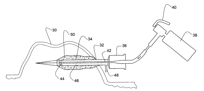
One embodiment of the method of the invention will now be described, with
reference to Fig. 3.
The skin surface of the hypertrophic scar or keloid 30 is first cleansed with
disinfectant solution. Then the penetrating area 32 into the scar is locally
anesthetised with lidocaine. The sterile cryoprobe 34 is inserted through the
anesthetised area into the hypertrophic scar or keloid. The probe is
preferably
inserted into the long axis of the scar so as to maximize the volume of scar
which is
frozen. The cutting tip of the probe is preferably inserted into the scar
tissue a few
3o millimeters below the scar surface (epidermis) without penetrating the
surrounding
CA 02414557 2003-O1-06
WO 02/02026 PCT/ILO1/00575
_7_
healthy tissue. The cryoprobe is then connected by the adapter 36 to a cryogun
38.
The cryogun valve 40 is opened and the liquid nitrogen flows into the inlet
tube 42
up to its distal outlet port 44 where it boils and becomes a gas. Thereafter,
it enters
the space 46 of the housing causing the wall of the housing to freeze. The
cryogas
s is vented to the environment through the vent 48.
An ice cylinder 50 having a thickness of 1-3 ruin forms around the
cryoprobe, freezing the surrounding tissue. Cryotherapy is generally performed
for
approximately 3 minutes. After closing the valve, the cryoprobe defrosts and
is
withdrawn. The treatment may be repeated every 2-4 weeks until the scar is
to flattened.
Example 3
patients aged 3 to 54 years and suffering from a hypertrophic scar or
keloid (HKS) of diverse cause older than 6 months were treated as described
below.
The trial, which extended over a 4-month period, evaluated the volume
reduction
(using dental impression material) of the HISS following one intralesional
cryotherapy as a monotherapy. In addition objective parameters (hardness and
color) and subjective complaints of pain/tenderness and itchiness/discomfort
were
examined in a scale of 1 to 5. Photographs were taken of all scars before and
after
treatment.
A cryoprobe as described in Fig. 1 was used in the treatments. The
penetrating area into the scar was locally anesthetized. Thereafter the
cryoprobe
was inserted into the long axis of the HKS so as to maximize the volume of the
HISS to be frozen.
The cryoprobe was connected by an adaptor to a cryogun filled with liquid
nitrogen. By depression of the activation trigger, the cryogun valve was
opened and
the liquid nitrogen was introduced into the cryoprobe thereby freezing the
HKS.
After the HKS became completely frozen the cryogun valve was closed and the
cryoprobe defrosted and withdrawn.
CA 02414557 2003-O1-06
WO 02/02026 PCT/ILO1/00575
_g_
The results are summarized in Table I. The preoperative HKS volume was
between 1 cc to 3.8 cc. Following one session of intralesional cryosurgery
treatment an average of 51% of scar volume reduction was achieved (33% to
67%).
Significant alleviation of objective and subjective symptoms as well as
softening of
s the scars was documented in 90% of patients. Minimal surface destruction was
noticed. The patients complained of very mild pain or discomfort during and
after
the procedure, which was easily managed. Only mild local edema was evident. No
active bleeding or infection was documented. One patient demonstrated late
hypopigmentation at the cryosurgical site.
1o
Table I: Volume Reduction following a Single Cryotherapy
Patient Age HKS Volume Volume after% Reduction
(years) Location before treatment
treatment(cc)(cc)
3 Cheek 1.5 0.5 66.7
14 Back 1.5 1 33.3
20 Lobule 1 0.55 45
22 Upper Post. 3.8 2.2 42.2
Auricle
22 Lower Post. 3.6 2 45.5
Auricle
23 Chest 0.3 0.1 66.7
30 Chest 2.5 1.25 50
34 Right Lobule 1 0.4 60
34 Left Lobule 1 0.4 60
54 Lobule 3.2 2 37.5
An increased efficiency of freezing scars as compared to conventional
15 cryosurgical methods for the treatment of HKS is illustrated.