Note: Descriptions are shown in the official language in which they were submitted.
CA 02414910 2002-12-20
ETH-1616
CANNULATED SCREW AND ASSOCIATED DRIVER SYSTEM
FIELD OF THE INVENTION
This invention relates to orthopedic screws and surgical
procedures using same, and, more particularly, to interference screws for
securing synthetic or biological tissue to bone.
BACKGROUND OF THE INVENTION
The knee joint is one of the strongest joints in the body because of
the powerful ligaments that bind the femur and tibia together.
Notwithstanding,
the knee is one of the most frequently injured joints, e.g., athletes
frequently
stress and tear knee ligaments. The large number of ligament injuries has
given
rise to numerous innovative surgical procedures and devices for replacing and
reconstructing torn or dislocated ligaments, typically involving grafting
autografts,
allografts, or a synthetic construct, to the site of a torn or dislocated
ligament.
For example, the replacement of an anterior cruciate ligament (ACL) may
involve
transplanting a portion of the patellar tendon, looped together portions of
semitendinosus-gracilis (hamstring) tendons, or donor achilles tendons, to
attachment sites in the region of the knee joint.
The most widely used technique for the reconstruction of the ACL
is known as the Jones procedure. The basic steps in the procedure include:
harvesting a graft made from a portion of the patellar tendon with attached
bone
blocks; preparing the graft attachment site (e.g., drilling holes in opposing
bones
of the joint in which the graft will be placed); placing the graft in the
graft
attachment site; and rigidly fixing the bone blocks in place within the graft
site,
i.e., the holes or "bone tunnels". The screws used to fix ahe graft in place
are
called "interference screws" because they wedge between the bone block and
the wall of the hole into which the bone block fits as they are screwed in.
Typically, there is very little space between the bone block and the hole in
the
bone at the fixation site.
Interference screws are typically driven into the space between the
bone block and the wall of the bone tunnel by placing the screw on a driver
and
CA 02414910 2002-12-20
then exerting a force on the driver and the screw in the direction of
insertion
while rotating the driver. The end of the driver rnay be received in a socket
or
groove located on the proximal end of the screw. More typically, the screw has
a
cannula or through hole in which the driver is inserted. The advantage of a
cannulated screw is that the force used to drive the screw into the space
between the bone block and the wall of the bone tunnel is spread over a larger
area of the screw. This reduces the risk of damage to the screw or slippage of
the driver relative to the screw as the screw is being driven into position.
Interference screws for anchoring ligaments to bone are typically
fabricated from medically approved metallic materials that are not naturally
absorbed by the body. A disadvantage of such screws is that once healing is
complete, an additional surgical procedure may be required to remove the screw
from the patient. Metallic screws may include a threaded shank joined to an
enlarged head having a transverse slot or hexagonal socket formed therein to
engage, respectively, a similarly configured, single blade or hexagonal
rotatable
driver for turning the screw into the bone. The enlarged heads on such screws
can protrude from the bone tunnel and can cause chronic irritation and
inflammation of surrounding body tissue.
Permanent metallic medical screws in movable joints can, in
certain instances, cause abrading of ligaments during normal motion of the
joint.
Screws occasionally back out after insertion, protruding into surrounding
tissue
and causing discomfort. Furthermore, permanent metallic screws and fixation
devices may shield the bone from beneficial stresses after healing. It has
been
shown that moderate periodic stress on bone tissue, such as the stress
?5 produced by exercise, helps to prevent decalcification of the bone. Under
some
conditions, the stress shielding which results from the long term use of metal
bone fixation devices can lead to osteoporosis.
Biodegradable or bioabsorbable interference screws have been
proposed to avoid the necessity of surgical removal after healing. Because the
degradation of a biodegradable screw occurs over a period of time, support
load
is transferred gradually to the bone as it heals. This reduces potential
stress
shielding effects. Conventional bioabsorbable interference screws are softer
and
weaker than metallic compositions, such that they are not self-tapping,
requiring
the holes drilled into the bone to be tapped. The necessity to tap holes in
the
CA 02414910 2002-12-20
injured bone adds to the complexity of the surgical procedure and lengthens
the
time required to complete the operation.
Accordingly, there is a need for a device and method that would
allow interference screws composed mainly of bioabsorbable materials to be
inserted into bone where the tapping of the bone is concurrent with the
insertion
of the screw into the bone.
SUMMARY OF THE INVENTION
The limitations of prior art apparatus for threading cannulated screws into
a substrate are overcome by the present invention which includes a driver for
threading a cannulated screw into a substrate. The driver has a tool with a
screw engaging portion and a threaded tip. The screw engaging portion is
extendable through a cannula formed in the cannulated screw and has a shape
that substantially prevents rotation of the screw engaging portion relative to
the
1 ~ cannulated screw when the screw engaging portion is extending through the
cannula of the cannulated screw. The threaded tip extends beyond the screw
when the screw engaging portion is inserted into the cannula of the cannulated
screw.
BRIEF DESCRIPTION OF THE FIGURES
Figure 1 is a perspective view of an exemplary embodiment of a driver and a
cannulated interference screw that may be inserted into a substrate by the
driver
in accordance with the present invention.
Figure 2 is a side view of a driver and a cannulated interference screw in
accordance with a second embodiment of the preserit invention with the
interference screw in position on the driver.
Figure 3 is a partially cross-sectional view of the driver and interference
screw of
FIG. 2.
.3
CA 02414910 2002-12-20
DETAILED DESCRIPTION OF THE INVENTION
The present invention relates to orthopedic surgical drivers for
screws for securing synthetic or biological connective tissue to a bone
surface,
such as, for example, attaching and maintaining a replacement anterior
cruciate
ligament (ACL) against a bone. The driver and method for using it would allow
cannulated interterence screws composed mainly of bioabsorbable materials to
be inserted into bone where the tapping of the bone tunnel is concurrent with
the
insertion of the screw into the bone. The driver has means for tapping threads
while it drives the screw into bone. The screw has threads to advance the
screw
into a position to establish an interference fit between the bone blocks and
the
bone tunnel, as well as, to hold the replacement anterior cruciate ligament
(ACL)
against the wall of the bone tunnel.
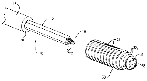
Figure 1 shows a rotatable driver 10 having a tool holder 14 and a
tool 16 with a threaded tip 18. A screw abutment surface 20 may be provided on
the tool holder 14 at the conjunction of the tool holder 14 and the tool 16.
As
shown, the tool holder 14 and the tool 16 mat be a monolithic structure, such
as
a unitary stainless steel forging, or it may be composite with the tool 16
inserting
into and interlocking with the tool holder 14 in a conventional manner. The
tool
holder 14 may be adapted to be grasped manually by virtue of a handle, a
wrench or a powered rotating tool (not shown) to rotate the driver 10.
The tool 16 shown has an elongated hexagonal body like a hex
wrench, but could be of another shape, such as polygonal, cross, star, or oval
shape. Threaded tip 18 is conicaily tapered and has helical lead-in screw
threads 22. The lead-in threads 22 are the means of tapping a previously
drilled
2S hole in bone to enable a screw 30 to be more easily threaded into the hole.
The rotatable driver 10 may be used to drive the interference
screw 30 into a hole in bone. Interference screw 30 is a longitudinally
elongated
cylindrical body with external threads 32 that are conically tapered running
from
approximately the middle of the interference screw 30 to the distal end 34
thereof thereby forming lead-in or starter threads 32~. A cannula 38 extends
through the interference screw 30. Threads 32 are like-handed relative to the
lead-in threads 22 of the toot 1fi such that they follow the lead-in threads
22 into
the hole in bone. Threads 32, in conjunction with the wedging effect of screw
30,
4
CA 02414910 2002-12-20
are the means of fixing the bone blocks in the bone tunnels during an ACL
reconstruction procedure. The cannula 38 shown is hexagonal in shape to match
the shape of tool 16 shown. One skilled in the art could envision screw
cannula
38 being other shapes for receiving a mating tool 16. These include, but are
not
limited to, polygonal, cross, star, or oval shapes.
Figures 2 and 3 show an interference screw 130 installed on the tool
116 of the rotatable driver 110. Reference numbers in Figures 2 and 3 are the
same as those used in Figure 1 for elements having the same or comparable form
and function, but increased by one hundred. The dimensions of tool 116 and
cannula 138 are preferably selected such that there is a close fit
therebetween and
preferably a friction fit. When the screw 130 is pressed against and abutting
the
tool holder 114, the threaded tip 118 extends beyond the distal end 134 of the
screw 130.
Although not shown, lead-in threads 132 may be provided with one
1 ~ or more flutes generally parallel to the axis of the threaded tip 118 as
are found on
conventional self-threading screws and taps to facilitate thread cutting. The
rotatable driver 110 is cannulated to allow passage of a guidewire 140
therethrough. Guidewire 140 allows accurate positioning of the threaded tip
118
during the ACL reconstruction procedure in accordance with known techniques.
The rotatable driver 10,110 of the present invention can be used in
the Jones procedure for the reconstruction of the ACL. After the steps of
harvesting and preparing the patellar tendon graft, preparing the graft site
by
drilling a hole through the tibia and femur and placing the graft, the
rotatable
driver 10, 110 of the present invention is used for turning the screw 30, 130
into
the bone tunnel, i.e., between the bone blocks and bone tunnels. Screws 30,
130 may be used to rigidly fix the upper and lower bone blocks in place within
the bone tunnel. When the screw 30, 130 is fully threaded into position within
the bone, a rearward pull on the driver 10, 110 will allow'the tool 16, 116 to
be
withdrawn from the cannula 38, 138. If the threaded 6p 18, 118 is threaded
into
bone prior to withdrawal, a small amount of bone will be withdrawn with the
thread tip 18, 118.
Screws 30, 130 for use with the driver 10, 110 of the present
invention may be formed from biocompatable, bioabsorbable materials, such as
bioabsorbable polymers, glasses or ceramics, autograft, allograft, or
xenograft
CA 02414910 2002-12-20
bone tissues, or combinations of absorbable ceramics, glasses and polymers. In
a preferred embodiment, the screw is formed from composites prepared by
incorporating bioabsorbable glass or ceramic reinforcements such as fibers or
particles in bioabsorbable polymer matrix.
s The following examples are illustrative of the principles and
practice of this invention, although the present invention is not limited to
these
specific embodiments. Numerous additional embodiments within the scope and
spirit of the invention will be apparent to those skilled in the art.
Example 1:
Drivers and mating composite screws were fabricated to test the self-
threading function of the driver of the present invention. Two drivers were
tested.
The first was a standard stainless steel hexagonal driver. The second was a
self-
threading driver. made from stainless steel, that had a leading tip with a
self-
threading design. The tip extended beyond the screw and incorporated all of
the
features of the present invention described above in reference to Figure 1.
The
cannulated screws were composed of a composite of 15/85 (volume percent) f3-
tricalcium phosphate (TCP) particles (10-micron average diameter) in
poly(lactic
acid), or PLA polymer. These screws were machined from billets of TCP/PLA
composites previously formed by injection molding.
The medial aspect of the medial femoral condyle on a porcine
femur was dissected to remove the soft tissue. A hole was drilled with a twist
bit
into the bone. The hole was positioned to be nearly perpendicular to the bone
surface and placed approximately at the insertion point of the medial
collateral
?5 ligament. The hole measured about two millimeters in diameter.
First, a cannulated composite bone screw mounted on a standard
hexagonal driver was used in an attempt to drive the screw into the bone. This
proved to be unsuccessful as the leading edge of the screw spun repeatedly and
failed to engage the bone. Next, the same type of screw was mounted onto the
self-threading driver. In this instance, the self-threading driver tip
immediately
engaged the bane and advanced the driver/screw complex forward so that the
leading edge of the screw gained purchase into the bone. The composite screw
was then able to advance into the bone.
6
CA 02414910 2002-12-20
Example 2:
A self-threading driver with a mounted composite screw was made for
simulation of a bone-tendon-bone ACL repair. The driver had a leading tip,
made
from stainless steel with a self-threading design. The tip extended beyond the
screw and incorporated all of the features of the present invention described
above in reference to Figures 2 and 3. The screw was composed of a composite
of 15185 (volume percent) f3-tricalcium phosphate (TCP) particles (10-micron
average diameter) in poly(lactic acid), or PLA polymer. The screw was machined
from a billet of TCP/PLA composite previously formed by injection molding.
A fresh frozen porcine knee model was used for simulation of a
bone-tendon-bone ACL repair. The shaft of the femur was securely clamped in a
bone vice. The capsular soft tissue structures of the knee were dissected and
removed, exposing the articular surfaces. A patellar bone plug, sized to 11-mm
in diameter, was harvested using an oscillating surgical saw. A portion of the
patellar tendon was left intact and attached to the bone plug. A bone tunnel
was
prepared in the femoral condyle by over-drilling a guide pin placed at the
insertion of the ACL and driven in an anterio-lateral direction. A 12-mm
diameter
tunnel was drilled. The patellar bone plug was inserted completely into the
tunnel
until its distal end intruded two to three millimeters beyond the tunnel
opening.
Since the bone plug was 11-mm in diameter and the tunnel was 12-mm in
diameter, there existed a 1-rnm gap into which the screw could be driven. The
self-tapping hexagonal driver with the mounted screw was positioned so that
during insertion, the threads of the screw would come into contact with the
cancellous surface of the bone plug. The driver/screw complex was then
advanced by screwing into the tunnel until the back end of the screw was
positioned nearly flush with the original cortical surface of the bone. During
the
insertion process, the composite screw was under torsional resistance. A
"biting"
sound was heard, indicating that the screw threads were cutting into the bone
materials. The bone plug was kept from advancing further into the tunnel
during
screw insertion by keeping constant tension on the patellar tendon.
In summary, the composite screw was successfully driven into the
bone tunnel/bone plug gap. The self-threading driver with mounted composite
7
CA 02414910 2002-12-20
screw was easily driven into the bone tunnel/bone plug gap once the threads on
the stainless steel front component of the driver caught onto the bone.