Note: Descriptions are shown in the official language in which they were submitted.
CA 02415422 2010-10-18
WO 02/04511 PCT/US01/21781
DESCRIPTION
CHROMOSOME 3p21.3 GENES ARE TUMOR SUPPRESSORS
BACKGROUND OF THE INVENTION
The U.S. Government has rights in the invention by virtue of P50-CA70907.
I. Field of the Invention
The invention generally relates to the fields of molecular biology and
oncology.
H. Related Art
Cancer is the result in the occurrence of multiple factors. Mutations may
occur in
proto-oncogenes that cause cellular proliferation to increase. Mutations also
may occur in
tumor suppressors whose normal function is to regulate cellular proliferation.
Mutations in
DNA repair enzymes impair the ability of the cell to repair damage before
proliferating.
Tumor suppressor genes are normal genes whose absence (loss or inactivation)
can lead to
cancer. Tumor suppressor genes encode proteins that slow cell growth and
division. Cancer
arises when there is a mutation in both alleles.
Tumor suppressor genes (TSGs) play a major role in the pathogenesis of human
lung
cancer and other cancers. Lung cancer cells harbor mutations and deletions in
multiple known
dominant and recessive oncogenes(Sekido et al., 1998, Virmani et al, 1998).
Known TSGs
such as Rb, p53, and putative TSGs have been found at chromosome regions 3p,
5q, 6p, 8p,
9p, and lip as well as other sites(Sekido et al., 1998, Gazdar et al., 1994,
Minna, 1994).
Cytogenetic and allelotyping studies of fresh lung tumors and tumor cells
showed tumor-cell
SUBSTITUTE SHEET (RULE 26)
CA 02415422 2003-01-08
WO 02/04511 PCT/US01/21781
allele loss at multiple sites, suggesting the existence of one or more such
TSGs(Sekido et al.,
1998, Virmani et al, 1998, Gazdar et al., 1994, Minna et al, 1997). However,
cytogenetic
changes and allele loss on the short arm of chromosome 3 (3p) have been shown
to be most
frequently involved in about 90% of small cell lung cancers (SCLCs) and >50%
of non-small
cell lung cancers (NSCLCs)(Sekido et al., 1998, Gazdar et al., 1994, Minna et
al, 1997, Daly
et al., 1993). SCLC and NSCLC are the two treatment groups of lung tumors and
are made
up of four histological types. Squamous cell-, adeno-, and large cell
carcinomas are in the
NSCLC group. Small cell lung cancer is in the SCLC group. Approximately 75% of
lung
tumors are NSCLCs. Metastases occur later with NSCLC than with SCLC. SCLC is
one of
the most metastatic of solid tumors (Mabry et al., 1998). In addition, similar
3p changes have
been seen in several other cancers in addition to lung, such as renal(Benmes
et al., 1998, Zbar
et al, 1987), breast(Gazdar et al, 1998, Sekido et al., 1998), head and neck
(Buchhagen et al.,
1996), pancreatic (Gorunova et al., 1998), kidney(Hughson et al., 1998),
oral(Uzawa et al.,
1998), and uterine cervical cancers(Kersemaekers et al., 1998. Furthermore, a
group of
TSGs, as defined by homozygous deletions in lung cancers, have been located
and isolated at
3p21.3 in a 450-kb-region(Sekido et al., 1998, Minna et al, 1997, Hung et al.,
1995, Sekido et
al., 1996, Wistuba et al.,1999). Studies of lung cancer preneoplasia indicate
that 3p21 allele
loss is the earliest genetic abnormality in lung cancer detected so far,
occurring in
hyperplastic lesions; this shows that one or more 3p-recessive oncogenes
function as
"gatekeepers" in the molecular pathogenesis of many human cancers, including
lung cancer,
where it is likely to be involved in >50% of all cases(Sekido et al., 1998,
Minna et al, 1997,
Hung et al., 1995, Sekido et al., 1996, Wistuba et al.,1999, Kohno et al.,
1999, et al., 1999,
Wistuba et al., 1999).
Recently, human chromosome band 3p21.3 has been shown to undergo overlapping
homozygous deletions in several SCLC and NSCLC lines; candidates of TSGs have
been
located in this critical region in several human cancers, further defining a
TSG region (Sekido
et al., 1998, Minna et al, 1997, Hung et al., 1995, van den Berg et al.,
1997). The evidence
shows that genes in this 3p21 critical region are involved in regulation of
the telomerase-
mediated cellular immortality pathway in lung, renal, and breast cancer cells
(Shay, 1997,
Shay, 1998). It has also been shown that 3p deletion occurs more frequently in
the lung
tumor tissues of patients who smoke. In addition, elevated sensitivity to the
carcinogen
benzo[a]pyrene diol epoxide at 3p21.3 has been associated with an increased
risk of lung
2
SUBSTITUTE SHEET (RULE 26)
CA 02415422 2003-01-08
WO 02/04511 PCT/US01/21781
cancer, suggesting that 3p21.3 is a molecular target of carcinogens in lung
cancer (Wu et al.,
1998). Despite those studies, there remains a need to further identify the
functions of these
genes and demonstrate their involvement with cancer.
3
SUBSTITUTE SHEET (RULE 26)
CA 02415422 2003-01-08
WO 02/04511 PCT/US01/21781
SUMMARY OF THE INVENTION
The tumor suppressor genes at 3p21.3 are now disclosed: Gene 26 (CACNA2D2)
(Gao et al., 2000), PL6, Beta* (BLU), LUCA-1 (HYAL1), LUCA-2 (HYAL2), 123F2
(RASSF1), Fusl, 101F6, Gene 21 (NPRL2), and SEM A3. The function of the
individual 3p
genes in suppression of tumor growth and tumor progression, induction of
apoptosis, alteration
of cell cycle kinetics, and repression of telomerase activity has been
characterized by the
liposome- and recombinant adenoviral vector-mediated transfer of 3p genes in
vitro and in vivo.
This also is the initial disclosure of the Beta* gene.
Therefore, it is an objective of the present invention to provide methods of
using
tumor suppressors having a chromosomal location of 3p21.3. It is also an
objective to
provide a tumor suppressor, Beta*. Further, it is an objective to provide
methods of
constructing recombinant adenovirus in which these tumor suppressors may be
inserted.
An embodiment of the present invention is an isolated polynucleotide encoding
a
polypeptide comprising an amino acid sequence of SEQ ID NO:2. There is also
provided a
nucleic acid with the sequence of SEQ ID NO: 1. Further provided is an
isolated polypeptide
comprising the amino acid sequence of SEQ ID NO:2. Another embodiment is a
nucleic acid
of 15 to about 100 base pairs comprising from 15 contiguous base pairs of SEQ
ID NO:1, or
the complement thereof. A further embodiment includes from about 20, 25, 30,
40, 50 or 100
contiguous base pairs of SEQ ID NO:1, or the complement thereof.
Another embodiment of the invention is an isolated peptide having between 10
and
about 50 consecutive residues of SEQ ID NO:2. Further, the peptide may
comprise 15, 20,
25, or 30 consecutive residues of SEQ ID NO:2. In this application, "about" is
defined as
within + or ¨2 amino acids.
Yet another embodiment is an expression cassette comprising a polynucleotide
encoding a polypeptide having the sequence of SEQ ID NO:2, wherein said
polynucleotide is
under the control of a promoter operable in eukaryotic cells. In another
embodiment, the
promoter of this expression cassette is heterologous to the coding sequence.
The promoter
may be a tissue specific and inducible promoter. In another embodiment, the
expression
cassette may be contained in a viral vector. The viral vector may be a
retroviral vector, an
4
SUBSTITUTE SHEET (RULE 26)
CA 02415422 2003-01-08
WO 02/04511 PCT/US01/21781
adenoviral vector, and adeno-associated viral vector, a vaccinia viral vector,
or a herpesviral
vector. In a further embodiment the expression cassette may comprise a
polyadenylation
signal.
Another embodiment is a cell comprising an expression cassette comprising a
polynucleotide encoding a polypeptide having the sequence of SEQ ID NO:2,
wherein said
polynucleotide is under the control of a promoter operable in eukaryotic
cells, said promoter
being heterologous to said polynucleotide.
Yet another embodiment of the invention is a monoclonal antibody that binds
immunologically to a polypeptide comprising SEQ ID NO:2, or an immunologic
fragment
thereof. Also provided is a monoclonal antibody with a detectable label. The
label may be a
fluorescent label, a chemiluminescent label, a radiolabel and an enzyme.
Another
embodiment of the invention is a hybridoma cell that produces a monoclonal
antibody that
binds immunologically to a polypeptide comprising SEQ ID NO:2, or an
immunologic
fragment thereof. A further embodiment is a polyclonal antisera, antibodies of
which bind
immunologically to a polypeptide comprising SEQ ID NO:2, or an immunologic
fragment
thereof.
Yet another embodiment is a isolated and purified nucleic acid that
hybridizes, under
high stringency conditions, to a DNA segment comprising SEQ ID NO:1, or the
complement
thereof. In a further embodiment the nucleic acid is about 15, 17, 20 or 25
bases in length.
Another embodiment of the invention is a method for constructing a recombinant
adenovirus comprising: (a) providing a shuttle vector, said shuttle vector
comprising an
adenoviral inverted terminal repeat (ITR) sequence, an expression cassette
comprising a
promoter and a poly-A sequence, a transgene under the control of said
promoter, and unique
restriction sites at the 5'- and 3'-ends of the ITR-promoter-transgene-poly-A
segment; (b)
cutting at said restriction enzyme sites; (c) ligating the released segment
into an adenoviral
vector lacking the entire El and E3 regions and transforming the resulting
vector a bacterial
host cell; (d) obtaining vector from said bacterial host cell and digesting
the vector to release
the El/E3-deleted adenovirus genome; and (e) transfecting the adenovirus
genome into El-
expressing host cells. In a further embodiment, the transgene is Gene 26, PL6,
Beta*,
LUCA-1, LUCA-2, 123F2, Fusl, 101F6, Gene 21, or SEM A3. In another embodiment,
the
SUBSTITUTE SHEET (RULE 26)
CA 02415422 2003-01-08
WO 02/04511 PCT/US01/21781
promoter may be a cytomegalovirus (CMV) promoter and said poly A sequence is
bovine
growth hormone (BGH) poly A sequence.
Yet another embodiment of the invention is a method for constructing a
recombinant
adenovirus comprising: (a) providing a shuttle vector comprising an adenoviral
inverted
terminal repeat (ITR) sequence, an expression cassette comprising a promoter
and poly-A
signal sequence, a transgene under the control of said promoter, a
tetracycline resistance-off
responsive element, and unique restriction sites at the 5' and 3' ends of the
IRT-promoter-
transgene-poly-A segment; (b) cutting at said restriction enzyme sites; (c)
ligating the
released segment into an adenoviral vector comprising a tetracyclin resistant-
off
transactivator gene and lacking the entire El and E3 regions, and transforming
the resulting
vector a bacterial host cell; (d) obtaining vector from said bacterial host
cell and digesting
the vector to release the El/E3-deleted adenovirus genome; and (e)
transfecting the
adenovirus genome into El-expressing host cells. In a further embodiment, the
transgene is
Gene 26, PL6, Beta*, LUCA-1, LUCA-2, 123F2, Fusl, 101F6, Gene 21 or SEM A3. In
another embodiment, the promoter may be a cytomegalovirus (CMV) promoter and
said poly
A sequence is bovine growth hormone (BGH) poly A sequence.
In yet another embodiment, also provided is a shuttle vector comprising an
adenoviral
inverted terminal repeat (ITR) sequence, an expression cassette comprising a
promoter and
poly-A sequence, a TetR-Off responsive element, and unique restriction sites
at the 5'- and
3'-ends of the ITR-promoter-poly-A segment. In another embodiment of the
invention the
promoter is a cytomegalovirus (CMV) promoter and said poly A sequence is
bovine growth
hormone (BGH) poly A sequence. Also provided is a multipurpose cloning site in
said
segment, positioned between said promoter and said poly-A sequence.
Yet another embodiment is an adenoviral vector comprising a tetracycline
resistant-
off transactivator gene and lacking the entire El and E3-regions.
Another embodiment of the invention is a method of diagnosing cancer in a
subject
comprising the steps of: (i) obtaining a biological sample from said subject;
and (ii)
assessing the expression of a functional Gene 26, PL6, Beta*, LUCA-1, LUCA-2,
123F2,
Fusl, 101F6, Gene 21, or SEM A3 product in sample. In a further embodiment the
sample is
a tissue sample. The tissue sample may be brain, lung, liver, spleen, kidney,
lymph node,
6
SUBSTITUTE SHEET (RULE 26)
CA 02415422 2003-01-08
WO 02/04511 PCT/US01/21781
small intestine, blood cells, pancreas, colon, stomach, cervix, breast,
endometrium, prostate,
testicle, ovary, skin, head and neck, esophagus, oral tissue, bone marrow or
blood tissue. In
another embodiment, the assessing comprises detecting a nucleic acid encoding
Gene 26,
PL6, Beta*, LUCA-1, LUCA-2, 123F2, Fusl, 101F6, Gene 21, or SEM A3. Detecting
may
comprise amplification said nucleic acid, nucleic acid hybridization, or
sequencing. In
another embodiment, assessing comprises detecting a Gene 26, PL6, Beta*, LUCA-
1, LUCA-
2, 123F2, Fusl, 101F6, Gene 21, or SEM A3 polypeptide. The detecting of a Gene
26, PL6,
Beta*, LUCA-1, LUCA-2, 123F2, Fusl, 101F6, Gene 21, or SEM A3 polypeptide may
comprise ELISA or immunohistochemistry. In yet another embodiment, the
assessing may
comprise wild-type or mutant oligonucleotide hybridization, with said
oligonucleotide
configured in an array on a chip or wafer. In another embodiment of the
invention, the
expression of Gene 26, PL6, Beta*, LUCA-1, LUCA-2, 123F2, Fusl, 101F6, Gene
21, or
SEM A3 is compared with the expression of Gene 26, PL6, Beta*, LUCA-1, LUCA-2,
123F2, Fusl, 101F6, Gene 21, or SEM A3 in normal samples. In another
embodiment, the
comparison involves evaluating the level of Gene 26, PL6, Beta*, LUCA-1, LUCA-
2, 123F2,
Fusl, 101F6, Gene 21, SEM A3 expression.
Another embodiment is a non-human transgenic animal lacking one or both
functional
alleles of Gene 26, PL6, Beta*, LUCA-1, LUCA-2, 123F2, Fusl, 101F6, Gene 21,
SEM A3.
Also provided is a non-human transgenic animal that overexpresses Gene 26,
PL6, Beta*,
LUCA-1, LUCA-2, 123F2, Fusl, 101F6, Gene 21, or SEM A3 as compared to a
similar non-
transgenic animal. In a further emodiment is a non-human transgenic animal,
the genome of
which comprises an expression cassette comprising a Gene 26, PL6, Beta*, LUCA-
1, LUCA-
2, 123F2, Fusl, 101F6, Gene 21, or SEM A3 under the control of an inducible
promoter.
An embodiment of the invention is a method for suppressing growth of a tumor
cell
comprising contacting said cell with an expression cassette comprising: (a) a
nucleic acid
encoding Gene 26, PL6, Beta*, LUCA-1, LUCA-2, 123F2, Fusl, 101F6, Gene 21, or
SEM
A3; and (b) a promoter active in said tumor cell, under conditions permitting
the uptake of
said nucleic acid by said tumor cell. In another embodiment, the tumor cell is
derived from a
brain tumor, lung tumor, liver tumor, spleen tumor, kidney tumor, lymph node
tumor, small
intestine tumor, blood cell tumor, pancreatic tumor, colon tumor, stomach
tumor, cervix
tumor, breast tumor, endometrial tumor, prostate tumor, testicle tumor,
ovarian tumor, skin
7
SUBSTITUTE SHEET (RULE 26)
CA 02415422 2003-01-08
WO 02/04511 PCT/US01/21781
tumor, head and neck tumor, esophageal tumor, oral tissue tumor, or bone
marrow tumor. In
a further embodiment, the nucleic acid is contained in a viral vector. The
viral vector may be
a retroviral vector, an adenoviral vector, and adeno-associated viral vector,
a vaccinia viral
vector, and a herpesviral vector. In yet another embodiment, the nucleic acid
is contained in
a liposome.
Another embodiment of the invention is a method of altering the phenotype of a
tumor cell comprising contacting said cell with an expression cassette
comprising: (a) a
nucleic acid encoding Gene 26, PL6, Beta*, LUCA-1, LUCA-2, 123F2, Fusl, 101F6,
Gene
21, SEM A3; and (b) a promoter active in said tumor cell, under conditions
permitting the
uptake of said nucleic acid by said tumor cell. In another embodiment, the
phenotype is
selected from the group consisting of proliferation, migration, contact
inhibition, soft agar
growth, cell cycling, invasiveness, tumorigenesis, and metastatic potential.
In yet another
embodiment, the promoter is a cytomegalovirus (CMV) promoter.
Another embodiment is a method of inhibiting cancer in a subject suffering
therefrom
comprising administering to said subject an expression cassette comprising:
(a) a nucleic
acid encoding Gene 26, PL6, Beta*, LUCA-1, LUCA-2, 123F2, Fusl, 101F6, Gene
21, or
SEM A3 polypeptide; and (b) a promoter active in tumor cells of said subject,
whereby
expression of said polypeptide inhibits said cancer. In a further embodiment,
the subject is a
human. In other embodiments, the nucleic acid encodes Gene 26, PL6, Beta*,
LUCA-1,
LUCA-2, 123F2, Fusl, 101F6, Gene 21, or SEM A3. In another embodiment, the
cancer is a
selected from the group consisting of brain cancer, lung cancer, liver cancer,
spleen cancer,
kidney cancer, lymph node cancer, small intestine cancer, blood cell cancer,
pancreatic
cancer, colon cancer, stomach cancer, cervix cancer, breast cancer,
endometrial cancer,
prostate cancer, testicle cancer, ovarian cancer, skin cancer, head and neck
cancer, esophageal
cancer, oral tissue cancer, and bone marrow cancer. In yet another embodiment,
the
expression cassette is contained in a viral vector. The viral vector may be a
retroviral vector,
an adenoviral vector, and adeno-associated viral vector, a vaccinia viral
vector, and a
herpesviral vector. In another embodiment, the expression cassette is
contained in a lipsome.
In another embodiment, the expression cassette further comprises a poly-A
sequence. The
poly-A sequence may be a bovine growth hormone (BGH) poly-A sequence. In a
further
8
SUBSTITUTE SHEET (RULE 26)
CA 02415422 2003-01-08
WO 02/04511 PCT/US01/21781
õ
embodiment, the expression cassette is administered intratumorally, in the
tumor vasculature,
local to the tumor, regional to the tumor, or systemically.
Also provided in the method of inhibiting cancer is the administering of a
chemotherapuetic agent to said subject. In another embodiment, the
chemotherapeutic
comprises cisplatin (CDDP), carboplatin, procarbazine, mechlorethamine,
cyclophosphamide, camptothecin, ifosfamide, melphalan, chlorambucil, busulfan,
nitrosurea,
dactinomycin, daunorubicin, doxorubicin, bleomycin, plicomycin, mitomycin,
etoposide
(VP16), tamoxifen, raloxifene, estrogen receptor binding agents, taxol,
gemcitabien,
navelbine, farnesyl-protein tansferase inhibitors, transplatinum, 5-
fluorouracil, vincristin,
vinblastin and methotrexate. Also provided is the administering radiation to
said subject. In
another embodiment, the radiation is delivered local to a cancer site or is
whole body
radiation. The radiation may comprise y-rays, X-rays, accelerated protons,
microwave
radiation, UV radiation or the directed delivery of radioisotopes to tumor
cells. In yet another
embodiment, a a second anticancer gene may be administered to said subject.
The second
anticancer gene may be a tumor suppressor. The second anticancer gene may be
an inhibitor
of apoptosis. In another embodiment, the second anticancer gene is an oncogene
antisense
construct.
An embodiment of the invention is a method of treating a subject with cancer,
comprising the step of administering to said subject a Gene 26, PL6, Beta*,
LUCA-1, LLTCA-
2, 123F2, Fusl, 101F6, Gene 21, SEM A3 polypeptide. In another embodiment, the
cancer is
a selected from the group consisting of brain cancer, lung cancer, liver
cancer, spleen cancer,
kidney cancer, lymph node cancer, small intestine cancer, blood cell cancer,
pancreatic
cancer, colon cancer, stomach cancer, cervix cancer, breast cancer,
endometrial cancer,
prostate cancer, testicle cancer, ovarian cancer, skin cancer, head and neck
cancer, esophageal
cancer, oral tissue cancer, and bone marrow cancer. In a further embodiment,
the polypeptide
is contained within a liposome. the liposome may be comprised of N-(142,3-
Dioleoyloxy]propy1)-N,N,N-trimethylammonium (DOTAP) and cholesterol. In
another
embodiment, the subject is human.
Another embodiment of the invention is a method of screening a candidate
substance
for anti-tumor activity comprising the steps of: (i) providing a cell lacking
a functional Gene
9
SUBSTITUTE SHEET (RULE 26)
CA 02415422 2003-01-08
WO 02/04511 PCT/US01/21781
26, PL6, Beta*, LUCA-1, LUCA-2, 123F2, Fusl , 101F6, Gene 21, or SEM A3
polypeptide;
(ii) contacting said cell with said candidate substance; and (iii) determining
the effect of
said candidate substance on said cell. In another embodiment, the cell is a
tumor cell. In
another embodiment, the determining may comprises comparing one or more
characteristics
of the cell in the presence of said candidate substance with the same one or
more
characteristics of a similar cell in the absence of said candidate substance.
In a further
embodiment, the characteristic is selected from the group consisting of Gene
26, PL6, Beta*,
LUCA-1, LUCA-2, 123F2, Fusl, 101F6, Gene 21, SEM A3 expression, phosphatase
activity,
proliferation, metastasis, contact inhibition, soft agar growth, cell cycle
regulation, tumor
formation, tumor progression, metastasis and tissue invasion. In another
embodiment, the
candidate substance is a chemotherapeutic or radiotherapeutic agent. Also
provided is a
candidate substance selected from a small molecule library. In further
embodiments, the cell
is contacted in vitro or in vivo.
An embodiment of the invention is a method of screening a candidate substance
for
anti-tumor activity comprising the steps of: (i) providing a cell; (ii)
contacting said cell
with said candidate substance; and (iii) determining the effect of said
candidate substance on
expression of a Gene 26, PL6, Beta*, LUCA-1, LUCA-2, 123F2, Fusl, 101F6, Gene
21, or
SEM A3 polypeptide.
Another embodiment is a method of producing a Beta* polypeptide in a host cell
comprising: (a) providing an expression cassette comprising a nucleic acid
encoding Beta*
operably linked to an promoter active in said host cell; (b) transferring said
expression
cassette into said host cell; and (c) culturing said host cell under
conditions permitting
expression of said Beta* polypeptide.
Yet another embodiment of the invention is a method of diagnosing cancer in a
subject comprising the steps of: (i) obtaining a biological sample from said
subject; and (ii)
detecting hypermethylation of the promoter region of Gene 26, PL6, Beta*, LUCA-
1, LUCA-
2, 123F2, Fusl, 101F6, Gene 21, or SEM A3.
Other objects, features and advantages of the present invention will become
apparent
from the following detailed description. It should be understood, however,
that the detailed
description and the specific examples, while indicationg preferred embodiments
o the
SUBSTITUTE SHEET (RULE 26)
CA 02415422 2003-01-08
WO 02/04511 PCT/US01/21781
invention, are given by way of illustration only, since various chanWArit
in011i`ficirons
within the spirit and scope of the invention will become apparent to those
skilled in the art
from this detailed description.
BRIEF SUMMARY OF THE DRAWINGS
The following drawings form part of the present specification and are included
to
further demonstrate certain aspects of the present invention. The invention
may be better
understood by reference to one or more of these drawings in combination with
the detailed
description of specific embodiments presented herein:
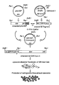
FIG. 1. Scheme of construction and production of recombinant adenovirus using
pAd-RAP and pAd-RAP-Shuttle system.
FIG. 2. Scheme of construction of recombinant adenovirus using pAd-RAP-Tet-Off
and pAd-RAP-TRE-CMV-Shuttle. TetR-Off = tetracyclin resistant-off
transactivator gene,
TRE = TetR-Off responsive elements.
FIG. 3. Timing of genetic changes found in preneoplastic lesions of the
respiratory
epithelium associated with primary non-small cell lung cancers.
FIG. 4. Allelotyping of 3p region in DNAs from human lung cancer cell lines
and
tumors. Filled ovals=loss of heterozygosity; open ovals=retaining of alleles;
and hatched
ovals=homozygous deletions.
FIG. 5. Scheme of the location of the 3p21 tumor suppressor region in human
chromosome 3p and the structure of recombinant adenoviral vectors of 3p genes.
The sizes
of the individual 3p genes and their corresponding amino acid residues, and
the active tumor
suppressor (TS) regions and known TSGs in the 3p are also indicated.
FIG. 6. Effects of overexpression of 3p genes on tumor cell growth in Ad-3p-
transduced lung cancer cells and normal human bronchial epithelial cells. MOIs
were
expressed as viral particles/cell (vp/c).
FIG. 7. Quantification of adenovirus-mediated 3p gene expression in H1299
cells by
Real Time RT-PCR. The MOIs are expressed as viral particles/cell (vp/c).
11
SUBSTITUTE SHEET (RULE 26)
CA 02415422 2003-01-08
WO 02/04511 PCT/US01/21781
FIG. 8. Induction of apoptosis by overexpression of 3p genes in Ad-3p-
transduced
lung cancer cells and normal HBEC. Apoptosis was analyzed by FACS with TUNEL
reaction.
FIG. 9. Effect of overexpression of 3p genes on cell cycle kinetics in Ad-3p-
transduced human lung cancer cells A549 and H1299.
FIG. 10. Effect of overexpression of 3p genes on A549 tumor growth by
intratumoral
injection of Ad-3p vectors in nude mice.
FIG. 11. Effect of overexpression of 3p genes on A549 lung metastatic tumor
growth
by systemic injectionof protamine-Ad-3p vector complexes in nude mice.
FIG. 12. Map of the RASSF1 locus, transcripts, and protein domains, A) The
exon¨
intron structure of the RASSF1 locus with the location of the CpG islands in
the predicted
promoter regions (the locations of which are shown by double-headed arrows) of
RASSF1A
and RASSF1C. RASSF1A transcription is predicted to come from the most
centromeric
promoter region located within a CpG island and begins with exon 1A. RASSEiF
also
commences at this promoter but is missing exon iC. Transcription of RASSFIC is
predicted
to begin in the most telorneric promoter region, which is approximately 2
kilobases from that
of RASSF1A and begins with exon 1. Blocks represent exons; lines represent
introns. B)
Schematic of the RASSF1A transcript and predicted protein-sequence domains.
The location
of the various primers (PKCDF, NF, R182, and R292) used for isoform-specific
reverse
transcription (RT)-polymerase chaln reaction (PCR) analyses are indicated.
Tick marks
identify the exon boundaries. The potential arc homology 3 (5H3)-binding
region, putative
diacylglycerol (DAG)-binding domain, PEST sequence, Rasassociation domain, and
ataxia-
telangiectasia-mutated (ATM) phosphorylation site are labeled. C) Schematic of
the
RASSFIC transcript and predicted protein-sequence domains. The locations of
the various
primers (NOX3, R182, and R292) used for isoform-specific RT¨PCR analyses are
indicated. D) Schematic of the RASSFIF transcript and predicted protein-
sequence domains.
FIG. 13. RASSF1A and RASSF1C messenger RNA levels detected by isoform-
specific reverse transcription¨polymerase chain reaction (RT¨PCR) in a
sampling of lung
cancer cell lines (A), breast cancer lines (B), and resected lung tumors and
normal human
12
SUBSTITUTE SHEET (RULE 26)
CA 02415422 2003-01-08
WO 02/04511 PCT/US01/21781
lung and breast epithelial cultures (C). All RT¨ PCR products were separated
on 2% agarose
gels and were identified by staining with ethidium bromide. Arrows indicate
location of
transcripts. A) Lung cancer lines tested in lanes: 1 ¨ 11157; 2 = 11358; 3 =
11727; 4 =
11740; 5 = 11748; 6 =11838; 7 = 111184; 8 = 111299; 9 = 111304; 10 =111437; 11
=
111450; 12 = 111770; 13 = 111792; 14 = 111963; 15 111993; 16 = 112009;
17=112077; iS
= 112108; 19 = 11HCC44; and 20 = HCC78. B) Breast cancer lines tested in
lanes: 1 =
11CC38;2 = 11CC1187;3 = HTB19;4 = HTB20; 5 = HTB22; 6 = 11TB23; 7 = 11TB24; 8
=
11TB25; 9 = 11TB26; 10 = 11TB27; 11 = HTB12I; 12 = HTB129; 13 HTB130; 14 =
HTBI31; 15 = HTB132; 16 = H'I'B133; 17 = 11CC 1395; iS = 11CC 1428; 19 =
11CC1569;
20 = 11CC1806; and 21 = 11CC2157. C) Resected lung adenocarcinoma samples (ADC
1-
5) and cultures of normal small-airway epithelial cells (SAECs), normal human
bronchial
epithelial (NHBE) cultures, and normal human breast epithelial (NHBRE)
cultures.
FIG. 14. Expression of RASSF1A after treatment of lung cancer cells with 5-aza-
2'-
deoxycytidine (SAza-CdR). NCI-11157, a non-small-cell lung carcinoma (NSCLC)
cell line
that expresses RASSF1C but not RASSFIA, was grown in the presence (+ lanes)
and absence
(¨ lanes) of 0.5 p.M SAza-CdR for 48 hours. Total RNA was isolated,
complementary DNA
was prepared, and isoformspecific reverse transcription¨polymerase chain
reaction was
performed for RASSF1A, RASSF1C, and glyceraldehyde-3-phosphate dehydrogenase
(GAPDH) as a control.
FIG. 15. . Methylation-specific polymerase chain reaction (PCR) for the
detection of
methylated RASSF1A 5, CpG sequences in primary resected non-small-cell lung
carcinomas
(NSCLCs) and their accompanying normal lung tissue (upper panel), small-cell
lung car-
cinoma (SCLC) cell lines (middle panel), and primary breast cancers (lower
panel).
Representative samples are shown. For resected NSCLCs, U = results with
primers specific
for unmethylated sequences; M = results with primers specific for methylated
sequences. NL
= normal lung tissue; T = tumor; P = results with peripheral blood lymphocyte
DNA, which
is unmethylated or in vitro methylated (IVMD); and 1120 = negative controls
with water
blanks. For SCLCs, each lane shows the PCR results for the methylated
sequences from a
different cell line. Lane 20 is negative control. For the breast cancers, each
lane shows the
PCR results for methylated sequences from a different sample. PCR products
were separated
on 2% agarose gels and bands were detected after staining with ethidium
bromide.
13
SUBSTITUTE SHEET (RULE 26)
CA 02415422 2003-01-08
WO 02/04511 PCT/US01/21781
FIG. 16. Kaplan¨Meier survival curve for 107 patients with resected non-small-
cell
lung carcinomas based on RASSF1A methylation status (32 methylated and 75 not
methylated), For the patients with unmethylated RASSF1A alleles, the number of
cases = 75,
censored = 39, and events = 36, with a mean overall survival of 52 months (95%
confidence
interval [CI] = 44 to 59) and a median overall survival of 49 months (95% CI =
44 to 59); for
the patients with methylated RASSF1A alleles, the number of cases = 32,
censored = nine,
and events = 23, with a mean overall survival of 37 months (95% CI = 27 to 46)
and a
median overall survival of 28 months (95% CI = 9 to 47). The log-rank test
statistic for
equality of survival distributions for RASSF1A methylation was 3.97, with df
1, P = .0463.
The patients at risk for each group were: RASSF1A unmethylated-12 months (n =
63), 36
months (n = 34), and 60 months (n = 16); RASSF1A methylated-12 months (n =
24), 36
months (n = 13), and 60 months (n = 5).
FIG. 17. . Effect of RASSF1A on the in vitro and in vivo growth of the non-
small-cell
lung carcinoma (NSCLC) cell line NCI-111299. A) Anchorage-dependent and
anchorage-
independent colony formation after transfection of NCI-H1299 cells with the ¨
ioo empty
vector (pcDNA3.1+) or peDNA3.1+ expression vectors containing wild-type p53 or
RASSF1A. For analysis of anchorage-dependent growth, after 2 days in
nonselective growth
medium, transfected NCI-111299 cells were diluted into 100-mm2 dishes with
selective
medium. Transfected cells were plated in liquid medium (for anchorage-
dependent assays) or
soft agar (for anchorage-independent assays) containing 800 p.g/mL of G418.
Colonies were
stained with methylene blue in anchorage-dependent experiments after 14 days.
Results
represent the average of eight to 12 experiments in liquid medium and three
soft-
agarexperiments. Standard deviations are shown or are less than 2%. Solid bars
anchorage-
dependent growth (95% confidence interval [CI] .0 to 36 for wt-p53 (wild-type)
and 52 to 60
for RASSFIA); open bars anchorage-independent growth (95% CI =0 to 6 for wild-
type (wt)-
p53 and 0 to 39 for RASSFIA). B) Northern blot analysis of the RASSF1A
expression in
stable clones of NCI-H1299 cells transfected with the pcDNA3.1+ vector or
pcDNA3.1+
containing RASSF1A complementary DNA (cDNA). The vector control (vector) and
four
separate clones with various RASSF1A messenger RNA levels are shown. Several
of these
clones were used in the anchorage-independent growth assay shown in D.
Ethidium bromide
staining of the ribosomal RNA is shown as a loading control. The clones were
also verified to
express the RASSF1A isoform by reverse transcription¨polymerase chain reaction
with the
14
SUBSTITUTE SHEET (RULE 26)
CA 02415422 2003-01-08
WO 02/04511 PCT/US01/21781
use of isoform-specific primers. C) Soft-agar (anchorage-independent) colony
formation in
stable clones of NCI-111299 cells transfected with the pcDNA3.1+ vector or
pcDNA3.1+
containing RASSF1A cDNA. The means and standard deviations are shown. For each
of the
RASSFIAexpressing clones, the 95% CI =0 to 4 for F1A.4, 2 to 16 for F1A.5, and
3 to 14 for
F1A.19. D) NCI111299 cells were infected with the pBABEpuro retrovirus
expression
vectors containing either the vector control or the RASSF1A or RASSFI C cDNAs.
Infected
cells (10000 per plate) were suspended in 0.33% agar, and the suspension was
layered over a
0.5% agar base. Colonies greater than 0.2 mm in diameter were counted after 21
days. The
lower right panel shows a representative western blot, developed with a rabbit
antibody to the
RASSF1-glutathione S-transferase fusion protein, to verify the expression of
the RASSF1
proteins. C = positive control generated by transient transfection of NCI-
111299 cells with
peDNA3.1+ containing RASSF1A cDNA; V = infection of NCI- H1299 cells with the
retroviral vector control (note runover from positive control; lA = infection
of NCI-H1299
cells with the retroviral vector containing RASSF1A; and 1C = infection of NCI-
H 1299 cells
with the retroviral vector containing RASSF1C. E) Effect of RASSF1A on the in
vivo growth
of NCI-111299 cells. Approximately 107 viable NCI-H 1299 cells expressing
RASSF1A were
injected into the flanks of each of five previously irradiated BALB/c (nulnu)
nude mice.
Tumor size was monitored overtime, and size is shown in cubic millimeters. The
average
volume of tumors grown in more than 20 mice that were given an injection of
vector-
transfected NCI-H 1299 cells is shown (H1299 parent). Mice that were given an
injection of
RASSFIA-infected NCI-H 1299 cells grew no measurable tumors.
FIG. 18. Schematic representation of the location of the putative 3p21.3 tumor
suppressor region in human chromosome 3p and the structure of the recombinant
adenoviral
vectors of 3p21.3 genes. The sizes of the individual 3p21.3 genes and their
corresponding
amino acid residues deduced from coding sequences of cDNAs, and the active
tumor
suppressor (TS) regions and known TSGs in the 3p are indicated. The
recombinant
adenoviral vectors of 3p21.3 genes (Ad-3ps) were constructed by inserting a
mammalian
expression cassette in which the 3p21.3 gene was driven by a CMV promoter and
tailed with
BGH poly A signal sequence into the El-deleted region of the replication
incompetent
adenovirus type 5 (Ad5) genome. The relative locations of El -deletion (AE1)
and E3-
SUBSTITUTE SHEET (RULE 26)
CA 02415422 2003-01-08
WO 02/04511 PCT/US01/21781
deletion (AE3), the inverted repeated terminal (IRT) sequences in the Ad5
genome are
indicated.
FIG. 19. Effects of exogenous expression of 3p21.3 genes on tumor cell growth
in
Ad-3p-transduced human lung cancer cells and normal bronchial epithelial
cells. Cells were
transduced with adenoviral vectors of 3p21.3 genes, 101F6, NPRL2, BLU, RASSF1C
FUSI ,
HYAL2, and HYALI, control genes, LacZ and p53, and empty vector, Ad-EV, at
highest
MOTs (vp/c), 5000 for A549, 1000 for H1299, 5000 for H460, 2500 for H358, and
1000 for
HBE, respectively, and PBS alone was used as a mock control. The cell
viability was
expressed as the percentage of viable adenoviral vector-transduced cells in
relation to PBS-
treated control cells (100%). The error bars represent standard deviations of
the mean in at
least three individual experiments. Treatments were given in quadruplicate for
each
experiment. The significance of the difference in cell viability between
vector-treated cells
and the Ad-EV-, Ad-LacZ-, or PBS-treated controls was analyzed by two-sided
Student's T-
test. P<0.05 was taken as significant. The differences between the cell
viability of the Ad-
EV- and Ad-LacZ-transduced cells versus PBS-treated controls were not
significant (P = 0.25
to P = 0.95 from different time points and cell lines). The differences
between the cell
viability of the Ad-101F6, Ad-Fusl, and Ad-NPRL2- transduced cells versus the
Ad-EV-,
Ad-LacZ-transduced, or PBS-treated controls at same MOIs were significant in
A549,
H1299, and in H460 at both 3 days and 5 days posttransduction. (P -0.0001 to P
0.005) but
not significant in H358 and HBEC cell lines at both 3 and 5 days
posttransduction (P 0.10
to P 0.95, from different time points and cell lines), respectively. The
effects of Ad-BLU,
Ad-HYAL2, and Ad-HYAL1 on cell viability were not significant in all cell
lines (P> 0.45)
compare to those of Ad-EV and Ad-LacZ.
FIG. 20. Quantification of adenovirus-mediated 3p21.3 gene expression in H1299
cells by real-time RT-PCR. The real-time RT-PCR was performed and PCR profiles
were
generated by an ABI Prism 7700 Sequence Detection system and equipped software
(Perkin
Elmer Applied Biosystems). Known concentrations of 13-Actin DNA were used as a
standard. The H1299 cells were transduced by adenoviral vectors of 3p21.3
genes, FUSI (A),
101F6 (B), NPRL2 (C), and HYALI (D) at a MOI of 1, 5, and 10 pfu/cell for 48
hr,
respectively, as indicated by arrows.
16
SUBSTITUTE SHEET (RULE 26)
CA 02415422 2003-01-08
WO 02/04511 PCT/US01/21781
FIG. 21. Induction of apoptosis by exogenous expression of 3p21.3 genes in Ad-
3p-
transduced human NSCLC cells and nounal HBECs. Apoptosis were analyzed by
FACS,
using TUNEL reaction with FITC-labeled dUTP. Cells were transduced with
adenoviral
vectors of 3p21.3 genes at an MOIs (vp/c) of 5000 for A549 (A), 1000 for
111299 (B), 5000
for H460 (C), 2500 for H358 (D), and 1000 for HBEC (E), respectively, and PBS,
Ad-EV,
and p53 were used as controls. Cell were harvested and analyzed for apoptosis
at the
indicated days posttransduction. The rate of apoptosis is expressed as the
percentage of
FITC-labeled cells in the total cell population. The error bars represent
standard deviations
of the mean in two or three repeated experiments with triplicate treatments
and TUNEL
reactions for each experiment. The significance of the difference in apoptosis
between vector-
treated cells and the Ad-EV-, Ad-LacZ-, or PBS-treated controls was analyzed
by two-sided
Student's T-test. P<0.05 was considered significant. The differences between
the apoptosis
induced by the Ad-By- and Ad-LacZ-transduced cells versus PBS-treated controls
were not
significant (P = 0.925 to P = 0.675 from different time points and cell
lines). The differences
between the apoptosis induced in the Ad-101F6, Ad-FUS1, and Ad-NPRL2-
transduced cells
versus the Ad-EV-, Ad-LacZ, or PBS-treated controls were significant in A549
and H460
cells at both 3 days and 5 days posttransduction (P --0.0001 to P 0.005), and
significant
versus the Ad-EV- and PBS-treated cells in H1299 at 5 days posttransduction (P
.Ø02), but
not significant in 11358 and HBEC cell lines at both 3 and 5 days
posttransduction at all time
points (P 0.85 to P 0.95), respectively. Induction of apoptosis in Ad-p53-
transduced
H358 cells were significant at all time points compared to all other
treatments (P < 0.0001).
Induction of apoptosis in cells treated with Ad-BLU, Ad-HYAL2, and Ad-HYALyall
was
not significant compared to those treated with PBS, Ad-By, or Ad-LacZ, in all
cell lines at all
time points (P> 0.85).
FIG. 22. Effects of intratumoral administration of adenoviral vectors of
3p21.3 genes
on growth of human lung cancer A549 (A) and H1299 (B) subcutaneous tumors in
nu/nu
mice. When the tumor reached 5 to 10 mm in diameter at about 2 weeks after
tumor
inoculation, the tumor was injected with individual adenoviral vectors of
3p21.3 genes,
101F6, NPRL2, BLU, RASSF1CFUS1, HAYL2, and HYAL1 or control vectors Ad-EV,
LacZ,
and p53, at a dose of 5 x 10¹⁰ vp/tumor each in 200 ill of PBS for three times
within a week,
respectively, and PBS alone was used as a mock control. Results were reported
as the mean
17
SUBSTITUTE SHEET (RULE 26)
CA 02415422 2003-01-08
WO 02/04511 PCT/US01/21781
SD in 5-10 mice for each treatment group. Tumor volumes were normalized by the
percentage increase of tumor sizes after treatment relative to those at the
beginning of the
treatment in each group. Mean tumor volumes SE from these experiments are
shown.
ANOVA was performed to determine statistical significance between each
treatment group
using a Statistica software (StatSoft Inc.) and P
0.05 was considered significant. The
differences betweof en the tumor volumes ofin the Ad-101F6, Ad-FUS1, Ad-NPRL2 -
treated mice versus in the Ad-EV- and Ad-LacZ- treated mouse controls were
statistically
significant in both A549 and H1299 tumor models (P < 0.0001), and the
difference in the Ad-
HYAL2-treated mice was significant in A549 (P = 0.024) but not in H1299 tumor
models,
after 5 days from the last injection (P < 0.0001), but not significant in Ad-
HYAL1, Ad-
HYAL2, Ad-RASSF1C, and Ad-BLU-treated (P> 0.05 in both A549 and H1299 tumor
models).
FIG. 23. Effect of systemic administration of protamine-Ad-3p complexes on
development of A549 experimental lung metastases in nu/nu mice. A., Relative
metastatic
tumors in mice treated with P-Ad-3p21.3 genes. All animals were i.v. injected
with various
protamine-adenoviral vector complexes every other two days for 3 times each at
a dose of 3 x
1010 viral particles plus 300 j.ig protamine in a total volume of 200 ill per
animal, and PBS
alone was used as a mock control. Each treatment group consisted of 5-10
animals. Lungs
were harvested two weeks after the last injection and metastastic colonies on
the surfaces of
lung were counted without knowledge of the treatment groups. Development of
metastases
were represented as the percentages of metastatic colonies formed in protamine-
adenovirus
complexes-treated groups in relation to those in the PBS-treated group (as
100%). Error bars
represent as standard error (SE). Non-parametric t-test (Wald-Wolfowitz Runs
Test) was
performed to determine statistical significance between each treatment group
using a
Statistica software (StatSoft Inc.) and P 0.05 was considered significant. A
significant
inhibition of development of metastases was observed in mice treated with P-Ad-
101F6 (P =
0.002), P-Ad-NPRL2 (P = 0.001), P-Ad-BLU (P = 0.018), P-Ad-FUS1 (P = 0.002),
and P-
Ad-HYAL2 (P = 0.014), respectively, compared to mice treated with PBS, P-Ad-
EV, or P-
Ad-LacZ, but no significant inhibition in mice treated with . P-Ad-BLU (P =
0.818) or P-Ad-
18
SUBSTITUTE SHEET (RULE 26)
CA 02415422 2003-01-08
WO 02/04511 PCT/US01/21781
HYAL1 (P = 0.904). B., the representative photos of lungs stained with India
ink for
metastases. The metastatic colonies were shown as white spots on the surfaces
of lung.
FIG. 24. (a) RT-PCR Analysis of NSCLCs cDNA HCC515 (Wild type FUSI ) and
H322 (smaller cDNA mutant form of FUS1). (b) Genomic structure of wild type
FUSI and
the mutant aberrant slicing form. Top line is genomic DNA from cosmid clone
LUCA#13
(#Z84492) and the indicated nucleotide sequence numbers. Arrowheads indicated
primers
for SSCP analysis. Boxes represent cDNA with the open reading frames (black)
and
untranslated regions (white) for the 110 amino acid wild type and 82 amino
acid aberrant
splice form of FUSI. Note the sequence for FUS1 and FUS1-aberrant is the same
for the first
80 amino acids. Three sets of primers were designed to cover the full FUS1
open reading
frame for PCR-SSCP analysis. The primers used were Si: GTTATGGTAGTGCGGACTG
and AS1, GGTGGAACCATTGCCCTTAC; S2. GACCTGTGACATTTGCCGTG and AS2,
CAACAGATCCCATCTGGGTC: S3; and CCTGAGCTGACCCCTTACA and AS3,
TCTGTCTGCCACCTCCCAG.
FIG. 25. (a.) Western blot analysis of endogenous and transient expression of
FUS1
in lung cancer cells. Transfection was performed according to the
manufacture's instruction
using DMRIE C (Life Technologies, Inc., GIBCO BRL Gaithersburg, MD). NSCLC
H1299
( 2x10⁵ cells) were plated in 3.5 cm dishes 24 hour before transfection and 2
tig of plasmid
and 4 1 of DMRIE C were used for each transfection. All of the plasmids were
resequenced
after PCR construction and the sequences of the various FUS1 open reading
frames were
verified. Ten 1 of lysate was made from 2x10⁴ cells using sample buffer
(100mM Tris
2%SDS 10% 13-mercaptoethanol 20% glycerol 0.03% PBP) and run in 12.5 SDS-PAGE
gels followed by transfer to nitrocellulose membranes. After blocking with 5 %
dry milk
and 0.2 % Tween 20 in PBS, the membranes were incubated at room temperature
for lh with
rabbit polyclonal antibodies. Anti FUS1 antibodies (1:300 dilution of sera)
were generated
by immunizing rabbits (Strategic Biosolution Ramona, CA) with peptides
corresponding to
amino acid 1 to 15 of the human FUS1 protein sequence. Anti-FLAG antibody M2
was
from Sigma (St. Louis, MO). The membranes were developed after incubation with
presence
of peroxidase-labeled anti-rabbit or anti-mouse IgG antibodies using Super
Signal
chemiluminescent substrate (Pierce Rockford, IL). The calculated molecular
weight of
19
SUBSTITUTE SHEET (RULE 26)
CA 02415422 2003-01-08
WO 02/04511 PCT/US01/21781
FLAG-tagged FUS1 is 15 kd and the size of the band that was recOiiTiied-
iqblitfi antibodies
is slightly higher than the calculated size. As expected the mutant FUS1
(predicted to be 82
amino acids) is slightly smaller than wild type FUS1 (110 amino acids). (b.)
Results of
colony formation assays in H1299 NSCLC cells. After transfection, the H1299
cells were
trypsinized, replated and cultured in G418 (600 lg/m1) supplemented medium
(RPMI 1640
5% fetal bovine serum) for 2 or 3 weeks and the number of G418 resistant
colonies counted
after staining with methylene blue in ethanol/PBS (50/50%). Note dramatic
suppression of
colony formation after transfection with FUSI and FUS/-FLAG but much less
suppression
with the 82 amino acid aberrant FUS1 construct. The mean and standard
deviations for an
average of 2-4 plates for 2 or more experiments for H1299 were: vector control
pcDNA3.1,
100 18% (100% --= 248 colonies), FUS/-FLAG 16 10%, FUSI 23 11%, FUSI mutant
77111%. Colony numbers of FUS1 and FUS/-FLAG transfected cells were
significantly
reduced (P < 0.01, student's t test) compared with vector control. H322 cells
had 40 34 %
colony faunation with FUS1-FLAG transfection compared to 100% for vector
control (P <
0.05).
FIG. 26. (a.) Induction of FUS1 protein by Ecdysone expression vector
(Invitrogen,
Carlsbad, CA) under the control of the Ponasterone A in NCI-H1299 stable
transfected
clones. The inventors transfected the regulatable hormone receptor vector
pVgRXR into
H1299 and obtained 20 Zeocin (selection marker of pVgRXR) resistant clones.
These stable
pVgRXR transfectants were screened for 13-gal activity following transfection
with pIND-
LacZ. From these clones the inventors selected clone ECR 9 as a parent cell
line in which 3-
gal activity was specifically regulated by Ponasterone A in H1299 cells. The
inventors made
an expression vector which contained FUS/-FLAG (pIND spl- FUS/-FLAG) and
transfected
this into ECR 9. Western analysis. Ten ug total cell lysate protein from each
cell line and
anti-FUS1 antibody were used for the analysis. The concentration (tiM) of
Ponasterone A
used for induction is indicated above the blots. ECR9 is H1299 parent cell
line transfected
with the regulatory vector alone; clones 13 and 16 represent H1299 clones
containing a
regulatable FUS1 vector. The in vitro growth of (b.) NSCLC H1299ECR 9
(control), (c.)
H1299FUS1C1one13 and (d.) H1299FUS1C1one16 was measured by the MTT assay.
Cells
(104) were plated in 1 ml of RPMI 1640 (Life Technologies Inc.) with 5 % fetal
bovine serum
SUBSTITUTE SHEET (RULE 26)
CA 02415422 2003-01-08
WO 02/04511 PCT/US01/21781
and cultured in the presence (1, 5 I_LM) or absence of Ponasterone A in a 24
well plates (added
at day 0) and wells were harvested for MTT assays at the days indicated. MTT
(Sigma) was
added to the cultures (500 )..tg/m1), incubated at 37 C for 2 hours, the
intracellular formazan
crystals solubilized with isopropanol containing 0.01 N HC1, and the
absorbance of the
solution at 560 nm was measured using a spectrophotometer. The OD 560 is
directly
proportioned to cell number in the range of 0-1.2. Data points represent an
average of 3
wells with SD (contained within the symbols) of each data point ¨5 %. For cell
cycle
distribution analysis of the FUS1 inducible H1299 clones, cells (2x10⁵) of ERC
9, CL.13 and
Cl. 16 were plated on 10 cm dishes and cultured in the presence (5 M) or
absence of
Ponasterone A for 2 days. Cells were harvested, fixed in 50 % ethanol/ PBS,
treated with
5mg/m1 RNase, stained with propidium iodide and analyzed for DNA content by
FACSCaliber instrument (Becton Dickinson San Jose, CA). FACS analysis was
performed
in three independent experiments with similar results. Under FUS1 induced
conditions the %
of cells in G1 increases significantly (P <0.05) compared to controls.
SEQUENCE SUMMARY
SEQ ID NO: 1 = Beta* (BLU) nucleotide sequence
SEQ ID NO: 2 = Beta* (BLU) amino acid sequence
DETAILED DESCRIPTION OF THE INVENTION
Tumor suppressor genes (TSGs) play a major role in the pathogenesis of human
lung
cancer and other cancers. Lung cancer cells harbor mutations and deletions in
multiple known
dominant and recessive oncogenes (Sekido et al., 1998, Virmani, et al., 1998).
Other TSGs
that have been found to be altered in lung cancer are p53, p16, Rb, and FHIT-1
(Mabry et al.,
1998). Known TSGs such as Rb, p53, and others have been found at chromosome
regions
3p, 5q, 6p, 8p, 9p, and lip as well as other sites (Sekido et al., 1998,
Gazdar et al., 1994,
Minna et al., 1994). Cytogenetic and allelotyping studies of fresh lung tumors
and tumor cells
showed tumor-cell allele loss at multiple sites, suggesting the existence of
one or more such
TSGs (Sekido et al., 1998, Virmani et al., 1998, Gazdar et al., 1994, Minna et
al., 1997).
21
SUBSTITUTE SHEET (RULE 26)
CA 02415422 2003-01-08
WO 02/04511 PCT/US01/21781
These loci are important in understanding predisposition to lung cancer among
smokers
(Mabry et al., 1998). Loss of heterozygosity (LOH) is common in lung cancers,
as in other
solid tumors. Some of the chromosomal loci that experience a loss of
heterozygosity in lung
cancer are: 9p21-p22, 13q14, 17p13.1, 3p12-p14, 3p21, 3p25, 5q21, 11q12-q24,
and 22q.
Vulnerability to lung cancer may be due to genetic differences occurring at
multiple loci.
These genes may play a role in the metabolization of tobacco carcinogens.
Cytogenetic
changes and allele loss on the short aiiii of chromosome 3 (3p) have been
shown to be most
frequently involved in about 90% of small cell lung cancers (SCLCs) and >50%
of non-small
cell lung cancers (NSCLCs) (Sekido et al., 1998, Gazdar et al., 1994, Minna et
al., 1997,
Daly et al., 1993). In addition, similar 3p changes have been seen in several
other cancers,
such as renal (Bernues et al., 1998, Zbar et al., 1987), breast (Gazdar et
al., 1998, Sekido et
al., 1998), head and neck (Buchhagen et al., 1996), pancreatic (Gorunova et
al., 1998) ,
kidney (Hughson et al., 1998), oral (Uzawa et al., 1998), and uterine cervical
cancers
(Kersemaekers et al., 1998, Wistuba et al., 1997).
Recently, human chromosome band 3p21.3 has been shown to undergo overlapping
homozygous deletions in several SCLC and NSCLC lines. Candidates of TSGs have
been
located in this critical region in several human cancers, further defining a
TSG region (Sekido
et al., 1998, Minna et al., 1997, Wistuba et al., 1999, van den Berg et al.,
1997). The evidence
shows that genes in this 3p21 critical region are involved in regulation of
the telomerase-
mediated cellular immortality pathway in lung, renal, and breast cancer cells
(Shay, 1997,
Shay, 1998). Cell hybrid and microcell chromosome 3 transfer studies have
demonstrated the
ability of human chromosome 3 genes to suppress malignancy in human lung,
renal, and
ovarian cancer cell lines (Sekido et al., 1998, Sanchez et al., 1994). It also
has been shown
that 3p deletion occurs more frequently in the lung tumor tissues of patients
who smoke. In
addition, elevated sensitivity to the carcinogen benzo[a]pyrene diol epoxide
at 3p21.3 has
been associated with an increased risk of lung cancer, suggesting that 3p21.3
can be a
molecular target of carcinogens in lung cancer (Wu et al., 1998).
This invention identifies genetic loci involved in lung cancer. A group of
TSGs
(Fusl, 101F6, Gene21, Gene26, PL6, Lucal, Luca2, 123F2, Beta* and SEM A3), as
defined
by homozygous deletions in lung cancers, have been located and isolated at
3p21.3 in a 450-
kb region (Sekido et al., 1998, Minna et al., 1997, Hung et al., 1997, Sekido
et al., 1996,
22
SUBSTITUTE SHEET (RULE 26)
CA 02415422 2003-01-08
WO 02/04511 PCT/US01/21781
Wistuba et al., 1999). Studies of lung cancer preneoplasia indicate that 3p21
allele loss is the
earliest genetic abnormality in lung cancer detected so far. One or more 3p-
recessive
oncogenes function as "gatekeepers" in the molecular pathogenesis of many
human cancers,
including lung cancer, where it is likely to be involved in >50% of all cases
(Sekido et al.,
1998, Minna et al., 1997, Hung et al., 1997, Sekido et al., 1996, Wistuba et
al., 1999, Kohno
et al., 1999, Wistuba et al., 1999) (FIG. 3).
Since (1) the 3p genes located at 3p21.3 in a 450 kb region are defined by
homozygous deletions in lung cancers; (2) the 3p21 allele loss is one of the
earliest genetic
abnormalities detected in lung cancer and other tumors; (3) the loss of
heterozygosity, the
homozygous deletion, and the abnormality of these 3p genes are associated with
the
pathogenesis of many human cancers including lung cancer where it is likely to
be involved
in >50% of all cases; and (4) the multiple 3p genes function as tumor
suppressor genes or the
3p21.3 region as a tumor suppressor region , the technologies and molecular
tools developed
based on the genetic/cytogenetic status and function of these 3p genes are
extremely valuable
for the early detection, diagnosis, and monitoring of prevention and
therapeutic efforts for
various human cancers.
I. Function of 3p Genes as Tumor Suppressor Gene Region
One of the criteria for defining the role of genes as tumor suppressor genes
is to
demonstrate that the tumor phenotype marked by inactivation of the genes can
be rescued by
the replacement of the wild-type alleles of these genes. If the frequent loss
of heterozygosity
(LOH), homozygous deletion, or, in some cases, abnormal transcripts and
mutations of genes
are the targets of carcinogens and the loss of function of genes leads to
human cancers, then
replacement of the abnormal genes with the wild-type genes would result in
tumor suppression
similar to that shown by the Rb or p53 tumor suppressor gene including
inhibition of tumor cell
growth in vitro, suppression of tumorigenicity and tumor growth, and
inhibition of tumor cell
invasion and metastasis in vivo (Pellegata et al., 1996, Polya et al., 1996,
Wang et al., 1996).
The identification of the 3p genes as tumor suppressor genes was based on the
cytogenetic and alleotyping studies of fresh tumors and tumor cell lines
showing tumor cell
allele loss at multiple sites and homozygous deletion in this region. Some of
these 3p genes
share varied degrees of homology in DNA and the predicted amino acid sequences
to some
23
SUBSTITUTE SHEET (RULE 26)
CA 02415422 2003-01-08
WO 02/04511 PCT/US01/21781
known genes in the presently available data bases; however, the function of
these 3p genes or
the 3p21.3 region in pathogenesis and tumorigenesis of cancers is previously
unknown. Cell
hybrid and microcell chromosome 3 transfer studies demonstrated the ability of
human
chromosome 3 genes to suppress malignancy in human lung, renal, and ovarian
cancer cell lines
and mouse A9 fibrosarcoma cells, however, only one example involving
introduction of a whole
chromosome 3 into A549 human lung carcinoma cells has been reported (Minna et
al., 1997,
Sanchez et al., 1994, Killary et al., 1992, Killary et al., 1995, Satoh et
al., 1993).
In the present invention, it is the first time that the function of the
individual 3p genes in
suppression of tumor growth and tumor progression, induction of apoptosis,
alteration of cell
cycle kinetics, as well as repression of telomerase activity has been
characterized by the
liposome- and the recombinant adenoviral vector-mediated transfer of 3p genes
in vitro and in
vivo, and that the concept of function of 3p genes as a tumor suppressor
region has been
developed based on the tumor suppressor activities involved in multiple 3p
genes in this critical
3p21.3 region. The finding of the 3p tumor suppressors permits new
therapeutics to be
developed for treating related cancers.
The adenoviral vector has been shown to be the most efficient gene delivery
system in
vitro and in vivo (Adams et al., 1996, Fang et al., 1999). Recombinant
adenovirus vectors
have been widely used for gene transfer in basic research as well as for
clinical applications
(Roth., 1998, Roth, 1998, Chengalvala et al., 1991). However, in vitro
manipulation of
adenoviral DNA is very difficult due to the large size of the genome and
limited unique and
useful restriction sites, making the construction of recombinant adenoviral
vectors relatively
time consuming and labor intensive. Two conventional methods for the
construction such
recombinant adenoviruses are well documented: an in vitro ligation method
(Berkner, 1988)
and an in vivo homologous recombination method (Bett et al., 1994). The in
vitro ligation
method consists of a first step of subcloning the transgene into a plasmid
vector to generate a
segment containing the left end of the viral genome and a mammalian gene
expression
cassette, and then the recombinant vector is produced by in vitro ligation of
the segment into
the viral genome, followed by transfection of the reconstituted recombinant
viral molecule
into permissive 293 cells. Hiroyuki and Kay disclose an in vitro ligation
method (Mizuguchi
et al., 1998). The other methods use two plasmids with overlaping fragments to
generate the
recombinant virus by homologous recombination in 293 cells. The major
limitations for these
24
SUBSTITUTE SHEET (RULE 26)
CA 02415422 2003-01-08
WO 02/04511 PCT/US01/21781
methods are the generation of a background of nonrecombinant virus, low
frequency of in
vivo homologous recombination, and repeated screening of plaque to isolate
pure
recombinant vectors. There are several alternative procedures for
construction of
recombinant adenoviral vectors based on homologous recombination of the two
plasmids
cotransfected in 293 cells (Bett et al., 1994), the targeted modification of
the adenoviral
genome in an infectious yeast artificial chromosome (YAC) in yeast cells
(Ketner et al.,
1994), the cosmid adenoviral vectors in cosmid packaging bacteria (Fu et al.,
1997), and
plasmids in recA+ bacteria strain (Chartier et al., 1996, He et al., 1998).
These methods while
more efficient, are more complex, require the use of an additional yeast hosts
or
nonconventional bacterial strain, face the low frequency of homologous
recombination in
these host and the instability of the recombinant adenoviral genome in
plasmids hosted by the
recA+ bacterial strain.
By comparison, the present Ad-RAP system is very simple, efficient, and rapid
for the
construction of recombinant adenoviral vector for gene therapy. This system
requires a
simple in vitro ligation using regular molecular biology reagents and commonly
used
bacterial strain. The resulting recombinant adenoviral genome containing
plasmids can be
easily screened and are stable. The subsquent transfection of the linearized
recombinant
adenovirus DNA mediated by liposome (DOTAP) into the permissive 293 cells is
very
efficient and a homogeneous population of recombinant adenovirus can be
produced rapidly.
The recombinant adenoviral vector, Ad-3ps, can be used to deliver 3p genes in
vitro
and in vivo with a much higher efficiency than any other available gene
delivery systems and
technologies. Due to the high efficiency of transduction and high level
expression of
transgenes in various cell types mediated by adenoviral vectors, the Ad-3p
vectors can be
used as a effective tool to study the biological function and mechanisms of
these tumor
suppressor genes in vitro and in vivo. The Ad-3ps can be used to limit
tumorigenicity, tumor
suppression, and restriction of metastatic processes in various tumors such as
lung, colon,
breast, stomach, cervix, and head and neck, prostate, and pancreas by either
intravenous or
intratumoral injection of the Ad-3p vector or protamine-Ad-3p complexes.
In many cases, expression of some genes such as Bak, Bax, FasL are highly
toxic to
the host 293 cells, making construction and production of the recombinant
adenovirus
bearing such genes extremely difficult and some times impossible by any of the
above
SUBSTITUTE SHEET (RULE 26)
CA 02415422 2003-01-08
WO 02/04511 PCT/US01/21781
methods and procedures. The present Ad-RAP-TetR-Off system can be used to
successfully
construct and produce such recombinant adenoviral vectors. The expression of
the transgene
in the adenoviral vector can be turned off by addition of tetracycline into
the cell culture
medium, and, consequently, the toxic effect of the gene on the host cells can
be avoided and
the recombinant adenovirus can be produced in the 293 cells as usual. Some
other systems
such as binary adenoviral vector systems (Kagawa et al., 1999) have been
developed to
successfully construct such recombinant adenovral vectors. However, the
expression of a
transgene in one viral vector depends on the expression of a trans-activator
gene in another
one, i.e., two adenoviral vectors are required for transgene expression in
vitro and in vivo,
which, in turn, limited the application of such a system in vivo. By
comparision, in the Ad-
TetR-Off vector system, the trans-activator TetR-Off gene and the TetR-Off
response
element (TRE) co-exist in the same adenoviral vector, and, therefore,
expression of transgene
can be turned on or off in one vector in the absence or presence of the
tetracycline inducer.
Furthermore, since the transgene is under the control of the TRE regulatory
promoter, the
level of expression of the transgene can be efficiently regulated by
administration of
tetracycline in vitro and in vivo. Together, these novel features of the Ad-
RAP-Tet-Off
system make it a useful new tool for rapid and successful construction and
production of a
recombinant adenoviral vector caryring cytotoxic genes.
Introduction of individual wild-type 3p21.3 genes by liposome- and adenovirus-
mediated transient transfection into lung cancer cell lines containing either
heterozygous or
homozygous deletion of the 3p region inhibited tumor cell growth, induced
apoptosis, and
altered cell cycle kinetics, suppressed tumor growth and tumor progression in
nude mice.
Varied levels of inhibition of cell growth, induction of apoptosis, and
alteration of cell cycle
kinetics were observed in Ad-Fusl, Ad101F6, and Ad-Gene 21-transduced human
lung cancer
cells H1299, A549, and H460, which are either lacking in 3p genes or have
abnormal ones.
However, no significant inhibitory effects on cell growth were observed in Ad-
Fusl, Ad-101F6,
and Ad-Gene 21-transduced normal HBEC and H358 cells, which contain wild type
3p genes.
Therefore, the observed cell growth inhibition was not due to the general
cytotoxicity of these
genes. The overexpression of 3p genes in these Ad-3p tranfectants was verified
by a
quantitative Real-Time RT-PCR. Tumor growth was significantly suppressed by
overexpression
of 101F6, Fusl, and Gene 21 via intratumoral injection of Ad-101F6, Ad-Fusl,
and Ad-Gene 21
vectors in H1299 and A549 xenografts in nude mice. Furthermore, the lung
metastatic tumor
26
SUBSTITUTE SHEET (RULE 26)
CA 02415422 2003-01-08
WO 02/04511 PCT/US01/21781
growth was also significantly inhibited by systematic injection of protamine-
complexed Ad-
101F6, Ad-Fusl, and Ad-Gene 21 in nude mice bearing the experimental A549
metastasis.
Together, these results show that multiple 3p genes fimction as tumor
suppressor genes or as a
tumor suppressor region in vitro and in vivo, and that these newly identified
and characterized 3p
tumor suppressor genes or this 3p tumor suppressor region can be used for
cancer gene therapy,
using molecular tools such as the liposome-3p complexes, recombinant
adenoviral vectors
containing 3p genes, and the local or systematic gene delivery systems
developed in this
invention. The identification and functional characterization of the wild-type
3p21.3 genes
and their mutated forms in lung cancer and other cancers provides a crucial
step in the
development of therapy for lung cancer and other tumors.
A. Background of 3p21.3
A group of TSGs, as defined by homozygous deletions in lung cancers, have been
located and isolated at 3p21.3 in a 450-kb region (Sekido et al., 1998, Minna
et al., 1997,
Hung et al., 1995, Sekido et al., 1996, Wistuba et al., 1999). Studies of lung
cancer
preneoplasia indicate that 3p21 allele loss is the earliest genetic
abnormality in lung cancer
detected so far, occurring in hyperplastic lesions. One or more 3p-recessive
oncogenes
function as "gatekeepers" in the molecular pathogenesis of many human cancers,
including
lung cancer, where it is likely to be involved in >50% of all cases (Sekido et
al., 1998, Minna
et al., 1997, Hung et al., 1995, Sekido et al., 1996, Wistuba et al., 1999,
Kohno et al., 1999,
Wistuba et al., 1999).
Recently, human chromosome band 3p21.3 has been shown to undergo overlapping
homozygous deletions in several SCLC and NSCLC lines. Candidates of TSGs have
been
located in this critical region in several human cancers, further defining a
TSG region (Sekido
et al., 1998, Minna et al., 1997, Wistuba et al., 1999, Kohn() et al., 1999,
Wistuba et al., 1999,
van den Berg et al., 1997). Genes in the 3p21 critical region are involved in
regulation of the
telomerase-mediated cellular immortality pathway in lung, renal, and breast
cancer cells
(Shay, 1997, Shay, 1998). It has also been shown that 3p deletion occurs more
frequently in
the lung tumor tissues of patients who smoke. In addition, elevated
sensitivity to the
carcinogen benzo[a]pyrene diol epoxide at 3p21.3 has been associated with an
increased risk
27
SUBSTITUTE SHEET (RULE 26)
CA 02415422 2003-01-08
WO 02/04511 PCT/US01/21781
of lung cancer, suggesting that 3p21.3 can be a molecular target of
carcinogens in lung cancer
(Wu et al., 1998).
B. 3p21.3 Proteins
In addition to the entire Fusl, 101F6, Gene 21, Gene 26, Beta*, Lucal, Luca2,
PL6,
123F2, and SEM A3 molecules, the present invention also relates to fragments
of the
polypeptides that may or may not retain the tumor suppressing activity. The
entire length of
each protein is Fus1=161, 101F6=222, Gene 21=203, Gene 26=1205, Beta*=440,
Lucal =435, Luca2=473, PL6=351, 123F2=431, and SEM A3=749 amino acids.
Fragments, including the N-terminus of the molecule may be generated by
genetic
engineering of translation stop sites within the coding region (discussed
below).
Alternatively, treatment of the Fusl, 101F6, Gene 21, Gene 26, Beta*, Lucal,
Luca2, PL6,
123F2, and SEM A3 molecules with proteolytic enzymes, known as proteases, can
produce a
variety of N-terminal, C-terminal and internal fragments. Examples of
fragments may
include contiguous residues of the Beta* sequence of 6,7, 8,9, 10, 11, 12, 13,
14, 15, 16, 17,
18, 19, 20, 21, 22, 23, 24, 25, 30, 35, 40, 45, 50, 55, 60, 65, 75, 80, 85,
90, 95, 100, or more
amino acids in length. These fragments may be purified according to known
methods, such as
precipitation (e.g., ammonium sulfate), HPLC, ion exchange chromatography,
affinity
chromatography (including immunoaffinity chromatography) or various size
separations
(sedimentation, gel electrophoresis, gel filtration).
I. Purification of 3p21.3 Proteins
It may be desirable to purify Fusl, 101F6, Gene 21, Gene 26, Beta*, Lucal,
Luca2,
PL6, 123F2, and SEM A3 or variants thereof. Protein purification techniques
are well known
to those of skill in the art. These techniques involve, at one level, the
crude fractionation of
the cellular milieu to polypeptide and non-polypeptide fractions. Having
separated the
polypeptide from other proteins, the polypeptide of interest may be further
purified using
chromatographic and electrophoretic techniques to achieve partial or complete
purification
(or purification to homogeneity). Analytical methods particularly suited to
the preparation of
a pure peptide are ion-exchange chromatography, exclusion chromatography;
sodium dodecyl
sulfate/polyacrylamide gel electrophoresis (SDS/PAGE); isoelectric focusing. A
particularly
28
SUBSTITUTE SHEET (RULE 26)
CA 02415422 2003-01-08
WO 02/04511 PCT/US01/21781
efficient method of purifying peptides is fast protein liquid chromatography
(FPLC) or even
Various methods for quantifying the degree of purification of the protein or
peptide
will be known to those of skill in the art in light of the present disclosure.
These include, for
example, determining the specific activity of an active fraction, or assessing
the amount of
polypeptides within a fraction by SDS/PAGE analysis.
Various techniques suitable for use in protein purification will be well known
to those
of skill in the art. These include, for example, precipitation with ammonium
sulphate, PEG,
antibodies and the like or by heat denaturation, followed by centrifugation;
chromatography
steps such as ion exchange, gel filtration, reverse phase, hydroxylapatite and
affinity
chromatography; isoelectric focusing; gel electrophoresis; and combinations of
such and
other techniques. As is generally known in the art, it is believed that the
order of conducting
the various purification steps may be changed, or that certain steps may be
omitted, and still
result in a suitable method for the preparation of a substantially purified
protein or peptide.
It is known that the migration of a polypeptide can vary, sometimes
significantly, with
different conditions of SDS/PAGE (Capaldi et al., 1977). It will therefore be
appreciated that
under differing electrophoresis conditions, the apparent molecular weights of
purified or
partially purified expression products may vary.
High Performance Liquid Chromatography (HPLC) is characterized by a very rapid
separation with extraordinary resolution of peaks. This is achieved by the use
of very fine
particles and high pressure to maintain an adequate flow rate. Separation can
be
accomplished in a matter of minutes, or at most an hour. Moreover, only a very
small
volume of the sample is needed because the particles are so small and close-
packed that the
void volume is a very small fraction of the bed volume. Also, the
concentration of the
sample can be low because the bands are so narrow that there is very little
dilution of the
sample.
Gel chromatography, or molecular sieve chromatography, is a special type of
partition
chromatography that is based on molecular size. The theory behind gel
chromatography is
that the column, which is prepared with tiny particles of an inert substance
that contain small
29
SUBSTITUTE SHEET (RULE 26)
CA 02415422 2010-10-18
WO 02/04511 PCT/US01/21781
pores, separates larger molecules from smaller molecules as they pass through
or around the
pores, depending on their size. As long as the material of which the particles
are made does
not adsorb the molecules, the sole factor determining rate of flow is the
size. Hence,
molecules are eluted from the column in decreasing size, so long as the shape
is relatively
constant. Gel chromatography is unsurpassed for separating molecules of
different size
because separation is independent of all other factors such as pH, ionic
strength, temperature,
etc. There also is virtually no adsorption, less zone spreading and the
elution volume is
related in a simple matter to molecular weight.
Affinity Chromatography is a chromatographic procedure that relies on the
specific
affinity between a substance to be isolated and a molecule that it can
specifically bind to.
This is a receptor-ligand type interaction. The column material is synthesized
by covalently
coupling one of the binding partners to an insoluble matrix. The column
material is then able
to specifically adsorb the substance from the solution. Elution occurs by
changing the
conditions to those in which binding will not occur (alter pH, ionic strength,
temperature,
etc.).
The matrix should be a substance that itself does not adsorb molecules to any
significant extent and that has a broad range of chemical, physical and
thermal stability. The
ligand should be coupled in such a way as to not affect its binding
properties. The ligand
should also provide relatively tight binding. It should be possible to elute
the substance
without destroying the sample or the ligand. One of the most common forms of
affinity
chromatography is immunoaffinity chromatography. The generation of antibodies
that would
be suitable for use in accord with the present invention is discussed below.
The present invention also describes smaller Fusl, 101F6, Gene 21, Gene 26,
Beta*,
Lucal, Luca2, PL6, 123F2, and SEM A3-related peptides for use in various
embodiments of
the present invention. Because of their relatively small size, the peptides of
the invention
also can be synthesized in solution or on a solid support in accordance with
conventional
techniques. Various automatic synthesizers are commercially available and can
be used in
accordance with known protocols. See, for example, Stewart and Young, (1984);
Tam et al.,
(1983); Merrifield, (1986); and Barany and Merrifield (1979). Short peptide
sequences, or
libraries of overlapping peptides, usually from about 6
SUBSTITUTE SHEET (RULE 26)
CA 02415422 2003-01-08
WO 02/04511 PCT/US01/21781
up to about 35 to 50 amino acids, which correspond to the selected regions
described herein,
can be readily synthesized and then screened in screening assays designed to
identify reactive
peptides. Alternatively, recombinant DNA technology may be employed wherein a
nucleotide sequence which encodes a peptide of the invention is inserted into
an expression
vector, transformed or transfected into an appropriate host cell and
cultivated under
conditions suitable for expression.
The present invention also provides for the use of Fusl, 101F6, Gene 21, Gene
26,
Beta*, Lucal, Luca2, PL6, 123F2, and SEM A3 proteins or peptides as antigens
for the
immunization of animals relating to the production of antibodies. A
biospecific or
multivalent composition or vaccine is produced. It is envisioned that the
methods used in the
preparation of these compositions will be familiar to those of skill in the
art and should be
suitable for administration to animals, i.e., pharmaceutically acceptable.
2. Variants of Fusl, 101F6, Gene 21, Gene 26, Beta*, Lucal, Luca2,
PL6,
123F2, and SEM A3
Amino acid sequence variants of these polypeptides can be substitutional,
insertional or
deletion variants. Deletion variants lack one or more residues of the native
protein that are not
essential for function or immunogenic activity. Another common type of
deletion variant is one
lacking secretory signal sequences or signal sequences directing a protein to
bind to a particular
part of a cell. Insertional mutants typically involve the addition of material
at a non-terminal
point in the polypeptide. This may include the insertion of an immunoreactive
epitope or simply
a single residue. Terminal additions are called fusion proteins.
Substitutional variants typically contain the exchange of one amino acid for
another at
one or more sites within the protein, and may be designed to modulate one or
more properties of
the polypeptide, such as stability against proteolytic cleavage, without the
loss of other functions
or properties. Substitutions of this kind preferably are conservative, that
is, one amino acid is
replaced with one of similar shape and charge. Conservative substitutions are
well known in the
art and include, for example, the changes of: alanine to serine; arginine to
lysine; asparagine to
glutamine or histidine; aspartate to glutamate; cysteine to serine; glutamine
to asparagine;
glutamate to aspartate; glycine to proline; histidine to asparagine or
glutamine; isoleucine to
leucine or valine; leucine to valine or isoleucine; lysine to arginine;
methionine to leucine or
31
SUBSTITUTE SHEET (RULE 26)
CA 02415422 2003-01-08
WO 02/04511 PCT/US01/21781
isoleucine; phenylalanine to tyrosine, leucine or methionine; serine to
threonine; threonine to
serine; tryptophan to tyrosine; tyrosine to tryptophan or phenylalanine; and
valine to isoleucine
or leucine.
The following is a discussion based upon changing of the amino acids of a
protein to
create an equivalent, or even an improved, second-generation molecule. For
example, certain
amino acids may be substituted for other amino acids in a protein structure
without appreciable
loss of interactive binding capacity with structures such as, for example,
antigen-binding regions
of antibodies or binding sites on substrate molecules. Since it is the
interactive capacity and
nature of a protein that defines that protein's biological functional
activity, certain amino acid
substitutions can be made in a protein sequence, and its underlying DNA coding
sequence, and
nevertheless obtain a protein with like properties. It is thus contemplated by
the inventors that
various changes may be made in the DNA sequences of genes without appreciable
loss of their
biological utility or activity, as discussed below. Table 1 shows the codons
that encode
particular amino acids.
Amino acid substitutions are generally based on the relative similarity of the
amino
acid side-chain substituents, for example, their hydrophobicity,
hydrophilicity, charge, size,
and the like. Exemplary substitutions that take various of the foregoing
characteristics into
consideration are well known to those of skill in the art and include:
arginine and lysine;
glutamate and aspartate; serine and threonine; glutamine and asparagine; and
valine, leucine,
and isoleucine.
C. Nucleic Acids
Fusl, 101F6, Gene 21, Gene 26, Beta*, Lucal, Luca2, PL6, 123F2, and SEM A3 are
found at a chromosomal position of 3p21.3 in a 450 kb critical region. They
are found in the
following order at 3p21.3: Gene 26, PL6, 101F6, Gene 21, Beta*, 123F2, Fusl,
Luca2,
Lucal, and SEM A3. The length of each is Fus1=1696, 101F6=1117, Gene 21=1696,
Gene
26=5482, Beta*-1746, Luca1=2565, Luca2=1783, PL6=1860, 123F2=1502, and SEM
A3=2919 nucleic acids(FIG. 5).
In addition, it should be clear that the present invention is not limited to
the specific
nucleic acids disclosed herein. As discussed below, "Fusl, 101F6, Gene 21,
Gene 26, Beta*,
32
SUBSTITUTE SHEET (RULE 26)
CA 02415422 2003-01-08
WO 02/04511 PCT/US01/21781
Lucal, Luca2, PL6, 123F2, or SEM A3 genes" may contain a variety of different
bases and
yet still produce a corresponding polypeptide that is functionally
indistinguishable, and in
some cases structurally, genes disclosed herein.
Nucleic acids according to the present invention may encode an entire Fusl,
101F6,
Gene 21, Gene 26, Beta*, Lucal, Luca2, PL6, 123F2, and SEM A3 genes, a domain
of Fusl,
101F6, Gene 21, Gene 26, Beta*, Lucal, Luca2, PL6, 123F2, and SEM A3, or any
other
fragment of the Fusl, 101F6, Gene 21, Gene 26, Beta*, Lucal, Luca2, PL6,
123F2, and SEM
A3 sequences set forth herein. The nucleic acid may be derived from genomic
DNA, i.e.,
cloned directly from the genome of a particular organism. In other
embodiments, however,
the nucleic acid would comprise complementary DNA (cDNA).
The term "cDNA" is intended to refer to DNA prepared using messenger RNA
(mRNA) as template. The advantage of using a cDNA, as opposed to genomic DNA
or DNA
polymerized from a genomic, non- or partially-processed RNA template, is that
the cDNA
primarily contains coding sequences of the corresponding protein. There may be
times when
the full or partial genomic sequence is preferred, such as where the non-
coding regions are
required for optimal expression or where non-coding regions such as introns
are to be
targeted in an antisense strategy.
It also is contemplated that a given Fusl, 101F6, Gene 21, Gene 26, Beta*,
Lucal,
Luca2, PL6, 123F2, or SEM A3 from a given species may be represented by
natural variants
that have slightly different nucleic acid sequences but, nonetheless, encode
the same protein
(Table 1).
As used in this application, the term "polynucleotide having the nucleic acid
sequence
of SEQ ID NO: 1" refers to a nucleic acid molecule that has been isolated free
of total cellular
nucleic acid. A functionally equivalent codon is a codon that encodes the same
amino acid,
such as the six codons for arginine or serine (Table 1), and also refers to
codons that encode
biologically equivalent amino acids.
33
SUBSTITUTE SHEET (RULE 26)
CA 02415422 2003-01-08
WO 02/04511 PCT/US01/21781
TABLE 1
Amino Acids Codons
Alanine Ala A GCA GCC GCG GCU
Cysteine Cys C UGC UGU
Aspartic acid Asp D GAC GAU
Glutamic acid Gin E GAA GAG
Phenylalanine Phe F LTUC UUU
Glycine Gly G GGA GGC GGG GGU
Histidine His H CAC CAU
Isoleucine Ile I AUA AUC AUU
Lysine Lys K AAA AAG
Leucine Leu L UUA UUG CUA CUC CUG CUU
Methionine Met M AUG
Asparagine Asn N AAC AAU
Proline Pro P CCA CCC CCG CCU
Glutamine Gin Q CAA CAG
Arginine Arg R AGA AGG CGA CGC CGG CGU
Serine Ser S AGC AGU UCA UCC UCG UCU
Threonine Thr T ACA ACC ACG ACU
Valine Val V GUA GUC GUG GLTU
Tryptophan Trp W UGG
Tyrosine Tyr Y UAC UAU
The DNA segments of the present invention include those encoding biologically
functional
equivalent Fusl, 101F6, Gene 21, Gene 26, Beta*, Lucal, Luca2, PL6, 123F2, and
SEM A3
proteins and peptides, as described above. Such sequences may arise as a
consequence of
34
SUBSTITUTE SHEET (RULE 26)
CA 02415422 2003-01-08
WO 02/04511 PCT/US01/21781
codon redundancy and amino acid functional equivalency that are known to occur
naturally
within nucleic acid sequences and the proteins thus encoded. Alternatively,
functionally
equivalent proteins or peptides may be created via the application of
recombinant DNA
technology, in which changes in the protein structure may be engineered, based
on
considerations of the properties of the amino acids being exchanged. Changes
designed by
man may be introduced through the application of site-directed mutagenesis
techniques or
may be introduced randomly and screened later for the desired function, as
described below.
D. Hybridization
Naturally, the present invention also encompasses DNA segments that are
complementary, or essentially complementary, to the sequences encoding Fusl,
101F6, Gene
21, Gene 26, Beta*, Lucal, Luca2, PL6, 123F2 and SEM A3. Nucleic acid
sequences that
are "complementary" are those that are capable of base-pairing according to
the standard
Watson-Crick complementary rules. As used herein, the term "complementary"
means
nucleic acid sequences that are substantially complementary, as may be
assessed by the same
nucleotide comparison set forth above, or as defined as being capable of
hybridizing to the
aforementioned nucleic acid segment under relatively stringent conditions such
as those
described herein. Such sequences may encode the entire Fusl, 101F6, Gene 21,
Gene 26,
Beta*, Lucal, Luca2, PL6, 123F2, and SEM A3 protein or functional or non-
functional
fragments thereof.
Alternatively, the hybridizing segments may be shorter oligonucleotides.
Sequences of
17 bases long should occur only once in the human genome and, therefore,
suffice to specify a
unique target sequence. Although shorter oligomers are easier to make and
increase in vivo
accessibility, numerous other factors are involved in determining the
specificity of hybridization.
Both binding affinity and sequence specificity of an oligonucleotide to its
complementary target
increases with increasing length. It is contemplated that exemplary
oligonucleotides of 8, 9, 10,
11, 12, 13, 14, 15, 16, 17, 18, 19, 20, 25, 30, 35, 40, 45, 50, 55, 60, 65,
70, 75, 80, 85, 90, 95,
100 or more base pairs will be used, although others are contemplated. Longer
polynucleotides
encoding 250, 500, or 1000 bases and longer are contemplated as well. Such
oligonucleotides
will find use, for example, as probes in Southern and Northern blots, in situ
tissue hybridization
and as primers in amplification reactions.
SUBSTITUTE SHEET (RULE 26)
CA 02415422 2003-01-08
WO 02/04511 PCT/US01/21781
Accordingly, the nucleotide sequences of the invention may be used for their
ability to
selectively form duplex molecules with complementary stretches of DNAs and/or
RNAs or to
provide primers for amplification of DNA or RNA from samples. Depending on the
application
envisioned, one would desire to employ varying conditions of hybridization to
achieve varying
degrees of selectivity of the probe or primers for the target sequence.
In certain applications, for example, substitution of amino acids by site-
directed
mutagenesis, it is appreciated that lower stringency conditions are required.
Under these
conditions, hybridization may occur even though the sequences of probe and
target strand are
not perfectly complementary, but are mismatched at one or more positions.
Conditions may
be rendered less stringent by increasing salt concentration and decreasing
temperature. For
example, a medium stringency condition could be provided by about 0.1 to 0.25
M NaC1 at
temperatures of about 37 C to about 55 C, while a low stringency condition
could be
provided by about 0.15 M to about 0.9 M salt, at temperatures ranging from
about 20 C to
about 55 C. Thus, hybridization conditions can be readily manipulated, and
thus will
generally be a method of choice depending on the desired results.
In other embodiments, hybridization may be achieved under conditions of, for
example, 50 mM Tris-HC1 (pH 8.3), 75 mM KC1, 3 mM MgC12, 10 mM dithiothreitol,
at
temperatures between approximately 20 C to about 37 C. Other hybridization
conditions
utilized could include approximately 10 mM Tris-HC1 (pH 8.3), 50 mM KC1, 1.5
mM
MgCl2, at temperatures ranging from approximately 40 C to about 72 C.
Formamide and
SDS also may be used to alter the hybridization conditions.
E. Primers and Probes
The term primer, as defined herein, is meant to encompass any nucleic acid
that is
capable of priming the synthesis of a nascent nucleic acid in a template-
dependent process.
Typically, primers are oligonucleotides from ten to twenty base pairs in
length, but longer
sequences can be employed. Primers may be provided in double-stranded or
single-stranded
form, although the single-stranded form is preferred. Probes are defined
differently, although
they may act as primers. Probes, while perhaps capable of priming, are
designed to binding
to the target DNA or RNA and need not be used in an amplification process.
36
SUBSTITUTE SHEET (RULE 26)
CA 02415422 2003-01-08
WO 02/04511 PCT/US01/21781
In other embodiments, the probes or primers are labeled with radioactive
species (32P,
14C, 35S, 3H, or other label), with a fluorophore (rhodamine, fluorescein) or
a chemillumiscent
(luciferase).
One method of using probes and primers of the present invention is in the
search for
genes related to Fusl, 101F6, Gene 21, Gene 26, Beta*, Lucal, Luca2, PL6,
123F2, and SEM
A3 or, more particularly, orthologs of Fusl, 101F6, Gene 21, Gene 26, Beta*,
Lucal, Luca2,
PL6, 123F2, and SEM A3 from other species. Normally, the target DNA will be a
genomic
or cDNA library, although screening may involve analysis of RNA molecules. By
varying
the stringency of hybridization, and the region of the probe, different
degrees of homology
may be discovered.
In certain embodiments, it will be advantageous to employ nucleic acids of
defined
sequences of the present invention in combination with an appropriate means,
such as a label,
for determining hybridization. A wide variety of appropriate indicator means
are known in the
art, including fluorescent, radioactive, enzymatic or other ligands, such as
avidin/biotin, which
are capable of being detected. In other embodiments, one may desire to employ
a fluorescent
label or an enzyme tag such as urease, alkaline phosphatase or peroxidase,
instead of radioactive
or other environmentally undesirable reagents. In the case of enzyme tags,
colorimetric
indicator substrates are known that can be employed to provide a detection
means that is visibly
or spectrophotometrically detectable, to identify specific hybridization with
complementary
nucleic acid containing samples.
Another way of exploiting probes and primers of the present invention is in
site-
directed, or site-specific mutagenesis. Site-specific mutagenesis is a
technique useful in the
preparation of individual peptides, or biologically functional equivalent
proteins or peptides,
through specific mutagenesis of the underlying DNA. The technique further
provides a ready
ability to prepare and test sequence variants, incorporating one or more of
the foregoing
considerations, by introducing one or more nucleotide sequence changes into
the DNA. Site-
specific mutagenesis allows the production of mutants through the use of
specific
oligonucleotide sequences which encode the DNA sequence of the desired
mutation, as well
as a sufficient number of adjacent nucleotides, to provide a primer sequence
of sufficient size
and sequence complexity to form a stable duplex on both sides of the deletion
junction being
37
SUBSTITUTE SHEET (RULE 26)
CA 02415422 2010-10-18
=
WO 02/04511 PCT/US01/21781
traversed. Typically, a primer of about 17 to 25 nucleotides in length is
preferred, with about
to 10 residues on both sides of the junction of the sequence being altered.
In general, it is envisioned that the probes or primers described herein will
be useful
as reagents in solution hybridization, as in PCRTM, for detection of
expression of
corresponding genes, as well as in embodiments employing a solid phase.
Representative
solid phase hybridization methods are disclosed in U.S. Patent Nos. 5,843,663,
5,900,481 and
5,919,626. Other methods of hybridization that may be used in the practice of
the present
invention are disclosed in U.S. Patent Nos. 5,849,481, 5, 849,486 and
5,851,772.
F. Template Dependent Amplification Methods
A number of template dependent processes are available to amplify the marker
sequences present in a given template sample. One of the best known
amplification methods
is the polymerase chain reaction (referred to as PCRTM) which is described in
detail in U.S.
Patent Nos. 4,683,195, 4,683,202 and 4,800,159, and in Innis et al., 1990.
Other methods of
amplication are ligase chain reaction (LCR), Qbeta Replicase, isothermal
amplification,
strand displacement amplification (SDA), PCRTIvi-like template- and enzyme-
dependent
synthesis using primers with a capture or detector moiety, transcription-based
amplification
systems (TAS), cylical synthesis of single-stranded and double-stranded DNA,
"RACE", one-
sided PCRTM, and dioligonucleotide amplification.
Briefly, in PCRTM, two primer sequences are prepared that are complementary to
regions on opposite complementary strands of the marker sequence. An excess of
deoxymicleoside triphosphates are added to a reaction mixture along with a DNA
polymerase, e.g., Taq polymerase. If the marker sequence is present in a
sample, the primers
will bind to the marker and the polymerase will cause the primers to be
extended along the
marker sequence by adding on nucleotides. By raising and lowering the
temperature of the
reaction mixture, the extended primers will dissociate from the marker to form
reaction
products, excess primers will bind to the marker and to the reaction products
and the process
is repeated.
38
SUBSTITUTE SHEET (RULE 26)
CA 02415422 2010-10-18
WO 02/04511 PCT/US01/21781
A reverse transcriptase PCRTM amplification procedure may be performed in
order to
quantify the amount of mR_NA amplified. Methods of reverse transcribing RNA
into cDNA
are well known and described in Sambrook et al., 1989. Alternative methods for
reverse
transcription utilize thermostable, RNA-dependent DNA polymerases. These
methods are
described in WO 90/07641 filed December 21, 1990. Polymerase chain reaction
methodologies are well known in the art.
G. Vectors
The term "vector" is used to refer to a carrier nucleic acid molecule into
which a
nucleic acid sequence can be inserted for introduction into a cell where it
can be replicated.
A nucleic acid sequence can be "exogenous," which means that it is foreign to
the cell into
which the vector is being introduced or that the sequence is homologous to a
sequence in the
cell but in a position within the host cell nucleic acid in which the sequence
is ordinarily not
found. Vectors include plasmids, cosmids, viruses (bacteriophage, animal
viruses, and plant
viruses), and artificial chromosomes (e.g., YACs). One of skill in the art
would be well
equipped to construct a vector through standard recombinant techniques, which
are described
in Maniatis et al , 1988 and Ausubel et al., 1994.
The term "expression cassette" refers to a vector containing a nucleic acid
sequence
coding for at least part of a gene product capable of being transcribed. In
some cases, RNA
molecules are then translated into a protein, polypeptide, or peptide. In
other cases, these
sequences are not translated, for example, in the production of antisense
molecules or
ribozymes. Expression vectors can contain a variety of "control sequences,"
which refer to
nucleic acid sequences necessary for the transcription and possibly
translation of an operably
linked coding sequence in a particular host organism. In addition to control
sequences that
govern transcription and translation, vectors and expression vectors may
contain nucleic acid
sequences that serve other functions as well and are described infra.
H. Promoters and Enhancers
A "promoter" is a control sequence that is a region of a nucleic acid sequence
at
which initiation and rate of transcription are controlled. It may contain
genetic elements at
which regulatory proteins and molecules may bind such as RNA polymerase and
other
39
SUBSTITUTE SHEET (RULE 26)
CA 02415422 2010-10-18
WO 02/04511 PCT/US01/21781
transcription factors. The phrases "operatively positioned," "operatively
linked," "under
control," and "under transcriptional control" mean that a promoter is in a
correct functional
location and/or orientation in relation to a nucleic acid sequence to control
transcriptional
initiation and/or expression of that sequence. A promoter may or may not be
used in
conjunction with an "enhancer," which refers to a cis-acting regulatory
sequence involved in
the transcriptional activation of a nucleic acid sequence.
A promoter may be one naturally associated with a gene or sequence, as may be
obtained by isolating the 5' non-coding sequences located upstream of the
coding segment
and/or exon. Such a promoter can be referred to as "endogenous." Similarly, an
enhancer
may be one naturally associated with a nucleic acid sequence, located either
downstream or
upstream of that sequence. Alternatively, certain advantages will be gained by
positioning
the coding nucleic acid segment under the control of a recombinant or
heterologous promoter,
which refers to a promoter that is not normally associated with a nucleic acid
sequence in its
natural environment. A recombinant or heterologous enhancer refers also to an
enhancer not
normally associated with a nucleic acid sequence in its natural environment.
Such promoters
or enhancers may include promoters or enhancers of other genes, and promoters
or enhancers
isolated from any other prokaryotic, viral, or eukaryotic cell, and promoters
or enhancers not
"naturally occurring," i.e., containing different elements of different
transcriptional
regulatory regions, and/or mutations that alter expression. In addition to
producing nucleic
acid sequences of promoters and enhancers synthetically, sequences may be
produced using
recombinant cloning and/or nucleic acid amplification technology, including
PCRTM, in
connection with the compositions disclosed herein (see U.S. Patent 4,683,202,
U.S. Patent
5,928,906. Such promoters may be used to drive p-galactosidase expression for
use as a
reporter gene. Furthermore, it is contemplated the control sequences that
direct transcription
and/or expression of sequences within non-nuclear organelles such as
mitochondria,
chloroplasts, and the like, can be employed as well.
Naturally, it will be important to employ a promoter and/or enhancer that
effectively
directs the expression of the DNA segment in the cell type, organelle, and
organism chosen
for expression. Those of skill in the art of molecular biology generally know
the use of
promoters, enhancers, and cell type combinations for protein expression, for
example, see
Sambrook et at, (1989). The promoters employed may be
SUBSTITUTE SHEET (RULE 26)
CA 02415422 2003-01-08
WO 02/04511 PCT/US01/21781
constitutive, tissue-specific, inducible, and/or useful under the appropriate
conditions to
direct high level expression of the introduced DNA segment, such as is
advantageous in the
large-scale production of recombinant proteins and/or peptides. The promoter
may be
heterologous or endogenous.
Table 2 lists several elements/promoters that may be employed, in the context
of the
present invention, to regulate the expression of a gene. This list is not
intended to be
exhaustive of all the possible elements involved in the promotion of
expression but, merely,
to be exemplary thereof. Table 3 provides examples of inducible elements,
which are regions
of a nucleic acid sequence that can be activated in response to a specific
stimulus.
TABLE 2
Promoter and/or Enhancer
Promoter/Enhancer References
Immunoglobulin Heavy Chain Banerji et al., 1983; Gilles et al., 1983;
Grosschedl et
al., 1985; Atchinson et al., 1986, 1987; Imler et al.,
1987; Weinberger et aL, 1984; Kiledjian et al., 1988;
Porton et al.; 1990
Immunoglobulin Light Chain Queen et al., 1983; Picard et al., 1984
T-Cell Receptor Luria et aL, 1987; Winoto et al., 1989; Redondo
et
al.; 1990
HLA DQ a and/or DQ 13 Sullivan et aL, 1987
13-Interferon Goodbourn et al., 1986; Fujita et aL, 1987;
Goodbourn et al, 1988
Interleukin-2 Greene et aL, 1989
Interleukin-2 Receptor Greene et aL, 1989; Lin et aL, 1990
MHC Class II 5 Koch et aL, 1989
MHC Class II HLA-DRa Sherman et al., 1989
13-Actin Kawamoto et aL, 1988; Ng et al.; 1989
41
SUBSTITUTE SHEET (RULE 26)
CA 02415422 2003-01-08
WO 02/04511
PCT/US01/21781
TABLE 2
Promoter and/or Enhancer
Promoter/Enhancer References
Muscle Creatine Kinase (MCK) Jaynes et at., 1988; Horlick et al., 1989;
Johnson et
al., 1989
Prealbumin (Transthyretin) Costa et al., 1988
Elastase I Omitz et al., 1987
Metallothionein (MTII) Karin et al., 1987; Culotta et al., 1989
Collagenase Pinkert et al., 1987; Angel et al., 1987
Albumin Pinkert et al., 1987; Tronche et aL, 1989, 1990
oc-Fetoprotein Godbout et al., 1988; Campere et al., 1989
t-Globin Bodine et al., 1987; Perez-Stable et al., 1990
13-G1obin Trudel et at., 1987
c-fos Cohen et at., 1987
c-HA-ras Triesman, 1986; Deschamps et al., 1985
Insulin Edlund et al., 1985
Neural Cell Adhesion Molecule Hirsh et al., 1990
(NCAM)
al-Antitrypain Latimer et at., 1990
H2B (TH2B) Histone Hwang et at., 1990
Mouse and/or Type I Collagen Ripe et at., 1989
Glucose-Regulated Proteins Chang et al., 1989
(GRP94 and GRP78)
Rat Growth Hormone Larsen et al., 1986
Human Serum Amyloid A (SAA) Edbrooke et al., 1989
Troponin I (TN I) Yutzey et at., 1989
42
SUBSTITUTE SHEET (RULE 26)
CA 02415422 2003-01-08
WO 02/04511
PCT/US01/21781
TABLE 2
Promoter and/or Enhancer
Promoter/Enhancer References
Platelet-Derived Growth Factor Pech et al., 1989
(PDGF)
Duchenne Muscular Dystrophy Klamut et al., 1990
SV40 Banerji et at., 1981; Moreau et at., 1981; Sleigh
et at.,
1985; Firak et at., 1986; Herr et al., 1986; Imbra et
at., 1986; Kadesch et at., 1986; Wang et at., 1986;
Ondek et al., 1987; Kuhl et al., 1987; Schaffner et at.,
1988
Polyoma Swartzendruber et at., 1975; Vasseur et at., 1980;
Katinka et at., 1980, 1981; Tyndell et at., 1981;
Dandolo et at., 1983; de Villiers et at., 1984; Hen et
at., 1986; Satake et at., 1988; Campbell and/or
Villarreal, 1988
Retroviruses Kriegler et at., 1982, 1983; Levinson et at.,
1982;
Kriegler et at., 1983, 1984a, b, 1988; Bosze et at.,
1986; Miksicek et at., 1986; Celander et al., 1987;
Thiesen et al., 1988; Celander et at., 1988; Chol et at.,
1988; Reisman et at., 1989
Papilloma Virus Campo et at., 1983; Lusky et at., 1983; Spandidos
and/or Wilkie, 1983; Spalholz et at., 1985; Lusky et
al., 1986; Cripe et al., 1987; Gloss et at., 1987;
Hirochika et al., 1987; Stephens et al., 1987; Glue et
at., 1988
Hepatitis B Virus Bulla et at., 1986; Jameel et al., 1986; Shaul et
al.,
1987; Spandau et at., 1988; Vannice et al., 1988
Human Immunodeficiency Virus Muesing et at., 1987; Hauber et al., 1988;
Jakobovits
et at., 1988; Feng et at., 1988; Takebe et at., 1988;
Rosen et al., 1988; Berkhout et al., 1989; Laspia et
at., 1989; Sharp et al., 1989; Braddock etal., 1989
Cytomegalovirus (CMV) Weber et at., 1984; Boshart et al., 1985; Foecking
et
at., 1986
Gibbon Ape Leukemia Virus Holbrook et al., 1987; Quinn et at., 1989
43
SUBSTITUTE SHEET (RULE 26)
CA 02415422 2003-01-08
WO 02/04511
PCT/US01/21781
TABLE 3
Inducible Elements
Element Inducer References
MT II Phorbol Ester (TFA) Palmiter et al., 1982;
Haslinger et al., 1985; Searle
Heavy metals et al., 1985; Stuart et al.,
1985; Imagawa et al., 1987,
Karin et al., 1987; Angel et
al., 1987b; McNeall et al.,
1989
MMTV (mouse mammary Glucocorticoids Huang et al., 1981; Lee et
al.,
tumor virus) 1981; Majors et al., 1983;
Chandler et al., 1983; Lee et
al., 1984; Ponta et al., 1985;
Sakai et al., 1988
(3-Interferon poly(rI)x Tavernier et al., 1983
poly(rc)
Adenovirus 5 E2 ElA Imperiale et al., 1984
Collagenase Phorbol Ester (TPA) Angel et al., 1987a
Stromelysin Phorbol Ester (TPA) Angel et al., 1987b
SV40 Phorbol Ester (TPA) Angel et al., 1987b
Murine MX Gene Interferon, Newcastle Hug et al., 1988
Disease Virus
GRP78 Gene A23187 Resendez etal., 1988
a-2-Macroglobulin IL-6 Kunz et al., 1989
Vimentin Serum Rittling et al., 1989
MHC Class I Gene H-2Kb Interferon Blanar etal., 1989
HSP70 ElA, SV40 Large T Taylor et al., 1989, 1990a,
Antigen 1990b
44
SUBSTITUTE SHEET (RULE 26)
CA 02415422 2003-01-08
WO 02/04511 PCT/US01/21781
TABLE 3
Inducible Elements
Element Inducer References
Proliferin Phorbol Ester-TPA Mordacq et al., 1989
Tumor Necrosis Factor PMA Hensel etal., 1989
Thyroid Stimulating Thyroid Hormone Chatterj ee et al., 1989
Hormone a Gene
The identity of tissue-specific promoters or elements, as well as assays to
characterize
their activity, is well known to those of skill in the art. Examples of such
regions include the
human LIMK2 gene (Nomoto et al,. 1999), the somatostatin receptor 2 gene
(Kraus et al.,
1998), murine epididymal retinoic acid-binding gene (Lareyre et al., 1999),
human CD4
(Zhao-Emonet et al., 1998), mouse alpha2 (XI) collagen (Tsumaki, et al.,
1998), DIA
dopamine receptor gene (Lee, et al., 1997), insulin-like growth factor II (Wu
et al., 1997),
human platelet endothelial cell adhesion molecule-1 (Almendro et al., 1996).
I. Initiation Signals
A specific initiation signal also may be required for efficient translation of
coding
sequences. These signals include the ATG initiation codon or adjacent
sequences.
Exogenous translational control signals, including the ATG initiation codon,
may need to be
provided. One of ordinary skill in the art would readily be capable of
determining this and
providing the necessary signals. It is well known that the initiation codon
must be "in-frame"
with the reading frame of the desired coding sequence to ensure translation of
the entire
insert. The exogenous translational control signals and initiation codons can
be either natural
or synthetic. The efficiency of expression may be enhanced by the inclusion of
appropriate
transcription enhancer elements.
.I. Splicing Sites
Most transcribed eukaryotic RNA molecules will undergo RNA splicing to remove
introns from the primary transcripts. Vectors containing genomic eukaryotic
sequences may
SUBSTITUTE SHEET (RULE 26)
CA 02415422 2010-10-18
WO 02/04511 PCT/US01/21781
require donor and/or acceptor splicing sites to ensure proper processing of
the transcript tor
protein expression. (See Chandler et al., 1997).
K. Polyadenylation Signals
In expression, one will typically include a polyadenylation signal to effect
proper
polyadenylation of the transcript. The nature of the polyadenylation signal is
not believed to
be crucial to the successful practice of the invention, and/or any such
sequence may be
employed. Specific embodiments include the SV40 polyadenylation signal and/or
the bovine
growth hormone polyadenylation signal, convenient and/or known to function
well in various
target cells. Also contemplated as an element of the expression cassette is a
transcriptional
termination site. These elements can serve to enhance message levels and/or to
minimize
read through from the cassette into other sequences.
L. Origins of Replication
In order to propagate a vector in a host cell, it may contain one or more
origins of
replication sites (often termed "on"), which is a specific nucleic acid
sequence at which
replication is initiated. Alternatively an autonomously replicating sequence
(ARS) can be
employed if the host cell is yeast.
M. Selectable and Screenable Markers
In certain embodiments of the invention, the cells contain nucleic acid
construct of the
present invention, a cell may be identified in vitro or in vivo by including a
marker in the
expression vector. Such markers would confer an identifiable change to the
cell permitting
easy identification of cells containing the expression vector. Generally, a
selectable marker is
one that confers a property that allows for selection. A positive selectable
marker is one in
which the presence of the marker allows for its selection, while a negative
selectable marker
is one in which its presence prevents its selection. An example of a positive
selectable
marker is a drug resistance marker. Examples of selectable and screenable
markers are well
known to one of skill in the art.
46
SUBSTITUTE SHEET (RULE 26)
CA 02415422 2003-01-08
WO 02/04511 PCT/US01/21781
N. Host Cells
In the context of expressing a heterologous nucleic acid sequence, "host cell"
refers to
a prokaryotic or eukaryotic cell, and it includes any transformable organisms
that is capable
of replicating a vector and/or expressing a heterologous gene encoded by a
vector. A host
cell can, and has been, used as a recipient for vectors. A host cell may be
"transfected" or
"transformed," which refers to a process by which exogenous nucleic acid is
transferred or
introduced into the host cell. A transformed cell includes the primary subject
cell and its
progeny.
Host cells may be derived from prokaryotes or eukaryotes, depending upon
whether
the desired result is replication of the vector or expression of part or all
of the vector-encoded
nucleic acid sequences. Numerous cell lines and cultures are available for use
as a host cell,
and they can be obtained through the American Type Culture Collection (ATCC),
which is an
organization that serves as an archive for living cultures and genetic
materials
(vvww.atcc.org). An appropriate host can be determined by one of skill in the
art based on the
vector backbone and the desired result. A plasmid or cosmid, for example, can
be introduced
into a prokaryote host cell for replication of many vectors. Bacterial cells
used as host cells
for vector replication and/or expression include DH5a, JM109, and KC8, as well
as a number
of commercially available bacterial hosts such as SURE Competent Cells and
SOLOPACKTM
Gold Cells (STRATAGENO, La Jolla). Alternatively, bacterial cells such as E.
con LE392
could be used as host cells for phage viruses.
Examples of eukaryotic host cells for replication and/or expression of a
vector include
HeLa, NIH3T3, Jurkat, 293, Cos, CHO, Saos, and PC12. Many host cells from
various cell
types and organisms are available and would be known to one of skill in the
art. Similarly, a
viral vector may be used in conjunction with either a eukaryotic or
prokaryotic host cell,
particularly one that is permissive for replication or expression of the
vector.
Some vectors may employ control sequences that allow it to be replicated
and/or
expressed in both prokaryotic and eukaryotic cells. One of skill in the art
would further
understand the conditions under which to incubate all of the above described
host cells to
maintain them and to permit replication of a vector. Also understood and known
are
techniques and conditions that would allow large-scale production of vectors,
as well as
47
SUBSTITUTE SHEET (RULE 26)
CA 02415422 2010-10-18
WO 02/04511 PCT/US01/21781
production of the nucleic acids encoded by vectors and their cognatep-dlYpe-
ptitla, proteins,
or peptides.
0. Expression Systems
Numerous expression systems exist that comprise at least a part or all of the
compositions discussed above. Prokaryote- and/or eukaryote-based systems can
be employed
for use with the present invention to produce nucleic acid sequences, or their
cognate
polypeptides, proteins and peptides. Many such systems are commercially and
widely
available.
The insect cell/baculovirus system can produce a high level of protein
expression of a
heterologous nucleic acid segment, such as described in U.S. Patent No.
5,871,986,
4,879,236 and which can be bought, for example, under the name MAXBAC 2.0
from
INVITROGEN and BACPACKTM BACULOVIRUS EXPRESSION SYSTEM FROM
CLONTECH .
Other examples of expression systems include STRATAGENe'S COMPLETE
CONTROLTm Inducible Mammalian Expression System, which involves a synthetic
ecdysone-
inducible receptor, or its pET Expression System, an E. colt expression
system. Another
example of an inducible expression system is available from INVITROGEN , which
carries the
T-RExTm (tetracycline-regulated expression) System, an inducible mammalian
expression
system that uses the full-length CMV promoter. INVITROGEN also provides a
yeast
expression system called the Pichia inethanolica Expression System, which is
designed for
high-level production of recombinant proteins in the methylotrophic yeast
Pichia
methanolica. One of skill in the art would know how to express a vector, such
as an
expression construct, to produce a nucleic acid sequence or its cognate
polypeptide, protein,
or peptide.
P. Delivery of Expression Vectors
There are a number of ways in which expression vectors may introduced into
cells. In
certain embodiments of the invention, the expression construct comprises a
virus or
engineered construct derived from a viral genome. The ability of certain
viruses to enter cells
via receptor-mediated endocytosis, to integrate into host cell genome and
express viral genes
48
SUBSTITUTE SHEET (RULE 26)
CA 02415422 2003-01-08
WO 02/04511 PCT/US01/21781
stably and efficiently have made them attractive candidates for theuffinsfer
of foreign genes
into mammalian cells (Ridgeway, 1988; Nicolas and Rubenstein, 1988; Baichwal
and
Sugden, 1986; Temin, 1986). The first viruses used as gene vectors were DNA
viruses
including the papovaviruses (simian virus 40, bovine papilloma virus, and
polyoma)
(Ridgeway, 1988; Baichwal and Sugden, 1986) and adenovirus es (Ridgeway, 1988;
Baichwal
and Sugden, 1986). These have a relatively low capacity for foreign DNA
sequences and
have a restricted host spectrum. Furthermore, their oncogenic potential and
cytopathic effects
in permissive cells raise safety concerns. They can accommodate only up to 8
kb of foreign
genetic material but can be readily introduced in a variety of cell lines and
laboratory animals
(Nicolas and Rubenstein, 1988; Temin, 1986).
One of the methods for in vivo delivery involves the use of an adenovirus
expression
vector. "Adenovirus expression vector" is meant to include those constructs
containing
adenovirus sequences sufficient to (a) support packaging of the construct and
(b) to express
an antisense polynucleotide that has been cloned therein. In this context,
expression does not
require that the gene product be synthesized.
1. Adenovirus expression vectors
The expression vector comprises a genetically engineered form of adenovirus.
Knowledge of the genetic organization of adenovirus, a 36 kb, linear, double-
stranded DNA
virus, allows substitution of large pieces of adenoviral DNA with foreign
sequences up to 7
kb (Grunhaus and Horwitz, 1992). In contrast to retrovirus, the adenoviral
infection of host
cells does not result in chromosomal integration because adenoviral DNA can
replicate in an
episomal manner without potential genotoxicity. Also, adenoviruses are
structurally stable,
and no genome rearrangement has been detected after extensive amplification.
Adenovirus
can infect virtually all epithelial cells regardless of their cell cycle
stage.
In one system, recombinant adenovirus is generated from homologous
recombination
between shuttle vector and proviru.s vector. Due to the possible recombination
between two
proviral vectors, wild-type adenovirus may be generated from this process.
Therefore, it is
critical to isolate a single clone of virus from an individual plaque and
examine its genomic
structure.
49
SUBSTITUTE SHEET (RULE 26)
CA 02415422 2003-01-08
WO 02/04511 PCT/US01/21781
Generation and propagation of the current adenovirus vectors, which are
replication
deficient, depend on a unique helper cell line, designated 293, which was
transformed from
human embryonic kidney cells by Ad5 DNA fragments and constitutively expresses
El
proteins (Graham et al., 1977).
Helper cell lines may be derived from human cells such as human embryonic
kidney
cells, muscle cells, hematopoietic cells or other human embryonic mesenchymal
or epithelial
cells. Alternatively, the helper cells may be derived from the cells of other
mammalian
species that are permissive for human adenovirus. Such cells include, e.g.,
Vero cells or
other monkey embryonic mesenchymal or epithelial cells. As stated above, the
preferred
helper cell line is 293.
Other than the requirement that the adenovirus vector be replication
defective, or at
least conditionally defective, the nature of the adenovirus vector is not
believed to be crucial
to the successful practice of the invention. The adenovirus may be of any of
the 42 different
known serotypes or subgroups A-F. Adenovirus type 5 of subgroup C is the
preferred
starting material in order to obtain the conditional replication-defective
adenovirus vector for
use in the present invention. This is because Adenovirus type 5 is a human
adenovirus about
which a great deal of biochemical and genetic information is known, and it has
historically
been used for most constructions employing adenovirus as a vector.
Adenovirus vectors have been used in eukaryotic gene expression (Levrero et
al.,
1991; Gomez-Foix et al., 1992) and vaccine development (Grunhaus and Horwitz,
1992;
Graham and Prevec, 1992). Recently, animal studies suggested that recombinant
adenovirus
could be used for gene therapy (Stratford-Perricaudet and Perricaudet, 1991;
Stratford-
Perricaudet et al., 1990; Rich et al., 1993). Studies in administering
recombinant adenovirus
to different tissues include trachea instillation (Rosenfeld et al., 1991;
Rosenfeld et al., 1992),
muscle injection (Ragot et al., 1993), peripheral intravenous injections (Herz
and Gerard,
1993) and stereotactic inoculation into the brain (Le Gal La Salle et al.,
1993).
2. Retrovirus expression vectors
The retroviruses are a group of single-stranded RNA viruses characterized by
an
ability to convert their RNA to double-stranded DNA in infected cells by a
process of
SUBSTITUTE SHEET (RULE 26)
CA 02415422 2003-01-08
WO 02/04511 PCT/US01/21781
reverse-transcription (Coffin, 1990). The resulting DNA then stably integrates
into cellular
chromosomes as a provirus and directs synthesis of viral proteins. The
integration results in
the retention of the viral gene sequences in the recipient cell and its
descendants. The
retroviral genome contains three genes, gag, pol, and env that code for capsid
proteins,
polymerase enzyme, and envelope components, respectively. A sequence found
upstream
from the gag gene contains a signal for packaging of the genome into virions.
Two long
terminal repeat (LTR) sequences are present at the 5' and 3' ends of the viral
genome. These
contain strong promoter and enhancer sequences and are also required for
integration in the
host cell genome (Coffin, 1990).
In order to construct a retroviral vector, a nucleic acid encoding a gene of
interest is
inserted into the viral genome in the place of certain viral sequences to
produce a virus that is
replication-defective. In order to produce virions, a packaging cell line
containing the gag,
pol, and env genes but without the LTR and packaging components is constructed
(Mann et
al., 1983). When a recombinant plasmid containing a cDNA, together with the
retroviral
LTR and packaging sequences is introduced into this cell line (by calcium
phosphate
precipitation for example), the packaging sequence allows the RNA transcript
of the
recombinant plasmid to be packaged into viral particles, which are then
secreted into the
culture media (Nicolas and Rubenstein, 1988; Temin, 1986; Mann et al., 1983).
The media
containing the recombinant retroviruses is then collected, optionally
concentrated, and used
for gene transfer. Retroviral vectors are able to infect a broad variety of
cell types. However,
integration and stable expression require the division of host cells (Paskind
et al., 1975).
3. Other viral vectors
Other viral vectors may be employed as expression constructs in the present
invention. Vectors derived from viruses such as vaccinia virus (Ridgeway,
1988; Baichwal
and Sugden, 1986; Coupar et al., 1988) adeno-associated virus (AAV) (Ridgeway,
1988;
Baichwal and Sugden, 1986; Hermonat and Muzycska, 1984) and herpesviruses may
be
employed. They offer several attractive features for various mammalian cells
(Friedmann,
1989; Ridgeway, 1988; Baichwal and Sugden, 1986; Coupar et al., 1988; Horwich
et al.,
1990).
51
SUBSTITUTE SHEET (RULE 26)
CA 02415422 2003-01-08
WO 02/04511 PCT/US01/21781
In order to effect expression of sense or antisense gene constructs, the
expression
construct must be delivered into a cell. This delivery may be accomplished in
vitro, as in
laboratory procedures for transforming cells lines, or in vivo or ex vivo, as
in the treatment of
certain disease states. One mechanism for delivery is via viral infection
where the expression
construct is encapsidated in an infectious viral particle.
4. Non-viral methods for transfer of expression constructs
Several non-viral methods for the transfer of expression constructs into
cultured
mammalian cells also are contemplated by the present invention. These include
calcium
phosphate precipitation (Graham and Van Der Eb, 1973; Chen and Okayama, 1987;
Rippe et
al., 1990) DEAE-dextran (Gopal, 1985), electroporation (Tur-Kaspa et al.,
1986; Potter et al.,
1984), direct microinjection (Harland and Weintraub, 1985), DNA-loaded
liposomes
(Nicolau and Sene, 1982; Fraley et al., 1979) and lipofectamine-DNA complexes,
cell
sonication (Fechheimer et al., 1987), gene bombardment using high velocity
microprojectiles
(Yang et al., 1990), and receptor-mediated transfection (Wu and Wu, 1987; Wu
and Wu,
1988). Some of these techniques may be successfully adapted for in vivo or ex
vivo use.
Once the expression construct has been delivered into the cell the nucleic
acid
encoding the gene of interest may be positioned and expressed at different
sites. In certain
embodiments, the nucleic acid encoding the gene may be stably integrated into
the genome of
the cell. This integration may be in the cognate location and orientation via
homologous
recombination (gene replacement) or it may be integrated in a random, non-
specific location
(gene augmentation). In yet further embodiments, the nucleic acid may be
stably maintained
in the cell as a separate, episomal segment of DNA. Such nucleic acid segments
or
"episomes" encode sequences sufficient to permit maintenance and replication
independent of
or in synchronization with the host cell cycle. How the expression construct
is delivered to a
cell and where in the cell the nucleic acid remains is dependent on the type
of expression
construct employed.
In yet another embodiment of the invention, the expression construct may
simply
consist of naked recombinant DNA or plasmids. Transfer of the construct may be
performed
by any of the methods mentioned above which physically or chemically
penn.eabilize the cell
membrane. This is particularly applicable for transfer in vitro but it may be
applied to in vivo
52
SUBSTITUTE SHEET (RULE 26)
CA 02415422 2010-10-18
WO 02/04511 PCT/US01/21781
use as well. Dubensky et al. (1984) successfully injected polyomavirus DNA in
the form of
calcium phosphate precipitates into liver and spleen of adult and newborn mice
demonstrating active viral replication and acute infection. Benvenisty and
Neshif (1986) also
demonstrated that direct intraperitoneal injection of calcium phosphate-
precipitated plasmids
results in expression of the transfected genes. It is envisioned that DNA
encoding a gene of
interest also may be transferred in a similar manner in vivo and express the
gene product.
In still another embodiment, the transferring a naked DNA expression construct
into
cells may involve particle bombardment. This method depends on the ability to
accelerate
DNA-coated microprojectiles to a high velocity allowing them to pierce cell
membranes and
enter cells without killing them (Klein et al., 1987). Several devices for
accelerating small
particles have been developed. One such device relies on a high voltage
discharge to
generate an electrical current, which in turn provides the motive force (Yang
et al., 1990).
The microprojectiles used have consisted of biologically inert substances such
as tungsten or
gold beads.
Selected organs including the liver, skin, and muscle tissue of rats and mice
have been
bombarded in vivo (Yang et al., 1990; Zelenin et al., 1991). This may require
surgical
exposure of the tissue or cells, to eliminate any intervening tissue between
the gun and the
target organ, i.e., ex vivo treatment. Again, DNA encoding a particular gene
may be
delivered via this method.
In a further embodiment of the invention, the expression construct may be
entrapped
in a liposome. Liposomes are vesicular structures characterized by a
phospholipid bilayer
membrane and an inner aqueous medium. Multilamellar liposonies have multiple
lipid layers
separated by aqueous medium. They form spontaneously when phospholipids are
suspended
in an excess of aqueous solution. The lipid components undergo self-
rearrangement before
the formation of closed structures and entrap water and dissolved solutes
between the lipid
bilayers (Ghosh and Bachhawat, 1991). Also contemplated are lipofeetarnine-DNA
complexes.
Liposome-mediated nucleic acid delivery and expression of foreign DNA in vitro
has
been very successful. Wong et-al., (1980) demonstrated the feasibility of
liposome-mediated
delivery and expression of foreign DNA in cultured chick embryo, HeLa and
hepatoma cells.
53
SUBSTITUTE SHEET (RULE 26)
CA 02415422 2003-01-08
WO 02/04511 PCT/US01/21781
Nicolau et al., (1987) accomplished successful liposome-mediated gene transfer
in rats after
intravenous injection.
In certain embodiments of the invention, the liposome may be complexed with a
hemagglutinating virus (HVJ). This has been shown to facilitate fusion with
the cell
membrane and promote cell entry of liposome-encapsulated DNA (Kaneda et al.,
1989). In
other embodiments, the liposome may be complexed or employed in conjunction
with
nuclear non-histone chromosomal proteins (HMG-1) (Kato et al., 1991). In yet
further
embodiments, the liposome may be complexed or employed in conjunction with
both HVJ
and HMG-1. In that such expression constructs have been successfully employed
in transfer
and expression of nucleic acid in vitro and in vivo, then they are applicable
for the present
invention. Where a bacterial promoter is employed in the DNA construct, it
also will be
desirable to include within the liposome an appropriate bacterial polymerase.
Other expression constructs which can be employed to deliver a nucleic acid
encoding
a particular gene into cells are receptor-mediated delivery vehicles. These
take advantage of
the selective uptake of macromolecules by receptor-mediated endocytosis in
almost all
eukaryotic cells. Because of the cell type-specific distribution of various
receptors, the
delivery can be highly specific (Wu and Wu, 1993).
Receptor-mediated gene targeting vehicles generally consist of two components:
a
cell receptor-specific ligand and a DNA-binding agent. Several ligands have
been used for
receptor-mediated gene transfer. The most extensively characterized ligands
are
asialoorosomucoid (ASOR) (Wu and Wu, 1987) and transferrin (Wagner et al.,
1990).
Recently, a synthetic neoglycoprotein, which recognizes the same receptor as
ASOR, has
been used as a gene delivery vehicle (Ferkol et al., 1993; Perales et al.,
1994) and epidermal
growth factor (EGF) has also been used to deliver genes to squamous carcinoma
cells
(Myers, EPO 0273085).
In other embodiments, the delivery vehicle may comprise a ligand and a
liposome.
For example, Nicolau et al., (1987) employed lactosyl-ceramide, a galactose-
terminal
asialganglioside, incorporated into liposomes and observed an increase in the
uptake of the
insulin gene by hepatocytes. Thus, it is feasible that a nucleic acid encoding
a particular gene
also may be specifically delivered into a cell type such as lung, epithelial
or tumor cells, by
54
SUBSTITUTE SHEET (RULE 26)
CA 02415422 2003-01-08
WO 02/04511 PCT/US01/21781
any number of receptor-ligand systems with or without liposomes. For example,
epidelmal
growth factor (EGF) may be used as the receptor for mediated delivery of a
nucleic acid
encoding a gene in many tumor cells that exhibit upregulation of EGF receptor.
Marmose can
be used to target the mannose receptor on liver cells. Also, antibodies to CD5
(CLL), CD22
(lymphoma), CD25 (T-cell leukemia) and MAA (melanoma) can similarly be used as
targeting moieties.
In certain embodiments, gene transfer may more easily be performed under ex
vivo
conditions. Ex vivo gene therapy refers to the isolation of cells from an
animal, the delivery
of a nucleic acid into the cells in vitro, and then the return of the modified
cells back into an
animal. This may involve the surgical removal of tissue/organs from an animal
or the
primary culture of cells and tissues.
Primary mammalian cell cultures may be prepared in various ways. In order for
the
cells to be kept viable while in vitro and in contact with the expression
construct, it is
necessary to ensure that the cells maintain contact with the correct ratio of
oxygen and carbon
dioxide and nutrients but are protected from microbial contamination. Cell
culture
techniques are well documented and are disclosed herein by reference
(Freshner, 1992).
One embodiment of the foregoing involves the use of gene transfer to
immortalize
cells for the production of proteins. The gene for the protein of interest may
be transferred as
described above into appropriate host cells followed by culture of cells under
the appropriate
conditions. The gene for virtually any polypeptide may be employed in this
manner. The
generation of recombinant expression vectors, and the elements included
therein, are
discussed above. Alternatively, the protein to be produced may be an
endogenous protein
normally synthesized by the cell in question.
Examples of useful mammalian host cell lines are Vero and HeLa cells and cell
lines
of Chinese hamster ovary, W138, BHK, COS-7, 293, HepG2, NIH3T3, RIN and MDCK
cells. In addition, a host cell strain may be chosen that modulates the
expression of the
inserted sequences, or modifies and process the gene product in the manner
desired. Such
modifications (e.g., glycosylation) and processing (e.g., cleavage) of protein
products may be
important for the function of the protein. Different host cells have
characteristic and specific
mechanisms for the post-translational processing and modification of proteins.
Appropriate
SUBSTITUTE SHEET (RULE 26)
CA 02415422 2003-01-08
WO 02/04511 PCT/US01/21781
cell lines or host systems can be chosen to insure the correct modification
and processing of
the foreign protein expressed.
A number of selection systems may be used including, but not limited to, HSV
thymidine kinase, hypoxanthine-guanine phosphoribosyltransferase and adenine
phosphoribosyltransferase genes, in tk-, hgprt- or aprt- cells, respectively.
Also, anti-
metabolite resistance can be used as the basis of selection for dhfi-, that
confers resistance to;
gpt, that confers resistance to mycophenolic acid; neo, that confers
resistance to the
aminoglycoside G418; and hygro, that confers resistance to hygromycin.
Animal cells can be propagated in vitro in two modes: as non-anchorage
dependent
cells growing in suspension throughout the bulk of the culture or as anchorage-
dependent
cells requiring attachment to a solid substrate for their propagation (i.e., a
monolayer type of
cell growth).
Q. Antibodies
The antibodies of the present invention are useful for the isolation of
antigens by
immunoprecipitation. Immunoprecipitation involves the separation of the target
antigen
component from a complex mixture, and is used to discriminate or isolate
minute amounts of
protein. For the isolation of membrane proteins cells must be solubilized into
detergent
micelles. Nonionic salts are preferred, since other agents such as bile salts,
precipitate at acid
pH or in the presence of bivalent cations. Antibodies are and their uses are
discussed further
below.
In another aspect, the present invention contemplates an antibody that is
immunoreactive with a Fusl, 101F6, Gene 21, Gene 26, Beta*, Lucal, Luca2, PL6,
123F2, or
SEM A3 molecule of the present invention, or any portion thereof An antibody
can be a
polyclonal or a monoclonal antibody. In one embodiment, an antibody is a
monoclonal
antibody. Means for preparing and characterizing antibodies are well known in
the art (see,
e.g., Howell and Lane, 1988).
Briefly, a polyclonal antibody is prepared by immunizing an animal with an
immunogen comprising a polypeptide of the present invention and collecting
antisera from
that immunized animal. A wide range of animal species can be used for the
production of
56
SUBSTITUTE SHEET (RULE 26)
CA 02415422 2003-01-08
WO 02/04511 PCT/US01/21781
antisera. Typically an animal used for production of anti-antisera is a non-
human animal
including rabbits, mice, rats, hamsters, pigs or horses. Because of the
relatively large blood
volume of rabbits, a rabbit is a preferred choice for production of polyclonal
antibodies.
Antibodies, both polyclonal and monoclonal, specific for isoforms of antigen
may be
prepared using conventional immunization techniques, as will be generally
known to those of
skill in the art. A composition containing antigenic epitopes of the compounds
of the present
invention can be used to immunize one or more experimental animals, such as a
rabbit or
mouse, which will then proceed to produce specific antibodies against the
compounds of the
present invention. Polyclonal antisera may be obtained, after allowing time
for antibody
generation, simply by bleeding the animal and preparing serum samples from the
whole
blood.
It is proposed that the monoclonal antibodies of the present invention will
find useful
application in standard immunochemical procedures, such as ELISA and Western
blot
methods and in immunohistochemical procedures such as tissue staining, as well
as in other
procedures which may utilize antibodies specific to Fusl, 101F6, Gene 21, Gene
26, Beta*,
Lucal, Luca2, PL6, 123F2, or SEM A3-related antigen epitopes. Additionally, it
is proposed
that monoclonal antibodies specific to the particular Fusl, 101F6, Gene 21,
Gene 26, Beta*,
Lucal, Luca2, PL6, 123F2, or SEM A3 of different species may be utilized in
other useful
applications
In general, both polyclonal and monoclonal antibodies against Fusl, 101F6,
Gene 21,
Gene 26, Beta*, Lucal, Luca2, PL6, 123F2, or SEM A3 may be used in a variety
of
embodiments. For example, they may be employed in antibody cloning protocols
to obtain
cDNAs or genes encoding other Fus1, 101F6, Gene 21, Gene 26, Beta*, Lucal,
Luca2, PL6,
123F2, or SEM A3. They may also be used in inhibition studies to analyze the
effects of
Fusl, 101F6, Gene 21, Gene 26, Beta*, Lucal, Luca2, PL6, 123F2, or SEM A3
related
peptides in cells or animals. Anti-Fusl, 101F6, Gene 21, Gene 26, Beta*,
Lucal, Luca2,
PL6, 123F2, or SEM A3 antibodies also will be useful in immunolocalization
studies to
analyze the distribution of Fusl, 101F6, Gene 21, Gene 26, Beta*, Lucal,
Luca2, PL6,
123F2, and SEM A3 during various cellular events, for example, to determine
the cellular or
tissue-specific distribution of Fusl, 101F6, Gene 21, Gene 26, Beta*, Lucal,
Luca2, PL6,
57
SUBSTITUTE SHEET (RULE 26)
CA 02415422 2010-10-18
WO 02/04511 PCT/US01/21781
123F2, and SEM A3 polypeptides under different points in the cell cycle. A
particularly
useful application of such antibodies is in purifying native or recombinant
Fusl, 101F6, Gene
21, Gene 26, Beta*, Lucal, Luca2, PL6, 123F2, or SEM A3, for example, using an
antibody
affinity column. The operation of all such immunological techniques will be
known to those
of skill in the art in light of the present disclosure.
Means for preparing and characterizing antibodies are well known in the art
(see, e.g.,
Harlow and Lane, 1988. More specific examples of monoclonal antibody
preparation are
given in the examples below.
As is well known in the art, a given composition may vary in its
immunogenicity. It
is often necessary therefore to boost the host immune system, as may be
achieved by
coupling a peptide or polypeptide immunogen to a carrier. Exemplary and
preferred carriers
are keyhole limpet hemocyanin (KLH) and bovine serum albumin (BSA). Other
albumins
such as ovalbumin, mouse serum albumin or rabbit serum albumin can also be
used as
carriers. Means for conjugating a polypeptide to a carrier protein are well
known in the art
and include glutaraldehyde, m-maleimidobencoyl-N-hydroxysuccinimide ester,
carbodiimide
and bis-biazotized benzidine.
As also is well known in the art, the immunogenicity of a particular immunogen
composition can be enhanced by the use of non-specific stimulators of the
immune response,
known as adjuvants. Exemplary and preferred adjuvants include complete
Freund's adjuvant
(a non-specific stimulator of the immune response containing killed
Mycobacterium
tuberculosis), incomplete Freund's adjuvants and aluminum hydroxide adjuvant.
The amount of immunogen composition used in the production of polyclonal
antibodies varies upon the nature of the immunogen as well as the animal used
for
immunization. A variety of routes can be used to administer the immunogen
(subcutaneous,
intramuscular, intradermal, intravenous and intraperitoneal). The production
of polyclonal
antibodies may be monitored by sampling blood of the immunized animal at
various points
following immunization. A second, booster, injection may also be given. The
process of
boosting and titering is repeated until a suitable titer is achieved. When a
desired level of
immunogenicity is obtained, the immunized animal can be bled and the serum
isolated and
stored, and/or the animal can be used to generate mAbs.
58
SUBSTITUTE SHEET (RULE 26)
CA 02415422 2010-10-18
WO 02/04511 PCT/US01/21781
MAbs may be readily prepared through use of well-known techniques, such as
those
exemplified in U.S. patent 4,196,265. Typically, this technique involves
immunizing a suitable
animal with a selected immunogen cornpostion, e.g., a purified or partially
purified Fusl,
101F6, Gene 21, Gene 26, Beta*, Lucal, Luca2, PL6, 123F2, or SEM A3 protein,
polypeptide
or peptide or cell expressing high levels of Fusl, 101F6, Gene 21, Gene 26,
Beta*, Lucal,
Luca2, PL6, 123F2, or SEM A3. The immunizing composition is administered in a
manner
effective to stimulate antibody producing cells. Rodents such as mice and rats
are preferred
animals, however, the use rabbit, sheep, and frog cells is also possible. The
use of rats may
provide certain advantages (Goding, 1986), but mice are preferred, with the
BALB/c mouse
being most preferred as this is most routinely used and generally gives a
higher percentage of
stable fusions.
Following immunization, somatic cells with the potential for producing
antibodies,
specifically B-lymphocytes (B-cells), are selected for use in the mAb
generating protocol.
These cells may be obtained from biopsied spleens, tonsils or lymph nodes, or
from a
peripheral blood sample. Spleen cells and peripheral blood cells are
preferred, the former
because they are a rich source of antibody-producing cells that are in the
dividing plasmablast
stage, and the latter because peripheral blood is easily accessible. Often, a
panel of animals
will have been immunized and the spleen of animal with the highest antibody
titer will be
removed and the spleen lymphocytes obtained by homogenizing the spleen with a
syringe.
Typically, a spleen from an immunized mouse contains approximately 5 x 10⁷ to
2 x 10⁸
lymphocytes.
The antibody-producing B lymphocytes from the immunized animal are then fused
with cells of an immortal myeloma cell, generally one of the same species as
the animal that
was immunized. Myelorna cell lines suited for use in hybridoma-producing
fusion
procedures preferably are non-antibody-producing, have high fusion efficiency,
and enzyme
deficiencies that render then incapable of growing in certain selective media
which support
the growth of only the desired fused cells (hybridomas).
Any one of a number of myeloma cells may be used, as are known to those of
skill in
the art (Goding, 1986; Campbell, 1984). For example, where the immunized
animal is a
mouse, one may use P3-X63/Ag8, P3-X63-Ag8.653, NS1/1.Ag 4 1, Sp210-Ag14, FO,
59
SUBSTITUTE SHEET (RULE 26)
CA 02415422 2003-01-08
WO 02/04511 PCT/US01/21781
NSO/U, MPC -11, MPC11- X45 -GTG 1.7 and S194/5XX0 Bul; for rats, one may use
R210.RCY3, Y3 - Ag 1.2.3, IR983F and 4B210; and U-266, GM1500 - GRG2, LICR ¨
LON
- HMy2 and UC729 - 6 are all useful in connection with cell fusions.
Methods for generating hybrids of antibody-producing spleen or lymph node
cells and
myeloma cells usually comprise mixing somatic cells with myeloma cells in a
2:1 ratio,
though the ratio may vary from about 20:1 to about 1:1, respectively, in the
presence of an
agent or agents (chemical or electrical) that promote the fusion of cell
membranes. Fusion
methods using Sendai virus have been described (Kohler and Milstein, 1975;
1976), and
those using polyethylene glycol (PEG), such as 37% (v/v) PEG, by Gefter et
al., (1977). The
use of electrically induced fusion methods is also appropriate (Goding, 1986).
Fusion procedures usually produce viable hybrids at low frequencies, around 1
x 10-6
to 1 x 10⁻⁸. However, this does not pose a problem, as the viable, fused
hybrids are
differentiated from the parental, unfused cells (particularly the unfused
myeloma cells that
would normally continue to divide indefinitely) by culturing in a selective
medium. The
selective medium is generally one that contains an agent that blocks the de
novo synthesis of
nucleotides in the tissue culture media. Exemplary and preferred agents are
aminopterin,
methotrexate, and azaserine. Aminopterin and methotrexate block de novo
synthesis of both
purines and pyrimidines, whereas azaserine blocks only purine synthesis. Where
aminopterin
or methotrexate is used, the media is supplemented with hypoxanthine and
thymidine as a
source of nucleotides (HAT medium). Where azaserine is used, the media is
supplemented
with hypoxanthine.
The preferred selection medium is HAT. Only cells capable of operating
nucleotide
salvage pathways are able to survive in HAT medium. The myeloma cells are
defective in
key enzymes of the salvage pathway, e.g., hypoxanthine phosphoribosyl
transferase (HPRT),
and they cannot survive. The B-cells can operate this pathway, but they have a
limited life
span in culture and generally die within about two weeks. Therefore, the only
cells that can
survive in the selective media are those hybrids formed from myeloma and B-
cells.
This culturing provides a population of hybridomas from which specific
hybridomas
are selected. Typically, selection of hybridomas is performed by culturing the
cells by single-
clone dilution in microtiter plates, followed by testing the individual clonal
supernatants
SUBSTITUTE SHEET (RULE 26)
CA 02415422 2003-01-08
WO 02/04511 PCT/US01/21781
(after about two to three weeks) for the desired reactivity. The ass4-
yl''slibWd-b6
simple and rapid, such as radioimmunoassays, enzyme immunoassays, cytotoxicity
assays,
plaque assays, dot immunobinding assays, and the like.
The selected hybridomas would then be serially diluted and cloned into
individual
antibody-producing cell lines, which clones can then be propagated
indefinitely to provide
mAbs. The cell lines may be exploited for mAb production in two basic ways. A
sample of
the hybridoma can be injected (often into the peritoneal cavity) into a
histocompatible animal
of the type that was used to provide the somatic and myeloma cells for the
original fusion.
The injected animal develops tumors secreting the specific monoclonal antibody
produced by
the fused cell hybrid. The body fluids of the animal, such as serum or ascites
fluid, can then
be tapped to provide mAbs in high concentration. The individual cell lines
could also be
cultured in vitro, where the mAbs are naturally secreted into the culture
medium from which
they can be readily obtained in high concentrations. mAbs produced by either
means may be
further purified, if desired, using filtration, centrifugation and various
chromatographic
methods such as HPLC or affinity chromatography.
R. Diagnosing Cancers Involving Fusl, 101F6, Gene 21, Gene 26, Beta*, Lucal,
Luca2, PL6, 123F2, or SEM A3
Fusl, 101F6, Gene 21, Gene 26, Beta*, Lucal, Luca2, PL6, 123F2, and SEM A3 and
their corresponding genes may be employed as a diagnostic or prognostic
indicator of cancer.
More specifically, point mutations, deletions, insertions or regulatory
perturbations relating to
Fusl, 101F6, Gene 21, Gene 26, Beta*, Lucal, Luca2, PL6, 123F2, and SEM A3 may
cause
cancer or promote cancer development, cause or promoter tumor progression at a
primary
site, and/or cause or promote metastasis. Other phenomena associated with
malignancy that
may be affected by Fusl, 101F6, Gene 21, Gene 26, Beta*, Lucal, Luca2, PL6,
123F2, and
SEM A3 expression include angiogenesis and tissue invasion.
I. Genetic Diagnosis
One embodiment of the instant invention comprises a method for detecting
variation
in the expression of Fusl, 101F6, Gene 21, Gene 26, Beta*, Lucal, Luca2, PL6,
123F2, or
SEM A3. This may comprise determining that level of Fusl, 101F6, Gene 21, Gene
26,
61
SUBSTITUTE SHEET (RULE 26)
CA 02415422 2003-01-08
WO 02/04511 PCT/US01/21781
Beta*, Lucal, Luca2, PL6, 123F2, or SEM A3 or determining specific alterations
in the
expressed product. Obviously, this sort of assay has importance in the
diagnosis of related
cancers. Such cancer may involve cancers of the brain, lung, liver, spleen,
kidney, lymph
node, small intestine, blood cells, pancreas, colon, stomach, cervix, breast,
endometrium,
prostate, testicle, ovary, skin, head and neck, esophagus, oral tissue, bone
marrow and blood
tissue.
The biological sample can be any tissue or fluid. Various embodiments include
cells
of the brain, lung, liver, spleen, kidney, lymph node, small intestine, blood
cells, pancreas,
colon, stomach, cervix, breast, endometrium, prostate, testicle, ovary, skin,
head and neck,
esophagus, oral tissue, bone marrow and blood tissue. Other embodiments
include fluid
samples such as peripheral blood, lymph fluid, ascites, serous fluid, pleural
effusion, sputum,
cerebrospinal fluid, lacrimal fluid, stool, or urine.
Nucleic acid used is isolated from cells contained in the biological sample,
according
to standard methodologies (Sambrook et al., 1989). The nucleic acid may be
genomic DNA
or fractionated or whole cell RNA. Where RNA is used, it may be desired to
convert the
RNA to a complementary DNA. In one embodiment, the RNA is whole cell RNA; in
another, it is poly-A RNA. Normally, the nucleic acid is amplified.
Depending on the format, the specific nucleic acid of interest is identified
in the
sample directly using amplification or with a second, known nucleic acid
following
amplification. Next, the identified product is detected. In certain
applications, the detection
may be performed by visual means (e.g., ethidium bromide staining of a gel).
Alternatively,
the detection may involve indirect identification of the product via
chemiluminescence,
radioactive scintigraphy of radiolabel or fluorescent label or even via a
system using
electrical or thermal impulse signals (Affymax Technology; Bellus, 1994).
Following detection, one may compare the results seen in a given patient with
a
statistically significant reference group of normal patients and patients that
have Fusl, 101F6,
Gene 21, Gene 26, Beta*, Lucal, Luca2, PL6, 123F2, or SEM A3-related
pathologies. In this
way, it is possible to correlate the amount or kind of Fusl, 101F6, Gene 21,
Gene 26, Beta*,
Lucal, Luca2, PL6, 123F2, or SEM A3 detected with various clinical states.
62
SUBSTITUTE SHEET (RULE 26)
CA 02415422 2003-01-08
WO 02/04511 PCT/US01/21781
Alterations of a gene include deletions, insertions, point mutations and
duplications.
Point mutations result in stop codons, frameshift mutations or amino acid
substitutions.
Somatic mutations are those occurring in non-germline tissues. Germ-line
tissue can occur in
any tissue and are inherited. Mutations in and outside the coding region also
may affect the
amount of Fus 1 , 101F6, Gene 21, Gene 26, Beta*, Lucal, Luca2, PL6, 123F2, or
SEM A3
produced, both by altering the transcription of the gene or in destabilizing
or otherwise
altering the processing of either the transcript (mRNA) or protein.
A variety of different assays are contemplated in this regard, including but
not limited
to, fluorescent in situ hybridization (FISH), direct DNA sequencing, PFGE
analysis, Southern
or Northern blotting, single-stranded conformation analysis (SSCA), RNAse
protection assay,
allele-specific oligonucleotide (ASO), dot blot analysis, denaturing gradient
gel
electrophoresis, RFLP and PCRTm-SSCP.
2. Southern/Northern Blotting
Blotting techniques are well known to those of skill in the art. Southern
blotting
involves the use of DNA as a target, whereas Northern blotting involves the
use of RNA as a
target. Each provide different types of information, although cDNA blotting is
analogous, in
many aspects, to blotting or RNA species.
Briefly, a probe is used to target a DNA or RNA species that has been
immobilized on
a suitable matrix, often a filter of nitrocellulose. The different species
should be spatially
separated to facilitate analysis. This often is accomplished by gel
electrophoresis of nucleic
acid species followed by "blotting" on to the filter.
Subsequently, the blotted target is incubated with a probe (usually labeled)
under
conditions that promote denaturation and rehybridization. Because the probe is
designed to
base pair with the target, the probe will binding a portion of the target
sequence under
renaturing conditions. Unbound probe is then removed, and detection is
accomplished as
described above.
63
SUBSTITUTE SHEET (RULE 26)
CA 02415422 2003-01-08
WO 02/04511 PCT/US01/21781
3. Separation Methods
It normally is desirable, at one stage or another, to separate the
amplification product
from the template and the excess primer for the purpose of determining whether
specific
amplification has occurred. In one embodiment, amplification products are
separated by
agarose, agarose-acrylamide or polyacrylamide gel electrophoresis using
standard methods.
See Sambrook et al., 1989.
Alternatively, chromatographic techniques may be employed to effect
separation.
There are many kinds of chromatography which may be used in the present
invention:
adsorption, partition, ion-exchange and molecular sieve, and many specialized
techniques for
using them including column, paper, thin-layer and gas chromatography
(Freifelder, 1982).
4. Detection Methods
Products may be visualized in order to confirm amplification of the marker
sequences.
One typical visualization method involves staining of a gel with ethidium
bromide and
visualization under UV light. Alternatively, if the amplification products are
integrally
labeled with radio- or fluorometrically-labeled nucleotides, the amplification
products can
then be exposed to x-ray film or visualized under the appropriate stimulating
spectra,
following separation.
5. Kit Components
All the essential materials and reagents required for detecting and sequencing
Fusl,
101F6, Gene 21, Gene 26, Beta*, Lucal, Luca2, PL6, 123F2, or SEM A3 and
variants
thereof may be assembled together in a kit. This generally will comprise
preselected primers
and probes. Also included may be enzymes suitable for amplifying nucleic acids
including
various polymerases (RT, Taq, SequenaseTM, etc.), deoxynucleotides and buffers
to provide
the necessary reaction mixture for amplification. Such kits also generally
will comprise, in
suitable means, distinct containers for each individual reagent and enzyme as
well as for each
primer or probe.
64
SUBSTITUTE SHEET (RULE 26)
CA 02415422 2003-01-08
WO 02/04511 PCT/US01/21781
6. RT-PCRTM (Relative Quantitative)
Reverse transcription (RT) of RNA to cDNA followed by relative quantitative
PCR TM
(RT-PCR TM) can be used to determine the relative concentrations of specific
mRNA species
isolated from patients. By determining that the concentration of a specific
mRNA species
varies, it is shown that the gene encoding the specific mRNA species is
differentially
expressed.
In PCR TM, the number of molecules of the amplified target DNA increase by a
factor
approaching two with every cycle of the reaction until some reagent becomes
limiting.
Thereafter, the rate of amplification becomes increasingly diminished until
there is no
increase in the amplified target between cycles. If a graph is plotted in
which the cycle
number is on the X axis and the log of the concentration of the amplified
target DNA is on
the Y axis, a curved line of characteristic shape is formed by connecting the
plotted points.
Beginning with the first cycle, the slope of the line is positive and
constant. This is said to be
the linear portion of the curve. After a reagent becomes limiting, the slope
of the line begins
to decrease and eventually becomes zero. At this point the concentration of
the amplified
target DNA becomes asymptotic to some fixed value. This is said to be the
plateau portion of
the curve.
The concentration of the target DNA in the linear portion of the PCR TM
amplification
is directly proportional to the starting concentration of the target before
the reaction began.
By determining the concentration of the amplified products of the target DNA
in PCR Tm
reactions that have completed the same number of cycles and are in their
linear ranges, it is
possible to determine the relative concentrations of the specific target
sequence in the original
DNA mixture. If the DNA mixtures are cDNAs synthesized from RNAs isolated from
different tissues or cells, the relative abundances of the specific mRNA from
which the target
sequence was derived can be determined for the respective tissues or cells.
This direct
proportionality between the concentration of the PCR Tm products and the
relative mRNA
abundances is only true in the linear range of the PCR Tmreaction.
The final concentration of the target DNA in the plateau portion of the curve
is
determined by the availability of reagents in the reaction mix and is
independent of the
SUBSTITUTE SHEET (RULE 26)
CA 02415422 2003-01-08
WO 02/04511 PCT/US01/21781
original concentration of target DNA. Therefore, the first condition that must
be met before
the relative abundances of a mRNA species can be determined by RT-PCR TM for a
collection
of RNA populations is that the concentrations of the amplified PCR Tm products
must be
sampled when the PCR TM reactions are in the linear portion of their curves.
The second condition that must be met for an RT-PCR TM experiment to
successfully
determine the relative abundances of a particular mRNA species is that
relative
concentrations of the amplifiable cDNAs must be normalized to some independent
standard.
The goal of an RT-PCR TM experiment is to determine the abundance of a
particular mRNA
species relative to the average abundance of all mRNA species in the sample.
In the
experiments described below, mRNAs for 13-actin, asparagine synthetase and
lipocortin II
were used as external and internal standards to which the relative abundance
of other mRNAs
are compared.
Most protocols for competitive PCR Tm utilize internal PCR Tm standards that
are
approximately as abundant as the target. These strategies are effective if the
products of the
PCR Tm amplifications are sampled during their linear phases. If the products
are sampled
when the reactions are approaching the plateau phase, then the less abundant
product
becomes relatively over represented. Comparisons of relative abundancies made
for many
different RNA samples, such as is the case when examining RNA samples for
differential
expression, become distorted in such a way as to make differences in relative
abundances of
RNAs appear less than they actually are. This is not a significant problem if
the internal
standard is much more abundant than the target. If the internal standard is
more abundant
than the target, then direct linear comparisons can be made between RNA
samples.
The above discussion describes theoretical considerations for an RT-PCR TM
assay for
clinically derived materials. The problems inherent in clinical samples are
that they are of
variable quantity (making normalization problematic), and that they are of
variable quality
(necessitating the co-amplification of a reliable internal control, preferably
of larger size than
the target). Both of these problems are overcome if the RT-PCR TM is performed
as a relative
quantitative RT-PCR Tm with an internal standard in which the internal
standard is an
amplifiable cDNA fragment that is larger than the target cDNA fragment and in
which the
abundance of the mRNA encoding the internal standard is roughly 5-100 fold
higher than the
66
SUBSTITUTE SHEET (RULE 26)
CA 02415422 2003-01-08
WO 02/04511 PCT/US01/21781
mRNA encoding the target. This assay measures relative abundance, hot absolute
abundance
of the respective mRNA species.
Other studies may be performed using a more conventional relative quantitative
RT-
PCR TM assay with an external standard protocol. These assays sample the PCR
TM products in
the linear portion of their amplification curves. The number of PCR TM cycles
that are optimal
for sampling must be empirically determined for each target cDNA fragment. In
addition, the
reverse transcriptase products of each RNA population isolated from the
various tissue
samples must be carefully normalized for equal concentrations of amplifiable
cDNAs. This
consideration is very important since the assay measures absolute mRNA
abundance.
Absolute mRNA abundance can be used as a measure of differential gene
expression only in
normalized samples. While empirical determination of the linear range of the
amplification
curve and normalization of cDNA preparations are tedious and time consuming
processes,
the resulting RT-PCR TM assays can be superior to those derived from the
relative quantitative
RT-PCR Tmassay with an internal standard.
One reason for this advantage is that without the internal
standard/competitor, all of
the reagents can be converted into a single PCR TM product in the linear range
of the
amplification curve, thus increasing the sensitivity of the assay. Another
reason is that with
only one PCR TM product, display of the product on an electrophoretic gel or
another display
method becomes less complex, has less background and is easier to interpret.
Still other studies may be performed using "real-time" RT-PCRTm (Higuchi et
al.,
1993). These assays detect PCRTM products as they accumulate instead of
detecting the
amount of PCRTM products accumulated after a fixed number of cycles. A method
of
detecting fluorescence after each PCRTM cycle is required. The fluorescence
signal is plotted
versus the cycle number. The cycle number is expressed as the threshold cycle
(CT). The
initial fluorescence defines the baseline for the plot and an accumulated
PCRTM product is
indicated by an increase in fluorescence above the baseline. Quantification of
the amount of
target in a sample is determined by measuring and comparing the CT to a
standard curve to
determine the starting copy number.
67
SUBSTITUTE SHEET (RULE 26)
CA 02415422 2003-01-08
WO 02/04511 PCT/US01/21781
"Real-Time" RT-PCRTm (Higuchi et at., 1993) provides more precise quantitation
of
the amount of target because it is determined during the exponential phase of
PCRTM, rather
than at the endpoint. It also allows higher throughput because the use of CT
values allow a
larger dynamic range. Dilutions of each sample are no longer required.
7. Immunodiagnosis
Antibodies of the present invention can be used in characterizing the Fusl,
101F6,
Gene 21, Gene 26, Beta*, Lucal, Luca2, PL6, 123F2,or SEM A3 content of healthy
and
diseased tissues, through techniques such as ELISAs and Western blotting. This
may provide
a screen for the presence or absence of malignancy or as a predictor of future
cancer.
The use of antibodies of the present invention, in an ELISA assay is
contemplated.
For example, anti-Fusl, 101F6, Gene 21, Gene 26, Beta*, Lucal, Luca2, PL6,
123F2, or
SEM A3 antibodies are immobilized onto a selected surface, preferably a
surface exhibiting a
protein affinity such as the wells of a polystyrene microtiter plate. After
washing to remove
incompletely adsorbed material, it is desirable to bind or coat the assay
plate wells with a
non-specific protein that is known to be antigenically neutral with regard to
the test antisera
such as bovine serum albumin (BSA), casein or solutions of powdered milk. This
allows for
blocking of non-specific adsorption sites on the immobilizing surface and thus
reduces the
background caused by non-specific binding of antigen onto the surface.
After binding of antibody to the well, coating with a non-reactive material to
reduce
background, and washing to remove unbound material, the immobilizing surface
is contacted
with the sample to be tested in a manner conducive to immune complex
(antigen/antibody)
formation.
Following formation of specific immunocomplexes between the test sample and
the
bound antibody, and subsequent washing, the occurrence and even amount of
immunocomplex formation may be determined by subjecting same to a second
antibody
having specificity for Fusl, 101F6, Gene 21, Gene 26, Beta*, Lucal, Luca2,
PL6, 123F2, or
SEM A3 that differs the first antibody. Appropriate conditions preferably
include diluting the
sample with diluents such as BSA, bovine gamma globulin (BGG) and phosphate
buffered
saline (PBS)/Tween . These added agents also tend to assist in the reduction
of nonspecific
68
SUBSTITUTE SHEET (RULE 26)
CA 02415422 2003-01-08
WO 02/04511 PCT/US01/21781
background. The layered antisera is then allowed to incubate for from about 2
to about 4 hr,
at temperatures preferably on the order of about 25 to about 27 C. Following
incubation,
the antisera-contacted surface is washed so as to remove non-immunocomplexed
material. A
preferred washing procedure includes washing with a solution such as
PBS/Tweene, or
borate buffer.
To provide a detecting means, the second antibody will preferably have an
associated
enzyme that will generate a color development upon incubating with an
appropriate
chromogenic substrate. Thus, for example, one will desire to contact and
incubate the second
antibody-bound surface with a urease, alkaline phosphatase, glucose oxidase,
or (horseradish)
peroxidase-conjugated anti-human IgG for a period of time and under conditions
which favor
the development of immunocomplex formation (e.g., incubation for 2 hr at room
temperature
in a PBS-containing solution such as PBS/Tweene).
After incubation with the second enzyme-tagged antibody, and subsequent to
washing
to remove unbound material, the amount of label is quantified by incubation
with a
chromogenic substrate such as urea and bromocresol purple or 2,2'-azino-di-(3-
ethyl-
benzthiazoline)-6-sulfonic acid (ABTS) and H202, in the case of peroxidase as
the enzyme
label. Quantitation is then achieved by measuring the degree of color
generation, e.g., using a
visible spectrum spectrophotometer.
The preceding format may be altered by first binding the sample to the assay
plate.
Then, primary antibody is incubated with the assay plate, followed by
detecting of bound
primary antibody using a labeled second antibody with specificity for the
primary antibody.
The antibody compositions of the present invention will find great use in
immunoblot
or Western blot analysis. The antibodies may be used as high-affinity primary
reagents for
the identification of proteins immobilized onto a solid support matrix, such
as nitrocellulose,
nylon or combinations thereof. In conjunction with immunoprecipitation,
followed by gel
electrophoresis, these may be used as a single step reagent for use in
detecting antigens
against which secondary reagents used in the detection of the antigen cause an
adverse
background. Immunologically-based detection methods for use in conjunction
with Western
blotting include enzymatically-, radiolabel-, or fluorescently-tagged
secondary antibodies
against the toxin moiety are considered to be of particular use in this
regard.
69
SUBSTITUTE SHEET (RULE 26)
CA 02415422 2003-01-08
WO 02/04511 PCT/US01/21781
8. Combination of Tumor Suppressors with Other Markers
Tumors are notoriously heterogeneous, particularly in advanced stages of tumor
progression (Morton et al., 1993; Fidler and Hart, 1982; Nowell, 1982; Elder
et al., 1989;
Bystryn et al., 1985). Although tumor cells within a primary tumor or
metastasis all may
express the same marker gene, the level of specific mRNA expression can vary
considerably
(Elder et al., 1989). It is, in certain instances, necessary to employ a
detection system that
can cope with an array of heterogeneous markers.
Thus, while the present invention exemplifies various tumor suppressors as a
markers,
any marker that is correlated with the presence or absence of cancer may be
used in
combination with these markers to improve the efficacy of tumor detection and
treatment. A
marker, as used herein, is any proteinaceous molecule (or corresponding gene)
whose
production or lack of production is characteristic of a cancer cell. Depending
on the
particular set of markers employed in a given analysis, the statistical
analysis will vary. For
example, where a particular combination of markers is highly specific for
melanomas or
breast cancer, the statistical significance of a positive result will be high.
It may be, however,
that such specificity is achieved at the cost of sensitivity, i.e., a negative
result may occur
even in the presence of melanoma or breast cancer. By the same token, a
different
combination may be very sensitive, i.e., few false negatives, but has a lower
specificity.
As new markers are identified, different combinations may be developed that
show
optimal function with different ethnic groups or sex, different geographic
distributions,
different stages of disease, different degrees of specificity or different
degrees of sensitivity.
Marker combinations also may be developed, which are particularly sensitive to
the effect of
therapeutic regimens on disease progression. Patients may be monitored after
surgery, gene
therapy, hyperthermia, immunotherapy, cytokine therapy, gene therapy,
radiotherapy or
chemotherapy, to determine if a specific therapy is effective.
One particularly useful combination of markers for melanoma is tyrosinase and
members of the MAGE family, particularly MAGE-1 or MAGE-3. Human tyrosinase is
an
essential enzyme which regulates the production of melanin (Nordlund et al.,
1989; Hoon et
SUBSTITUTE SHEET (RULE 26)
CA 02415422 2003-01-08
WO 02/04511 PCT/US01/21781
al., 1993), a group of brown or black pigments in the skin and eyes of humans.
More
specifically, tyrosinase catalyzes the conversion of tyrosine to Dopa and of
Dopa to
dopaquinone.
There are many other markers that may be used in combination with these, and
other,
markers. For example, b-human chorionic gonadotropin (b-HCG). b-HCG is
produced by
trophoblastic cells of placenta of pregnant woman and is essential for
maintenance of
pregnancy at the early stages (Pierce et al., 1981; Talmadge et al., 1984). b-
HCG is known to
be produced by trophoblastic or germ cell origin tumors, such as
choriocarcinoma or
testicular carcinoma cells (Madersbacher et al., 1994; Cole et al., 1983).
Also ectopic
expression of b-HCG has been detected by a number of different immunoassays in
various
tumors of non-gonadal such as breast, lung, gastric, colon, and pancreas, etc.
(McManus et
al., 1976; Yoshimura et al., 1994; Yamaguchi et al., 1989; Marcillac et al.,
1992; Alfthan et
al., 1992). Although the function of b-HCG production in these tumors is still
unknown, the
atavistic expression of b-HCG by cancer cells and not by normal cells of non-
gonadal origin
suggests it may be a potentially good marker in the detection of melanoma and
breast cancer
(Hoon et al., 1996; Sarantou et al., 1997).
Another exemplary example of a marker is glycosyltransferase b-1, 4-N-
acetylgalacto-saminyltransferase (GalNAc).
GalNAc catalyzes the transfer of N-
acetylgalactosamine by b1(r) 4 linkage onto both gangliosides GM3 and GD3 to
generate
GM2 and GD2, respectively (Nagata et al., 1992; Furukawa et al., 1993). It
also catalyzes
the transfer of N-acetylgalactosamine to other carbohydrate molecules such as
mucins.
Gangliosides are glycosphingolipids containing sialic acids which play an
important role in
cell differentiation, adhesion and malignant transformation. In melanoma,
gangliosides GM2
and GD2 expression, are often enhanced to very high levels and associate with
tumor
progression including metastatic tumors (Hoon et al., 1989; Ando et al., 1987;
Carubia et al.,
1984; Tsuchida et al., 1987a). Gangliosides are also expressed in melanoma,
renal, lung,
breast carcinoma cancer cells. The gangliosides GM2 and GD2 are immunogenic in
humans
and can be used as a target for specific immunotherapy such as human
monoclonal antibodies
or cancer vaccines (Tsuchida et al., 1987b; Irie, 1985.)
71
SUBSTITUTE SHEET (RULE 26)
CA 02415422 2003-01-08
WO 02/04511 PCT/US01/21781
GalNAc mRNA may be used as a marker of GM2 and GD2 expression and
consequently a marker of either melanoma or breast cancer cells. GalNAc is
generally not
expressed in normal lymphocytes, epithelial cells, melanocytes, connective
tissue or lymph
node cells. If detected, it is in very low levels. Prostate specific antigen
is a well
characterized marker for prostate cancer (Gomella et al., 1997). bcr/abl gene
for leukemia is
a further well characterized marker that is contemplated to be useful in
combination with
HOJ-1.
Other markers contemplated by the present invention include cytolytic T
lymphocyte
(CTL) targets. MAGE-3 is a marker identified in melanoma cells and breast
carcinoma.
MAGE-3 is expressed in many melanomas as well as other tumors and is a (CTL)
target
(Gaugler et al., 1994). MAGE-1, MAGE-2, MAGE-4, MAGE-6, MAGE-12, MAGE-Xp,
and are other members of the MAGE gene family. MAGE-1 gene sequence shows 73%
identity with MAGE-3 and expresses an antigen also recognized by CTL (Gaugler
et al.,
1994). MART-1 is another potential CTL target (Robbins et al., 1994) and also
may be
included in the present invention.
MUC18 is another marker that is useful in the identification of melanoma cells
(Lehman et al., 1989; Lehman et al., 1987). MUC18 is a cell surface
glycoprotein that is a
member of the immunoglobulin superfamily and possesses sequence homology to
neural cell
adhesion molecules (NCAM). Other mucin family members include MUC1, MUC2, MUC3
and MUC4. These were found to be expressed at a high level in certain tumor
cell lines
(Hollingsworth et aL, 1994) and also may be used as markers in combination
with the novel
HOJ-1 marker of the present invention.
Other members of the immunoglobulin superfamily of adhesion molecules
associated
with the development of melanoma metastasis (Denton et al., 1992) may be
utilized in the
present invention. Examples include intercellular adhesion molecule-1 (ICAM-1)
NCAM,
VCAM-1 and ELAM. Another embodiment includes carcinoma cell related molecules
and
molecules associated with other metastatic diseases such as carcinoembryonic
antigen (CEA;
Lin and Guidotti, 1989).
Other carcinoma or skin cancer associated proteins and their corresponding
nucleic
acids also may be utilized in the present invention. Preferred examples
include melanoma
72
SUBSTITUTE SHEET (RULE 26)
CA 02415422 2003-01-08
WO 02/04511 PCT/US01/21781
antigen gp75 (Vijayasardahi et al., 1990), human cytokeratin 20, high
molecular weight
melanoma antigen (Natali et al., 1987) and cytokeratin 19 (K19) (Datta et al.,
1994). Other
markers that may be useful herein include inhibitors of the cyclin-dependent
kinases, (CDK).
For example, CDK4 regulates progression through the G1 phase of the cell
cycle. The
activity of CDK4 is controlled by an activating subunit, D-type cyclin, and by
an inhibitory
subunit, the pl6INK4 has been biochemically characterized as a protein that
specifically
binds to and inhibits CDK4 (Serrano et al., 1993; Serrano et al., 1995). Other
CDK-
inhibitory proteins that also includes p16, p21WAF1, and p27KIP1. This list is
not intended
to be exhaustive, but merely exemplary, for the type and number of potential
markers which
may be used in the present invention.
Other proteins and their corresponding nucleic acids related to the melanin
synthesis
pathway may be used as markers, such as tyrosinase related protein 1 and 2 and
members of
the pMel 17 gene family (Kwon et al., 1993).
Preferred embodiments of the invention involve many different combinations of
markers for the detection of cancer cells. Any marker that is indicative of
neoplasia in cells
may be included in this invention.
S. Transgenic Animals/Knockout Animals
In one embodiment of the invention, transgenic animals are produced which
contain a
functional transgene encoding a functional Fusl, 101F6, Gene 21, Gene 26,
Beta*, Lucal,
Luca2, PL6, 123F2, or SEM A3 polypeptide or variants thereof. Transgenic
animals
expressing Fusl, 101F6, Gene 21, Gene 26, Beta*, Lucal, Luca2, PL6, 123F2, or
SEM A3
transgenes, recombinant cell lines derived from such animals and transgenic
embryos may be
useful in methods for screening for and identifying agents that induce or
repress function of
Fusl, 101F6, Gene 21, Gene 26, Beta*, Lucal, Luca2, PL6, 123F2, or SEM A3.
Transgenic
animals of the present invention also can be used as models for studying
indications such as
cancers.
In one embodiment of the invention, a Fusl, 101F6, Gene 21, Gene 26, Beta*,
Lucal,
Luca2, PL6, 123F2, or SEM A3 transgene is introduced into a non-human host to
produce a
transgenic animal expressing a human or murine Fusl, 101F6, Gene 21, Gene 26,
Beta*,
73
SUBSTITUTE SHEET (RULE 26)
CA 02415422 2010-10-18
WO 02/04511 PCT/US01/21781
Lucal, Luca2, PL6, 123F2, or SEM A3 gene. The transgenic animal is produced by
the
integration of the transgene into the genome in a manner that permits the
expression of the
transgene. Methods for producing transgenic animals are generally described by
Wagner and
Hoppe (U.S. Patent No. 4,873,191, Brinster et al. 1985 and in "Manipulating
the Mouse
Embryo; A Laboratory Manual" 2nd edition (eds., Hogan, Beddington. Costantimi
and Long,
Cold Spring Harbor Laboratory Press, 1994).
It may be desirable to replace the endogenous Fusl, 101F6, Gene 21, Gene 26,
Beta*,
Lucal, Luca2, PL6, 123F2, or SEM A3 by homologous recombination between the
transgene
and the endogenous gene; or the endogenous gene may be eliminated by deletion
as in the
preparation of "knock-out" animals. Typically, a Fusl, 101F6, Gene 21, Gene
26, Beta*,
Lucal, Luca2, PL6, 123F2, or SEM A3 gene flanked by genomic sequences is
transferred by
microinjection into a fertilized egg. The microinjected eggs are implanted
into a host female,
and the progeny are screened for the expression of the transgene. Transgenic
animals may be
produced from the fertilized eggs from a number of animals including, but not
limited to
reptiles, amphibians, birds, mammals, and fish. Within a particular
embodiment, transgenic
mice are generated which overexpress Fusl, 101F6, Gene 21, Gene 26, Beta*,
Lucal, Luca2,
PL6, 123F2, or SEM A3 or express a mutant form of the polypeptide.
Alternatively, the
absence of a Fusl, 101F6, Gene 21, Gene 26, Beta*, Lucal, Luca2, PL6, 123F2,
or SEM A3
in "knock-out" mice permits the study of the effects that loss of Fusl, 101F6,
Gene 21, Gene
26, Beta*, Lucal, Luca2, PL6, 123F2, or SEM A3 protein has on a cell in vivo.
Knock-out
mice also provide a model for the development of Fusl, 101F6, Gene 21, Gene
26, Beta*,
Lucal, Luca2, PL6, 123F2, or SEM A3-related cancers.
As noted above, transgenic animals and cell lines derived from such animals
may find
use in certain testing experiments. In this regard, transgenic animals and
cell lines capable of
expressing wild-type or mutant Fusl, 101F6, Gene 21, Gene 26, Beta*, Lucal,
Luca2, PL6,
123F2, or SEM A3 may be exposed to test substances. These test substances can
be screened
for the ability to enhance wild-type Fusl, 101F6, Gene 21, Gene 26, Beta*,
Lucal, Luca2,
PL6, 123F2, or SEM A3 expression and or function or impair the expression or
function of
mutant Fusl, 101F6, Gene 21, Gene 26, Beta*, Lucal, Luca2, PL6, 123F2, or SEM
A3.
74
SUBSTITUTE SHEET (RULE 26)
CA 02415422 2003-01-08
WO 02/04511 PCT/US01/21781
Promoter sequences mentioned within this document may be used to drive 13 -
galactosidase expression. The use of a 13-galactosidase reporter construct in
transgenic mice
may be used to identify factors which regulate Fusl, 101F6, Gene 21, Gene 26,
Beta*, Lucal,
Luca2, PL6, 123F2, or SEM A3 expression.
T. Methods for Treating Cancers Using Fusl, 101F6, Gene 21, Gene 26, Beta*,
Lucal, Luca2, PL6, 123F2, or SEM A3
The present invention also involves, in another embodiment, the treatment of
cancer.
The types of cancer that may be treated, according to the present invention,
is limited only by
the involvement of Fusl, 101F6, Gene 21, Gene 26, Beta*, Lucal, Luca2, PL6,
123F2, or
SEM A3. By involvement, it is not even a requirement that Fus1, 101F6, Gene
21, Gene 26,
Beta*, Lucal, Luca2, PL6, 123F2, or SEM A3 be mutated or abnormal - the
overexpression
of Fusl, 101F6, Gene 21, Gene 26, Beta*, Lucal, Luca2, PL6, 123F2, or SEM A3
may
actually overcome other lesions within the cell. Thus, it is contemplated that
a wide variety
of cancer cells may be treated using Fusl, 101F6, Gene 21, Gene 26, Beta*,
Lucal, Luca2,
PL6, 123F2, or SEM A3 therapy, including brain, lung, liver, spleen, kidney,
lymph node,
small intestine, blood cells, pancreas, colon, stomach, cervix, breast,
endometrium, prostate,
testicle, ovary, skin, head and neck, esophagus, oral tissue, bone marrow and
blood tissue.
In many contexts, it is not necessary that the cancer cell be killed or
induced to
undergo normal cell death or "apoptosis." Rather, to accomplish a meaningful
treatment, all
that is required is that the tumor growth be slowed to some degree. It may be
that the tumor
growth is partially or completely blocked, however, or that some tumor
regression is
achieved. Clinical terminology such as "remission" and "reduction of tumor"
burden also are
contemplated given their normal usage.
I. Genetic Based Therapies
One of the therapeutic embodiments contemplated by the present inventors is
the
intervention, at the molecular level, in the events involved in the
tumorigenesis of some
cancers. Specifically, the present inventors intend to provide, to a cancer
cell, an expression
cassette capable of providing Fusl, 101F6, Gene 21, Gene 26, Beta*, Lucal,
Luca2, PL6,
123F2, or SEM A3 to that cell. The lengthy discussion of expression vectors
and the genetic
SUBSTITUTE SHEET (RULE 26)
CA 02415422 2003-01-08
WO 02/04511 PCT/US01/21781
elements employed therein is incorporated into this section by reference.
Particularly
preferred expression vectors are viral vectors such as adenovirus, adeno-
associated virus,
herpesvirus, vaccinia virus and retrovirus. Also preferred is liposomally-
encapsulated
expression vector.
Various routes are contemplated for various tumor types. The section below on
routes
contains an extensive list of possible routes. For practically any tumor,
systemic delivery is
contemplated. This will prove especially important for attacking microscopic
or metastatic
cancer. Where discrete tumor mass may be identified, a variety of direct,
local and regional
approaches may be taken. For example, the tumor may be directly injected with
the
expression vector. A tumor bed may be treated prior to, during or after
resection. Following
resection, one generally will deliver the vector by a catheter left in place
following surgery.
One may utilize the tumor vasculature to introduce the vector into the tumor
by injecting a
supporting vein or artery. A more distal blood supply route also may be
utilized.
In a different embodiment, ex vivo gene therapy is contemplated. This approach
is
particularly suited, although not limited, to treatment of bone marrow
associated cancers. In
an ex vivo embodiment, cells from the patient are removed and maintained
outside the body
for at least some period of time. During this period, a therapy is delivered,
after which the
cells are reintroduced into the patient; hopefully, any tumor cells in the
sample have been
killed.
2. Protein Therapy
Another therapy approach is the provision, to a subject, of Fusl, 101F6, Gene
21,
Gene 26, Beta*, Lucal, Luca2, PL6, 123F2, or SEM A3 polypeptide, active
fragments,
synthetic peptides, mimetics or other analogs thereof. The protein may be
produced by
recombinant expression means or, if small enough, generated by an automated
peptide
synthesizer. Formulations would be selected based on the route of
administration and
purpose including, but not limited to, liposomal formulations and classic
pharmaceutical
preparations.
76
SUBSTITUTE SHEET (RULE 26)
CA 02415422 2003-01-08
WO 02/04511 PCT/US01/21781
3. Combined Therapy with Immunotherapy, Traditional Chemo- or
Radiotherapy
Tumor cell resistance to DNA damaging agents represents a major problem in
clinical
oncology. One goal of current cancer research is to find ways to improve the
efficacy of
chemo- and radiotherapy. One way is by combining such traditional therapies
with gene
therapy. For example, the herpes simplex-thymidine kinase (HS-tk) gene, when
delivered to
brain tumors by a retroviral vector system, successfully induced
susceptibility to the antiviral
agent ganciclovir (Culver et al., 1992). In the context of the present
invention, it is
contemplated that Fusl, 101F6, Gene 21, Gene 26, Beta*, Lucal, Luca2, PL6,
123F2, or
SEM A3 replacement therapy could be used similarly in conjunction with chemo-
or
radiotherapeutic intervention. It also may prove effective to combine Fusl,
101F6, Gene 21,
Gene 26, Beta*, Lucal, Luca2, PL6, 123F2, or SEM A3 gene therapy with
immunotherapy,
as described above.
To kill cells, inhibit cell growth, inhibit metastasis, inhibit angiogenesis
or otherwise
reverse or reduce the malignant phenotype of tumor cells, using the methods
and
compositions of the present invention, one would generally contact a "target"
cell with a
Fusl, 101F6, Gene 21, Gene 26, Beta*, Lucal, Luca2, PL6, 123F2, or SEM A3
expression
construct and at least one other agent. These compositions would be provided
in a combined
amount effective to kill or inhibit proliferation of the cell. This process
may involve
contacting the cells with the expression construct and the agent(s) or
factor(s) at the same
time. This may be achieved by contacting the cell with a single composition or
pharmacological formulation that includes both agents, or by contacting the
cell with two
distinct compositions or formulations, at the same time, wherein one
composition includes
the expression construct and the other includes the agent.
Alternatively, the gene therapy treatment may precede or follow the other
agent
treatment by intervals ranging from minutes to weeks. In embodiments where the
other agent
and expression construct are applied separately to the cell, one would
generally ensure that a
significant period of time did not expire between the time of each delivery,
such that the
agent and expression construct would still be able to exert an advantageously
combined effect
on the cell. In such instances, it is contemplated that one would contact the
cell with both
77
SUBSTITUTE SHEET (RULE 26)
CA 02415422 2003-01-08
WO 02/04511 PCT/US01/21781
modalities within about 12-24 hours of each other and, more preferably, within
about 6-12
hours of each other, with a delay time of only about 12 hours being most
preferred. In some
situations, it may be desirable to extend the time period for treatment
significantly, however,
where several days (2, 3, 4, 5, 6 or 7) to several weeks (1, 2, 3, 4, 5, 6, 7
or 8) lapse between
the respective administrations.
It also is conceivable that more than one administration of either Fusl,
101F6, Gene
21, Gene 26, Beta*, Lucal, Luca2, PL6, 123F2, or SEM A3 or the other agent
will be
desired. Various combinations may be employed, where Fusl, 101F6, Gene 21,
Gene 26,
Beta*, Lucal, Luca2, PL6, 123F2, or SEM A3 is "A" and the other agent is "B",
as
exemplified below:
A/B/A B/A/B B/B/A A/A/B B/A/A A/B/B B/B/B/A B/B/A/B
A/A/B/B A/B/A/B A/B/B/A B/B/A/A B/A/B/A B/A/A/B B/B/B/A
A/A/A/B B/A/A/A A/B/A/A A/A/B/A A/B/B/B B/A/B/B B/B/A/B
Other combinations are contemplated. Again, to achieve cell killing, both
agents are
delivered to a cell in a combined amount effective to kill the cell.
Agents or factors suitable for use in a combined therapy are any chemical
compound
or treatment method that induces DNA damage when applied to a cell. Such
agents and
factors include radiation and waves that induce DNA damage such as, 'y-
irradiation, X-rays,
accelerated protons, UV-irradiation, microwaves, electronic emissions, and the
like. A
variety of chemical compounds, also described as "chemotherapeutic agents,"
function to
induce DNA damage, all of which are intended to be of use in the combined
treatment
methods disclosed herein. Chemotherapeutic agents contemplated to be of use,
include, e.g.,
cisplatin (CDDP), carboplatin, procarbazine, mechlorethamine,
cyclophosphamide,
camptothecin, ifosfamide, melphalan, chlorambucil, busulfan, nitrosurea,
dactinomycin,
daunorubicin, doxorubicin, bleomycin, plicomycin, mitomycin, etoposide (VP16),
tamoxifen,
raloxifene, estrogen receptor binding agents, taxol, gemcitabien, navelbine,
farnesyl-protein
tansferase inhibitors, transplatinum, 5-fluorouracil, vincristin, vinblastin
and methotrexate
and even hydrogen peroxide. The invention also encompasses the use of a
combination of
one or more DNA damaging agents, whether radiation-based or actual compounds,
such as
78
SUBSTITUTE SHEET (RULE 26)
CA 02415422 2003-01-08
WO 02/04511 PCT/US01/21781
the use of X-rays with cisplatin or the use of cisplatin with eteNside: j" In
certain
embodiments, the use of cisplatin in combination with a Fusl, 101F6, Gene 21,
Gene 26,
Beta*, Lucal, Luca2, PL6, 123F2, or SEM A3 expression construct is
particularly preferred
as this compound.
In treating cancer according to the invention, one would contact the tumor
cells with
an agent in addition to the expression construct. This may be achieved by
irradiating the
localized tumor site with radiation such as X-rays, accelerated protons, UV-
light, 'y-rays or
even microwaves. Alternatively, the tumor cells may be contacted with the
agent by
administering to the subject a therapeutically effective amount of a
pharmaceutical
composition comprising a compound such as, adriamycin, 5-fluorouracil,
etoposide,
camptothecin, actinomycin-D, mitomycin C, or more preferably, cisplatin. The
agent may be
prepared and used as a combined therapeutic composition, or kit, by combining
it with a
Fusl, 101F6, Gene 21, Gene 26, Beta*, Lucal, Luca2, PL6, 123F2, or SEM A3
expression
construct, as described above.
Agents that directly cross-link nucleic acids, specifically DNA, are envisaged
to
facilitate DNA damage leading to a synergistic, antineoplastic combination
with Fusl, 101F6,
Gene 21, Gene 26, Beta*, Lucal, Luca2, PL6, 123F2, or SEM A3. Agents such as
cisplatin,
and other DNA alkylating agents may be used. Cisplatin has been widely used to
treat
cancer, with efficacious doses used in clinical applications of 20 mg/m2 for 5
days every three
weeks for a total of three courses. Cisplatin is not absorbed orally and must
therefore be
delivered via injection intravenously, subcutaneously, intratumorally or
intraperitoneally.
Agents that damage DNA also include compounds that interfere with DNA
replication, mitosis and chromosomal segregation. Such chemotherapeutic
compounds
include adriamycin, also known as doxorubicin, etoposide, verapamil,
podophyllotoxin, and
the like. Widely used in a clinical setting for the treatment of neoplasms,
these compounds
are administered through bolus injections intravenously at doses ranging from
25-75 mg/m2
at 21 day intervals for adriamycin, to 35-50 mg/rn2 for etoposide
intravenously or double the
intravenous dose orally.
Agents that disrupt the synthesis and fidelity of nucleic acid precursors and
subunits
also lead to DNA damage. As such a number of nucleic acid precursors have been
79
SUBSTITUTE SHEET (RULE 26)
CA 02415422 2003-01-08
WO 02/04511 PCT/US01/21781
developed. Particularly useful are agents that have undergone extensive
testing and are
readily available. As such, agents such as 5-fluorouracil (5-FU), are
preferentially used by
neoplastic tissue, making this agent particularly useful for targeting to
neoplastic cells.
Although quite toxic, 5-FU, is applicable in a wide range of carriers,
including topical,
however intravenous administration with doses ranging from 3 to 15 mg/kg/day
being
commonly used.
Other factors that cause DNA damage and have been used extensively include
what
are commonly known as y-rays, X-rays, accelerated protons, and/or the directed
delivery of
radioisotopes to tumor cells. Other forms of DNA damaging factors are also
contemplated
such as microwaves, and UV-irradiation. It is most likely that all of these
factors effect a
broad range of damage DNA, on the precursors of DNA, the replication and
repair of DNA,
and the assembly and maintenance of chromosomes. Dosage ranges for X-rays
range from
daily doses of 50 to 200 roentgens for prolonged periods of time (3 to 4
weeks), to single
doses of 2000 to 6000 roentgens. Dosage ranges for radioisotopes vary widely,
and depend
on the half-life of the isotope, the strength and type of radiation emitted,
and the uptake by
the neoplastic cells.
The skilled artisan is directed to "Remington's Pharmaceutical Sciences" 15th
Edition,
chapter 33, in particular pages 624-652. Some variation in dosage will
necessarily occur
depending on the condition of the subject being treated. The person
responsible for
administration will, in any event, deteimine the appropriate dose for the
individual subject.
Moreover, for human administration, preparations should meet sterility,
pyrogenicity, general
safety and purity standards as required by FDA Office of Biologics Standards.
The inventors propose that the regional delivery of Fusl, 101F6, Gene 21, Gene
26,
Beta*, Lucal, Luca2, PL6, 123F2, or SEM A3 expression constructs to patients
with 3p21.3-
linked cancers will be a very efficient method for delivering a
therapeutically effective gene
to counteract the clinical disease. Similarly, the chemo- or radiotherapy may
be directed to a
particular, affected region of the subjects body. Alternatively, systemic
delivery of
expression construct and/or the agent may be appropriate in certain
circumstances, for
example, where extensive metastasis has occurred.
SUBSTITUTE SHEET (RULE 26)
CA 02415422 2003-01-08
WO 02/04511 PCT/US01/21781
In addition to combining Fusl, 101F6, Gene 21, Gene 26, Beta*, Lucal, Luca2,
PL6,
123F2, or SEM A3-targeted therapies with chemo- and radiotherapies, it also is
contemplated
that combination with other gene therapies will be advantageous. For example,
targeting of
Fusl, 101F6, Gene 21, Gene 26, Beta*, Lucal, Luca2, PL6, 123F2, or SEM A3 and
p53 or
p16 mutations at the same time may produce an improved anti-cancer treatment.
Any other
tumor-related gene conceivably can be targeted in this manner, for example,
p21, Rb, APC,
DCC, NF-1, NF-2, BCRA2, p16, FHIT, WT-1, MEN-I, MEN-II, BRCA1, VHL, FCC, MCC,
ras, myc, neu, raf, erb, src, fins, fun, trk, ret, gsp, hst, bcl and abl.
It also should be pointed out that any of the foregoing therapies may prove
useful by
themselves in treating a Fusl, 101F6, Gene 21, Gene 26, Beta*, Lucal, Luca2,
PL6, 123F2,
or SEM A3-related disorder. In this regard, reference to chemotherapeutics and
non-Fus1,
101F6, Gene 21, Gene 26, Beta*, Lucal, Luca2, PL6, 123F2, or SEM A3 gene
therapy in
combination should also be read as a contemplation that these approaches may
be employed
separately.
4. Formulations and Routes for Administration to Patients
Where clinical applications are contemplated, it will be necessary to prepare
pharmaceutical compositions - expression vectors, virus stocks, proteins,
antibodies and
drugs - in a form appropriate for the intended application. Generally, this
will entail
preparing compositions that are essentially free of pyrogens, as well as other
impurities that
could be harmful to humans or animals.
One will generally desire to employ appropriate salts and buffers to render
delivery
vectors stable and allow for uptake by target cells. Buffers also will be
employed when
recombinant cells are introduced into a patient. Aqueous compositions of the
present
invention comprise an effective amount of the vector to cells, dissolved or
dispersed in a
pharmaceutically acceptable carrier or aqueous medium. Such compositions also
are referred
to as inocula. The phrase "pharmaceutically or pharmacologically acceptable"
refer to
molecular entities and compositions that do not produce adverse, allergic, or
other untoward
reactions when administered to an animal or a human. As used herein,
"pharmaceutically
acceptable carrier" includes any and all solvents, dispersion media, coatings,
antibacterial and
antifungal agents, isotonic and absorption delaying agents and the like. The
use of such
81
SUBSTITUTE SHEET (RULE 26)
CA 02415422 2003-01-08
WO 02/04511 PCT/US01/21781
media and agents for pharmaceutically active substances is well know in the
art. Except
insofar as any conventional media or agent is incompatible with the vectors or
cells of the
present invention, its use in therapeutic compositions is contemplated.
Supplementary active
ingredients also can be incorporated into the compositions.
The active compositions of the present invention may include classic
pharmaceutical
preparations. Administration of these compositions according to the present
invention will be
via any common route so long as the target tissue is available via that route.
This includes
oral, nasal, buccal, rectal, vaginal or topical. Alternatively, administration
may be by
orthotopic, intradermal, subcutaneous, intramuscular, intraperitoneal or
intravenous injection.
Such compositions would normally be administered as pharmaceutically
acceptable
compositions. Upon formulation, solutions will be administered in a manner
compatible with
the dosage fammlation and in such amount as is therapeutically effective.
Examples The following examples are included to demonstrate preferred
embodiments
of the invention. It should be appreciated by those of skilled the art that
the techniques
disclosed in the examples which follow represent techniques discovered by the
inventor to
function well in the practice of the invention, and thus can be considered to
constitute
preferred modes for its practice. However, those of skill in the art should,
in light of the
present disclosure, appreciate that many changes can be made in the specific
embodiments
which are disclosed and still obtain a like or similar result without
departing from the
concept, spirit and scope of the invention. More specifically, it will be
apparent that certain
agents which are both chemically and physiologically related may be
substituted for the
agents described herein while the same or similar results would be achieved.
All such similar
substitutes and modifications apparent to those skilled in the art are deemed
to be within the
spirit, scope and concept of the invention as defined by the appended claims.
Example 1. Identification of 3p Genes in 3p21.3 Critical Region and Isolation
of cDNA
of 3p Genes
The 3p tumor suppressor region was identified by allelotyping designed to
search for
areas of LOH in matched tumor/normal tissue pairs, and examine uncommon
examples of
homozygous deletions (FIG. 4). The most frequently involved region showing
allele loss in
lung cancer was mapped to the 3p21.3 region. Furthermore, multiple overlapping
82
SUBSTITUTE SHEET (RULE 26)
CA 02415422 2003-01-08
WO 02/04511 PCT/US01/21781
homozygous deletions have been found in the 3p21.3 chromosome region in SCLC
lines
H740 and H1450, which narrowed down the search for the tumor suppressor genes
flanking
about 750 kb in 3p21.3 region. Nine genes, Fusl, 101F6, NPRL2 (Gene 21),
CACNA2D2(Gene 26), HYAL1 (Luca 1), HYAL2 (Luca 2), PL6, 123F2, and Beta*, were
either disrupted or immediately flanking a ¨35 kb homozygous deletion found in
the 3p21.3
region. SEM A3 is also present in the 3p21.3 region. The cDNAs of these genes
were
isolated and cloned, and mutations in these genes were determined in various
tumor and
tumor cell lines by single strand conformation polymorphism (SSCP) and DNA
sequencing
analysis (Table 4). Some of the cDNA clones showed ¨50% amino acid homologies
to
known genes in the GeneBank, some demonstrated complete DNA sequence homology
to
random sequence tagged sites in the GeneBank, and one gene, Beta*, was
previously
unknown (Table 4).
83
SUBSTITUTE SHEET (RULE 26)
CA 02415422 2003-01-08
WO 02/04511 PCT/US01/21781
Table 4. Genes Identified in the 125 kb 3p21.3 Critical Region and Status of
Their
cDNA Sequencing and Mutation Analysis
Gene* GenBAnk CDNA
Sequence (bp) Mutation Mutations
Number (aa) Analysis **
(Numbers)
CACNA2D2 (Gene 26) A1F040709 5,482 bp (1,205 aa) Yes (60) none
PL6 U09584 1,860 bp (351 aa) Yes (38) none
101F6 AF040709 1,117 bp (222 aa) Yes (38) none
NPRL2 (Gene 21) AF040707, 1,696 bp (203 aa) Yes (38) 1 stop
AF040708
Beta* (BLU) none 1,739 bp (440 aa) Yes (61) 3 missense
123F2 (RAS SF1) AF040703 1,502 bp (431 aa) Yes (37) none
FUS-1 AF055479 1,696 bp (161 aa) Yes (79) 2 stop
HYAL2 (LUCA-2) U09577 1,783 bp (473 aa) Yes (40) none
HYAL1 (LUCA-1) U03056 2,565 bp (435 aa) Yes (40) 2 missense
*The predicted amino acid sequence homologies to other known genes include:
CACNA2D2 (Gene 26), voltage
gated Ca2+ channel alpha 2 delta regulatory subunit; NPRL2 (Gene 21), nitrogen
permease regulator; 123F2,
Maxpl/Norel homologue of a Ras binding protein; HYAL2 (LUCA-2) and HYAL1 (LUCA-
1), a family of
hyaluronidases.
**Only mutations altering the amio acid sequence are shown. In addition,
polymorphisms found in
more than one tumor and that did not alter the amino acid sequence are not
given.
Example 2. Construction of Recombinant Adenoyiral Vectors of 3p Genes
Adenoviral vectors have been widely used for gene delivery in vitro, in animal
models, preclinical research, and human clinical gene therapy trials. The high
efficiency of
-transduction and high-level expression of transgenes mediated by adenovirus
vectors in
various cell types are reasons why the recombinant adenoviral vectors
expressing the 3p
genes (Ad-3ps) are effective tools for introduction of the functional wild-
type 3p genes into
tumors or tumor cell lines with abnormalities of 3p or 3p genes.
Recombinant adenoviral vectors of 3p21 genes, including Gene21, Fusl, 101F6,
Gene
26, 123F2S, Lucal, and Beta*, have been constructed using the inventors'
recently developed
84
SUBSTITUTE SHEET (RULE 26)
CA 02415422 2003-01-08
WO 02/04511 PCT/US01/21781
ligation-mediated plasmid-adenovirus vector construction system, pAd-RAP and
pAd-RAP-
Shuttle. The inventors have successfully and rapidly constructed recombinant
adenoviral
vectors for all ten genes in the 3p21.3 region and many other recombinant
vectors using this
system. Recombinant Ad-3ps can efficiently deliver 3p genes into and express
them in various
cell types in vitro by directly infecting target cells and in vivo by
intravenous or local injection of
vectors. The relative genomic locations of the tumor suppressor 3p21.3 genes
in chromosome
3p and the structure of the recombinant adenoviral vectors of 3p genes are
schematically
demonstrated (FIG. 5).
The inventors have developed a novel ligation-mediated plasmid-adenovirus
vector
construction system, named pAd-RAP and pAd-RAP-Shuttle. This system can be
used to
rapidly construct recombinant adenovirus-containing plasmids in bacterial
Escherichia coli,
and then successfully produce homogeneous adenovirus in mammalian host 293
cells (FIG.
1). In this system, the transgene (X) is first placed in a plasmid shuttle
vector, pAd-RAP-
Shuttle, containing the adenoviral inverted repeated terminal (IRT) sequence,
an expression
cassette of a cytomegalovirus (CMV) promoter and bovine growth hormone (BGH)
poly (A)
signal sequence, and two unique restriction sites BstBI and ClaI at the 5' and
3'ends of the
IRT-CMV-multiple cloning sites-BGH sequence, respectively. The BstBI/ClaI-
released
DNA fragment containing IRT-CMV-X-BGH is then inserted into an adenoviral
plasmid
vector, pAd-RAP, which contains a complete El and E3-deleted adenovirus type 5
genome
and three unique restriction sites (Pad, BstBI, and ClaI), by in vitro
ligation using BstBI and
ClaI sites. After transformation into Escherichia coli, 90% of the
transformants have the
correct insert. Finally, PacI/BstBI digestion of the resulting plasmid allows
release of the
entire adenovirus genome-containing the 3p gene. The recombinant Ad-X DNA is
then
transfected into 293 cells, resulting in a homogeneous population of
recombinant Ad-X.
Other promoters, poly A sequences, and restriction sites can be used. This
system can be
used to rapidly construct a recombinant adenovirus within 2-4 weeks. By
comparison, the
conventional methods, such as that using homologous recombination in mammalian
cells,
will usually take 3-12 months.
In case of failure to produce a specific recombinant Adenovirus due to the
possible
cytotoxicity of the transgenes in the host 293 cells, a similar system named
pAd-RAP-TetR-
Off and pAd-RAP-TRE-CMV-Shuttle, as demonstrated in FIG. 2, with tetracycline
SUBSTITUTE SHEET (RULE 26)
CA 02415422 2003-01-08
WO 02/04511 PCT/US01/21781
regulatory elements (TRE) that can turn off transgene expression in the
presence of
tetracycline has been developed and can be used for the production of such
vectors.
The Ad-CMV-GFP (Ad-GFP) and Ad-CMV-LacZ vectors were used to monitor
transduction efficiency by the viral vectors and as nonspecific transgene
expression controls.
Ad-E1- (Ad-By), an empty El- vector, is used as a negative control. Viral
titers were
determined by both optical density measurement and plaque assay. Potential
contamination of
the viral preparation by the wild-type virus was monitored by polymerase chain
reaction
(PCR) analysis. Sequences of 3p genes in the viral vectors were confirmed by
automated
DNA sequencing. The resulting Ad-3ps are named, Ad-101F6, Ad-Gene26, Ad-
Gene21, Ad-
Fusl, Ad-PL6, Ad-Lucal, Ad-Luca2, Ad-123F2S, Ad-Beta*, and Ad-SEM A3,
respectively.
Example 3. Preparation of PAD3ps
The preparation of protamine-adenovirus (PAD) complexes and enhancement of
transduction efficiency by PADs in vitro and in vivo have been reported (Clark
et al., 1999,
Lanuti et al., 1999). The protamine-adenovirus complexes were prepared by
simply mixing 1
x 1010 viral particles with 50 jig of protamine sulfate (10 mg/ml). The
complexes were
incubated for 10 mm at room temperature, and then the complexed adenovirus
were diluted
in an appropriate volume of PBS for designated in vitro or in vivo
experiments.
Example 4. Preparations of LDC3ps and LPD3ps
The liposome (DOTAP:Cholesterol) (LDC), plasmid DNA, and LDC-3p DNA
complexes (LDC3p5) were prepared as described by Templeten et al. (Templeton
et al.,
1997). LDC3ps were formulated as 80 nmol liposome:50 pg DNA in 5% Dextral
water
(D 5W) at a total volume of 100 1.1.1 for intravenous injection to one mouse.
Lipo some
(DOTAP:Cholesterol):Protamine:DNA (LPD) were prepared based on the method of
Hung
(li et al., 1998).
Example 5. Effects of overexpression of 3p genes on Tumor Cell Growth
To study biological function of new tumor suppressor genes, experiments are
conventionally performed in tumor cell lines either transiently or stably
transfected by wild
type gene-expressing plasmids. The Ad-3p vectors can offer several advantages
over
86
SUBSTITUTE SHEET (RULE 26)
CA 02415422 2003-01-08
WO 02/04511 PCT/US01/21781
plasmids for 3p gene delivery in vitro and in vivo: 1) high efficienCy (>80%)
of transduction
and high level of 3p gene expression can be easily achieved in a wide spectrum
of cell types
by simply adjusting the multiplicity of infection (MOI) of viral particles to
target cells,
consequently, the Ad-3ps can be used to evaluate effects of 3p genes as a
individual or as a
whole region 2) Ad-3ps-transduction can be directly applied to tumor cells to
study their
effect on tumorigenicity in animals without selection of stably transduced
colonies, by which
problems associated with colony selection process and unknown effectors or
factors
generated in resulting cell colonies can be avoided; and 3) Ad-3ps can be
directly used to
evaluate the role of 3p genes as a tumor suppressor gene region in vivo by
either intravenous
or intratumoral injection of animals with the individual or combined Ad-3p
vectors.
The biological function of these newly isolated 3p genes is characterized in
this
invention by liposome- and recombinant adenoviral vector-mediated gene
transfer both in
vitro and in vivo. Human lung cancer cell lines (H1299, H358, H460, and A549),
with varied
status of chromosome 3p or individual 3p genes and a normal human bronchial
epithelial cell
(HBEC) line were used to evaluate the effects of 3p genes on cell growth
arrest, proliferation,
apoptosis, and cell cycle kinetics in vitro and on growth of the primary and
metastatic tumors
in animal models.
To test the hypothesis that the 3p genes function as tumor suppressors in
vitro, the
inventors performed a series of experiments to study the effects of
overexpression of the 3p
genes on cell proliferation in various human non-small cell lung cancer cells
and a normal
human bronchial epithelial cell line (HBEC) varying in status of 3p
chromosomal structure or
genes and gene products (Table 5) by liposome- or adenoviral vector-mediated
3p gene
transfer. One of these lines is H1299, a NSCLC cell line that contains an
internal
homozygous deletion of p53, and has no normal copy of chromosome 3 with LOH of
3p
alleles and has very high levels of telomerase expression and activity. A549,
is a lung
carcinoma cell line that contains wild-type p53 with abnormal 3p alleles; H358
is a lung
cancer cell line that contains wild-type p53 with two 3p alleles; and H460 is,
a lung cancer
cell line that contains wild-type p53 with loss of noe allele of the 3p21.3
region (Table 5).
Normal HBECs or fibroblast cells (Clonetics Inc., Walkersville, MD) were also
used to
evaluate the general toxicity of the 3p genes and Ad-3ps. The 293 cell line
was used in the
87
SUBSTITUTE SHEET (RULE 26)
CA 02415422 2003-01-08
WO 02/04511 PCT/US01/21781
construction, amplification, and titration of adenoviral vectors. The cells
were maintained in
Dulbecco's Modified Eagle Medium (DMEM) containing 4.5 g/1 of glucose with 10%
FBS.
88
SUBSTITUTE SHEET (RULE 26)
CA 02415422 2003-01-08
WO 02/04511
PCT/US01/21781
Table 5. Status of 3p Genes in Human Lung Cancer Cell Lines iiia'Nhiirla1LHBEC-
*1
Cell Line Origin 3p Genes P53 hTERT
111299 Lung, large LOH Deletion High Activity
A549 NSCL LOH WT Active
11460 Lung, Large LOH WT Active
11358 NSCL WT Deletion Active
HBEC Bronchial Epithelia Normal Normal undetected
*Abbreviation: HBEC, human bronchial epithelia cell, NSCL, non-small cell lung
cancer, LOH, loss of
heterozygosity, WT, wild type.
The Ad-3p vectors, protamine-Ad-3p complexes (PAD3ps) or liposome (DOTAP)-3p
plasmid DNA complexes (LPD3ps) developed in this invention can be used to
deliver 3p
genes efficiently to the tumor cells in vitro by direct transduction and to
the primary and
distant lung or other metastatic tumor sites in vivo by systemic
administration. The
spontaneous or experimental pulmonary metastasis models of human lung cancers
H1299 and
A549, as well as other cancers, can be used to study the effects of 3p genes
on tumor
progression and metastasis by systemic treatment of lung metastatic tumors in
mice through
intravenous injection of either PAD3p or LPD3p complexes.
In experiments with liposome-mediated 3p gene transfer in H1299 cells, six
genes out
of the nine, Fusl, 101F6, Lucal, 123F2S, Beta*, and Gene 21, demonstrated
varied degrees (20-
65%) of cell growth inhibition in H1299 transfectants after 48 hr of
transfection, compared to
untransfected and empty CMV vector-transfected controls. Three other genes
Gene 26, PL6,
and Luca 2 showed no significant effects on H1299 cell growth under the same
experimental
condition (Table 6). The observed inhibitory effect of Fusl, Beta*, 123F2S,
and Gene 21 on
H1299 cell growth were comparable to that of highly cytotoxic gene Bak under
the same
experiment conditions (Table 6). The three other genes Gene 26, PL6, and Luca
2 showed no
significant effects on H1299 cell growth under the same experimental
conditions. Varied
degrees (10-40%) of induction of apoptosis and altered cell cycle kinetics
(changes of cell
populations at GO, G1 and S phases) were observed in H1299 cells transfected
with plasmids
containing genes Fusl, 101F6, 123F2S, Luca, and Beta* by FACS analysis with
TUNEL
reaction and PI staining.
89
SUBSTITUTE SHEET (RULE 26)
CA 02415422 2003-01-08
WO 02/04511 PCT/US01/21781
Table 6. Effects of DOTAP-mediated 3p Gene Expression on Growth of 111299
Cells
(48 h)
Data from MDACC (Transfection) Data from UTSMC (Colony
(% Cell Viability STDEV) Formation)t (% Cell Viability
STDEV)
PBS 100 ND
CMV-EV 85 9.5 100
GFP 84 7.2 ND
Bak 54 5.1 ND
101F6 76 3.5 52 10.0
Fusl 78 2.1 49 14.0
Gene 21 45 6.5 83.7 17.7
Gene 26 88 12.5 40 0.00
Luca 1 81 2.8 66 27.2
Luca 2 100 9.8 80 27.6
PL6 100 13.6 95 53.8
123F25 67 3.8 58 0.0
Beta* 35 2.3 51 8.5
'Data from colony formation assay in 3p gene-expressing plasmid DNA-ti-
anfected cells, relative to that of
empty plasmid-transfected cells. ND, undetermined; STDEV, standard deviation
from the mean of the repeated
experiments.
Effects of 3p genes on tumor cell growth were further characterized by
recombinant-
adenoviral vector-mediated 3p gene transfer in various lung cancer cell lines
and a normal
HBEC line. To test the specificity of the observed inhibitory effects of
101F6, Fusl, and
Gene21 overexpression on tumor cell proliferation and the potential
cytotoxicity of the
overexpressed 3p genes, the inventors analyzed the effect of these 3p genes on
cell
proliferation in Ad-3p-transduced wild type 3p-containing H358 cells and
normal HBEC
cells (FIG. 6). Cells in each line were transduced in vitro by Ad-101F6, Ad-
fusl and Ad-
Gene21 vectors administered at various multiplicity of infections (MOIs) in
viral
SUBSTITUTE SHEET (RULE 26)
CA 02415422 2003-01-08
WO 02/04511 PCT/US01/21781
particles/cell (vp/c); cells treated with PBS were used as mock, empty vector
Ad-By as
negative, Ad-LacZ as nonspecific, and Ad-p53 as positive controls,
respectively. A less than
10% loss of cell viability in Ad-3p-transduced HBEC and a less than 20% loss
in H358 cells
at various MOIs, were observed when compared with that in untransduced control
cells.
Similar losses were also observed in Ad-EV- and Ad-LacZ transduced cells and
slightly
higher loses in Ad-p53-transduced cells throughout the posttransduction time
course,
suggesting that no generalized cytotoxicity was associated with overexpression
of these 3p
genes. The transduction efficiency was determined by examining the GFP-
expressing cells in
the Ad-GFP transduced cell population under a fluorescence microscope.
The transduction efficiency of the adenoviral vectors was greater than 80% at
the
highest MOI applied for each cell line. Cell proliferation was analyzed by
determining the
viability of cells at 1, 3 and 5 days posttransduction, respectively. Cell
viability was
significantly reduced in Ad-101F6, Ad-Fusl, and Ad-Gene21 transduced A549 and
H460
cells which exhibit LOH in 3p region but contain wild-type p53 and H1299 cells
which
contains homozygous deletions of 3p region and p53 (FIG. 6). In all cases, the
viability of
transduced cells was compared with that of untransduced (PBS-treated) control
cells (whose
viability was set at 100%).
The overexpression of 3p genes in these Ad-3p tranfectants was verified by a
quantitative Real Time RT-PCR, and known concentrations of human total RNA,
primers,
and TaqMan probes for 13-actin DNA were used as standards and as a internal
control (FIG.
7). TaqMan probes and primers of 3p genes were designed using a Primerexpress
software
(Perkin Elmer Applied Biosystems, Foster City, CA). Human genomic DNA or total
RNAs
were used as template standards and human I3-actin or glyceraldehyde-3-
phosphate
dehydrogenase (GAPDH) TaqMan probes and primers as controls. Total RNA was
isolated
from Ad-3p transduced tumor cells or tumor specimen using TRIZOL. Real time RT-
PCR
and quantification of RT-PCR products were performed and analyzed using a
TaqMan Gold
RT-PCR Kit, an ABI Prism 7700 Sequence Detection System and equipped software.
These
results show that overexpression of these 3p genes can inhibit tumor cell
growth in vitro.
91
SUBSTITUTE SHEET (RULE 26)
CA 02415422 2003-01-08
WO 02/04511 PCT/US01/21781
Example 6. Effects of 3p Genes on Tumor Cell Growth and Proliferation
To test whether the growth properties of various lung cancer cells with
abnormalities
of 3p or 3p genes could be altered by the introduction of wild-type 3p genes,
cell viability in
Ad-3p-transduced tumor cells at varied MOIs at designated posttransduction
time intervals
are assayed by XTT staining as described previously, (He et al., 1998) and the
untransduced
and Ad-EV-, Ad-GFP-, or Ad-LacZ-transduced cells were used as controls. Each
experiment
was repeated at least three times, with each treatment given in duplicate or
triplicate.
Proliferation of the Ad-3p-transduced cells were analyzed by an
immunofluorescence-
enzyme-linked immunosorbent assay for incorporation of bromodeoxyuridine
(BrdU) into
cellular. Ad-3p-transduced normal HBECs were used to evaluate the possible
general toxicity
of the 3p genes and Ad-3ps in vitro. Transcription and expression of 3p genes
in Ad-3p-
transduced cells were examined by reverse transcriptase-polymerase chain
reaction or
Northern- and Western-blot analysis with anti-3p protein polyclonal
antibodies.
Example 7. Western Blot Analysis of Expression of 3p Genes in Ad-3p-transduced
Cells
Expression of 3p genes in Ad-3p-transduced cells was analysed by Western blot,
using polyclonal antibodes aganist polypeptides derived from predicted 3p
amino acid
sequences or monoclonal antibodies against c-myc of FLAG tags in 3p fusion
proteins. Cells
grown in 60 mm-dishes (1-5 x 10⁶/well) were treated with Ad-3ps, and PBS alone
was used
as a control. Proteins were separated by SDS-PAGE. Each lane was loaded with
about 60 lag
cell lysate protein and electrophoresed at 100 V for 1-2 h. Proteins were then
transferred
from gels to Hybond-ECLTM membranes. Membranes were blocked in blocking
solution
(3% dry milk, 0.1% Tween 20 in PBS) for 1 h at room temperature. Membranes
were then
incubated with 1:1000 dilution of rabbit anti-human 3p peptides or anti-myc or
FLAG
monocolonal antibodies, and 1:1000 dilution of mouse anti-13-actin monocolonal
antibodies.
Immunocomplexes were detected with secondary HRP-labeled rabit anti-mouse IgG
or goat
anti-rabit IgG antibodies using an ECLTM kit (Amersham International).
92
SUBSTITUTE SHEET (RULE 26)
CA 02415422 2003-01-08
WO 02/04511 PCT/US01/21781
Example 8. Induction of Apoptosis by 3p Genes in Ad-3p-transduced Tumor Cells
The ability of exogenous 3p genes to induce apoptosis and their impact on cell-
cycle
processes in the Ad-3p-transduced H1299, A549, H460, H358, and HBEC cells were
analyzed by FACS using the terminal deoxynucleotidyl transferase-mediated dUTP
nick end
labeling (TUNEL) reaction (FIG. 8). Induction of apoptosis was detected in Ad-
3p-
transduced H1299 (FIG. 8A), A549 (FIG. 8B), and H460 (FIG. 8C) cells, but not
in H358
(FIG. 8D) and HBEC (FIG. 8E) cells. More than 15-20%, 40-65%, and 75% of cells
were
apoptotic at day 5 after transduction with Ad-101F6, Ad-Fusl, and Ad-Gene21 in
the
transduced H1299, A549, and H460 cells, respectively, whereas only about 7%
and 10% of
cells treated with PBS alone and transduced with Ad-EV vector, respectively,
were TUNEL-
positive at the same time periods. The level of induction of apoptosis in the
Ad-3p-transduced
cells increased with time posttransduction and correlated with the viability
of cells (FIG. 6).
The inhibition of tumor cell proliferation by 3p genes are mediated directly
or indirectly
through induction of apoptosis.
Example 9. Induction of Apoptosis and Alteration of Cell Cycle Kinetics Ad-3p-
transduced Cells
Inhibition of tumor cell growth and proliferation by tumor suppressor genes is
usually
characterized by induction of apoptosis and alteration of cell cycle
processes. Thus, 3p gene-
induced apoptosis and cell cycle kinetics were analyzed by flow cytometry
using the terminal
deoxy transferase deoxyuridine triphosphate (dUTP) nick-end labelling (TUNEL)
reaction
with fluorescein isothiocyanate-labeled dUTP (Roche Molecular Biochemicals)
and
propidium iodide staining, respectively. In brief, cells (1 x 10⁶/well) are
seeded on six-well
plates and transduced with Ad-3p constructs; untreated and Ad-EV-, Ad-GFP-, or
Ad-LacZ-
transduced cells were used as controls. Cells were harvested at designated
posttransduction
times and then analyzed for DNA fragmentation and apoptosis by TUNEL reaction
and for
DNA content and cell cycle status by propidium iodide staining using flow
cytometry,
respectively, as described previously. The cell -cycle profiles in the Ad-
101F6, Ad-Fusl, and
Ad-Gene21-transduced cells appeared to be significantly affected by
overexpression of these
genes at later G2 and S phases stages compared to those in the untransduced
and Ad-By-
transduced controls at 3 days posttransduction (FIG. 9).
93
SUBSTITUTE SHEET (RULE 26)
CA 02415422 2003-01-08
WO 02/04511 PCT/US01/21781
Example 10. Suppression of Tumor Growth by Overexpression of 3p Genes In Vivo
The tumor suppressor function of 3p genes, 101F6, Fusl, and Gene21 were
evaluated
in vivo by direct intratumoral injection of these Ad-3p vectors into the A549
subcutaneous
tumors in nude mice (FIG. 10). The growth of tumors was recorded from first
injection until
20 days after last injection. All of the tumors in the mice treated with Ad-
101F6, Ad-Fusl,
and Ad-Gene21 showed significantly suppressed growth compared with tumors.
Example 11. Effects of 3p Gene Expression on Tumorigenicity and Tumor Growth
In
Vivo
For the tumorigenicity study, H1299 or A549 cells were transduced in vitro
with Ad-
3p at an appropriate MOI with phosphate-buffered saline (PBS) alone as a mock
control, Ad-
EV as a negative control, and Ad-LacZ as a nonspecific control. The transduced
cells were
harvested at 24 h and 48 h posttransduction, respectively. The viability of
the cells was
determined by trypan blue exclusion staining. Viable cells (1 x 10⁷) were then
injected
subcutaneously into the right flank of 6- to 8-week-old female nude mice.
Tumor formation
in mice was observed two or three times weekly for up to 3 months. Tumor
dimensions were
measured every 2 or 3 days.
To study the effect of 3p genes on tumor growth, H1299 or A549 cells were used
to
establish subcutaneous tumors in nude mice. Briefly, 1 x 10⁷ cells were
injected into the right
flank of 6- to 8-week-old female nude mice. When the tumors reached 5 to 10 mm
in
diameter (at about 2 weeks postinjection), the animals were intratumorally
injected with Ad-
3p and control vectors, respectively, 4 to 5 times within 10 to 12 days for at
a total dose of 3
to 5 x 10¹⁰ pfu per tumor. Tumor size was measured and calculated as described
above. At
the end of the experiment, the animals were killed and the tumors were excised
and processed
for pathological and immunohistochemical analysis.
Example 12. Inhibition of Lung Metastatic Tumor Growth by Protamine-Adenovirus
Complex-mediated 3p Gene Transfer In Vivo
The inventors have developed a novel Protamine/Adenovirus complex for
enhancement of the efficiency of adenovirus-mediated gene transfer in vitro
and for systemic
delivery of recombinant adenovirus to lung and other organs in vivo by
intravenous injection
94
SUBSTITUTE SHEET (RULE 26)
CA 02415422 2003-01-08
WO 02/04511 PCT/US01/21781
of the complex. The Protemine-Ad-3p complexes (PAd3ps) were used to study the
effects of
overexpression of 3p genes on pulmonary metastatic tumor growth in A549
experimental
lung metastasis model in nude mice (FIG. 11). The metastatic tumor growth was
significantly inhibited in PAd-101F6, PAd-Fusl, and PAd-Gene21-treated mice,
compared
with those in control groups. These data are consistent with results obtained
from Ad-3p-
treated subcutaneous tumors. Therefore, the 3p genes play a role in
suppression of tumor
growth and inhibition of tumor progression in vivo.
Example 13. Effect of 3p genes on metastatic tumor growth by LPD3p- or PAD3p-
mediated 3p Genes Transfer In Vivo
The experimental lung metastasis models of H1299 and A549 cells were used to
study
the effects of 3p genes on tumor progression and metastasis by systematic
treatment of lung
metastatic tumors through intravenous injection of either PAD3p or LPD3p
complexes.
A549 cells (1-2 x 10⁶) in 200 pi PBS were intra venially inoculated with nude
mice and
H1299 cells (1-2 x 10⁶) with SCID mice, respectively. Experimental metastatic
tumor
colonies were formed 7-10 days post-inoculation. PAD3ps and control complexes
were
administered to animals by intravenous injection every other two days for 3
times each at a
dose of 2-5 x 10¹⁰ viral particles/200-500 tig protamine, in a total volume of
200 tl per
animal. Alternatively, LPD3ps were applied by intravenous injection every day
for 6 times
each at a dose of 120 nmol liposome:61.1.g protamine:50 lig DNA, in a total
volume of 200 tl
per animal. Animals were killed two weeks post last injection. Lung metastasis
tumors were
stained with Indian ink (Li et al., 1998), tumor colonies on the surfaces of
lung were counted
under an anatomic microscope, and then the lung tissue were sectioned for
further pathologic
and immunohistochemical analysis.
Example 14. Analysis of Telomerase Activity and Cellular Immortality
Activation of the enzyme telomerase, which has been associated with cellular
immortality, may constitute a key step in the development of human cancer.
Because of the
nearly universal deregulated expression of telomerase in lung cancer cells and
the evidence
for involvement of 3p genes in the telomerase repression regulatory pathway,
it will be
important to study whether the alteration of tumor cell growth and
proliferation implied by
the introduction of wild-type 3p21.3 genes is associated with repressed
telomerase activity in
SUBSTITUTE SHEET (RULE 26)
CA 02415422 2003-01-08
WO 02/04511 PCT/US01/21781
Ad-3p transductants. To assay telomerase activity, untransduced and Ad-3p- and
control
vector-transduced cells (105) are harvested and prepared as described
previously. (Wu et al.,
1998) The cell extract equivalent to approximately 103, 102,or 101 cells is
used for each
telomerase assay. A standard telomeric repeat amplification protocol
procedure, which is
capable of detecting telomerase activity in as few as 10 to 100 lung cancer
cells, is performed
with modifications as described. (Pellegata et al., 1996, Mizuguchi et al.,
1998, Kagawa et
al., 1999)
Example 15. 123F2 (RASSF1A) in Lung and Breast Cancers and Malignant Phenotype
Suppression (339)
I. Characterization of the 123F2 (RASSF1) Gene
To determine if the 123F2 (RASSF1A) gene was mutated in lung and breast
cancers,
the inventors performed extensive mutational analysis of the RASSF1A isoform
with the use
of single-strand conformation polymorphism assays on genomic DNA. The
inventors had
previously found no RASSF1C mutations in 77 lung cancer cell line samples
(346). By use of
the RASSFIA sequence as a reference, the inventors found several
polymorphisms, including
the following: codon 21 (AAG to CAG), Lys to Gln; codon 28 (CGT to CGA), no
amino
acid change; codon 49 (GGC to GGT), no amino acid change; codon 53 (CGC to
TGC), Arg
to Cys; codon 129 (GAC to GAG), Asp to Glu; codon 133 (GCT to TCT), Ala to
Ser; and
codon 325 (TAT to TGT), Tyr to Cys. The 123F2 (RASSF1) gene is shown in Fig.
12.
Expression of RASSFIA and RASSF1C in Lung and Breast Cancer Cell Lines
123F2 (RASSF1) is located within a region frequently affected by allele loss
during
growth of lung, breast, head and neck, kidney, and cervical tumors (341-345).
The inventors
investigated whether 123F2 (RASSF1A) and RASSF1C are expressed in lung and
breast
cancer cell lines. The inventors used isoform-specific RT¨PCR to examine the
expression of
123F2 (RASSF1A) and RASSF1C in lung and breast tumor cell lines and in normal
lung and
breast epithelial cultures (Fig. 13).
96
SUBSTITUTE SHEET (RULE 26)
CA 02415422 2003-01-08
WO 02/04511 PCT/US01/21781
Isoform-specific RT-PCR assays were used for analysis of RASSF1A and RASSF1C
expression. Primers for RASSFIC were Nox3 (5'-CTGCAGCCAAGAGGACTCGG-3') and
R182 and for RASSF1A were either PKCDF or NF (5'-TGCAAGTTCACCTGCCAC-3')
and R182 (Fig. 12. C). Total RNA was isolated from previously described lung
and breast
cancer cell lines grown in RPMI-1640 medium supplemented with 5% fetal bovine
serum
(complete medium) by Trizol extraction . Four micrograms of total RNA was
reverse
transcribed by use of GIBCO-BRL Superscript First Strand cDNA Kit. All cDNA
preparations were tested for the ability to amplify a nontranscribed genomic
sequence
immediately upstream of the first exon of the RASSF1A transcript. Any cDNAs
that
produced a product from this sequence were discarded because they were
contaminated with
genomic DNA.
The inventors also assessed the expression of RASSF1A after exposure to 5-aza-
2'-
deoxycytidine, a drug that inhibits DNA methylation. The inventors exposed
subconfluent
cultures of the RASSF1A-nonexpressing NSCLC line NCI-H157 to 0.5 p.M 5-aza-2'-
deoxycytidine for 48 hours, after which the inventors isolated total RNA and
performed
RT¨PCR for RASSF1A, RASSF1C, and glyceraldehyde-3-phosphate dehydrogenase
(GAPDH). RT-PCR of GAPDH transcripts was performed with the use of forward
primer
GAPDH-C (5'-CATGACAACTTTGGTATCGTG-3') and reverse primer GAPDH-D (5'-
GTGTCGCTGTTGAAGTCAGA-3'). RTPCR products were separated by agarose gel
electrophoresis and visualized after staining with ethidium bromide.
123F2 (RASSF1A) was expressed in normal lung epithelial cultures (NHBE and SAB
cultures), in a normal breast epithelial culture (Fig. 13, C), but not in 32
(100%) of 32 SCLC
lines, in 17 (65%) of 26 NSCLC cell lines, and in 15 (60%) of 25 (60%) breast
cancer cell
lines. Representative data are shown in Fig. 13. By contrast, RASSF1C was
expressed in
nearly all of the lung and breast cancer cell lines tested, with the
exceptions of several lung
and breast cancer lines with known homozygous deletions that include the 123F2
(RASSF1)
locus. In' resected lung adenocarcinomas, 123F2 (RASSF1A) was expressed in
only two of
five cancers, while RASSF1C was expressed in all cancers (Fig. 13, C).
During RT¨PCR analysis for 123F2 (RASSF1A), the inventors frequently noted two
closely spaced bands in RASSF1A-expressing tumors and in NHBE cultures (Fig.
13). The
97
SUBSTITUTE SHEET (RULE 26)
CA 02415422 2003-01-08
WO 02/04511 PCT/US01/21781
inventors sequenced these RT¨PCR products and found that the larger band
corresponded to
123F2 (RASSF1A), while the smaller product represented a different transcript,
RASSF1F
(GenBank Accession #AF286217). This transcript skips exon 1C to produce an
mRNA
encoding a predicted truncated peptide of 92 amino acids ending within the DAG-
binding
domain (Fig. 12. D). In nearly all of the samples, RASSF1F is expressed when
123F2
(RASSF1A) is expressed. However, in some breast cancers and normal breast
epithelial
cultures (Fig. 13), 123F2 (RASSF1A) is expressed without RASSF1F expression.
III. Methylation Status of the 123F2 (RASSF1A) Promoter Region
Aberrant promoter methylation in tumors has been found to lead to the loss of
gene
expression of several tumor suppressor genes in human cancers (348). To assess
whether the
loss of 123F2 (RASSF1A) expression in lung cancer was the result of promoter
hypermethylation, the inventors determined the CpG methylation status in the
5' region of
123F2 (RASSF1A) (from -800 to +600 bp of the predicted 123F2 (RASSF1A)
transcript start
site) by sequencing sodium bisulfite-modified DNA from eight lung cancer cell
lines.
The methylation status of the presumed RASSF1A and RASSF1C promoter regions
was determined by methylation-specific PCR. Genomic DNAs from lung cancer cell
lines not
expressing RASSF1A (NCI lines H1299, H1184, H1304, H841, H2108, and H128) or
expressing RASSF1A (H1792 and H2009) were modified by sodium bisulfite
treatment (352,
353). Bisulfite treatment converts cytosine bases to uracil bases but has no
effect on
methylcytosine bases. PCR amplification followed by sequencing of the PCR
fragments
identifies specific CpG dinucleotides in the promoter region that are modified
by methylation
(352, 354, 355). PCR primers were designed to amplify genomic sequences in the
presumed
promoter regions of RASSF1A (cosmid Lucal2; GenBank Accession #AC002481
nucleotides
17730-18370) and RASSF1C (GenBank Accession #AC002481 nucleotides 21022-21152
and 21194-21332). The resulting PCR fragments were sequenced by automated
fluorescence-based DNA sequencing to determine the methylation status.
The data on CpG methylation in RASSF1A-nonexpressing lung cancer cell lines
were
used to design methylation-specific PCR (352) primers for the RASSF1A 5'
promoter region:
The primers to detect the methylated form were 5'-GGGTTTTGCGAGAGCGCG-3'
(forward) and 5'-GCTAACAAACGCGAACCG-3' (reverse), and the primers to detect
the
98
SUBSTITUTE SHEET (RULE 26)
CA 02415422 2003-01-08
WO 02/04511 PCT/US01/21781
unmethylated form were 5'-GGTTTTGTGAGAGTGTGTTTAG-3' (forward) and 5'-
CACTAACAAACACAAACCAAAC-3' (reverse). Each primer set generated a 169-base-
pair (bp) product. Methylation-specific PCR cycling conditions consisted of
one incubation
of 15 minutes at 95 'C, followed by 40 cycles of a 30-second denaturation at
94 C, 50
seconds at an annealing temperature (64 C for methylation-specific and 59 C
for
unmethylated-specific primers), a 30-second extension at 72 C, and a final
extension at
72 C for 10 minutes. PCR products were separated in 2% agarose gels.
Lymphocyte DNA,
methylated in vitro by CpG (Sss/) methylase (New Engiand Biolabs, Inc.,
Beverly, MA)
following the manufacturer's directions, was used as a positive control. A
water blank was
used as a negative control.
All of the six lung cancer cell lines not expressing 123F2 (RASSF1A) exhibited
methylation of almost all CpG dinucleotide sites in the putative promoter
region. The two
lung cancer cell lines that did express 123F2 (RASSF1A either were not
methylated at these
CpG sites or showed limited methylation. By contrast, no methylation was found
in CpG sites
in the presumed RASSF1C promoter region of these eight cell lines.
To confirm that promoter hypermethylation contributes to the lack of
expression of
123F2 (RASSF1A) in the lung cancer cell lines, the inventors assessed the
effect of 5-aza-2'-
deoxycytidine, a drug that inhibits DNA methylase, on 123F2 (RASSF1A)
expression. The
inventors exposed the RASSF1A-nonexpressing NSCLC line NCI-H157 to 5-aza-2'-
deoxycytidine and found re-expression of 123F2 (RASSF1A) by this cell line but
little or no
change in the expression of the housekeeping gene GAPDH or in the expression
of
RASSF1C (Fig. 14).
IV. Methylation-Specific PCR Analysis of the Promoter Region of 123F2
(RASSF1A)
in Lung and Breast Cancers
To determine the methylation status of the promoter region of RASSE1A in
primary
lung and breast cancers, the inventors used methylation-specific PCR analysis.
Genomic
DNA from a large number of primary resected NSCLCs, paired lung tissues
resected from
the same patients but not involved with the cancer, primary resected breast
cancers, and a
large panel of lung and breast cancer cell lines were treated with sodium
bisulfite and tested
for the presence of methylated and unmethylated CpG dinucleotides in the
promoter region of
99
SUBSTITUTE SHEET (RULE 26)
CA 02415422 2003-01-08
WO 02/04511 PCT/US01/21781
123F2 (RASSF1A) (Fig. 15). All of the primary resected NSCI;C -and non-tumor-
paired
samples contained unmethylated promoter sequences, which were expected because
these
resected tumors were not microdissected and were contaminated with stromal
cells. However,
32 (30%) of 107 primary NSCLCs, 47 (100%) of 47 SCLC lines, and 19(49%) of 39
primary
breast cancers exhibited the methylated RASSFIA allele (Fig. 15; Table 7). By
contrast, no
methylated alleles were detected in 104 paired resected nonmalignant lung
tissues (Fig. 15;
Table 7).
100
SUBSTITUTE SHEET (RULE 26)
CA 02415422 2003-01-08
WO 02/04511 PCT/US01/21781
Table 7. Frequency of methylation-specific polymerase chain reaction assay for
detection of
RASSF1A CpU island-methylated alleles in lung and breast cancers
DNA sample source* No. tested No. of methylation alleles
(positive) (%)
Primary resected NSCLCs 107 32(30%)
Corresponding nonmalignant lung 104 0(0%)
NSCLC lines 27 17 (63%)
SCLC lines 47 47 (100%)
Primary resected breast cancers 39 19(49%)
Breast cancer lines 22 14(64%)
*NSCLC = non-small-cell lung carcinoma; SCLC = small-cell lung carcinoma.
The inventors found a high frequency of methylated 123F2 (RASSF1A) alleles in
the
panel of lung and breast cell cancer lines (Table 7). Because the lung and
breast cancer cell
lines represent essentially clonal populations of cancer cells without
contaminating normal
cells, the inventors tabulated the frequency of the methylated and
unmethylated 123F2
(RASSF1A) alleles (Table 8). While the lung and breast cancer lines often
derive from
clinically more aggressive lesions than the average population of tumors (349-
351), the
inventors previous studies (350, 351) have shown that cancer cell lines
continue to retain the
genetic alterations found in the uncultured cancer specimens from which they
were derived.
The presence of only the methylated allele is consistent with either the
methylation of both
parental alleles or the retention of the methylated allele and the loss of the
unmethylated 3p
allele. All of the SCLC cell lines howed only the methylated allele or lacked
123F2
(RASSF1A) entirely because of a homozygous deletion, consistent with the
nearly universal
3p21.3 allele loss in SCLC (341, 350, 356). Of the NSCLC cell lines, 13 (48%)
of 27 (Table 8)
had only the methylated 123F2 (RASSF1A) allele, and 10 (37%) of 27 had only
the
unmethylated allele, consistent with a lower rate of 3p21.3 allele loss in
this tumor type (341).
101
SUBSTITUTE SHEET (RULE 26)
CA 02415422 2003-01-08
WO 02/04511 PCT/US01/21781
Likewise, 10 (45%) of 22 samples (Table 8) of breast cancer cell lines had
only the
methylated allele, and seven (32%) of 22 had only the unmethylated allele,
again consistent
with the rate of 3p21.3 allele loss found in breast cancer (351). As expected,
two tumor lines
shown previously to have homozygous deletions involving the 3p21.3 region were
negative
for both the methylated and the unmethylated allele (Table 8) (346, 347).
102
SUBSTITUTE SHEET (RULE 26)
CA 02415422 2003-01-08
WO 02/04511 PCT/US01/21781
Table 8. Presence of methylated and unmethylated RASSF1A alleles in 97 lung
and breast
cancer cell lines*
RASSF1A CpG genotype
Methylated allele Unmethylated allele SCLC NSCLC BCCL Total
0 4 4 8
47 13 10 70
0 10 7 17
1 0 1 2f
Total 48 27 22 97
*SCLC = small-cell lung cancer¨ NSCLC = non-small-cell lung cancer; BCCL =
breast cancer cell lines.
tThe two tumor cell lines with methylation-specific polymerase chain reaction
genotypes lacking both
methylated and unmethylated alleles (SCLC line NCIH740 and breast cancer line
11CC 1500) were known to
have homozygous deletions including the RASSFI locus in chromosome region
3p21.3.
For a subset of 61 lung and breast cancer cell lines, the inventors performed
both
expression and methylation analysis and found a statistically significant
association (P<.001,
Fisher's exact test) between the presence of methylated RASSF1A alleles and
the loss of
123F2 (RASSF1A) expression. In 12 samples, 123F2 (RASSF1A) was expressed in
the
absence of a methylated allele; in 44 samples, 123F2 (RASSF1A) was not
expressed in the
presence of a methylated allele; in four samples, 123F2 (RASSF1A) was not
expressed in the
absence of methylated allele; and in one sample (a breast cancer cell line),
123F2
(RASSF1A) was expressed in the presence of both a methylated and an
unmethylated allele.
These data show the critical association of 123F2 (RASSF1A) methylation with
loss of
123F2 (RASSF1A) expression.
103
SUBSTITUTE SHEET (RULE 26)
CA 02415422 2003-01-08
WO 02/04511 PCT/US01/21781
The inventors next assessed whether there was any association between 123F2
(RASSF1A) promoter methylation and clinical findings in the patients with
primary NSCLC.
The inventors found no statistically significant association between 123F2
(RASSF1A)
methylation and age, sex, tumor¨node¨metastasis (TNM) pathologic stage, or
tumor
histology in 107 resected NSCLCs. In addition, the inventors found no
statistically significant
association between 123F2 (RASSF1A) methylation and age, TNM pathologic stage,
tumor
histology, estrogen or progesterone receptor status, or HER2/Neu expression in
39 primary
resected breast cancers.
Survival among lung cancer patients differed by the methylation status of
123F2
(RASSF1A) (P = .046) (Fig. 16). Also, by univariate analysis, in this group of
107 patients
with NSCLC treated with an attempt at curative surgical resection, tumor (Ti,
T2, and T3),
lymph node stage (Ni and N2), and reported weight loss were statistically
significant
predictors of adverse survival. Neither smoking history (yes/no or pack-years
with 40 pack-
year cutoff) nor treatment differences (all patients had surgical resection of
lobectomy or
pneumonectomy, and only five had prior radiotherapy or chemotherapy) accounted
for the
adverse survival. Because a multivariate analysis is of limited use with a
small sample size,
the inventors performed a Cox proportional hazards regression analysis by use
of 123F2
(RASSF1A) methylation and the main univariate factors (tumor, lymph node
stage, and
weight loss). 123F2 (RASSF1A) methylation was not found to be an independent
prognostic
factor of survival. However, this result could be due to small numbers because
even lymph
node stage (a known prognostic factor) was also no longer an independent
factor in the
analysis.
V. Effect of Exogenous Expression of 123F2 (RASSF1A) on Tumor Cell
Phenotype
The inventors examined the effect of RASSF1A on the tumor cell phenotype by
three
methods. The inventors used anchorage-dependent colony formation as a measure
of
proliferation and anchorage-independent colony formation as a measure of
malignant poten-
tial. The inventors also directly assessed in vivo tumor formation.
104
SUBSTITUTE SHEET (RULE 26)
CA 02415422 2003-01-08
WO 02/04511 PCT/US01/21781
The in vitro growth characteristics of NSCLC NCI-H 1299, clones that express
RASSF1A were tested for anchorage-dependent and anchorage-independent (soft
agar)
growth. After 48 hours of growth in nonselective medium, transiently
transfected NSCLC
NCI-H 1299 cells were detached with trypsin and diluted, usually 10-to 25-
fold, in complete-
medium containing 800 pg/mL of G418 and plated into fresh 100-mm dishes. The
medium
was changed twice weekly. After 14 days, the medium was removed, the plates
were washed
with phosphate-buffered saline (PBS), and the colonies were stained with 1%
methylene blue
in 50% (vol/vol) ethanol. For the anchorage-independent, soft agar-growth
assays, 1000
RASSFIA-expressing cells were suspended and plated in 0.33% Noble agar (Sigma
Chemical
Co.. St. Louis, MO) in complete medium supplemented with 600 p.g/mL G418 and
layered
over a 0.50% agar base in complete medium. After 21 days, colonies greater
than 0.2 mm in
diameter were counted.
For retrovirally infected cells, anchorage-independent growth assays were
performed
as follows: 10000 viable selected cells from each infection were plated in
0.33% soft agar
over a 0.50% agar base in Dulbecco's modified Eagle medium (Life Technologies,
Inc.) with
10% heat-inactivated fetal bovine serum. After 21 days, colonies greater than
0.2 mm in
diameter were counted.
The inventors also tested the ability of RASSF1A-infected cells to grow in
vivo in
nude mice. Male BALB/c nude (nu/nu) 3- to 6-week-old mice were irradiated on
day 0 of the
experiment in groups of five aaimals by a 5-minute exposure to 350 cGy from a
cesium
source. The next day, each mouse was given an injection subcutaneously on its
flank with 0.2
mL of sterile PBS containing 107 viable parental, vector control, or RASSF1A
retroviral-
infected NSCLC NCI-H1299 tumor cells. Mice were monitored every 2-3 days for
tumor
size; once tumors reached greater than 1500 mm3, the mice were killed.
The inventors first cloned RASSF1A cDNA into pcDNA3.1+, an expression vector
that contains a selectable marker, and transfected NCI-H1299 cells, which lack
endogenous
123F2 (RASSF1A) expression. After selection for 14-21 days, the inventors
determined
colony formation of NCI-H1299 cells in both anchorage-dependent and anchorage-
independent assays. Expression of 123F2 (RASSF1A) in NCI-H 1299 cells resulted
in a
40%-60% decrease in anchorage-dependent colony formation and in an approximate
90%
105
SUBSTITUTE SHEET (RULE 26)
CA 02415422 2003-01-08
WO 02/04511 PCT/US01/21781
decrease in anchorage-independent colony formation compared with cells
transfected with
the pcDNA3. 1 vector alone (Fig. 17, A). Because NCI-111299 cells have an
intragenic p53
homozygous deletion (34), transient expression of wild-type p53 can serve as a
positive
control for growth inhibition. Indeed, expression of wild-type p53 in NCI-H
1299 cells
resulted in a 80% and 95% reduction in colony formation in anchorage-dependent
and
anchorage-independent assays, respectively (Fig. 17, A). Several clones of NCI-
H1299 cells
transfected with 123F2 (RASSF1A) were isolated in selective medium and were
found to
express 123F2 (RASSF1A) by northern blot analysis (Fig; 17, B). Although the
clones grew
well in vitro, each had reduced anchorage-independent colony formation by
approximately
90% compared with the vector-transfected control clones (Fig. 17, C).
To eliminate the possibility that the pcDNA3.1+ vector mediated the growth-
suppression effects, the inventors infected NCI-H 1299 cells with retroviral-
expression
vectors containing 123F2 (RASSF1A) or RASSF1C and tested the ability of these
cells to
grow in an anchorage-independent manner. Cells expressing 123F2 (RASSF1A) had
a
marked reduction in the ability to form soft-agar colonies compared with cells
infected with
the retroviral empty vector or the retroviral vector containing RASSF1C (Fig.
17, D). Cells
expressing the retroviral vector formed 3200 colonies per 10000 cells plated.
123F2
(RASSF1A)-expressing cells formed only 19% of the vector control colonies,
while
RASSF1C formed 108% of the vector control. RASSF1A- and RASSF1C-infected cells
grew
well in vitro and showed no signs of toxicity or apoptosis.
Finally, the inventors tested the ability of the retrovirally infected
NCIH1299 cells to
form tumors in nude mice. Cells transfected with the vector (parental cells)
formed tumors
rapidly (Fig. 17, E). By contrast, cells infected with 123F2 (RASSF1A)
retroviral vector and
expressing the 123F2 (RASSF1A) protein had much lower tumorigenicity in vivo
(Fig. 17,
E).
106
SUBSTITUTE SHEET (RULE 26)
CA 02415422 2003-01-08
WO 02/04511 PCT/US01/21781
Example 16. Several Genes in the Human Chromosome 3p21.3 Homozygous Deletion
Region Exhibit Tumor Suppressor Activities in vitro and in vivo
I. Effects of forced expression of 3p genes on Tumor Cell Growth.
To test the hypothesis that one or more of the 3p genes function as tumor
suppressors
in vitro, the inventors performed a series of experiments to study the effects
of expression of
the 3p21.3 genes on cell proliferation in several types of Ad-3p-transduced
human NSCLC
cells and a normal HBEC line. Cells in each line were transduced in vitro by
Ad-101F6, Ad-
FUS1, Ad-NPRL2, Ad-BLU, Ad-RASSF1, Ad-HYAL2 and Ad-HYAL1 vectors at various
MOIs in units of vp/c; cells were treated with PBS, Ad-EV, Ad-LacZ, or Ad-p53
as mock,
negative, non-specific, or positive controls, respectively. The transduction
efficiency was
determined by examining GFP-expressing cells in the Ad-GFP-transduced cell
population
under a fluorescence microscope and was found to be greater than 80% at the
highest MOI
applied for each cell line.
Cell proliferation was analyzed by using the XTT assay to determine the number
of
viable cells remained at 1, 2, 3, and 5 days after transduction {only data for
day 5 at highest
MOIs (5000 vp/c for A549, 1000 vp/c for H1299, 5000 vp/c for H460, 2500 vp/c
for H358,
and 1000 vp/c for HBE, respectively) are shown} (Fig. 19). In all cases, the
viability of
transduced cells was compared with that of untransduced (PBS-treated) control
cells (whose
viability was set at 100%). As can be seen in Figure 22, cell viability was
significantly
reduced in Ad-101F6-, Ad-Fusl-, and Ad-NPRL2-transduced A549 and H460 cells,
which
show homozygousity for multiple 3p21.3 markers and contain wild-type p53, and
111299
cells, which exhibit 3p21.3 homozygous but also have a homozygous deletion of
p53. A
modest reduction of cell viability was shown in Ad-RASSF1C-transduced H1299
cells.
However, no significant effect on growth was observed in any of these cells
transduced with
Ad-HYAL1, Ad-HYAL2, Ad-BLU, Ad-By or Ad-LacZ. These results suggest that
exogenous expression of some wild-type 3p21.3 genes could inhibit 3p-deficient
tumor cell
growth or restore the tumor suppressor function of these 3p21.3 genes in
vitro.
To clarify the specificity of the observed inhibitory effects on tumor cell
growth and
examine the potential cytotoxicity of the exogenously expressed 3p21.3 genes
on normal
cells, the inventors analyzed the effects of these 3p21.3 genes on cell
proliferation in 3p21.3
107
SUBSTITUTE SHEET (RULE 26)
CA 02415422 2003-01-08
WO 02/04511 PCT/US01/21781
heterozygous H358 cells and normal HBECs (Fig. 19). As shown in Fig. 19, HBECs
transduced with all Ad-3p genes at highest MOIs had losses of cell viability
of less than 10%,
while H358 cells transduced with the same vectors had losses of cell viability
less than 20%
when compared with the untransduced control cells. Similar levels of losses of
cell numbers
were observed in H358 and HBEC cells transduced with Ad-By and Ad-LacZ. H358
cells
which are deleted for p53 showed reduced cell viability when transduced with
the Ad-p53
control. These results couple with the lack of effect with Ad-LacZ, Ad-HYAL2,
Ad-
HYAL1, Ad-RASSF1, and Ad-BLU, demonstrate the specificity of the tumor-
suppressing
function of 3p21.3 genes, FUS1,NPRL2, 101F6 in 3p-deficient tumor cells and
indicate that
no generalized cytotoxicity was associated with exogenous expression of these
wild-type
3p21.3 genes.
Expression of 3p21.3 genes in Ad-3p transfectants was verified by quantitative
real-
time RT-PCR, and known concentrations of human total RNA and primers and
TaqMan
probe for 13-actin DNA and for GAPDH cDNA were used as standards and internal
controls,
respectively (Fig. 20). The transcription of FUSI (Fig. 20A), 101F6 (Fig.
20B), NPRL2 (Fig.
20C), and HYALI (Fig. 20D) was demonstrated quantitatively by showing the
association
between increased levels of expression of these 3p21.3 genes with increased
MOIs of the
corresponding Ad-3p vectors in transduced 111299 cells. The transcription of
other 3p21.3
genes, HYAL2, HYAL1, BLU, and RASSFI, was also detected by real-time RT-PCR.
The
expression of FUS1 and 101F6 proteins was detected also by western blot
analysis using
available polyclonal antibodies raised against the oligopeptides derived from
their deduced
amino acid sequences.
II. Induction of Apoptosis by 3p Genes in Ad-3p-transduced Tumor Cells.
The ability of exogenously expressed 3p21.3 genes to induce apoptosis in the
Ad-3p-
transduced 141299, A549, 11460, H358, and HBEC cells was analyzed by FACS
using the
TUNEL reaction (Fig. 21). Induction of apoptosis was detected in Ad-101F6-, Ad-
FUS1-,
and Ad-NPRL2-transduced A549 (Fig. 21A), H1299 (Fig. 21B), and H460 (Fig. 21C)
cells,
but not in H358 (Fig. 21D) and HBEC (Fig. 21E) cells. The apoptotic cell
populations
increased with increased duration of transduction; more than 15-20%, 40-65%,
and 75% of
108
SUBSTITUTE SHEET (RULE 26)
CA 02415422 2003-01-08
WO 02/04511 PCT/US01/21781
cells were apoptotic 5 days after transduction with Ad-101F6, Ad-FUS1, and Ad-
NPRL2 in
the transduced H1299, A549, and H460 cells, respectively, whereas only about
7% and 10%
of cells treated with PBS alone and transduced with Ad-By vector,
respectively, were
TUNEL-positive after the same time interval. The levels of apoptosis induction
by Ad-
101F6, Ad-FUS1, and Ad-NPRL2 appeared 20-50% more significant in A549 and H460
cell
lines with wild-type p53 genes (Fig. 21A and 21C) than that in H1299 cell line
deleted for
p.5.3 gene (Fig. 21B). Levels of apoptosis in A549 and H460 cells were
comparable to those
induced by Ad-p53 in p53-deficient H1299 and H358 cells (Fig. 21B and D).
However, no
significant induction of apoptosis was observed in any tumor cell line
transduced by Ad-
BLU, Ad-RASSF1, Ad-HYAL2, and Ad-HYAL1 (Fig. 21). The levels and time of
induction
of apoptosis in cells transduced by these Ad-3p vectors were well correlated
with those of
cell proliferation inhibition in cells treated with the same vectors (Fig.
19), suggesting that
suppression of tumor cell proliferation by 3p21.3 genes is mediated directly
or indirectly
through a mechanism of apoptosis induction.
III. Suppression of Tumor Growth by Intratumoral Injection of Ad-3p Vectors.
To determine whether the observed inhibitory effects of these 3p21.3 genes on
tumor
cell proliferation in vitro could be demonstrated on tumor growth in vivo, the
inventors
evaluated the efficacy of 3p21.3 genes in suppressing tumor growth by direct
intratumoral
injection of Ad-3p21.3 gene vectors, along with PBS and Ad-By, Ad-LacZ, and Ad-
p53
vectors as controls, into A549 or H1299 tumor xenografts in nu/nu mice (Fig.
22). The
growth of tumors was recorded from the first injection until 20 days after the
last injection.
Tumor volumes were normalized by calculating the percentage increase in tumor
volume
after treatment relative to volume at the beginning of treatment in each
group. In both A549
(Fig. 22A). and H1299 (Fig. 22B) tumor models, all of the tumors treated with
Ad-101F6, Ad-
FUS1, or Ad-NPRL2 showed significantly suppressed growth (P < 0.001), compared
with
mouse groups treated with Ad-LacZ or Ad-By controls, whereas no significant
effect was
observed in Ad-BLU, Ad-RASSF1, and Ad-HYALl-treated tumors. 111299 A549 tumor
xenografts but not A549 H1299 tumors treated with Ad-HYAL2 showed significant
reduction only at the end points of treatment (P =0.036). Moreover, a
significantly stronger
109
SUBSTITUTE SHEET (RULE 26)
CA 02415422 2003-01-08
WO 02/04511 PCT/US01/21781
inhibition of tumor growth was shown in A549 tumors treated with Ad-101F6 and
Ad-
NPRL2 vectors than in tumors treated with the Ad-p53 vector (Fig. 22A).
IV. Inhibition of Development of Experimental Lung Metastases by Protamine-
Adenovirus Complex-mediated 3p21.3 Gene Transfer.
A novel formulation using protamine/adenovirus complexes (designated P-Ad) for
enhanced systemic delivery of recombinant adenovirus in vivo was developed to
further
explore the potential of 3p21.3 genes in suppressing systemic metastases. An
experimental
A549 metastatic human lung cancer model was used to study the effects of
3p21.3 gene
transfer on the development of lung metastases in nu/nu mice (Fig. 23). The
adenoviral
3p21.3 gene vectors were complexed to protamine and delivered via intravenous
injection.
The development of A549 metastases was significantly inhibited and the
formation of
metastatic tumor colonies on the surfaces of lungs from mice inoculated with
A549 was
reduced more than 80% in animals treated with P-Ad-101F6, P-Ad-FUS1, P-Ad-
NPRL2, P-
Ad-BLUor P-Ad-HYAL2 compared with those in control treatment groups (Fig.
23A).
However, no significant reduction of metastatic colony formation was observed
in animals
treated with P-Ad-HYAL1 and P-Ad-RASSF1P-Ad-BLU. These data are consistent
with
results obtained from Ad-3p-treated subcutaneous tumors, further supporting
the roles of
these 3p21.3 genes in suppression of tumor growth and inhibition of tumor
progression in
vivo.
Example 17. Overexpression of candidate tumor suppressor gene FUS1 isolated
from
the 3p 21.3 homozygous deletion region leads to G1 arrest and growth
inhibition of lung
cancer cells
Very frequent loss of one allele of chromosome arm 3p in both small lung
cancer
(SCLC) and non-small cell lung cancer (NSCLC) provides strong evidence for the
existence
of tumor suppressor genes (TSGs) in this chromosome region (363; 364; 367;
371; 372).
Multiple different 3p regions showing isolated allele loss were identified by
detailed
allelotyping studies suggesting there are several different TSGs located on 3p
suggesting
110
SUBSTITUTE SHEET (RULE 26)
CA 02415422 2003-01-08
WO 02/04511 PCT/US01/21781
there are several different TSGs located on 3p (361; 362; 372). Nested
homozygous
deletions in lung cancer and breast cancer cell lines have been found at
3p21.3 that focused
our search on a 630 kb region including the identification, annotation, and
evaluation of 25
new genes as TSG candidates (357; 365; 366; 368; 369; 370). A breast cancer
deletion
narrowed this region further to 120 kb and 9 TSG candidates (CACNA2D2, PL6,
106F6,
NPRL2/g21, BLU, RASSFI , FUS1, HYAL2, HYAL1) were located in or bordering this
region
(369). One of these candidate TSGs, FUS1 (AF055479), did not show homology
with any
known genes, was found to have only few mutations in lung cancers, and usually
was
expressed at the mRNA level in lung cancers (366). Several NSCLCs (NCI-H322
and NCI-
H1334) exhibited the same nonsense mutation, which arose from aberrant mRNA
splicing.
This aberrant form lacked 28 bp of mRNA at the 3' terminus of FUS1 exon 2
resulting in a
truncated predicted protein of 82 amino acids compared to 110 amino acids in
the wild-type
(Fig. 24). To confirm the inventors mutational analysis, which previously had
been
conducted on lung cancer cell line DNAs, they searched for other mutations in
FUS1 in
primary uncultured lung cancers. Single strand conformation polymorphism
(SSCP) analysis
was performed using genomic DNA of 40 primary uncultured lung cancers (9 SCLCs
and 31
NSCLCs) (Fig. 24) (360). No mutations were detected although the inventors
found a single
nucleotide polymorphism in intron 2 that did not alter the amino acid sequence
of FUS1 .
The inventors next considered CpG island promoter region methylation as an
epigenetic mechanism leading to TSG inactivation. In fact, such tumor acquired
promoter
region methylation was found to occur for the RASSFIA mRNA isoform residing
immediately centromeric to FUS1 (354; 358). However, FUS1 mRNA was expressed
in
most lung cancers making such CpG methylation an unlikely method of
inactivation of FUSI
(366). In addition, the 5' putative promoter region containing CpG islands of
FUS1 was
sequenced using sodium bisulfite treated (355) DNA from 6 lung cancers were
the inventors
did not detect FUS1 protein expression (see below) and found no CpG
methylation.
The possibility of haploinsufficiency or reduced expression of FUS1 was
considered
as another mechanism for this gene to participate in lung cancer pathogenesis
(356; 359;
373). The inventors first performed western blot analysis of a panel of lung
cancer cell lines
using an anti-Fusl anti peptide antibody which readily detected exogenously
expressed Fusl
(Fig. 25) but could not detect any endogenous FUS1 expression in lung cancers
(Fig. 25 for
111
SUBSTITUTE SHEET (RULE 26)
CA 02415422 2003-01-08
WO 02/04511 PCT/US01/21781
H1299 NSCLC given as an example of negative data). This lack of detection
could be due to
a variety of factors including the quality of the antibodies. Nevertheless, if
loss or low levels
of FUS1 protein expression was involved in lung cancer pathogenesis the
inventors reasoned
that exogenous introduction and expression of Fusl might suppress the
malignant phenotype.
Colony foimation assays were performed after transfection of FUS1 expression
vectors. The
inventors made a C terminal FLAG-tagged FUS1 construct by PCR and ligated it
into
expression vector pcDNA3.1 (Invitrogen, Carlsbad CA). Empty vector and an
expression
vector containing wild-type FUS1, FLAG-tagged FUS1 , and the 82 aa mutant FUS1
werewas transfected into NSCLC NCI-H1299 cells which has suffered allele loss
for the
3p21.3 630 kb region and does not express detectable FUS1 protein (Fig. 25),
and NSCLC
NCI-H322 cells containing a expressing the endogenoushomozygous nonsense
truncation
mutation of FUS1 1 and also not expressing detectable FUS1 protein. Expression
of the
FUSI constructs in H1299 cells after transient transfection was confirmed by
Western blot
analysis using anti-Flag and anti-N terminal FUS1 antibodies (Fig. 25). The
effect of FUS1
transfection with a neo resistance gene on lung cancer colony formation was
tested. The
numbers of G418 resistant colonies in the FUSI transfections were dramatically
reduced in
comparison with transfection with the empty vector (Fig. 25). By contrast, the
number of
colonies formed in the mutant FUS1 transfectants was only slightly reduced,
suggesting that
this lung cancer-associated mutant FUSI was functionally inactive (Fig. 25).
An ecdysone inducible mammalian expression system in H1299 cells was developed
to confirm that overexpression of FUS1 could inhibit tumor cell growth. In
this system,
FUS1 expression is induced in the presence of Ponasterone A. H1299 parent ECR9
cells
with the regulatable hormone receptor vector pVgRXR alone served as an
additional control.
H1299 ECR9 cells were transfected with pINDspl-FUS1-FLAG(neo), selected with
G418 in
the presence or absence of Ponasterone A, and compared the numbers of G418
resistant
colonies. The number of colonies formed in cells with induced expression of in
the FUS1
induced condition was decreased an average of 75 8 % compared with number of
colonies
in cells underthe uninduced condition, providing another confirming ation of
the growth
inhibitory activity of FUS1. Twenty stable G418 resistant clones were isolated
in the
uninduced condition and , the inducible expression of FUS1-FLAG was examined.,
Among
them, 6 clones showed some FUS1 induction and two stable clones were selected
(C1.13 and
112
SUBSTITUTE SHEET (RULE 26)
CA 02415422 2003-01-08
WO 02/04511 PCT/US01/21781
C1.16) in which expression of FUS1-Flag wasas clearly inducible by Ponasterone
A (Fig. 26).
However, both cell lines expressed some FUS1 in the uninduced condition,
indicating that
regulation of FUS1 expression was leaky.
The cell growth rate was examined in induced and uninduced conditions by the
MTT
assay. Ponasterone A has no effect on the growth of parental cell line H1299
ECR 9 cells,
but the growth of Cl. 13 and Cl. 16 cells were inhibited in the presence of
Ponasterone A
(Fig. 26). The induction of Fusl expression and inhibition of tumor cell
growth appeared to
be dependent on the dose of Ponasterone A both increasing with the increased
concentrations
of Ponasterone A (Fig. 26). With Fusl induction, the doubling times of the
tumor cells were
also increased in both clones, from 22 to 46 hrs for Cl. 13 and from 21 to 45
hrs for C1.16,
respectively. These results also indicated that overexpression of Fus1
suppresses H1299 lung
cancer cell growth in vitro.
An increase of apoptosis in H1299 cells under induced condition by TUNEL assay
was not observed. However, when cells were induced by Ponasterone A to express
Fusl for
48 hrs and analyzed by fluorscent activated cell sorter (FACS) analysis (see
legend of Fig 26
for details) by FACS analysis the inventors found: parental H1299-ECR9 cells
to have
unchanged cell cycle parameters (G1 51%, S 18%, G2/M 31% uninduced and G1 50%,
S
18%, G2/M 32% induced); while Fusl induced clones showed G1 arrest (H1299
clonel3
showed G1 50%, S 17%, G2/M 33% uninduced and G1 65%, S 10%, G2/M 25% induced;
and H1299 clonel6 G1 56%, S 16%, G2/M 28% uninduced and G1 65%, S 12%, G2/M
23%
induced). The increase in Gl% was significant (P < 0.05, students t test).
These results
suggest of cell cycle analysis showedthat overexpression of FUSI in H1299
cells is
associated with G1 arrest and alteration of cell cycle kinetics.
Lung cancer cell lines do not express detectable endogenous levels of Fusl
protein,
and exogenous introduction of Fusl with overexpression inhibited lung cancer
cell growth in
vitro. This growth inhibition was seen in a lung cancer line suffering allele
loss for the region
and in another carrying a homozygous truncating mutation of FUSI. In addition,
the
inventors found that this truncated Fusl protein had lost tumor growth
suppressing activity.
Besides the acute transfection studies, the inventors established a Fusl
inducible system and
showed that tumor growth inhibition was correlated with the level of
expression of Fusl
113
SUBSTITUTE SHEET (RULE 26)
CA 02415422 2013-06-13
WO 02/04511 PCT/US01/21781
protein. In addition, cell cycle analysis using the same expression-
regulatable system
showed that the mechanism for the inhibition of cell growth was associated
with G1 arrest
and not with induction of apoptosis. Finally, the inventors confirmed that
somatic mutation
of FUSI was rare in primary lung cancers (0/40), in agreement with previous
studies which
showed 3/79 lung cancers with alterations in the FUS1 gene (2 nonsense
mutations and 1
deletion). In fact the frequency of mutation in any of the 22 out of 25
candidate genes the
inventors have studied in detail in this 600 kb 3p21.3 region is low compared
to the high
frequency LOH at this locus. One possibility to account for the low mutation
frequency is
loss of expression of PUS] or other of the 3p21.3 genes by tumor promoter
acquired
methylation. The expression of RASSF1A mR_NA isoform isolated from the same
3p21.3
deletion region and 15.5 kb centrorneric of FUS1 was repressed in many lung
cancers by
acquired CpG island promoter DNA methylation for this gene (354; 358).
Replacement of
R1ISSF1A inhibited tumor cell growth in vitro and in vivo indicating RASSF1A
is another
candidate tumor suppresser gene in this locus. However the inventors have not
found loss of
FUS1 mRNA expression (366) or 5' region CpG methylation for FUS1 in lung
cancers thus
excluding tumor acquired promoter methylation as an inactivating mechanism for
the FUS1
gene. FUS1 may act as haploinsufficient tumor suppressor gene (356). The
inventors
experiments showed that overexpression of FUSI caused G1 arrest in H1299.
Although
some signal or environmental cue may induce the expression of Fusl and lead to
G1 arrest in
normal cells, 3p allelic loss and some other alteration of FUS1 in malignant
cells may lead to
haploinsufficiency and/or loss of expression of FUSI in lung tumors and escape
from cell
cycle arrest.
114
CA 02415422 2013-06-13
WO 02/04511 PCT/US01/21781
The scope of the claims should not be limited by the preferred embodiments and
examples,
but should be given the broadest interpretation consistent with the
description as a whole.
115
CA 02415422 2010-10-18
WO 02/04511 PCT/US01/21781
References
The following references, to the extent that they provide exemplary procedural
or other
details supplementary to those set forth herein:
1. Roth JA. Gene replacement strategies for lung cancer. Curr Opin Oncol 1998;
10:127-
132.
2. Roth JA. Restoration of tumour suppressor gene expression for cancer. Forum
1998;
8:368-376.
3. Chengalvala MV, Lubeck MD, Selling BJ, et a/. Adenovirus vectors for gene
expression. Curr.Opin.Biotechnol.. 1991; 2:718-722.
4. Adams DH, Hubscher SG, Fisher NC, Williams A, Robinson M. Expression of E-
selectin and E-selectin ligands in human liver inflammation. Hepatol 1996;
24:533-8.
5. Ji L, Fang B, Yen N, Fong K, Minna JD, Roth JA. Induction of apoptosis and
inhibition of tumorigenicity and tumor growth by adenovirus vector-mediated
fragile histidine triad (FHIT) gene overexpression. Cancer Res. 1999; 59:3333-
3339.
6. Said Y, Fong KM, Minna JD. Progress in understanding the molecular
pathogenesis
of human lung cancer. Biochimica Biophysica Acta 1998; 1378:F21-F59
7. Virmani AK, Fong KM, Kodagoda D, McIntire D, Hung J, Tonk V. et al.
Allelotyping
demonstrates common and distinct patterns of chromosomal loss in human lung
cancer types. Genes, Chrom Cancer 1998; 21:308-319.
8. Gazdar AF, Bader S, Hung J, Kishimoto Y, Sekido Y, Sugio K, et al.
Molecular
genetic changes found in human lung cancer and its precursor lesions. Cold
Spring Harbor Sym Quant Biol 1994; 59:565-572.
116
SUBSTITUTE SHEET (RULE 26)
CA 02415422 2003-01-08
WO 02/04511
PCT/US01/21781
9. Minna JD. Summary of the role of dominant and recessive oncogenes in the
pathogenesis of lung cancer and the application of this knowledge in
translational research. In: Pass H, Mitchell J, Johnson D, Turrisi A, editors.
Summary of the role of dominant and recessive oncogenes in the pathogenesis
of lung cancer and the application of this knowledge in translational
research.
Philadelphia: 18 Lippincott Co., 1994:
10. Minna JD, Sekido Y, Fong K, Gazdar AF. Molecular Biology of Lung Cancer.
In:
DeVita VT, Jr., Hellman S, Rosenberg SA, editors. Cancer: Principles and
Practice of Oncology. 5 ed. Philadelphia: Lippincott, 1997:849-857.
11. Daly MC, Xiang RH, Buchhagen D, Hensel CH, Garcia DK, Killary AM, et al. A
homozygous deletion on chromosome 3 in a small cell lung cancer cell line
correlates with a region of tumor suppressor activity. Oncogene 1993; 8:1721-
1729.
12. Bernues M, Casadevall C, Miro R, Caballin MR, Gelabert A, Ejarque MJ, et
al.
Analysis of 3p allelic losses in renal cell carcinomas: Comparison with
cytogenetic results. Cancer Genet Cytogenet 1998; 107:121-124.
13. Zbar B, Brauch H, Talmadge C, Linehan M. Loss of alleles of loci on the
short arm of
chromosome 3 in renal cell carcinoma. Nature 1987; 327:721-724.
14. Gazdar AF, Kurvari V, Virmani A, Gollahon L, Sakaguchi M, Westerfield M,
et al.
Characterization of paired tumor and non-tumor cell lines established from
patients with breast cancer. Internatl J Cancer 1998; 78:766-774.
15. Sekido Y, Ahmadian M, Wistuba II, Latif F, Bader S, Wei MH, et al.
Cloning of a
breast cancer homozygous deletion junction narrows the region of search for a
3p21.3 tumor suppressor gene. Oncogene 1998; 16:3151-3157.
16. Buchhagen DL, Worsham MJ, Dyke DL, Carey TE. Two regions of homozygosity
on
chromosome 3p in squamous cell carcinoma of the head and neck: comparison
with cytogenetic analysis. Head & Neck 1996; 18:529-537.
117
SUBSTITUTE SHEET (RULE 26)
CA 02415422 2003-01-08
WO 02/04511 PCT/US01/21781
17. Gorunova L, Hoglund M, Andren-Sandberg A, Dawiskiba S, Jin YS, Mitelman,
et at.
Cytogenetic analysis of pancreatic carcinomas: Intratumor heterogeneity and
nonrandom pattern of chromosome aberrations. Genes Chrom Cancer 1998;
23:81-99.
18. Hughson MD, Dickman K, Bigler SA, Meloni AM, Sandberg AA. Clear-cell and
papillary carcinoma of the kidney: An analysis of chromosome 3, 7, and 17
abnormalities by microsatellite amplification, cytogenetics, and fluorescence
in
situ hybridization. Cancer Genet Cytogenet 1998; 106:93-104.
19. Uzawa N, Yoshida MA, Hosoe S, Oshimura M, Amagasa T, Ikeuchi T. Functional
evidence for involvement of multiple putative tumor suppressor genes on the
short arm of chromosome 3 in human oral squamous cell carcinogenesis.
Cancer Genet Cytogenet 1998; 107:125-131.
20. Kersemaekers AM, Kenter GG, Hermans J, Fleuren GJ , van d, V. Allelic loss
and
prognosis in carcinoma of the uterine cervix. Internatl J Cancer 1998; 79:411-
417.
21. Wistuba II, Montellano FD, Milchgrub S, Vianani AK, Behrens C, Chen H, et
at.
Deletions of chromosome 3p are frequent and early events in the pathogenesis
of uterine cervical carcinoma. Cancer Res 1997; 57:3154-3158.
22. Hung J, Kishimoto Y, Sugio K, Virmani A, McIntire DD, Minna JD, et at.
Allele-
specific chromosome 3p deletions occur at an early stage in the pathogenesis
of
lung carcinoma. JAMA 1995; 273:1908
23. Sekido Y, Bader S, Latif F, Chen JY, Duh FM, Wei MH, et at. Human
semaphorins
A(V) and IV reside in the 3p21.3 small cell lung cancer deletion region and
demonstrate distinct expression patterns. Proc Natl Acad Sci,U.S.A 1996;
93 :4120-4125.
24. Wistuba II, Behrens C, Milchgrub S, Bryant D, Hung J, Minna JD, et at.
Sequential
molecular abnormalities are involved in the multistage development of
squamous cell lung carcinoma. Oncogene 1999; 18:643-650.
118
SUBSTITUTE SHEET (RULE 26)
CA 02415422 2003-01-08
WO 02/04511 PCT/US01/21781
25. Kohno H, Hiroshima K, Toyozaki T, Fujisawa T, Ohwada H. p53 mutation and
allelic
loss of chromosome 3p, 9p of preneoplastic lesions in patients with nonsmall
cell lung carcinoma. Cancer 1999; 85:341-347.
26. Wistuba II, Behrens C, Virmani AK, Milchgrub S, Syed S, Lam S, et al.
Allelic losses
at chromosome 8p21-23 are early and frequent events in the pathogenesis of
lung cancer. Cancer Res 1999; 59:1973-1979.
27. van den Berg A, Dijkhuizen T, Draaijers TG, Hulsbeek MM, Maher ER, van, et
al.
Analysis of multiple renal cell adenomas and carcinomas suggests allelic loss
at
3p21 to be a prerequisite for malignant development. Genes Chrom Cancer
1997; 19:228-232.
28. Shay JW. Telomerase in human development and cancer., J Cell Physiol 1997;
173:266-270.
29. Shay JW. Telomerase in cancer: diagnostic, prognostic, and therapeutic
implications.
Cancer J Sci American 1998; 4 Suppl 1:S26-S34
30. Sanchez Y, El-Naggar A, Pathak S, Killary AM. A tumor suppressor locus
within
3p14-p12 mediates rapid cell death of renal cell carcinoma in vivo. Proc Natl
Acad Sci,U.S.A 1994; 91:3383-3387.
31. Wu X, Zhao Y, Honn SE, Tomlinson GE, Minna JD, Hong WK, et al.
Benzo[a]pyrene
diol epoxide-induced 3p21.3 aberrations and genetic predisposition to lung
cancer. Cancer Res 1998; 58:1605-1608.
32. Pellegata NS, Ranzani GN. The significance of p53 mutations in human
cancers. Eur J
Histochem 1996; 40:273-282.
33. Polyak K, Waldman T, He TC, Kinzler KW, Vogelstein B. Genetic determinants
of
p53-induced apoptosis and growth arrest. Genes Devel 1996; 10:
34. Wang XW, Harris CC. Tp53 tumour suppressor gene - clues to molecular
carcinogenesis and cancer therapy. Cancer Surv 1996; 28:169-196.
119
SUBSTITUTE SHEET (RULE 26)
CA 02415422 2003-01-08
WO 02/04511 PCT/US01/21781
35. Quigley JP, Armstrong PB. Tumor cell intravasation alu-cidatect heiliy
opens the window. Cell 1998; 94:281-284.
36. Killary AM, Wolf ME, Giambemardi TA, Naylor SL. Definition of a tumor
suppressor locus within human chromosome 3p21-p22. Proc Natl Acad
Sci,U.S.A. 1992; 89:10877-10881.
37. Killary AM, Fournier RE. Microcell fusion. Methods in Enzymology 1995;
254:133-
152.
38. Satoh H, Lamb PW, Dong JT, Everitt J, Boreiko C, Oshimura M, et al.
Suppression of
tumorigenicity of A549 lung adenocarcinoma cells by human chromosomes 3
and 11 introduced via microcell-mediated chromosome transfer. Mol
Carcinogenesis 1993; 7:157-164.
39. Berkner KL. Development of adenovirus vectors for the expression of
heterologous
genes. Biotech 1988; 6:616-629.
40. Bett AJ, Haddara W, Prevec L, Graham FL. An efficient and flexible system
for
construction of adenovirus vectors with insertions or deletions in early
regions 1
and 3. Proc.Natl.Acad.Sci.U.S.A. 1994; 91:8802-8806.
41. Ketner G, Spencer F, Tugendreich S, Connelly C, Hieter, P. Efficient
manipulation of
the human adenovirus genome as an infectious yeast artificial chromosome
clone. Proc.Natl.Acad.Sci.U.S.A. 1994; 91:6186-6190.
42. Fu S, Deisseroth AB. Use of the cosmid adenoviral vector cloning system
for the in
vitro construction of recombinant adenoviral vectors. Human Gene Ther 1997;
8:1321-1330.
43. Chartier, C., Degryse, E., Gantzer, M., Dieterle, A., Pavloff, N., and
Meier-Tackmann,
D. Efficient generation of recombinant adenovirus vectors by homologous
recombination in Escherichia coli. J.Virol. 70, 4805-4810. 1996.
120
SUBSTITUTE SHEET (RULE 26)
CA 02415422 2003-01-08
WO 02/04511 PCT/US01/21781
44. He TC, Zhou S , da Costa LT, Yu J, Kinzler KW, Vogelstein B. A simplified
system
for generating recombinant adenoviruses. Proc.Natl.Acad.Sci.U.S.A. 1998;
95:2509-2514.
45. Mizuguchi H, Kay MA. Efficient construction of a recombinant adenovirus
vector by
an improved in vitro ligation method. Human Gene Ther 1998; 9:2577-2583.
46. Kagawa S, Pearson SA, Ji L, Xu K, McDonnell TJ, Swisher S, et at. A binary
adenoviral vector for expressing high levels of the proapoptotic gene bax.
Gene
Ther. 1999;
47. Clark PR, Stopeck AT, Brailey JL, Wang Q, McArthur J, Finer MH, et al.
Polycations
and cationic lipids enhance adenovirus transduction and transgene expression
in
tumor cells. Cancer Gene Ther 1999; 6:437-446.
48. Lanuti M, El Kouri C, Force SD, Chang MY, Amin K, Xu K, et al. Use of
protamine
to augment adenovirus-mediated cancer gene therapy. Gene Ther 1999;
6:1600-1610.
49. Templeton NS, Lasic DD, Frederik PM, Strey HH, Roberts, DD, et al.
Improved
DNA: liposome complexes for increased systemic delivery and gene expression.
Nat Biotechnol 1997; 15:647-652.
50. Li S, Rizzo MA, Bhattacharya S, Huang L. Characterization of cationic
lipid-
protamine-DNA (LPD) complexes for intravenous gene delivery. Gene Ther
1998; 5:930-937.
51. Li S, Rizzo MA, Bhattacharya S, Huang L. Characterization of cationic
lipid-
protamine-DNA (LPD) complexes for intravenous gene delivery. Gene Ther
1998; 5:930-937.
52. Mabry M, Nelkin BD, Baylin SB. Lung Cancer in The Genetic Basis of
Human
Cancer; (Vogelstein B and Kinzler KW, eds., McGraw Hill 1998) p.671-679.
121
SUBSTITUTE SHEET (RULE 26)
CA 02415422 2003-01-08
WO 02/04511 PCT/US01/21781
53. Baichwal and Sugden, "Vectors for gene transfer derived from animal DNA
viruses: Transient and stable expression of transferred genes", In: Gene
Transfer,
Kucherlapati R, ed., New York, Plenum Press, pp. 117-148, 1986.
54. Barmy and Merrifield, The Peptides, Gross and Meienhofer, eds.,
Academic Press,
New York, pp. 1-284, 1979.
55. Bellus,J. Macromol. Sci. Pure Appl. Chem, A31(1): 1355-1376, 1994.
56. Benvenisty and Neshif, "Direction introduction of genes into rats and
expression of the
genes", Proc. Nat. Acad. Sci. USA, 83:9551-9555, 1986.
57. Bishop, J.M., "Molecular themes in oncogenesis", Cell, 64:2351-248,
1991.
58. Boring et al., Cancer Statistics, 1994 CA, 43:7-26, 1994.
59. Brinster et al., Proc. Nat'l Acad. Sci. USA, 82: 4438-4442, 1985.
60. Capaldi et al., Biochem. Biophys. Res. Comm., 76:425, 1977
61. Chang et al., "Foreign gene delivery and expression in hepatocytes
using a hepatitis B
virus vector", Hepatology, 14:124A, 1991.
62. Chen and Okayama, "High-efficiency transfection of mammalian cells by
plasmid DNA",
MoL Cell Biol., 7:2745-2752, 1987.
63. Coffin, Retroviridae and Their Replication. In: Virology, Fields et
al., eds., Raven Press,
New York, pp. 1437-1500, 1990.
64. Cohen, P., "The discovery of protein phOsphatases: From chaos and
confusion to an
understanding of their role in cell regulation and human disease", Bioessays,
61-583-588,
1994.
65. Collet et al., "Protein kinase activity associated with the avian
sarcoma virus src gene
product", Proc. Natl. Acad. Sci. USA, 75:2021-2024, 1978.
66. Cook et al., "In vitro splicing of the ribosomal RNA precursor of
Tetrahymena:
involvement of a guanosine nucleotide in the excision of the intervening
sequence,"
Cell, 27:487-496, 1981.
122
SUBSTITUTE SHEET (RULE 26)
CA 02415422 2003-01-08
WO 02/04511 PCT/US01/21781
67. Couch et al., "Immunization with types 4 and 7 adenovirus by selective
infection of the
intestinal tract," Am. Rev. Resp. Dis., 88:394-403, 1963.
68. Coupar et al., "A general method for the construction of recombinant
vaccinia virus
expressing multiple foreign genes", Gene, 68:1-10, 1988.
69. Culver et al., In vivo gene transfer with retroviral vector-producer
cells for treatment of
experimental brain tumors. Science, 256:1550-1552, 1992.
70. Daly et at., "A homozygous deletion on chromosome 3 in small cell lung
cancer cell
line correlates with a region of tumor suppressive activity", Oncogene 8:1721-
1729,
1993.
71. Davey et at., EPO No. 329 822.
72. Denu et at., "Form and function in protein dephosphorylation", Cell
87:361-364, 1996.
73. Dub ensky et al., "Direct transfection of viral and plasmid DNA into
the liver or spleen of
mice", Proc. Nat. Acad. Sci. USA, 81:7529-7533, 1984.
74. EP 329 822, Davey et at.
75. Farming and Anderson, "Protein-protein interactions: PDZ domain
networks.", Curr
Biol, 6:(11)1385-1388, 1996.
76. Fechheimer et al., "Transfection of mammalian cells with plasmid DNA by
scrape loading
and sonication loading", Proc. Natl. Acad. Sci. USA, 84:8463-8467, 1987.
77. Ferkol et al., "Regulation of the phosphoenolpyruvate
carboxykinase/human factor IX
gene introduced into the livers of adult rats by receptor-mediated gene
transfer", FASEB
J., 7:1081-1091, 1993.
78. Fodor et at., "Light-directed, spatially addressable parallel chemical
synthesis",
Science, 251 :767-773, 1991.
79. Forster & Symons, "Self-cleavage of plus and minus RNAs of a virusoid
and a
structural model for the active sites," Cell, 49:211-220, 1987.
80. Foulds, The natural history of cancer. I Chronic Dis., 8:2-37, 1958.
123
SUBSTITUTE SHEET (RULE 26)
CA 02415422 2003-01-08
WO 02/04511 PCT/US01/21781
81. Fraley et al., "Entrapment of a bacterial plasmid in phospholipid
vesicles: Potential for
gene transfer", Proc. Natl. Acad. Sci. USA, 76:3348-3352, 1979.
82. Freifelder, Physical Biochemistry Applications to Biochemistry and
Molecular Biology,
2nd ed. Wm. Freeman and Co., New York, NY, 1982.
83. Freshner, Animal Cell Culture: A Practical Approach, 2nd ed.,
Oxford/New York, IRL
Press, Oxford University Press, 1992.
84. Friedmann, "Progress toward human gene therapy", Science, 244:1275-
1281, 1989.
85. Frohrnan, In: PCRTM Protocols: A Guide To Methods And Applications,
Academic
Press, N.Y., 1990.
86. Gefter et al., Somatic Cell Genet., 3: 231-236, 1977.
87. Gerlach et al., "Construction of a plant disease resistance gene from
the satellite RNA
of tobacco rinspot virus," Nature (London), 328:802-805, 1987.
88. Ghosh-Choudhury et al., EMBO 1, 6:1733-1739, 1987.
89. Ghosh and Bachhawat, Targeting of Liposomes to Hepatocytes. In: Liver
Diseases,
Targeted Diagnosis and Therapy Using Specific Receptors and Ligands. Wu et aL,
eds.,
Marcel Dekker, New York, pp. 87-104, 1991.
90. Goding, 1986, In: Monoclonal Antibodies: Principles and Practice, 2d
ed., Academic
Press, Orlando, Fla., pp. 60-61, and 71-74, 1986.
91. Gomez-Foix et al., J. Biol. Chem., 267:25129-25134, 1992.
92. Gopal, "Gene transfer method for transient gene expression, stable
transfection, and
cotransfection of suspension cell cultures", Mol. Cell Biol., 5:1188-1190,
1985.
93. Graham and Prevec, In: Methods in Molecular Biology: Gene Transfer and
Expression
Protocol, E.J. Murray, ed., Humana Press, Clifton, NJ, 7:109-128, 1991.
124
SUBSTITUTE SHEET (RULE 26)
CA 02415422 2003-01-08
WO 02/04511 PCT/US01/21781
94. Graham and van der Eb, "A new technique for the assay of infectivity of
human
adenovirus 5 DNA", Virology, 52:456-467, 1973.
95. Graham et al., "Characteristics of a human cell line transformed by DNA
from human
adenovirus type 5", J. Gen. ViroL, 36:59-72, 1977.
96. Grunhaus and Horwitz, "Adenovirus as cloning vector", Seminar in
Virology, 3:237-252,
1992.
97. Hardie and Hanks, In: The Protein Kinase Facts Book, 1995
98. Harland and Weintraub, "Translation of mammalian mRNA injected into
Xenopus
oocytes is specifically inhibited by antisense RNA", I Cell Biol., 101:1094-
1099, 1985.
99. Harlow and Lane, Antibodies: A Laboratory manual, Cold Spring Harbor
Laboratory,
1988.
100. Hermonat and Muzycska, "Use of adenoassociated virus as a mammalian DNA
cloning
vector: Transduction of neomycin resistance into mammalian tissue culture
cells", Proc.
Nat. Acad. Sci. USA, 81:6466-6470, 1984.
101. Hersdorffer et at., DNA Cell Biol., 9:713-723, 1990.
102. Herz and Gerard, Proc. Nat'l Acad. Sci. USA, 90:2812-2816, 1993.
= 103. Horwich, et at., "Synthesis of hepadnavirus particles that contain
replication-defective
duck hepatitis B virus genomes in cultured HuH7 cells", I ViroL, 64:642-650,
1990.
104. Hunter, T., "Cooperation between oncogenes", Cell, 64-249-270, 1991.
105. Innis et al., PCRTM Protocols, Academic Press, Inc., San Diego CA, 1990.
106. Johnson et al., Peptide Turn Mimetics" IN: Biotechnology And Pharmacy,
Pezzuto et
al., eds., Chapman and Hall, New York, 1993.
107. Jones and Shenk, Cell, 13:181-188, 1978.
108. Kaneda et al., "Increased expression of DNA cointroduced with nuclear
protein in adult
rat liver", Science, 243:375-378, 1989.
125
SUBSTITUTE SHEET (RULE 26)
CA 02415422 2003-01-08
WO 02/04511 PCT/US01/21781
109. Karlsson et aL,EMBO 1, 5:2377-2385, 1986.
110. Kato et al., "Expression of hepatitis B virus surface antigen in adult
rat liver", J. Biol.
Chem., 266:3361-3364, 1991.
111. Kim & Cook, "Three dimensional model of the active site of the self-
splicing rRNA
precursor or Tetrahymena," Proc. Natl. Acad. Sci. USA, 84:8788-8792, 1987.
112. Klein et al., "High-velocity microprojectiles for delivering nucleic
acids into living cells",
Nature, 327:70-73, 1987.
113. Kohler and Milstein, Eur. J. ImmunoL, 6:511-519, 1976.
114. Kohler and Milstein, Nature, 256:495-497, 1975.
115. Kok et al., "A homozygous deletion in a small cell lung cancer cell line
involving a
3p21 region with a marked instability in yeast artificial chromosomes", Cancer
Res.
54:4183-4187, 1994.
116. Komiya et al., "Allelic losses at loci on chromosome 10 are associated
with metastasis
and progression of human prostate cancer", Genes Chromo. Cancer 17:245-253,
1996.
117. Kruglyak, et al., "Parametric and nonparametric linkage analysis: a
unified multipoint
approach," Am. J. Hum. Genet., 58:347-1363, 1996.
118. Kwoh et aL, Proc. Nat. Acad. Sci. USA, 86: 1173, 1989.
119. Kyte and Doolittle, 1 MoL BioL, 157(1):105-132, 1982.
120. Lathrop, et al., "Strategies for multilocus linkage analysis in humans,"
Proc. Natl.
Acad. Sci. USA, 81:3443-3446, 1984.
121. Le Gal La Salle etal., Science, 259:988-990, 1993.
122. Lee et al., "Human retinoblastoma susceptibility gene: cloning,
identification, and
sequence", Science, 235:1394-1399, 1987.
123. Levrero et al.,' Gene, 101:195-202, 1991.
124. Macejak and Sarnow, Nature, 353:90-94, 1991.
126
SUBSTITUTE SHEET (RULE 26)
CA 02415422 2003-01-08
WO 02/04511 PCT/US01/21781
125. Manipulating the Mouse Embryo: A Laboratory Manual, 2nd ed., Hogan et
al., eds.,
Cold Spring Harbor Laboratory Press, 1994.
126. Mann et al., "Construction of a retroyirus packaging mutant and its use
to produce helper-
free defective retrovirus", Cell, 33:153-159, 1983.
127. Markowitz et al., 1 Virol., 62:1120-1124, 1988.
128. Merrifield, Science, 232: 341-347, 1986.
129. Michel & Westhof, "Modeling of the three-dimensional architecture of
group I catalytic
introns based on comparative sequence analysis," J Mol. Biol., 216:585-610,
1990.
130. Mulligan, Science, 260:926-932, 1993.
131. Myers, EP 0273085
132. Nicolas and Rubinstein, In: Vectors: A survey of molecular cloning
vectors and their
uses, Rodriguez and Denhardt, eds., Stoneham: Butterworth, pp. 494-513, 1988.
133. Nicolau and Sene, "Liposome-mediated DNA transfer in eukaryotic cells",
Biochim.
Biophys. Acta, 721:185-190, 1982.
134. Nicolau et al., "Liposomes as carriers for in vivo gene transfer and
expression,"
Methods Enzymol., 149:157-176, 1987.
135. Ohara et al., Proc. Nat' Acad. Sci. USA, 86: 5673-5677, 1989.
136. PCT/1JS87/00880
137. PCT/1J589/01025
138. Pease et al., "Light-generated oligonucleotide arrays for rapid DNA
sequence
analysis", Proc. Natl. Acad. Sci. USA, 91:5022-5026, 1994.
139. Pelletier and Sonenberg, Nature, 334:320-325, 1988.
127
SUBSTITUTE SHEET (RULE 26)
CA 02415422 2003-01-08
WO 02/04511 PCT/US01/21781
140. Perales et al., "Gene transfer in vivo: Sustained expression and
regulation of genes
introduced into the liver by receptor-targeted uptake", Proc. Natl. Acad. Sci.
91:4086-
4090, 1994.
141. Petersen et al., "Small-cell lung cancer is characterized by a high
incidence of deletions
on chromosomes 3p, 4q, 5q, 13q, and 17p", Brit. J. Cancer 75:79-86, 1997.
142. Pignon et al., Hum. Mutat., 3: 126-132, 1994.
143. Potter et al., "Enhancer-dependent expression of human k immunoglobulin
genes
introduced into mouse pre-B lymphocytes by electroporation," Proc. Nat. Acad.
Sci.
USA, 81:7161-7165, 1984.
144. Racher et al., Biotechnology Techniques, 9:169-174, 1995.
145. Ragot et al., Nature, 361:647-650, 1993.
146. Ransom et al., "Correlation of cytogenetic analysis and loss of
heterozygosity studies in
human diffuse astrocytomas and mixed oligo-astrocytomas", Genes Chromosom.
Cancer 5:357-374, 1992.
147. Rasheed et al., Oncogene,11:2243-2246, 1995.
148. Reinhold-Hurek & Shub, "Self-splicing introns in tRNA genes of widely
divergent
bacteria," Nature, 357:173-176, 1992.
149. Remington's Pharmaceutical Sciences, 15th ed., pp. 1035-1038 and 1570-
1580.
150. Renan, "Cancer genes: Current status, future prospects and applications
in
radiotherapy/oncology," Radiother. Oncol., 19:197-218, 1990.
151. Rich et al., Hum. Gene Ther., 4:461-476, 1993.
152. Ridgeway, Mammalian Expression Vectors, In: Vectors: A Survey of
Molecular Cloning
Vectors and Their Uses, Rodriguez et al., eds., Stoneham: Butterworth, pp. 467-
492,
1988.
128
SUBSTITUTE SHEET (RULE 26)
CA 02415422 2003-01-08
WO 02/04511 PCT/US01/21781
153. Rippe et al., "DNA-mediated gene transfer into adult rat hepatocytes in
primary
culture," Mol. Cell Biol., 10:689-695, 1990.
154. Rosenfeld et al., In vivo transfer of the human cystic fibrosis
transmembrane conductance
regulator gene to the airway epithelium. Cell, 68:143-155, 1992.
155. Rosenfeld et al., Science, 252:431-434, 1991.
156. Roux et al., "A versatile and potentially general approach to the
targeting of specific cell
types by retroviruses: Application to the infection of human cells by means of
major
histocompatibility complex class I and class II antigens by mouse ecotropic
murine
leukemia virus-derived viruses", Proc. Nat'l Acad. Sci. USA, 86:9079-9083,
1989.
157. Sambrook et al., In: Molecular Cloning: A Laboratory Manual, 2d Ed., Cold
Spring
Harbor Laboratory Press, Cold Spring Harbor, NY, 1989.
158. Sanchez, et al., "Microparticle immunoenzymatic assay for detection of
prostate
specific antigen: characterization of the technique and comparative analysis
with a
monoclonal immunoradiometric assay", Mil Med., 160:(8)416-419, 1995.
159. Saras and Heldin CH "PDZ domains bind carboxy-terminal sequences of
target
proteins", Trends Biochem Sci. 21(12) 455-458, 1996.
160. Sarver et al., "Ribozymes as potential anti-HIV-1 therapeutic agents,"
Science,
247:1222-1225, 1990.
161. Scanlon et al., "Ribozyme-mediated cleavages of c-fos mRNA reduce gene
expression
of DNA synthesis enzymes and metallothionein," Proc Natl Acad Sci USA,
88:10591-
10595, 1991.
162. Shoemaker et al., "Quantitative phenotypic analysis of yeast deletion
mutants using a
highly parallel molecular bar-coding strategy", Nature Genetics 14:450-456,
1996.
163. Sonoda et al., Cancer Res., 55:2166-2168, 1995.
129
SUBSTITUTE SHEET (RULE 26)
CA 02415422 2003-01-08
WO 02/04511 PCT/US01/21781
164. Spruck, et al., "p16 gene in uncultured tumours [letter] [see comments]"
Nature,:370:(6486)183-184, (see also Comment in Nature 1994 Jul
21;370(6486):180
1994
165. Stewart and Young, Solid Phase Peptide Synthesis, 2d. ed., Pierce
Chemical Co., 1984.
166. Stratford-Perricaudet and Perricaudet, Gene transfer into animals: the
promise of
adenovirus. In: Human Gene Transfer, 0. Cohen-Haguenauer et al., eds., John
Libbey
Eurotext, France, pp. 51-61, 1991.
167. Stratford-Perricaudet et al., "Evaluation of the transfer and expression
in mice of an
enzyme-encoding gene using a human adenovirus vector", Hum. Gene. Ther., 1:241-
256, 1990.
168. Tam et aL, J Am. Chem. Soc., 105:6442, 1983.
169. Temin, Retrovirus vectors for gene transfer: Efficient integration into
and expression of
exogenous DNA in vertebrate cell genome. In: Gene Transfer, Kucherlapati R,
ed., New
York, Plenum Press, pp. 149-188, 1986.
170. Tonks and Neel, "From form to function: signaling by protein tyrosine
phosphatases"
Cell 87:(3)365-368, (see also Comment in Cell 1996 Nov 1;87(3):361-4,) 1996.
171. Top et al., "Immunization with live types 7 and 4 adenovirus vaccines.
II. Antibody
response and protective effect against acute respiratory disease due to
adenovirus type
7," J. Infect. Dis., 124:155-160, 1971.
172. Tur-Kaspa et al., "Use of electroporation to introduce biologically
active foreign genes
into primary rat hepatocytes", MoL Cell Biol., 6:716-718, 1986.
173. U.S. 4,873,191, Wagner and Hoppe
174. U.S. 5,279,721
175. Varmus et al., Cell, 25:23-36, 1981.
130
SUBSTITUTE SHEET (RULE 26)
CA 02415422 2003-01-08
WO 02/04511 PCT/US01/21781
176. Vogelstein, et al., "RAS gene mutations in childhood acute myeloid
leukemia: a
Pediatric Oncology Group study", Genes Chromosomes Cancer, 2:(2)159-162, 1990.
177. Wagner et al., Proc. NatL Acad. Sci. 87(9):3410-3414, 1990.
178. Wagner et al., Science, 260:1510-1513, 1993.
179. Walker et al., Proc. Nat'l Acad. Sci. USA, 89:392-396 1992.
180. Wei et al., "Construction of a 600-kilobase cosmid clone contig and
generation of a
transcriptional map surrounding the lung cancer tumor suppressor gene (TSG)
locus on
human chromosome 3p21.3: progress toward the isolation of a lung cancer TSG",
Cancer Res. 56:1487-1494, 1996.
181. Weinberg, "Positive and negative controls on cell growth", Biochemistry,
28:8263-
8269, 1989.
182. WO 88/10351, Gingeras et al.
183. WO 89/06700, Miller et al.
184. WO 90/07641, filed December 21, 1990.
185. Wong et aL, "Appearance of b-lactamase activity in animal cells upon
liposome mediated
gene transfer", Gene, 10:87-94, 1980.
186. Wu and Wu, Adv. Drug Delivery Rev., 12:159-167, 1993.
187. Wu and Wu, "Evidence for targeted gene delivery to HepG2 hepatoma cells
in vitro"
Biochemistry, 27:887-892, 1988.
188. Wu and Wu, "Receptor-mediated in vitro gene transfections by a soluble
DNA carrier
system", I Biol. Chem., 262:4429-4432, 1987.
189. Wu et al., Genomics, 4:560, 1989.
190. Yang et aL, In vivo and in vitro gene transfer to mammalian somatic cells
by particle
bombardment. Proc. Natl. Acad. Sci. USA, 87:9568-9572, 1990.
131
SUBSTITUTE SHEET (RULE 26)
CA 02415422 2003-01-08
WO 02/04511
PCT/US01/21781
191. Zelenin et al., "High-velocity mechanical DNA transfer of the
chloramphenicol
acetyltransferase gene into rodent liver, kidney and mammary gland cells in
organ
explants and in vivo", FEBS Lett., 280:94-96, 1991.
192. Abbondanzo et al., Breast Cancer Res. Treat., 16: 182(#151), 1990.
193. Alfthan et al., Cancer Res., 52:4628-4633, 1992.
194. Allred et al., Breast Cancer Res. Treat., 16: 182(#149), 1990.
195. Altschul et al., J. Mol. Biol., 215:403-410, 1990.
196. Ando et al., Int. J. Cancer, 40:12-17, 1987.
197. Antibodies: A Laboratory Manual, Cold Spring Harbor Laboratory, Cold
Spring
Harbor Press, Cold Spring Harbor, New York, 1988.
198. Ausubel, Brent, Kingston, Moore, Seidman, Smith, Struhl, eds., Current
Protocols in
Molecular Biology (Wiley, New York), 1994.
199. Barmy and Merrifield, "The Peptides, Gross and Meienhofer, eds", Academic
Press,
New York, 1-284, 1979
200. Basombrio, Cancer Res., 30:2458-2462, 1970.
201. Bittner et al., Methods in Enzymol, 153:516-544, 1987.
202. Boel et al., Immunity, 2(2):167-75, 1995.
203. Boon et al., J. Exp. Med., 152:1184-1193, 1980.
204. Boring et al., Cancer Statistics, 1994.
205. Brown et al., Breast Cancer Res. Treat., 16: 192(#191), 1990.
206. Brunner et al., J. Immunol., 124:1627-1634, 1980.
207. Bystryn et al., Cancer Res., 45:5603-5607, 1985.
132
SUBSTITUTE SHEET (RULE 26)
CA 02415422 2003-01-08
WO 02/04511
PCT/US01/21781
208. Campbell, In: Monoclonal Antibody Technology, Laboratory Techniques in
Biochemistry and Molecular Biology, Vol. 13, Burden & Von Knippenberg (Eds.),
Elseview, Amsterdam, pp. 71-74; 75-83, 1984.
209. Capaldi et al., Biochem. Biophys. Res. Comm., 76:425, 1977.
210. Carubia et al., Biochem. Biophys. Res. Commun., 120:500-504, 1984.
211. Chen et al., Proc. Am. Urol. Assn., 153: 267A, 1995.
212. Chien et al., Proc. Nat. Acad. Sci. USA, 88:9578-9582, 1991.
213. Chinault and Carbon, Gene, 5:111-126, 1979.
214. Chomczynski and Mackey, Anal. Biochem., 225:163-164, 1995.
215. Cohen, Science, 259:1691-1692, 1993
216. Cole et al., Endocrinology, 113:1176-1178, 1983.
217. Cox et al., J. Virol., 67(9):5664-5667, 1993.
218. Datta et al., J. Clin. Oncol., 12:475-482, 1994.
219. Denton et al., J Pathol, 167(2):187-91, 1992.
220. Diamond et al., J. Urol., 128:729-734, 1982.
221. Donahue et al., J. Biol. Chem., 269: 8604-8609, 1994
222. Dzau et al., Proc. Natl. Acad. Sci. USA, 93:11421-11425, 1996.
223. Elder et al., Cancer Res., 49:5091-5096, 1989.
224. EP 431,523
225. EPO 329,822
226. Fearon et al., Am J Clin Nutr, 47 (1):42-48, 1988.
133
SUBSTITUTE SHEET (RULE 26)
CA 02415422 2003-01-08
WO 02/04511 PCT/US01/21781
227. Fidler and Hart, Science, 217:998-1001, 1982.
228. Fidler, et al., Res Immunol.,144:(4)284-7; discussion 294-8, 1983.
229. Fitzpatrick, T.B. , In: The American Cancer Society Cancer Handbook. Ch.
30, pp.
532-547, Doubleday & Co., Garden City, NY (Arthur I. Holleb, M.D., ed.) 1986.
230. Forrest, A.P., J. Natl. Cancer Inst., 82:1525, 1990.
231. Frohman, In: PCR Protocols: A Guide To Methods And Applications, Academic
Press,
N.Y., 1990.
232. Furukawa et al., Proc. Natl. Acad. Sci. (USA), 90:1972-1976, 1993.
233. Fynan et al., Proc. Natl. Acad. Sci. USA, 90:11478-11482, 1993.
234. Gal et. al., Lab. Invest., 68(1):18, 1993.
235. Gaugler et al., J. Exper. Med., 179:921-930, 1994.
236. GB 2,202,328
237. Gefter et al., Somatic Cell Genet., 3: 231-236, 1977
238. Goding, In: Monoclonal Antibodies: Principles and Practice, 2d ed.,
Academic Press,
Orlando, Fla., pp. 65-66, and 71-74, 1986.
239. Gomella et al., J. Urolology, 158:326-337, 1997.
240. Gross, Cancer Res., 3:326-333, 1943.
241. Hess et al., J. Adv. Enzyme Reg., 7:149, 1968.
242. Hewitt et al., Br J Cancer, 33 (3) p241-59, 1976.
243. Hewitt et al., Br Med J, 2 (6033):477, 1976.
244. Hitzeman et al., J. Biol. Chem., 255:2073, 1980.
245. Holland et al., Biochemistry, 17:4900, 1978.
134
SUBSTITUTE SHEET (RULE 26)
CA 02415422 2003-01-08
WO 02/04511
PCT/US01/21781
246. Hollingsworth et al., Int J Cancer, 57(2):198-203, 1994.
247. Hoon et al., Int J Cancer, 69(5):369-74, 1996.
248. Hoon et al., J. Immunol., 154:730-737, 1995.
249. Hoon et al., J. Urol., 150(6):2013-2018, 1993.
250. Hoon et al., Int. J. Cancer, 43:857-862, 1989.
251. Innis et al., PCR Protocols, Academic Press, Inc., San Diego CA, 1990.
252. Inouye et al., Nucleic Acids Res., 13: 3101-3109, 1985.
253. Irie, In: M. Torisu and T. Yoshida (eds), Basic mechanisms and clinical
treatment of
tumor metastasis, pp. 371-384, Academic Press, Tokyo, 1985.
254. Johnson et al., Peptide Turn Mimetics" IN: Biotechnology And Pharmacy,
Pezzuto et
al., eds., Chapman and Hall, New York, 1993.
255. Jones, Genetics, 85: 12, 1977.
256. Kingsman et al., Gene, 7: 141, 1979.
257. Klein et al., Cancer Res., 20:1561-1572, 1960.
258. Kohler and Milstein, Bur. J. Immunol. 6:511-519, 1976.
259. Kohler and Milstein, Nature, 256:495-497, 1975
260. Kripke, J. Natl. Canc. Inst., 53:333-1336, 1974.
261. Kwoh et al., Proc. Nat. Acad. Sci. USA, 86: 1173, 1989.
262. Kwon, B.S, J Invest. Dermatol., 100(2 Suppl):134S-140S, 1993.
263. Kyte and Doolittle, J. Mol. Biol., 157:105-132, 1982.
264. Lehmann et al., Proc. Nat'l Acad. Sci. USA, 86:9891-9895, 1989.
135
SUBSTITUTE SHEET (RULE 26)
CA 02415422 2003-01-08
WO 02/04511 PCT/US01/21781
265. Lehmann,et al., Cancer Res., 47:841-845, 1987.
266. Levy et al., Adv. Cancer Res., 24:1-59, 1977.
267. Liang and Pardee, Science, 257: 967-971, 1992.
268. Liang et al., Cancer Res., 52:6966-6968, 1992.
269. Lin and Guidotti, J. Biol. Chem., 264:14408-14414, 1989.
270. Lowy et al., Cell, 22: 17, 1980.
271. Madersbacher et al.,Cancer Res., 54:5096-5100, 1994.
272. Marcillac et al., Cancer Res., 52:3901-3907, 1992.
273. Maryanski et al., Eur. J. Immunol., 124:1627-1634, 1980.
274. Maryanski et al., Eur. J. Immunol., 12:406-412, 1982.
275. McManus et al., Cancer Res., 36:3476-3481, 1976.
276. Melcher and Johnson, Mol. Cell Biol., 15:2839-2848, 1995.
277. Merrifield, Science, 232: 341-347, 1986.
278. Mok et al., Gynecol. Oncol., 52: 247-252, 1994.
279. Morton etal., Cancer, 71:3737-3743, 1993.
280. Mosmann, J. Immunol. Methods, 65:55-63, 1983.
281. Mulligan, Science, 260:926-932, 1993.
282. Nagata et al., J. Biol. Chem, 267:12082-12089, 1992.
283. Nakamura et al., In: Handbook of Experimental Immunology (4th Ed.), Weir,
E.,
Herzenberg, L.A., Blackwell, C., Herzenberg, L. (eds). Vol. 1, Chapter 27,
Blackwell
Scientific Pub!., Oxford, 1987.
136
SUBSTITUTE SHEET (RULE 26)
CA 02415422 2003-01-08
WO 02/04511 PCT/US01/21781
284. Natali et al., Cancer, 59:55-63, 1987.
285. Nordlund et al., J. Invest. Dermatol,. 92:53S-60S, 1989.
286. Nowell, P.C. Genetic instability in cancer cells: relationship to tumor
cell
heterogeneity. TUMOR CELL HETEROGENEITY, Owens, A.H., Coffey, D.S.,
Baylin, S.B. (eds.). New York, Academic Press (1982) pp. 351-365.
287. O'Hare et al., Proc. Nat'l Acad. Sci. USA, 78: 1527, 1981.
288. Ohara et al., Proc. Nat'l Acad. Sci. USA, 86: 5673-5677, 1989.
289. Palladino et al., Canc. Res. , 47:5074-5079, 1987.
290. PCT/US87/00880
291. PCT/US89/01025
292. Pinkel, et al., Proc Natl Acad Sci U S A, 83(9):2934-8, 1986.
293. Prehn, et al., J. Natl. Canc. Inst., 18:769-778, 1957.
294. Remington's Pharmaceutical Sciences, 15th ed., pp. 1035-1038 and 1570-
1580; 624-
652.
295. Robbins et al., Cancer Res, 54(12):3124-6, 1994.
296. Robzyk and Kassir, "A simple and highly efficient procedure for rescuing
autonomous
plasmids from yeast," Nucl. Acids Res., 20:3790 ,1992.
297. Rubinstein et al., J. Natl. Cancer Inst., 82:1113-1120, 1990.
298. Sager et al., FASEB J., 7: 964-970, 1993.
299. Sambrook et al., Molecular Cloning, 2nd ed., Cold Spring Harbor
Laboratory Press,
CSH, 1.38-1.39, 1989.
300. Santerre et al., Gene, 30:147, 1984.
137
SUBSTITUTE SHEET (RULE 26)
CA 02415422 2003-01-08
WO 02/04511 PCT/US01/21781
301. Sarantou et al., Cancer Res, 57(7):1371-6, 1997.
302. See Hewitt, et al., Brit. J. Cancer, 33:241-259, 1976.
303. Serrano et al., Nature, 366:704-707, 1993.
304. Serrano et al., Science, 267:249-252, 1995.
305. Smith and Johnson, "Single-step purification of polypeptides expressed in
Escherichia
Coli as fusions with glutathione 5-transferase," Gene, 67:31-40, 1988.
306. Stewart and Young, Solid Phase Peptide Synthesis, 2d. ed., Pierce
Chemical Co., 1984.
307. Stinchcomb et al., Nature, 282: 39, 1979.
308. Sun and Cohen, Gene, 137:127-132, 1993.
309. Szybalska et al., Proc. Nat'l Acad. Sci. USA, 48: 2026, 1962.
310. Talmadge et al., Nature, 307:37-40, 1984.
311. Tam et al., J. Am. Chem. Soc., 105:6442, 1983.
312. Tang et al., Nature, 356:152-154, 1992.
313. Traversari et al., Immunogenetics , 35(3):145-52, 1992.
314. Traversari et al , J Exp Med., 176(5):1453-7, 1992.
315. Tschemper et al., Gene, 10: 157, 1980.
316. Tsuchida et al., J. Natl. Cancer Inst., 78:45-54, 1987a.
317. Tsuchida et al., J. Natl. Cancer Inst., 78:55-60, 1987b.
318. Ulmer et al., Science, 259:1745-1749, 1993.
319. Van den Eynde, et al., Biochem Soc Trans, 23(3):681-6, 1995.
320. Van den Eynde et al., J Exp Med, 182(3):689-98, 1995.
138
SUBSTITUTE SHEET (RULE 26)
CA 02415422 2003-01-08
WO 02/04511 PCT/US01/21781
321. Van Der Bruggen, Traversari, Chomez, Lurquin, De Plaen, Van Den Eynde,
Knuth,
Boon, "A gene encoding an antigen recognized by cytolytic T lymphocytes on a
human
melanoma," Science, 254:1643-1647, 1991.
322. Van Pel et al., J. Exp. Med., 157:1992-2001, 1983.
323. Vijayasardahi et al., J. Experimental Medicine, 171(4):1375-1380, 1990.
324. Visualization of Nucleic Acids" Gerad Morel! Ed., CRC pub!., 1995.
325. Wagner et al., Science, 260:1510-1513, 1993.
326. Walker et al., Proc. Nat'l Acad. Sci. USA, 89:392-396 1992.
327. Wang et al., In: Animal Cell Technology: Basic & Applied Aspects, S.
Kaminogawa et
al., (eds), vol. 5, pp463-469, Kluwer Academic Publishers, Netherlands, 1993.
328. Watson et al., Cancer Res., 54: 4598-4602, 1994.
329. Weitzel and Patel, GATA, 11(5-6) 165-170, 1994.
330. Weitzel et al., Genomics, 14:309-319, 1992.
331. Welsh etal., Nucleic Acids Res., 20: 4965-4970, 1992.
332. Whitton et al., J. Virol., 67:(1)348-352,1993.
3,33. Wigler et al., Cell, 11: 223, 1977.
334. Wigler et al., Proc. Nat'l Acad. Sci. USA, 77: 3567, 1980.
335. Wong et al., Int. J. Oncol., 3: 13-17, 1993.
336. Wu et al., Genomics, 4:560, 1989
337. Yamaguchi et al., Br. J. Cancer, 60:382-384, 1989.
338. Yoshimura et al., Cancer, 73:2745-2752, 1994.
139
SUBSTITUTE SHEET (RULE 26)
CA 02415422 2003-01-08
WO 02/04511 PCT/US01/21781
339. Burbee et al., J. Natl. Cancer Inst,, 93: 691-699, 2001.340. Gao et al.,
J. Biol. Chem.,
275: 12237-12242, 2000.
341. Wistuba 1. Bebrens C, Vinnani A, Mele 0, Milchgrub 5, Girard L. c, al.
High
resolution chromosome 3p allelotyping of human lung cancer and
preneoplastic/preinvasive
bronchial epithelium reveals multiple, discontinuous sites of 3p allele loss
and three regions
of frequent breakpoints. Cancer Res 2000;60:1949-60.
342. Sekido Y, Fong KM. Minna JD. Progress in understanding the molecular
pathogenesis of human lung cancer. Biochim Biophys Acts 1998;1378:F21-59.
343. Wistuba 11, Lam 5, Behrens C, Virmani AK, Fong KM. LeRiche 3, et al.
Molecular
damage in the bronchial epithelium of current and former smokers. J Nati
Cancer Inst
1997;89:1366-73.
344. Wistuba Ii, Behrens C, Milchgrub 5, Bryant D, Hung J, Minna iD. Ct al.
Sequential
molecular abnormalities are involved in the multistage development of squamous
cell lung
carcinoma. Oncogene 1999; 18:643-50.
345. Kok K, Naylor SL, Buys CH. Deletions of the short atm of chromosome 3 in
solid
tumors and the search for suppressor genes. Adv Cancer Res 1997;71:27-92.
346. Lerman Ml, Minna ID. The 630-kb lung cancer homozygous deletion region on
human chromosome 3p21.3: identification and evaluation of the resident
candidate tumor
suppressor genes. The International Lung Cancer Chromosome 3p21.3 Tumor
Suppressor
Gene Consortium. Cancer Res 2000;60:61 16-33.
347. Sekido Y, Ahmadian M, Wistuba Ii, Latif F, Bader 5, Wei MH, et al.
Cloning of a
breast cancer homozygous deletion junction narrows the region of search for a
3p21.3 tumor
suppressorgene. Oncogene 1998;16:3151-7.
348. Baylin SB, Herman JO, Graff JR. Vertino PM, Issa JP. Alterations in DNA
methylation: a fundamental aspect of neoplasia. Adv Cancer Res 1998;72:141-96.
140
SUBSTITUTE SHEET (RULE 26)
CA 02415422 2003-01-08
WO 02/04511 PCT/US01/21781
349. Phelps RM, Johnson BE, Ibde DC, Oazdar AF, Carbone DP, McClintock PR, ct
al.
NCI-Navy Medical Oncology Branch cell line database. J Cell Biochem Suppi
1996;24:32-
91.
350. Wistuba II. Bryant D, Behrens C, Milchgrub S, Virmani AK, Ashfaq R, et
al.
Comparison of features of human lung cancer cell lines and their corresponding
tumors. Chin
Cancer Res 1999;5:991-1000.
351. Gazdar AF, Kurvari V, Virmani A, Gollal¨on L, Sakaguchi M, Westerfield M,
et al.
Characterization of paired tumor and non-tumor cell lines established from
patients with
breast cancer. Int 3 Cancer 1998;78:766-74.
352. Herman JO, Graff JR, Myohanen 5, Nelkin BD, Baylin SB.
Methylationspecific PCR:
a novel PCR assay for methylation status of Cp0 islands. Proc Natl Acad Sci U
S A
1996;93:9821-6.
353. Zochbauer-Muller 5, Fong KM, Virmani AK, Geradts 3, Gazdar AF, Minna 3D.
Aberrant promoter methylation of multiple genes in non-small cell lung
cancers. Cancer Res
2001;61:249-55.
354. Burbee, D., Forgacs, E., Zochbauer-Miiller, S., Shivakuma, L., Fong, K.,
Gao, B.,
Randle, D., Virmani, A., Bader, S., Sekido, Y., Latif, F., Milchgrub, S.,
Gazdar, A., Lerman,
M., Zabarovsky, E., White, M. & Minna, J. (2001). J Natl Cancer Inst, (In
Press).
355. Clark, S.J., Harrison, J., Paul, C.L. & Frommer, M. (1994). Nucleic Acids
Res, 22,
2990-7.
356. Cook, W.D. & McCaw, B.J. (2000). Oncogene, 19, 3434-8.
357. Daly, M.C., Xiang, R.H., Buchhagen, D., Hensel, C.H., Garcia, D.K.,
Killary, A.M.,
Minna, J.D. & Naylor, S.L. (1993). Oncogene, 8, 1721-9.
358. Dammann, R., Li, C., Yoon, J.H., Chin, P.L., Bates, S. & Pfeifer, G.P.
(2000). Nat
Genet, 25, 315-9.
141
SUBSTITUTE SHEET (RULE 26)
CA 02415422 2003-01-08
WO 02/04511 PCT/US01/21781
359. Di Cristofano, A., Pesce, B., Cordon-Cardo, C. & Pandolfi, P.P. (1998).
Nat Genet,
19, 348-55.
360. Forgacs, E., Biesterveld, E.J., Sekido, Y., Fong, K., Muneer, S.,
Wistuba, II,
Milchgrub, S., Brezinschek, R., Virmani, A., Gazdar, A.F. & Minna, J.D.
(1998). Oncogene,
17, 1557-65.
361. Hibi, K., Takahashi, T., Yamakawa, K., Ueda, R., Sekido, Y., Ariyoshi,
Y., Suyama,
M., Takagi, H., Nakamura, Y. & Takahashi, T. (1992). Oncogene, 7, 445-449.
362. Killary, A.M., Wolf, M.E., Giambernardi, T.A. & Naylor, S.L. (1992). Proc
Natl
Acad Sci USA, 89, 10877-81.
363. Kok, K., Naylor, S.L. & Buys, C.H. (1997). Adv Cancer Res, 71, 27-92.
364. Kok, K., Osinga, J., Carritt, B., Davis, M., van der Hout, A., van der
Veen, A.,
Landsvater, R., de Leij, L., Berendsen, H., Postmus, P., Poppema, S. & Buys,
C. (1987).
Nature (London), 330, 578-581.
365. Kok, K., van den Berg, A., Veldhuis, P., van der Veen, A., Franke, M.,
Schoenmakers, E., Hulsbeek, M., van der Hout, A., de Leij, L., van de Yen, W.
& Buys, C.
(1994). Cancer Res, 54, 4183-4187.
366. Lerman, M. & Minna, J. (2000). Cancer Res, 60, 6116-6133.
367. Naylor, S.L., Johnson, B.E., Minna, J.D. & Sakaguchi, A.Y. (1987).
Nature, 329,
451-4.
368. Roche, J., Boldog, F., Robinson, M., Robinson, L., Varella-Garcia, M.,
Swanton, M.,
Waggoner, B., Fishel, R., Franklin, W., Gemmill, R. & Drabkin, H. (1996).
Oncogene, 12,
1289-97.
369. Sekido, Y., Ahmadian, M., Wistuba, II, Latif, F., Bader, S., Wei, M.H.,
Duh, F.M.,
Gazdar, A.F., Lerman, M.I. & Minna, J.D. (1998). Oncogene, 16, 3151-7,
142
SUBSTITUTE SHEET (RULE 26)
CA 02415422 2003-01-08
WO 02/04511 PCT/US01/21781
370. Wei, M.H., Latif, F., Bader, S., Kashuba, V., Chen, J.Y., Duh, F.M.,
Sekido, Y., Lee,
C.C., Geil, L., Kuzmin, I., Zabarovsky, E., Klein, G., Zbar, B., Minna, J.D. &
Lerman, M.I.
(1996). Cancer Res, 56, 1487-92.
371. Whang-Peng, J., Bunn, P.J., Kao, S.C., Lee, E.C., Carney, D.N., Gazdar,
A. & Minna,
J.D. (1982). Cancer Genet Cytogenet, 6, 119-134.
372. Wistuba, II, Behrens, C., Virmani, A.K., Mele, G., Milchgrub, S., Girard,
L., Fondon,
J.W., 3rd, Garner, H.R., McKay, B., Latif, F., Lennan, M.I., Lam, S., Gazdar,
A.F. & Minna,
J.D. (2000). Cancer Res, 60, 1949-60.
373. Xu, X., Brodie, S.G., Yang, X., Im, Y.H., Parks, W.T., Chen, L., Zhou,
Y.X.,
Weinstein, M., Kim, S.J. & Deng, C.X. (2000). Oncogene, 19, 1868-74.
374. Zochbauer-Muller, S., Fong, K., Virmani, A., Geradts, J., Gazdar, A. &
Minna, J.
(2001). Cancer Res, 61, 249-255.
143
SUBSTITUTE SHEET (RULE 26)
CA 02415422 2003-06-23
SEQUENCE LISTING
<110> Board of Regents, The Univesity of Texas System;
The Government of the United States of America as Represented by
the Secretary, Dept. of Health and Human Services, The National
Institutes of Health
<120> Chromosome 3P21.3 Genes Are Tumor Suppressors
<130> 11548-135
<140> CA 2,415,422
<141> 2001-07-10
<150> US 60/217,112
<151> 2000-07-10
<160> 2
<170> PatentIn version 3.1
<210> 1
<211> 1746
<212> DNA
<213> Homo sapiens
<400> 1
gatccccaac cgtcccgcaa ctgtcctgtc ccagactttg gcaccgtcgg ggtccgtcgt 60
ccccgaatgt gacagcatcc ccaccccggc tgctgcccag gatccgccgg accccggcct 120
cgatatggga gacctggaac tgctgctgcc cggggaagct gaagtgctgg tgcggggtct 180
gcgcagcttc ccgctacgcg agatgggctc cgaagggtgg aaccagcagc atgagaacct 240
ggagaagctg aacatgcaag ccatcctcga tgccacagtc agccagggcg agcccattca 300
ggagctgctg gtcacccatg ggaaggtccc aacactggtg gaggagctga tcgcagtgga 360
gatgtggaag cagaaggtgt tccctgtgtt ctgcagggtg gaggacttca agccccagaa 420
144
CA 02415422 2003-06-23
caccttcccc atctacatgg tggtgcacca cgaggcctcc atcatcaacc tcttggagac 480
agtgttcttc cacaaggagg tgLgtgagtc agcagaagac actgtcttgg acttggtaga 540
ctattgccac cgcaaactga ccctgctggt ggcccagagt ggctgtggtg gcccccctga 600
gggggaggga tcccaggaca gcaaccccat gcaggagctg cagaagcagg cagagctgat 660
ggaatttgag attgcactga aggccctctc agtactacgc tacatcacag actgtgtgga 720
cagcctctct ctcagcacct tgagccgtat gcttagcaca cacaacctgc cctgcctcct 780
ggtggaactg ctggagcata gtccctggag ccggcgggaa ggaggcaagc tgcagcagtt 840
cgagggcagc cgttggcata ctqtggcccc ctcagagcag caaaagctga gcaagttgga 900
cgggcaagtg tggatcgccc tgtacaacct gctgctaagc cctgaggctc aggcgcgcta 960
ctgcctcaca agttttgcca agggacggct actcaagctt cgggccttcc tcacagacac 1020
actgctggac cagctgccca acctggccca cttgcagagt ttcctggccc atctgaccct 1080
aactgaaacc cagcctccta agaaggacct ggtgttggaa cagatcccag aaatctggga 1140
gcggctggag cgagaaaaca gaggcaagtg gcaggcaatt gccaagcacc agctccagca 1200
tgtgttcagc ccctcagagc aggacctgtg gctgcaggcg cgaaggtggg ctgagaccta 1260
caggctggat gtgctagagg caatggctcc agagcggccc cgctgtgctt actgcagtgc 1320
agaggcttct aagcgctgct cacgatgcca gaatgagtgg tattgctgca gggagtgcca 1380
agtcaagcac tggqaaaagc atggaaagac ttgtgtcctg gcagcccagg gtgacagagc 1440
caaatgaggg ctgcagttgc tgagqgccga ccacccatgc caagggaatc cacccagaat 1500
gcacccctga acctcaagat cacgqtccag cctctgccgg agccccagtc tccgcagtgg 1560
agagcagagc gggcggtaaa gctgctgacc gatctccctc ctcctcaccc caagtgaagg 1620
ctcgagactt cctgccccac ccagtgggta ggccaagtgt gttgcttcag caaaccggac 1680
caggagggcc agggccggat gtggggaccc tcttcctcta gcacagtaaa gctggcctcc 1740
agatcg 1746
<210> 2
<211> 362
<212> PRT
<213> Homo sapiens
<400> 2
Met Gin His Pro His Pro Gly Cys Cys Pro Lys Pro Arg Pro Arg Tyr
1 5 10 15
145
CA 02415422 2003-06-23
Gly Arg Pro Gly Thr Ala Ala Ala Arg Gly Ser Her Ala Gly Ala Gly
20 ?5 30
Ser Ala Gin Leu Pro Ala Thr Arg Arg Val Glu Pro Ala Ala Glu Pro
35 40 45
Gly Glu Ala His Pro Arg Cys His Ser Gin Pro Gly Arg Ala His Ser
50 55 60
Gly Ala Ala Gly His Pro Trp Glu Gly Pro Asn Thr Gly Gly Gly Ala
65 70 75 80
Asp Val Glu Ala Glu Gly Val Pro Cys Val Leu Gin Gly Ala Pro Glu
85 90 95
His Leu Pro His Leu His Gly Gly Ala Pro Arg Gly Leu His His Gin
100 105 110
Pro Leu Gly Asp Ser Val Leu Pro Gin Gly Val Her Arg Arg His Cys
115 120 125
Leu Gly Leu Gly Arg Leu Lou Pro Pro Ala Gly Gly Pro Glu Trp Leu
130 135 140
Trp Trp Pro Pro Gly Gly Gly Ile Pro Gly Gln Gln. Pro His Ala Gly
145 150 155 160
Ala Ala Glu Ala Gly Ile Asp Cys Thr Glu Gly Pro Lou Ser Thr Thr
165 170 175
Leu Cys Gly Gin Pro Leu Her Gin His Lou Glu Pro Tyr Ala His Thr
180 185 190
Gin Pro Ala Leu Pro Pro Gly Gly Thr Ala Gly Ala Ser Leu Glu Pro
195 200 205
Ala Gly Arg Arg Gin Ala Ala Ala Val Arg Gly Gin Cys Gly Pro Lou
210 215 220
Arg Ala Ala Lys Ala Glu Gin Val Gly Arg Ala Ser Val Asp Arg Pro
225 230 235 240
Val Gin Pro Ala Ala Lys Pro Gly Leu Pro His Lys Phe Cys Gin Gly
245 250 255
146
CA 02415422 2003-06-23
Thr Ala Thr Gln Ala His Arg His Thr Ala Gly Pro Ala Ala Gin Pro
260 265 270
Gly Pro Leu Ala Glu Phe Pro Gly Pro Ser Asp Pro Asn Asn Pro Ala
275 280 285
Ser Glu Gly Pro Gly Val Gly Thr Asp Pro Arg Asn Leu Gly Ala Ala
290 295 300
Gly Arg Gin Val Ala Gly Asn Cys Gin Ala Pro Ala Pro Ala Cys Val
305 310 315 320
Gin Pro Leu Arg Ala Gly Pro Val Ala Ala Gly Ala Lys Val Asp Leu
325 330 335
Gin Ala Gly Cys Ala Arg Gly Ser Gly Ser Arg Ala Ala Leu Gin Cys
340 345 350
Arg Gly Phe Ala Leu Leu Thr Met Pro Glu
355 360
147