Note: Descriptions are shown in the official language in which they were submitted.
CA 02415828 2003-O1-14
WO 02/05897 PCT/SE01/01626
1.
Device for mini-invasive ultrasound treatment of disc
disease.
The present invention relates to a device for mini-
-invasive ultrasound treatment of disc disease, wherein
at least one therapeutic ultrasound transducer is provi-
ded for treatment of the disc, preferably nucleus pulpo-
sus, of a patient by generating by means of said thera-
peutic ultrasound transducer an ultrasonic field, the
temperature focus of which is located in the disc, pre-
ferably nucleus pulposus, for heating thereof.
The intervertebral disc consists of an outer fibrous
tissue ring, anulus fibrosus, and an inner, more viscous
part, nucleus pulposus. The disc functions as a shock ab-
sorber and if anulus fibrosus breaks, e.g. a small fissu-
ring, disc matter may find its way out and cause a comp-
ression of nerve roots and induce an inflammatory reac-
tion.
Prolapsed intervertebral discs have been treated sur-
gically since the thirties by removal of the displaced
disc matter and/or a part of the bulging disc. Later, the
surgical treatment has developed towards less invasive
operations and now, microscopes and percutaneous techni-
ques are used for removing disc matter. An alternative
method for surgical treatment is chemonucleolys, where
the enzyme chymopapain is injected into nucleus pulposus,
the central part of the disc. The enzyme polymerizes the
long proteoglycan chains in nucleus pulposus with subse-
quent loss of the hygroscopicity. This reduces the volume
and pressure in nucleus pulposus and the bulging part of
the disc, which explains the pain relief patients with
sciatica experience after chemonucleolys. The method has
proven to give pain relief in 75 per cent of the cases
and has a well documented cost efficiency. Unfortunately,
the method has caused serious allergic reactions in about
1 per cent of the cases. Next step in the development could
be a non-invasive treatment or therapy of prolapsed inter-
CA 02415828 2003-O1-14
WO 02/05897 PCT/SE01/01626
2.
vertebral discs, which preferably should be painless,
avoid the risk for infections and carried through ambu-
latory.
A method for thermotherapy and coagulation of tissue
involves use of focused ultrasound with high intensity.
The ultrasound pass well through soft tissue and can be
focused on remote spots within a surface of a few milli-
meters. The energy absorption in the tissue increases the
temperature with a sharp temperature gradient such that
the boundaries of the treated volume are clearly limited
without causing any damages on the surrounding tissue
(US 5 291 890, US 5 501 655). Ultrasound treatment or
therapy of prolapsed intervertebral discs is previously
known (EP 0 872 262).
Heat treatment or thermotherapy of discs has proven
successful in a method called IDET (US 6 073 051, US
6 007 570, US 5 980 504). The method has as its aim to
insert a catheter into the disc by means of a cannula.
Farthest out on the catheter there is a spool which is
heated by applying a radio frequency voltage thereon
(US 5 785 705). The heat is increased to about 90°C in
nucleus pulposus where the heating element of the cathe-
ter has been located and treatment or therapy is carried
through for about 15 minutes.
Surgery with focused ultrasound has several advan-
tages compared with other thermal techniques. In the
first place, it is non-invasive, secondly, focus can be
made movable and thirdly, the energy can be supplied
in a few seconds. The limitation of ultrasound is its
absorption in bone and its poor penetration through gas-
-filled passages. Clinical applications of ultrasound
surgery are today mostly used in ophtalmic surgery, uro-
logy and oncology. The effect of ultrasound can be divi-
ded into thermal and non-thermal effects.
The thermal effects of ultrasound are caused by ab-
sorption of ultrasound in the tissue. This leads to a
temperature increase which is dependent on the parameters
CA 02415828 2003-O1-14
WO 02/05897 PCT/SE01/01626
3.
of the ultrasound (frequency and intensity) and the acous-
tic properties of the tissue. The absorption of ultrasound
in musculoskeletal tissues increases with the apatite and
protein content, which means high absorption in bone, car-
s tilage, tendons and ligaments. Water however, has a low
ultrasound absorption capacity and can for this reason be
used as an acoustic medium between the ultrasound trans-
ducer and the tissue. Higher absorption can be expected
in anulus fibroses (high collagen content) than in nu-
clews pulposus (high water content). This will lead to
higher temperatures in the outer part of the interverte-
bral disc than in the central part. In order to avoid
that the temperature in anulus fibroses exceeds a detri-
mental level at the same time as the temperature in nu-
clews pulposus reaches a sufficient level, the ultra-
sound can be transmitted from several ultrasound sources.
In this manner, the fields will overlap each other and
increase the effect in nucleus pulposus at the same time
as the intensity in the surrounding tissue including anu-
lus fibroses can be kept low.
The object of the present invention has been to faci-
litate, at the abovementioned devices, location of the
temperature focus of the ultrasonic field of the ultra-
sound transducer on a desired point in the disc, prefer-
ably in nucleus pulposus. This is arrived at according to
the invention by means of a device having the characteri-
zing features of subsequent claim 1.
By means of the device defined in the claims, it is
achieved that the temperature focus of the ultrasonic
field of the therapeutic ultrasound transducer can be lo-
cated and maintained on the desired point in the disc,
preferably in nucleus pulposus.
The invention will be further described below with
reference to the accompanying drawings, in which
fig. 1 schematically illustrates a structural embodi-
ment of the device according to the invention;
CA 02415828 2003-O1-14
WO 02/05897 PCT/SE01/01626
4.
fig. 2 schematically illustrates a therapeutic ultra-
sound transducer forming part of the device according to
fig. 1; and
fig. 3 schematically illustrates a calibrating device
which may form part of a device according to fig. 1.
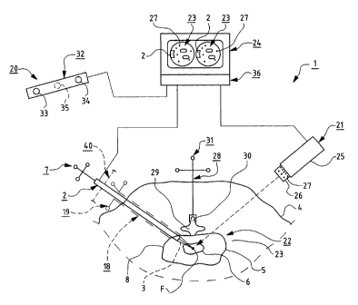
The treatment device 1 schematically illustrated in
fig. 1 is adapted to generate, by means of a therapeutic
ultrasound transducer ~ (so called therapeutic transducer),
an ultrasonic field 3, the temperature focus F of which is
intended to be located in the intervertebral disc 5, pre-
ferably in nucleus pulposus 6, of the patient 4 for treat-
ment thereof. The therapeutic ultrasound transducer 2 comp-
rises a plurality of, preferably three or more position
transmitters 7 for determining its position.
The therapeutic ultrasound transducer 2 is adapted
to be inserted through the patient's 4 skin and engage
the disc 5, preferably anulus fibrosus 8, to provide a
local temperature increase in nucleus pulposus 6 so that
enzymes such as collagenase present in the disc are acti-
vated and cause decomposition of collagen and proteogly-
canes, which results in shrinking of nucleus pulposus 6
primarily because of less hygroscopicity. The therapeu-
tic ultrasound transducer 2 can be placed against the
disc 5 without perforating anulus fibrosus 8 and thereby
transmit the ultrasonic field 3 focused in temperature
focus F towards the treatment volume. The transmitter ele-
ment 9 of the therapeutic ultrasound transducer 2, e.g. a
piezoelectric element, may be cooled with water for coo-
ling the crystal and the tissue closest to the therapeu-
tic ultrasound transducer 2 in a similar way as one today
does in microwave therapy of cancer in the prostate gland
(US 5 964 791).
In order to provide said cooling, the therapeutic
ultrasound transducer 2 is provided at its distal end 10
with at least one cooling chamber 11 with cooling liquid
12. This cooling chamber 11 is located between the trans-
mitter element 9 and a membrane-like wall 13 of such
CA 02415828 2003-O1-14
WO 02/05897 PCT/SE01/01626
5.
flexible material that said wall is able to adapt to the
surface of anulus fibrosus 8 when it is brought in con-
tact therewith.
The therapeutic ultrasound transducer 2 further comp-
s rises at least one temperature sensor 14 for measuring
the temperature before and/or during treatment. In order
to increase the volume of therapy or treatment, the direc-
tion or setting of the therapeutic ultrasound transducer 2
can be varied such that temperature focus F is scanned
over a larger area. The temperature sensor 14 is provi
ded to measure the temperature at the inner side of the
flexible wall 13 and it is preferably connected to said
wall 13 such that it follows the wall 13 when said wall
is deformed when brought in contact with the surface of
anulus fibrosus 8.
The cooling liquid 12 is preferably water which is
distributed through an inlet passage 15 to the cooling
chamber 11 and through an outlet passage 16 therefrom
such that the water can circulate through the cooling
chamber 11. A sealing means 17 is provided within the
transmitter element 9 for preventing cooling liquid 12
from finding its way out of the cooling chamber 11.
In more detail, the therapeutic ultrasound transdu-
cer 2 is adapted to cause a local temperature increase in
nucleus pulposus 6 so that enzymes such as collagenase
present in the disc 5, are activated and cause decomposi-
tion of collagen and proteoglycanes, which results in
shrinking of nucleus pulposus 6 primarily because of less
hygroscopicity.
The treatment device 1 may comprise a rigid tube 18
with associated inner portion and several position trans-
mitters 19, preferably three such transmitters. The tube
18 may, by means of optical navigation technique, be in-
serted dorsolaterally towards the disc 5. The inner por-
tion of the tube 18 is then replaced by the therapeutic
ultrasound transducer 2 and said tube 18 is schematically
illustrated in fig. 1 with broken lines.
CA 02415828 2003-O1-14
WO 02/05897 PCT/SE01/01626
6.
The treatment device 1 also comprises an optical
navigating device 20 to navigate the therapeutic ultra-
sound transducer 2 (US 5 772 594). This optical naviga-
ting device 20 comprises at least one diagnostic camera 21
which is adapted to produce at least one picture or image
of the anatomic structure 23 of the treatment area 22 in
a monitor 24. The diagnostic camera 21 may be an X-ray
camera 25 taking two pictures of the anatomic structure
23 of the treatment area 22 from different directions
with preferably a 90° intermediate angle and showing or
displaying these in the monitor 24. At the optical naviga-
ting device 20, the X-ray camera 25 is used together with
an optical analogue-digital-converter for obtaining or
producing a real time image or picture in the monitor 24
of the position and direction of the therapeutic ultra-
sound transducer 2 (US 6 021 343, US 5 834 759, US 5 383
454).
The X-ray camera 25 comprises a calibrating device
26 - e.g. a calibrating hood - which is located in front
of the objective of the X-ray camera 25 and having mar-
kers 27 the mutual distances of which are known. The mar-
kers 27 may be round and consist e.g. of tantalum.
The optical navigating device 20 further comprises
a reference device 28 which is provided to be attached to
the spinous process 30 of a vertebra 29 or in a correspon-
ding position such that it gets a determined or fixed po-
sition relative to the treatment area 22. The reference
device 28 has several position transmitters 31, namely
preferably at least three, and these may consist of metal-
lic material, e.g. tantalum.
Furthermore, the optical navigating device 20 comp-
rises a signal receiving and/or signal sending unit 32.
This includes a suitable number of signal receivers 33,
34 for receiving reflected or other signals from the posi-
tion transmitters 7 and 31 of the therapeutic ultrasound
transducer 2 and the reference device 28 respectively.
The signal receiving and/or signal sending unit 32 may
CA 02415828 2003-O1-14
WO 02/05897 PCT/SE01/01626
7.
eventually comprise one or more signal transmitters 35
for sending or transmitting signals to said position
transmitters 7 and 31, which are provided to receive
these signals.
The signals transmitted by the position transmitters
7 and 31 may e.g. be in the form of infrared light and
the signal receivers 33, 34 may in such case be receivers
of infrared light.
In the treatment device 1 there may also be inclu-
ded a calibrating unit 37 for calibrating the temperature
effect of the temperature focus F of the therapeutic
ultrasound transducer 2. The calibrating unit 37 has one
or more thermoelements 38 by means of which the effect
at said temperature focus F can be measured for calibra
tion. The thermoelements 38 are connected to a schemati
cally illustrated measure instrument 39.
Prior to treatment of the disc 5, preferably nucleus
pulposus 6, the reference device 28 is located on the
patient°s 4 vertebra 29 and the therapeutic ultrasound
transducer 2 is calibrated in the calibrating unit 37.
Two X-ray pictures are taken of the patient°s 4 ana-
tomic structure 23 at the disc 5 and these X-ray pictures
are shown on the monitor 24. On these X-ray pictures, the
position of the reference device 28 relative to the disc 5
may then be determined by means of the markers 27 of the
calibrating device 26.
During treatment of the disc 5, preferably nucleus
pulposus 6, the therapeutic ultrasound transducer 2 is
navigated by means of the signal receiving or signal sen-
ding unit 32, whereby the navigation is presented in the
X-ray pictures or images on the monitor 24. This is ac-
complished while the position transmitters 7 of the thera-
peutic ultrasound transducer 2 cooperate through signals
with the signal transmitters 33, 34 of the signal recei-
ving or signal sending unit 32. By means of said naviga-
tion, the therapeutic ultrasound transducer 2 can be posi-
tioned such that the temperature focus F of its ultrasonic
CA 02415828 2003-O1-14
WO 02/05897 PCT/SE01/01626
8.
field 3 will lie in the disc 5, preferably nucleus pulpo-
sus 6. The temperature in the temperature focus F prefer-
ably exceeds 45°C.
The treatment can be automatically interrupted if
the patient 4 moves to an incorrect position relative to
the therapeutic ultrasound transducer 2 or vice versa.
The invention is not limited to the embodiment de-
scribed above, but may vary within the scope of the fol-
lowing claims. Thus, the treated disc 5 may e.g. be any
disc in the body.
The diagnostic camera 21 may be a computerized tomo-
graphy (CT) scanner which is provided to produce images
of said anatomic structure 23 and these images can be
processed in a computer program or software for obtaining
a 3D-image in the monitor 24.
The therapeutic ultrasound transducer 2 may be provi-
ded to be positioned manually or be located on a positio-
ning device 40 for positioning thereof relative to the
disc 5 to be treated.