Note: Descriptions are shown in the official language in which they were submitted.
CA 02416146 2003-O1-23
WO 02/09584 PCT/USO1/23417
1
ELECTRODE ARRAY AND SENSOR
ATTACHMENT SYSTEM FOR
NONINVASIVE NERVE LOCATION AND IMAGING DEVICE
Cross-Reference to Related Applications
This application is a Continuation-in-Part of and claims priority from United
States
Application Serial No. 09/624,397, filed July 27, 2000.
Technical Field
This invention relates to a medical device for the noninvasive location and
imaging of
peripheral nerves. Specifically, the present invention is a sensor system for
use at the skin
surface comprising an electrode array assembly with one or more electrodes and
a sensor
attachment system. Each electrode in the electrode array assembly maintains a
connection to
peripheral nerve detection and imaging instrumentation. One or more return
wires are
attached to the electrode array assembly and to a skin surface electrode
during use of the
sensor system. A disposable, sterile sensor attachment system allows
conductance between
the electrode array and the skin surface of the subj ect. The sensor
attachment system
contains individual conductor islands, each adapted to align accurately with a
specific
electrode of the electrode array. The layer of the sensor attachment system
that adheres to
the skin surface of the subject may be left on the skin at the end of sampling
to provide a skin
marking guide. This facilitates the positioning of needles for subsequent
nerve stimulation or
therapy.
Background of the Invention
The use of direct current skin surface conductance or resistance measurements
has been
employed in many forms for the identification of peripheral nerves, myofascial
trigger points,
and acupuncture points (Cory, et al., Characterization of cutaneous electrical
hyperconductivity sites associated with myfascial trigger points and taxsal
tunnel syndrome.
CA 02416146 2003-O1-23
WO 02/09584 PCT/USO1/23417
2
In: Abstracts, 8th Woy~ld Coug~ess on Pain (Seattle: IASP Press, 1996), p. 54;
Kaslow AL
and Lowenschuss O. Dragon chasing: a new technique for acupuncture point
finding and
stimulation. Anze~ican Jou~~nal of Acupuntu~e, 3(2):157-160, 1975); Kejariwal,
et al.,
Development of a device for non-invasive nerve location. In: Abstracts, 8th
World Congress
o~ Pain (Seattle: IASP Press, 1996), p.279-280; Kwolc, et al., Mapping
acupunture points
using multi channel device. Aust~alas Phys Eng Sci Med, 21(2):68-72, 1998;
Lykken,
Square-wave analysis of skin impedance. Psychophysiology, 7(2):262-275, 1971.
An early
example of this was the use of a transcutaneous electrical nerve stimulation
(TENS) unit to
identify acupuncture points. When a TENS unit is coupled between examiner and
subject,
the finger of the examiner acts as a sampling electrode (Kaslow, et al.,
Dragon chasing: a
new technique for acupuncture point fording and stimulation. American Journal
of
Acupunture, 3(2):157-160, 1975)). However, the literature in the field
illustrates
inconsistency in locating these sites through electrical conductance
measurements
(Reichmanis et al., Electrical correlates of acupuncture points. IEEE
Ti°ansactions oy~
Bio~aedical Ehgihee~iug, BME 22:533-535, 1975).
U.S. Pat. No. 4,016,870 to Lock describes a system for acupuncture point
location in which a
single, hand-held probe of undisclosed composition is used to determine sites
of high skin
surface conductance. U.S. Pat. No. 5,897,505 to Feinberg, et al., describes a
system for
measuring selective tissue conductance and temperature utilizing chrome-
plated, brass
electrodes in a handheld embodiment. Although each of these systems measures
conductance at the skin surface, they suffer two main drawbaclcs. First,
metallic electrodes
display uneven current densities at the skin surface-electrode interface,
which is largely
dependent on the underlying moisture pattern of the skin. Devices for
measuring skin
surface conductance and resistance that do not employ aqueous interfaces are
particularly
subject to this effect and, in some cases, current densities became high
enough to produce a
painful sensation. Second, handheld devices are subject to uncontrolled
application
pressures. This is complicated in larger diameter electrode systems, such as
that of U.S.
Patent 5,897,505 to Feinberg, where the angle of application causes pressure
to be unequally
distributed on the skin surface. The use of electrical conductance
measurements at the skin
CA 02416146 2003-O1-23
WO 02/09584 PCT/USO1/23417
3
surface to locate nerve tissue is facilitated by the use of aqueous
electrodes, rather than
metallic or dry silver-silver chloride electrodes, a.nd by the use of non-
sinusoidal, alternating
current waveforms. Based upon observations such as these, a device that
locates peripheral
nerves transcutaneously was disclosed in the commonly owned U.S. Pat. No.
5,560,372 to
Cory (the disclosure of which is incorporated herein by reference).
FIG. 9 is a circuit diagram of the non-invasive, peripheral nerve mapping
device according to
the U.S. Pat. No. 5,560,372 to Cory as it is positioned over the forearm of a
patient. The
sampling electrode (10) depicted herein comprises eight electrodes (l0a-h)
having leads (41)
arranged in a linear array and applied to the volar surface of the forearm on
the epidermal
surface (80). The reference electrode (70) is placed on the dorsal forearm. A
constant
current output is applied between the two electrodes (10, 70) on the epidermal
surface (80).
The voltage difference V between the two electrodes is measured and varies
from adjacent
skin sites as the electrical conductance of the skin changes. The reference
electrode (70) may
comprise a conductive carbon impregnated silastic pad provided with an
insulated metal foil
sheet laminated thereto. The metal foil sheet is in electrical contact with a
connector
element. The reference electrode may further contain adhesive layer laminated
to the bottom
of the silastic pad provided with a silicon release sheet attached to the
adhesive layer.
Reference electrode may comprise a carbon-impregnated silastic pad provided
with a layer of
pharmaceutical electrode gel placed on the bottom of the pad to be positioned
against the
skin.
FIG. 10(A) depicts the constant current input (I) for each sub-electrode (10a
through 10h),
numbers 1-8 respectively, as shown in FIG. 9. FIG. 10(B) depicts the voltage
output V for
each sub-electrode. With reference to FIG. 9 and FIG. 10(B), electrode number
(10b),
number 2 in FIG. .10(B) is positioned over ulnar nerve (88). As shoran in FIG.
10(B),
electrode (I Ob) indicates the position of the ulnar nerve (88) by a decrease
in output voltage.
Similarly, electrodes (10d) and (10e), numbers 4 and 5 in FIG. 10(B), display
a similar
output voltage decrease as they are positioned over median nerve (84). Thus,
the non-
invasive, peripheral nerve mapping device according to the present invention
accurately
identifies the location of subcutaneous nerves. Voltage minima (conductance
maxima) are
CA 02416146 2003-O1-23
WO 02/09584 PCT/USO1/23417
4
observed over the ulnar and median nerves (88, 84) at constant current. Sites
of decreased
skin voltage differentials are mapped and have been shown by nerve stimulator
technique,
direct dissection and local anesthetic blockage, in animal and human models,
to correspond
to the location of subcutaneous nerves.
The problem of avoiding metallic interfaces with the skin surface is addressed
by the use of
water-saturated felt electrodes in U.S. Pat. No. 5,560,372 to Cory and by the
use of hydrogels
(Jossinet and McAdams, Hydrogel Electrodes in Biosignal Recording. A~hual
Ihte~hatio~al
Confef ence on the IEEE Engineering ih MedicifZe ahd Biology Society,
12(4):1490-1491,
1990). The ability to obtain reproducible skin surface conductance and
resistance readings
allows the recognition of skin surface sites that correspond to underlying
peripheral nerves.
While this approach circumvents the problems of current density disparities,
of the formation
of thin oxidation films on the electrodes, and of subsequent back
electromotive force,
additional problems remain that are associated with the interface between the
sampling
electrodes and the slcin surface.
Summary of the Invention
It is an object of the present invention to provide a sensor system comprising
an electrode
aiTay and a sensor attachment system for use with an electrical field
generating device that
can non-invasively detect peripheral nerves.
It is a further object of the present invention to provide a method for
detecting peripheral
nerves using the aforementioned sensor system.
It is a further object of the present invention to provide for an electrode
array, which is
flexible, reusable, and suitable for use, either alone or in combination with
a sensor
attachment system as herein described.
It is a further object of the present invention to provide for a sensor
attachment system,
comprising conductor islands, which is disposable and suitable for use in
combination with
an electrode array as herein described.
Further objects and advantages of the invention will be apparent from the
following
description of the invention.
CA 02416146 2003-O1-23
WO 02/09584 PCT/USO1/23417
In satisfaction of the foregoing objects and advantages, the present invention
provides an
electrode array comprising:
a sheet of electrically non-conductive material having a sensor electrode
region, an
instrumentation connector region and a flexible stem region mechanically
joining the
electrode sensor region and the instrumentation connector region;
an electrode array having one or more electrodes, which are disposed within
the sensor
electrode region,
a connection lead corresponding to each electrode disposed within the
instrumentation
connector region,
a return lead disposed within the instrumentation connector region,
an electrically conductive connection between each electrode and its
corresponding
connection lead; and
features on the sensor electrode region for alignment with a skin attachment
system, and for
alignment with the image displayed by a nerve location device.
Brief Description of the Drawings
FIG. 1 is a basic depiction of an electrode array of a first embodiment of the
present
invention in a 16-electrode conformation, view from side not facing skin (top
view).
FIG. 2 is a cross-sectional side view of an electrode array of the present
invention in a 16-
electrode conformation.
FIG. 3A shows a sensor attachment system in a 16-electrode conformation, top
view.
FIG. 3B shows a sensor attachment system in a 16-electrode conformation, side
view.
FIG. 4 shows an exemplary electrode array of a second embodiment of the
present invention
in a 64-electrode conformation, top view.
FIG. 5A shows an assembly of an electrode array and a sensor attachment system
according
the present invention, side view.
FIG. 5B shows an assembly of an electrode array and sensor attachment system
of FIG SA,
view from the sensor attachment side.
CA 02416146 2003-O1-23
WO 02/09584 PCT/USO1/23417
6
FIG. 6 shows a side view of an electrode array and sensor attachment system
according to the
present invention attached to an area of slcin.
FIG. 7A-7D illustrate the steps of removing an electrode array from a skin
marking guide
according to the present invention, marking a location on an area of skin
through a hole in the
skin marking guide, and removal of the skin marking guide from the area of
skin.
Fig. $ illustrates the use of the present invention with an electronic
measiu~ement device, such
as a nerve mapping device.
Fig. 9 illustrates a prior art embodiment of an array sensor in a nerve
mapping device.
Figs. 10A and l OB illustrate a prior art operation of an array sensor to
sense various types of
nerves in a nerve mapping device.
Detailed Descr~tion of the Invention
The medical device of the present invention is a sensor system that comprises
two
components. A sensor system of the present invention thus comprises an
electrode array and
a sensor attachment system to attach to the skin. When combined to form the
sensor system,
both the electrode array and the sensor attachment system are presented in the
form of
complementary arrays of electrodes and conductor islands, respectively. The
electrode array
comprises one or more electrodes, advantageously four or more electrodes. The
electrodes
may be arranged randomly, in a single line, or in another fixed order.
Advantageously, the electrodes of the array may be arranged in plural rows.
The adjacent
rows may be in line with one another or offset with respect to their nearest
neighbor(s). A
preferred arrangement is for the array to comprise a minimum of four
electrodes arranged as
two or more rows, where adjacent rows are in line with one another. Another
preferred
arrangement is for the array to comprise a minimum of four electrodes arranged
as two or
more rows of electrodes, where adjacent rows are offset with respect to one
another.
A further preferred arrangement is for there to be a minimum of two rows of
four or more
electrodes each.
A further preferred arrangement is for there to be a minimum of two rows of
eight or more
electrodes each.
CA 02416146 2003-O1-23
WO 02/09584 PCT/USO1/23417
7
Another fiu-ther preferred arrangement is for there to be a minimum of eight
rows of eight
electrodes each.
An exemplary embodiment according to the present invention is a two row array,
as depicted
in FIG. 1, where an offset arrangement of adjacent rows is used. Another
exemplary
embodiment according to the present invention is an eight row arrangement, as
depicted in
FIG. 4, where adjacent rows are in line with one another.
By the foregoing, it should be apparent that any number of conformations is
possible with
this invention. The important consideration in constructing an electrode array
assembly of
the present invention is that the electrodes of the electrode array line up
with the conductor
islands of the sensor attachment system so that they can operate together as
the herein
described sensor system.
An electrode array assembly of the present invention may advantageously be
made flexible
so that the electrode array assembly may conform to a wide variety of body
surfaces,
locations, and circumferences. To achieve this flexibility, the electrode
array should
comprise as a support structure a flexible, electrically non-conductive sheet.
Also, it is useful
to employ very thin, narrow, electrically conductive paste or adhesive as an
electrical
connection between the electrodes in the electrode array region and the leads
in the electrical
connector region of the electrode array. The electrical connections may also
comprise
suitably applied metallic inlc, metallic wire, or conductive trace.
The electrode array of the present invention may be reused, a feature which is
particularly
achieved when the electrode array is used with a separate sensor attachment
system of the
present invention.
The electrode surface should be chemically, as well as biologically, inert. In
other words, the
electrode surface should not chemically react with, or be degraded by,
surfaces which it will
contact during normal use. To obtain reproducible measurements, the formation
of thin,
oxidation films on the electrode surface must be avoided. At the same time,
the electrode
array must be resistant to damage by bending and twisting. The electrode array
must also be
stable when cleaned with common sterilizing agents, such as isopropyl alcohol.
The
electrode array must also be stable upon sterilization by ethylene oxide,
gamma radiation, or
CA 02416146 2003-O1-23
WO 02/09584 PCT/USO1/23417
8
autoclaving. Suitable materials for the electrode surfaces include gold, gold-
plated copper,
nickel-plated metal, platinum, palladium, silver-silver chloride or another
conductive metal,
metal salt combination or conductive polymers) such as polyaniline.
In preparing the electrode array assemblies of the present invention, thought
must be given to
the reduction of sampling eiTOr. The present inventors have performed
experiments to
determine the size parameters that minimize sampling error. The present
invention may be
advantageously practiced by constructing electrode arrays in which the
electrode diameters
are in the range of about 2-3 mm, with edge-to-edge spacing of about 3 mm.
Smaller
electrode diameters and closer electrode spacing may result in excessive
variation between
sample readings. It is believed that, at smaller electrode diameters,
conductor resistance
increases due to decreasing cross-sectional area in relation to the electrical
field path, which
may introduce variation in sample readings. However, where such variations are
tolerable,
smaller electrode diameters may be used. Of course, larger electrode diameters
and spacings
may be advantageously employed and are contemplated as being within the scope
of the
present invention, although smaller diameters are generally preferred due to
their generally
more favorable resolution characteristics. The ordinary artisan will
appreciate that a wide
variety of electrode diameters and spacings may be used and are contemplated
as being
within the scope of the present invention.
The sensor attachment system of the present invention provides an interface
between the
electrode array and the slcin surface of a living being, preferably a mammal,
more preferably
a human patient. The sensor attachment system comprises a plurality of layers.
One layer,
hereinafter the support layer, contains a plurality of conductor-islands
arranged in a support.
The conductor-islands are formed from a suitable conductive material such as a
hydrogel, or
silver-silver chloride gel. Another layer, hereinafter the skin marlcing guide
layer, is
fenestrated (i.e. has holes) so that the conductor islands protrude through
the holes therein.
The support layer and the skin marking guide layer are held together by
awadhesive that is
easily brolcen, so that after imaging the peripheral nerves, the practitioner
may then peel the
support layer away from the slcin marlcing guide layer, leaving the latter
attached to the skin.
CA 02416146 2003-O1-23
WO 02/09584 PCT/USO1/23417
9
The hydrogel or silver-silver chloride gel may be any electrically conductive
hydrogel or
silver-silver chloride gel known in the medicinal, biofeedback or biological
testing ants.
The hydrogel polymers are desirably water interactive andlor hydrophilic in
nature and are of
a molecular weight or structure, or have been modified such that they absorb
significant
quantities of water and may form hydrogels when placed in contact with water
or aqueous
media for a period of time sufficient to reach equilibrium with water, but
which do not fully
dissolve at equilibrium in water.
The hydrogel polymers include polymers from the group of homo- and copolymers
based on
various combinations of the following vinyl monomers: acrylic and methacrylic
acids,
acrylamide, methacrylamide, hydroxyethylacrylate ormethacrylate,
vinylpyrrolidones, as
well as polyvinyalcohol and its co- and terpolymers, polyvinylacetate, its co-
and terpolymers
with the above listed monomers and 2-acrylamido-2-methyl-propanesulfonic acid
(AMPS~)
and sulfonated styrenes. Very useful are copolymers of the above listed
monomers with
copolymerizable functional monomers such as acryl or methacryl amide acrylate
or
methacrylate esters where the ester groups are derived from straight or
branched chain alkyl,
aryl having up to four aromatic rings which may contain alkyl substituents of
1 to 6 carbons.
Most preferably the hydrogels of the invention should be composed of synthetic
copolymers
from the group of acrylic and methacrylic acids, acrylamide, methacrylamide,
hydroxyethylacrylate (HEA) or methacrylate (HEMA), vinylpyrrolidones, and
polyacrylonitriles. Specific illustrative examples of useful polymers are the
following types
of polymers: hydroxyethyl methacrylate, crosslinlced polyvinyl alcohol,
crosslinlced N-
vinylpyrrolidone/acrylic acid copolymers, crosslinlced poly(N-vinyl lactam),
crosslinked
polyacrylamide, crosslinked polyacrylic acid, and crosslinlced poly(2-
acrylamide-2-
methylpropane) sulfonic acid, or "Procam" or Hydrogel A11926, tradenames of
Ludlow
Technical Products hydrogel material.
The foam used in the support layer may be any foam known in the art for such
applications.
The foam support layer should be flexible so as to conform to the surface to
which it is
applied. Any type of foam layer may be used but preferred foams are closed
cell foams such
as polyethylene. Closed cell foams are foams which have generally spherical
discrete pores
CA 02416146 2003-O1-23
WO 02/09584 PCT/USO1/23417
which are not connected. However, equivalent support layers may be used as
lcnown in the
medical arts.
The skin marking guide layer may be made of any polymeric material lcnown in
the medical
arts. Particularly advantageous are those polymeric materials that are clear
or translucent.
Polyurethane, polypropylene, polyvinyl chloride, and copolymers thereof, all
of which are
known in the art, are preferred. The polymeric materials may be colored in
order to enhance
their visibility against skin. Particularly preferred are bright colors that
offer enhanced
contrast on any colored slcin. Color such as blue and white are particularly
preferred for the
skin marking guide, however other colors, such a fluorescent yellow, orange,
green and
magenta may also be used.
The nerve imaging instrument to be employed is not critical to the present
invention and may
be any suitable instrument known in the art, such as the multiplexed system
disclosed in
commonly owned U.S. Pat. No. 5,560,372 to Cory, incorporated herein by
reference.
The following non-limiting advantages may be realized by practicing this
invention:
1. Sterility. The new sensor attaclunent system directly contacts the skin of
the subject
and should be a sterile, disposable, adhesive patch. The electrode array, to
which the sensor
attachment system operatively attaches to the slcin, may extend about six
inches away from
the skin of the subject, may be disposable or may be reusable and cleansed
with isopropyl
alcohol or sterilized under ethylene oxide, gamma radiation, or autoclaving.
2. Slcin marking. Once samples have been taken with the sensor, all but the
bottom
(skin marking guide) layer of the sensor attachment system may be removed from
the slcin of
the subject. This bottom layer is fenestrated, with holes that correspond to
the location of the
electrodes in the electrode array, and provides a slcin marlcing guide. Slcin
can be marked
through this skin marking guide to facilitate subsequent injections) at the
sites) chosen by
the practitioner based upon the readings obtained, or the skin marking guide
may be left in
place on the skin to provide a convenient template for guiding a nerve
stimulation needle or
other needle.
3. Pressure applied to electrodes. The electrode array and sensor attachment
system,
joined together, are placed on the skin surface before sampling. The joined
devices are held
CA 02416146 2003-O1-23
WO 02/09584 PCT/USO1/23417
11
on the slcin by virtue of an adhesive on the sensor attachment system. This
arrangement was
designed in part to circumvent the possibility that unequal pressures applied
to each of the
electrodes by the practitioner would interfere with the readings obtained at
sampling.
4. Motion artifacts. Stable adherence of the sensor attachment system to the
skin of the
subject and to the electrode array decreases the possibility of motion
artifacts.
5. Quality of image. The number of electrodes in the electrode array assembly
may be
increased in order to improve the resolution possible with the device.
6. Flexibility. The sensor attaclunent system and electrode array may be
manufactured
with different numbers of electrodes in different arrangements tb address
multiple uses and
user preferences.
7. Imaging of a two-dimensional area. To image a two-dimensional area, as
required for
neurodiagnostic applications of the device, the sensor attachment system and
electrode array
may be manufactured in a two-dimensional rather than a linear format (for
example, an 8x8
array). This circumvents the need to move the device along a line on the skin
surface, which
is cumbersome for the operator and subject to inaccuracy.
8. Parts replacement. The sensor attachment system is disposable after each
use, but is
designed as a sterile part that is inexpensive to produce. The electrode
arrays may be
designed as reusable parts, but would be subject to wear and tear during use
and sterilization.
This invention has the practical advantage of separating the electrode array
from the
reminder of the device so that the electrode arrays, if nondisposable, may
easily be replaced
at minimal cost.
9. Operator's hands. The new invention offers an important practical advantage
in
freeing up the hands ~of the practitioner while samples are taken and
displayed.
10. Acceptability in practice. The new invention significantly decreases the
steps
required to sample the skin surface, reducing the time required for nerve
localization.
11. Size. The bulls of the device that is in proximity to the subject has been
reduced
significantly by this invention, facilitating use of the device and acceptance
by the subject.
CA 02416146 2003-O1-23
WO 02/09584 PCT/USO1/23417
12
12. Placement of the return electrode(s). The attachment of the return
electrode wires)
of limited length to the electrode array minimizes errors in the placement of
the return
electrode(s).
Further uses, benefits and features of the present invention will be seen from
a review of the
detailed description of the preferred embodiments in conjunction with the
various fgures of
the drawings.
Preferred Embodiments
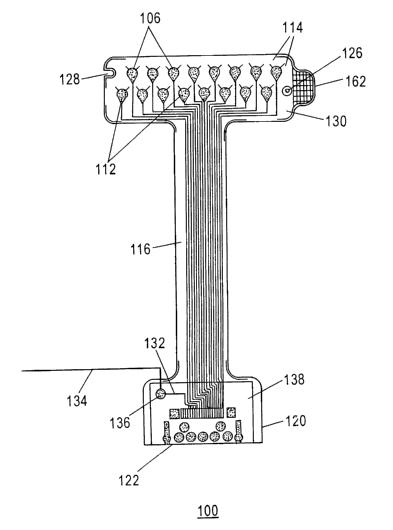
An exemplary electrode array 100 according to the present invention is
depicted in Figure 1.
The electrode atTay 100 has 16 electrodes 106, arranged in two rows, which are
offset with
respect to one another. Another electrode array 400 is depicted in Figure 4.
Electrode array
400 has 64 electrodes 106, arranged in eight rows and eight columns. Surfaces
(not shown)
of electrodes 106 are exposed through a non-conductive sheet (not shown)
facing the skin
surface (view not shown). The nonconductive sheet is advantageously a
polyimide, however
the composition of the nonconductive sheet need not be limited to this
material. Other
suitable nonconductive sheet materials include polycarbonates, polyurethanes,
polyvinylchlorides, polybutylenes, vinyl, silastic, and polyethylene.
The elects odes 106 are advantageously fabricated using a subtraction
technique for
production of printed circuit boards. An image of electrodes and traces is
first developed on
a copper-plated KAPTON° brand (duPont) polyimide polymer sheet. A
photoresist layer is
applied over the image. After exposure to ultraviolet radiation, the copper
surrounding the
photoresist protected regions is etched away with a ferric chloride solution.
The photoresist
is removed with an organic solvent, such as acetone. Following masking of the
copper
traces, nickel is electroplated onto the copper electrode pads. Gold is then
electroplated onto
the nickel electroplate. A final KAPTON° polyimide layer is laminated
over the traces.
Following soldering of the connector and integrated circuit elements, the
electrode assembly
is complete.
To ensure non-reactivity with a sensor attachment system 300 (Figs. 3A, 3B) or
with a skin
layer itself, the exposed surfaces of electrodes 106 are plated with gold in
some embodiments
CA 02416146 2003-O1-23
WO 02/09584 PCT/USO1/23417
13
according to the present invention. However, other conductive materials that
do not readily
react with skin are used in other embodiments according to the present
invention. Such
conductive materials include suitable metals, metal alloys, metal-metal salt
combinations,
a.nd conductive polymers. Between the gold surfaces of electrodes 106 and
underlying metal
paste traces 112 is an interposed layer of nickel (not shown) to ensure
adequate plating of the
gold. Other interposed metal layers, e.g. tin, may be used. Opposite the gold-
plated surface
of the electrode the metal paste traces 114 in a Y-configuration provide
stability and strength
for the electrodes 106. The metal paste traces 112 extend from each electrode
106, through a
stem 116 of the electrode array 100, 400, to attach to the instrumentation
connector 122. The
width and thicl~ness of the metal paste traces 112 vary from 5-15 mil
depending on the
number of electrodes 106 in the array. In some embodiments according to the
present
invention, the metal paste traces 112 may be substituted with metallic inks,
metallic wires, or
conductive polymers.
Figure 4 illustrates a second embodiment of the present invention in which
integrated circuit
elements are mounted on the instrumentation connector portion 120. The
electrode array 400
in Figure 4 is illustrated as 64 electrode two dimensional array, although any
number of
electrodes may be included in the array. The electrode array in Figure 4 is
operated by
integrated circuit elements consisting of shift registers 470 and multiplexers
472 mounted on
the instrumentation connector portion 120 of the electrode array 400. In the
embodiment
depicted in Figure 4, there is one shift register 470 and four multiplexers
472, however other
configurations are possible and are contemplated as being within the general
scope of the
present invention. Moreover, although the embodiment illustrates shift
registers 470 and
multiplexers 472 mounted separately from each other, those of skill in the art
will appreciate
that they may fabricated on the same integrated circuit chip. Those of skill
in the art will also
appreciate that additional circuitry, such as a control device, may also
preferably be included
with the shift registers 470 and multiplexers 472.
The shift registers 470 and mutiplexers 472 preferably cause individual
electrode elements in
the array to provide a sensing signal, such a voltage, in a time division
manner according to a
predetermined cycling frequency, as understood by those of skill in the art.
Moreover, those
CA 02416146 2003-O1-23
WO 02/09584 PCT/USO1/23417
14
of skill in the art will appreciate that the shift registers 470 and
multiplexers 472 may be
configured to sample the sensing signal from one electrode or from a plurality
of electrodes
at any given sampling time period. The sensing signals provided from the
electrodes may
then be provided to an analyzing device, such as a nerve location device,
through
instrumentation connector 122. Instrumentation connector 122 may be a plug
type
connector, or any other known type of electrical connector.
Figure 8 illustrates the electrode array 100 joined with the sensor attachment
system 300 and
mounted on the skin of a subject 700. As illustrated in Figure 8, the
electrode array is
connected to an analyzing device 500 via instrumentation connector 122. The
analyzing
device 500 preferably drives a display device 600 to display the results of
the analysis
performed by the analyzing device 500. In the preferred embodiment, the
analyzing device
500 may be a nerve location device, which determines the location of nerves
based on the
signals received from the electrode array 100, as described in commonly owned
U.S. Patent
No. 5,560,372 to Cory. However, the analyzing device 500 is not limited to a
nerve location
device, and may be any device configured to receive electrical sensing signals
from a test
organism. The analyzing device 500 and the display device 600 may be
collectively referred
to as a medical imaging instrument.
The electrode arrays 100, 400 are wide at both the electrode sensor region 130
and the
instrumentation connector region 120. Between the instrumentation connector
region 120
and the electrode sensor region 130, the stem region 116 is narrow to promote
flexibility and
convenience of use. The electrode sensor region 130 of the electrode array 100
preferably
contains a registration hole 126 and a registration notch 128. These design
characteristics
allow for accurate positioning with the sensor attachment system 300. A tab
162 is on one
side of the electrode sensor portion 130 of the electrode array 100 for ease
of removal of the
electrode array 100 from a slcin marlcing guide 308 (Fig. 3B) after sampling
is complete. In
some embodiments according to the present invention, the electrode arrays 100,
400 have
one registration hole 126 and registration notch 128. In other embodiments
according to the
present invention, the number and position of the registration notches 128 and
registration
holes 126 vary, depending upon the dimensions of the sensor portion 130 of the
electrode
CA 02416146 2003-O1-23
WO 02/09584 PCT/USO1/23417
array 100, 400. Some larger two-dimensional electrode arrays employ additional
registration
elements. Others require no registration notches 128 or registration holes
126.
The instrumentation comzector portion 120 of the electrode array 100, 400
preferably has a
plastic rigidizer 138 (which may made of any suitable material other than
plastic) positioned
on the side of the electrode array opposite the exposed electrodes. The
rigidizer 138 provides
additional support for the instrumentation connector 122 and any attached
integrated circuit
elements such as shift registers 470 and multiplexers 472. A return lead 134
comlects via a
soldered union 136 to a metal trace 132 running to the instrumentation
connector 122. The
instrumentation connector portion 120 of the electrode array 100, 400 is
encapsulated in
molded medicinal grade silicone polymer or polyethylene. In soW a embodiments
according
to the present invention, metal paste material is applied at points of
curvature and stress on
the electrode array 100, 400 to provide additional shear-resistance and
prolong the useable
life span of the electrode array.
Figure 2 depicts a side cross-sectional view of the electrical components of
the embodiment
depicted in Figure 1. Within the instrumentation connector region 120 is
instrumentation
connector 122, which connects to metal paste traces 112. The return electrode
wires) 134
are connected to instrumentation connector 122 through metal paste trace 132
at soldered
union 136. The metal paste traces 112 comlect to the electrode sensor region
130.
The electrode array of the invention is preferably used in combination with a
skin sensor
attachment system as described herein. However, the electrode array can also
be used
independently of the skin sensor attachment system, for example, as a
diagnostic device to
screen peripheral nerves for abnormalities.
Sensor attachment systems. An embodiment of a sensor attachment system 300 for
use with
an electrode array 100 as depicted in Figure 1 is depicted in Figures 3A and
3B. Sensor
attachment system 300 is shaped to conform exactly to a particular electrode
array
configuration such as electrode array 100 in Figure 1. A suitable sensor
attachment system
300 according to the present invention consists of seven layers:
CA 02416146 2003-O1-23
WO 02/09584 PCT/USO1/23417
16
1. Top cover 302 of the sensor attachment system 300 is preferably composed of
polyethylene, polystyrene, polyvinylchloride, polybutylene, polyurethane or
other material
and provides protection for the underlying materials.
2. A top adhesive layer 304 allows solid connection of the sensor attachment
system 300
with the electrode array 100. In the preferred embodiment, the top adhesive
layer 304 does
not extend over the conductor islands 314.
3. Beneath the top adhesive layer 304 is a support layer 306 comprised of, but
not
limited in composition to, closed-cell foam. The thickness of the support
layer 306 may be
vaxied depending on the application intended.
4. Between the support layer 306 and a skin marlcing guide 308 is an
intermediate
adhesive layer 310, which joins the support layer 306 and the skin marlcing
guide 308.
5. The skin maxlcing guide 308 is formed of a material such as 4 mil
polyethylene, which
is preferably colored so as to be easily visible on all skin types (e.g, blue
or white).
6. A bottom adhesive layer 316 allows the skin marking guide 308, and thus the
entire
sensor attachment system with the electrode array 100 on top of it, to adhere
to the skin of
the subject. The skin marlcing guide 308 allows the skin to be marlced at
sites) of interest
before its removal.
7. Bottom cover 312 of the sensor attachment system 300 is preferably composed
of
polyethylene, polystyrene, polyvinylchloride, polybutylene, polyurethane or
other material
and provides protection for the underlying materials.
Holes 314 are formed through all layers of the sensor attachment system 300
except for the
top cover 302 and the bottom cover 312. The holes 314 are filled with a
conductive material
318 comprising, but not limited in composition to, an organic hydrogel.
Registration
elements are preferably positioned on the sensor attachment system to provide
for accurate
placement of the electrode array 100 on the sensor attachment system 300 and
to indicate
orientation on the nerve location device display. Tabs 320, aligned with the
electrode array
tab 162 of electrode array 100, are found on the support layer 306 and the
skin marking guide
308. The tabs 320 aid in removal of the support layer 306 and skin marlcing
guide 308. In
some embodiments, the sensor attachment system 300 may be packaged in a rigid
container
CA 02416146 2003-O1-23
WO 02/09584 PCT/USO1/23417
17
or aluminized pouch (not shown) which is sealed in an airtight fashion. The
sensor
attachment system 300, in its container, is preferably capable of withstanding
sterilization by
gamma-irradiation. In its sealed container, the sensor attachment system 300
preferably has
a shelf life of approximately I 8 months.
A combination of sensor attaclnnent system 300 and electrode array 100 is
shown in Figures
SA and SB. In Figure SA sensor attachment system 300 is applied to electrode
sensor region
130 of electrode array 100. In Figure SB, the electrode array 100 and sensor
attaclnnent
system 300 are seen from the side facing the skin during attachment. The
sensor attaclunent
system 300 is seen after bottom cover 312 has been peeled off. Visible are
registration notch
128, registration hole 126, tab 162, conductor islands 314 and adhesive layer
316, which
covers skin marking guide 308. Return lead 134 connects to instrumentation
connector
region 120, which has an instrumentation connector I22 for connection to an
appropriate
instrument.
An electrode array and sensor attachment system according to the present
invention, when
connected to an appropriate nerve location device, may be used to identify
peripheral nerves,
neuromas, myofascial trigger points, nerve entrapments, and acupuncture
points. To use the
invention, one preferably attaches the sensor attachment system 300 to an
appropriately
configured electrode array 100, which is then connected to the nerve location
device. When
used for sensing nerves, the electrode array may preferably operate in the
same manner as the
electrode array described in commonly owned U.S. Pat. No. 5,560,372.
Particularly, with
reference to Fig. 10(A), an input current may be applied to each electrode,
then, as shown in
Fig. 10(B) an output voltage is sensed from each electrode. The variations in
the output
voltage received indicate the underlying tissue electrical conductance.
Figure 6 depicts an embodiment according to the present invention, wherein the
sensor
attachment system 300 is then attached to skin 602. Electrode array 100 has
been attached to
the sensor attachment system 300, from which top layer 302 and bottom layer
312 have been
removed.
A method of using the electrode array 100 and sensor attachment system 300
according to
the present invention is depicted in Figures 7A-7D. In Figure 7A, an electrode
array 100 is
CA 02416146 2003-O1-23
WO 02/09584 PCT/USO1/23417
18
shown as it is attached to skin 602 through a sensor attachment system (not
shown). Visible
in this view is the electrode sensor region 130 of the electrode array 100,
which comprises
electrodes 106, metal paste traces 112 and 114, registration notch 128,
registration hole 126
and tab 162. The electrode sensor region 130 is attached to the
instrumentation connector
region (not shown) through stem 116.
Figure 7B depicts removal of electrode array 100 from skin marking guide 308.
The support
layer 306 goes with electrode array 100 as it is peeled back from the skin
marking guide 308
via tab 162.
Figure 7C depicts marking of slcin 602 through a hole 702 in skin marking
guide 308 with a
pen 710.
Figure 7D depicts peeling away of skin marking guide 308 from skin 602 to
reveal marls 712.
Steps for carrying out a method of using an electrode array and sensor
attachment system
according to the present invention include the following.
1. Connect the electrode array 100 to an instrumentation connector (not shown)
of a
nerve location device.
2. Remove the top cover 302 from the electrode array side of the sensor
attachment
system 300.
3. Align the registration features of sensor attachment system 300 with the
registration
notch 128 and the registration hole 126 of electrode array 100, position and
securely attach
the sensor attachment system 300 to the electrode array 100.
4. Remove bottom cover 312 from skin surface side of the sensor attaclnnent
system
300.
5. Attach the slcin marking guide 308, now on the combined electrode array and
sensor
attachment system, to intact skin of a suitable subject. The skin is
advantageously prepared
by stripping 3-5 times with adhesive tape.
6. Attach the return electrodes) (for example, a standard ECG electrode) (not
shown) to
the skin of subject within 10-20 cm of the electrode array assembly 100.
7. Attach the return electrode wires) 134, for instance with an alligator
clip, to the
return electrodes) of the instrument (not shown).
CA 02416146 2003-O1-23
WO 02/09584 PCT/USO1/23417
19
8. Obtain samples with the nerve location instrument (not shown).
9. Once skin surface has been sampled with the nerve location device, there
are two
options:
a. Using the tab 162 on the electrode array 100 and the tabs) 320 on the
sensor
attachment system 300, remove the entire electrode array 100 and the sensor
attachment
system 300 from skin surface, or
b. Using the tabs 320 on the sensor attachment system 300 as an aid, remove
all
but the skin yarlcing guide 308 from the skin surface. At this point, one may
mark the skin
through the skin marking guide 308 at the points) of interest determined by
the nerve
location device. Once the skin surface has been marked, the skin marking guide
308 is
removed and the skin surface prepared for positioning of a nerve stimulation
needle and/or a
needle for therapeutic injection (e.g., regional anesthesia or pain relief).
10. All portions of the sensor attachment system 300 are discarded.
11. The electrode array 100 is discarded, if in a disposable embodiment, or if
in a
reusable embodiment, is cleansed with isopropyl alcohol or, if desired, may be
sterilized
under ethylene oxide, gamma radiation, or autoclaving. The latter method may
decrease the
longevity of the electrode array.
While the foregoing preferred embodiments serve to illustrate the present
invention and the
best mode of operation thereof, other suitable embodiments, arrangements and
uses may be
envisioned by the ordinary artisan and as such are contemplated as being
within the scope of
the herein described invention.