Note: Descriptions are shown in the official language in which they were submitted.
CA 02417134 2003-O1-23
WO 02/08449 PCT/CA01/01066
1
HO-1 SUPPRESSOR AS A DIAGNOSTIC AND PROGNOSTIC
TEST FOR DEMENTING DISEASES
FIEhD OF THE INVENTION
Applicant's related US Patent No. 6,210,895 (April 3,
2001), herein incorporated by reference, relates to a method
for predicting, diagnosing, andlor prognosticating dementing
diseases such as Alzheimer Disease (AD) and Age-Associated
Cognitive Decline (AACD). The invention relates to improved
methods for predicting, diagnosing, prognosticating and/or
treating dementing diseases such as Alzheimer Disease (AD) and
Age-Associated Cognitive Decline (AACD) or Mild Cognitive
Impairment (MCI)~ as well as methods and reagents to facilitate
the study of the cause and progression of these diseases.
BACKGROUND OF THE INVENTION
Alzheimer Disease (AD) is a neurodegenerative disease
which causes dementia. The terms "Alzheimer Disease" and
"Alzheimer's Disease" are both utilized in the art, these terms
being equivalent and are used interchangeably here and
elsewhere. The period from first detection of AD to
termination can range from a few years to 15 years, during
which time the patient progressively suffers loss of both
mental function and control of bodily functions. There is
significant variability in the progress of the disease. While
the majority of patients have a gradual, inexorable progression
(losing on average 3 to 4 points~on the 30 point Folstein
mini-mental state score annually), approximately 300 of AD
cases have a prolonged stable initial plateau phase lasting
several years, as described in Haxby et a1. (1992), herein
incorporated by reference. A subgroup of patients has a
fulminant, rapidly progressive downhill course over several
years, as described in Mann et al. (1992), herein incorporated
by reference. Other patients (about 100 of cohorts) remain
slowly progressive, showing only gradual decline from year to
year, as described in Grossi et a1. (1988), herein incorporated
by reference. The pathological, chemical and molecular bases
of this heterogeneity remain undetermined. Recognition of the
CA 02417134 2003-O1-23
WO 02/08449 PCT/CA01/01066
2
variability of AD progression represents an important clinical
insight, and may explain the diagnostic difficulties presented
by "atypical" cases.
Attempts at predicting the onset of AD or monitoring
its progression have met with limited success. ~nlhile in
certain cases, there is a familial manifestation of the
disease, it.appears that the majority of AD cases are
non-familial, and until recently (see below), no simple genetic
marker for the disease had been determined. Much research has
focused on the protein beta-amyloid, deposits of which are
found in the brains of AD victims.
However, recently, as described in our related US
Patent (No. 6,210,895 April 3, 2001) and publication (Schipper
et al., 2000), both herein incorporated by reference, we have
devised a diagnostic method, useful in the prediction,
diagnosis, and prognostication of AD, AACD/MCI, and related
neurological diseases. This diagnostic method is based on the
determination that patients suffering from AD have a
significantly lower concentration of heme oxygenase-1 (HO-1) in
their lymphocytes and plasma, and, accordingly, a significantly
lower concentration of ribonucleotide sequences encoding H0-1
in their lymphocytes.
HO-1: Heme oxygenase-1 (HO-1) is an enzyme that
catalyses the rapid degradation of heme to biliverdin in brain
and other tissues. This 32 kDa member of the heat shock
protein superfamily contains a heat-shock element in its
promoter region and is rapidly up regulated in response to
oxidative stress, metal ions, amino acid analogues, sulfhydryl
agents, and hyperthermia. In response to oxidative stress,
induction of HO-1 may result in protection of cells by
catabolizing pro-oxidant metalloporphyrins, such as heme, into
bile pigments (biliverdin, bilirubin), with free radical
scavenging capabilities. Heme and other intracellular ferrous
iron chelates are capable of converting hydrogen peroxide to
the highly cytotoxic hydroxyl radical.
CA 02417134 2003-O1-23
WO 02/08449 PCT/CA01/01066
3
Using immunostaining techniques in conjunction with
laser scanning confocal microscopy, intense HO-1
immunoreactivity in neurons and astrocytes of post-mortem
hippocampus and temporal cortex derived from AD subjects has
been observed, whereas neural HO-1 staining was faint or non-
existent in the hippocampus and temporal cortex of control
specimens matched for age and post-mortem interval, as noted in
Schipper et al. (1995), herein incorporated by reference.
Furthermore, consistent co-localization of HO-1 to
neurofibrillary tangles and senile plaques in the AD specimens
has been demonstrated. Finally, robust 32 kDa bands
corresponding to HO-1 were observed by Western blotting of
protein extracts derived from AD temporal cortex and
hippocampus after SDS-PAGE, whereas control HO-1 bands were
faint or absent. These results indicate that HO-1 is
significantly over-expressed in neurons and astrocytes of AD
hippocampus and cerebral cortex relative to control brains and
support the contention that AD-affected tissues are
experiencing chronic oxidative stress.
AACD/MCI: AACD and MCI are terms used to identify
individuals who experience a cognitive decline that falls short
of dementia. These terms are equivalent, MCI being a more
recently adopted term, and are used interchangeably throughout
this application. Satisfaction of criteria (World Health
Organization) for this diagnosis requires a report by the
individual or family of a decline in cognitive function, which
is gradual, and present at least 6 months. There may be
difficulties across any cognitive domains (although memory is
impaired in the vast majority of cases), and these must be
supported by abnormal performance on quantitative cognitive
assessments for which age and education norms are available for
relatively healthy individuals (ie., the patient is compared to
normal subjects his/her own age). Performance must be at least
1 SD below the mean value for the appropriate population on
such tests. Neither dementia, nor significant depression or
drug effects may be present. No cerebral or systemic disease
or condition known to cause cerebral cognitive dysfunction may
be present. In Applicant's experience, all patients who were
CA 02417134 2003-O1-23
WO 02/08449 PCT/CA01/01066
4
classified as CDR.5 ("questionable dementia") on the Clinical
Dementia rating scale and who met these exclusions, also met
the criteria for AACD/MCT. About 1/3 of Alzheimer's patients
have had a clearly definable period of isolated memory deficit
which preceded their more global cognitive decline, as noted by
Haxby et a1. (1992), herein incorporated by reference. Using
AACD/MCI criteria which look at other domains in addition to
memory, the percentage with an identifiable prodrome is likely
higher. Fortunately, not all AACD/MCI individuals seem to
decline. It appears that a significant number of these
subjects show a stable, non-progressive memory deficit on
testing.
Related Disorders: Determining HO-1 concentration can
also assist. in predicting, diagnosing, or prognosticating other
dementing diseases having similar manifestations and/or in
distinguishing such diseases from AD. Such other diseases
include Parkinson disease with dementia, Progressive
Supranuclear Palsy, Vascular (i.e. multi-infarct) Dementia,
Lewy Body Dementia, Huntington's Disease, Down's syndrome,
normal pressure hydrocephalus, corticobasal ganglionic
degeneration, multisystem atrophy, head trauma, neurosyphilis,
Creutzfeld-Jacob disease and other prion diseases, HIV and
other encephalitides, and metabolic disorders such as
hypothyroidism and vitamin B12 deficiency. The method may also
prove useful in differentiating the "pseudodementia" of
depression from Alzheimer disease.
The determination of a relationship between HO-1
levels and AD represents a very significant advance in this
field, and may be utilized for the development of methods of
predicting, diagnosing in its very early stage, and
prognosticating AD and other dementing diseases. However,
identification of the factors) and mechanisms) which control
HO-1 expression in the normal versus the diseased state are
needed, to provide even earlier diagnosis, as well as
therapeutic methods and reagents or substances, and methods and
reagents for the study of AD and other dementing diseases. In
addition, the reduction or absence of HO-1 in patients
suffering from AD represents a negative test, and, particularly
CA 02417134 2003-O1-23
WO 02/08449 PCT/CA01/01066
for the purposes of diagnosis, it would be more desirable to
have a positive indicator of disease, i.e. a factor whose
presence (rather than absence) correlates with disease.
Further, the decrease in HO-1 expression may represent an
5 effect, rather than a cause of AD and other dementing diseases,
therefore the identification of factors) and mechanisms)
which control HO-1 expression in the normal versus the diseased
state are also needed to identify components and events which
have an active causative role in the onset and progression of
these diseases.
SUN~SARY OF THE INVENTION
It is a goal of the present invention to provide
improved methods for predicting, diagnosing, prognosticating
and/or treating AD and other dementing diseases, as well as
methods and reagents to facilitate the study of the cause and
progression of these diseases.
Advantageously, embodiments of this invention provide
an easily administered blood or cerebrospinal fluid test which
is used to predict, diagnose, or prognosticate AD and other
dementing diseases.
One aspect of the present invention is a heme
oxygenase-1 suppressor (HOS) factor, wherein said factor
attenuates the increase in the level of heme oxygenase-1
(HO-1). In an embodiment, such an increase occurs in response
to exposure to an experimental agent or treatment which is
capable of increasing the level of HO-1. For example, such
experimental agents or treatments comprise exposure to any one
or more of oxidative stress, metal ions, amino acid analogues,
sulfhydryl agents (e. g., cysteamine, homocysteine),
interleukin-1(3, tumour necrosis factor-a (TNF-a) and
hyperthermia.
Another aspect of the present invention is a method
for assessing dementing diseases in a patient which comprises:
determining the level of heme oxygenase-1 suppressor (HOS)
factor or activity, in tissue or a body fluid obtained from a
patient; and comparing said level of HOS factor or activity
CA 02417134 2003-O1-23
WO 02/08449 PCT/CA01/01066
6
with the corresponding level of HOS factor or activity in
corresponding tissue or body fluid obtained from at least one
control person, whereby if said level of HOS factor or activity
is greater than said corresponding level of HOS factor or
activity in said tissue or body fluid obtained from at least
one control person then said patient suffers from a dementing
disease; wherein such method is used to predict the onset of,
diagnose, or prognosticate dementing diseases.
Yet another aspect of the present invention is a
diagnostic method for differentiating, in a patient suffering
from a dementing disease, between a dementing disease which is
HO-1-dependent and a dementing disease which is HO-1-
independent, said method comprising: determining the level of
heme oxygenase-1 suppressor (HOS) factor or activity, in tissue
or a body fluid obtained from a patient suffering from a
dementing diseased and comparing said level of HOS factor or
activity with the corresponding level of HOS factor or activity
in corresponding tissue or body fluid obtained from at least
one control person, whereby if said level of HOS factor or
activity differs significantly from said corresponding level of
HOS factor or activity in said tissue or body fluid obtained
from at least one control person then said patient suffers from
a dementing disease which is HO-1-dependent, and if said level
of HOS factor or activity does not differ significantly from
said corresponding level of HOS factor or activity in said
tissue or body fluid obtained from at least one control person
then said patient suffers from a dementing disease which is HO-
1-independent.
In an embodiment, another aspect of the present
invention is a method for differentiating the pseudodementia of
depression from other dementing diseases in a patient which
comprises: determining the level of heme oxygenase-1
suppressor (HOS) factor or activity, in tissue or body fluid
obtained from a patient; and comparing said level of HOS factor
or activity with the corresponding level of HOS factor or
activity in'corresponding tissue or body fluid obtained from at
least one control person; whereby if said level of HOS factor
or activity is greater than said corresponding level of HOS
CA 02417134 2003-O1-23
WO 02/08449 PCT/CA01/01066
7
factor or activity in said corresponding tissue or body fluid
obtained from at least one control person then said patient
suffers from a dementing disease other than the pseudodementia
of depression; wherein such method is used to differentiate
the pseudodementia of depression from other dementing diseases.
The dementing diseases assessed using the methods
described above include, but are not limited to, Alzheimer
Disease, Age-Associated Cognitive Decline, Mild Cognitive
Impairment, Parkinson disease with dementia, Progressive
Supranuclear Palsy, Vascular (i.e. multi-infarct) Dementia,
Lewy Body Dementia, Huntington's Disease, Down's syndrome,
normal pressure hydrocephalus, corticobasal ganglionic
degeneration, multisystem atrophy, head trauma, neurosyphilis,
Creutzfeld-Jacob disease and other prion diseases, HIV and
other encephalitides, and metabolic disorders such as
hypothyroidism and vitamin B12 deficiency. Further, as noted
above, the methods may also prove useful in differentiating the
"pseudodementia" of depression from Alzheimer disease.
Examples of the above mentioned tissue or body fluids
include, but are not limited to, blood, plasma, lymphocytes,
cerebrospinal fluid, urine, saliva, epithelia, and fibroblasts.
The above-mentioned control tissue or body fluid, for
example, may be obtained from at least one normal age-matched
control person or from the patient at another time, in an
embodiment, at an earlier time.
Yet another aspect of the present invention is a
method for assaying the level of heme oxygenase-1 (HO-1)
suppresser (HOS) factor or activity in a sample which
comprises: exposing the sample to a cell culture; subjecting
the cell culture to exposure to an experimental agent or
treatment which may increase the level of HO-1 protein or mRNA
encoding HO-1; determining the level of HO-1 protein or mRNA
encoding HO-1; and comparing said level of HO-1 protein or mRNA
encoding HO-1 with a corresponding control level of HO-1
protein or mRNA encoding HO-1;
CA 02417134 2003-O1-23
WO 02/08449 PCT/CA01/01066
8
whereby the level of said HO-1 protein or mRNA
encoding HO-1 inversely correlates with the level of HOS factor
or activity.
The present invention also provides evidence for the
existence of a putative heme oxygenase-1 (HO-1) suppressor
(HOS) factor in the samples derived from a patient suffering a
dementing disease, as well as a partially purified fraction
comprising HOS activity and a corresponding putative HOS
factor.
Accordingly, a further aspect of the present
invention is a method for assaying the level of heme
oxygenase-1 (HO-1) suppressor (HOS) factor or activity in a
sample which comprises: exposing the sample to a cell culture;
subjecting'the cell culture to exposure to an experimental
agent or treatment which may increase the level of HO-1 protein
or mRNA encoding HO-1; determining the level of HO-1 protein
or mRNA encoding HO-1; and comparing said level of HO-1 protein
or mRNA encoding HO-1 with a corresponding control level of HO-
1 protein or mRNA encoding HO-1; whereby the level of said HO-
1 protein or mRNA encoding HO-1 inversely correlates with the
level of HOS factor or activity.
The above-mentioned corresponding control level of
HO-1 protein or mRNA may be obtained, for example, by assaying
the level of HO-1 protein or mRNA in a corresponding cell
culture which has been subjected to exposure to the above-
mentioned experimental agent or treatment, but has not been
exposed to the above-mentioned sample prior to exposure to the
above-mentioned experimental agent or treatment.
Additional aspects of the present invention are
polyclonal and monoclonal antibodies which recognize the HOS
factor, as well as hybridoma cells which produce the latter
monoclonal antibodies.
Yet a further aspect of the present invention is a
method for assaying the level of heme oxygenase-1 (HO-1)
suppressor (HOS) factor or activity in a sample comprising:
exposing said sample to an antibody which recognizes the HOS
CA 02417134 2003-O1-23
WO 02/08449 PCT/CA01/01066
9
factor; isolating immune complexes; and determining the level
of HOS factor or activity in the immune complex.
Since HOS affects the levels of HO-1 mRNA and
protein, therefore the invention also contemplates a method for
assaying the level of HOS activity or factor using a reporter
construct comprising transcriptional regulatory elements)
(e.g., a promoter region) of the HO-1 gene operably linked to a
suitable reporter gene.
Accordingly, a further aspect of the present
invention is a method for assaying the level of heme
oxygenase-1 (HO-1) suppressor (HOS) activity in a sample
comprising: exposing said sample to a reporter construct,
wherein said reporter construct comprises the HO-1 promoter
region and a reporter gene, wherein said reporter gene encodes
a protein which possesses a detectable reporter activity;
determining the level of said reporter activity, and comparing
said level of said reporter activity with a corresponding
control level of said reporter activity; whereby the level of
said reporter activity inversely correlates with the level of
HOS factor or activity.
The above-mentioned control level of reporter
activity may be obtained, for example, by measuring the
reporter activity produced by a corresponding reporter
construct that has not been exposed to ahe above-mentioned
sample.
The HOS activity of the present invention may also be
used for the elucidation of other factors and mechanisms
involved in the onset and progression of AD and other dementing
diseases. These factors and mechanisms may yield therapeutic
agents and methods, as well as contribute to our understanding
of the molecular events which are involved in the onset and
progression of AD and other dementing diseases.
Therefore, a further aspect of the present invention
is a method for screening a candidate compound for the presence
of an inhibitor or activator of HOS activity or HOS factor
comprising: exposing said candidate compound to a sample known
CA 02417134 2003-O1-23
WO 02/08449 PCT/CA01/01066
to comprise HOS activity or HOS factor; assaying the level of
HOS activity or HOS factor using a method selected from the
group consisting of: (a) a method for assaying the level of
heme oxygenase-1 (HO-1) suppressor (HOS) factor or activity in
5 a sample which comprises: exposing the sample to a cell
culture; subjecting the cell culture to exposure to an
experimental agent or treatment which may increase the level of
mRNA encoding HO-1; determining the level of HO-1 protein or
mRNA encoding HO-1; and comparing said level of HO-1 protein or
10 mRNA encoding HO-1 with a corresponding control level of HO-1
protein or mRNA encoding HO-l; whereby the level of said HO-1
protein or mRNA encoding HO-1 inversely correlates with the
level of HOS factor or activity; (b) a method for assaying the
level of heme oxygenase-1 (HO-1) suppressor (HOS) factor or
activity in a sample comprising: exposing said sample to an
antibody which recognizes the HOS factor; isolating immune
complexes; and determining the level of HOS factor or activity
in the immune complex; and (c) a method for assaying the level
of heme oxygenase-1 (HO-1) suppressor (HOS) factor or activity
in a sample comprising: exposing said sample to a reporter
construct, wherein said reporter construct comprises the HO-1
promoter region and a reporter gene, wherein said reporter gene
encodes a protein which possesses a detectable reporter
activity; and determining the level of said reporter activity;
and comparing said level of said reporter activity with a
corresponding control level, of said reporter activity; whereby
the level of said reporter activity inversely correlates with
the level of HOS factor or activity; and comparing said level
of HOS activity or HOS factor with a corresponding control
level of HOS activity or HOS factor in a corresponding control
sample, wherein said control sample comprises said sample
known to comprise HOS activity that has not been exposed to
said candidate compound.
A further aspect of the present invention is a
commercial package comprising means for determining the level
of heme oxygenase-1 (HO-1) suppressor (HOS) factor or activity,
in tissue o,r body fluid obtained from a patient, and
instructions for comparing said level of HOS factor or activity
CA 02417134 2003-O1-23
WO 02/08449 PCT/CA01/01066
11
with an established standard of the corresponding HOS activity
in corresponding control tissue or body fluid. Such control
tissue or body fluid, for example, may be obtained from at
least one normal age-matched control person or from the patient
at another time, in an embodiment, at an earlier time.
Since levels of HO-1 mRNA, protein and/or activity as
well as HOS factor and/or activity may be altered in patients
suffering from a dementing disease, inhibitors or activators of
HOS factor or HOS activity represent potential substances or
compounds which may be utilized for the treatment of a
dementing disease.
Accordingly, a further aspect of the present
invention is a compound for the treatment of a dementing
disease, wherein the compound alleviates the dementing disease
by increasing or decreasing the level of hems oxygenase-1
(HO-1) mRNA, protein or activity.
A further aspect of the present invention is a
compound for the treatment of a dementing disease, wherein the
compound alleviates the dementing disease by increasing or
decreasing the level of hems oxygenase-1 (HO-1) suppressor
(HOS) factor or activity.
Yet a further aspect of the present invention is a
pharmaceutical composition for the treatment of a dementing
disease which comprises the substance or compound described
above in admixture with a suitable pharmaceutically acceptable
diluent or carrier.
Yet a further aspect of the present invention is a
method of treating a dementing disease in a patient, comprising
administering to said patient the compound or pharmaceutical
composition described above in an amount effective to treat a
dementing disease, wherein said method results in the
alleviation of the dementing disease by increasing or
decreasing the level of hems oxygenase-1 (HO-1) mRNA, protein
or activity.
CA 02417134 2003-O1-23
WO 02/08449 PCT/CA01/01066
12
Yet a further aspect of the present invention is a
method of treating a dementing disease in a patient, comprising
administering to said patient the compound or pharmaceutical
composition described above in an amount effective to treat a
dementing disease, wherein said method results in the
alleviation of the dementing disease by increasing or
decreasing the level of heme oxygenase-1 (HO-1) suppressor
(HOS) factor or activity.
Yet a further aspect of the present invention is a
use of the above-mentioned compound or pharmaceutical
composition for the treatment of a dementing disease.
Yet a further aspect of the present invention is a
commercial package containing as an active pharmaceutical
ingredient the compound or pharmaceutical composition described
above together with instructions for its use in the treatment
of a dementing disease.
The substance or compound, composition, method and
commercial package noted above may, for example, be utilized
for the treatment of a dementing disease selected from the
group consisting of Alzheimer Disease, Age-Associated Cognitive
Decline, Mild Cognitive Impairment, Parkinson disease with
dementia, Progressive Supranuclear Palsy, Vascular (i.e. multi-
infarct) Dementia, Lewy Body Dementia, Huntington's Disease,
Down's syndrome, normal pressure hydrocephalus, corticobasal
ganglionic degeneration, multisystem atrophy, head trauma,
neurosyphilis, Creutzfeld-Jacob disease and other prion
diseases, HIV and other encephalitides, and metabolic disorders
such as hypothyroidism and vitamin B12 deficiency.
BRIEF DESCRIPTION OF THE DRAWINGS
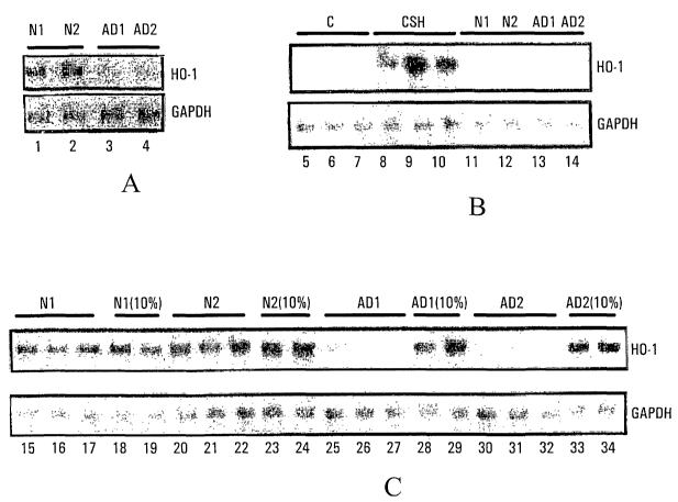
Figure 1 is a Northern analysis of HO-1 mRNA
implicating the presence of a circulating HO-1 suppressor (HOS)
factor in sporadic AD, as described in Example 1. Control
GAPDH bands used to ensure uniformity of RNA loading are
depicted below the HO-1 bands.
CA 02417134 2003-O1-23
WO 02/08449 PCT/CA01/01066
13
Figure 2 depicts tabular results of studies of
demographics and HOS activity in normal young control (NYC),
normal elderly control (NEC), mild cognitive impairment (MCI)
and sporadic Alzheimer disease (AD) subjects, as described in
Example 2. Suppression by 24h incubation in human plasma of
CSH-induced (880~,M x 6 h) glial HO-1 mRNA band (Northern blot)
relative to CSH-treated astrocytes grown in standard culture
media; 0=0-25o suppression, 1=26-50o suppression, 2=51-750
suppression, 3=76-100% suppression. HOS = HOS activity; MMSE
- Folstein Mini-mental State Exam Score; Cortisol = Plasma
cortisol levels (nMol/L); AD Meds = cholinesterase inhibitors
used for treatment of Alzheimer disease; E400 and E800 = 400
and 800 units vitamin E, respectively; C500 = 500 mg vitamin C.
Figure 3 depicts graphical results of HOS activity in
normal control (NC), mild cognitive impairment (MCI) and
sporadic Alzheimer disease (AD) subjects. HOS
activity=percentage suppression (quartiles) by 24h incubation
in human plasma of CSH-induced (880~,M x 6 h) glial HO-1 mRNA
band (Northern blot) relative to CSH-treated astrocytes grown
in standard culture media, as described in Example 3.
Figure 4 depicts analysis of plasma cortisol levels
(mean ~ SD) in normal control (NC), mild cognitive impairment
(MCI) and sporadic Alzheimer disease (AD) subjects (panel A),
as described in Example 4. ( )=number of cases per group.
Differences between groups are not statistically significant
(1-way ANOVA). Correlation between plasma cortisol levels and
HOS activity in the MCI (panel B) and AD (panel C) groups is
not significant (linear regression analysis).
Figure 5 is a Northern analysis of HO-1 mRNA
demonstrating the effects of sample storage time and
antioxidant exposure on plasma HOS activity, as described in
Example 8. .C=Control (untreated) astrocyte cultures,
CSH=cysteamine-treated astrocyte culture, AD=Alzheimer,
MCI=Mild Cognitive Impairment, NEC=normal elderly control,
N=normal subject on antioxidants. Control GAPDH bands are used
as noted in Figure 1.
CA 02417134 2003-O1-23
WO 02/08449 PCT/CA01/01066
14
Figure 6 is a Northern analysis of HO-1 mRNA
demonstrating the effects of plasma dilution on HOS activity,
as described in Example 6.
Figure 7 is a Northern analysis of HO-1 mRNA
demonstrating the effect of heat treatment on HOS activity, as
described in Example 7. Control GAPDH bands are used as noted
in Figure 1.
Figure 8 is a Northern analysis of HO-1 mRNA
demonstrating the partial purification of HOS factor by
heparin-agarose chromatography, as described in Example 8.
Control GAPDH bands are used as noted in Figure 1.
Figure 9 is a Northern analysis of HO-1 mRNA
demonstrating further HOS purification of the heparin agarose
eluate by Concanavalin-A (Con-A) Agarose affinity column
chromatography, as described in Example 9. Control GAPDH bands
are used as noted in Figure 1.
Figure 10 is a Northern analysis of HO-1 mRNA
demonstrating further HOS purification of the heparin agarose-
conconavalin A eluate derived from 4 pooled AD plasma samples
(29 cc starting material) on a SuperoseTM 12 HR FPLC Column, as
described in Example 10. Control GAPDH bands are used as noted
in Figure 1.
Figure 11 presents graphical results of relative
protein concentrations in SuperoseTM 12 HR FPLC Column fractions
derived from pooled AD plasma samples described in Fig. 10, as
described in Example 10. Arrow denotes protein concentration
in fraction (number 20-22) exhibiting robust HOS activity.
Figure 12 depicts results of a chromatogram from a
function test of SuperoseTM 12 HR FPLC 1-cm diameter column
(Catl. # 17-0538-01, Lot # 8283034) [Amersham Pharmacia
Biotech, Inc Quebec Canada] using standard protein mixtures, as
described in Example 10.
Figure 13 is a Northern analysis of HO-1 mRNA
demonstrating the effects of NEC and AD plasma on astrocyte HO-
CA 02417134 2003-O1-23
WO 02/08449 PCT/CA01/01066
1 mRNA induction by multiple stimuli. The HOS bioassay was
performed as described for Figure 1. Northern blots for HO-1
mRNA (top) and respective GAPDH mRNA (bottom) are shown.
Control GAPDH bands are used as noted in Figure 1.
5 DESCRIPTION OF THE EMBODIMENTS OF THE INVENTION
Applicant has devised an improved diagnostic method,
useful in the prediction, diagnosis, and prognostication of AD,
AACD/MCI, and related neurological diseases, as well as methods
and reagents which are useful in the treatment and study of AD,
10 AACD/MCI, and related neurological diseases. These methods are
based on the discovery that patients suffering from AD have an
activity and corresponding factor in their plasma which
significantly suppresses the expression of heme oxygenase-1
(HO-1). This HO-1 suppressor activity is assayed via the
15 inability to upregulate the concentration of nucleotide
sequences encoding HO-1, in response to exposure to a suitable
experimental agent or treatment, in a suitable cell culture
system pre-incubated with a tissue or body fluid derived from
patients suffering from AD or other dementing diseases. This
suppressor activity and corresponding factor shall be referred
to as HO-1 suppressor (HOS) activity and factor, respectively.
Applicant has identified an activity, namely HOS
activity, which is present in tissue or body fluids derived
from patients suffering from AD as well as possibly those
suffering from other dementing diseases, but is absent in
normal age-matched control subjects. This activity may be
detected in a tissue or body fluid obtained from these
patients. Examples of possible sources of suitable tissue or
body fluids include blood, plasma, lymphocytes, cerebrospinal
fluid, urine, saliva, epithelia (such as skin epithelia) and
fibroblast cell lines derived from patients.
An aspect of the invention is a HOS activity, which
is an activity that suppresses the upregulation of HO-1
expression. Such upregulation occurs, for example, following
exposure to an experimental agent or treatment which is, in the
absence of HOS activity, capable of increasing HO-1 expression,
CA 02417134 2003-O1-23
WO 02/08449 PCT/CA01/01066
16
as detected.by increases in HO-1 protein or HO-1 mRNA. In
patients suffering from AD, as well as possibly those suffering
from other dementing diseases, HOS activity suppresses the
expression of HO-1, which is expressed at significantly higher
levels in lymphocytes and possibly other non-neural tissue or
body fluids in normal aged-matched control subjects.
A further aspect of the invention is a method of
assaying HOS activity in a sample. Examples of possible
sources of suitable samples include tissues and body fluids .
such as, for example, blood, plasma, lymphocytes, cerebrospinal
fluid, urine, saliva, epithelia and fibroblast cell lines
derived from patients, or fractions derived from these samples.
The assay involves exposing the sample to be tested to a cell
culture that is capable of undergoing an induction in HO-1
expression in response to exposure to a certain experimental
agent or treatment. An example of such a cell culture is a rat
astroglial culture, however, many other useful possibilities
exist. Examples of such exposure to an experimental agent or
treatment include exposure to oxidative stress, metal ions,
amino acid analogues, sulfhydryl agents, interleukin-1(3, tumour
necrosis factor-a (TNF-a) and hyperthermia. Examples of
suitable sulfhydryl reagents include, but are not limited to,
cysteamine and homocysteine. Following such exposure to an
experimental agent or treatment, the level of HO-1 protein or
HO-1 mRNA may be detected using suitable methods. The level of
HO-1 may for example be detected by an immunoassay. The level
of HO-1 mRNA may for example be detected by Northern analysis
using an appropriate probe(s). Detection of HO-1 mRNA of
greater sensitivity may be achieved for example using the
reverse transcriptase-polymerase chain reaction (RT-PCR)
method, described in Abraham (1998) and Mawal et al. (2000),
both herein incorporated by reference. The activity assay may
be adapted to a large scale level for analyzing a large number
of samples simultaneously, possibly in a suitable array format,
possibly with the automated execution (e.g., by robotics) of
some or all of the steps therein.
CA 02417134 2003-O1-23
WO 02/08449 PCT/CA01/01066
17
A further possibility may be the development of a
reporter-based assay for assaying HOS activity. Such an assay
may involve the preparation of a suitable reporter construct,
e.g. comprising a transcriptional regulatory element, such as
the 5' untranslated promoter region, of the HO-1 gene,.operably
linked to a suitable reporter gene, i.e., capable of regulating
the expression of a suitable reporter gene. Such a construct
may additionally comprise the 3' untranslated region of the
HO-1 gene or another suitable 3' sequence. In another
embodiment, the construct may comprise an in frame fusion of a
suitable reporter gene within the open reading frame of the
HO-1 gene. The reporter gene may be chosen as such to
facilitate the detection of its expression, e.g. by the
detection of the presence and/or activity of its gene product.
Many such suitable reporters may be used, which provide
detectable signals. Most preferred embodiments in this class
are those that provide a conveniently detectable signal, which
may be detected by, for example, spectroscopic methods.
Examples of suitable reporter genes include those encoding
luciferase,~beta-galactosidase, green fluorescent protein,
alkaline phosphatase, chloramphenicol acetyltransferase, as
well as others. Such a reporter construct may be introduced
into a suitable system capable of exhibiting an increase in the
level of expression of the reporter gene in response to
exposure to an experimental agent or treatment which is capable
of increasing HO-1 expression as noted above. Such an assay
would also be adaptable to a possible large scale, high-
throughput, automated format as noted above, and would allow
more convenient detection due to the presence of its reporter
component.
Using methods of assaying HOS activity as described
above, applicant has determined that the level of HOS activity
'"in a sample decreases with the increasing dilution of the
sample, suggesting that HOS activity is attributed to the
presence of a corresponding HOS factor. Using the same assay
methods, applicant has further determined that pre-heating the
sample to be tested abrogates HOS activity, suggesting that HOS
activity is attributed to a protein or' complex of proteins.
CA 02417134 2003-O1-23
WO 02/08449 PCT/CA01/01066
18
Since, to applicant's knowledge, glucocorticoids are the only
known suppressors of HO-1 expression (Lavrovsky et al., 1996;
Deramaudt et al., 1999), the discovery of a protein-like HOS
factor is novel. Applicant has further demonstrated that
cortisol levels are not increased in AD or MCI samples with
respect to normal samples, thus demonstrating that suppression
of HO-1 expression in AD and MCI samples is not attributed to
glucocorticoids, but rather, is a result of the activity of a
(non-glucocorticoid) HOS factor.
Applicant has accomplished a partial purification of HOS
activity and therefore HOS factor using one or multiple
chromatographic methods in sequential fashion. An example of a
suitable chromatographic method is affinity chromatography
using a heparin-agarose matrix or a concanavallin-A (Con-A)
agarose matrix or gel filtration chromatography using for
example a SuperoseTM-12 matrix. Applicant has accomplished
further purification of HOS factor using heparin-agarose,
concanavallin-A (Con-A) agarose and SuperoseTM-12
chromatography, in sequence, further suggesting that HOS factor
comprises a protein or complex of proteins, and, based on
binding to the Con-A matrix, likely comprises a glycoprotein,
in an embodiment, a mannoprotein. This suggests that HOS
activity and the corresponding HOS factor may be obtained in a
more highly purified form using various chromatographic
methods. Such purification is for example shown in Figure 11,
where the peak of HOS activity elutes later'that most of the
protein in the sample, thus indicating that the SuperoseTM-12
column has removed the majority of protein contaminants from
the HOS factor-containing sample. Calibration of the column
using known protein molecular weight standards (Figure 12)
indicates that HOS factor is a protein or complex of proteins
having an approximate molecular weight in the range of 80-100
kDa, in an embodiment, having a molecular weight of
approximately 90 kDa. These data thus provide further support
that HOS factor is a protein-like molecule. Applicant has
further shown that HOS factor and associated HOS activity are
stable during prolonged storage.
CA 02417134 2003-O1-23
WO 02/08449 PCT/CA01/01066
l9
Accordingly, the invention further provides a HOS
factor, as described above.
Applicant has further demonstrated that HOS activity
is not due to simple antioxidant behaviour, since both AD and
normal plasma exhibit equivalent levels of partial suppression
of the HO-1 mRNA response to a pro-oxidant, for example,
menadione. Further, typical doses of antioxidants have no
effect on the induction of HO-1 mRNA expression, and exposure
of multiple, high dose, antioxidants only results in partial
suppression.
A~further aspect of the present invention is an
improved diagnostic method, potentially useful in the
prediction, diagnosis, and prognostication of AD, AACD/MCI, and
related neurological diseases. This diagnostic method is based
on the detection of HOS activity, using for example the assay
methods described above, in a tissue or body fluid obtained
from a patient. Because the presence of HOS activity precedes
any decrease in HO-1 expression in a patient, this diagnostic
method provides an even earlier diagnosis of AD, AACD/MCI, and
related neurological diseases. In addition, the
immunodetection of HOS factor or activity (see below) may
provide an improved method of diagnosis over the detection of
decreases in HO-1 expression using methods such as Northern
analysis or the reverse transcriptase-polymerase chain reaction
(RT-PCR) method, described in Abraham (1998) and Mawal et al.
(2000), both herein incorporated by reference. Further, the
correlation of the presence of HOS activity with the disease
state represents a positive test for diagnosis. This is more
desirable than a negative test, used for diagnosis based on the
reduction or absence of HO-1 expression in a patient suffering
from one of the dementing diseases described above.
It is known in the art that certain dementing
diseases, for example, AD, correlate with changes in HO-1
levels while others do not. Such dementing diseases may be
categorized as HO-1-dependent and HO-1-independent. As
described in the instant application, such changes in HO-1
levels are a result of changes in the levels of HOS factor or
CA 02417134 2003-O1-23
WO 02/08449 PCT/CA01/01066
activity. Therefore, the invention further relates to methods,
reagents, compounds and commercial packages to differentiate a
dementing disease which exhibits a significantly altered level
of HO-1 protein, HO-1 mRNA, HOS factor, or HOS activity, i.e.,
5 an HO-1-dependent dementing disease, from a dementing disease
which does not exhibit such a significantly altered level of
HO-1 protein, HO-1 mRNA, HOS factor, or HOS activity, i.e., an
HO-1-independent dementing disease. The term "significantly"
as used here means that the levels are altered from control
10 levels beyond the range of experimental error, as known in the
art.
The HOS activity of the present invention may also be
used to develop therapeutic agents and methods for the
treatment of AD and other dementing diseases. Since the
15 appearance of HOS activity correlates with the presence of the
disease state, the HOS activity and HOS factor is expected to
play a causative role in the onset and/or progression of AD and
other dementing diseases. Therefore, identification of factors
or mechanisms which inhibit or activate HOS activity may be
20 utilized for the development of therapeutic agents and methods
for the treatment of AD and other dementing diseases. If an
increase in HOS activity is a causative event in the onset
and/or progression of AD and other dementing diseases, an
inhibitor of HOS activity is expected to have therapeutic
potential. Conversely, an activator of HOS activity is
expected to represent an upstream causative agent of the onset
and/or progression of AD and other dementing diseases, which
may provide even earlier and improved methods of diagnosis.
Further, all factors which effect HOS activity will lead to a
better understanding of the mechanisms of the onset and/or
progression of AD and other dementing diseases, and ultimately
contribute to the development of improved therapeutic methods
and agents. In addition, other factors which affect levels of
HO-1 mRNA, protein and activity are also useful to the
invention, similar to the above, and are thus a further aspect
of the invention.
Accordingly, it is a further aspect of the present
invention to provide a HOS activity-based screening method to
CA 02417134 2003-O1-23
WO 02/08449 PCT/CA01/01066
21
identify putative compounds which either inhibit or augment HOS
activity. Such screening may be performed using for example
the HOS activity assays described above, and may be adapted to
a large scale, and possibly automated format. Such a method
may comprise exposing a known HOS activity-containing sample to
the compound to be tested, and subsequently determining the
level of HOS activity present, which is then compared to a
control sample that was not exposed to the compound to be
tested. In a high-throughput, automated format, this screening
method may be used for the rapid analysis of libraries
containing a large number of compounds for their effects on HOS
activity. In an embodiment, examples of such libraries include
chemical libraries prepared by combinatorial synthesis.
The partially purified fraction comprising HOS factor
and HOS activity, obtained, for example, from heparin-agarose
and/or Con-A agarose and/or SuperoseTM-12 column chromatography,
may be used to immunize a small mammal, e.g., a mouse or a
rabbit, in order to raise antibodies which recognize this
activity. ~n an embodiment the above mentioned fraction is
obtained from sequential heparin-agarose, Con-A agarose and
SuperoseTM-12 column chromatography. Accordingly, a further
aspect of the invention provides an antibody that recognizes
the HOS factor of the invention.
An antibody of the invention is either polyclonal or
monoclonal. Antibodies may be recombinant, e.g., chimeric
(e. g., constituted by a variable region of murine origin
associated with a human constant region), humanized (a human
immunoglobulin constant backbone together with hypervariable
region of animal, e.g., murine, origin), and/or single chain.
Both polyclonal and monoclonal antibodies may also be in the
form of immunoglobulin fragments, e.g., F(ab)'2, Fab or Fab'
fragments. The antibodies of the invention are of any isotype,
e.g., IgG or IgA, and polyclonal antibodies are of a single
isotype or a mixture of isotypes.
Antibodies against the HOS factor of the present
invention are generated by immunization of a mammal with a
partially purified fraction comprising HOS factor. In an
CA 02417134 2003-O1-23
WO 02/08449 PCT/CA01/01066
22
embodiment the above mentioned fraction is obtained from
sequential heparin-agarose, Con-A agarose and SuperoseTM-12
column chromatography. Such antibodies may be polyclonal or
monoclonal. Methods to produce polyclonal or monoclonal
antibodies are well known in the art. For a review, see Harlow
and Lane (1988) and Yelton et a1. (1981), both of which are
herein incorporated by reference. For monoclonal antibodies,
see Kohler and Milstein (1975), herein incorporated by
reference. '
The antibodies of the invention, which are raised to
a partially purified fraction comprising HOS factor of the
invention, are produced and identified using standard
immunological assays, e.g., Western blot analysis, dot blot
assay, or ELISA (see, e.g., Coligan et al. (1994), herein
incorporated by reference). The antibodies are used in
diagnostic methods to detect the presence of a HOS factor and
activity in a sample, such as a tissue or body fluid. The
antibodies are also used in affinity chromatography for
obtaining a purified fraction comprising the HOS factor and
activity of the invention.
Accordingly, a further aspect of the invention
provides (i) a reagent for detecting the presence of HOS factor
and activity in a tissue or body fluids and (ii) a diagnostic
method for detecting the presence of HOS factor and activity in
a tissue or body fluid, by contacting the tissue or body fluid
with an antibody of the invention, such that an immune complex
is formed, and by detecting such complex to indicate the
presence of HOS factor and activity in the sample or the
organism from which the sample is derived.
Those skilled in the art will readily understand that
the immune complex is formed between a component of the sample
and the antibody, and that any unbound material is removed
prior to detecting the complex. It is understood that an
antibody of the invention is used for screening a sample, such
as, for example, blood, plasma, lymphocytes, cerebrospinal
fluid, urine, saliva, epithelia and fibroblasts, for the
presence of'HOS activity.
CA 02417134 2003-O1-23
WO 02/08449 PCT/CA01/01066
23
For diagnostic applications, the reagent (i.e., the
antibody of the invention) is either in a free state or
immobilized on a solid support, such as a tube, a bead, or any
other conventional support used in the field. Immobilization
is achieved using direct or indirect means. Direct means
include passive adsorption (non-covalent binding) or covalent
binding between the support and the reagent. By "indirect
means" is meant that an anti-reagent compound that interacts
with a reagent is first attached to the solid support.
Indirect means may also employ a ligand-receptor system, for
example, where a molecule such as a vitamin is grafted onto the
reagent and the corresponding receptor immobilized on the solid
phase. This is illustrated by the biotin-streptavidin system.
Alternatively, a peptide tail is added chemically or by
genetic engineering to the reagent and the grafted or fused
product immobilized by passive adsorption or covalent linkage
of the peptide tail.
Such diagnostic agents may be included in a kit which
also comprises instructions for use. The reagent is labeled
with a detection means which allows for the detection of the
reagent when it is bound to its target. The detection means
may be a fluorescent agent such as fluorescein isocyanate or
fluorescein isothiocyanate, or an enzyme such as horse radish
peroxidase or luciferase or alkaline phosphatase, or a
radioactive element such as 12~I or 5lCr.
Accordingly, a further aspect of the invention
provides a process for purifying, from a tissue or body fluid,
the HOS factor of the invention, which involves carrying out
antibody-based affinity chromatography with the tissue or body
fluid, wherein the antibody is an antibody of the invention.
For use in a purification process of the invention,
the antibody is either polyclonal or monoclonal, and preferably
is of the IgG type. Purified IgGs are prepared from an
antiserum using standard methods (see, e.g., Coligan et a1.
(1994), herein incorporated by reference). Conventional
chromatography supports, as well as standard methods for
CA 02417134 2003-O1-23
WO 02/08449 PCT/CA01/01066
24
grafting antibodies, are described in, e.g., Harlow and Zane
(1988), herein incorporated by reference, and outlined below.
Briefly, a tissue or body fluid, such as plasma from
a patient suffering from AD, preferably in a buffer solution,
is applied to a chromatography material, preferably
equilibrated with the buffer used to dilute the tissue or body
fluid so that the HOS factor of the invention (i.e., the
antigen) is allowed to adsorb onto the material. The
chromatography material, such as a gel or a resin coupled to an
antibody of the invention, is in either a batch form or a
column. The unbound components are washed off and the antigen
is then eluted with an appropriate elution buffer, such~as a
glycine buffer or a buffer containing a chaotropic agent, e.g.,
guanidine HC1, or high salt concentration (e. g., 3 M MgCl2).
Eluted fractions are recovered and the presence of the antigen
is detected, e.g., by measuring the absorbance at 280 nm.
A further aspect of the present invention is a
diagnostic imaging method, which comprises introducing into a
biological system, an antibody of the invention, which is used
in conjunction with an appropriate detection system to identify
areas where HOS factor or activity is present or absent.
The following examples are provided in order to
illustrate the methods of the present invention and are not
meant to limit the scope of the invention.
Example 1: Determination of the presence of HOS activity in
plasma derived from AD patients
Whole blood is collected from normal elderly (N1, N2)
subjects or~patients with probable sporadic AD (AD1, AD2) in
heparinized tubes. This is then layered over a Ficoll PaqueTM
density gradient and centrifuged at 1800 rpm for 20 minutes.
The top plasma layer is then collected and saved for incubation
with rat astroglia as described below. The lymphocyte
fractions are collected and used for the isolation of mRNA for
Northern analysis as described below.
CA 02417134 2003-O1-23
WO 02/08449 PCT/CA01/01066
Determination of lymphocyte HO-1 mRNA levels:
Lymphocyte fractions were obtained by differential
centrifugation of whole blood on Ficoll PaqueT~' gradients as
described above. Cytoplasmic RNA was isolated from the
5 lymphocytes using an acid guanidinium thiocyanate-phenol-
chloroform extraction method, as described by Chomezynski
et a1. (1997), herein incorporated by reference. Six
micrograms of RNA was denatured and size-separated by
electrophoresis on 1% agarose/formaldehyde gels. RNA integrity
10 was confirmed by ethidium bromide staining. The RNA was
transferred onto Hybond-NT~' filter paper and covalently cross-
linked to the membrane by UV light for two minutes. The
hybridization probe (HO-1; l.Okb) was prepared by random
priming using the Random Primer DNA Labelling System, as
15 described by Feinberg et al. (1984), herein incorporated by
reference. Prehybridization was performed for 12 hours at 42°C
in a buffer containing formamide deionized, 5 x Denhardt's
reagent, 6 x SSPE and 0.5o SDS. The hybridization buffer
consisted of the prehybridization buffer without 5 x Denhardt's
20 reagent, and 32P-labelled denatured DNA probe, as described in
Noonberg et a1. (1994), herein incorporated by reference.
Equal loading of RNA was confirmed by hybridization with a cDNA
for the (housekeeping) gene, glyceraldehyde-3-phosphate
dehydrogenase (GAPDH). All washes were performed under
25 stringent conditions (1 x SSC and 0.2o SDS for 45 minutes at
room temperature, 0.4 x SSC and 0.2o SDS for 15 minutes at
65°C). The RNA hybridizing with cDNA probes was visualized by
autoradiography using an intensifying screen at -80°C, as
described in Church et a1. (1984), herein incorporated by
reference.
As noted in our related US Patent (No. 6,210,895;
April 13, 2001) and publication (Schipper et al., 2000), both
herein incorporated by reference, and as reiterated in Panel A
of Figure 1, lymphocytes isolated from normal subjects N1 and
N2 exhibit significant levels of HO-1 mRNA (lanes 1 and 2),
which is not detectable in lymphocytes isolated from AD
patients AD1 and AD2 (lanes 3 and 4).
CA 02417134 2003-O1-23
WO 02/08449 PCT/CA01/01066
26
Assay of plasma HOS activity via the induction of
HO-1 expression upon cysteamine (CSH) treatment of rat
astroglia:
Brain cell cultures: Rat astroglia were prepared as
described in Schipper et al. (1999), herein incorporated'by
reference, as follows:
Pregnant Sprague-Dawley rats were obtained from Charles
River Breeding Farms. Primary neural cell cultures were
prepared from 1-day old neonates by mechanoenzymatic
dissociation of cerebral tissue or body fluid as previously
described by Chopra et al., (1997), herein incorporated by
reference. Cells were grown in Ham's F-12 and high-glucose
DMEM (50:50 vol/vol) supplemented with 10 mM HEPES. 5o heat-
inactivated horse serum, 5o heat-inactivated fetal bovine
serum, and penicillin/streptomycin (50 U/ml and 50 ~,g/ml,
respectively). The cells were plated in 75-cm2 tissue or body
fluid culture flasks at a density of 1 x 10⁶ cells/ml. Cultures
were incubated at 37°C in humidified 95o air/5o C02 for 6h, at
which time they were vigorously shaken 20-30 times with
replacement of fresh medium to remove adherent oligodendroglia
and microglia from the astrocytic monolayers. The cultures
were then incubated under the above-mentioned conditions for
6 days, at which time >980 of the cells composing the monolayer
were astroglia, as determined by immunohistochemical labeling
for the astrocyte-specific marker glial fibrillary acidic
protein, as described by Chopra et al. (1995), herein
incorporated by reference. These astroglia cultures were grown
under different conditions and subjected to different
treatments (see below), and subsequently mRNA was isolated for
Northern analysis of HO-1 mRNA levels as described in Schipper
et al. (1999), herein incorporated by reference, as follows:
RNA isolation and Northern analysis: Cultured
astrocytes were harvested using a rubber policeman, and
cytoplasmic RNA was isolated using an acid guanidinium
thiocyanate/phenol/chloroform extraction method, as described
by Chomczynski and Sacchi (1987), herein incorporated by
reference. Ten micrograms of RNA was denatured and size-
CA 02417134 2003-O1-23
WO 02/08449 PCT/CA01/01066
27
separated by electrophoresis on 1% agarose/formaldehyde gels.
RNA integrity was confirmed by ethidium bromide staining. The
RNA was transferred onto Hybond-NTM filter paper and covalently
cross-linked to the membrane by UV light for 2 min. The
hybridization probe (HO-1; 1.0 kb) was prepared by random
primer-generated double-strand DNA probes using the random
primer DNA labeling system, as described by Feinberg and
Vogelstein (1984), herein incorporated by reference.
Prehybridization was performed for 12 h at 42°C in a buffer
containing formamide-deionized, 5X Denhardt's reagent,
6X saline-sodium phosphate-EDTA, and 0.5o sodium dodecyl
sulfate (SDS). The hybridization buffer consisted of the
prehybridization buffer without 5X Denhardt's reagent and
32P-labeled denatured DNA probe, as described by Noonberg et a1.
(1994), herein incorporated by reference. Equal loading of RNA
was confirmed by hybridization with a cDNA for the
(housekeeping) gene, glyceraldehyde-3-phosphate dehydrogenase
(GAPDH), or 18S mRNA. All washes were performed under
stringent conditions [1X saline-sodium citrate (SSC) and 0.2o
SDS for 45 min at room temperature, 0.4X SSC and 0.2o SDS for
15 min at 65°C, and 0.1X SSC and 0.2o SDS for 15 min at 65°C].
The RNA hybridizing with cDNA probes was SDS for 15 min at
65°C, and 0.1X SSC and 0.2o SDS for 15 min at 65°C]. The RNA
hybridizing with cDNA probes was visualized by autoradiography
using an intensifying screen at -80°C, as described by Church
and Gilbert,(1984), herein incorporated by reference.
Resulting bands on the autoradiograph were analyzed using a
phosphorimager S1 densitometer. Densitometry data were
normalized by calculating the ratios of the HO-1 mRNA signals
to control GAPDH or 18S mRNA signals.
Figure 1, panel A: Northern blot of lymphocyte HO-1
mRNA (and control GAPDH mRNA) derived from 2 normal elderly
individuals (N1, N2) and 2 patients with probable sporadic AD
AD1, AD2). As noted in our related US Patent (No. 6,210,895;
April 13, 2001) and publication (Schipper et al., 2000), both
herein incorporated by reference, lymphocyte HO-1 mRNA bands
are visible in the controls (lanes 1 and 2), and not detectable
CA 02417134 2003-O1-23
WO 02/08449 PCT/CA01/01066
28
(lanes 3 and 4) in the AD subjects, indicating the presence of
HOS activity in the latter.
Using the methods described above, Northern analysis
of HO-1 mRNA levels of rat astroglia grown under different
conditions and subjected to different treatments was performed,
the results of which are shown in panels B and C of Figure 1.
Panel B: Control (unchallenged) rat astroglia grown in
standard culture media for 6 days exhibit faint or no HO-1 mRNA
bands (lanes 5-7). Cysteamine (CSH) treatment (880~M x 6hr)
induces robust HO-1 mRNA bands in these cells (lanes 8-10).
Twenty-four hour incubation of the rat astroglia with the
plasma derived from the same 2 normal subjects (N1, N2; lanes
11-12) and the 2 AD patients (ADl, AD2; lanes 13-14) noted
above (see panel A) has no appreciable affect on baseline HO-1
mRNA levels. Panel C: In contrast to plasma derived from the
2 normal subjects (lanes 15-24), undiluted plasma from the 2 AD
patients markedly suppresses the rat astroglial HO-1 mRNA
response to CSH (lanes 25-27; 30-32). Dilution of the AD
plasma (1:9 in standard culture media; "10%") greatly
diminishes its inhibitory effect on CSH-induced HO-1 mRNA
expression (lanes 28-29; 33-34). Therefore, there exists in
the plasma of AD patients an HOS activity, which is not present
in the plasma of normal subjects, and which is assayable by the
determination of HO-1 mRNA levels in rat astroglia incubated
with the relevant plasma sample and subjected to CSH treatment.
Example 2: Demographics and HOS activity in normal young
control (NYC), normal elderly control (NEC), mild cognitive
impairment (MCI) and sporadic Alzheimer disease (AD) subjects.
Results are shown in tabular form in Figure 2.
Suppression by 24h incubation in human plasma of CSH-induced
(880~M x 6 h) glial HO-1 mRNA band (Northern blot) relative to
CSH-treated astrocytes grown in standard culture media; 0=0-250
suppression, 1=26-50o suppression, 2=51-75o suppression, 3=76-
1000 suppression. HOS = HOS activity; MMSE = Folstein Mini-
mental State Exam Score; Cortisol = Plasma cortisol levels
(nMollL). AD Meds = cholinesterase inhibitors used for
treatment of Alzheimer disease. E400 and E800 = 400 and 800
CA 02417134 2003-O1-23
WO 02/08449 PCT/CA01/01066
29
units vitamin E, respectively; C500 = 500 mg vitamin C. HOS
activity was assayed as described in Example 1. Measurement of
cortisol levels were performed using the GammaCoat [I-125]
Cortisol Radioimmunoassay (RIA) Kit based on the competitive
binding principles of RIA.
Example 3: HOS activity in normal control (NC), mild cognitive
impairment (MCI) and sporadic Alzheimer disease (AD) subjects.
Results are shown in Figure 3. HOS activity =
percentage suppression (quartiles) by 24h incubation in human
plasma of CSH-induced (880~M x 6 h) glial HO-1 mRNA band
(Northern blot) relative to CSH-treated astrocytes grown in
standard culture media. HOS activity was assayed as described
in Example 1.
Example 4: Plasma cortisol levels (mean ~ SD) in normal
control (NC), mild cognitive impairment (MCI) and sporadic
Alzheimer disease (AD) subjects.
Results are shown in Figure 4. Panel A shows mean (~
SD) plasma cortisol levels of NC, MCI and AD subjects. ( ) -
number of cases per group. Differences between groups are not
statistically significant (1-way ANOVA). Correlations between
plasma cortisol levels and HOS activity in the MCI (panel B)
and AD (panel C) groups are not significant (linear regression
analysis). Although glucocorticoids are known suppressors of
the HO-1 gene, these data indicate that elevated cortisol
levels are not responsible for HOS activity in the MCI and AD
plasma.
Example 5: Effects of sample storage time and antioxidant
exposure on plasma HOS activity.
Results are shown in Figure 5. HOS activity was
assayed as described in Example 1. C = Control (untreated)
astrocyte cultures, CSH = cysteamine-treated astrocyte culture,
AD = Alzheimer, MCI = Mild Cognitive Impairment, NEC = normal
elderly control, N = normal subject on antioxidants. Protease
inhibitors (Complete Protease Inhibitor Cocktail, Cat. #
1836153, Roche, Mannheim) were added to all plasma samples
CA 02417134 2003-O1-23
WO 02/08449 PCT/CA01/01066
prior to freezing. HOS activity is retained in AD and MCI
plasma samples stored at -85° C for up to 15 months. In normal
subjects, low-dose vitamin E (400Ulday), a dose of vitamin E
commonly taken by AD patients, does not affect the astrocyte
5 HO-1 mRNA response to CSH (N1). In normal individuals, exposure
to multiple, very high-dose antioxidants partially attenuates
the filial HO-1 mRNA response to CSH (N2, N3).
Example 6: Effects of plasma dilution.on HOS activity
Plasma HOS activity was assayed via the determination
10 of HO-1 mRNA levels in treated rat astroglia as described in
Example 1. In this case, the effects of plasma dilution were
examined, as documented in Figure 6.
Lane 1: Absence of HO-1 mRNA in unchallenged rat
astrocytes grown in standard culture media. Lane 2:
15 Cysteamine (CSH; 880~,M x 6h)induces strong HO-1 mRNA bands in
cultured astroglia grown in standard media. Lane 3: Absence
of HO-1 mRNA bands in unchallenged astrocytes grown in
Alzheimer (AD) plasma (A: patient 1; B: patient 2). Lanes 4-6:
undiluted AD plasma markedly suppresses the HO-1 mRNA response
20 to CSH in cultured astroglia (intense HOS activity present).
The filial HO-1 mRNA response to CSH progressively increases
(abrogation of HOS activity) with increasing dilutions of AD
plasma using standard media (lanes 7-15).
Panel A and panel B of Figure 6 represent data
25 obtained using plasma obtained from two different AD patients
(A: patient 1; B: patient 2). As noted above and shown again
in Figure 6, untreated rat astroglia grown in standard culture
media exhibit little or no detectable HO-1 mRNA (lane 1),
however, HO-1 expression increases significantly upon CSH
30 treatment (lane 2). In the absence of CSH treatment, rat
astroglia incubated with AD plasma show no detectable HO-1 mRNA
(lane 3), also as noted in Example 1. CSH treatment of rat
astroglia incubated with undiluted AD plasma ("1000") failed to
induce any significant induction of HO-1 expression (lanes
4-6), due to the intense HOS activity present in the undiluted
AD plasma. However, the rat astroglial response to CSH
CA 02417134 2003-O1-23
WO 02/08449 PCT/CA01/01066
31
progressively increases (abrogation of HOS activity) with
increasing dilutions of the AD plasma using standard media
(lanes 7-15). Therefore, there appears to exist in AD plasma 'a
HOS factor whose plasma concentration correlates with HOS
activity.
Example 7: Effect of heat treatment on HOS activity
Plasma HOS activity was assayed via the determination
of HO-1 mRNA levels in treated rat astroglia as described in
Example 1. In this case, the effects of prior heat treatment
of AD plasma were examined, as documented in Figure 7.
As noted above, control rat astroglia grown in
standard media or exposed to human plasma (from normal [NEC]
subjects or AD patients) exhibit little or no HO-1 mRNA via
Northern analysis in the absence of CSH treatment (lanes 1, 5,
7 and 10). CSH treatment (880 ~,M CSH x 6h) of astroglia grown
in standard media results in the observation of an intense HO-1
mRNA signal (lane 2). Also as noted above, this induction of
HO-1 expression in response to CSH treatment is significantly
attenuated in astroglia incubated in AD plasma for 24 h (lanes
3 and 4). However, this attenuation is no longer observed when
the AD plasma is heated (100°C for 10 min.) prior to incubation
with rat astroglia, indicating that as a result of this pre-
heating AD plasma HOS activity is abrogated, as observed in the
robust HO-l~mRNA signal seen in lane 6. CSH treatment of rat
astroglia with normal plasma either untreated or pre-heated
results in a robust observed HO-1 mRNA signal, since HOS
activity is absent in either case (lanes 8 and 9). Therefore,
these data indicate that HOS activity in AD plasma is mediated
by a protein.
Example 8: Partial purification of HOS factor by heparin-
agarose affinity column chromatography
Plasma from one normal subject (NEC) and one AD
patient (AD) was subjected to affinity purification on a
heparin-agarose column as described in Sasaki et al. (1987),
herein incorporated by reference, as follows:
CA 02417134 2003-O1-23
WO 02/08449 PCT/CA01/01066
32
Plasma preparation for loading onto Heparin Agarose
column: The NEC and AD plasma tubes were thawed at 4°C. The
samples were then dialyzed against Heparin Agarose column
loading buffer [HALB: 20 mM Hepes (SIGMA Chemical Co., Saint
Louis, MI, USA, Catl # H-4034) pH 7.2, 150 mM NaCl, protease
inhibitor tablet (Roche Diagnostics, Laval, PQ, CANADA Catl. #
1 873 580)] for 2h with gentle stirring. The samples were then
centrifuged at 15,000 g at 4°C for 20 minutes and supernatants
collected.
Heparin Agarose affinity--column chromatography: The
Heparin Agarose column (1 cm X 2 cm; SIGMA Chemical Co., Saint
Louis, MI, USA, Catl # H-0402) was prewashed with 20 ml of
HALB. Plasma supernatants were loaded on the column. The
column was washed with 4-6 volumes of HALB and 1 ml fractions
collected. The flow-through fractions containing protein were
pooled. The column was eluted with elution buffer [EB: 20 mM
Hepes pH 7.2, 1 M NaCl, protease inhibitor] and 1 ml eluates
containing proteins were pooled and dialyzed against HALB for
2-4h.
The preparation of protein (e. g. plasma or column
fractions) for the rat astroglia/HOS activity assay was
performed as described in Sasaki et al. (1987), herein
incorporated by reference, as follows:
Media was removed from 70 ml, 25 cm~ flasks containing
confluent astrocyte monolayer (7-10 days in culture). To each
individual flask, 1.4 ml of NEC or AD plasma was added. To
each individual flask, approximately 1.4 ml (10 mg protein
based on the Bradford BioRad Protein assay kit using BSA as
control) of the Heparin Agarose flow-through fraction derived
from NEC or AD subjects was added with 0.6 ml of complete DMEM
medium. To each individual flask, approximately 1.4 ml (0.5 mg
protein) of the Heparin Agarose eluate fractions derived from
NEC or AD subjects was added with 0.6 ml of complete DMEM
medium.
Subsequently, the various samples were assayed for
HOS activity as described in Example 1. Briefly, the eluate
CA 02417134 2003-O1-23
WO 02/08449 PCT/CA01/01066
33
samples were incubated with rat astroglia, which were then
subjected to CSH treatment. Subsequent mRNA isolation and
Northern analysis to determine the level of HO-1 mRNA was
performed, with the results shown~in Figure 8.
Figure 8: Panel A: Northern blots of HO-1 mRNA. Panel B:
Control GAPDH mRNA. Plasma from one NEC and one AD patient
was affinity purified on a heparin-agarose column and the
eluate, collected using a high salt solution, was dialyzed. In
the absence of CSH treatment, control rat astroglia pre-
incubated with heparin eluates from NEC or AD plasma for 24h
did not exhibit an increase in HO-1 expression, as observed by
the relatively faint HO-1 mRNA bands which correspond to these
samples (lanes 1 and 4). CSH treatment (880 ~.M CSH x 6h) of
rat astroglia incubated with the heparin eluate from NEC plasma
results in an induction of HO-1 expression, as observed by
intense HO-1 mRNA bands (lanes 2 and 3). Conversely, no
augmentation of HO-1 mRNA bands in response to CSH treatment
was observed in rat astroglia incubated for 24h with the
heparin eluate fraction derived from the plasma of the AD
patient. These data support the presence of a HOS factor in
the plasma of AD patients, but not normal (NEC) subjects, and
indicate that the factor binds to heparin-agarose affinity
columns.
Example 9: Further HOS purification of heparin agarose eluate
by Concanavalin-A (Con-A) Agarose affinity column
chromatography.
Heparin Agarose column eluate (as described in Fig.
8) was dialyzed against loading buffer: 50 mM Hepes, pH 7.2
containing 150 mM NaCl, 1 mM MgCl~, 1mM MnCl2, 1 mM CaCl2 and
CompleteTM EDTA-free Protease inhibitor cocktail for 4 h at 4 °C.
The dialysate was loaded onto Con-A Agarose column. The column
was washed with four bed volumes of loading buffer. The HOS
fraction was eluted with loading buffer containing 0.2M a-D-
methyl mannopyranoside. The eluate was dialyzed against loading
buffer. The HOS bioassay (glial HO-1 mRNA response to 880~,M CSH
x 6 h) was performed as described in Example 1 and Figure 1.
Results are shown in figure 9. Glial HO-1 mRNA bands were
CA 02417134 2003-O1-23
WO 02/08449 PCT/CA01/01066
34
faint in all specimens not exposed to CSH. Robust HO-1 mRNA
responses to CSH were observed in control astroglial cultures
(grown in standard culture media) and astrocytes incubated for
24 hours in NEC plasma. In contrast, HO-1 mRNA responses to CSH
were markedly suppressed in astrocytes incubated for 24 hours
in (i) whole AD plasma, (ii) dialyzed AD plasma prior to
heparin-ConA chromatography and (iii) heparin Agarose--ConA
eluate derived from AD plasma These data indicate that the AD
plasma HOS factor binds to ConA columns and is therefore likely
a glycoprotein.
Example 10: Further HOS purification of heparin agarose-
conconavalin A eluate derived from 4 pooled AD plasma samples
(29 cc starting material) on Superose'~ 12 HR FPLC Column.
Results are shown in Figures 10 to 12. Heparin
Agarose - ConA Agarose purified AD plasma (1-ml) was dialyzed
against 20 mM Hepes, pH 7.2 containing 150 mM NaCl and
CompleteTM EDTA-free Protease inhibitor cocktail (1 tablet/100-
ml; Catl. # 1873580, Lot 61320101; Roche Diagnostics, Quebec,
Canada) for four hours at 4 °C. The dialyzed fraction was loaded
on SuperoseTM 12 HR FPLC 1-cm diameter column (Catl. # 17-0538-
01, Lot # 8283034) [Amersham Pharmacia Biotech, Inc Quebec
Canada]. HOS activity was measured by bioassay in each fraction
as described in Example 1 and Figure 1. As shown in Figure 10,
robust HOS activity was observed in fraction number 20-22.
Figure 11: Relative protein concentrations in
SuperoseTM 12 HR FPLC Column fractions derived from pooled AD
plasma samples described in Fig. 10. Each 0.5-ml fraction of
the flow-through was collected and absorbance (O. D.) measured
at 280 nm by spectrophotometer. A graph of O.D. versus fraction
number is plotted. Arrow denotes protein concentration in
fraction (number 20-22) exhibiting robust HOS activity (Figure
11) .
Figure 12: A chromatogram from a function test of
SuperoseTM 12 HR FPLC 1-cm diameter column (Catl. # 17-0538-01,
Lot # 8283034) [Amersham Pharmacia Biotech, Inc Quebec Canada]
using standard protein mixtures. Plasma samples were derived
CA 02417134 2003-O1-23
WO 02/08449 PCT/CA01/01066
from the same pooled AD plasmas described in Fig. 10. Each 0.5-
ml fraction of the flow-through was collected and absorbance
measured at 280 nm. A graph of peak molecular weight for each
protein standard versus fraction number was plotted. Based on
5 the elution profile of standards with known molecular weight,
the molecular weight of the HOS-positive fraction (number 20-
22) is estimated at approximately 90 kDa.
Example 11: Effects of NEC and AD plasma on astrocyte HO-1
mRNA induction by multiple stimuli.
10 The HOS bioassay was performed as described in
Example 1 and Figure 1. Northern blots for HO-1 mRNA (top) and
respective GAPDH mRNA (bottom) are shown in Figure 13. AD
plasma strongly suppressed the HO-1 mRNA response to CSH
(880~M), interleukin-1(3 (I1-1(3 50 and 100 ng/ml), and
15 homocysteine (HC 200~M). NEC plasma showed no HOS activity in
the face of these stimuli. AD plasma completely suppressed,
whereas NEC plasma partially suppressed, the HO-l mRNA response
to tumour necrosis factor-a (TNF-a 50 and 100 ng/ml). AD and
NEC plasma exhibited partial and equal suppression of the glial
20 HO-1 mRNA response to menadione (Mena 100~M). These data
indicate that: A) The HOS protein in AD plasma is active
against multiple inducers of astroglial HO-1 mRNA. B) HOS
activity in AD plasma is particularly potent in the face of
TNF-a challenge. Since partial HOS activity against TNF-a also
25 occurs in NEC plasma, differences in HOS protein expression
between AD and NEC may be quantitative rather than qualitative.
C) Simple antioxidant behaviour does not account for HOS
activity in AD plasma because both AD and NEC plasma exhibit
partial and equal suppression of the glial HO-1 mRNA response
30 to the pro-oxidant, menadione.
The above examples illustrate application of the
testing method to detect HOS activity. Further, the above
examples illustrate the testing method using plasma as the
tissue or body fluid sample. The method can also be applied to
35 other tissue or body fluids such as blood, cerebrospinal fluid,
CA 02417134 2003-O1-23
WO 02/08449 PCT/CA01/01066
36
urine, saliva, epithelia and to fibroblast cell lines derived
from patients.
The test can be applied to compare the level of HOS
activity in a specific patient over a period of time, or to
compare the level of HOS activity in a patient with the
corresponding level in a normal control population.
The above examples further demonstrate the presence of a
HOS factor which is a glycoprotein, in an embodiment a
mannoprotein, having an approximate molecular weight in the
range of 80-100 kDa, in an embodiment having a molecular weight
of approximately 90 kDa, and which is not a glucocorticoid.
CA 02417134 2003-O1-23
WO 02/08449 PCT/CA01/01066
37
REFERENCES
Abraham, N.G. (1998) Quantitation of hems oxygenase
(HO-1) copies in human tissue or body fluids by competitive
RT/PCR, in Methods in Molecular Biology, vol. 108: Free
Radical and Antioxidant Protocols (Armstrong, D., ed.),
pp. 199-209, Humana Press Inc., Totowa, NJ.
Coligan J.E., et al., (1994) Current Protocols in
Immunology, John Wiley & Sons, Inc., New York, NY.
Chomczynski P. and Sacchi N. (1987) Single-step
method of RNA isolation by acid guanidinium thiocyanate-phenol-
chlorophorm extraction. Analytical Biochem. 162:156-159.
Chopra, V.S., et al. (1995) Differential effects of
cysteamine on heat shock protein induction and cytoplasmic
granulation in astrocytes and glioma cells. Mol. Brain Res.
31:173-184.
Chopra, V.S., et a1. (1997) A cellular stress model
for the differential expression of glial lysosomal cathepsins
in the aging nervous system. Exp. Neurol. 147:221-228.
Church G.M. and Gilbert W. (1984) Genomic
sequencing. Proc. Natl. Acad. Sci. USA 81:1991-1995.
Deramaudt, T.B., et a1. (1999) Negative regulation of
human hems oxygenase in microvessel endothelial cells by
dexamethasone. Proc. Soc. Exp. Biol. Med. 222:185-193.
Feinberg AP and Vogelstein B. (1984) A Technique for
radiolabelling DNA restriction endonuclease fragments to high
specific activity. Analytical Biochem. 137:266-267.
Grossi, D., et a1. (1988) Senile demential.
II International Symposium (pp. 97-99). Paris: John Libbey
Eurotext.
Harlow, E. and Lane, D (1988) Antibodies:
A Laboratory Manual, Cold Spring Harbor Laboratory Press, Cold
Spring Harbor, NY.
CA 02417134 2003-O1-23
WO 02/08449 PCT/CA01/01066
38
Haxby J.V., et al. (1992) Individual trajectories of
cognitive decline in patients with dementia of the Alzheimer
type. J. Clin. Exp. Neuropsychol. 14:575-592.
Kohler G. and Milstein C. (1975) Continuous cultures
of fused cells secreting antibody of predefined specificity.
Nature 256:495-497.
Lavrovsky, Y., et a1. (1996) Downregulation of the
human heme oxygenase gene by glucocorticoids and identification
of 56b regulatory elements. Biochem. Biophys. Res. Comm.
218:759-765.
Mann, U., et a1. (1992) Heterogeneity in Alzheimer's
disease: Progression rate segregated by distinct
neuropsychological and cerebral metabolic profiles. J. Neurol.
Neurosurg. Psychiatry 55:956-959.
Mawal, Y., et al. (2000) RT-PCR confirmation of
suppressed HO-1 mRNA levels in Alzheimer lymphocytes. Abstract
to be presented at the 1St International Symposium on HO/C0,
July 14-17, 2000.
Noonberg S.B., et a1. (1994) Detection of triplex-
forming RNA oligonucleotides by triplex blotting.
BioTechniques 16:1070-1072.
Sasaki, H., et al., (1987) Improved method for the
immobilization of heparin. J. Chromatog. 400:123-132.
Schipper H.M., et a1. (1995). Expression of heme
oxygenase-1 in the senescent and Alzheimer-diseased brain.
Ann. Neurol. 37:758-768.
Schipper H.M., et al. (1999) Mitochondrial iron
sequestration in dopamine-challenged astroglia: role of heme
oxygenase-1 and the permeability transition pore. J.
Neurochem. 72:1802-1811.
Schipper H.M., et al. (2000) Evaluation of Heme
Oxygenase-1'as a Systemic Biological Marker in AD. Neurology
54:1297-1304.
CA 02417134 2003-O1-23
WO 02/08449 PCT/CA01/01066
39
Yelton D.E. and Scharff M.D. (1981) Monoclonal
Antibodies: a powerful new tool in biology and medicine. Ann.
Rev. Biochem. 50:657-680.