Note: Descriptions are shown in the official language in which they were submitted.
CA 02417988 2003-02-12
WO 02/13711 PCT/USO1/25354
Steerable Sphincterotome and
Methods for Cannulation, Papillotomy and Sphincterotomy
BACKGROUND OF THE INVENTION
1. Field of the Invention
This invention generally relates to apparatus that is useful in performing
diagnostic and therapeutic modalities in the biliary tree and more
particularly to apparatus
that is adapted for facilitating the diagnosis of gallstones in the bile duct
and other
portions of the biliary tree and the removal of such gallstones.
2. Description of Related Art
Historically the migration of gallstones into an individual's common bile duct
was
corrected by general surgical procedures. A surgeon would incise the bile duct
and
remove the gallstones and normally remove the gallbladder. In recent years
less invasive
treatment modalities have replaced these general surgical procedures and
reduced patient
trauma, long hospital stays and recovery periods.
For example, U.S. Pat. No. 4,696,668 and U.S. Pat. No. 4,781,677, both to
Wilcox, disclose a treatment modality involving the administration of a
dissolution agent
in the bile duct to essentially dissolve any gallstones. More specifically, a
catheter
contains several lumens for inflating and deflating each of two balloons,
venting bile, and
infusing and aspirating the dissolution agent. Inflating the balloons occludes
the bile duct
at two spaced sites and creates a sealed spaced that receives the dissolution
agent. As the
space is sealed from the remaining biliary tree, the dissolution agent fords
access to the
gallbladder and any gallstones therein through the cystic duct with the
exclusion of bile
from the gallbladder fundus. The dissolution agent also will be confined in
high
concentration around bile duct gallstones. After the gallstones dissolve the
balloons are
deflated and the catheter can be withdrawn. In this particular approach, the
catheter is
directed into the biliary tree using a standard duodenoscope that passes
through the
alimentary tract. Although this and analogous approaches have the potential of
minimizing patient trauma, such treatments require extended placement of the
1
CA 02417988 2003-02-12
WO 02/13711 PCT/USO1/25354
duodenoscope in the patient, exhibit low efficacy and introduce a potential
for adverse
reactions to the dissolution agents.
In an alternative approach, a surgeon directs a surgical extractor into the
biliary
tree through at least an incision in the bile duct. For example, in U.S. Pat.
No. 3,108,593
to Glassman a surgeon incises both the bile duct and duodenum. Then the
surgeon directs
an extractor through the bile duct incision, biliary tree, sphincter of Oddi
and duodenum
to exit through the duodenum incision. This extractor includes a series of
longitudinally
spaced cages for trapping any gallstones in the bile duct and removing them
through
either of the incisions.
U.S. Pat. No. 4,627,837 to Gonzalo discloses a catheter device with a pair of
inflatable balloons at its distal end. This catheter is led through an
incision in the bile duct
toward the duodenum. After the distal balloon passes through the sphincter of
Oddi, both
balloons axe expanded to anchor the catheter in place. This enables the
catheter to be used
---for-i-rn-'gating and flushing through other lumens in or_der_to capture any
gallstone_in the
second balloon for removal through the incised bile duct.
In accordance with still another modality as for the treatment of strictures,
a
surgeon may insert a catheter device through the bile duct or duodenum for the
purpose
of dilating or enlarging the sphincter of Oddi. For example, U.S. Pat. No.
4,705,041 to
I~im discloses a dilator that is directed through an incision in the bile duct
and the
sphincter of Oddi. An expandable tip dilates the sphincter of Oddi. U.S. Pat.
No.
5,035,696 to Rydell discloses an electrosurgical instrument that is directed
through the
duodenum and to the sphincter of Oddi for performing a sphincterotomy. This
apparatus
contains a cutting wire that is heated to cut the sphincter muscle. U.S. Pat.
No. 5,024,617
to I~arpiel, discloses a similar device that can be directed through a
duodenoscope. U.S.
Pat. No. 5,152,772 to Sewell, Jr. discloses a device for performing a
sphincterotomy that
is directed through an incision in the bile duct and includes a knife for
cutting the
sphincter muscle.
The use of the duodenoscope and sphincterotomy devices, such as shown in the
Rydell and Karpiel patents, enables an internist to diagnose and treat
problems in the
biliary tree with minimal patient invasion. For example, modalities as
described in these
2
CA 02417988 2003-02-12
WO 02/13711 PCT/USO1/25354
patents eliminates the surgery needed for incising the bile duct.
Consequently, these
modalities can be performed as outpatient or day surgical procedures. These
procedures
greatly reduce patient trauma, the length of a hospital stay and recovery
times. For
example, if an internist determines that gallstones are present in the biliary
tree,
particularly the common bile duct, the internist can insert a duodenoscope
into the
duodenum to view the sphincter of Oddi. Then a first catheter can be advanced
through
the working channel of the duodenoscope with or without a guidewire and
directed
through the sphincter of Oddi into the biliary tree. Contrast agent injected
through the
catheter enables fluoroscopy or other imaging procedures to confirm the
presence of
gallstones within the biliary tree. Next the internist exchanges the first
catheter for a
second catheter for performing a sphincterotomy such as the types disclosed in
the above-
identified Rydell and Karpiel patents. The second catheter is then exchanged
for a third
catheter such as shown in the Glassman patent or some other equivalent
retrieval catheter
for drawings gallstones through the enlarged sphincter of Oddi. Thereafter the
retrieval
catheter is manipulated to release the gallstone into the duodenum. The
catheter, any
guidewire and the duodenoscope can then be removed to complete the procedure.
This procedure is significantly less traumatic to the patient than other prior
art
procedures because the only incision occurs during the sphincterotomy.
However, this
procedure as presently practiced requires three separate catheters and two
catheter
exchanges. These exchanges are required because the first, second and third
catheters
function solely to inject contrast agent to perform the sphincterotomy and to
dislodge
gallstones, respectively. The time required for performing each catheter
exchange can
increase patient trauma and increase the duration of the procedure and reduce
efficiency.
Moreover, each such procedure requires the use of two or three separate
catheter devices.
SUMMARY
Therefore, an object of this invention is to provide apparatus for performing
both
diagnosis and additional therapeutic treatment without requiring a catheter
exchange.
Yet another object of this invention is to provide apparatus that enables the
removal of gallstones from the biliary tree by a procedure that reduces the
number of
required catheters and catheter exchanges.
3
CA 02417988 2003-02-12
WO 02/13711 PCT/USO1/25354
Still another object of this invention is to provide a single catheter
apparatus that
can perform a sphincterotomy and remove gallstones in the common bile duct.
Yet another object of this invention is to provide a single catheter apparatus
that
can perform a sphincterotomy and inject contrast material into the biliary
tree.
Still yet another object of this invention is to provide a single catheter
apparatus
that can inj ect contrast agent into the biliary tree, performing a
sphincterotomy and
remove gallstones in the bile duct into the duodenum.
Presently available products that may be modified according to the present
invention include the Boston Scientific Ultratome, Ultratome XL, Stonetome,
Flourotome, Tapertome, RX "C" Channel Sphincterotome, RX "U" Channel
Sphincterotome, and RX Tapertome. Other products that may be modified
according to
the present invention include the Wilson Cook Canulatome, Wiltex Accuratome,
Bard
ProForma, and Olympus Clever Clevercut.
Accordingly, there is provided according to the present invention a method for
cannulation of a common bile duct comprising threading a catheter through an
appropriately placed endoscope, wherein said catheter comprises at least two
and
preferably three lumens, preferably a guide wire lumen, a contrast lumen, and
a cutting
wire lumen, whereby the handle of the device, secured to the cutting wire, may
rotate
independently of the catheter shaft and whereby the handle assembly is rotated
to change
the position of the distal tip independently of the scope position to achieve
desired
position for cannulation of the common bile duct. A rotation marking may be
used to
indicate the amount of rotation present and a rotation lock may be used to
maintain the
orientation of the tip.
The present invention also provides a method for sphincterotomy, whereby
following cannulation, the handle of the mechanism may be rotated again, to
the extent
necessary to achieve the desired cutting position and cutting is effected by
application of
current to the cutting wire. Rotation lock and rotation markings may also be
incorporated.
4
CA 02417988 2003-02-12
WO 02/13711 PCT/USO1/25354
According to the invention, there is also provided a device comprising a
catheter
comprising two or preferably three lumens, preferably a guide wire lumen, a
contrast
fluid Lumen, and a cutting wire lumen, whereby the catheter is rotatably
attached to a
handle fixed to the proximal end of the cutting wire. The proximal end of the
catheter
may terminate in a molded luer port assembly comprising entry points for the
guide wire
and for inj ection of contrast fluid. The guide wire and contrast lumens
terminate at the
distal end of the catheter. The handle and the catheter or molded luer port
assembly may
be designed to snap together to facilitate fast and inexpensive manufacture.
Rotation lock
and markings may also be included in this embodiment.
The present invention is an improvement of the devices and methods disclosed
in
U.S. Patent No. 5,547,469, U.S. Patent No. 5,868,698 and U.S. Patent No.
5,683,362 and
in U.S. Patent Application serial no. 09/154,834 in the name of Rowland, et
al., all owned
by the owner of the present application, the common disclosure of which is
incorporated
herein and the subject matter of which is considered part of the present
invention as set
forth below. Figures 1 and 2 herein are original to the present application.
Accordingly,
original figures 1-9 of the Rowland, et al. applications are renumbered herein
as Figures 3
through 11.
In accordance with one aspect of this invention, apparatus can be used in a
treatment modality including an enlargement procedure and another procedure to
be
performed. This apparatus includes a catheter with proximal and distal ends
and proximal
and distal portions. The catheter includes first, second and third generally
parallel
lumens. The first lumen has a greater diameter than either of the second and
third lumens
and the lumens each extend between proximal and distal portions of the
catheter. The
apparatus for performing the enlargement procedure extends through the second
lumen
for operating distally of the catheter in response to manipulations of an
operator at the
proximal end of the catheter. The first lumen has a proximal port fox enabling
access to
the first lumen and the third lumen has a proximal port and a distal port for
enabling the
remote control of some other procedure.
In accordance with another aspect of this invention, apparatus is provided for
removing objects from the biliary tree. This apparatus includes a catheter
that is directed
CA 02417988 2003-02-12
WO 02/13711 PCT/USO1/25354
through the working channel of a duodenoscope and the sphincter of Oddi into
the biliary
tree. The catheter includes first, second and third lumens with the first
lumen being larger
than either the second or third lumens and the lumens generally extending
between
proximal and distal portions of the catheter along parallel axes. Apparatus
for cutting the
sphincter of Oddi includes a cutting wire extending through the second lumen
and
externally of the catheter means through a distal port along a length that is
coextensive
with part of the distal portion of the catheter. A handle attaches to the
catheter at the
proximal portion and to the proximal wire portion to control the position and
orientation
of the cutting wire. A rotation lock and marking may be incorporated to fix
the
orientation of the distal tip and to indicate the orientation of the distal
tip respectively.
An expansible balloon is mounted on the distal portion spaced from the cutting
wire and
can be inflated through the third lumen in order to move any gallstone in the
biliary tree
through the enlarged sphincter of Oddi.
In accordance with still another aspect of this invention, the apparatus is
provided
for directing contrast agent into the biliary tree and performing a
sphincterotomy through
the working channel of a duodenoscope. This apparatus includes a catheter that
is
directed through the working channel of the duodenoscope and the sphincter of
Oddi into
the biliary tree. The catheter includes first, second and third lumens with
the first lumen
being larger than either the second or third lumens and the lumens generally
extending
between proximal and distal portions of the catheter along parallel axes.
Apparatus for
cutting the sphincter of Oddi includes a cutting wire extending through the
second lumen
and externally of the catheter means through a distal port along a length that
is
coextensive with part of said distal portion of the catheter. A handle
attaches to the
catheter into the proximal wire portion to control the position and
orientation of the
cutting wire. A rotation lock and marking may be incorporated to fix the
orientation of
the distal tip and to indicate the orientation of the distal tip respectively.
The proximal
port of the third lumen connects to a contrast agent source and the third
lumen delivers
contrast agent into the biliary tree through a distal port in the distal end
of the catheter.
6
CA 02417988 2003-02-12
WO 02/13711 PCT/USO1/25354
BRIEF DESCRIPTION OF THE DRAWINGS
The various objects, advantages and novel features of this invention will be
more
fully apparent from a reading of the following detailed description in
conjunction with
the accompanying drawings in which like reference numerals refer to like
parts, and in
which:
FIG. 1 is a plan view of one embodiment of apparatus constructed in accordance
with the present invention with a rotatable handle attached to a cutting wire;
FIG. la is a plan view of a snap in handle connection for the apparatus of
FIG. 1;
FIG. 2 is a view of an alternative embodiment of the rotatable handle of the
present invention;
FIG. 3 is a plan view of one embodiment of apparatus constructed in accordance
with this invention;
FIG. 4 is a cross-section taken along lines 2--2 in FIG. 3;
FIG. 5 is a cross-section taken along lines 3--3 in FIG. 4;
FIG. 6 is a cross-section taken along lines 4--4 in FIG. 5;
FIG. 7 depicts the apparatus of FIG. 3 positioned through a duodenoscope for
injecting contrast agent into the biliary tree.
FIG. 8 is an enlarged view that depicts the orientation of the apparatus in
FIG. 3
for performing a sphincterotomy;
FIG. 9 depicts the apparatus of FIG. 3 positioned through a duodenoscope for
dislodging material within the common bile duct;
FIG. 10 is a cross-section of an alternative embodiment of the apparatus as
viewed generally along lines 3--3 in FIG. 4.;
FIG. 11 is a cross-section of still another embodiment of this invention taken
along lines 3--3 in FIG. 4;
FIG. 12 is a view of the rotatable handle of the present invention including a
rotation lock;
7
CA 02417988 2003-02-12
WO 02/13711 PCT/USO1/25354
FIG. 13 is a detailed view of the rotation lock of FIG. 12;
FIG. 13a is a sectional view along line A-A of FIG. 13;
FIG. 14 shows an alignment between the rotatable handle and the bifurcation
connector showing zero rotation of the rotatable handle;
FIGS. 15a-d show alternative embodiments of the rotation lock of the present
invention;
FIGS. 16a-d show cross-sectional areas of the alternate embodiment of FIGS.
15a-d;
FIGS. 17a-c show three alternative embodiments of rotation markings for the
present invention;
FIGS. 18a & b illustrate alternatives of bifurcation connectors; and
FIGS. 19a & b illustrate a bowing lock included in the present invention.
DESCRIPTION OF ILLUSTRATED EMBODIMENTS
FIG. 3 depicts a catheter apparatus 10 that has the capability of injecting a
contrast agent into the biliary tree, of performing a sphincterotomy and of
dislodging a
gallstone into the duodenum. The apparatus 10 includes a catheter 11 which,
for purposes
of definition, includes a proximal end portion 13 extending from a proximal
end 12 and a
distal end 14 with a distal portion 15 extending a short distance from the
distal end 14. In
a typical application, the catheter will have a working length of 200 cm and
the distal end
portion 15 will have a length of 6 cm to 9 cm. Normally the distal portion 15
will have a
diameter that is smaller than the diameter of the proximal portion to increase
the
flexibility of the distal portion 15. The reduction in diameter also makes the
tip less
traumatic and allows the tip portion to reach smaller passages while allowing
the larger
proximal portion to provide necessary hoop strength and rigidity, particularly
where the
proximal portion 13 is coextensive with the working channel of a duodenoscope.
For
example, the proximal and distal portions might have diameters corresponding
to 7 Fr
and 5.5 Fr catheter sizes (i.e., 0.09"and 0.07"respectively).
8
CA 02417988 2003-02-12
WO 02/13711 PCT/USO1/25354
As shown particularly in FIG. 4, the catheter 11 has three lumens. A first
lumen
16 has a diameter that is greater than either a second lumen 17 or a third
lumen 20. In one
particular embodiment the lumen 16 has a diameter of 0.040" in the proximal
portion 13
that reduces to about 0.037" in the distal portion 15 to receive a standard
0.035"
guidewire. In addition the lumen 16 is offset from the center of the catheter
11.
The lumens 17 and 20 are each smaller in diameter than the lumen 16 and are
radially offset from the centerline of the catheter, from each other and from
the lumen 16.
In one particular embodiment the lumens 17 and 20 each have internal diameters
of
0.028" in the proximal portions 13 that reduces to about 0,020" in the distal
portion 15.
As described later, this lumen 20 carnes a cutting wire for performing a
sphincterotomy
and for allowing the infusion of a contrast agent at reasonable rates. The
angular spacing
between the lumens 17 and 20 is about 45 degrees and the angular spacing
between the
first lumen 16 and each of the lumens 17 and 20 each is about 157.5 degrees.
In this
configuration and with these dimensions the proximal portion 13 readily passes
through
the working channel of any duodenoscope.
Referring again to FIGS. 3 and 4, each of the lumens 16, 17 and 20 includes an
entry port in the proximal portion 13 and an exit port in the distal portion
15. Generally,
and as described in more detail later, the first lumen 16 has an exit port
through the distal
end 14 while the exit ports for the lumens 17 and 20 can be sited at different
locations in
the distal portion 15 depending upon a particular application.
In FIG. 3, the entry ports in proximal portion 13 adj acent the proximal end
12
include an entry port 21 that provides access to the lumen 16 and includes an
optional
Leur lock fitting 22. A proximally positioned entry port 23 provides access to
the lumen
17 and includes an optional Leur lock fitting 24. A proximal entry port 25 for
the lumen
20 is located coextensively with a portion of a handle 26 attached to the
proximal end 12.
Referring to the distal end portion 15, the catheter 11 in this particular
embodiment carries an expansible balloon 30 proximally of the excursion of a
cutting
wire 31 externally of the catheter 11. As shown in FIG. 5, the lumen 17
emerges at a
distal exit port 32 through the side of the catheter 11 with the interior of
the expansible
balloon 30. An extension of the lumen 17 beyond the distal port 32 is sealed
by known
9
CA 02417988 2003-02-12
WO 02/13711 PCT/USO1/25354
methods of manufacture. Consequently, fluid forced through the entrance port
23, as by a
syringe (not shown) attached to the Leur lock fitting 24, expands the balloon
30 into an
occluding orientation as shown in FIG. 5 with an inflated diameter in the
range up to 20
mm.
As will also be apparent from viewing FIGS. 5 and 6, the first lumen 16
extends
through the catheter 11 and terminates with an exit port 33 in the distal end
14. Thus the
lumen 16 is adapted for receiving a guidewire through the entrance port 21
that will
extend through the catheter 11 and exit the distal end 14 and allow the
catheter to slide
over that guidewire.
Referring to FIG. 6, a distal end 34 of the cutting wire 31 attaches to a
clamp 35
formed at the distal end of the lumen 20. Spaced skived ports 36A and 36B
allow an
active portion 37 of the cutting wire 31 to emerge from the catheter 11
through the skived
aperture 36A, parallel the catheter 11 exteriorly thereof and return into the
lumen 20
through the port 36B and a reinforcing sleeve 3~. The cutting wire 31 then
extends
through the lumen 20 to the handle 26 shown in FIG. 1 where it emerges as a
proximal
end portion 40.
The handle 26, as shown in FTG. 3, includes a central member 41 terminating
with
a thumb ring 42. The central member 41 extends through and slides with respect
to a
body section 43 having opposed finger rings 44. The central member 41 also
attaches to
the catheter 11, and is therefore an extension of the catheter 11. The member
43
additionally includes an internal connector 45 for clamping the proximal end
40 of the
cutting wire 31. Thus, when the body 43 is at its distal position as shown in
FIG. 3, the
distal portion of the catheter 15 is in essentially straight line as shown in
FIG. 3 with the
active portion 37 of the cutting wire 31 being closely adjacent the catheter
11. Retracting
the body portion 43, causes the cutting wire 31 to bend the distal end
upwardly as shown
in FIG. 3 to a position that is essentially at right angles to the main axis
of the catheter, as
will be shown later.
The connector block 45 and the cutting wire 31 are generally conductive
members
that attach through an RF connector 46 to an RF heating source 47. The use of
such RF
heating sources 47 for energizing a cutting wire 31 thereby to cut the
sphincter muscle is
CA 02417988 2003-02-12
WO 02/13711 PCT/USO1/25354
well known in the art and represents one possible sphincterotomy procedure
that can be
adapted for the apparatus of this invention and is not described further.
With this description of the apparatus structure, it will now be possible to
understand its use in a particular application. FIG. 7 discloses, in a
partially broken and
schematic view, the positioning of a duodenoscope 50 in the duodenum 51
adjacent the
sphincter of Oddi 52. A catheter 11 such as constructed in FIG. 3 passes
through the
sphincter of Oddi 52 into the common bile duct 53, bypassing the pancreatic
duct 54. The
distal end 14 does not extend to the gallbladder 55.
Fluoroscopy allows the appropriate positioning by utilizing a series of radio-
opaque markers 56 at the distal portion 15 that may include the clamp 35 and
the
reinforcing sleeve 38 in FIG. 6. The catheter 11 can be positioned with or
without the
presence of a guidewire 57 in the lumen 16 shown in FIGS. 4, 5 and 6. For
purposes of
injecting the contrast agent, any guidewire 57 can be withdrawn to allow the
contrast
agent to be injected through the lumen 16 for purposes of fluoroscopic
examination to
confirm the presence of one or more gallstones 5~. It is also possible during
the operation
to expand the balloon 30 to occlude the bile duct 53 and block any migration
of contrast
agent into the duodenum 51 or the pancreatic duct 54.
FIG. 8 is an enlarged view showing the duodenum 51, sphincter of Oddi 52,
portions of the pancreatic duct 54 and the common bile duct 53. In FIG. 8 the
catheter 11
has been positioned relative to the duodenoscope 50 through the opening of the
sphincter
of Oddi 52. The handle 43 in FIG. 3 has been drawn proximally to deflect the
distal
portion 15 into essentially a right angle configuration such that the cutting
wire 31 abuts a
portion of the sphincter of Oddi 52. The application of RF heating to the
cutting wire 31
then will cut the sphincter of Oddi 52 and enlarge the opening therethrough.
As will be
apparent, the sphincterotomy is performed with direct visualization of the
sphincter of
Oddi through the duodenoscope.
Moreover, as has been observed by others, catheters having guidewire and
cutting
wire lumens tend to assume a particular angular orientation when the distal
portion 15
emerges from the duodenoscope. This orientation is essentially independent of
the
angular position of the catheter when it is inserted into the duodenoscope.
The offset
11
CA 02417988 2003-02-12
WO 02/13711 PCT/USO1/25354
nature of the lumen 20 as shown in FIG. 4, improves the location of the
cutting wire 31 as
the distal portion 15 passes through the sphincter of Oddi 52. Specifically
the angularly
offset brings the cutting wire 31 into better alignment with the common bile
duct 53 and
displaces the cutting wire from the pancreatic duct 54.
FIG. 9 depicts the catheter after the sphincterotomy and after the catheter 11
is
advanced over the guidewire 57, if used. FIG. 9 also discloses the catheter 11
after the
balloon 30 has been moved beyond a gallstone 58 in the bile duct 53. The
balloon 30 is
expanded so that upon withdrawal of the catheter 11 the balloon 30 will
dislodge the
gallstones 5 S and sweep them through the sphincter of Oddi 52 into the
duodenum 51.
As will now be apparent from the description of the particular catheter
apparatus
shown in FIG. 3 and its use as discussed with respect to FIGS. 7, ~, and 9,
the single
catheter apparatus of this invention is capable of providing diagnostic
contrast agent
injection, of performing a sphincterotomy and of dislodging gallstones in the
common
_..._bile_duct.. _or _other portions _ of the biliary tree without having to
exchange a catheter.
Moreover, positioning and sizing of the lumens enables these functions to be
performed
with a catheter apparatus that is readily adapted for use in the working
channels of
standard duodenoscopes. Consequently the gallstones can be removed from the
biliary
tree without bile duct incisions and accompanying surgical procedures, as
duodenoscope
can be introduced through the alimentary tract. Consequently the entire
procedure is
adapted for being performed more rapidly than prior art procedures and with
fewer
components. The net effect is to reduce patient trauma and the overall time
and cost of
conducting the procedure.
In FIG. 3 the balloon 30 is located proximally of the cutting wire 31. FIG. 10
discloses an alternative embodiment in which a balloon 60 is located distally
of the
cutting wire 31. More specifically, the distal end of a lumen 17A,
corresponding to the
lumen 17 in FIGS. 5 and 6, is sealed. A side facing exit port 61 skived or
otherwise
formed in the catheter 11 opens into a chamber 62 formed by the balloon 60. A
first
sealing portion 63 and a sealing portion 64 of the balloon 60 connect
proximally and
distally of the aperture 61 respectively and seal the chamber 62.
12
CA 02417988 2003-02-12
WO 02/13711 PCT/USO1/25354
Introduction of a balloon inflation fluid through the lumen 17A expands the
balloon 60 into an occluding orientation corresponding to the orientation of
the balloon
30 shown in FIG. 5. Retraction of the catheter 11 with the distal balloon 60
inflated
enables withdrawal of a gallstone from the bile duct. This particular
embodiment is
particularly adapted when it is determined that a gallstone is located high in
the biliary
tree to minimize the incursion of the distal portion 15 through the biliary
tree beyond the
gallstone or in any application in which the internist desires to minimize the
length of the
distal portion 15 that extends beyond the occluding balloon.
FIG. 11 discloses another embodiment of this invention for enlarging the
sphincter of Oddi and performing another procedure, such as injecting a
contrast agent
into the biliary tree, as might be used in the diagnosis and treatment of a
stricture in the
biliary tree. In this particular embodiment an exit port 65 from the lumen 17B
is located
in the distal end 14 of the distal portion 15. The lumen 16 then can be used
for a
guidewire and the lumen 17B, for inj ecting the contrast agent directly into
the biliary tree
while the guidewire remains in place. The apparatus would then be positioned
to perform
a sphincterotomy without having to exchange a catheter should the procedure be
warranted.
As still another alternative, the internist could utilize a conventional
catheter for
purposes of injecting the contrast agent to determine the need for gallstone
removal. If
treatment were indicated, the internist could then utilize apparatus as shown
in FIG. 3
with a single exchange over the guidewire that would pass through the lumen 16
as
previously described.
Therefore, it will now be apparent that apparatus constructed in accordance
with
this invention attains the several objects and the advantages of this
invention. More
particularly, catheter apparatus constructed in accordance with this invention
allows the
injection of a contrast agent, the performance of a sphincterotomy and
dislodging
gallstones from the common bile duct through the enlarged sphincter of Oddi
into the
duodenum all without requiring any catheter exchanges. Moreover, this
apparatus allows
such a procedure to occur through a duodenoscope to minimize patient trauma.
The use
13
CA 02417988 2003-02-12
WO 02/13711 PCT/USO1/25354
of a single catheter with an elimination of catheter exchanges further reduces
the time and
costs associated with the use of multiple, single-function catheter devices.
As will be apparent from the foregoing description, many alterations can be
made
to the specifically disclosed embodiments. Different balloon structures can be
used and
located at alternative positions. Different cutting wire embodiments and
orientations can
be used. Thus, although this invention has been disclosed in terms of certain
embodiments, it will be apparent that many modifications can be made to the
disclosed
apparatus without departing from the invention. In particular, it is
considered that all of
the foregoing embodiments may be used in conjunction with a handle fixed to
the cutting
wire but rotatable relative to the catheter. A rotation lock fixing the
orientation of the
cutting wire andlor a rotation marking, indicating the amount of rotation may
be included
with the current invention. Therefore, it is the intent of the appended claims
to cover all
such variations and modifications as come within the true spirit and scope of
this
invention.
Consistent therewith, the following subject matter claimed in the Rowland, et
al
patents and applications is specifically claimed in connection with the
subject matter
specific to the present application, namely, a handle fixed to the cutting
wire and
rotatable relative to the shaft of the catheter, whereby turning of the handle
independently
of the catheter and independently of the endoscope causes the distal tip of
the device to
rotate independently of the endoscope allowing the surgical team greater
control over the
position of the device for cannulation and subsequently for sphincterotomy A
rotation
lock fixing the orientation of the cutting wire and/or a rotation marking,
indicating the
amount of rotation may be included with the current invention.
Due to inconsistencies in the sphincterotome, anatomy, and endoscope
manipulation, it is difficult to accurately and consistently position the
sphincterotome for
proper cannulation. The steerable sphincterotome of the present invention
allows the
physician to control the position of the distal tip of the device
independently of the
endoscope and adjust for inconsistencies in the device and the anatomy.
According to the
present invention, the handle to which the cutting wire is attached is freely
rotatable
relative to the catheter. Rotating the handle of the present invention induces
a twisting of
14
CA 02417988 2003-02-12
WO 02/13711 PCT/USO1/25354
the attached cutting wire which allows orientation of the distal end without
rotating the
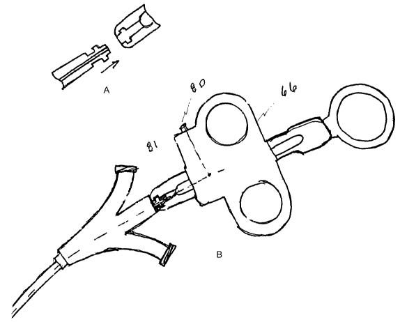
proximal end of the attached catheter. See Figs. 1 and 2. Handle 66, secured
to the
cutting wire at 80 but rotatable relative to the shaft of the catheter at 81,
provides a
mechanism to rotate the wire, transmitting the force to rotate the device tip.
With the
handle rotating independently of the shaft at the proximal end, the force can
be applied
directly to the distal tip without twisting the entire shaft. Also a rotation
lock to maintain
the orientation of the tip and/or a rotation marking, to indicate the amount
of rotation may
be included. An integrated molded luer port assembly for 2 and 3 lumen
catheters may
be provided to snap into the rotatable handle, to facilitate fast and
economical
manufacturing, as shown in Figs. l and 1 a. Alternatively, prior art serial
lumen ports
may be configured to snap into the rotatable handle as shown in Fig. 2.
Refernng to FIG. 12, the present invention also contains a feature known as a
rotation lock. Rotation lock 68 allows the user to maintain the orientation of
the tip at all
times. This is done by maintaining the position of handle 66 relative to
bifurcation
connector 67 after the handle has been rotated. Rotation lock 68 allows the
user to
release handle 66 at any time during the procedure, while maintaining the
orientation of
handle 66 and preventing further rotation while the lock is engaged.
Maintaining the
position of handle 66 maintains the orientation of the distal tip in the
desired orientation.
Maintaining the orientation of the distal tip reduces the amount of time and
effort
required to cannulate if the distal tip moved. Preventing undesired movement
of the
distal tip may also prevent patient injury.
Refernng to FIG. 13, two pair of mating detents 69 and slots 70 may be used to
create this rotation lock. Detents 69 and slots 70 are located along the
central axis of
body 71, at the intersection of body 71 and bifurcation connector 67. In FIG.
13, the two
pair of detents 69 and slots 70 are located 180° apart, relative to the
central axis. This
creates a lock position every half rotation of handle 66. During use of the
device, as
handle 66 is rotated, detents 69 become disengaged from slots 70. As detents
69 become
disengaged, they compress slightly. As handle 66 reaches a position
180° from where
rotation began, detents 69 recover from their compressed state, and engage
with slots 70
once again. As detents 69 traverse from one position to the next, there is a
noticeable
amount of friction between the mating components. This friction is great
enough that
CA 02417988 2003-02-12
WO 02/13711 PCT/USO1/25354
handle 66 can be released at any time without fear of losing the orientation
position of the
distal tip.
Rotation lock 68 also serves a secondary function of keeping the distal tip
locked
in the home position while the catheter is being removed from the package,
inserted into
the endoscope, and manipulated through the endoscope. Without this feature,
the initial
orientation position of the distal tip would become unpredictable. FIG. 13a
shows a
detailed diagram of the interaction between detents 69 and slots 70.
Referring to FIG. 14, when detents 69 and slots 70 axe engaged, bifurcation
connector 67 and finger rings 44 all lie in the same plane. This acts as the
rotation
marker. Whenever finder rings 44 are rotated into the same plane as
bifurcation
connector 67, the rotation lock is engaged, thus signaling 180° of
rotation from the last
position. The use of a marker such as this allows the user to more easily keep
track of
how much handle 66 has been rotated. This is helpful if the user desires to
move the
distal tip back to its original position. In effect, the user will know, for
example, that
handle 66 has been rotated three clicks from the original position. Therefore,
to return
handle 66 to the original position, it must be rotated three clicks in the
opposite direction.
FIGURES 15a-15d show alternative embodiments of rotation lock 68. FIGURE
15a shows a pure frictional lock. The connection of bifurcation connector 69
to the
handle 66 could be designed such that rotation lock 68 is purely a function of
frictional
interference between the two components. Alternative embodiments could include
different types of assembly joints to create this fiiction. In the primary
embodiment, the
assembly of the two components is accomplished by mating a male post of the
bifurcation connector to a female hole of the same size and shape. Alternative
embodiments could reverse this, so that the male protrusion is part of the
main body of
handle 66. The friction lock could also be built into the mating faces of main
body and
bifurcation connector, which are perpendicular to the major axis. FIGURE 16a
shows a
cross section of the rotation lock along Z-Z of FIGURE 15a.
FIGURE 15b shows a oval post lock embodiment of the present invention. The
connection of bifurcation connector 67 to handle 66 could also be designed
incorporating
an ovalized male post 73 and female hole 72. In this embodiment, as handle 66
is rotated
16
once again. As detents 6
CA 02417988 2003-02-12
WO 02/13711 PCT/USO1/25354
relative to bifurcation connector 67, ovalized hole 72 would deform, allowing
oval post
73 to rotate. As handle 66 reached a rotation of 1 ~0°, ovalized hole
72 would conform
back to its original shape, thus locking handle 66 in place. As shown in
FIGURES 15c
and 15d, this basic concept may be expanded to incorporate other shapes rather
than oval
as shown in FIGURE 15b. One of ordinary skill in the art would appreciate that
the
shape of the geometry however, governs the degrees of rotation between locked
positions. For example, if post 73 and ovalized hole 72 configuration were
made up of
mating equilateral triangles (FIGURE 15c), there would be 120° of
rotation between
locked positions. Using a square configuration (FIGURE 15d), would give
90° between
locked positions. FIGURE 16b, illustrates the cross-sectional area across Y-Y
of
FIGURE 15b. FIGURE 16c illustrates the cross-sectional area of FIGURE lSc
across X-
X and FIGURE 16d illustrates the cross-sectional area of FIGURE 15d along
cross-
section W-W.
FIGURES 17a-c show alternative embodiments by which a rotation marker may
be created and included in the present invention. One of ordinary skill in the
art would
understand these embodiments may be expanded. To aid the user in knowing
exactly
how much handle 66 has been rotated from its original andlor last position,
several forms
of visual markers can be incorporated into the design. One alternative
embodiment is
comprised of a set of lines placed radially, around the major axis, at the
area where the
main body and bifurcation connector 67 meets (FIG. 17a). A single line on the
stationary
component, bifurcation connector 67, would match up with a corresponding line
on body
43. As handle 66 is rotated relative to bifurcation connector 67, the series
of lines on the
body would rotate past the stationary line on bifurcation 67. Each line would
indicate an
incremental amount of movement. For example, if there were four, equally
spaced lines
on the body, each line that passed the marker on the bifurcation connection
would signify
90° of rotation.
This feature could be further enhanced by many methods. A series of numbers
rather than lines could be used to signify the amount of rotation (FIG. 17b).
Alternating
colors could also be used to signify the amount of rotation. Alternating line
patterns
could be used as well (FIG. 17c).
17
CA 02417988 2003-02-12
WO 02/13711 PCT/USO1/25354
Another alternative embodiment may use audible tones to make the user aware of
the amount of rotation. One method for doing this would be able to design the
rotation
lock features so that a click is clearly audible at predetermined points along
the rotational
travel of the body.
Referring to FIGURES 18a and 18b, there axe several alternative means by which
a bifurcation connector can be created. One of ordinary skill would understand
these
embodiments may be expanded from those presented in the current application.
Although the present invention is comprised of a connector with two lumens,
the
connector design could easily be modified to accommodate three or more lumens
(FIG.
18a). This would allow future designs to incorporate both guidewire post
connector 74
and injection port connector 75 into one component.
Another alternative to the bifurcation connector of the present design would
be
one, which also houses the electrical connector 76 (FIG. 18b). Electrical
connector 76,
presently incorporated-into the-finger ring, could be moved to the bifurcation
connector.
Referring now to FIGURES 19a and 19b, other embodiments of the present
invention may consist of handle 66 similar to that previously described, but
with the
addition of a bowing lock. A bowing lock would aid the user in that handle 66
could be
released at any time, and the catheter tip would maintain its bowed position.
Just as the
rotation lock provides for a safer and more efficient procedure, the bowing
lock would do
the same.
The bowing lock could be incorporated into the design in many ways. The
bowing lock, in its simplest form, would consist of friction lock 77 created
between
finger rings 44 and main body 43 (FIG. 19a). An alternative to this design
would create a
similar friction lock, but would use the surfaces between wire termination 78
and main
body 43 (FIG. 19b). The friction lock shown in figure 19b is enhanced by
incorporating
several lock ribs 79. Lock ribs 79 would be used to hold the catheter tip at a
specific,
predetermined angle. In effect, locking handle 66 into the first position
would, for
example, deflect the tip 30°. The next position would deflect the tip
60°. This feature
would give the user even more control when positioning the catheter tip within
the
anatomy. In both cases, as finger rings 44 are actuated along main body 43,
and catheter
18
CA 02417988 2003-02-12
WO 02/13711 PCT/USO1/25354
tip 80 is bowed, the friction between the mating components would hold the
position of
the handle, and thus hold the position of the bow.
19