Note: Descriptions are shown in the official language in which they were submitted.
CA 02418896 2003-02-07
WO 02/11639 PCT/US01/24916
GYNECOLOGICAL ABLATION PROCEDURE AND SYSTEM
USING AN ABLATION NEEDLE
FIELD OF THE INVENTION
The present invention relates to a procedure and system for treating
gynecological
disorders. More particularly, the present invention relates to the treatment
of pelvic
tumors.
BACKGROUND OF THE INVENTION
Benign and malignant tumors can occur in the pelvis. For example, uterine
leiomyomata, are muscle cell tumors that occur in 77% of women in the
reproductive
years. Although uterine leiomyomata rarely (0.1%) progress to cancer, these
tumors can
cause excessive menstrual bleeding, irregular bleeding, pregnancy loss,
infertility, urinary
frequency, and pelvic pressure or pain with sexual activity, menses, or daily
activities.
Women with uterine leiomyomata frequently incur surgical procedures (e.g.,
hysterectomy, dilatation and curettage, myomectomy, and hysteroscopy), medical
and
hormonal therapies, office visits, and a variety of radiologic procedures
(e.g., ultrasounds,
CAT scans, and MRIs), in an effort to treat these tumors. Uterine leiomyomata
account'
for approximately 200,000 hysterectomies per year in the United States alone,
at a direct
cost of well Over $2 billion. Hysterectomies carry a morbidity rate of 1%,
with 2,000
deaths per year and 240,000 complications per year in North America.
Uterine leiomyomata are most often multiple, and may be subserosal (i.e.,
bulging
externally from the uterus), intramural (i.e., growing entirely within the
wall of the
uterus), submucosal (i.e., hidden within the uterine cavity), or pedunculated
(i.e., growing
outward with a stalk-like base). Because patients may have multiple uterine
leiomyomata
at different locations, conservative surgeries may involve both an abdominal
and a
vaginal (hysteroscopic) approach, thereby necessitating two procedures. -
Investigators have utilized a laser or bipolar cautery to perform myolysis or
destruction of these tumors, although neither of these methods is performed in
significant
numbers today. These methods necessarily destroy normal overlying tissue in
order to
-1-
CA 02418896 2003-02-07
WO 02/11639 PCT/US01/24916
treat the underlying tumor. As a result, the integrity of the uterus is
compromised, and
harmful scar tissue (e.g., adhesions) may occur. Thus, there is a need for an
improved
method of treating benign and malignant pelvic tumors that does not damage the
overlying tissue. Such an improved method could be used on women who wish to
later
conceive and subsequently deliver. There is also a need for a single method
capable of
treating all sizes of subserosal, intramural, submucuosal, and pedunculated
tumors in all
*locations. A single method, which would relieve most or all symptoms of
abdominal or
pelvic pain/pressure, abnormal uterine bleeding, urinary frequency,
infertility, and
miscarriage, is also needed. In addition, it would be desirable for the method
to be less
invasive, cheaper, and safer than conventional methods of treating pelvic
tumors, and also
to allow for uterine preservation.
SUMMARY OF THE INVENTION
The present invention, also referred to as "the Halt procedure," is an
innovative,
outpatient procedure that utilizes electromagnetic energy to effectively
ablate pelvic
tumors. The invention employs an ablation device that uses radio-frequency
(RF) energy
to treat pelvic tumors, while sparing the surrounding normal tissue. Although
the
ablation device utilized in the present invention has FDA approval for
ablation of soft
tissue tumors, no known reports exist in the medical literature of the
ablation device's
application to uterine leiomyomata or other pelvic tumors. In addition,
current results
indicate that, compared to other conservative therapies, the present method is
very
effective. Thus far, the present invention has provided relief from all of the
types of
symptoms caused by pelvic tumors, such as uterine leiomyomata. Furthermore,
the
present invention is versatile, safe, and well-accepted by patients.
Advantages of the
present invention include a quick recovery time; typically no more than a
week, and
significant cost savings. More importantly, the present invention provides a
practical and
efficient way to achieve uterine conservation on an out-patient basis.
In accordance with one embodiment of the present invention, a method of
treating
a pelvic tumor includes inserting an ablation device into a pelvic region and
positioning
the ablation device proximate the pelvic tumor, using a laparoscope and an
imaging
device to confirm placement of the ablation apparatus. Various ablation
devices may be
used. For example, the ablation device may include no arms, a plurality of
deployable
arms, or separate needles that are inserted into the pelvic tumor. The method
further
-2-
CA 02418896 2003-02-07
WO 02/11639 PCT/US01/24916
includes delivering energy through the ablation device to the pelvic tumor to
ablate the
tumor. The method uses RF energy, however, other forms of energy, such as
microwave,
light (e.g., laser), or acoustic (e.g., ultrasound) energy may also be used to
ablate the
pelvic tumors.
In accordance with another embodiment of the present invention, a method of
treating pelvic tumors includes providing a patient on an operating table, and
at least one
monitor for a laparoscope and an imaging device, with the at least one monitor
located
across the operating table from a surgeon and proximate the patient's waist.
The at least
one monitor may be mounted on a tower located proximate the patient's waist.
An
energy source and the imaging device are provided adjacent to the at least one
monitor,
with the energy source and imaging device being located proximate the
patient's knees.
The method further includes inserting an ablation device into a pelvic region
of the
patient and positioning the device proximate a pelvic tumor. The location and
placement
of the ablation device with respect to the pelvic tumor is confirmed using the
laparoscope
and the imaging device. The method also includes delivering energy to the
pelvic tumor
to ablate the tumor. The tumor may be maintained at a temperature in the range
of
approximately 65 C and 100 C for at least 7 minutes to ablate the tumor.
In accordance with still another embodiment of the present invention, a
surgical
system for treating pelvic tumors in a patient lying on an operating table
includes an
ablation device, an energy source, a laparoscope, and an imaging device. The
energy
source is coupled to the ablation device and provides energy to the device to
ablate a
pelvic tumor. The laparoscope and the imaging device are connected to at least
one
monitor. The at least one monitor is located the operating table from a
surgeon and
proximate the patient's waist, while the energy source and imaging device are
located
alongside the at least one monitor and proximate the patient's knees.
The present invention procedure may be performed by laparoscopy (i.e., open
abdominal incision), percutaneously, or hysteroscopically. The Halt procedure
has most
often utilized conventional laparoscopy with the additional placement of (1) a
supra-
pubic port or sleeve (10 mm) at the top of the uterus for an intra-abdominal
ultrasound
probe and (2) an ablation device, also usually in the lower abdominal region.
The Halt
procedure has also been performed by a trans-abdominal technique, utilizing
-3-
CA 02418896 2012-03-15
54312-1
conventional trans-abdominal ultrasound and placement of the ablation device
trans-abdominally with laparoscopic confirmation, as well as by a trans-
cervical
technique.
In accordance with another embodiment of the invention, there is a
surgical system for ablating pelvic tumors, comprising: (a) an elongated
ablation
device, comprising: (i) a sharp tip; and (ii) an electrode mounted internally
of said
ablation device, said electrode being slideably mounted to be deployed from
said
device and positioned within a uterine fibroid; (b) an imaging device separate
from
the ablation device adapted to be positioned with respect to the ablation
device to
image the ablation device within the uterine fibroid; and (c) an energy source
coupled
to the ablation device comprising an energy output, said energy output being
coupled
to the electrode to directly ablate the uterine fibroid.
In accordance with still another embodiment of the invention, there is a
surgical system for ablating uterine fibroids in a patient, the system
comprising: (a)
an ablation device adapted to be disposed in a position in a uterine fibroid
of a
patient, wherein the ablation device comprises: (i) a tip; and (ii) a
plurality of
electrodes for direct ablation of the uterine fibroid; (b) an energy source
comprising
an energy output, said energy output being coupled to the ablation device; (c)
a
laparoscope adapted to be disposed in a position spaced apart from said
ablation
device; (d) said ablation device being configured and dimensioned to be
disposed
within a uterine fibroid of the patient to avoid contact with normal tissue
outside of the
uterine fibroid, and (e) a manipulator for mechanically engaging the uterus
and for
changing the position of one of said uterine fibroids.
In accordance with a further embodiment of the invention, there is a
surgical system for ablating a uterine fibroid in a patient, comprising: an
elongated
and pointed ablation device positionable and configured for insertion into a
uterine
fibroid in a patient, the ablation device comprising at least three electrodes
for direct
ablation of the uterine fibroid, said electrodes being pointed in
configuration for
- 4 -
CA 02418896 2012-03-15
54312-1
insertion into and engagement with the uterine fibroid within the fibroid and
when
deployed extending into a predictable and defined volume to avoid contact with
normal tissue outside of the uterine fibroid, said electrodes being mounted
within the
ablation device for deployment from the ablation device and retraction into
the
ablation device; an RF energy source coupled to the ablation device for
providing RF
energy to said electrodes; a laparoscope providing an optical image, and
adapted to
be disposed within the patient in a configuration to confirm placement of the
electrodes within the uterine fibroid, said laparoscope being spaced apart
from said
ablation device; an intra-abdominal ultrasound imaging probe, separate from
the
ablation device, adapted to be disposed within the patient in a configuration
to also
confirm placement of the electrodes within the uterine fibroid, the intra-
abdominal
ultrasound probe being separated by a distance from said ablation device to
result in
the intra-abdominal ultrasound probe imaging the uterus of the patient to
image a
location of the ablation device within the uterine fibroid; and an
insufflation device
disposed in a configuration for insufflating the abdomen and having a
sufficiently high
air pressure output to create an air pressure within the abdomen of the
patient with
the uterus hosting the uterine fibroid, allowing said laparoscope to visualize
the
uterus.
In accordance with a further embodiment of the invention, there is a
surgical system for ablating a uterine fibroid in a patient, comprising: an
ablation
device positionable and configured for insertion into a uterine fibroid in a
patient, the
ablation device comprising at least three electrodes for direct ablation of
the uterine
fibroid, said electrodes being configured for engagement with the uterine
fibroid within
the fibroid and to avoid contact with normal tissue outside of the uterine
fibroid, said
electrodes being mounted within the ablation device for deployment from the
ablation
device and retraction into the ablation device; an RF energy source coupled to
the
ablation device for providing RF energy to said electrodes; a laparoscope
adapted to
be disposed within the patient in a configuration to confirm placement of the
electrodes within the uterine fibroid, said laparoscope being spaced apart
from said
ablation device; an intra-abdominal ultrasound imaging probe, separate from
the
- 4a -
CA 02418896 2012-03-15
=
54312-1
ablation device, adapted to be disposed within the patient in a configuration
to also
confirm placement of the electrodes within the uterine fibroid, the intra-
abdominal
ultrasound probe being separated by a distance from said ablation device to
result in
the intra-abdominal ultrasound probe imaging the uterus of the patient to
image a
location of the ablation device within the uterine fibroid; and an
insufflation device
disposed in a configuration for insufflating the abdomen and creating an air
pressure
within the abdomen of the patient with the uterus hosting the uterine fibroid,
allowing
said laparoscope to visualize the uterus.
- 4b -
CA 02418896 2012-03-15
54312-1
BRIEF DESCRIPTION OF THE DRAWINGS
FIG. 1 is a perspective diagram of a surgical system for ablating pelvic
tumors, in
accordance with the present invention.
FIG. 2 is a top plan view of the surgical system of FIG. 1, illustrating an
arrangement of certain equipment with respect to a patient lying on an
operating table.
FIG. 3 is a flowchart illustrating a closed laparotomy method of ablating
pelvic
tumors in accordance with the present invention.
DETAILED DESCRIPTION OF TUE INVENTION
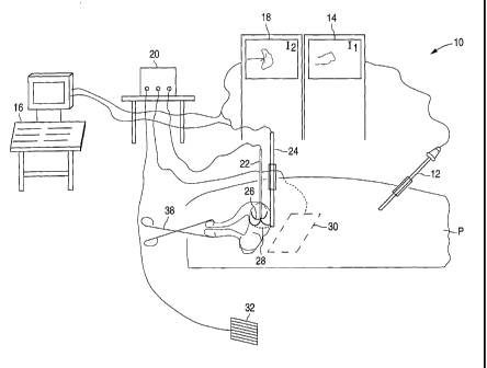
Referring first to FIG. 1, a surgical system 10 for ablating pelvic tumors
includes
a laparoscope 12, a video monitor 14 associated with laparoscope 12, an
imaging device
16, a video monitor 18 associated with imaging device 16, an energy source 20
and an
ablation device 22. Laparoscope 12, which is inserted into a patient P. is
electrically
connected to video monitor 14, which displays an image from laparoscope 12. As
will be
explained in greater detail below, laparoscope 12 enables a surgeon to view
the insertion
' and placement of ablation device 22 into a pelvic region of the patient.
Imaging device 16 is electrically connected to video monitor 18 and provides
images of the patient's pelvic region. These images, which are displayed on
video
monitor 18, enable the surgeon to determine the presence and location of any
pelvic
tuniors. Imaging device 16 shown in FIG. 1 is an ultrasound machine, and
includes an
intra-abdominal ultrasound probe 24. Instead of intra-abdominal ultrasound
probe 24, a
transducer (not shown) may be coupled to the ultrasound machine for trans-
abdominal
ultrasound imaging. In addition, other imaging devices, such as an 1\01
machine or a CT
device, may also be used instead of an ultrasound machine.
Ablation device 22 is a sterile, electrosurgical device that may include a
plurality
of retractable arms 26. FIG. 1 shows arms 26 of ablation device 22 deployed in
a pelvic
tumor 28. Examples of the ablation device include the Model 30 Electrosurgical
Device
and the RITA StarBurstTM XL, both available from RITA Medical Systems, Inc.
Each
- 4c -
CA 02418896 2003-02-07
WO 02/11639 PCT/US01/24916
arm 26 of ablation device 22 is a retractable curved electrode for delivering
energy and
has a thermocouple (not shown) located at the distal end. Although FIG. 1
shows
ablation device 22 as including deployable arms, an ablation device without
any arms
may also be used. Alternatively, the ablation device may include two or more
needles
that may be inserted into the tumor.
Ablation device 22 is coupled to energy source 20, which supplies energy to
each
of the arms 26 of ablation device 22. Energy source 20 may be an RF generator,
such as
the Model 500 Generator or the RITA Model 1500 RF Generator, both available
from
RITA Medical Systems, Inc. The supply of RF energy from energy source 20 to
ablation
device 22 and to a dispersive electrode 30 is controlled by an operator
control, such as by
a foot pedal 32. The application of RF energy causes an increase in tumor
temperature.
At sufficiently high temperatures, cell death occurs, thereby destroying the
tumor.
Energy source 20 may further include a mono-polar or bipolar energy source,
which allows the ablation device 22 to utilize traditional mono-polar or
bipolar cautery to
treat very small, superficial tumors and to ablate the track formed during
insertion of
ablation device 22. Cauterizing the ablation device track reduces or prevents
bleeding
upon withdrawal of ablation device 22 from the patient.
As better illustrated in FIG. 2, in accordance with the present invention, the
equipment of surgical system 10 is set up about the patient in a non-
traditional
arrangement. FIG. 2 illustrates the patient P lying in a dorsal position on an
operating
table 34. A tower 36, which supports video monitor 14 for laparoscope 12 and
imaging
device monitor 18, is located proximate the patient's waist, rather than at
the foot of
operating table 34. Since the surgeon S is located on the other side of
operating table 34
across from tower 36, the surgeon S has a direct view of the monitors 14 and
18. Video
monitors 14 and 18 need not be provided on tower 36; they may be suspended
from the
ceiling and located on the other side of operating table 34 across from the
surgeon S.
During longer surgical procedures, the placement of video monitors 14 and 18
directly
across from the surgeon is more comfortable for the surgeon, as the surgeon
need not turn
his/her head toward the foot of operating table 34 to view monitors 14 and 18.
Although FIGS. 1 and 2 show separate video monitors 14 and 18 for laparoscope
12 and imaging device 16, respectively, a single monitor capable of
simultaneously
-5-
CA 02418896 2003-02-07
WO 02/11639 PCT/US01/24916
displaying multiple images from the laparoscope and the imaging device, such
as a
picture-in-picture monitor, may also be used. The single monitor would be
located across
the table from the surgeon S and may be mounted on tower similar to tower 36,
suspended from the ceiling, or otherwise located across the patient from the
surgeon for
easy viewing by the surgeon.
=
Tower 36 may include additional equipment (not shown), such as an insufflation
machine, a printer, and a light source. Tower 36 may be provided with wheels
so that it
may be easily moved about the operating room. An additional monitor 37 for
laparoscope 12 may also be provided across from a surgical assistant A, who is
seated
across the table from the surgeon S, at approximately the patient's chest
level. Thus,
additional monitor 37 would be located adjacent the surgeon S. Additional
monitor 37
may mounted on a movable tower (not shown), suspended from the ceiling, or
otherwise
appropriately located.
Imaging device 16 and energy source 20, which are not located on tower 36, are
positioned along operating table 34, across from the surgeon S, and toward the
foot of
operating table 34. For example, imaging device 16 and energy source 20 may be
located
proximate the patient's knees.
A method of treating pelvic tumors, in accordance with one embodiment of the
present invention, will now be described, with reference to the flow chart
illustrated in
FIG. 3. This method 50 employs a laparoscopic technique for ablating pelvic
tumors.
First, at step 52, the patient is prepared for laparoscopy by placing and
properly adhering
dispersive electrode 30 to the lower back of the patient. At step 54, the
patient is then
placed under general anesthesia, and the surgeon performs an examination of
the pelvic
region. A manipulator 38 (FIG. 1), such as a tenaculum, is placed on the
patient's cervix,
and a 14 french foley catheter is inserted into the patient's bladder for
emptying the
bladder during the surgical procedure.
At step 56, the patient is placed in a dorsal position with her arms at her
sides,
rather than extended out as an airplane, and a blanket and a surgical drape
are placed over
the patient. This position provides the surgeon and surgical assistant with
more room to
move about. The dorsal position is also a safer position for the patient than
a frog-leg or
lithotomy position, as the dorsal position reduces the instance of nerve
injuries and
-6-
CA 02418896 2003-02-07
WO 02/11639 PCT/US01/24916
provides better circulation. In addition, the dorsal position does not require
the use of
custom drapes and stirrups. The surgical drape contains pouches for at least
one
laparoscopic cord. Serial compression devices (not shown) are placed on the
patient's
legs to improve circulation during the surgical procedure and reduce the
possibility of
thromboembolism. In addition, the patient may be placed in a bear hugger
system (not
shown) to maintain the patient's body temperature while under general
anesthesia.
At step 58, the equipment is arranged about operating table 34. As illustrated
in
FIG. 2, tower 36, which includes video monitors 14 and 18, an insufflation
machine, a
printer and a light source, is placed proximate the patient's waist and across
from the
surgeon S. The surgical assistant A is seated across the table from the
surgeon at about
the patient's chest level, with tower 34 located behind the assistant and
further toward the
foot of operating table 34. Imaging device 16 and energy source 20 are
situated alongside
operating table 34 on the same side as the assistant A and toward the foot of
operating
table 34. The additional monitor 37 is positioned across from the surgical
assistant A at
about the patient's chest level.
At step 60, the patient P is placed in a trendelenburg position. The surgeon
then
makes an infra-umbilical or sub-umbilical incision. A verres needle is then
inserted into
the incision and into the peritoneal cavity. The insufflation machine is then
used to
insufflate the abdomen with carbon dioxide gas until the abdominal pressure is
approximately 15 mm Hg.
Next, at step 62, a 5 mm trocar and sleeve are inserted through the infra-
umbilical
or sub-umbilical incision. The trocar is then removed and laparoscope 12 is
inserted into
the sleeve. Laparosope 12 and monitor 14 are then used to verify correct
placement of
laparoscope 12 within the peritoneal cavity and the absence of any trauma. The
sleeve is
attached to the carbon dioxide gas supply and includes a valve for controlling
the
abdominal pressure of the peritoneal cavity.
Steps 60 and 62 discussed above describe a closed laparoscopy procedure. For
those patients, for whom the surgeon feels an open laparoscopy would be
advantageous,
the surgeon would make an infra- or sub-umbilical incision and use a
combination of
blunt and sharp dissection through subcutaneous tissue. The surgeon would then
retract
the instruments for exposure. When the fascia is visualized, it is grasped
with one or
-7-
CA 02418896 2003-02-07
WO 02/11639 PCT/US01/24916
more clamps, elevated and incised. This provides a view of the peritoneum
below, which
may be bluntly or sharply incised. An appropriate laparoscopic sleeve is then
placed, and
the abdomen is insufflated with carbon dioxide gas. The laparoscope is then
inserted into
the sleeve.
At step 64, the surgeon then uses laparoscope 12, while palpating a top of the
uterine fundus, to determine an optimal location for an intra-abdominal
ultrasound probe.
The optimal location is generally at the top of the uterus, rather than supra-
pubic. An
incision is then made at this location and a 10 mm trocar and sleeve are
inserted. The
trocar is removed and ultrasound probe 24 is inserted into the sleeve. By way
of
example, the ultrasound probe 24 may be an Aloka model no. UST-5526L-7.5 probe
for
use with an Aloka model no. SSD140U ultrasound machine. Ultrasound probe 24
transmits an image of the pelvic region to ultrasound machine 16. The image is
displayed
on ultrasound video monitor 18, which is located on tower 36 proximate video
monitor
14 for laparoscope 12. Thus, the surgeon may simultaneously view the images on
video
monitors 14 and 18. As discussed above, a single monitor that simultaneously
displays
images from laparoscope 12 and imaging device 16 may be used instead of
separate
monitors 14 and 18.
At step 66, the surgeon examines the entire pelvis and abdomen to confirm the
presence or absence of any pathologies. The surgeon also uses laparoscope 12
and
ultrasound probe 24 to visualize any tumors, such as uterine leiomyomata. In
particular,
the surgeon takes note of the number of tumors, and the location and size of
each, and
compares that information with previously acquired data.
At step 68, the surgeon determines an order for treating the tumors. This
order is
determined based on the locations of the various tumors, and whether or not
the tumors
are accessible from a single midline location or require different locations
from which to
access the tumors. For example, if two tumors are generally along the same
track of
ablation device 22, the surgeon will first ablate the deeper tumor and, upon
retraction of
ablation device 22, ablate the remaining tumor. In addition, the surgeon may
choose to
ablate first a portion of the tumor that is furthest away from the vasculature
and work
toward the vasculature, or vice versa.
-8-
CA 02418896 2003-02-07
WO 02/11639 PCT/US01/24916
At step 70, the surgeon tests ablation device 22 to ensure that it is
operating
properly. Ablation device 22 is connected to generator 20, and proper feedback
from the
thermocouples, if any, is observed. In particular, the surgeon operates foot
pedal 32, or
any other appropriate operator control, to activate the supply of RF energy
from generator
20 and notes an appropriate rise in temperature and any peaks.
At step 72, if the surgeon decides that all of the tumors are approachable via
a
single midline location, the surgeon makes an incision, approximately 2.5 to
3.0 mm
long, and inserts ablation device 22. Entry of ablation device 22 is observed
using
laparoscope 12. The surgeon uses ultrasound probe 24 to visualize the size and
location
of the tumors with respect to ablation device 22.
Next, at step 74, the surgeon manipulates the patient's uterus using other
techniques to stabilize the uterus..
At step 76, after the surgeon has stabilized the uterus and located the
tumors, the
surgeon guides ablation device 22 into the uterus and the into a wall of the
uterus. The
surgeon may guide ablation device 22 by changing the position of the uterus
relative to
ablation device 22. In addition, the surgeon may rotate the ablation device
for better
penetration of the uterine wall with less movement of the uterus. Ablation
device 22 has
a plurality of markings (not shown) that enable the surgeon to note the depth
of
penetration of device 22. Confirmation of the location and placement of
ablation device
22 are provided by both laparoscope 12 and ultrasound probe 24.
Next, at step 78, the surgeon advances the tip of ablation device 22 to an
appropriate depth for treating a tumor. In doing so, the needle makes only a
very small
puncture. For example, an ablation device having a needle of 16 gauge may
produce a
puncture site of approximately 1 mm to 2 mm in diameter. The appropriate depth
depends on the size of the tumor. When ablation device 22 has been inserted to
the
appropriate depth, arms 26 of ablation device 22 are deployed to the
appropriate extent in
the tumor 28, as illustrated in FIG. 1. A 30 scope is used to ensure that all
of the arms
26 remain within the confines of the tumor and do not extend outside of the
organ. Arms
26 may effectively anchor ablation device 22 in tumor 28.
-9-
CA 02418896 2003-02-07
WO 02/11639 PCT/US01/24916
At step 80, the surgeon then records a baseline starting temperature of the
tumor.
The temperature of the tumor is obtained by the thermocouples located at the
distal ends
of arms 26 of ablation device 22.
At step 82, the surgeon then ablates the tumor by supplying RF energy from
generator 20 to ablation device 22. While generator 20 is activated, it is
important to
monitor the temperature or impedance of all parts of the ablation device. If
the
temperature or impedance for any part of ablation device 22 is abnormal, it
could indicate
that that part of the device is external to the organ.
RF energy is supplied to the tumor to raise the temperature of the tumor, such
that
it is in the range of between approximately 65 C and 100 C, for about 14
minutes. Cell
death occurs at a temperature of about 65 C. However, since these tumors are
heterogeneous and, therefore, can differ in density, vasculature and content,
a preferred
target temperature range for ablating pelvic tumors is between 85 C and 100
C. For
small tumors the target time may be between approximately 7 minutes and 14
minutes.
One of ordinary skill in the art, however, will appreciate that ablation times
of less than 7
minutes may also be adequate.
The temperature of the tumor, as provided by the thermocouples, is monitored
and
recorded at least at a 7 minutes and a 14 minutes interval. Thus, at least a
baseline
starting temperature, half-time temperature, and end-of-ablation-period
temperature are
recorded for each tumor. While RF energy is being delivered to the tumor, the
surgeon
keeps an eye on the monitors 14 and 18 to ensure that none of the arms 26 of
ablation
device 22 inadvertently extends through the tumor. The uterus can contract as
it is
heated, causing arms 26 of ablation device 22 to project from the tumor and
contact
normal tissue, which may be damaged by the RF energy. When the tumor has been
sufficiently ablated, energy source 20 is turned off.
After each ablation, at step 84 the uterus is irrigated with fluid. The fluid
prevents
the serosa from drying out as a result of the carbon dioxide gas that is
pumped into the
abdomen.
If the tumor is larger than the ablation field for the given ablation device,
then at
step 86, the surgeon may need to reposition ablation device 22 within another
part of the
tumor and reapply RF energy, repeating steps 76 through 84. Thus, if the
tumors are
-10-
_
CA 02418896 2003-02-07
WO 02/11639 PCT/US01/24916
greater in size than the ablation capacity of ablation device 22, multiple
applications of
energy, of overlapping ablation areas, may be necessary to ablate the bulk of
the tumor.
For tumors less than 3 cm, however, a single application of the RF energy
should be
sufficient to ablate the tumor.
At step 88, the surgeon then repositions ablation device 22 at the next tumor.
The
surgeon may leave ablation device 22 in the same track, if the next tumor is
along the
same line of approach. The surgeon would retract arms 26 and advance or
withdraw
ablation device 22 as needed for entry into another tumor. The surgeon would
then repeat
the ablation sequence of step 76 through step 86 described above.
If the subsequent tumor is in a different location, the surgeon may retract
arms 26
of ablation device 22 and withdraw ablation device 22, while applying a mono-
polar
cautery to reduce or prevent bleeding from the ablation device track.
Alternatively, rather
than completely withdraw ablation device 22 and re-insert ablation device 22
through
another incision, repeating steps 72 through 86, the surgeon may withdraw
ablation
device 22 until it is only 0.5 cm to 1 cm deep and adjust the uterus until the
desired angle
of approach is obtained and properly locating ablation device 22 with
ultrasound probe 24
or applying traction or pushing inward with uterine manipulator 38.
Small, superficial, subserosal fibroids (e.g., less than 1 cm) may be ablated
with a
mono-polar cautery at step 90. Bipolar paddles may also be used if the fibroid
extends
from the wall of the uterus. Similarly, if the tumor is pedunculated, the
surgeon may treat
or incise the stalk. Mono-polar or bipolar cautery may be applied to
subserosal,
intramural, and submucuos leiomyomata. In addition, other pelvic pathologies
are treated
as appropriate.
After all of the tumors have been ablated, at step 92, the surgeon confirms
hemostasis, withdraws ablation device 22, and applies a mono-polar cautery
with ablation
device 22 to the puncture sites, if necessary. A small amount of irrigation
fluid may be
left in the pelvis.
Finally, at step 94, documentation, including videotapes, ultrasound
photographs,
and photographs from the laparoscope are obtained. The sleeves are opened to
allow the
escape of the carbon dioxide gas. The patient is then removed from the
trendelenburg
position, and a local anesthetic agent is injected into the incisions. The
surgeon then
-11-
CA 02418896 2003-02-07
WO 02/11639
PCT/US01/24916
repairs the fascia of the 10 mm incision using an absorbable suture, S-
retractors to
facilitate visualization of the fascial edges. A1i5TM clamps are used to
facilitate grasping
for elevating the fascial edges for suturing, re-approximating the
subcutaneous tissue with
sutures, closing the skin, and placing SteristripTM bandages. The surgeon then
removes
the dispersive electrode 30 and examines the surrounding skin.
The patient is transported to a recovery room, where she will remain until she
is
tolerating liquids, ambulating with assistance, and voiding adequately.
If the patient's uterus is very large (e.g., 16 weeks or greater), the above-
described
laparoscopic technique may be less effective. Accordingly, a direct trans-
abdominal
insertion of ablation device 22 is performed with laparoscopic confirmation
only (e.g., no
intia-abdominal ultrasound confirmation). In this method the patient is
prepared in the
same manner as that described above at step 52. The surgeon also performs a
pelvic
examination, positions the patient, arranges the equipment, forms an infra-
umbilical
incision, insufflates the patient's abdomen, and inserts laparoscope 12, as in
step 54
through to step 62 above. Specifically, the surgeon inspects the abdomen and
documents
the presence or absence of bowel adhesions or other pathologic conditions that
would
render this method inappropriate.
Next, the surgeon releases the gas from the patient's abdomen, allowing the
abdominal wall to contact an anterior portion of the uterus. A sterile cover
drape over a
transducer allows for trans-abdominal ultrasound imaging using a non-sterile
transducer
(not shown). The ultrasound is used to locate and measure the tumors.
The surgeon then makes an incision for ablation device 22 and inserts ablation
device 22, using abdominal ultrasonography to guide its insertion. Ablation
device 22
may be inserted percutaneously, or trans-abdominally, into the tumor in the
uterus.
Ablation device 22 is positioned at a tumor and arms 26 are deployed in the
tumor, just as described above with respect to the laparoscopic method. Prior
to applying
RF energy to the tumor, the surgeon insufflates the abdomen and performs a
laparoscopy
to confirm that none of the arms 26 of ablation device 22 extend beyond the
uterine
tissue.
-12-
CA 02418896 2003-02-07
WO 02/11639 PCT/US01/24916
The surgeon then applies RF energy to the tumor, in the same manner as
described at step 80 through step 84 above, including recording the baseline,
half-time,
and end-of-ablation-period temperatures. The surgeon may use the same approach
as
described above to ablate multiple pelvic tumors. Upon withdrawal of the
ablation device
22, the surgeon fulgurates the ablation device track with a mono-polar
cautery. Thus,
remaining steps are the same as step 86 through step 94 described above.
The above-described methods enable the surgeon to ablate substantially all of
a
tumor from a single, ablation device puncture site. In addition, depending on
the location
of the tumors, multiple tumors may be ablated from a puncture site. The
methods further
enable the surgeon to treat all sizes of tumors in any area of the pelvic
region.
The foregoing description of the preferred embodiments of the present
invention
have been provided for illustrative purposes only. They are not intended to be
exhaustive
or to limit the invention to the precise forms disclosed. Various
modifications may be
made without departing from the spirit and scope of the inventions as set
forth in the
appended claims. For example, although the present invention has been
described with
respect to the treatment of uterine leiomyomata, the present invention may
also be used to
treat other pelvic tumors, such as those present in the ovaries. The present
invention may
be performed using a trans-cervical technique or a hysteroscopic technique, in
addition to
the laparoscopic and trans-abdominal techniques described above. The scope of
the
invention is defined by the following claims.
-13-