Note: Descriptions are shown in the official language in which they were submitted.
CA 02419205 2007-12-21
ANTI- TNF ANTIBODIES, COMPOSITIONS, METHODS AND USES
BACKGROUND OF THE INVENTION
FIELD OF THE INVENTION
The present invention relates to antibodies, including specified portions or
variants,
specific for at least one tumor necrosis factor alpha (TNF) protein or
fragment thereof, as well as
nucleic acids encoding such anti-TNF antibodies, complementary nucleic acids,
vectors, host
cells, and methods of making and using thereof, including therapeutic
formulations,
administration and devices.
RELATED ART
TNF alpha is a soluble homotrimer of 17 kD protein subunits (Smith et al., J.
Biol.
Chem. 262:6951-6954 (1987)). A membrane-bound 26 kD precursor form of TNF also
exists
(Kriegler et al., Cell 53:45-53 (1988)). For reviews of TNF, see Beutler et
al., Nature 320:584
(1986); Old, Science 230:630 (1986); and Le et al., Lab. Invest. 56:234
(1987).
Cells other than monocytes or macrophages also produce TNF alpha. For example,
human non-monocytic tumor cell lines produce TNF alpha (Rubin et al., J. Exp.
Med.' 164:1350
(1986); Spriggs et al., Proc. Natl. Acad. Sci. USA 84:6563 (1987)). CD4+ and
CD8+
peripheral blood T lymphocytes and some cultured T and B cell lines (Cuturi et
al., J. Exp. Med.
165:1581 (1987); Sung et al., J. Exp. Med. 168:1539 (1988); Turner et al.,
Eur. J. Inununol.
17:1807-1814 (1987)) also produce TNF alpha.
TNF alpha causes pro-inflammatory actions which result in tissue injury, such
as
degradation of cartilage and bone (Saklatvala, Nature 322:547-549 (1986);
Bertolini, Nature
319:516-518 (1986)), induction of adhesion molecules, inducing procoagulant
activity on
vascular endothelial cells (Pober et al., J. Immunol. 136:1680 (1986)),
increasing the adherence
of neutrophils and lymphocytes (Pober et al., J. Immunol. 13 8:3319 (1987)),
and stimulating the
release of platelet activating factor from macrophages, neutrophils and
vascular endothelial cells
(Camussi et-al., J. Exp. Med. 166:1390 (1987)).
Recent evidence associates TNF alpha with infections (Cerami et al., Immunol.
Today
9:28 (1988)), immune disorders, neoplastic pathologies (Oliff et al., Cell
50:555 (1987)),
autoimmune pathologies and graft-versus-host pathologies (Piguet et al., J.
Exp. Med. 166:1280
(1987)). The association of TNF alpha with cancer and infectious pathologies
is
7
CA 02419205 2003-02-07
WO 02/12502 PCT/US01/24785
often related to the host's catabolic state. Cancer patients suffer from
weight loss, usually
associated with anorexia.
The extensive wasting which is associated with cancer, and other diseases, is
known as "cachexia" (Kern et al., J. Parent. Enter. Nutr. 12:286-298 (1988)).
Cachexia
includes progressive weight loss, anorexia, and persistent erosion of lean
body mass in
response to a malignant growth. The cachectic state causes much cancer
morbidity and
mortality. There is evidence that TNF alpha is involved in cachexia in cancer,
infectious
pathology, and other catabolic states (see, e.g., Beutler and Cerami, Ann.
Rev. Immunol.
7:625-655 (1989)).
TNF alpha is believed to play a central role in gram-negative sepsis and
endotoxic shock (Michie et al., Br. J. Surg. 76:670-671 (1989); Debets et al.,
Second Vienna
Shock Forum, p. 463-466 (1989); Simpson et al., Crit. Care Clin. 5:27-47
(1989)), including
fever, malaise, anorexia, and cachexia. Endotoxin strongly activates
monocyte/macrophage
production and secretion of TNF alpha and other cytokines (Kornbluth et al.,
J. Immunol.
137:2585-2591 (1986)). TNF alpha and other monocyte-derived cytokines mediate
the
metabolic and neurohormonal responses to endotoxin (Michie et al., New Engl.
J. Med.
318:1481-1486 (1988)). Endotoxin administration to human volunteers produces
acute illness
with flu-like symptoms including fever, tachycardia, increased metabolic rate
and stress
hormone release (Revhaug et al., Arch. Surg. 123:162-170 (1988)). Circulating
TNF alpha
increases in patients suffering from Gram-negative sepsis (Waage et al.,
Lancet 1:355-357
(1987); Hammerle et al., Second Vienna Shock Forum, p. 715-718 (1989); Debets
et al., Crit.
Care Med. 17:489-497 (1989); Calandra et al., J. Infect. Dis. 161:982-987
(1990)).
Thus, TNF alpha has been implicated in inflammatory diseases, autoimmune
diseases,
viral, bacterial and parasitic infections, malignancies, and/or
neurogenerative diseases and is a
useful target for specific biological therapy in diseases, such as rheumatoid
arthritis and
Crohn's disease. Beneficial effects in open-label trials with a chimeric
monoclonal antibody to
TNF alpha (cA2) have been reported with suppression of inflammation and with
successful
retreatment after relapse in rheumatoid arthritis (Elliott et al., Arthritis
Rheum. 36:1681-1690
(1993); and Elliott et al., Lancet 344:1125-1127 (1994)) and in Crohn's
disease (Van Dullemen
et al., Gastroenterology 109:129-135 (1995)). Beneficial results in a
randomized, double-
blind, placebo-controlled trial with cA2 have also been reported in rheumatoid
arthritis with
suppression of inflammation (Elliott et al., Lancet 344:1105-1110 (1994)).
Antibodies to
a "modulator" material which was characterized as cachectin (later found to be
identical to
TNF) were disclosed by Cerami et al. (EPO Patent Publication 0212489, March 4,
1987).
Such antibodies were said to be useful in diagnostic immunoassays and in
therapy of shock in
bacterial infections. Rubin et al. (EPO Patent Publication 0218868, April 22,
1987) disclosed
2
CA 02419205 2003-02-07
WO 02/12502 PCT/US01/24785
monoclonal antibodies to human TNF, the hybridomas secreting such antibodies,
methods of
producing such antibodies, and the use of such antibodies in immunoassay of
TNF. Yone et al.
(EPO Patent Publication 0288088, October 26, 1988) disclosed anti-TNT
antibodies, including
mAbs, and their utility in immunoassay diagnosis of pathologies, in particular
Kawasaki's
pathology and bacterial infection. The body fluids of patients with Kawasaki's
pathology
(infantile acute febrile mucocutaneous lymph node syndrome; Kawasaki, T.,
Allergy 16:178
(1967); Kawasaki, T., Shonica (Pediatrics) 26:935 (1985)) were said to contain
elevated TNF
levels which were related to progress of the pathology (Yone et al., supra).
Other investigators have described mAbs specific for recombinant human TNF
which had neutralizing activity in vitro (Liang, C-M. et al. (Biochem.
Biophys. Res. Comm.
137:847-854 (1986); Meager, A. et al., Hybridoma 6:305-311 (1987); Fendly et
al., Hybridoma
6:359-369 (1987); Bringman, T.S. et al., Hybridoma 6:489-507 (1987); Hirai, M.
et al., J.
Immunol. Meth. 96:57-62 (1987); Moller, A. et al. (Cytokine 2:162-169 (1990)).
Some of
these mAbs were used to map epitopes of human TNF and develop enzyme
immunoassays
(Fendly et al., supra; Hirai et al., supra; Moller et al., supra) and to
assist in the purification of
recombinant TNF (Bringman et al., supra). However, these studies do not
provide a basis for
producing TNF neutralizing antibodies that can be used for in vivo diagnostic
or therapeutic
uses in humans, due to immunogenicity, lack of specificity and/or
pharmaceutical suitability.
Neutralizing antisera or mAbs to TNF have been shown in mammals other than man
to
abrogate adverse phaysiological changes and prevent death after lethal
challenge in
experimental endotoxemia and bacteremia. This effect has been demonstrated,
e.g., in rodent
lethality assays and in primate pathology model systems (Mathison, J.C. et
al., J. Clin. Invest.
81:1925-1937 (1988); Beutler, B. et al., Science 229:869-871 (1985); Tracey,
K.J. et al.,
Nature 330:662-664 (1987); Shimamoto, Y. et al., Immunol. Lett. 17:311-318
(1988); Silva,
A.T. et al., J. Infect. Dis. 162:421-427 (1990); Opal, S.M. et al., J. Infect:-
Dis. 161:1148-1152
(1990); Hinshaw, L.B. et al., Circ. Shock 30:279-292 (1990)).
Putative receptor binding loci of hTNF has been disclosed by Eck and Sprang
(J. Biol.
Chem. 264(29), 17595-17605 (1989), who identified the receptor binding loci of
TNF-a as
consisting of amino acids 11-13, 37-42, 49-57 and 155-157. PCT application
W091/02078
(priority date of August 7, 1989) discloses TNF ligands which can bind to
monoclonal
antibodies having the following epitopes: at least one of 1-20, 56-77, and 108-
127; at least two
of 1-20, 56-77, 108-127 and 138-149; all of 1-18, 58-65; 115-125 and 138-149;
all of 1-18, and
108-128; all of 56-79, 110-127 and 135- or 136-155; all of 1-30, 117-128 and
141-153; all of
1-26, 117-128 and 141-153; all of 22-40, 49-96 or -97, 110-127 and 136-153;
all of 12-22, 36-
45, 96-105 and 132-157; all of both of 1-20 and 76-90; all of 22-40, 69-97,
105-128 and 135-
3
CA 02419205 2003-02-07
WO 02/12502 PCT/US01/24785
155; all of 22-31 and 146-157; all of 22-40 and 49-98; at least one of 22-40,
49-98 and 69-97,
both of 22-40 and 70-87.
Non-human mammalian, chimeric, polyclonal (e.g., anti-sera) and/or monoclonal
antibodies (Mabs) and fragments (e.g., proteolytic digestion or fusion protein
products thereof)
are potential therapeutic agents that are being investigated in some cases to
attempt to treat
certain diseases. However, such antibodies or fragments can elicit an immune
response when
administered to humans. Such an immune response can result in an immune
complex-
mediated clearance of the antibodies or fragments from the circulation, and
make repeated
administration unsuitable for therapy, thereby reducing the therapeutic
benefit to the patient
and limiting the readministration of the antibody or fragment. For example,
repeated
administration of antibodies or fragments comprising non-human portions can
lead to serum
sickness and/or anaphalaxis. In order to avoid these and other problems, a
number of
approaches have been taken to reduce the immunogenicity of such antibodies and
portions
thereof, including chimerization and humanization, as well known in the art.
These and other
approaches, however, still can result in antibodies or fragments having some
immunogenicity,
low affinity, low avidity, or with problems in cell culture, scale up,
production, and/or low
yields. Thus, such antibodies or fragments can be less than ideally suited for
manufacture or
use as therapeutic proteins.
Accordingly, there is a need to provide anti-TNT antibodies or fragments that
overcome one more of these problems, as well as improvements over known
antibodies or
fragments thereof.
SUMMARY OF THE INVENTION
The present invention provides isolated human, primate, rodent, mammalian,
chimeric,
humanized and/or CDR-grafted anti-TNF antibodies, immunoglobulins,,cleavage
products and
other specified portions and variants thereof, as well as anti-TNF antibody
compositions,
encoding or complementary nucleic acids, vectors, host cells, compositions,
formulations,
devices, transgenic animals, transgenic plants, and methods of making and
using thereof, as
described and enabled herein, in combination with what is known in the art.
The present invention also provides at least one isolated anti-TNF antibody as
described herein. An antibody according to the present invention includes any
protein or
peptide containing molecule that comprises at least a portion of an
immunoglobulin molecule,
such as but not limited to at least one complementarity determinng region
(CDR) of a heavy or
light chain or a ligand binding portion thereof, a heavy chain or light chain
variable region, a
heavy chain or light chain constant region, a framework region, or any portion
thereof, that can
be incorporated into an antibody of the present invention. An antibody of the
invention can
4
CA 02419205 2003-02-07
WO 02/12502 PCT/US01/24785
include or be derived from any mammal, such as but not limited to a human, a
mouse, a rabbit,
a rat, a rodent, a primate, or any combination thereof, and the like.
The present invention provides, in one aspect, isolated nucleic acid molecules
comprising, complementary, or hybridizing to, a polynucleotide encoding
specific anti-TNF
antibodies, comprising at least one specified sequence, domain, portion or
variant thereof. The
present invention further provides recombinant vectors comprising said anti-
TNF antibody
nucleic acid molecules, host cells containing such nucleic acids and/or
recombinant vectors, as
well as methods of making and/or using such antibody nucleic acids, vectors
and/or host cells.
At least one antibody of the invention binds at least one specified epitope
specific to at
least one TNF protein, subunit, fragment, portion or any combination thereof.
The at least one
epitope can comprise at least one antibody binding region that comprises at
least one portion
of said protein, which epitope is preferably comprised of at least 1-5 amino
acids of at least
one portion thereof, such as but not limited to, at least one functional,
extracellular, soluble,
hydrophillic, external or cytoplasmic domain of said protein, or any portion
thereof.
The at least one antibody can optionally comprise at least one specified
portion of at
least one complementarity determining region (CDR) (e.g., CDR1, CDR2 or CDR3
of the
heavy or light chain variable region) and/or at least one constant or variable
framework region
or any portion thereof. The at least one antibody amino acid sequence can
further optionally
comprise at least one specified substitution, insertion or deletion as
described herein or as
known in the art.
The present invention also provides at least one isolated anti-TNF antibody as
described herein, wherein the antibody has at least one activity, such as, but
not limited to
inhibition of TNF-induced cell adhesion molecules, inhibition of TNF binding
to
receptor, Arthritic index improvement in mouse model, (see, e.g., Examples 3-
7). A(n)
anti-TNF antibody can thus be screened for a corresponding activity according
to known
methods, such as but not limited to, at least one biological activity towards
a TNF protein.
The present invention further provides at least one TNF anti-idiotype antibody
to at
least one TNF antibody of the present invention. The anti-idiotype antibody
includes any
protein or peptide containing molecule that comprises at least a portion of an
immunoglobulin
molecule, such as but not limited to at least one complementarity determinng
region (CDR) of
a heavy or light chain or a ligand binding portion thereof, a heavy chain or
light chain variable
region, a heavy chain or light chain constant region, a framework region, or
any portion
thereof, that can be incorporated into an antibody of the present invention.
An antibody of the
invention can include or be derived from any mammal, such as but not limited
to a human, a
mouse, a rabbit, a rat, a rodent, a primate, and the like.
5
CA 02419205 2009-04-27
The present invention provides, in one aspect, isolated nucleic acid molecules
comprising, complementary, or hybridizing to, a polynucleotide encoding at
least one TNF
anti-idiotype antibody, comprising at least one specified sequence, domain,
portion or
variant thereof. The present invention further provides recombinant vectors
comprising
said TNF anti-idiotype antibody encoding nucleic acid molecules, host cells
containing
such nucleic acids and/or recombinant vectors, as well as methods of making
and/or using
such anti-idiotype antibody nucleic acids, vectors and/or host cells.
The present invention also provides at least one method for expressing at
least one
anti-TNF antibody, or TNF anti-idiotype antibody, in a host cell, comprising
culturing a
host cell as described herein under conditions wherein at least one anti-TNF
antibody is
expressed in detectable and/or recoverable amounts.
The present invention also provides at least one composition comprising (a) an
isolated anti-TNF antibody encoding nucleic acid and/or antibody as described
herein; and
(b) a suitable carrier or diluent. The carrier or diluent can optionally be
pharmaceutically
acceptable, according to known carriers or diluents. The composition can
optionally further
comprise at least one further compound, protein or composition.
The present invention further provides at least one anti-TNF antibody method
or
composition, for administering a therapeutically effective amount to modulate
or treat at
least one TNF related condition in a cell, tissue, organ, animal or patient
and/or, prior to,
subsequent to, or during a related condition, as known in the art and/or as
described herein.
The present invention also provides at least one composition, device and/or
method
of delivery of a therapeutically or prophylactically effective amount of at
least one anti-
TNF antibody, according to the present invention.
The present invention further provides at least one anti-TNF antibody method
or
composition, for diagnosing at least one TNF related condition in a cell,
tissue, organ,
animal or patient and/or, prior to, subsequent to, or during a related
condition, as known in
the art and/or as described herein.
The present invention also provides at least one composition, device and/or
method
of delivery for diagnosing of at least one anti-TNF antibody, according to the
present
invention.
6
CA 02419205 2010-03-31
More particularly, in one aspect, the invention provides an antibody, or an
antigen-
binding fragment thereof, having three heavy chain complementarity determining
regions
(CDRs) and three light chain CDRs, wherein
each heavy chain CDR comprises the amino acid sequence of the corresponding
heavy chain CDR of mAb TNV 148 as described in Figure 4; and
each light chain CDR comprises the amino acid sequence of the corresponding
light
chain CDR of mAb TNV 148 as described in Figure 5.
In another aspect, the invention provides an antibody, or an antigen-binding
fragment thereof, comprising:
the heavy chain CDRs and variable framework regions (FRs) of mAb TNV 148 as
described in Figure 4; and
the light chain CDRs and variable FRs of mAb TNV148 as described in Figure 5;
optionally, further comprising the specified substitution from proline to
serine in
FR3 of mAb TNV 148B as described in Figure 4.
In one aspect, there is provided an antibody, or an antigen-binding fragment
thereof,
having three heavy chain complementarity determining regions (CDRs) and three
light
chain CDRs, wherein
each heavy chain CDR comprises the amino acid sequence of the corresponding
heavy chain CDR of mAb TNV 148: SYAMH, FMSYDGSNKKYADSVKG and
DRGIAAGGNYYYYGMDV; and
each light chain CDR comprises the amino acid sequence of the corresponding
light
chain CDR of mAb TNV 148: RASQSVYSYLA, DASNRAT, and
QQRSNWPPFT.
In one aspect, there is provided an isolated nucleic acid molecule comprising
a
polynucleotide encoding the antibody, or an antigen-binding fragment thereof,
described
herein.
In one aspect, there is provided a recombinant vector comprising the nucleic
acid
molecule described herein.
6a
CA 02419205 2010-03-31
In one aspect, there is provided a host cell comprising the nucleic acid
molecule of
claim 4 or the vector described herein.
In one aspect, there is provided a composition comprising the antibody, or an
antigen-binding fragment thereof, described herein and a pharmaceutically
acceptable
carrier or diluent.
In one aspect, there is provided the antibody, or an antigen-binding fragment
thereof, described herein or the composition described herein for use in
diagnosis or
therapy involving a TNF alpha related disorder.
In one aspect, there is provided the antibody, or an antigen-binding fragment
thereof, described herein or the composition described herein for use in
treating an immune
related disease attributed to TNF alpha.
In one aspect, there is provided an antibody, or an antigen-binding fragment
thereof,
of described herein wherein said antibody, or the antigen-binding fragment
thereof, binds to
tumor necrosis factor.
DESCRIPTION OF THE FIGURES
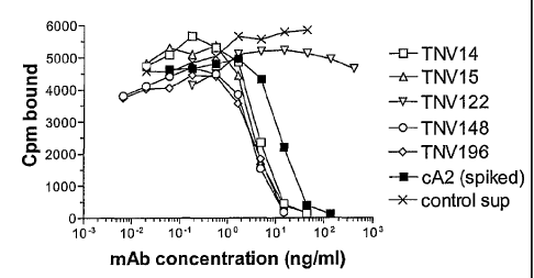
Figure 1 shows a graphical representation showing an assay for ability of TNV
mAbs in hybridoma cell supernatants to inhibit TNFV binding to recombinant TNF
receptor. Varying amounts of hybridoma cell supernatants containing known
amounts of
TNV mAb were preincubated with a fixed concentration (5 ng/ml) of 1251-labeled
TNFV.
The mixture was transferred to 96-well Optiplates that had been previously
coated with
p55-sf2, a recombinant TNF receptor/IgG fusion protein. The amount of TNFV
that bound
to the p55
6b
CA 02419205 2007-12-21
receptor in the presence of the mAbs was determined after washing away the
unbound
material and counting using a gamma counter. Although eight TNV mAb samples
were
tested in these experiments, for simplicity three of the mAbs that were shown
by DNA
sequence analyses to be identical to one of the other TNV mAbs (see Section
5.2.2) are not
shown here. Each sample was tested in duplicate. The results shown are
representative of two
independent experiments.
Figure 2A-B shows DNA and amino acid sequences of the TNV mAb heavy chain
variable regions. Figure 2A shows the n-terminal portion of the heavy chain
variable regions
including CDR1 and CDR2 as underlined. Figure 2B continues the heavy chain
variable
region sequences and includes the remaining c-terminal sequences including
CDR3. The
germline gene shown is the DP-46 gene. 'TNVs' indicates that the sequence
shown is the
sequence of TNVI4, TNVI5, TNV148, and TNV196. The first three nucleotides in
the TNV
sequence define the translation initiation Met codon. Dots in the TNV mAb gene
sequences
indicate the nucleotide is the same as in the germline sequence. The first 19
nucleotides
(underlined) of the TNV sequences correspond to the oligonucleotide used to
PCR-amplify
the variable region. An amino acid translation (single letter abbreviations)
starting with the
mature mAb is shown only for the germline gene. The three CDR domains in the
germline
amino acid translation are marked in bold and underlined. Lines labeled TNV
148(B) indicate
that the sequence shown pertains to both TN- V148 and TNVI48B. Gaps in the
germline DNA
sequence (CDR3) are due to the sequence not being known or not existing in the
germline
gene. The TNV mAb heavy chains use the J6 joining region.
Figure 3 shows DNA sequences of the TNV mAb light chain variable regions. The
germline gene shown is a representative member of the Vg/38K family of human
kappa
germline variable region genes. Dots in the TNV mAb gene sequences indicate
the
nucleotide is the same as in the germline sequence. The first 16 nucleotides
(underlined) of
the TNV sequences correspond to the oligonucleotide used to PCR-amplify the
variable
region. An amino acid translation of the mature mAb (single letter
abbreviations) is shown
only for the germline gene. The three CDR domains in the germline amino acid
translation
are marked in bold and underlined. Lines labeled TNV148(B) indicate that the
sequence
shown pertains to both TNV148 and TNV148B. Gaps in the germline DNA sequence
(CDR3) are due to the sequence not being known or not existing in the germline
gene. The
TNV mAb light chains use the J3 joining sequence.
Figure 4 shows deduced amino acid sequences of the TNV mAb heavy chain
variable regions. The amino acid sequences shown (single letter abbreviations)
were deduced
from DNA sequence determined from both uncloned PCR products and cloned PCR
products.
7
CA 02419205 2007-12-21
The amino sequences are shown partitioned into the secretory signal sequence
(signal),
framework (FW), and complementarity determining region (CDR) domains. The
amino acid
sequence for the DP-46 germline gene is shown on the top line for each domain.
Dots
indicate that the
7a
CA 02419205 2003-02-07
WO 02/12502 PCT/US01/24785
amino acid in the TNV mAb is identical to the germline gene. TNV 148(B)
indicates that the
sequence shown pertains to both TNV 148 and TNV148B. 'TNVs' indicates that the
sequence
shown pertains to all TNV mAbs unless a different sequence is shown. Dashes in
the germline
sequence (CDR3) indicate that the sequences are not known or do not exist in
the germline
gene.
Figure 5 shows deduced amino acid sequences of the TNV mAb light chain
variable
regions. The amino acid sequences shown (single letter abbreviations) were
deduced from
DNA sequence determined from both uncloned PCR products and cloned PCR
products. The
amino sequences are shown partitioned into the secretory signal sequence
(signal), framework
(FW), and complementarity determining region (CDR) domains. The amino acid
sequence for
the Vg/38K-type light chain germline gene is shown on the top line for each
domain. Dots
indicate that the amino acid in the TNV mAb is identical to the germline gene.
TNV148(B)
indicates that the sequence shown pertains to both TNV148 and TNV 148B. 'All'
indicates that
the sequence shown pertains to TNV14, TNV15, TNV148, TNV148B, and TNV186.
Figure 6 shows schematic illustrations of the heavy and light chain expression
plasmids used to make the rTNV148B-expressing C466 cells. p1783 is the heavy
chain
plasmid and p1776 is the light chain plasmid. The rTNV148B variable and
constant region
coding domains are shown as black boxes. The immunoglobulin enhancers in the J-
C introns
are shown as gray boxes. Relevant restriction sites are shown. The plasmids
are shown
oriented such that transcription of the Ab genes proceeds in a clockwise
direction. Plasmid
p1783 is 19.53 kb in length and plasmid p1776 is 15.06 kb in length. The
complete nucleotide
sequences of both plasmids are known. The variable region coding sequence in
p1783 can be
easily replaced with another heavy chain variable region sequence by replacing
the
BsiWI/BstBI restriction fragment. The variable region coding sequence in p1776
can be
replaced with another variable region sequence by replacing the SaIUAflII
restriction fragment.
Figure 7 shows graphical representation of growth curve analyses of five
rTNV148B-
producing cell lines. Cultures were initiated on day 0 by seeding cells into
T75 flasks in
I5Q+MHX media to have a viable cell density of 1.0 X 10⁵ cells/ml in a 30 ml
volume. The
cell cultures used for these studies had been in continuous culture since
transfections and
subclonings were performed. On subsequent days, cells in the T flasks were
thoroughly
resuspended and a 0.3 ml aliquot of the culture was removed. The growth curve
studies were
terminated when cell counts dropped below 1.5 X 10⁵ cells/ml. The number of
live cells in the
aliquot was determined by typan blue exclusion and the remainder of the
aliquot stored for
later mAb concentration determination. An ELISA for human IgG was performed on
all
sample aliquots at the same time.
8
CA 02419205 2003-02-07
WO 02/12502 PCT/US01/24785
Figure 8 shows a graphical representation of the comparison of cell growth
rates in the
presence of varying concentrations of MHX selection. Cell subclones C466A and
C466B were
thawed into MHX-free media (IMDM. 5% FBS, 2 mM glutamine) and cultured for two
additional days. Both cell cultures were then divided into three cultures that
contained either
no MHX, 0.2X MHX, or IX MHX. One day later, fresh T75 flasks were seeded with
the
cultures at a starting density of 1 X 10' cells/ml and cells counted at 24
hour intervals for one
week. Doubling times during the first 5 days were calculated using the formula
in SOP
PD32.025 and are shown above the bars.
Figure 9 shows graphical representations of the stability of mAb production
over time
from two rTNV 148B-producing cell lines. Cell subclones that had been in
continuous culture
since performing transfections and subclonings were used to start long-term
serial cultures in
24-well culture dishes. Cells were cultured in 15Q media with and without MHX
selection.
Cells were continually passaged by splitting the cultures every 4 to 6 days to
maintain new
viable cultures while previous cultures were allowed to go spent. Aliquots of
spent cell
supernatant were collected shortly after cultures were spent and stored until
the mAb
concentrations were determined. An ELISA for human IgG was performed on all
sample
aliquots at the same time.
Figure 10 shows arthritis mouse model mice Tg 197 weight changes in response
to
anti-TNF antibodies of the present invention as compared to controls in
Example 4. At
approximately 4 weeks of age the Tg197 study mice were assigned, based on
gender and body
weight, to one of 9 treatment groups and treated with a single intraperitoneal
bolus dose of
Dulbecco's PBS (D-PBS) or an anti-TNF anatibody of the present invention
(TNV14, TNV148
or TNV 196) at either 1 mg/kg or 10 mg/kg. When the weights were analyzed as a
change from
pre-dose, the animals treated with 10 mg/kg cA2 showed consistently higher
weight gain than
the D-PBS-treated animals throughout the study. This weight gain
was"significant at weeks 3-
7. The animals treated with 10 mg/kg TNV148 also achieved significant weight
gain at week
7 of the study.
Figures 11A-C represent the progression of disease severity based on the
arthritic
index as presented in Exanple 4. The 10 mg/kg cA2-treated group's arthritic
index was lower
then the D-PBS control group starting at week 3 and continuing throughout the
remainder of
the study (week 7). The animals treated with 1 mg/kg TNV 14 and the animals
treated with 1
mg/kg cA2 failed to show significant reduction in AI after week 3 when
compared to the D-
PBS-treated Group. There were no significant differences between the 10 mg/kg
treatment
groups when each was compared to the others of similar dose (10 mg/kg cA2
compared to 10
mg/kg TNV14, 148 and 196). When the 1 mg/kg treatment groups were compared,
the I
9
CA 02419205 2003-02-07
WO 02/12502 PCT/US01/24785
mg/kg TNV148 showed a significantly lower Al than I mg/kg cA2 at 3, 4 and 7
weeks. The I
mg/kg TNV 148 was also significantly lower than the 1 mg/kg TNV 14-treated
Group at 3 and 4
weeks. Although TNV 196 showed significant reduction in Al up to week 6 of the
study (when
compared to the D-PBS-treated Group), TNV148 was the only 1 mg/kg treatment
that
remained significant at the conclusion of the study.
Figure 12 shows arthritis mouse model mice Tg 197 weight changes in response
to
anti-TNF antibodies of the present invention as compared to controls in
Example 5. At
approximately 4 weeks of age the Tg197 study mice were assigned, based on body
weight, to
one of 8 treatment groups and treated with a intraperitoneal bolus dose of
control article (D-
PBS) or antibody (TNV 14, TNV 148) at 3 mg/kg (week 0). Injections were
repeated in all
animals at weeks 1, 2, 3, and 4. Groups 1-6 were evaluated for test article
efficacy. Serum
samples, obtained from animals in Groups 7 and 8 were evaluated for immune
response
induction and pharmacokinetic clearance of TNV14 or TN- V148 at weeks 2, 3 and
4.
Figures 13A-C are graphs representing the progression of disease severity in
Example
5 based on the arthritic index. The 10 mg/kg cA2-treated group's arthritic
index was
significantly lower then the D-PBS control group starting at week 2 and
continuing throughout
the remainder of the study (week 5). The animals treated with 1 mg/kg or 3
mg/kg of cA2 and
the animals treated with 3 mg/kg TNV14 failed to achieve any significant
reduction in Al at
any time throughout the study when compared to the d-PBS control group. The
animals treated
with 3 mg/kg TNV 148 showed a significant reduction when compared to the d-PBS-
treated
group starting at week 3 and continuing through week 5. The 10 mg/kg cA2-
treated animals
showed a significant reduction in AI when compared to both the lower doses (1
mg/kg and 3
mg/kg) of cA2 at weeks 4 and 5 of the study and was also significantly lower
than the TN-
V14-treated animals at weeks 3-5. Although there appeared to be no significant
differences between
any of the 3mg/kg treatment groups, the Al for the animals treated witll3
mg/kg TNV14 were
significantly higher at some time points than the 10 mg/kg whereas the animals
treated with
TNV148 were not significantly different from the animals treated with 10 mg/kg
of cA2.
Figure 14 shows arthritis mouse model mice Tg 197 weight changes in response
to
anti-TNF antibodies of the present invention as compared to controls in
Example 6. At
approximately 4 weeks of age the Tg197 study mice were assigned, based on
gender and body
weight, to one of 6 treatment groups and treated with a single intraperitoneal
bolus dose of
antibody (cA2, or TNV 148) at either 3 mg/kg or 5 mg/kg. This study utilized
the D-PBS and
10 mg/kg cA2 control Groups.
Figure 15 represents the progression of disease severity based on the
arthritic index as
presented in Example 6. All treatment groups showed some protection at the
earlier time
CA 02419205 2003-02-07
WO 02/12502 PCT/US01/24785
points, with the 5 mg/kg cA2 and the 5 mg/kg TNV 148 showing significant
reductions in AI at
weeks 1-3 and all treatment groups showing a significant reduction at week 2.
Later in the
study the animals treated with 5 mg/kg cA2 showed some protection, with
significant
reductions at weeks 4, 6 and 7. The low dose (3 mg/kg) of both the cA2 and the
TNV148
showed significant reductions at 6 and all treatment groups showed significant
reductions at
week 7. None of the treatment groups were able to maintain a significant
reduction at the
conclusion of the study (week 8). There were no significant differences
between any of the
treatment groups (excluding the saline control group) at any time point.
Figure 16 shows arthritis mouse model mice Tg 197 weight changes in response
to
anti-TNF antibodies of the present invention as compared to controls in
Example 7. To
compare the efficacy of a single intraperitoneal dose of TNV 148 (derived from
hybridoma
cells) and rTNV 148B (derived from transfected cells). At approximately 4
weeks of age the
Tg197 study mice were assigned, based on gender and body weight, to one of 9
treatment
groups and treated with a single intraperitoneal bolus dose of Dulbecco's PBS
(D-PBS) or
antibody (TNV148, rTNV 148B) at 1 mg/kg.
Figure 17 represents the progression of disease severity based on the
arthritic index as
presented in Example 7. The 10 mg/kg cA2-treated group's arthritic index was
lower then the
D-PBS control group starting at week 4 and continuing throughout the remainder
of the study
(week 8). Both of the TNV148-treated Groups and the 1 mg/kg cA2-treated'Group
showed a
significant reduction in Al at week 4. Although a previous study (P-099-0 17)
showed that
TNV148 was slightly more effective at reducing the Arthritic Index following a
single 1 mg/kg
intraperitoneal bolus, this study showed that the Al from both versions of the
TNV antibody-
treated groups was slightly higher. Although (with the exception of week 6)
the 1 mg/kg cA2-
treated Group was not significantly increased when compared to the 10'ing/kg
cA2 group and
the TNV148-treated Groups were significantly higher at weeks 7 and 8, there
were no
significant differences in Al between the 1 mg/kg cA2, 1 mg/kg TNV148 and 1
mg/kg
TNV148B at any point in the study.
DESCRIPTION OF THE INVENTION
The present invention provides isolated, recombinant and/or synthetic anti-
TNF human, primate, rodent, mammalian, chimeric, humanized or CDR-grafted,
antibodies
and TNF anti-idiotype antibodies thereto, as well as compositions and encoding
nucleic acid
molecules comprising at least one polynucleotide encoding at least one anti-
TNF antibody or
anti-idiotype antibody. The present invention further includes, but is not
limited to, methods
11
CA 02419205 2003-02-07
WO 02/12502 PCT/US01/24785
of making and using such nucleic acids and antibodies and anti-idiotype
antibodies, including
diagnostic and therapeutic compositions, methods and devices.
As used herein, an "anti-tumor necrosis factor alpha antibody," "anti-TNF
antibody," "anti-TNF antibody portion," or "anti-TNF antibody fragment" and/or
"anti-TNF
antibody variant" and the like include any protein or peptide containing
molecule that
comprises at least a portion of an immunoglobulin molecule, such as but not
limited to at least
one complementarity determinng region (CDR) of a heavy or light chain or a
ligand binding
portion thereof, a heavy chain or light chain variable region, a heavy chain
or light chain
constant region, a framework region, or any portion thereof, or at least one
portion of an TNF
receptor or binding protein, which can be incorporated into an antibody of the
present
invention. Such antibody optionally further affects a specific ligand, such as
but not limited to
where such antibody modulates, decreases, increases, antagonizes, angonizes,
mitigates,
aleviates, blocks, inhibits, abrogates and/or interferes with at least one TNF
activity or binding,
or with TNF receptor activity or binding, in vitro, in situ and/or in vivo. As
a non-limiting
example, a suitable anti-TNF antibody, specified portion or variant of the
present invention can
bind at least one TNF, or specified portions, variants or domains thereof . A
suitable anti-TNF
antibody, specified portion, or variant can also optionally affect at least
one of TNF activity or
function, such as but not limited to, RNA, DNA or protein synthesis, TNF
release, TNF
receptor signaling, membrane TNF cleavage, TNF activity, TNF production and/or
synthesis.
The term "antibody "is further intended to encompass antibodies, digestion
fragments,
specified portions and variants thereof, including antibody mimetics or
comprising portions of
antibodies that mimic the structure and/or function of an anitbody or
specified fragment or
portion thereof, including single chain antibodies and fragments thereof.
Functional fragments
include antigen-binding fragments that bind to a mammalian TNF. For example,
antibody
fragments capable of binding to TNF or portions thereof, including, but'not
limited to Fab
(e.g., by papain digestion), Fab' (e.g., by pepsin digestion and partial
reduction) and F(ab')2
(e.g., by pepsin digestion), facb (e.g., by plasmin digestion), pFc' (e.g., by
pepsin or plasmin
digestion), Fd (e.g., by pepsin digestion, partial reduction and
reaggregation), Fv or scFv (e.g.,
by molecular biology techniques) fragments, are encompassed by the invention
(see, e.g.,
Colligan, Immunology, supra).
Such fragments can be produced by enzymatic cleavage, synthetic or recombinant
techniques, as known in the art and/or as described herein. antibodies can
also be produced in a
variety of truncated forms using antibody genes in which one or more stop
codons have been
introduced upstream of the natural stop site. For example, a combination gene
encoding a
F(ab')2 heavy chain portion can be designed to include DNA sequences encoding
the CH,
domain and/or hinge region of the heavy chain. The various portions of
antibodies can be
12
CA 02419205 2007-12-21
joined together chemically by conventional techniques, or can be prepared as a
contiguous
protein using genetic engineering techniques.
As used herein, the term "human antibody" refers to an antibody in which
substantially
.every part of the protein (e.g., CDR, framework, CL, CH domains (e.g., CHI,
CH2, CH3), hinge,
(VL, VH)) is substantially non-immunogenic in humans, with only minor sequence
changes or
variations. Similarly, antibodies designated primate (monkey, babboon,
chimpanzee, etc.),
rodent (mouse, rat, rabbit, guinea pid, hamster, and the like) and other
mammals designate
such species, sub-genus, genus, sub-family, family specific antibodies.
Further, chimeric
antibodies include any combination of the above. Such changes or variations
optionally and
preferably retain or reduce the immunogenicity in humans or other species
relative to non-
modified antibodies. Thus, a human antibody is distinct from a chimeric or
humanized
antibody. It is pointed out that a human antibody can be produced by a non-
hurrian animal or
prokaryotic or eukaryotic cell that is capable of expressing functionally
rearranged human
immunoglobulin (e.g., heavy chain and/or light chain) genes. Further, when a
human antibody
is a single chain antibody, it can comprise a linker peptide that is not found
in native human
antibodies. For example, an Fv can comprise a linker peptide, such as two to
about eight
glycine or other amino acid residues, which connects the variable region of
the heavy chain
and the variable region of the light chain. Such linker peptides are
considered to be of human
origin.
Bispecific, heterospecific, heteroconjugate or similar antibodies can also be
used that
are monoclonal, preferably human or humanized, antibodies that have binding
specificities for
at least two different antigens. In the present case, one of the binding
specificities is for at least
one TNF protein, the other one is for any other antigen. Methods for making
bispecific
antibodies are known in the art. Traditionally, the recombinant production of
bispecific
antibodies is based on the co-expression of two immunoglobulin heavy chain-
light chain pairs,
where the two heavy chains have different specificities (Milstein and Cuello,
Nature 305:537
(1983)). Because of the random assortment of immunoglobulin heavy and light
chains, these
hybridomas (quadromas) produce a potential mixture of 10 different antibody
molecules, of
which only one has the correct bispecific structure. The purification of the
correct molecule,
which is usually done by affinity chromatography steps, is rather cumbersome,
and the product
yields are low. Similar procedures are disclosed, e.g., in WO 93/08829, US
Patent Nos,
6210668, 6193967, 6132992, 6106833, 6060285, 6037453, 6010902, 5989530,
5959084,
5959083, 5932448, 5833985, 5821333, 5807706, 5643759, 5601819, 5582996,
5496549,
4676980, WO 91/00360, WO 92/00373, EP 03089, Traunecker et al., EMBO J.
10:3655
(1991), Suresh et al., Methods in Enzymology 121:210 (1986).
13
CA 02419205 2007-12-21
Anti-TNF antibodies (also termed TNF antibodies) useful in the methods and
compositions of the present invention can optionally be characterized by high
affinity binding
to TNF and optionally and preferably having low toxicity. In particular, an
antibody, specified
fragment or variant of the invention, where the individual components, such as
the variable
region, constant region and framework, individually and/or collectively,
optionally and
preferably possess low immunogenicity, is useful in the present invention. The
antibodies that
can be used in the invention are optionally characterized by their ability to
treat patients for
extended periods with measurable alleviation of symptoms and low and/or
acceptable toxicity.
Low or acceptable immunogenicity and/or high affinity, as well as other
suitable properties,
can contribute to the therapeutic results achieved. "Low immunogenicity" is
defined herein as
raising significant HAHA, HACA or HAMA responses in less than about 75%, or
preferably
less than about 50% of the patients treated and/or raising low titres in the
patient treated (less
than about 300, preferably less than about 100 measured with a double antigen
enzyme
immunoassay) (Elliott et at., Lancet 344:1125-1127 (1994).
Utility
The isolated nucleic acids of the present invention can be used for production
of at least
one anti-TNF antibody or specified variant thereof, which can be used to
measure or effect in
an cell, tissue, organ or animal (including mammals and humans), to diagnose,
monitor,
modulate, treat, alleviate, help prevent the incidence of, or reduce the
symptoms of, at least
one TNF condition, selected from, but not limited to, at least one of an
immune disorder or
disease, a cardiovascular disorder or disease, an infectious, malignant,
and/or neurologic
disorder or disease.
Such a method can comprise administering an effective amount of a composition
or a
pharmaceutical composition comprising at least one anti-TNF antibody'to a
cell, tissue, organ,
animal or patient in need of such modulation, treatment, alleviation,
prevention, or reduction in
symptoms, effects or mechanisms. The effective amount can comprise an amount
of about
0.001 to 500 mg/kg per single (e.g., bolus), multiple or continuous
administration, or to
achieve a serum concentration of 0.01-5000 g/ml serum concentration per
single, multiple, or
continuous adminstration, or any effective range or value therein, as done and
determined
using known methods, as described herein or known in the relevant arts.
Citations
All publications or patents cited herein show the state of the art at the time
of the
present invention and/or provide description relevant to the present
invention. Publications
refer to any scientific or patent
14
CA 02419205 2007-12-21
publications, or any other information available in any media format,
including all recorded,
electronic or printed formats. The following references are relevant: Ausubel,
et al., ed.,
Current Protocols in Molecular Biology, John Wiley & Sons, Inc., NY, NY (1987-
2001);
Sambrook, et al., Molecular Cloning: A Laboratory Manual, 2"d Edition, Cold
Spring Harbor,
NY (1989); Harlow and Lane, antibodies, a Laboratory Manual, Cold Spring
Harbor, NY
(1989); Colligan, et al., eds., Current Protocols in Immunology, John Wiley &
Sons, Inc., NY
(1994-2001); Colligan et al., Current Protocols in Protein Science, John Wiley
& Sons, NY,
NY, (1997-2001).
Antibodies of the Present Invention
At least one anti-TNF antibody of the present invention can be optionally
produced
by a cell line, a mixed cell line, an immortalized cell or clonal population
of immortalized
cells, as well known in the art. See, e.g., Ausubel, et al., ed., Current
Protocols in Molecular
Biology, John Wiley & Sons, Inc., NY, NY (1987-2001); Sambrook, et al.,
Molecular
Cloning: A Laboratory Manual, 2"d Edition, Cold Spring Harbor, NY (1989);
Harlow and
Lane, antibodies, a Laboratory Manual, Cold Spring Harbor, NY (1989);
Colligan, et al., eds.,
Current Protocols in Immunology, John Wiley & Sons, Inc., NY (1994-200 1);
Colligan et al.,
Current Protocols in Protein Science, John Wiley & Sons, NY, NY, (1997-200 1).
Human antibodies that are specific for human TNF proteins or fragments thereof
can
be raised against an appropriate immunogenic antigen, such as isolated and/or
TNF protein or
a portion thereof (including synthetic molecules, such as synthetic peptides).
Other specific or
general mammalian antibodies can be similarly raised. Preparation of
immunogenic antigens,
and monoclonal antibody production can be performed using any suitable
technique.
In one approach, a hybridoma is produced by fusing a suitable immortal cell
line
(e.g., a myeloma cell line such as, but not limited to, Sp2/0, Sp2/0-AG14,
NSO, NS 1, NS2,
AE-l, L.5, >243, P3X63Ag8.653, Sp2 SA3, Sp2 MAI, Sp2 SS1, Sp2 SAS, U937, MLA
144,
ACT IV, MOLT4, DA-1, JURKAT, WEHI, K-562, COS, RAJI, NIH 3T3, HL-60, MLA 144,
NAMAIWA, NEURO 2A, or the like, or heteromylomas, fusion products thereof, or
any cell
or fusion cell derived therefrom, or any other suitable cell line as known in
the art, with
antibody producing cells, such as, but not limited to, isolated or cloned
spleen, peripheral
blood, lymph, tonsil, or other immune or B cell containing cells, or any other
cells expressing
heavy or light chain constant or variable or framework or CDR sequences,
either as
endogenous or heterologous nucleic acid, as recombinant or endogenous, viral,
bacterial,
algal, prokaryotic, amphibian, insect, reptilian, fish, mammalian, rodent,
equine, ovine, goat,
sheep, primate, eukaryotic, genomic DNA, cDNA, rDNA, mitochondrial DNA or RNA,
chloroplast DNA or RNA, hnRNA, mRNA,
CA 02419205 2007-12-21
tRNA, single, double or triple stranded, hybridized, and the like or any
combination thereof.
See, e.g., Ausubel, supra, and Colligan, Immunology, supra, chapter 2.
Antibody producing cells can also be obtained from the peripheral blood or,
preferably the spleen or lymph nodes, of humans or other suitable animals that
have been
immunized with the antigen of interest. Any other suitable host cell can also
be used for
expressing heterologous or endogenous nucleic acid encoding an antibody,
specified fragment
or variant thereof, of the present invention. The fused cells (hybridomas) or
recombinant cells
can be isolated using selective culture conditions or other suitable known
methods, and cloned
by limiting dilution or cell sorting, or other known methods. Cells which
produce antibodies
with the desired specificity can be selected by a suitable assay (e.g.,
ELISA).
Other suitable methods of producing or isolating antibodies of the requisite
specificity
can be used, including, but not limited to, methods that select recombinant
antibody from a
peptide or protein library (e.g., but not limited to, a bacteriophage,
ribosome, oligonucleotide,
RNA, cDNA, or the like, display library; e.g., as available from Cambridge
antibody
Technologies, Cambridgeshire, UK; MorphoSys, Martinsreid/Planegg, DE;
Biovation,
Aberdeen, Scotland, UK; BioInvent, Lund, Sweden; Dyax Corp., Enzon,
Affymax/Biosite;
Xoma, Berkeley, CA; Ixsys. See, e.g., EP 368,684, PCT/GB91/01134;
PCT/GB92/01755;
PCT/GB92/002240; PCT/GB92/00883; PCT/GB93/00605; US 08/350260(5/12/94);
PCT/GB94/01422; PCT/GB94/02662; PCT/GB97/01835; (CAT/MRC); W090/14443;
WO90/14424; WO90/14430; PCT/US94/1234; WO92/18619; WO96/07754; (Scripps); EP
614989 (MorphoSys); W095/16027 (BioInvent); W088/06630; W090/3809 (Dyax); US
4,704,692 (Enzon); PCT/US91/02989 (Affymax); W089/06283; EP 371 998; EP 550
400;
(Xoma); EP 229 046; PCT/US91/07149 (Ixsys); or stochastically generated
peptides or
proteins - US 5723323, 5763192, 5814476, 5817483, 5824514, 5976862, WO
86/05803, EP
590 689 (Ixsys, now Applied Molecular Evolution (AME)), or that rely upon
immunization of
transgenic animals (e.g., SCID mice, Nguyen et al., Microbiol. Immunol. 41:901-
907 (1997);
Sandhu et al., Crit. Rev. Biotechnol. 16:95-118 (1996); Eren et al., Immunol.
93:154-161
(1998), as well as related patents and applications) that are capable of
producing a repertoire
of human antibodies, as known in the art and/or as described herein. Such
techniques, include,
but are not limited to, ribosome display (Hanes et al., Proc. Natl. Acad. Sci.
USA, 94:4937-
4942 (May 1997); Hanes et al., Proc. Natl. Acad. Sci. USA, 95:14130-14135
(Nov. 1998));
single cell antibody producing technologies (e.g., selected lymphocyte
antibody method
("SLAM") (US pat. No. 5,627,052, Wen et al., J. Immunol. 17:887-892 (1987);
Babcook et
al., Proc. Natl. Acad. Sci. USA 93:7843-7848 (1996)); gel microdroplet and
flow cytometry
(Powell et al.,
16
CA 02419205 2007-12-21
Biotechnol. 8:333-337 (1990); One Cell Systems, Cambridge, MA; Gray et al., J.
Imm. Meth.
182:155-163 (1995); Kenny et al., Bio/Technol. 13:787-790 (1995)); B-cell
selection
(Steenbakkers et al., Molec. Biol. Reports 19:125-134 (1994); Jonak et al.,
Progress Biotech,
Vol. 5, In Vitro Immunization in Hybridoma Technology, Borrebaeck, ed.,
Elsevier Science
Publishers BY., Amsterdam, Netherlands (1988)).
Methods for engineering or humanizing non-human or human antibodies can also
be
used and are well known in the art. Generally, a humanized or engineered
antibody has one or
more amino acid residues from a source which is non-human, e.g., but not
limited to mouse,
rat, rabbit, non-human primate or other mammal. These human amino acid
residues are often
referred to as "import" residues, which are typically taken from an "import"
variable, constant
or other domain of a known human sequence. Known human Ig sequences are
disclosed,
on websites and on-line databases known to those of skill in the art.
17
CA 02419205 2007-12-21
Lonberg et al.; Jakobovits et al. WO 98/50433, Jakobovits et al. WO 98/24893,
Lonberg et al.
WO 98/24884, Lonberg et al, WO 97/13852, Lonberg et al. WO 94/25585,
Kucherlapate et al.
WO 96/34096, Kucherlapate et al. EP 0463 151 B 1, Kucherlapate et a!. EP 0710
719 Al,
Surani et al. US. Pat. No. 5,545,807, Bruggemann et at. WO 90/04036,
Bruggemann et al. EP
0438 474 B 1, Lonberg et al. EP 0814 259 A2, Lonberg et al. GB 2 272 440 A,
Lonberg et al.
Nature 368:856-859 (1994), Taylor et al., Int. Inununol. 6(4)579-591 (1994),
Green et al,
Nature Genetics 7:13-21 (1994), Mendez et at., Nature Genetics 15:146-156
(1997), Taylor et
al., Nucleic Acids Research 20(23):6287-6295 (1992), Tuaillon et al., Proc
Natl Acad Sol USA
90(8)3720-3724 (1993), Lonberg et at., Int Rev linmunol 13(1):65-93 (1995) and
Fishwald et
al., Nat Biotechnol 14(7):845-851 (1996).
Generally, these mice comprise at least one transgene comprising DNA from at
least one human immunoglobulin locus that is functionally rearranged, or which
can undergo
functional rearrangement. The endogenous immunoglobulin loci in such mice can
be
disrupted or deleted to eliminate the capacity of the animal to produce
antibodies encoded by
endogenous genes.
Screening antibodies for specific binding to similar proteins or fragments can
be
conveniently achieved using peptide display libraries. This method involves
the screening of
large collections of peptides for individual members having the desired
function or structure.
antibody screening of peptide display libraries is well known in the art. The
displayed peptide
sequences can be from 3 to 5000 or more amino acids in length, frequently from
5-100 amino
acids long, and often from about 8 to 25 amino acids long. In addition to
direct chemical
synthetic methods for generating peptide libraries, several recombinant DNA
methods have been
described. One type involves the display of a peptide sequence on the surface
of a bacteriophage
or cell. Each bacteriophage or cell contains the nucleotide sequence encoding
the particular
displayed peptide sequence. Such methods are described in PCT Patent
Publication Nos.
91/17271, 91/18980, 91/19818, and 93/08278. Other systems for generating
libraries of peptides
have aspects of both in vitro chemical synthesis and recombinant methods. See,
PCT Patent
Publication Nos. 92/05258, 92/14843, and 96/19256. See also, U.S. Patent Nos,
5,658,754; and
5,643,768. Peptide display libraries, vector, and screening kits are
commercially available from
such suppliers as Invitrogen (Carlsbad, CA), and Cambridge antibody
Technologies
(Cambridgeshire, UK). 'See, e.g., U.S. Pat. Nos. 4704692,4939666,4946778,
5260203,
5455030, 5518889, 5534621, 5656730, 5763733, 5767260, 5856456, assigned to
Enzon;
5223409, 5403484, 5571698, 5837500, assigned to Dyax, 5427908, 5580717,
assigned to
Affymax; 5885793, assigned to Cambridge antibody Technologies; 5750373,
assigned to
Genentech, 5618920, 5595898, 5576195, 5698435, 5693493, 5698417, assigned to
Xoma,
19
CA 02419205 2007-12-21
Such imported sequences can be used to reduce immunogenicity or reduce,
enhance or
modify binding, affinity, on-rate, off-rate, avidity, specificity, half-life,
or any other suitable
characteristic, as known in the art. Generally part or all of the non-human or
human CDR
sequences are maintained while the non-human sequences of the variable and
constant regions
are replaced with human or other amino acids. antibodies can also optionally
be humanized
with retention of high affinity for the antigen and other favorable biological
properties. To
achieve this goal, humanized antibodies can be optionally prepared by a
process of analysis of
the parental sequences and various conceptual humanized products using three-
dimensional
models of the parental and humanized sequences. Three-dimensional
immunoglobulin models
are commonly available and are familiar to those skilled in the art. Computer
programs are
available which illustrate and display probable three-dimensional
conformational structures of
selected candidate immunoglobulin sequences. Inspection of these displays
permits analysis of
the likely role of the residues in the functioning of the candidate
immunoglobulin sequence,
i.e., the analysis of residues that influence the ability of the candidate
immunoglobulin to bind
its antigen. In this way, FR residues can be selected and combined from the
consensus and
import sequences so that the desired antibody characteristic, such as
increased affinity for the
target antigen(s), is achieved. In general, the CDR residues are directly and
most substantially
involved in influencing antigen binding. Humanization or engineering of
antibodies of the
present invention can be performed using any known method, such as but not
limited to those
described in, Winter (Jones et al., Nature 321:522 (1986); Riechmann et al.,
Nature 332:323
(1988); Verhoeyen et al., Science 239:1534 (1988)), Sims et al., J. Immunol.
151: 2296 (1993);
Chothia and Lesk, J. Mol. Biol. 196:901 (1987), Carter et al., Proc. Natl.
Acad. Sci. U.S.A.
89:4285 (1992); Presta et al., J. Immunol. 151:2623 (1993), US patent Nos:
5723323,
5976862, 5824514, 5817483, 5814476, 5763192, 5723323, 5,766886, 5714352,
6204023,
6180370, 5693762, 5530101, 5585089, 5225539; 4816567, PCT/: US98/16280,
US96/18978,
US91/09630, US91/05939, U594/01234, GB89/01334, GB91/01134, GB92/01755;
W090/14443, W090/14424, W090/14430, EP 229246.
The anti-TNF antibody can also be optionally generated by immunization of a
transgenic animal (e.g., mouse, rat, hamster, non-human primate, and the like)
capable of
producing a repertoire of human antibodies, as described herein and/or as
known in the art.
Cells that produce a human anti-TNF antibody can be isolated from such animals
and
immortalized using suitable methods, such as the methods described herein.
Transgenic mice that can produce a repertoire of human antibodies that bind to
human
antigens can be produced by known methods (e.g., but not limited to, U.S. Pat.
Nos: 5,770,428,
5,569,825, 5,545,806, 5,625,126, 5,625,825, 5,633,425, 5,661,016 and 5,789,650
issued to
18
CA 02419205 2007-12-21
Colligan, supra; Ausubel, supra; or Sambrook, supra.
Antibodies of the present invention can also be prepared using at least one
anti-TNF
antibody encoding nucleic acid to provide transgenic animals or mammals, such
as goats,
cows, horses, sheep, and the like, that produce such antibodies in their milk.
Such animals can
be provided using known methods. See, e.g., but not limited to, US patent nos.
5,827,690;
5,849,992; 4,873,316; 5,849,992; 5,994,616; 5,565,362; 5,304,489, and the
like..
Antibodies of the present invention can additionally be prepared using at
least one
anti-TNF antibody encoding nucleic acid to provide transgenic plants and
cultured plant cells
(e.g., but not limited to tobacco and maize) that produce such antibodies,
specified portions or
variants in the plant parts or in cells cultured therefrom. As a non-limiting
example, transgenic
tobacco leaves expressing recombinant proteins have been successfully used to
provide large
amounts of recombinant proteins, e.g., using an inducible promoter. See, e.g.,
Cramer et al.,
Curr. Top. Microbol. Immunol. 240:95-118 (1999) and references cited therein.
Also,
transgenic maize have been used to express mammalian proteins at commercial
production
levels, with biological activities equivalent to those produced in other
recombinant systems or
purified from natural sources, See, e.g., Hood et al., Adv. Exp. Med. Biol.
464:127-147 (1999)
and references cited therein. antibodies have also been produced in large
amounts from
transgenic plant seeds including antibody fragments, such as single chain
antibodies (scFv's),
including tobacco seeds and potato tubers, See, e.g., Conrad et al., Plant
Mol. Biol. 38:101-
109 (1998) and reference cited therein. Thus, antibodies of the present
invention can also be
produced using transgenic plants, according to know methods. See also, e.g.,
Fischer et al.,
Biotechnol. Appl. Biochem. 30:99-108 (Oct., 1999), Ma et al., Trends
Biotechnol. 13:522-7
(1995); Ma et al., Plant Physiol. 109:341-6 (1995); Whitelam et al., Biochem.
Soc. Trans.
22:940-944 (1994); and references cited therein. See, also generally for plant
expression of
antibodies.
The antibodies of the invention can bind human TNF with a wide range of
affinities
(KD), In a preferred embodiment, at least one human mAb of the present
invention can
optionally bind human TNF with high affinity. For example, a human mAb can
bind human
TNF with a KD equal to or less than about 10' M, such as but not limited to,
0.1-9.9 (or any
range or value therein) X 10"7, 10.8, 10"9,10'`0, 10"", 10"12 , 10'13 or any
range or value therein.
The affinity or avidity of an antibody for an antigen can be determined
experimentally
using any suitable method. (See, for example, Berzofsky, et al., "Antibody-
Antigen
Interactions," In Fundamental Immunology, Paul, W. E., Ed., Raven Press: New
York, NY
CA 02419205 2003-02-07
WO 02/12502 PCT/US01/24785
(1984); Kuby, Janis Lnrnunology, W. H. Freeman and Company: New York, NY
(1992); and
methods described herein). The measured affinity of a particular antibody-
antigen interaction
can vary if measured under different conditions (e.g.,, salt concentration,
pH). Thus,
measurements of affinity and other antigen-binding parameters (e.g., KD, Kõ
Kd) are preferably
made with standardized solutions of antibody and antigen, and a standardized
buffer, such as
the buffer described herein.
Nucleic Acid Molecules
Using the information provided herein, such as the nucleotide sequences
encoding at
least 70-100% of the contiguous amino acids of at least one of SEQ ID NOS:1,
2, 3, 4, 5, 6, 7,
8, specified fragments, variants or consensus sequences thereof, or a
deposited vector
comprising at least one of these sequences, a nucleic acid molecule of the
present invention
encoding at least one anti-TNF antibody can be obtained using methods
described herein or as
known in the art.
Nucleic acid molecules of the present invention can be in the form of RNA,
such as
mRNA, hnRNA, tRNA or any other form, or in the form of DNA, including, but not
limited to,
cDNA and genomic DNA obtained by cloning or produced synthetically, or any
combinations
thereof. The DNA can be triple-stranded, double-stranded or single-stranded,
or any
combination thereof. Any portion of at least one strand of the DNA or RNA can
be the coding
strand, also known as the sense strand, or it can be the non-coding strand,
also referred to as
the anti-sense strand.
Isolated nucleic acid molecules of the present invention can include nucleic
acid
molecules comprising an open reading frame (ORF), optionally with one or more
introns, e.g.,
but not limited to, at least one specified portion of at least one CDR, as
CDR1, CDR2 and/or
CDR3 of at least one heavy chain (e.g., SEQ ID NOS:1-3) or light chain (e.g.,
SEQ ID NOS:
4-6); nucleic acid molecules comprising the coding sequence for an anti=TNF
antibody or
variable region (e.g., SEQ ID NOS:7,8); and nucleic acid molecules which
comprise a
nucleotide sequence substantially different from those described above but
which, due to the
degeneracy of the genetic code, still encode at least one anti-TNF antibody as
described herein
and/or as known in the art. Of course, the genetic code is well known in the
art. Thus, it
would be routine for one skilled in the art to generate such degenerate
nucleic acid variants
that code for specific anti-TNF antibodies of the present invention. See,
e.g., Ausubel, et al.,
supra, and such nucleic acid variants are included in the present invention.
Non-limiting
examples of isolated nucleic acid molecules of the present inveniton include
SEQ ID NOS:10,
11, 12, 13, 14, 15, corresponding to non-limiting examples of a nucleic acid
encoding,
respectively, HC CDR1, HC CDR2, HC CDR3, LC CDR1, LC CDR2, LC CDR3, HC
variable
region and LC variable region.
21
CA 02419205 2003-02-07
WO 02/12502 PCT/US01/24785
In another aspect, the invention provides isolated nucleic acid molecules
encoding a(n)
anti-TNF antibody having an amino acid sequence as encoded by the nucleic acid
contained in
the plasmid deposited as designated clone names and
ATCC Deposit Nos. , respectively, deposited
on
As indicated herein, nucleic acid molecules of the present invention which
comprise a
nucleic acid encoding an anti-TNF antibody can include, but are not limited
to, those encoding
the amino acid sequence of an antibody fragment. by itself; the coding
sequence for the entire
antibody or a portion thereof; the coding sequence for an antibody, fragment
or portion, as well
as additional sequences, such as the coding sequence of at least one signal
leader or fusion
peptide, with or without the aforementioned additional coding sequences, such
as at least one
intron, together with additional, non-coding sequences, including but not
limited to, non-
coding 5' and 3' sequences, such as the transcribed, non-translated sequences
that play a role
in transcription, mRNA processing, including splicing and polyadenylation
signals (for
example - ribosome binding'and stability of mRNA); an additional coding
sequence that codes
for additional amino acids, such as those that provide additional
functionalities. Thus, the
sequence encoding an antibody can be fused to a marker sequence, such as a
sequence
encoding a peptide that facilitates purification of the fused antibody
comprising an antibody
fragment or portion.
Polynucleotides Which Selectively Hybridize to a Polynucleotide as Described
Herein
The present invention provides isolated nucleic acids that hybridize under
selective
hybridization conditions to a polynucleotide disclosed herein. Thus, the
polynucleotides of this
embodiment can be used for isolating, detecting, and/or quantifying nucleic
acids comprising
such polynucleotides. For example, polynucleotides of the present invention
can be used to
identify, isolate, or amplify partial or full-length clones in a deposited
library. In some
embodiments, the polynucleotides are genomic or cDNA sequences isolated, or
otherwise
complementary to, a cDNA from a human or mammalian nucleic acid library.
Preferably, the cDNA library comprises at least 80% full-length sequences,
preferably at
least 85% or 90% full-length sequences, and more preferably at least 95% full-
length sequences.
The cDNA libraries can be normalized to increase the representation of rare
sequences. Low or
moderate stringency hybridization conditions are typically, but not
exclusively, employed with
sequences having a reduced sequence identity relative to complementary
sequences. Moderate
and high stringency conditions can optionally be employed for sequences of
greater identity.
Low stringency conditions allow selective hybridization of sequences having
about 70%
sequence identity and can be employed to identify orthologous or paralogous
sequences.
22
CA 02419205 2007-12-21
Optionally, polynucleotides of this invention will encode at least a portion
of an antibody
encoded by the polynucleotides described herein. The polynucleotides of this
invention embrace
nucleic acid sequences that can be employed for selective hybridization to a
polynucleotide
encoding an antibody of the present invention. See, e.g., Ausubel, supra;
Colligan, supra.
Construction of Nucleic Acids
The isolated nucleic acids of the present invention can be made using (a)
recombinant
methods, (b) synthetic techniques, (c) purification techniques, or
combinations thereof, as well-
known in the art.
The nucleic acids can conveniently comprise sequences in addition to a
polynucleotide of
the present invention. For example, a multi-cloning site comprising one or
more endonuclease
restriction sites can be inserted into the nucleic acid to aid in isolation of
the polynucleotide.
Also, translatable sequences can be inserted to aid in the isolation of the
translated polynucleotide
of the present invention. For example, a hexa-histidine marker sequence
provides a convenient
means to purify the proteins of the present invention. The nucleic acid of the
present invention -
excluding the coding sequence - is optionally a vector, adapter, or linker for
cloning and/or
expression of a polynucleotide of the present invention.
Additional sequences can be added to such cloning and/or expression sequences
to
optimize their function in cloning and/or expression, to aid in isolation of
the polynucleotide, or
to improve the introduction of the polynucleotide into a cell. Use of cloning
vectors, expression
vectors, adapters, and linkers is well known in the art. (See, e.g., Ausubel,
supra; or Sambrook,
supra)
Recombinant Methods for Constructing Nucleic Acids
The isolated nucleic acid compositions of this invention, such as RNA, cDNA,
genomic
DNA, or any combination thereof, can be obtained from biological sources using
any number of
cloning methodologies known to those of skill in the art. In some embodiments,
oligonucleotide
probes that selectively hybridize, under stringent conditions, to the
polynucleotides of the present
invention are used to identify the desired sequence in a cDNA or genomic DNA
library. The
isolation of RNA, and construction of cDNA and genomic libraries, is well
known to those of
ordinary skill in the art. (See, e.g., Ausubel, supra; or Sambrook, supra)
Nucleic Acid Screening and Isolation Methods
A cDNA or genomic library can be screened using a probe based upon the
sequence of a
polynucleotide of the present invention, such as those disclosed herein.
Probes can be used to
hybridize with genomic DNA or cDNA sequences to isolate homologous genes in
the same or
different organisms. Those, of skill in the art will appreciate that various
degrees of stringency of
hybridization can be employed in the assay; and either the hybridization or
the wash medium can
23
CA 02419205 2007-12-21
be stringent. As the conditions for hybridization become more stringent, there
must be a greater
degree of complementarity between the probe and the target for duplex
formation to occur. The
degree of stringency can be controlled by one or more of temperature, ionic
strength, pH and the
presence of a partially denaturing solvent such as formamide. For example, the
stringency of
hybridization is conveniently varied by changing the polarity of the reactant
solution through, for
example, manipulation of the concentration of formamide within the range of 0%
to 50%. The
degree of complementarity (sequence identity) required for detectable binding
will vary in
accordance with the stringency of the hybridization medium and/or wash medium.
The degree of
complementarity will optimally be 100%, or 70-100%, or any range or value
therein. However,
it should be understood that minor sequence variations in the probes and
primers can be
compensated for by reducing the stringency of the hybridization and/or wash
medium.
Methods of amplification of RNA or DNA are well known in the art and can be
used
according to the present invention without undue experimentation, based on the
teaching and
guidance presented herein.
Known methods of DNA or RNA amplification include, but are not limited to,
polymerase chain reaction (PCR) and related amplification processes (see,
e.g., U.S. Patent
Nos. 4,683,195, 4,683,202, 4,800,159, 4,965,188, to Mullis, et al.; 4,795,699
and 4,921,794 to
Tabor, et al; 5,142,033 to Innis; 5,122,464 to Wilson, et al.; 5,091,310 to
Innis; 5,066,584 to
Gyllensten, et al; 4,889,818 to Gelfand, et al; 4,994,370 to Silver, et al;
4,766,067 to Biswas;
4,656,134 to Ringold) and RNA mediated amplification that uses anti-sense RNA
to the target
sequence as a template for double-stranded DNA synthesis (U.S. Patent No.
5,130,238 to
Malek, et al, with the tradename NASBA). (See, e.g., Ausubel, supra; or
Sambrook, supra.)
For instance, polymerase chain reaction (PCR) technology can be used to
amplify the
sequences of polynucleotides of the present invention and related genes
directly from genomic
DNA or eDNA libraries. PCR and other in vitro amplification methods call also
be useful, for
example, to clone nucleic acid sequences that code for proteins to be
expressed, to make nucleic
acids to use as probes for detecting the presence of the desired mRNA in
samples, for nucleic
acid sequencing, or for other purposes. Examples of techniques sufficient to
direct persons of
skill through in vitro amplification methods are found in Berger, supra,
Sambrook, supra, and
Ausubel, supra, as well as Mullis, et al., U.S. Patent No. 4,683,202 (1987);
and Innis, et al., PCR
Protocols A Guide to Methods and Applications, Eds., Academic Press Inc., San
Diego, CA
(1990). Commercially available kits for genomic PCR amplification are known in
the art. See,
e.g., Advantage-GC Genomic PCR Kit (Clontech). Additionally, e.g., the T4 gene
32 protein
(Boehringer Mannheim) can be used to improve yield of long PCR products,
Synthetic Methods for Constructing Nucleic Acids
24
CA 02419205 2007-12-21
WO 02/12502 PCT/USO1/24785
The isolated nucleic acids of the present invention can also be prepared by
direct
chemical synthesis by known methods (see, e.g., Ausubel, et al., supra).
Chemical synthesis
generally produces a single-stranded oligonucleotide, which can be converted
into double-
stranded DNA by hybridization with a complementary sequence, or by
polymerization with a
DNA polymerase using the single strand as a template. One of skill in the art
will recognize that
while chemical synthesis of DNA can be limited to sequences of about 100 or
more bases, longer
sequences can be obtained by the ligation of shorter sequences.
Recombinant Expression Cassettes
The present invention further provides recombinant expression cassettes
comprising a
nucleic acid of the present invention. A nucleic acid sequence of the present
invention, for
example a cDNA or a genomic sequence encoding an antibody of the present
invention, can be
used to construct a recombinant expression cassette that can be introduced
into at least one
desired host cell. A recombinant expression cassette will typically comprise a
polynucleotide of
the present invention operably linked to transcriptional initiation regulatory
sequences that will
direct the transcription of the polynucleotide in the intended host cell. Both
heterologous and
non-heterologous (i.e., endogenous) promoters can be employed to direct
expression of the
nucleic acids of the present invention.
In some embodiments, isolated nucleic acids that serve as promoter, enhancer,
or other
elements can be introduced in the appropriate position (upstream, downstream
or in intron) of a
non-heterologous form of a polynucleotide of the present invention so as to up
or down regulate
expression of a polynucleotide of the present invention. For example,
endogenous promoters can
be altered in vivo or in vitro by mutation, deletion and/or substitution.
Vectors And Host Cells
The present invention also relates to vectors that include isolated nucleic
acid
molecules of the present invention, host cells that are genetically engineered
with the
recombinant vectors, and the production of at least one anti-TNF antibody by
recombinant
techniques, as is well known in the art. See, e.g., Sambrook, et al., supra;
Ausubel, et al.,
supra.
The polynucleotides can optionally be joined to a vector containing a
selectable
marker for propagation in a host. Generally, a plasmid vector is introduced in
a precipitate,
such as a calcium phosphate precipitate, or in a complex with a charged lipid.
If the vector is a
virus, it can be packaged in vitro using an appropriate packaging cell line
and then transduced
into host cells.
The DNA insert should be operatively linked to an appropriate promoter. The
expression constructs will further contain sites for transcription initiation,
termination and, in
the transcribed region, a ribosome binding site for translation. The coding
portion of the
CA 02419205 2007-12-21
mature transcripts expressed by the constructs will preferably include a
translation initiating at
the beginning and a termination codon (e.g., UAA, UGA or UAG) appropriately
positioned at
the end of the rnRNA to be translated, with UAA and UAG preferred for
mammalian or
eukaryotic cell expression.
Expression vectors will preferably but optionally include at least one
selectable
marker. Such markers include, e.g., but not limited to, methotrexate (MTX),
dihydrofolate
reductase (DHFR, US Pat.Nos. 4,399,216; 4,634,665; 4,656,134; 4,956,288;
5,149,636;
5,179,017, ampicillin, neomycin (G418), mycophenolic acid, or glutamine
synthetase (GS, US
Pat.Nos. 5,122,464; 5,770,359; 5,827,739) resistance for eukaryotic cell
culture, and
tetracycline or ampicillin resistance genes for culturing in E. coil and other
bacteria or
prokaryotics. Appropriate culture mediums and conditions for the above-
described host cells
are known in the art. Suitable vectors will be readily apparent to the skilled
artisan.
Introduction of a vector construct into a host cell can be effected by calcium
phosphate
transfection, DEAE-dextran mediated transfection, cationic lipid-mediated
transfection,
electroporation, transduction, infection or other known methods. Such methods
are described
in the art, such as Sambrook, supra, Chapters 1-4 and 16-18; Ausubel, supra,
Chapters 1, 9, 13,
15, 16.
At least one antibody of the present invention can be expressed in a modified
form,
such as a fusion protein, and can include not only secretion signals, but also
additional
heterologous functional regions. For instance, a region of additional amino
acids, particularly
charged amino acids, can be added to the N-terminus of an antibody to improve
stability and
persistence in the host cell, during purification, or during subsequent
handling and storage.
Also, peptide moieties can be added to an antibody of the present invention to
facilitate
purification. Such regions can be removed prior to final preparation of an
antibody or at least
one fragment thereof. Such methods are described in many standard laboratory
manuals, such
as Sambrook, supra, Chapters 17.29-17.42 and 18.1-18.74; Ausubel, supra,
Chapters 16, 17
and 18.
Those of ordinary skill in the art are knowledgeable in the numerous
expression systems
available for expression of a nucleic acid encoding a protein of the present
invention.
Alternatively, nucleic acids of the present invention can be expressed in a
host cell by
turning on (by manipulation) in a host cell that contains endogenous DNA
encoding an antibody
of the present invention. Such methods are well known in the art, e.g., as
described in US patent
Nos. 5,580,734, 5,641,670, 5,733,746, and 5,733,761.
Illustrative of cell cultures useful for the production of the antibodies,
specified portions
or variants thereof, are mammalian cells. Mammalian cell systems often will be
in the form of
monolayers of cells although mammalian cell suspensions or bioreactors can
also be used. A
26
CA 02419205 2003-02-07
WO 02/12502 PCT/US01/24785
number of suitable host cell lines capable of expressing intact glycosylated
proteins have been
developed in the art, and include the COS-1 (e.g., ATCC CRL 1650), COS-7
(e.g., ATCC CRL-
1651), HEK293, BHK21 (e.g., ATCC CRL-10), CHO (e.g., ATCC CRL 1610) and BSC-1
(e.g.,
ATCC CRL-26) cell lines, Cos-7 cells, CHO cells, hep G2 cells, P3X63Ag8.653,
SP2/0-Agl4,
293 cells, HeLa cells and the like, which are readily available from, for
example, American
Type Culture Collection, Manassas, Va (www.atcc.org). Preferred host cells
include cells of
lymphoid origin such as myeloma and lymphoma cells. Particularly preferred
host cells are
P3X63Ag8.653 cells (ATCC Accession Number CRL-1580) and SP2/0-Ag14 cells (ATCC
Accession Number CRL-1851). Ina particularly preferred embodiment, the
recombinant cell
is a P3X63Ab8.653 or a SP2/0-Ag14 cell.
Expression vectors for these cells can include one or more of the following
expression
control sequences, such as, but not limited to an origin of replication; a
promoter (e.g., late or
early SV40 promoters, the CMV promoter (US Pat.Nos. 5,168,062; 5,385,839), an
HSV tk
promoter, a pgk (phosphoglycerate kinase) promoter, an EF-1 alpha promoter (US
Pat.No.
5,266,491), at least one human immunoglobulin promoter; an enhancer, and/or
processing
information sites, such as ribosome binding sites, RNA splice sites,
polyadenylation sites (e.g., an
SV40large T Ag poly A addition site), and transcriptional terminator
sequences. See, e.g.,
Ausubel et al., supra; Sambrook, et al., supra. Other cells useful for
production of nucleic acids
or proteins of the present invention are known and/or available, for instance,
from the American
Type Culture Collection Catalogue of Cell Lines and Hybridomas (www.atcc:org)
or other
known or commercial sources.
When eukaryotic host cells are employed, polyadenlyation or transcription
terminator
sequences are typically incorporated into the vector. An example of a
terminator sequence is the
polyadenlyation sequence from the bovine growth hormone gene. Sequences for
accurate
splicing of the transcript can also be included. An example of a splicing
sequence is the VP1
intron from SV40 (Sprague, et al., J. Virol. 45:773-781 (1983)). Additionally,
gene sequences to
control replication in the host cell can be incorporated into the vector, as
known in the art.
Purification of an Antibody
An anti-TNF antibody can be recovered and purified from recombinant cell
cultures by
well-known methods including, but not limited to, protein A purification,
ammonium sulfate
or ethanol precipitation, acid extraction, anion or cation exchange
chromatography,
phosphocellulose chromatography, hydrophobic interaction chromatography,
affinity
chromatography, hydroxylapatite chromatography and lectin chromatography. High
performance liquid chromatography ("HPLC") can also be employed for
purification. See,
e.g., Colligan, Current Protocols in Immunology, or Current Protocols in
Protein Science, John
27
CA 02419205 2007-12-21
Wiley & Sons, NY, NY, (1997-2001), e.g., Chapters 1, 4, 6, 8, 9, 10.
Antibodies of the present invention include naturally purified products,
products
of chemical synthetic procedures, and products produced by recombinant
techniques
from a eukaryotic host, including, for example, yeast, higher plant, insect
and_
mammalian cells. Depending upon the host employed in a recombinant production
procedure, the antibody of the present invention can be glycosylated or can be
non-
glycosylated, with glycosylated preferred. Such methods are described in many
standard laboratory manuals, such as Sambrook, supra, Sections 17.37-17.42;
Ausubel,
supra, Chapters 10, 12, 13, 16, 18 and 20, Colligan, Protein Science, supra,
Chapters
12-14.
Anti- TNF Antibodies
The isolated antibodies of thepresent invention comprise an antibody amino
acid
sequences disclosed herein encoded by any suitable polynucleotide, or any
isolated or prepared
antibody. Preferably, the human antibody or antigen-binding fragment binds
human TNF and,
thereby partially or substantially neutralizes at least one biological
activity of the protein. An
antibody, or specified portion or variant thereof, that partially or
preferably substantially
neutralizes at least one biological activity of at least one TNF protein or
fragment can bind the
protein or fragment and thereby inhibit activitys mediated through the
bindingof TNF to the TNF
receptor or through other TNF-dependent or mediated mechanisms. As used
herein, the term
"neutralizing antibody" refers to an antibody that can inhibit an TNF-
dependent activity by about
20-120%, preferably by at least about 10, 20, 30, 40, 50, 55, 60, 65, 70, 75,
80, 85, 90, 91, 92, 93,
94, 95, 96, 97, 98, 99, 100% or more depending on the assay. The capacity of
an anti-TNF
antibody to inhibit an TNF-dependent activity is preferably assessed by at
least one suitable TNF
protein or receptor assay, as described herein and/or as known in the art. A
human antibody of
the invention can be of any class (IgG, IgA, IgM, IgE, IgD, etc.) or isotype
and can comprise a
kappa or lambda light chain. In one embodiment, the human antibody comprises
an IgG heavy
chain or defined fragment, for example, at least one of isotypes, IgGI, IgG2,
IgG3 or IgG4.
Antibodies of this type can be prepared by employing a transgenie mouse or
other trangenic non-
human mammal comprising at least one human light chain (e.g., IgG, IgP and IgM
(e.g., yl, y2,
y3, y4) transgenes as described herein and/or as known in the art. In another
embodiment, the
anti-human TNF human antibody comprises an IgGi heavy chain and a IgGi light
chain.
At least one antibody of the invention binds at least one specified epitope
specific to at
least one TNF protein, subunit, fragment, portion or any combination thereof.
The at least one
28
CA 02419205 2003-02-07
WO 02/12502 PCT/US01/24785
epitope can comprise at least one antibody binding region that comprises at
least one portion
of said protein, which epitope is preferably comprised of at least one
extracellular, soluble,
hydrophillic, external or cytoplasmic portion of said protein. The at least
one specified epitope
can comprise any combination of at least one amino acid sequence of at least 1-
3 amino acids
to the entire specified portion of contiguous amino acids of the SEQ ID NO:9.
Generally, the human antibody or antigen-binding fragment of the present
invention
will comprise an antigen-binding region that comprises at least one human
complementarity
determining region (CDRI, CDR2 and CDR3) or variant of at least one heavy
chain variable
region and at least one human complementarity determining region (CDRI, CDR2
and CDR3)
or variant of at least one light chain variable region. As a non-limiting
example, the antibody
or antigen-binding portion or variant can comprise at least one of the heavy
chain CDR3
having the amino acid sequence of SEQ ID NO:3, and/or a light chain CDR3
having the amino
acid sequence of SEQ ID NO:6, In a particular embodiment, the antibody or
antigen-binding
fragment can have an antigen-binding region that comprises at least a portion
of at least one
heavy chain CDR (i.e., CDR1, CDR2 and/or CDR3) having the amino acid sequence
of the
corresponding CDRs 1, 2 and/or 3 (e.g., SEQ ID NOS:1, 2, and/or 3). In another
particular
embodiment, the antibody or antigen-binding portion or variant can have an
antigen-binding
region that comprises at least a portion of at least one light chain CDR
(i.e., CDRI, CDR2
and/or CDR3) having the amino acid sequence of the corresponding CDRs 1, 2
and/or 3 (e.g.,
SEQ ID NOS: 4, 5, and/or 6). In a preferred embodiment the three heavy chain
CDRs and the
three light chain CDRs of the anitbody or antigen-binding fragment have the
amino acid
sequence of the corresponding CDR of at least one of mAb TNV148, TNV14, TNV15,
TNV196, TNV15, TNV118, TNV32, TNV86, as described herein. Such antibodies can
be
prepared by chemically joining together the various portions (e.g., CDRs,
framework) of the
antibody using conventional techniques, by preparing and expressing a (i.e.,
one or more)
nucleic acid molecule that encodes the antibody using conventional techniques
of recombinant
DNA technology or by using any other suitable method.
The anti-TNF antibody can comprise at least one of a heavy or light chain
variable
region having a defined amino acid sequence. For example, in a preferred
embodiment, the
anti-TNF antibody comprises at least one of at least one heavy chain variable
region,
optionally having the amino acid sequence of SEQ ID NO:7 and/or at least one
light chain
variable region, optionally having the amino acid sequence of SEQ ID NO:8.
antibodies that
bind to human TNF and that comprise a defined heavy or light chain variable
region can be
prepared using suitable methods, such as phage display (Katsube, Y., et al.,
Int JMo1. Med,
1(5):863-868 (1998)) or methods that employ transgenic animals, as known in
the art and/or as
described herein. For example, a transgenic mouse, comprising a functionally
rearranged
29
CA 02419205 2003-02-07
WO 02/12502 PCT/USO1/24785
human immunoglobulin heavy chain transgene and a transgene comprising DNA from
a
human immunoglobulin light chain locus that can undergo functional
rearrangement, can be
immunized with human TNF or a fragment thereof to elicit the production of
antibodies. If
desired, the antibody producing cells can be isolated and hybridomas or other
immortalized
antibody-producing cells can be prepared as described herein and/or as known
in the art.
Alternatively, the antibody, specified portion or variant can be expressed
using the encoding
nucleic acid or portion thereof in a suitable host cell.
The invention also relates to antibodies, antigen-binding fragments,
immunoglobulin
chains and CDRs comprising amino acids in a sequence that is substantially the
same as an
amino acid sequence described herein. Preferably, such antibodies or antigen-
binding
fragments and antibodies comprising such chains or CDRs can bind human TNF
with high
affinity (e.g., KD less than or equal to about 10-9 M). Amino acid sequences
that are
substantially the same as the sequences described herein include sequences
comprising
conservative amino acid substitutions, as well as amino acid deletions and/or
insertions. A
conservative amino acid substitution refers to the replacement of a first
amino acid by a second
amino acid that has chemical and/or physical properties (e.g, charge,
structure, polarity,
hydrophobicity/ hydrophilicity) that are similar to those of the first amino
acid. Conservative
substitutions include replacement of one amino acid by another within the
following groups:
lysine (K), arginine (R) and histidine (H); aspartate (D) and glutamate (E);
asparagine (N),
glutamine (Q), serine (S), threonine (T), tyrosine (Y), K, R, H, D and E;
aldnine (A), valine
(V), leucine (L), isoleucine (I), proline (P), phenylalanine (F), tryptophan
(W), methionine
(M), cysteine (C) and glycine (G); F, W and Y; C, S and T.
Amino Acid Codes
The amino acids that make up anti-TNF antibodies of the present invention are
often
abbreviated. The amino acid designations can be indicated by designating the
amino acid by
its single letter code, its three letter code, name, or three nucleotide
codon(s) as is well
understood in the art (see Alberts, B., et al., Molecular Biology of The Cell,
Third Ed., Garland
Publishing, Inc.,New York, 1994):
SINGLE LETTER THREE LETTER NAME THREE NUCLEOTIDE
CODE CODE CODON(S)
A Ala Alanine GCA, GCC, GCG, GCU
C Cys Cysteine UGC, UGU
D Asp Aspartic acid GAC, GAU
E Glu Glutamic acid GAA, GAG
F Phe Phenylanine UUC, UUU
G Gly Glycine GGA, GGC, GGG, GGU
H His Histidine CAC, CAU
CA 02419205 2003-02-07
WO 02/12502 PCT/US01/24785
I Ile Isoleucine AUA, AUC, AUU
K Lys Lysine AAA, AAG
L Leu Leucine UUA, UUG, CUA, CUC,
CUG, CUU
M Met Methionine AUG
N Asn As ara ine AAC, AAU
P Pro Proline CCA, CCC, CCG, CCU
Gln Glutamine CAA, CAG
R Arg Arginine AGA, AGG, CGA, CGC,
CGG, CGU
S Ser Serine AGC, AGU, UCA, UCC,
UCG,UCU
T Thr Threonine ACA, ACC, ACG, ACU
V Val Valine GUA, GUC, GUG, GUU
W T T to han UGG
Y Tyr Tyrosine UAC, UAU
An anti-TNF antibody of the present invention can include one or more amino
acid
substitutions, deletions or additions, either from natural mutations or human
manipulation, as
specified herein.
Of course, the number of amino acid substitutions a skilled artisan would make
depends on many factors, including those described above. Generally speaking,
the number of
amino acid substitutions, insertions or deletions for any given anti-TNF
antibody, fragment or
variant will not be more than 40, 30, 20, 19, 18, 17, 16, 15, 14, 13, 12, 11,
10, 9, 8, 7, 6, 5, 4, 3,
2, 1, such as 1-30 or any range or value therein, as specified herein.
Amino acids in an anti-TNF antibody of the present invention that are
essential for
function can be identified by methods known in the art, such as site-directed
mutagenesis or
alanine-scanning mutagenesis (e.g., Ausubel, supra, Chapters 8, 15; Cunningham
and Wells,
Science 244:1081-1085 (1989)). The latter procedure introduces single alanine
mutations at
every residue in the molecule. The resulting mutant molecules are then-tested
for biological
activity, such as, but not limited to at least one TNF neutralizing activity.
Sites that are critical
for antibody binding can also be identified by structural analysis such as
crystallization,
nuclear magnetic resonance or photoaffinity labeling (Smith, et al., J. Mol.
Biol. 224:899-904
(1992) and de Vos, et al., Science 255:306-312 (1992)).
Anti-TNF antibodies of the present invention can include, but are not limited
to, at
least one portion, sequence or combination selected from 5 to all of the
contiguous amino acids
of at least one of SEQ ID NOS:1, 2, 3, 4, 5, 6.
A(n) anti-TNF antibody can further optionally comprise a polypeptide of at
least one
of 70-100% of the contiguous amino acids of at least one of SEQ ID NOS:7, 8.
In one embodiment, the amino acid sequence of an immunoglobulin chain, or
portion
thereof (e.g., variable region, CDR) has about 70-100% identity (e.g., 70, 71,
72, 73, 74, 75,
31
CA 02419205 2003-02-07
WO 02/12502 PCT/USO1/24785
76, 77, 78, 79, 80, 81, 82, 83, 84, 85, 86, 87, 88, 89, 90, 91, 92, 93, 94,
95, 96, 97, 98, 99, 100
or any range or value therein) to the amino acid sequence of the corresponding
chain of at least
one of SEQ ID NOS:7, 8. For example, the amino acid sequence of a light chain
variable
region can be compared with the sequence of SEQ ID NO:8, or the amino acid
sequence of a
heavy chain CDR3 can be compared with SEQ ID NO:7. Preferably, 70-100% amino
acid
identity (i.e., 90, 91, 92, 93, 94, 95, 96, 97, 98, 99, 100 or any range or
value therein) is
determined using a suitable computer algorithm, as known in the art.
Exemplary heavy chain and light chain variable regions sequences are provided
in SEQ
ID NOS: 7, 8. The antibodies of the present invention, or specified variants
thereof, can
comprise any number of contiguous amino acid residues from an antibody of the
present
invention, wherein that number is selected from the group of integers
consisting of from 10-
100% of the number of contiguous residues in an anti-TNF antibody. Optionally,
this
subsequence of contiguous amino acids is at least about 10, 20, 30, 40, 50,
60, 70, 80, 90, 100,
110, 120, 130, 140, 150, 160, 170, 180, 190, 200, 210, 220, 230, 240, 250 or
more amino acids in
length, or any range or value therein. Further, the number of such
subsequences can be any
integer selected from the group consisting of from 1 to 20, such as at least
2, 3, 4, or 5.
As those of skill will appreciate, the present invention includes at least one
biologically
active antibody of the present invention. Biologically active antibodies have
a specific activity at
least 20%, 30%, or 40%, and preferably at least 50%, 60%, or 70%, and most
preferably at least
80%, 90%, or 95%-1000% of that of the native (non-synthetic), endogenous. dr
related and
known antibody. Methods of assaying and quantifying measures of enzymatic
activity and
substrate specificity, are well known to those of skill in the art.
In another aspect, the invention relates to human antibodies and antigen-
binding
fragments, as described herein, which are modified by the covalent attachment
of an organic
moiety. Such modification can produce an antibody or antigen-binding fragment
with
improved pharmacokinetic properties (e.g., increased in vivo,serum half-life).
The organic
moiety can be a linear or branched hydrophilic polymeric group, fatty acid
group, or fatty acid
ester group. In particular embodiments, the hydrophilic polymeric group can
have a molecular
weight of about 800 to about 120,000 Daltons and can be a polyalkane glycol
(e.g.,
polyethylene glycol (PEG), polypropylene glycol (PPG)), carbohydrate polymer,
amino acid
polymer or polyvinyl pyrolidone, and the fatty acid or fatty acid ester group
can comprise from
about eight to about forty carbon atoms.
The modified antibodies and antigen-binding fragments of the invention can
comprise
one or more organic moieties that are covalently bonded, directly or
indirectly, to the antibody.
Each organic moiety that is bonded to an antibody or antigen-binding fragment
of the
invention can independently be a hydrophilic polymeric group, a fatty acid
group or a fatty
32
CA 02419205 2003-02-07
WO 02/12502 PCT/US01/24785
acid ester group. As used herein, the term "fatty acid" encompasses mono-
carboxylic acids
and di-carboxylic acids. A "hydrophilic polymeric group," as the term is used
herein, refers to
an organic polymer that is more soluble in water than in octane. For example,
polylysine is
more soluble in water than in octane. Thus, an antibody modified by the
covalent attachment
of polylysine is encompassed by the invention. Hydrophilic polymers suitable
for modifying
antibodies of the invention can be linear or branched and include, for
example, polyalkane
glycols (e.g., PEG, monomethoxy-polyethylene glycol (mPEG), PPG and the like),
carbohydrates (e.g., dextran, cellulose, oligosaccharides, polysaccharides and
the like),
polymers of hydrophilic amino acids (e.g., polylysine, polyarginine,
polyaspartate and the
like), polyalkane oxides (e.g., polyethylene oxide, polypropylene oxide and
the like) and
polyvinyl pyrolidone. Preferably, the hydrophilic polymer that modifies the
antibody of the
invention has a molecular weight of about 800 to about 150,000 Daltons as a
separate
molecular entity. For example PEG5000 and PEG,0.000, wherein the subscript is
the average
molecular weight of the polymer in Daltons, can be used. The hydrophilic
polymeric group
can be substituted with one to about six alkyl, fatty acid or fatty acid ester
groups. Hydrophilic
polymers that are substituted with a fatty acid or fatty acid ester group can
be prepared by
employing suitable methods. For example, a polymer comprising an amine group
can be
coupled to a carboxylate of the fatty acid or fatty acid ester, and an
activated carboxylate (e.g.,
activated with N, N-carbonyl diimidazole) on a fatty acid or fatty acid ester
can be coupled to a
hydroxyl group on a polymer.
Fatty acids and fatty acid esters suitable for modifying antibodies of the
invention can
be saturated or can contain one or more units of unsaturation. Fatty acids
that are suitable for
modifying antibodies of the invention include, for example, n-dodecanoate
(C12, laurate), n-
tetradecanoate (C14, myristate), n-octadecanoate (C18, stearate), n-
eicosanoate (C20, arachidate)
, n-docosanoate (C22, behenate), n-triacontanoate (C30), n-tetracontanoate
(C40), cis-A9-
octadecanoate (C18, oleate), all cis-A5,8,11,14-eicosatetraenoate (C20,
arachidonate),
octanedioic acid, tetradecanedioic acid, octadecanedioic acid, docosanedioic
acid, and the like.
Suitable fatty acid esters include mono-esters of dicarboxylic acids that
comprise a linear or
branched lower alkyl group. The lower alkyl group can comprise from one to
about twelve,
preferably one to about six, carbon atoms.
The modified human antibodies and antigen-binding fragments can be prepared
using
suitable methods, such as by reaction with one or more modifying agents. A
"modifying
agent" as the term is used herein, refers to a suitable organic group (e.g.,
hydrophilic polymer,
a fatty acid, a fatty acid ester) that comprises an activating group. An
"activating group" is a
chemical moiety or functional group that can, under appropriate conditions,
react with a
second chemical group thereby forming a covalent bond between the modifying
agent and the
33
CA 02419205 2007-12-21
second chemical group. For example, amine-reactive activating groups include
electrophilic
groups such as tosylate, mesylate, halo (chloro, bromo, fluoro, iodo), N-
hydroxysuccinimidyl
esters (NHS), and the like. Activating groups that can react with thiols
include, for example,
maleimide, iodoacetyl, acrylolyl, pyridyl disulfides, 5-thiol-2-nitrobenzoic
acid thiol (TNB-
thiol), and the like. An aldehyde functional group can be coupled to amine- or
hydrazide-
containing molecules, and an azide group can react with a trivalent
phosphorous group to form
phosphoramidate or phosphorimide linkages. Suitable methods to introduce
activating groups
into molecules are known in the art (see for example, Hermanson, G. T.,
Bioconjugate
Techniques. Academic Press: San Diego, CA (1996)). An activating group can be
bonded
directly to the organic group (e.g., hydrophilic polymer, fatty acid, fatty
acid ester), or through
a linker moiety, for example a divalent C,-C,, group wherein one or more
carbon atoms can be
replaced by a heteroatom such as oxygen, nitrogen or sulfur. Suitable linker
moieties include,
for example, tetraethylene glycol, -(CHZ)3-, -NH-(CH2)6-NH-, -(CHZ)Z-NH- and -
CHZ-O-CHZ-
CHZ-O-CH,-CHZ-O-CH-NH-. Modifying agents that comprise a linker moiety can be
produced, for example, by reacting a mono-Boc-alkyldiamine (e.g., mono-Boc-
ethylenediamine, mono-Boc-diaminohexane) with a fatty acid in the presence of
1-ethyl-3-(3-
dimethylaminopropyl) carbodiimide (EDC) to form an amide bond between the free
amine and
the fatty acid carboxylate. The Boc protecting group can be removed from the
product by
treatment with trifluoroacetic acid (TFA) to expose a primary amine that can
be coupled to
another carboxylate as described, or can be reacted with maleic anhydride and
the resulting
product cyclized to produce an activated maleimido derivative of the fatty
acid. (See, for
example, Thompson, et al., WO 92/16221).
The modified antibodies of the invention can be produced by reacting a human
antibody or antigen-binding fragment with a modifying agent. For example, the
organic
t
moieties can be bonded to the antibody in a non-site specific manner by
employing an amine-
reactive modifying agent, for example, an NHS ester of PEG. Modified human
antibodies or
antigen-binding fragments can also be prepared by reducing disulfide bonds
(e.g., intra-chain
disulfide bonds) of an antibody or antigen-binding fragment. The reduced
antibody or antigen-
binding fragment can then be reacted with a thiol-reactive modifying agent to
produce the
modified antibody of the invention. Modified human antibodies and antigen-
binding
fragments comprising an organic moiety that is bonded to specific sites of an
antibody of the
present invention can be prepared using suitable methods, such as reverse
proteolysis (Fisch et
al., Biocon jugate Chem., 3:147-153 (1992); Werlen et al., Biooor jugate
Chem., 5:411-417
35' (1994); Kumaran et al., Protein Sci. 6(10):2233-2241 (1997); Itoh et al.,
Bioorg. Chem., 24(l):
59-68 (1996); Capellas et al., Biotechnol. Bioeng., 56(4):456-463 (1997)), and
the methods
34
CA 02419205 2003-02-07
WO 02/12502 PCT/US01/24785
described in Hermanson, G. T., Bioconjugate Techniques, Academic Press: San
Diego, CA
(1996).
ANTI-IDIOTYPE ANTIBODIES TO ANTI-TNF ANTIBODY COMPOSITIONS
In addition to monoclonal or chimeric anti-TNF antibodies, the present
invention is also directed to an anti-idiotypic (anti-Id) antibody specific
for such antibodies of
the invention. An anti-Id antibody is an antibody which recognizes unique
determinants
generally associated with the antigen-binding region of another antibody. The
anti-Id can be
prepared by immunizing an animal of the same species and genetic type (e.g.
mouse strain) as
the source of the Id antibody with the antibody or a CDR containing region
thereof. The
immunized animal will recognize and respond to the idiotypic determinants of
the immunizing
antibody and produce an anti-Id antibody. The anti-Id antibody may also be
used as an
"immunogen" to induce an immune response in yet another animal, producing a so-
called
anti-anti-Id antibody.
ANTI-TNF ANTIBODY COMPOSITIONS
The present invention also provides at least one anti-TNF antibody composition
comprising at least one, at least two, at least three, at least four, at least
five, at least six or
more anti-TNF antibodies thereof, as described herein and/or as known in the
art that are
provided in a non-naturally occurring composition, mixture or form. Such
compositions
comprise non-naturally occurring compositions comprising at least one or two
full length, C-
and/or N-terminally deleted variants, domains, fragments, or specified
variants, of the anti-
TNF antibody amino acid sequence selected from the group consisting of 70-100%
of the
contiguous amino acids of SEQ ID NOS:1, 2, 3, 4, 5, 6, 7, 8, or specified
fragments, domains
or variants thereof. Preferred anti-TNF antibody compositions include at least
one or two full
length, fragments, domains or variants as at least one CDR or LBR containing
portions of the
anti-TNF antibody sequence of 70-100% of SEQ ID NOS:1, 2, 3, 4, 5, or
specified
fragments, domains or variants thereof. Further preferred compositions
comprise 40-99% of at
least one of 70-100% of SEQ ID NOS:1, 2, 3, 4, 5, 6, or specified fragments,
domains or
variants thereof. Such composition percentages are by weight, volume,
concentration,
molarity, or molality as liquid or dry solutions, mixtures, suspension,
emulsions or colloids, as
known in the art or as described herein.
Anti-TNF antibody compositions of the present invention can further comprise
at least
one of any suitable and effective amount of a composition or pharmaceutical
composition
comprising at least one anti-TNF antibody to a cell, tissue, organ, animal or
patient in need of
such modulation, treatment or therapy, optionally further comprising at least
one selected from
at least one TNF antagonist (e.g., but not limited to a TNF antibody or
fragment, a soluble
TNF receptor or fragment, fusion proteins thereof, or a small molecule TNF
antagonist), an
CA 02419205 2007-12-21
antirheumatic (e.g., methotrexate, auranofin, aurothioglucose, azathioprine,
etanercept, gold
sodium thiomalate, hydroxychloroquine sulfate, leflunomide, sulfasalzine), a
muscle relaxant,
a narcotic, a non-steroid anti-inflammatory drug (NSAID), an analgesic, an
anesthetic, a
sedative, a local anethetic, a neuromuscular blocker, an antimicrobial (e.g.,
aminoglycoside, an
antifungal, an antiparasitic, an antiviral, a carbapenem, cephalosporin, a
flurorquinolone, a
macrolide, a penicillin, a sulfonamide, a tetracycline, another
antimicrobial), an antipsoriatic, a
corticosteriod, an anabolic steroid, a diabetes related agent, a mineral, a
nutritional, a thyroid
agent, a vitamin, a calcium related hormone, an antidiarrheal, an antitussive,
an antiemetic, an
antiulcer, a laxative, an anticoagulant, an erythropieitin (e.g., epoetin
alpha), a fiigrastim (e.g.,
G-CSF, Neupogen), a sargramostim (GM-CSF, Leukine), an immunization, an
immunoglobulin, an immunosuppressive (e.g., basiliximab, cyclosporine,
daclizumab), a
growth hormone, a hormone replacement drug, an estrogen receptor modulator, a
mydriatic, a
cycloplegic, an allcylating agent, an antimetabolite, a mitotic inhibitor, a
radiopharmaceutical,
an antidepressant, antimanic agent, an antipsychotic, an anxiolytic, a
hypnotic, a
sympathomimetic, a stimulant, donepezil, tacrine, an asthma medication, a beta
agonist, an
inhaled steroid, a leukotriene inhibitor, a methylxanthine, a cromolyn, an
epinephrine or
analog, dornase alpha (Pulmozyme), a cytokine or a cytokine antagonist. Non-
limiting
examples of such cytokines include, but are not limted to, any of IL-1 to IL-
23. Suitable
dosages are well known in the art. See, e.g., Wells et al., eds.,
Pharmacotherapy Handbook, 2d
Edition, Appleton and Lange, Stamford, CT (2000); PDR Pharmacopoeia, Tarascon
Pocket
Pharmacopoeia 2000, Deluxe Edition, Tarascon Publishing, Loma Linda, CA
(2000).
36
CA 02419205 2007-12-21
Such anti-cancer or anti-infectives can also include toxin molecules that are
associated, bound, co-formulated or co-administered with at least one antibody
of the present
invention. The toxin can optionally act to selectively kill the pathologic
cell or tissue. The
pathologic cell can be a cancer or other cell. Such toxins can be, but are not
limited to,
purified or recombinant toxin or toxin fragment comprising at least one
functional cytotoxic
domain of toxin, e.g., selected from at least one of ricin, diphtheria toxin,
a venom toxin, or a
bacterial toxin. The term toxin also includes both endotoxins and exotoxins
produced by any
naturally occurring, mutant or recombinant bacteria or viruses which may cause
any
pathological condition in humans and other mammals, including toxin shock,
which can result
in death. Such toxins may include, but are not limited to, enterotoxigenic E.
coli heat-labile
enterotoxin (LT), heat-stable enterotoxin (ST), Shigella cytotoxin, Aerotnonas
enterotoxins,
toxic shock syndrome toxin-1 (TSST-1), Staphylococcal enterotoxin A (SEA), B
(SEB), or C
(SEC), Streptococcal enterotoxins and the like. Such bacteria include, but are
not limited to,
strains of a species of enterotoxigenic E, coli (ETEC), enterohemorrhagic E.
coli (e.g., strains
of serotype 0157:H7), Staphylococcus species (e.g., Staphylococcus aureus,
Staphylococcus
pyogenes), Shigella species (e.g., Shigella dysenteriae, Shigellaflexneri,
Shigella boydii, and
Shigella sonnei), Salmonella species (e.g., Salmonella t)phi, Salmonella
cholera-suis,
Salmonella enteritidis), Clostridium species (e.g., Clostridium pefringens,
Clostridium
dijcile, Clostridium botulinum), Camphlobacter species (e.g., Camphlobacter
jejuni,
Camphlobacter fetus), Heliobacter species, (e.g., Heliobacter pylori),
Aeromonas species (e.g.,
Aeromonas sobria, Aeromonas hydrophila, Aeromonas caviae), Pleisomonas
shigelloides,
Yersina enterocolitica, Vibrlos species (e.g., Vibrios cholerae, Vibrios
parahemalyticus),
Klebsiella species, Pseudoinonas aeruginosa, and Streptococci. See, e.g.,
Stein, ed.,
INTERNAL MEDICINE, 3rd ed., pp 1-13, Little, Brown and Co., Boston, (1990);
Evans et al.,
eds., Bacterial Infections of Humans: Epidemiology and Control, 2d. Ed., pp
239-254, Plenum
Medical Book Co., New York (1991); Mandell et al, Principles and Practice of
Infectious
Diseases, 3d. Ed., Churchill Livingstone, New York (1990); Berkow et al, eds.,
The Merck
Manual, lfzth edition, Merck and Co., Rahway, N.J., 1992; Wood et al, FEMS
Microbiology
Immunology, 76:121-134 (1991); Mar-rack et al, Science, 248:705-711 (1990).
37
CA 02419205 2003-02-07
WO 02/12502 PCT/US01/24785
Anti-TNF antibody compounds, compositions or combinations of the present
invention can further comprise at least one of any suitable auxiliary, such
as, but not limited to,
diluent, binder, stabilizer, buffers, salts, lipophilic solvents,
preservative, adjuvant or the like.
Pharmaceutically acceptable auxiliaries are preferred. Non-limiting examples
of, and methods
of preparing such sterile solutions are well known in the art, such as, but
limited to, Gennaro,
Ed., Remington's Pharmaceutical Sciences, 18`h Edition, Mack Publishing Co.
(Easton, PA)
1990. Pharmaceutically acceptable carriers can be routinely selected that are
suitable for the
mode of administration, solubility and/or stability of the anti-TNF antibody,
fragment or
variant composition as well known in the art or as described herein.
Pharmaceutical excipients and additives useful in the present composition
include but
are not limited to proteins, peptides, amino acids, lipids, and carbohydrates
(e.g., sugars,
including monosaccharides, di-, tri-, tetra-, and oligosaccharides;
derivatized sugars such as
alditols, aldonic acids, esterified sugars and the like; and polysaccharides
or sugar polymers),
which can be present singly or in combination, comprising alone or in
combination 1-99.99%
by weight or volume. Exemplary protein excipients include serum albumin such
as human
serum albumin (HSA), recombinant human albumin (rHA), gelatin, casein, and the
like.
Representative amino acid/antibody components, which can also function in a
buffering
capacity, include alanine, glycine, arginine, betaine, histidine, glutamic
acid, aspartic acid,
cysteine, lysine, leucine, isoleucine, valine, methionine, phenylalanine,
aspartame, and the
like. One preferred amino acid is glycine.
Carbohydrate excipients suitable for use in the invention include, for
example,
monosaccharides such as fructose, maltose, galactose, glucose, D-mannose,
sorbose, and the
like; disaccharides, such as lactose, sucrose, trehalose, cellobiose, and the
like;
polysaccharides, such as raffinose, melezitose, maltodextrins, dextrans,
starches, and the like;
and alditols, such as mannitol, xylitol, maltitol, lactitol, xylitol sorbitol
(glucitol), myoinositol
and the like. Preferred carbohydrate excipients for use in the present
invention are mannitol,
trehalose, and raffinose.
Anti-TNF antibody compositions can also include a buffer or a pH adjusting
agent;
typically, the buffer is a salt prepared from an organic acid or base.
Representative buffers
include organic acid salts such as salts of citric acid, ascorbic acid,
gluconic acid, carbonic
acid, tartaric acid, succinic acid, acetic acid, or phthalic acid; Tris,
tromethamine
hydrochloride, or phosphate buffers. Preferred buffers for use in the present
compositions are
organic acid salts such as citrate.
Additionally, anti-TNF antibody compositions of the invention can include
polymeric
excipients/additives such as polyvinylpyrrolidones, ficolls (a polymeric
sugar), dextrates (e.g.,
38
CA 02419205 2007-12-21
cyclodextrins, such as 2-hydroxypropyl-(3-cyclodextrin), polyethylene glycols,
flavoring
agents, antimicrobial agents, sweeteners, antioxidants, antistatic agents,
surfactants (e.g.,
polysorbates such as "TWEEN 20" and "TWEEN 80"), lipids (e.g., phospholipids,
fatty acids),
steroids (e.g., cholesterol), and chelating agents (e.g., EDTA).
These and additional known pharmaceutical excipients and/or additives suitable
for
use in the anti-TNF antibody, portion or variant compositions according to the
invention are
known in the art, e.g., as listed in "Remington: The Science & Practice of
Pharmacy", 19`h ed.,
Williams & Williams, (1995), and in the "Physician's Desk Reference", 52" ed.,
Medical
Economics, Montvale, NTJ (1998). Preferred carrier or excipient materials are
carbohydrates
(e.g., saccharides and alditols) and buffers (e.g., citrate) or polymeric
agents.
Formulations
As noted above, the invention provides for stable formulations, which is
preferably a phosphate buffer with saline or a chosen salt, as well as
preserved solutions and
formulations containing a preservative as well as multi-use preserved
formulations suitable for
pharmaceutical or veterinary use, comprising at least one anti-TNF antibody in
a
pharmaceutically acceptable formulation. Preserved formulations contain at
least one known
preservative or optionally selected from the group consisting of at least one
phenol, m-cresol,
p-cresol, o-cresol, chlorocresol, benzyl alcohol, phenylmercuric nitrite,
phenoxyethanol,
formaldehyde, chlorobutanol, magnesium chloride (e.g., hexahydrate),
alkylparaben (methyl,
ethyl, propyl, butyl and the like), benzalkonium chloride, benzethonium
chloride, sodium
dehydroacetate and thimerosal, or mixtures thereof in an aqueous diluent. Any
suitable
concentration or mixture can be used as known in the art, such as 0.001-5%, or
any range or
value therein, such as, but not limited to 0.001, 0.003, 0.005, 0.009, 0.01,
0.02, 0.03, 0.05,
0.09, 0.1, 0.2, 0.3, 0.4., 0.5, 0.6, 0.7, 0.8, 0.9, 1.0, 1.1, 1.2, 1.3, 1.4,
1.5,,1.6, 1.7, 1.8, 1.9, 2.0,
2.1, 2.2, 2.3, 2.4, 2.5, 2.6, 2.7, 2.8, 2.9, 3.0, 3.1, 3.2, 3.3, 3.4, 3.5,
3.6, 3.7, 3.8, 3.9, 4.0, 4.3, 4.5,
4.6, 4.7, 4.8, 4.9, or any range or value therein. Non-limiting examples
include, no
preservative, 0.1-2% m-cresol (e.g., 0.2, 0.3. 0.4, 0.5, 0.9, 1.0%), 0.1-3%
benzyl alcohol (e.g.,
0.5, 0.9, 1.1., 1.5, 1.9, 2.0, 2.5%), 0.001-0.5% thimerosal (e.g., 0.005,
0.01), 0.001-2.0%
phenol (e.g., 0.05, 0.25, 0.28, 0.5, 0.9, 1.0%), 0.0005-1.0% alkylparaben(s)
(e.g., 0.00075,
0.0009, 0.001, 0.002, 0.005, 0.0075, 0.009, 0.01, 0.02, 0.05, 0.075, 0.09,
0.1, 0.2, 0.3, 0.5, 0.75,
0.9, 1.0%), and the like.
As noted above, the invention provides an article of manufacture, comprising
packaging material and at least one vial comprising a solution of at least one
anti-TNF
antibody with the prescribed buffers and/or preservatives, optionally in an
aqueous diluent,
wherein said packaging material comprises a label that indicates that such
solution can be held
39
CA 02419205 2003-02-07
WO 02/12502 PCT/US01/24785
over a period of 1, 2, 3, 4, 5, 6, 9, 12, 18, 20, 24, 30, 36, 40, 48, 54, 60,
66, 72 hours or greater.
The invention further comprises an article of manufacture, comprising
packaging material, a
first vial comprising lyophilized at least one anti-TNF antibody, and a second
vial comprising
an aqueous diluent of prescribed buffer or preservative, wherein said
packaging material
comprises a label that instructs a patient to reconstitute the at least one
anti-TNF antibody in
the aqueous diluent to form a solution that can be held over a period of
twenty-four hours or
greater.
The at least one anti-TNFantibody used in accordance with the present
invention can
be produced by recombinant means, including from mammalian cell or transgenic
preparations, or can be purified from other biological sources, as described
herein or as known
in the art.
The range of at least one anti-TNF antibody in the product of the present
invention
includes amounts yielding upon reconstitution, if in a wet/dry system,
concentrations from
about 1.0 gg/ml to about 1000 mg/ml, although lower and higher concentrations
are operable
and are dependent on the intended delivery vehicle, e.g., solution
formulations will differ from
transdermal patch, pulmonary, transmucosal, or osmotic or micro pump methods.
Preferably, the aqueous diluent optionally further comprises a
pharmaceutically
acceptable preservative. Preferred preservatives include those selected from
the group
consisting of phenol, m-cresol, p-cresol, o-cresol, chlorocresol, benzyl
alcohol, alkylparaben
(methyl, ethyl, propyl, butyl and the like), benzalkonium chloride,
benzethonium chloride,
sodium dehydroacetate and thimerosal, or mixtures thereof. The concentration
of preservative
used in the formulation is a concentration sufficient to yield an anti-
microbial effect. Such
concentrations are dependent on the preservative selected and are readily
determined by the
skilled artisan.
Other excipients, e.g. isotonicity agents, buffers, antioxidants, preservative
enhancers,
can be optionally and preferably added to the diluent. An isotonicity agent,
such as glycerin, is
commonly used at known concentrations. A physiologically tolerated buffer is
preferably
added to provide improved pH control. The formulations can cover a wide range
of pHs, such
as from about pH 4 to about pH 10, and preferred ranges from about pH 5 to
about pH 9, and a
most preferred range of about 6.0 to about 8Ø Preferably the formulations of
the present
invention have pH between about 6.8 and about 7.8. Preferred buffers include
phosphate
buffers, most preferably sodium phosphate, particularly phosphate buffered
saline (PBS).
Other additives, such as a pharmaceutically acceptable solubilizers like Tween
20 (polyoxyethylene (20) sorbitan monolaurate), Tween 40 (polyoxyethylene (20)
sorbitan
monopalmitate), Tween 80 (polyoxyethylene (20) sorbitan monooleate), Pluronic
F68
(polyoxyethylene polyoxypropylene block copolymers), and PEG (polyethylene
glycol) or
CA 02419205 2003-02-07
WO 02/12502 PCT/US01/24785
non-ionic surfactants such as polysorbate 20 or 80 or poloxamer 184 or 188,
Pluronic polyls,
other block co-polymers, and chelators such as EDTA and EGTA can optionally be
added to
the formulations or compositions to reduce aggregation. These additives are
particularly
useful if a pump or plastic container is used to administer the formulation.
The presence of
pharmaceutically acceptable surfactant mitigates the propensity for the
protein to aggregate.
The formulations of the present invention can be prepared by a process which
comprises mixing at least one anti-TNF antibody and a preservative selected
from the group
consisting of phenol, m-cresol, p-cresol, o-cresol, chlorocresol, benzyl
alcohol, alkylparaben,
(methyl, ethyl, propyl, butyl and the like), benzalkonium chloride,
benzethonium chloride,
sodium dehydroacetate and thimerosal or mixtures thereof in an aqueous
diluent. Mixing the
at least one anti-TNF antibody and preservative in an aqueous diluent is
carried out using
conventional dissolution and mixing procedures. To prepare a suitable
formulation, for
example, a measured amount of at least one anti-TNF antibody in buffered
solution is
combined with the desired preservative in a buffered solution in quantities
sufficient to provide
the protein and preservative at the desired concentrations. Variations of this
process would be
recognized by one of ordinary skill in the art. For example, the order the
components are
added, whether additional additives are used, the temperature and pH at which
the formulation
is prepared, are all factors that can be optimized for the concentration and
means of
administration used.
The claimed formulations can be provided to patients as clear solutions or as
dual vials comprising a vial of lyophilized at least one anti-TNF antibody
that is reconstituted
with a second vial containing water, a preservative and/or excipients,
preferably a phosphate
buffer and/or saline and a chosen salt, in an aqueous diluent. Either a single
solution vial or
dual vial requiring reconstitution can be reused multiple times and can
suffice for a single or
multiple cycles of patient treatment and thus can provide a more convenient
treatment regimen
than currently available.
The present claimed articles of manufacture are useful for administration over
a period of immediately to twenty-four hours or greater. Accordingly, the
presently claimed
articles of manufacture offer significant advantages to the patient.
Formulations of the
invention can optionally be safely stored at temperatures of from about 2 to
about 40 C and
retain the biologically activity of the protein for extended periods of time,
thus, allowing a
package label indicating that the solution can be held and/or used over a
period of 6, 12, 18,
24, 36, 48, 72, or 96 hours or greater. If preserved diluent is used, such
label can include use
up to 1-12 months, one-half, one and a half, and/or two years.
The solutions of at least one anti-TNF antibody in the invention can be
prepared by a process that comprises mixing at least one antibody in an
aqueous diluent.
41
CA 02419205 2007-12-21
Mixing is carried out using conventional dissolution and mixing procedures. To
prepare a
suitable diluent, for example, a measured amount of at least one antibody in
water or buffer is
combined in quantities sufficient to provide the protein and optionally a
preservative or buffer
at the desired concentrations. Variations of this process would be recognized
by one of
ordinary skill in the art. For example, the order the components are added,
whether additional
additives are used, the temperature and pH at which the formulation is
prepared, are all factors
that can be optimized for the concentration and means of administration used.
The claimed products can be provided to patients as clear solutions or as dual
vials comprising a vial of lyophilized at least one anti-TNF antibody that is
reconstituted with
a second vial containing the aqueous diluent. Either a single solution vial or
dual vial
requiring reconstitution can be reused multiple times and can suffice for a
single or multiple
cycles of patient treatment and thus provides a more convenient treatment
regimen than
currently available.
The claimed products can be provided indirectly to patients by providing to
pharmacies, clinics, or other such institutions and facilities, clear
solutions or dual vials
comprising a vial of lyophilized at least one anti-TNF antibody that is
reconstituted with a
second vial containing the aqueous diluent. The clear solution in this case
can be up to one
liter or even larger in size, providing a large reservoir from which smaller
portions of the at
least one antibody solution can be retrieved one or multiple times for
transfer into smaller vials
and provided by the pharmacy or clinic to their customers and/or patients:.
J,
Recognized devices comprising these single vial systems include those pen-
injector devices for delivery of a solution such as BD Pens, BD Autojector ,
Humaject =
NovoPen , B-D Pen, AutoPen , and OptiPen , GenotropinPen , Genotronorm Pen ,
Humatro
Pen , Reco-Pen , Roferon Pen , Biojector , iject , 1-tip Needle-Free Injector
, Intraject ,
Medi-Ject , e.g., as made or developed by Becton Dickensen (Franklin,Lakes,
NJ),
Disetronic (Burgdorf, Switzerland; Bioject, Portland, Oregon; National Medical
Products,
Weston Medical (Peterborough, UK), Medi-Ject Corp (Minneapolis, MN).
Recognized devices
comprising a dual vial system include those pen-injector systems for
reconstituting a
lyophilized drug in a cartridge for delivery of the reconstituted solution
such as the
HumatroPen .
The products presently claimed include packaging material. The packaging
material provides, in addition to the information required by the regulatory
agencies, the
conditions under which the product can be used. The packaging material of the
present
invention provides instructions to the patient to reconstitute the at least
one anti-TNF antibody
in the aqueous diluent to form a solution and to use the solution over a
period of 2-24 hours or
42
CA 02419205 2003-02-07
WO 02/12502 PCT/US01/24785
greater for the two vial, wet/dry, product. For the single vial, solution
product, the label
indicates that such solution can be used over a period of 2-24 hours or
greater. The presently
claimed products are useful for human pharmaceutical product use.
The formulations of the present invention can be prepared by a process that
comprises mixing at least one anti-TNF antibody and a selected buffer,
preferably a phosphate
buffer containing saline or a chosen salt. Mixing the at least one antibody
and buffer in an
aqueous diluent is carried out using conventional dissolution and mixing
procedures. To
prepare a suitable formulation, for example, a measured amount of at least one
antibody in
water or buffer is combined with the desired buffering agent in water in
quantities sufficient to
provide the protein and buffer at the desired concentrations. Variations of
this process would
be recognized by one of ordinary skill in the art. For example, the order the
components are
added, whether additional additives are used, the temperature and pH at which
the formulation
is prepared, are all factors that can be optimized for the concentration and
means of
administration used.
The claimed stable or preserved formulations can be provided to patients as
clear solutions or as dual vials comprising a vial of lyophilized at least one
anti-TNF antibody
that is reconstituted with a second vial containing a preservative or buffer
and excipients in an
aqueous diluent. Either a single solution vial or dual vial requiring
reconstitution can be
reused multiple times and can suffice for a single or multiple cycles of
patient treatment and
thus provides a more convenient treatment regimen than currently available.
At least one anti-TNF antibody in either the stable or preserved formulations
or
solutions described herein, can be administered to a patient in accordance
with the present
invention via a variety of delivery methods including SC or IM injection;
transdermal,
pulmonary, transmucosal, implant, osmotic pump, cartridge, micro pump, or
other means
appreciated by the skilled artisan, as well-known in the art.
Therapeutic Applications
The present invention also provides a method for modulating or treating at
least one TNF related disease, in a cell, tissue, organ, animal, or patient,
as known in the art or
as described herein, using at least one dual integrin antibody of the present
invention.
The present invention also provides a method for modulating or treating at
least one
TNF related disease, in a cell, tissue, organ, animal, or patient including,
but not limited to, at
least one of obesity, an immune related disease, a cardiovascular disease, an
infectious disease,
a malignant disease or a neurologic disease.
The present invention also provides a method for modulating or treating at
least one
immune related disease, in a cell, tissue, organ, animal, or patient
including, but not limited to,
at least one of rheumatoid arthritis, juvenile rheumatoid arthritis, systemic
onset juvenile
43
CA 02419205 2003-02-07
WO 02/12502 PCT/US01/24785
rheumatoid arthritis, psoriatic arthritis, ankylosingspondilitis, gastric
ulcer, seronegative
arthropathies, osteoarthritis, inflammatory bowel disease, ulcerative colitis,
systemic lupus
erythematosis, antiphospholipid syndrome, iridocyclitis/uveitis/optic
neuritis, idiopathic
pulmonary fibrosis, systemic vasculitis/wegener's granulomatosis, sarcoidosis,
orchitis/vasectomy reversal procedures, allergic/atopic diseases, asthma,
allergic rhinitis,
eczema, allergic contact dermatitis, allergic conjunctivitis, hypersensitivity
pneumonitis,
transplants, organ transplant rejection, graft-versus-host disease, systemic
inflammatory
response syndrome, sepsis syndrome, gram positive sepsis, gram negative
sepsis, culture
negative sepsis, fungal sepsis, neutropenic fever, urosepsis, meningococcemia,
trauma/hemorrhage, burns, ionizing radiation exposure, acute pancreatitis,
adult respiratory
distress syndrome, rheumatoid arthritis, alcohol-induced hepatitis, chronic
inflammatory
pathologies, sarcoidosis, Crohn's pathology, sickle cell anemia, diabetes,
nephrosis, atopic
diseases, hypersensitity reactions, allergic rhinitis, hay fever, perennial
rhinitis, conjunctivitis,
endometriosis, asthma, urticaria, systemic anaphalaxis, dermatitis, pernicious
anemia,
hemolytic disesease, thrombocytopenia, graft rejection of any organ or tissue,
kidney
translplant rejection, heart transplant rejection, liver transplant rejection,
pancreas transplant
rejection, lung transplant rejection, bone marrow transplant (BMT) rejection,
skin allograft
rejection, cartilage transplant rejection, bone graft rejection, small bowel
transplant rejection,
fetal thymus implant rejection, parathyroid transplant rejection, xenograft
rejection of any
organ or tissue, allograft rejection, anti-receptor hypersensitivity
reactions, Graves disease,
Raynoud's disease, type B insulin-resistant diabetes, asthma, myasthenia
gravis, antibody-
meditated cytotoxicity, type III hypersensitivity reactions, systemic lupus
erythematosus,
POEMS syndrome (polyneuropathy, organomegaly, endocrinopathy, monoclonal
gammopathy, and skin changes syndrome), polyneuropathy, organomegaly,
endocrinopathy,
monoclonal gammopathy, skin changes syndrome, antiphospholipid syndrome,
pemphigus,
scleroderma, mixed connective tissue disease, idiopathic Addison's disease,
diabetes mellitus,
chronic active hepatitis, primary billiary cirrhosis, vitiligo, vasculitis,
post-MI cardiotomy
syndrome, type IV hypersensitivity, contact dermatitis, hypersensitivity
pneumonitis, allograft
rejection, granulomas due to intracellular organisms, drug sensitivity,
metabolic/idiopathic,
Wilson's disease, hemachromatosis, alpha-l-antitrypsin deficiency, diabetic
retinopathy,
hashimoto's thyroiditis, osteoporosis, hypothalamic-pituitary-adrenal axis
evaluation, primary
biliary cirrhosis, thyroiditis, encephalomyelitis, cachexia, cystic fibrosis,
neonatal chronic lung
disease, chronic obstructive pulmonary disease (COPD), familial
hematophagocytic
lymphohistiocytosis, dermatologic conditions, psoriasis, alopecia, nephrotic
syndrome,
nephritis, glomerular nephritis, acute renal failure, hemodialysis, uremia,
toxicity,
preeclampsia, okt3 therapy, anti-cd3 therapy, cytokine therapy, chemotherapy,
radiation
44
CA 02419205 2007-12-21
therapy (e.g., including but not limited toasthenia. anemia, cachexia, and the
like), chronic
salicylate intoxication, and the like. See, e.g., the Merck Manual, 12th-17th
Editions, Merck
& Company, Rahway, NJ (1972, 1977, 1982, 1987, 1992, 1999), Pharmacotherapy
Handbook,
Wells et al., eds., Second Edition, Appleton and Lange, Stamford, Conn. (1998,
2000).
The present invention also provides a method for modulating or treating at
least one
cardiovascular disease in a cell, tissue, organ, animal, or patient,
including, but not limited to,
at least one of cardiac stun syndrome, myocardial infarction, congestive heart
failure, stroke,
ischemic stroke, hemorrhage, arteriosclerosis, atherosclerosis, restenosis,
diabetic
ateriosclerotic disease, hypertension, arterial hypertension, renovascular
hypertension,
syncope, shock, syphilis of the cardiovascular system, heart failure, cor
pulmonale, primary
pulmonary hypertension, cardiac arrhythmias, atrial ectopic beats, atrial
flutter, atrial
fibrillation (sustained or paroxysmal), post perfusion syndrome,
cardiopulmonary bypass
inflammation response, chaotic or multifocal atrial tachycardia, regular
narrow QRS
tachycardia, specific arrythmias, ventricular fibrillation, His bundle
arrythmias,
atrioventricular block, bundle branch block, myocardial ischemic disorders,
coronary artery
disease, angina pectoris, myocardial infarction, cardiomyopathy, dilated
congestive
cardiomyopathy, restrictive cardiomyopathy, valvular heart diseases,
endocarditis, pericardial
disease, cardiac tumors, aordic and peripheral aneuryisms, aortic dissection,
inflammation of
the aorta, occulsion of the abdominal aorta and its branches, peripheral
vascular disorders,
occulsive arterial disorders, peripheral atherlosclerotic disease,
thromboangitis obliterans,
functional peripheral arterial disorders, Raynaud's phenomenon and disease,
acrocyanosis,
erythromelalgia, venous diseases, venous thrombosis, varicose veins,
arteriovenous fistula,
lymphederma, lipedema, unstable angina, reperfusion injury, post pump
syndrome, ischemia-
reperfusion injury, and the like. Such a method can optionally comprise:
administering an
effective amount of a composition or pharmaceutical composition comprising at
least one anti-
TNF antibody to a cell, tissue, organ, animal or patient in need of such
modulation, treatment
or therapy.
The present invention also provides a method for modulating or treating at
least one
infectious disease in a cell, tissue, organ, animal or patient, including, but
not limited to, at
least one of. acute or chronic bacterial infection, acute and chronic
parasitic or infectious
processes, including bacterial, viral and fungal infections, HIV infection/HIV
neuropathy,
meningitis, hepatitis (A,B or. C, or the like), septic arthritis, peritonitis,
pneumonia, epiglottitis,
e. coli 0157:h7, hemolytic uremic syndrome/thrombolytic thrombocytopenic
purpura, malaria,
dengue hemorrhagic fever, leishmaniasis, leprosy, toxic shock syndrome,
streptococcal
myositis, gas gangrene, mycobacterium tuberculosis, mycobacterium avium
intracellulare,
CA 02419205 2003-02-07
WO 02/12502 PCT/US01/24785
pneumocystis carinii pneumonia, pelvic inflammatory disease,
orchitis/epidydimitis,
legionella, lyme disease, influenza a, epstein-barr virus, vital-associated
hemaphagocytic
syndrome, vital encephalitis/aseptic meningitis, and the like;
The present invention also provides a method for modulating or treating at
least one
malignant disease in a cell, tissue, organ, animal or patient, including, but
not limited to, at
least one of: leukemia, acute leukemia, acute lymphoblastic leukemia (ALL), B-
cell, T-cell or
FAB ALL, acute myeloid leukemia (AML), chromic myelocytic leukemia (CML),
chronic
lymphocytic leukemia (CLL), hairy cell leukemia, myelodyplastic syndrome
(MDS), a
lymphoma, Hodgkin's disease, a malignant lymphoma, non-hodgkin's lymphoma,
Burkitt's
lymphoma, multiple myeloma, Kaposi's sarcoma, colorectal carcinoma, pancreatic
carcinoma,
nasopharyngeal carcinoma, malignant histiocytosis, paraneoplastic
syndrome/hypercalcemia of
malignancy, solid tumors, adenocarcinomas, sarcomas, malignant melanoma,
hemangioma,
metastatic disease, cancer related bone resorption, cancer related bone pain,
and the like.
The present invention also provides a method for modulating or treating at
least one
neurologic disease in a cell, tissue, organ, animal or patient, including, but
not limited to, at
least one of: neurodegenerative diseases, multiple sclerosis, migraine
headache, AIDS
dementia complex, demyelinating diseases, such as multiple sclerosis and acute
transverse
myelitis; extrapyramidal and cerebellar disorders' such as lesions of the
corticospinal system;
disorders of the basal ganglia or cerebellar disorders; hyperkinetic movement
disorders such as
Huntington's Chorea and senile chorea; drug-induced movement disorders, such
as those
induced by drugs which block CNS dopamine receptors; hypokinetic movement
disorders,
such as Parkinson's disease; Progressive supranucleo Palsy; structural lesions
of the
cerebellum; spinocerebellar degenerations, such as spinal ataxia, Friedreich's
ataxia, cerebellar
cortical degenerations, multiple systems degenerations (Mencel, Dejerine-
Thomas, Shi-
Drager, and Machado-Joseph); systemic disorders (Refsum's disease,
abetalipoprotemia,
ataxia, telangiectasia, and mitochondrial multi.system disorder);
demyelinating core disorders,
such as multiple sclerosis, acute transverse myelitis; and disorders of the
motor unit' such as
neurogenic muscular atrophies (anterior horn cell degeneration, such as
amyotrophic lateral
sclerosis, infantile spinal muscular atrophy and juvenile spinal muscular
atrophy); Alzheimer's
disease; Down's Syndrome in middle age; Diffuse Lewy body disease; Senile
Dementia of
Lewy body type; Wernicke-Korsakoff syndrome; chronic alcoholism; Creutzfeldt-
Jakob
disease; Subacute sclerosing panencephalitis, Hallerrorden-Spatz disease; and
Dementia
pugilistica, and the like. Such a method can optionally comprise administering
an effective
amount of a composition or pharmaceutical composition comprising at least one
TNF antibody
or specified portion or variant to a cell, tissue, organ, animal or patient in
need of such
46
CA 02419205 2007-12-21
modulation, treatment or therapy. See, e.g., the Merck Manual, 16`h Edition,
Merck &
Company, Rahway, NJ (1992)
Any method of the present invention can comprise administering an effective
amount
of a composition or pharmaceutical composition comprising at least one anti-
TNF antibody to
a cell, tissue, organ, animal or patient in need of such modulation, treatment
or therapy. Such
a method can optionally further comprise co-administration or combination
therapy for
treating such immune diseases, wherein the administering of said at least one
anti-TNF
antibody, specified portion or variant thereof, further comprises
administering, before
concurrently, and/or after, at least one selected from at least one TNF
antagonist (e.g., but not
limited to a TNF antibody or fragment, a soluble TNF receptor or fragment,
fusion proteins
thereof, or a small molecule TNF antagonist), an antirheumatic (e.g.,
methotrexate, auranofin,
aurothioglucose, azathioprine, etanercept, gold sodium thiomalate,
hydroxychloroquine
sulfate, leflunomide, sulfasalzine), a muscle relaxant, a narcotic, a non-
steroid anti-
inflammatory drug (NSAID), an analgesic, an anesthetic, a sedative, a local
anethetic, a
neuromuscular blocker, an antimicrobial (e.g., aminoglycoside, an antifungal,
an antiparasitic,
an antiviral, a carbapenem, cephalosporin, a flurorquinolone, a macrolide, a
penicillin, a
sulfonamide, a tetracycline, another antimicrobial), an antipsoriatic, a
corticosteriod, an
anabolic steroid, a diabetes related agent, a mineral, a nutritional, a
thyroid agent, a vitamin, a
calcium related hormone, an antidiarrheal, an antitussive, an antiemetic, an
antiulcer, a
laxative, an anticoagulant, an erythropieitin (e.g., epoetin alpha), a
filgrastim (e.g., G-CSF,
Neupogen), a sargramostim (GM-CSF, Leukine), an immunization, an
immunoglobulin, an
immunosuppressive (e.g., basiliximab, cyclosporine, daclizumab), a growth
hormone, a
hormone replacement drug, an estrogen receptor modulator, a mydriatic, a
cycloplegic, an
alkylating agent, an antimetabolite, a mitotic inhibitor, a
radiopharmaceutical, an
antidepressant, antimanic agent, an antipsychotic, an anxiolytic, a hypnotic,
a
sympathomimetic, a stimulant, donepezil, tacrine, an asthma medication, a beta
agonist, an
inhaled steroid, a leukotriene inhibitor, a methylxanthine, a cromolyn, an
epinephrine or
analog, dornase alpha (Pulmozyme), a cytokine or a cytokine antagonist.
Suitable dosages are
well known in the art. See, e.g., Wells et al., eds., Pharmacotherapy
Handbook, 2nd Edition,
Appleton and Lange, Stamford, CT (2000); PDR Pharmacopoeia, Tarascon Pocket
Pharmacopoeia 2000, Deluxe Edition, Tarascon Publishing, Loma Linda, CA
(2000).
TNF antagonists suitable for compositions, combination therapy, co-
administration,
devices and/or methods of the present invention (further comprising at least
one anti body,
specified portion and variant thereof, of the present invention), include, but
are not limited to,
anti-TNF antibodies, antigen-binding fragments thereof, and receptor molecules
which bind
47
CA 02419205 2007-12-21
specifically to TNF; compounds which prevent and/or inhibit TNF synthesis, TNF
release or
its action on target cells, such as thalidomide, tenidap, phosphodiesterase
inhibitors (e.g,
pentoxifylline and rolipram), Alb adenosine receptor agonists and Alb
adenosine receptor
enhancers; compounds which prevent and/or inhibit TNF receptor signalling,
such as mitogen
activated protein (MAP) kinase inhibitors; compounds which block and/or
inhibit membrane
TNF cleavage, such as metalloproteinase inhibitors; compounds which block
and/or inhibit
TNF activity, such as angiotensin converting enzyme (ACE) inhibitors (e.g.,
captopril); and
compounds which block and/or inhibit TNF production and/or synthesis, such as
MAP kinase
inhibitors.
As used herein, a "tumor necrosis factor antibody," "TNF antibody," "TNFa
antibody," or fragment and the like decreases, blocks, inhibits, abrogates or
interferes with
TNFa activity in vitro, in situ and/or preferably in vivo. For example, a
suitable TNF human
antibody of the present invention can bind TNFct and includes anti-TNF
antibodies, antigen-
binding fragments thereof, and specified mutants or domains thereof that bind
specifically to
TNFa. A suitable TNF anttibody or fragment can also decrease block, abrogate,
interfere,
prevent and/or inhibit TNF RNA, DNA or protein synthesis, TNF release, TNF
receptor
signaling, membrane TNF cleavage, TNF activity, TNF production and/or
synthesis.
Chimeric antibody cA2 consists of the antigen binding variable region of the
high-
affinity neutralizing mouse anti-human TNFa IgG1 antibody, designated A2, and
the constant
regions of a human IgGI, kappa immunoglobulin. The human IgGi Fc region
improves
allogeneic antibody effector function, increases the circulating serum half-
life and decreases
the immunogenicity of the antibody. The avidity and epitope specificity of the
chimeric
antibody cA2 is derived from the variable region of the murine antibody A2. In
a particular
embodiment, a preferred source for nucleic acids encoding the variable region
of the murine
antibody A2 is the A2 hybridoma cell line.
Chimeric A2 (cA2) neutralizes the cytotoxic effect of both natural and
recombinant
human TNFa in a dose dependent manner. From binding assays of chimeric
antibody cA2 and
recombinant human TNFc , the affmity constant of chimeric antibody cA2 was
calculated to be
1.04xl010M''. Preferred methods for determining monoclonal antibody
specificity and affinity
by competitive inhibition can be found in Harlow, et al., antibodies: A
Laboratory Manual,
Cold Spring Harbor Laboratory Press, Cold Spring Harbor, New York, 1988;
Colligan et al.,
eds., Current Protocols in Immunology, Greene Publishing Assoc. and Wiley
Interscience,
New York, (1992-2000); Kozbor et al., Immunol. Today, 4:72-79 (1983); Ausubel
et al., eds.
Current. Protocols in Molecular Biology, Wiley Interscience, New York (1987-
2000); and
Muller, Meth. Enzymol., 92:589-601 (1983).
48
CA 02419205 2007-12-21
In a particular embodiment, murine monoclonal antibody A2 is produced by a
cell line
designated c134A. Chimeric antibody cA2 is produced by a cell line designated
c168A.
Additional examples of monoclonal anti-TNF antibodies that can be used in the
present invention are described in the art (see, e.g., U.S. Patent No.
5,231,024; Moller, A. et
al., Cytokine 2(3):162-169 (1990); U.S. Application No. 07/943,852 (filed
September 11,
1992); Rathjen et al., International Publication No. WO 91/02078 (published
February 21,
1991); Rubin et al., EPO Patent Publication No. 0 218 868 (published April 22,
1987); Yone et
al., EPO Patent Publication No. 0 288 088 (October 26, 1988); Liang, et al.,
Biochem. Biophys.
Res. Conim. 137:847-854 (1986); Meager, et al., Hybridoma 6:305-311 (1987);
Fendly et al.,
Hybridoma 6:359-369 (1987); Bringman, et al., Hybridoma 6:489-507 (1987);.and
Hirai, et
al., J. Immunol. Meth. 96:57-62 (1987)).
TNF Receptor Molecules
Preferred TNF receptor molecules useful in the present invention are those
that bind
TNFa with high affinity (see, e.g., Feldmann et al., International Publication
No. WO
92/07076 (published April 30, 1992); Schall et al., Cell 61:361-370 (1990);
and Loetscher et
at., Cell 61:351-359 (1990)), and optionally possess low immunogenicity. In
particular, the 55
kDa (p55 TNF-R) and the 75 kDa (p75 TNF-R) TNF cell surface receptors are
useful in the
present invention. Truncated forms of these receptors, comprising the
extracellular domains
(ECD) of the receptors or functional portions thereof (see, e.g., Corcoran et
al., Eur. J.
Biochem. 223:831-840 (1994)), are also useful in the present invention.
Truncated forms of the
TNF receptors, comprising the ECD, have been detected in urine and serum as 30
kDa and 40
kDa TNFa inhibitory binding proteins (Engelmann, H. et al., J. Biol. Chem.
265:1531-1536
(1990)). TNF receptor multimeric molecules and TNF immunoreceptor fusion
molecules, and
derivatives and fragments or portions thereof, are additional examples of TNF
receptor
molecules which are useful in the methods and compositions of the present
invention. The
TNF receptor molecules which can be used in the invention are characterized by
their ability to
treat patients for extended periods with good to excellent alleviation of
symptoms and low
toxicity. Low immunogenicity and/or high affinity, as well as other undefined
properties, can
contribute to the therapeutic results achieved.
TNF receptor multimeric molecules useful in the present invention comprise all
or a
functional portion of the ECD of two or more TNF receptors linked via one or
more
polypeptide linkers or other nonpeptide linkers, such as polyethylene glycol
(PEG). The
multimeric molecules can further comprise a signal peptide of a secreted
protein to direct
49
CA 02419205 2007-12-21
expression of the multimeric molecule. These multimeric molecules and methods
for their
production have been described in U.S. Application No. 08/437,533 (filed May
9, 1995).
TNF immunoreceptor fusion molecules useful in the methods and compositions of
the
present invention comprise at least one portion of one or more immunoglobulin
molecules and
all or a functional portion of one or more TNF receptors. These immunoreceptor
fusion
molecules can be assembled as monomers, or hetero- or homo-multimers. The
immunoreceptor fusion molecules can also be monovalent or multivalent. An
example of such
a TNF immunoreceptor fusion molecule is TNF receptor/IgG fusion protein. TNF
immunoreceptor fusion molecules and methods for their production have been
described in the
art (Lesslauer et al., Eur. J. Immunol. 21:2883-2886 (1991); Ashkenazi et al.,
Proc. Natl.
Acad. Sci. USA 88:10535-10539 (1991); Peppel et al., J. Exp. Med. 174:1483-
1489.(1991);
Kolls et al., Proc. Natl. Acad. Sci. USA 91:215-219 (1994); Butler et al.,
Cytokine 6(6):616-
623 (1994); Baker et al., Eur. J. Lnmunol. 24:2040-2048 (1994); Beutler et
al., U.S. Patent No.
5,447,851; and U.S. Application No. 08/442,133 (filed May 16, 1995). Methods
for producing
immunoreceptor fusion molecules can also be found in Capon et al., U.S. Patent
No. 5,116,964;
Capon et al., U.S. Patent No. 5,225,538; and Capon et al., Nature 337:525-531
(1989).
A functional equivalent, derivative, fragment or region of TNF receptor
molecule
refers to the portion of the TNF receptor molecule, or the portion of the TNF
receptor molecule
sequence which encodes TNF receptor molecule, that is of sufficient size and
sequences to
functionally resemble TNF receptor molecules that can be used in the present
invention (e.g.,
bind TNF with high affinity and possess low immunogenicity). A functional
equivalent of
TNF receptor molecule also includes modified TNF receptor molecules-that
functionally
resemble TNF receptor molecules that can be used in the present invention
(e.g., bind TNF
with high affinity and possess low immunogenicity). For example, a functional
equivalent of
TNF receptor molecule can contain a "SILENT" codon or one or more amino acid
substitutions, deletions or additions (e.g., substitution of one acidic amino
acid for another
acidic amino acid; or substitution of one codon encoding the same or different
hydrophobic
amino acid for another codon encoding a hydrophobic amino acid). See Ausubel
et al.,
Current Protocols in Molecular Biology, Greene Publishing Assoc. and Wiley-
Interscience,
New York (1987-2000).
Cytokines include any known cytokine. See, e.g., CopewithCytokines.com.
Cytokine
antagonists include, but are not limited to, any antibody, fragment or
mimetic, any soluble
receptor, fragment or mimetic, any small molecule antagonist, or any
combination thereof.
CA 02419205 2003-02-07
WO 02/12502 PCT/USO1/24785
Therapeutic Treatments. Any method of the present invention can comprise a
method for treating a TNF mediated disorder, comprising administering an
effective amount of
a composition or pharmaceutical composition comprising at least one anti-TNF
antibody to a
cell, tissue, organ, animal or patient in need of such modulation, treatment
or therapy. Such a
method can optionally further comprise co-administration or combination
therapy for treating
such immune diseases, wherein the administering of said at least one anti-TNF
antibody,
specified portion or variant thereof, further comprises administering, before
concurrently,
and/or after, at least one selected from at least one at least one selected
from at least one TNF
antagonist (e.g., but not limited to a TNF antibody or fragment, a soluble TNF
receptor or
fragment, fusion proteins thereof, or a small molecule TNF antagonist), an
antirheumatic (e.g.,
methotrexate, auranofin, aurothioglucose, azathioprine, etanercept, gold
sodium thiomalate,
hydroxychloroquine sulfate, leflunomide, sulfasalzine), a muscle relaxant, a
narcotic, a non-
steroid anti-inflammatory drug (NSAID), an analgesic, an anesthetic, a
sedative, a local
anethetic, a neuromuscular blocker, an antimicrobial (e.g., aminoglycoside, an
antifungal, an
antiparasitic, an antiviral, a carbapenem, cephalosporin, a flurorquinolone, a
macrolide, a
penicillin, a sulfonamide, a tetracycline, another antimicrobial), an
antipsoriatic, a
corticosteriod, an anabolic steroid, a diabetes related agent, a mineral, a
nutritional, a thyroid
agent, a vitamin, a calcium related hormone, an antidiarrheal, an antitussive,
an antiemetic, an
antiulcer, a laxative, an anticoagulant, an erythropieitin (e.g., epoetin
alpha), a filgrastim (e.g.,
G-CSF, Neupogen), a sargramostim (GM-CSF, Leukine), an immunization,'an
immunoglobulin, an immunosuppressive (e.g., basiliximab, cyclosporine,
daclizumab), a
growth hormone, a hormone replacement drug, an estrogen receptor modulator, a
mydriatic, a
cycloplegic, an alkylating agent, an antimetabolite, a mitotic inhibitor, a
radiopharmaceutical,
an antidepressant, antimanic agent, an antipsychotic, an anxiolytic, a
hypnotic, a
sympathomimetic, a stimulant, donepezil, tacrine, an asthma medication, a beta
agonist, an
inhaled steroid, a leukotriene inhibitor, a methylxanthine, a cromolyn, an
epinephrine or
analog, dornase alpha (Pulmozyme), a cytokine or a cytokine antagonist.
Typically, treatment of pathologic conditions is effected by administering an
effective
amount or dosage of at least one anti-TNF antibody composition that total, on
average, a range
from at least about 0.01 to 500 milligrams of at least one anti-TNFantibody
per kilogram of
patient per dose, and preferably from at least about 0.1 to 100 milligrams
antibody /kilogram of
patient per single or multiple administration, depending upon the specific
activity of contained in
the composition. Alternatively, the effective serum concentration can comprise
0.1-5000.tg/ml
serum concentration per single or multiple adminstration. Suitable dosages are
known to medical
practitioners and will, of course, depend upon the particular disease state,
specific activity of the
composition being administered, and the particular patient undergoing
treatment. In some
51
CA 02419205 2003-02-07
WO 02/12502 PCT/US01/24785
instances, to achieve the desired therapeutic amount, it can be necessary to
provide for repeated
administration, i.e., repeated individual administrations of a particular
monitored or metered
dose, where the individual administrations are repeated until the desired
daily dose or effect is
achieved.
Preferred doses can optionally include 0.1, 0.2, 0.3, 0.4, 0.5, 0.6, 0.7, 0.8,
0.9, 1, 2, 3, 4,
5, 6, 7, 8, 9, 10, 11, 12, 13, 14, 15, 16, 17, 18, 19, 20, 21, 22, 23, 24, 25,
26, 27, 28, 29, 30, 31, 32,
33, 34, 35, 36, 37, 38, 39, 40, 41, 42, 43, 44, 45, 46, 47, 48, 49, 50, 51,
52, 53, 54, 55, 56, 57, 58,
59, 60, 62, 63, 64, 65, 66, 67, 68, 69, 70, 71, 72, 73, 74, 75, 76, 77, 78,
79, 80, 81, 82, 83, 84, 85,
86, 87, 88, 89, 90, 91, 92, 93, 94, 95, 96, 97, 98, 99 and/or 100-500
mg/kg/administration, or any
range, value or fraction thereof, or to achieve a serum concentration of 0.1,
0.5, 0.9, 1.0, 1.1, 1.2,
1.5, 1.9, 2.0, 2.5, 2.9, 3.0, 3.5, 3.9, 4.0, 4.5, 4.9, 5.0, 5.5, 5.9, 6.0,
6.5, 6.9, 7.0, 7.5, 7.9, 8.0, 8.5,
8.9, 9.0, 9.5, 9.9, 10, 10.5, 10.9, 11, 11.5, 11.9, 20, 12.5, 12.9, 13.0,
13.5, 13.9, 14.0, 14.5, 4.9,
5.0, 5.5., 5.9, 6.0, 6.5, 6.9, 7.0, 7.5, 7.9, 8.0, 8.5, 8.9, 9.0, 9.5, 9.9,
10, 10.5, 10.9, 11, 11.5, 11.9,
12, 12.5, 12.9, 13.0, 13.5, 13.9, 14, 14.5, 15,15.5, 15.9, 16, 16.5, 16.9, 17,
17.5, 17.9, 18, 18.5,
18.9, 19, 19.5, 19.9, 20, 20.5, 20.9, 21, 22, 23, 24, 25, 26, 27, 28, 29, 30,
35, 40, 45, 50, 55, 60,
65, 70, 75, 80, 85, 90, 96, 100, 200, 300, 400, 500, 600, 700, 800, 900, 1000,
1500, 2000, 2500,
3000, 3500, 4000, 4500, and/or 5000 gg/ml serum concentration per single or
multiple
administration, or any range, value or fraction thereof.
Alternatively, the dosage administered can vary depending upon known factors,
such
as the pharmacodynamic characteristics of the particular agent, and its mode
and route of
administration; age, health, and weight of the recipient; nature and extent of
symptoms, kind of
concurrent treatment, frequency of treatment, and the effect desired. Usually
a dosage of
active ingredient can be about 0.1 to 100 milligrams per kilogram of body
weight. Ordinarily
0.1 to 50, and preferably 0.1 to 10 milligrams per kilogram per administration
or in sustained
release form is effective to obtain desired results.
As a non-limiting example, treatment of humans or animals can be provided as a
one-
time or periodic dosage of at least one antibody of the present invention 0.1
to 100 mg/kg, such
as 0.5, 0.9, 1.0, 1.1, 1.5, 2, 3, 4, 5, 6, 7, 8, 9, 10, 11, 12, 13, 14, 15,
16, 17, 18, 19, 20, 21, 22,
23, 24, 25, 26, 27, 28, 29, 30, 40, 45, 50, 60, 70, 80, 90 or 100 mg/kg, per
day, on at least one
of day 1, 2, 3, 4, 5, 6, 7, 8, 9, 10, 11, 12, 13, 14, 15, 16, 17, 18, 19, 20,
21, 22, 23, 24, 25, 26,
27, 28, 29, 30, 31, 32, 33, 34, 35, 36, 37, 38, 39, or 40, or alternatively or
additionally, at least
one of week 1, 2, 3, 4, 5, 6, 7, 8, 9, 10, 11, 12, 13, 14, 15, 16, 17, 18, 19,
20, 21, 22, 23, 24, 25,
26, 27, 28, 29, 30, 31, 32, 33, 34, 35, 36, 37, 38, 39, 40, 41, 42, 43, 44,
45, 46, 47, 48, 49, 50,
51, or 52, or alternatively or additionally, at least one of 1, 2, 3, 4, 5,
6,, 7, 8, 9, 10, 11, 12, 13,
14, 15, 16, 17, 18, 19, or 20 years, or any combination thereof, using single,
infusion or
repeated doses.
52
CA 02419205 2003-02-07
WO 02/12502 PCT/US01/24785
Dosage forms (composition) suitable for internal administration generally
contain
from about 0.1 milligram to about 500 milligrams of active ingredient per unit
or container. In
these pharmaceutical compositions the active ingredient will ordinarily be
present in an
amount of about 0.5-99.999% by weight based on the total weight of the
composition.
For parenteral administration, the antibody can be formulated as a solution,
suspension, emulsion or lyophilized powder in association, or separately
provided, with a
pharmaceutically acceptable parenteral vehicle. Examples of such vehicles are
water, saline,
Ringer's solution, dextrose solution, and 1-10% human serum albumin. Liposomes
and
nonaqueous vehicles such as fixed oils can also be used. The vehicle or
lyophilized powder
can contain additives that maintain isotonicity (e.g., sodium chloride,
mannitol) and chemical
stability (e.g., buffers and preservatives). The formulation is sterilized by
known or suitable
techniques.
Suitable pharmaceutical carriers are described in the most recent edition of
Remington's Pharmaceutical Sciences, A. Osol, a standard reference text in
this field.
Alternative Administration
Many known and developed modes of can be used according to the present
invention
for administering pharmaceutically effective amounts of at least one anti-TNF
antibody
according to the present invention. While pulmonary administration is used in
the following
description, other modes of administration can be used according to the
present invention with
suitable results.
TNF antibodies of the present invention can be delivered in a carrier, as a
solution,
emulsion, colloid, or suspension, or as a dry powder, using any of a variety
of devices and
methods suitable for administration by inhalation or other modes described
here within or
known in the art.
Parenteral Formulations and Administration
Formulations for parenteral administration can contain as common excipients
sterile
water or saline, polyalkylene glycols such as polyethylene glycol, oils of
vegetable origin,
hydrogenated naphthalenes and the like. Aqueous or oily suspensions for
injection can be
prepared by using an appropriate emulsifier or humidifier and a suspending
agent, according to
known methods. Agents for injection can be a non-toxic, non-orally
administrable diluting
agent such as aquous solution or a sterile injectable solution or suspension
in a solvent. As the
usable vehicle or solvent, water, Ringer's solution, isotonic saline, etc. are
allowed; as an
ordinary solvent, or suspending solvent, sterile involatile oil can be used.
For these purposes,
any kind of involatile oil and fatty acid can be used, including natural or
synthetic or
semisynthetic fatty oils or fatty acids; natural or synthetic or
semisynthtetic mono- or di- or tri-
glycerides. Parental administration is known in the art and includes, but is
not limited to,
53
CA 02419205 2007-12-21
conventional means of injections, a gas pressured needle-less injection device
as described in
U.S. Pat. No. 5,851,198, and a laser perforator device as described in U.S.
Pat. No. 5,839,446.
Alternative Delivery
5* The invention further relates to the administration of at least one anti-
TNF antibody by
parenteral, subcutaneous, intramuscular, intravenous, intrarticular,
intrabronchial,
intraabdominal, intracapsular, intracartilaginous, intracavitary, intracelial,
intracelebellar,
intracerebroventricular, intracolic, intracervical, intragastric,
intrahepatic, intramyocardial,
intraosteal, intrapelvic, intrapericardiac, intraperitoneal, intrapleural,
intraprostatic,
intrapulmonary, intrarectal, intrarenal, intraretinal, intraspinal,
intrasynovial, intrathoracic,
intrauterine, intravesical, bolus, vaginal, rectal, buccal, sublingual,
intranasal, or transdermal
means. At least one anti-TNF antibody composition can be prepared for use for
parenteral
(subcutaneous, intramuscular or intravenous) or any other administration
particularly in the
form of liquid solutions or suspensions; for use in vaginal or rectal
administration particularly
in semisolid forms such as, but not limited to, creams and suppositories; for
buccal, or
sublingual administration such as, but not limited to, in the form of tablets
or capsules; or
intranasally such as, but not limited to, the form of powders, nasal drops or
aerosols or certain
agents; or transdermally such as not limited to a gel, ointment, lotion,
suspension or patch
delivery system with chemical enhancers such as dimethyl sulfoxide to either
modify the skin
structure or to increase the drug concentration in the transdermal patch
(Junginger, et al. In
"Drug Permeation Enhancement"; Hsieh, D. S., Eds., pp. 59-90 (Marcel Dekker,
Inc. New
York 1994.), or with oxidizing agents that enable the application of
formulations containing
proteins and peptides onto the skin (WO 98/53847), or applications of electric
fields to create
transient transport pathways such as electroporation, or to increase the
mobility of charged
drugs through the skin such as iontophoresis, or application of ultrasound
such as sonophoresis
(U.S. Pat. Nos. 4,309,989 and 4,767,402).
PulmonarylNasal Administration
For pulmonary administration, preferably at least one anti-TNF antibody
composition
is delivered in a particle size effective for reaching the lower airways of
the lung or sinuses.
According to the invention, at least one anti-TNF antibody can be delivered by
any of a variety
of inhalation or nasal devices known in the art for administration of a
therapeutic agent by
inhalation. These devices capable of depositing aerosolized formulations in
the sinus cavity or
alveoli of a patient include metered dose inhalers, nebulizers, dry powder
generators, sprayers,
and the like. Other devices suitable for directing the pulmonary or nasal
administration of
antibodies are also known in the art. All such devices can use of formulations
suitable for the
54
CA 02419205 2007-12-21
administration for the dispensing of antibody in an aerosol. Such aerosols can
be comprised of
either solutions (both aqueous and non aqueous) or solid particles, Metered
dose inhalers like
the Ventolin metered dose inhaler, typically use a propellent gas and require
actuation during
inspiration (See, e.g., WO 94/16970, WO 98/35888). Dry powder inhalers like
Turbuhal.erTM
(Astra), Rotahaler (Glaxo), Diskus (Glaxo), SpirosT''' inhaler (Dura),
devices marketed by
Inhale Therapeutics, and the Spinhaler powder inhaler (Fisons), use breath-
actuation of a
mixed powder (US 4668218 Astra, EP 237507 Astra, WO 97/25086 Glaxo,, WO
94/08552
Dura, US 5458135 Inhale, WO 94/06498 Fisons). Nebulizers like AERxTM Aradigm,
the
Ultravent nebulizer (Mallinckrodt),, and the Acorn II nebulizer (Marquest
Medical Products)
(U.S. 5404871 Aradigm, WO 97/22376) produce aerosols from solutions, while
metered dose
inhalers, dry powder inhalers, etc. generate small particle aerosols. These
specific examples of
commercially available inhalation devices are intended to be representative of
specific devices
suitable for the practice of this invention, and are not intended as limiting
the scope of the
invention. Preferably, a composition comprising at least one anti-TNF antibody
is delivered by
a dry powder inhaler or a sprayer. There are several desirable features of an
inhalation device
for administering at least one antibody of the present invention. For example,
delivery by the
inhalation device is advantageously reliable, reproducible, and accurate. The
inhalation device
can optionally deliver small dry particles, e.g. less than about 10 m,
preferably about 1-5 m,
for good respirability.
Administration of TNF antibody Compositions,as a Spray
A spray including TNF antibody composition protein can be producea oy forcing
a
suspension or solution of at least one anti-TNF antibody through a nozzle
under pressure. The
nozzle size and configuration, the applied pressure, and the liquid feed rate
can be chosen to
achieve the desired output and particle size. An electrospray can be produced,
for example, by
an electric field in connection with a capillary or nozzle feed.
Advantageously, particles of at
least one anti-TNF antibody composition protein delivered by a sprayer have a
particle size
less than about 10 m, preferably in the range of about 1 m to about 5 m, and
most
preferably about 2 m to about 3 m.
Formulations of at least one anti-TNF antibody composition protein suitable
for use
with a sprayer typically include antibody composition protein in an aqueous
solution at a
concentration of about 0.1 mg to about 100 mg of at least one anti-TNF
antibody composition
protein per ml of solution or mg/gm, or any range or value therein, e.g., but
not lmited to, .1,
.2.,.3,.4,.5,.6,.7,.8,.9, 1, 2, 3, 4, 5, 6, 7, 8, 9, 10, 11, 12, 13, 14, 15,
16, 17, 18, 19, 20, 21,
22, 23, 24, 25, 26, 27, 28, 29, 30, 40, 45, 50, 60, 70, 80, 90 or 100 mg/ml or
mg/gm. The
formulation can include agents such as an excipient, a buffer, an isotonicity
agent, 'a
CA 02419205 2003-02-07
WO 02/12502 PCT/US01/24785
preservative, a surfactant, and, preferably, zinc. The formulation can also
include an excipient
or agent for stabilization of the antibody composition protein, such as a
buffer, a reducing
agent, a bulk protein, or a carbohydrate. Bulk proteins useful in formulating
antibody
composition proteins include albumin, protamine, or the like. Typical
carbohydrates useful in
formulating antibody composition proteins include sucrose, mannitol, lactose,
trehalose,
glucose, or the like. The antibody composition protein formulation can also
include a
surfactant, which can reduce or prevent surface-induced aggregation of the
antibody
composition protein caused by atomization of the solution in forming an
aerosol. Various
conventional surfactants can be employed, such as polyoxyethylene fatty acid
esters and
alcohols, and polyoxyethylene sorbitol fatty acid esters. Amounts will
generally range
between 0.00 1 and 14% by weight of the formulation. Especially preferred
surfactants for
purposes of this invention are polyoxyethylene sorbitan monooleate,
polysorbate 80,
polysorbate 20, or the like. Additional agents known in the art for
formulation of a protein
such as TNF antibodies, or specified portions or variants, can also be
included in the
formulation.
Administration of TNF antibody compositions by a Nebulizer
antibody composition protein can be administered by a nebulizer, such as jet
nebulizer
or an ultrasonic nebulizer. Typically, in a jet nebulizer, a compressed air
source is used to
create a high-velocity air jet through an orifice. As the gas expands beyond
the nozzle, a low-
pressure region is created, which draws a solution of antibody composition
protein through a
capillary tube connected to a liquid reservoir. The liquid stream from the
capillary tube is
sheared into unstable filaments and droplets as it exits the tube, creating
the aerosol. A range
of configurations, flow rates, and baffle types can be employed to achieve the
desired
performance characteristics from a given jet nebulizer. In an ultrasonic
nebulizer, high-
frequency electrical energy is used to create vibrational, mechanical energy,
typically
employing a piezoelectric transducer. This energy is transmitted to the
formulation of
antibody composition protein either directly or through a coupling fluid,
creating an aerosol
including the antibody composition protein. Advantageously, particles of
antibody
composition protein delivered by a nebulizer have a particle size less than
about 10 m,
preferably in the range of about 1 m to about 5 m, and most preferably about
2 gm to about
3 m.
Formulations of at least one anti-TNF antibody suitable for use with a
nebulizer, either
jet or ultrasonic, typically include a concentration of about 0.1 mg to about
100 mg of at least
one anti-TNF antibody protein per ml of solution. The formulation can include
agents such as
an excipient, a buffer, an isotonicity agent, a preservative, a surfactant,
and, preferably, zinc.
The formulation can also include an excipient or agent for stabilization of
the at least one anti-
56
CA 02419205 2003-02-07
WO 02/12502 PCT/US01/24785
TNF antibody composition protein, such as a buffer, a reducing agent, a bulk
protein, or a
carbohydrate. Bulk proteins useful in formulating at least one anti-TNF
antibody composition
proteins include albumin, protamine, or the like. Typical carbohydrates useful
in formulating
at least one anti-TNF antibody include sucrose, mannitol, lactose, trehalose,
glucose, or the
like. The at least one anti-TNF antibody formulation can also include a
surfactant, which can
reduce or prevent surface-induced aggregation of the at least one anti-TNF
antibody caused by
atomization of the solution in forming an aerosol. Various conventional
surfactants can be
employed, such as polyoxyethylene fatty acid esters and alcohols, and
polyoxyethylene
sorbital fatty acid esters. Amounts will generally range between 0.001 and 4%
by weight of
the formulation. Especially preferred surfactants for purposes of this
invention are
polyoxyethylene sorbitan mono-oleate, polysorbate 80, polysorbate 20, or the
like. Additional
agents known in the art for formulation of a protein such as antibody protein
can also be
included in the formulation.
Administration of TNF antibody compositions By A Metered Dose Inhaler
In a metered dose inhaler (MDI), a propellant, at least one anti-TNF antibody,
and any
excipients or other additives are contained in a canister as a mixture
including a liquefied
compressed gas. Actuation of the metering valve releases the mixture as an
aerosol, preferably
containing particles in the size range of less than about 10 .Lm, preferably
about 1 tm to about
5 m, and most preferably about 2 m to about 3 m. The desired aerosol
particle size can be
obtained by employing a formulation of antibody composition protein produced
by various
methods known to those of skill in the art, including jet-milling, spray
drying, critical point
condensation, or the like. Preferred metered dose inhalers include those
manufactured by 3M
or Glaxo and employing a hydrofluorocarbon propellant.
Formulations of at least one anti-TNF antibody for use with a metered-dose
inhaler
device will generally include a finely divided powder containing at least one
anti-TNF
antibody as a suspension in a non-aqueous medium, for example, suspended in a
propellant
with the aid of a surfactant. The propellant can be any conventional material
employed for this
purpose, such as chlorofluorocarbon, a hydrochlorofluorocarbon, a
hydrofluorocarbon, or a
hydrocarbon, including trichlorofluoromethane, dichlorodifluoromethane,
dichlorotetrafluoroethanol and 1,1,1,2-tetrafluoroethane, HFA-134a
(hydrofluroalkane-134a),
HFA-227 (hydrofluroalkane-227), or the like. Preferably the propellant is a
hydrofluorocarbon. The surfactant can be chosen to stabilize the at least one
anti-TNF
antibody as a suspension in the propellant, to protect the active agent
against chemical
degradation, and the like. Suitable surfactants include sorbitan trioleate,
soya lecithin, oleic
acid, or the like. In some cases solution aerosols are preferred using
solvents such as ethanol.
57
CA 02419205 2003-02-07
WO 02/12502 PCT/US01/24785
Additional agents known in the art for formulation of a protein such as
protein can also be
included in the formulation.
One of ordinary skill in the art will recognize that the methods of the
current invention
can be achieved by pulmonary administration of at least one anti-TNF antibody
compositions
via devices not described herein.
Oral Formulations and Administration
Formulations for oral rely on the co-administration of adjuvants (e.g.,
resorcinols and
nonionic surfactants such as polyoxyethylene oleyl ether and n-
hexadecylpolyethylene ether)
to increase artificially the permeability of the intestinal walls, as well as
the co-administration
of enzymatic inhibitors (e.g., pancreatic trypsin inhibitors,
diisopropylfluorophosphate (DFF)
and trasylol) to inhibit enzymatic degradation. The active constituent
compound of the solid-
type dosage form for oral administration can be mixed with at least one
additive, including
sucrose, lactose, cellulose, mannitol, trehalose, raffinose, maltitol,
dextran, starches, agar,
arginates, chitins, chitosans, pectins, gum tragacanth, gum arabic, gelatin,
collagen, casein,
albumin, synthetic or semisynthetic polymer, and glyceride. These dosage forms
can also
contain other type(s) of additives, e.g., inactive diluting agent, lubricant
such as magnesium
stearate, paraben, preserving agent such as sorbic acid, ascorbic acid,
.alpha.-tocopherol,
antioxidant such as cysteine, disintegrator, binder, thickener, buffering
agent, sweetening
agent, flavoring agent, perfuming agent, etc.
Tablets and pills can be further processed into enteric-coated preparations.
The liquid
preparations for oral administration include emulsion, syrup, elixir,
suspension and solution
preparations allowable for medical use. These preparations can contain
inactive diluting agents
ordinarily used in said field, e.g., water. Liposomes have also been described
as drug delivery
systems for insulin and heparin (U.S. Pat. No. 4,239,754). More recently,
microspheres of
artificial polymers of mixed amino acids (proteinoids) have been used to
deliver
pharmaceuticals (U.S. Pat. No. 4,925,673). Furthermore, carrier compounds
described in U.S.
Pat. No. 5,879,681 and U.S. Pat. No. 5,5,871,753 are used to deliver
biologically active agents
orally are known in the art.
Mucosal Formulations and Administration
For absorption through mucosal surfaces, compositions and methods of
administering
at least one anti-TNF antibody include an emulsion comprising a plurality of
submicron
particles, a mucoadhesive macromolecule, a bioactive peptide, and an aqueous
continuous
phase, which promotes absorption through mucosal surfaces by achieving
mucoadhesion of the
emulsion particles (U.S. Pat. Nos. 5,514,670). Mucous surfaces suitable for
application of the
emulsions of the present invention can include corneal, conjunctival, buccal,
sublingual, nasal,
vaginal, pulmonary, stomachic, intestinal, and rectal routes of
administration. Formulations
58
CA 02419205 2003-02-07
WO 02/12502 PCT/US01/24785
for vaginal or rectal administration, e.g. suppositories, can contain as
excipients, for example,
polyalkyleneglycols, vaseline, cocoa butter, and the like. Formulations for
intranasal
administration can be solid and contain as excipients, for example, lactose or
can be aqueous
or oily solutions of nasal drops. For buccal administration excipients include
sugars, calcium
stearate, magnesium stearate, pregelinatined starch, and the like (U.S. Pat.
Nos. 5,849,695).
Transdermal Formulations and Administration
For transdermal administration, the at least one anti-TNF antibody is
encapsulated in a
delivery device such as a liposome or polymeric nanoparticles, microparticle,
microcapsule, or
microspheres (referred to collectively as microparticles unless otherwise
stated). A number of
suitable devices are known, including microparticles made of synthetic
polymers such as
polyhydroxy acids such as polylactic acid, polyglycolic acid and copolymers
thereof,
polyorthoesters, polyanhydrides, and polyphosphazenes, and natural polymers
such as
collagen, polyamino acids, albumin and other proteins, alginate and other
polysaccharides, and
combinations thereof (U.S. Pat. Nos. 5,814,599).
Prolonged Administration and Formulations
It can be sometimes desirable to deliver the compounds of the present
invention to the
subject over prolonged periods of time, for example, for periods of one week
to one year from
a single administration. Various slow release, depot or implant dosage forms
can be utilized.
For example, a dosage form can contain a pharmaceutically acceptable non-toxic
salt of the
compounds that has a low degree of solubility in body fluids, for example, (a)
an acid addition
salt with a polybasic acid such as phosphoric acid, sulfuric acid, citric
acid, tartaric acid, tannic
acid, pamoic acid, alginic acid, polyglutamic acid, naphthalene mono- or di-
sulfonic acids,
polygalacturonic acid, and the like; (b) a salt with a polyvalent metal cation
such as zinc,
calcium, bismuth, barium, magnesium, aluminum, copper, cobalt, nickel, cadmium
and the
like, or with an organic cation formed from e.g., N,N'-dibenzyl-
ethylenediamine or
ethylenediamine; or (c) combinations of (a) and (b) e.g. a zinc tannate salt.
Additionally, the
compounds of the present invention or, preferably, a relatively insoluble salt
such as those just
described, can be formulated in a gel, for example, an aluminum monostearate
gel with, e.g.
sesame oil, suitable for injection. Particularly preferred salts are zinc
salts, zinc tannate salts,
pamoate salts, and the like. Another type of slow release depot formulation
for injection would
contain the compound or salt dispersed for encapsulated in a slow degrading,
non-toxic, non-
antigenic polymer such as a polylactic acid/polyglycolic acid polymer for
example as
described in U.S. Pat. No. 3,773,919. The compounds or, preferably, relatively
insoluble salts
such as those described above can also be formulated in cholesterol matrix
silastic pellets,
particularly for use in animals. Additional slow release, depot or implant
formulations, e.g. gas
or liquid liposomes are known in the literature (U.S. Pat. Nos. 5,770,222 and
"Sustained and
59
CA 02419205 2003-02-07
WO 02/12502 PCT/US01/24785
Controlled Release Drug Delivery Systems", J. R. Robinson ed., Marcel Dekker,
Inc., N.Y.,
1978).
Having generally described the invention, the same will be more readily
understood by
reference to the following examples, which are provided by way of illustration
and are not
intended as limiting.
Example 1: Cloning and Expression of TNF antibody in Mammalian Cells
A typical mammalian expression vector contains at least one promoter element,
which
mediates the initiation of transcription of mRNA, the antibody coding
sequence, and signals
required for the termination of transcription and polyadenylation of the
transcript. Additional
elements include enhancers, Kozak sequences and intervening sequences flanked
by donor and
acceptor sites for RNA splicing. Highly efficient transcription can be
achieved with the early
and late promoters from SV40, the long terminal repeats (LTRS) from
Retroviruses, e.g., RSV,
HTLVI, HIVI and the early promoter of the cytomegalovirus (CMV). However,
cellular
elements can also be used (e.g., the human actin promoter). Suitable
expression vectors for
use in practicing the present invention include, for example, vectors such as
pIRES lneo,
pRetro-Off, pRetro-On, PLXSN, or pLNCX (Clonetech Labs, Palo Alto, CA),
pcDNA3.1 (+1-),
pcDNA/Zeo (+/-) or pcDNA3.l/Hygro (+/-) (Invitrogen), PSVL and PMSG
(Pharmacia,
Uppsala, Sweden), pRSVcat (ATCC 37152), pSV2dhfr (ATCC 37146) and pBC12MI
(ATCC
67109). Mammalian host cells that could be used include human Hela 293,-H9 and
Jurkat
cells, mouse NIH3T3 and C127 cells, Cos 1, Cos 7 and CV 1, quail QC1-3 cells,
mouse L cells
and Chinese hamster ovary (CHO) cells.
Alternatively, the gene can be expressed in stable cell lines that contain the
gene
integrated into a chromosome. The co-transfection with a selectable marker
such as dhfr, gpt,
neomycin, or hygromycin allows the identification and isolation of the
transfected cells.
The transfected gene can also be amplified to express large amounts of the
encoded
antibody. The DHFR (dihydrofolate reductase) marker is useful to develop cell
lines that carry
several hundred or even several thousand copies of the gene of interest.
Another useful
selection marker is the enzyme glutamine synthase (GS) (Murphy, et al.,
Biochem. J. 227:277-
279 (1991); Bebbington, et al., Bio/Technology 10:169-175 (1992)). Using these
markers, the
mammalian cells are grown in selective medium and the cells with the highest
resistance are
selected. These cell lines contain the amplified gene(s) integrated into a
chromosome. Chinese
hamster ovary (CHO) and NSO cells are often used for the production of
antibodies.
The expression vectors pCl and pC4 contain the strong promoter (LTR) of the
Rous
Sarcoma Virus (Cullen, et al., Molec. Cell. Biol. 5:438-447.(1985)) plus a
fragment of the
CA 02419205 2003-02-07
WO 02/12502 PCT/US01/24785
CMV-enhancer (Boshart, et al., Cell 41:521-530 (1985)). Multiple cloning
sites, e.g., with the
restriction enzyme cleavage sites BamHI, Xbal and Asp718, facilitate the
cloning of the gene
of interest. The vectors contain in addition the 3' intron, the
polyadenylation and termination
signal of the rat preproinsulin gene.
Cloning and Expression in CHO Cells
The vector pC4 is used for the expression of TNF antibody. Plasmid pC4 is a
derivative of the plasmid pSV2-dhfr (ATCC Accession No. 37146). The plasmid
contains the
mouse DHFR gene under control of the SV40 early promoter. Chinese hamster
ovary- or other
cells lacking dihydrofolate activity that are transfected with these plasmids
can be selected by
growing the cells in a selective medium (e.g., alpha minus MEM, Life
Technologies,
Gaithersburg, MD) supplemented with the chemotherapeutic agent methotrexate.
The
amplification of the DHFR genes in cells resistant to methotrexate (MTX) has
been well
documented (see, e.g., F. W. Alt, et al., J. Biol. Chem. 253:1357-1370 (1978);
J. L. Hamlin and
C. Ma, Biochem. et Biophys. Acta 1097:107-143 (1990); and M. J. Page and M. A.
Sydenham,
Biotechnology 9:64-68 (1991)). Celts grown in increasing concentrations of MTX
develop
resistance to the drug by overproducing the target enzyme, DHFR, as a result
of amplification
of the DHFR gene. If a second gene is linked to the DHFR gene, it is usually
co-amplified and
over-expressed. It is known in the art that this approach can be used to
develop cell lines
carrying more than 1,000 copies of the amplified gene(s). Subsequently, when
the
methotrexate is withdrawn, cell lines are obtained that contain the amplified
gene integrated
into one or more chromosome(s) of the host cell.
Plasmid pC4 contains for expressing the gene of interest the strong promoter
of the
long terminal repeat (LTR) of the Rous Sarcoma Virus (Cullen, et al., Molec.
Cell. Biol.
5:438-447 (1985)) plus a fragment isolated from the enhancer of the immediate
early gene of
human cytomegalovirus (CMV) (Boshart, et al., Cell 41:521-530 (1985)).
Downstream of the
promoter are BamHI, XbaI, and Asp718 restriction enzyme cleavage sites that
allow
= integration of the genes. Behind these cloning sites the plasmid contains
the 3' intron and
polyadenylation site of the rat preproinsulin gene. Other high efficiency
promoters can also be
used for the expression, e.g., the human b-actin promoter, the SV40 early or
late promoters or
the long terminal repeats from other retroviruses, e.g., HIV and HTLVI.
Clontech's Tet-Off
and Tet-On gene expression systems and similar systems can be used to express
the TNF in a
regulated way in mammalian cells (M. Gossen, and H. Bujard, Proc. Natl. Acad.
Sci. USA 89:
5547-5551 (1992)). For the polyadenylation of the mRNA other signals, e.g.,
from the human
growth hormone or globin genes can be used as well. Stable cell lines carrying
a gene of
interest integrated into the chromosomes can also be selected upon co-
transfection with a
61
CA 02419205 2003-02-07
WO 02/12502 PCT/USO1/24785
selectable marker such as gpt, G418 or hygromycin. It is advantageous to use
more than one
selectable marker in the beginning, e.g., G418 plus methotrexate.
The plasmid pC4 is digested with restriction enzymes and then dephosphorylated
using calf intestinal phosphatase by procedures known in the art. The vector
is then isolated
from a 1% agarose gel.
The isolated variable and constant region encoding DNA and the
dephosphorylated
vector are then ligated with T4 DNA ligase. E. coli HB101 or XL-1 Blue cells
are then
transformed and bacteria are identified that contain the fragment inserted
into plasmid pC4
using, for instance, restriction enzyme analysis.
Chinese hamster ovary (CHO) cells lacking an active DHFR gene are used for
transfection. 5 g of the expression plasmid pC4 is cotransfected with 0.5 g of
the plasmid
pSV2-neo using lipofectin. The plasmid pSV2neo contains a dominant selectable
marker, the
neo gene from Tn5 encoding an enzyme that confers resistance to a group of
antibiotics
including G418. The cells are seeded in alpha minus MEM supplemented with 1 g
/ml
G418. After 2 days, the cells are trypsinized and seeded in hybridoma cloning
plates (Greiner,
Germany) in alpha minus MEM supplemented with 10, 25, or 50 ng/ml of
methotrexate plus 1
g /ml G418. After about 10-14 days single clones are trypsinized and then
seeded in 6-well
petri dishes or 10 ml flasks using different concentrations of methotrexate
(50 nM, 100 nM,
200 nM, 400 nM, 800 nM). Clones growing at the highest concentrations of
methotrexate are
then transferred to new 6-well plates containing even higher concentrations of
methotrexate
(1 mM, 2 mM, 5 mM, 10 mM, 20 mM). The same procedure is repeated until clones
are
obtained that grow at a concentration of 100 - 200 mM. Expression of the
desired gene
product is analyzed, for instance, by SDS-PAGE and Western blot or by reverse
phase HPLC
analysis.
Example 2: Generation of High Affinity Human IgG Monoclonal Antibodies
Reactive
With Human TNF Using Transgenic Mice
Summary
Transgenic mice have been used that contain human heavy and light chain
immunoglobulin genes to generate high affinity, completely human, monoclonal
antibodies
that can be used therapeutically to inhibit the action of TNF for the
treatment of one or more
TNF-mediated disease. (CBA/J x C57/BL6/J) F2 hybrid mice containing human
variable and
constant region antibody transgenes for both heavy and light chains are
immunized with
human recombinant TNF (Taylor et al., Intl. Immunol. 6:579-591 (1993);
Lonberg, et al.,
Nature 368:856-859 (1994); Neuberger, M., Nature Biotech. 14:826 (1996);
Fishwild, et al.,
Nature Biotechnology 14:845-851 (1996)). Several fusions yielded one or more
panels of
completely human TNF reactive IgG monoclonal antibodies. The completely human
anti-TNF
62
CA 02419205 2003-02-07
WO 02/12502 PCT/US01/24785
antibodies are further characterized. All are IgG1 . Such antibodies are found
to have
affinity constants somewhere between lx 109 and 9x10¹². The unexpectedly high
affinities of
these fully human monoclonal antibodies make them suitable candidates for
therapeutic
applications in TNF related diseases, pathologies or disorders.
Abbreviations
BSA - bovine serum albumin
CO2 - carbon dioxide
DMSO - dimethyl sulfoxide
EIA - enzyme immunoassay
FBS - fetal bovine serum
H,O, - hydrogen peroxide
HRP - horseradish peroxidase\
ID - interadermal
Ig - immunoglobulin
TNF - tissue necrosis factor alpha
IP - intraperitoneal
IV - intravenous
Mab - monoclonal antibody
OD - optical density
OPD - o-Phenylenediamine dihydrochloride
PEG - polyethylene glycol
PSA - penicillin, streptomycin, amphotericin
RT - room temperature
SQ - subcutaneous
v/v - volume per volume
w/v - weight per volume
Materials and Methods
Animals
Transgenic mice that can express human antibodies are known in the art (and
are
commecially available (e.g., from GenPharm International, San Jose, CA;
Abgenix, Freemont,
CA, and others) that express human immunoglobulins but not mouse IgM or Ig .
For
example, such transgenic mice contain human sequence transgenes that undergo
V(D)Jjoining,
heavy-chain class switching, and somatic mutation to generate a repertoire of
human sequence
immunoglobulins (Lonberg, et al., Nature 368:856-859 (1994)). The light chain
transgene can
be derived, e.g., in part from a yeast artificial chromosome clone that
includes nearly half of
the germline human V region. In addition, the heavy-chain transgene can encode
both
human and human l(Fishwild, et al., Nature Biotechnology 14:845-851 (1996))
and/or 3
constant regions. Mice derived from appropriate genotopic lineages can be used
in the
immunization and fusion processes to generate fully human monoclonal
antibodies to TNF.
Immunization
One or more immunization schedules can be used to generate the anti-TNF human
hybridomas. The first several fusions can be performed after the following
exemplary
63
CA 02419205 2003-02-07
WO 02/12502 PCT/US01/24785
immunization protocol, but other similar known protocols can be used. Several
14-20 week
old female and/or surgically castrated transgenic male mice are immunized IP
and/or ID with
1-1000 g of recombinant human TNF emulsified with an equal volume of TITERMAX
or
complete Freund's adjuvant in a final volume of 100-400 L (e.g., 200). Each
mouse can also
optionally receive 1-10 g in 100 L physiological saline at each of 2 SQ
sites. The mice can
then be immunized 1-7, 5-12, 10-18, 17-25 and/or 21-34 days later IP (1-400
g) and SQ (1-
400 gg x 2) with TNF emulsified with an equal volume of TITERMAX or incomplete
Freund's adjuvant. Mice can be bled 12-25 and 25-40 days later by retro-
orbital puncture
without anti-coagulant. The blood is then allowed to clot at RT for one hour
and the serum is
collected and titered using an TNF EIA assay according to known methods.
Fusions are
performed when repeated injections do not cause titers to increase. At that
time, the mice can
be given a final IV booster injection of 1-400 g TNF diluted in 100 L
physiological saline.
Three days later, the mice can be euthanized by cervical dislocation and the
spleens removed
aseptically and immersed in 10 mL of cold phosphate buffered saline (PBS)
containing 100
U/mL penicillin, 100 gg/mL streptomycin, and 0.25 gg/mL amphotericin B (PSA).
The
splenocytes are harvested by sterilely perfusing the spleen with PSA-PBS. The
cells are
washed once in cold PSA-PBS, counted using Trypan blue dye exclusion and
resuspended in
RPMI 1640 media containing 25 mM Hepes.
Cell Fusion
Fusion can be carried out at a 1:1 to 1:10 ratio of murine myeloma cells to
viable
spleen cells according to known methods, e.g., as known in the art. As a non-
limiting
example, spleen cells and myeloma cells can be pelleted together. The pellet
can then be
slowly resuspended, over 30 seconds, in 1 mL of 50% (w/v) PEG/PBS solution
(PEG
molecular weight 1,450, Sigma) at 37 C. The fusion can then be stopped by
slowly adding
10.5 mL of RPMI 1640 medium containing 25 mM Hepes (37 C) over'1 minute. The
fused
cells are centrifuged for 5 minutes at 500-1500 rpm. The cells are then
resuspended in HAT
medium (RPMI 1640 medium containing 25 mM Hepes, 10% Fetal Clone I serum
(Hyclone),
1 mM sodium pyruvate, 4 mM L-glutamine, 10 gg/mL gentamicin, 2.5% Origen
culturing
supplement (Fisher), 10% 653-conditioned RPMI 1640/Hepes media, 50 M
2-mercaptoethanol, 100 M hypoxanthine, 0.4 gM aminopterin, and 16 gM
thymidine) and
then plated at 200 L/well in fifteen 96-well flat bottom tissue culture
plates. The plates are
then placed in a humidified 37 C incubator containing 5% CO2 and 95% air for 7-
10 days.
Detection of Human IgG Anti-TNF Antibodies in Mouse Serum
Solid phase EIA's can be used to screen mouse sera for human IgG antibodies
specific
for human TNF. Briefly, plates can be coated with TNF at 2 g/mL in PBS
overnight. After
washing in 0.15M saline containing 0.02% (v/v) Tween 20, the wells can be
blocked with 1%
64
CA 02419205 2003-02-07
WO 02/12502 PCT/US01/24785
(w/v) BSA in PBS, 200 gL/well for 1 hour at RT. Plates are used immediately or
frozen at -20
C for future use. Mouse serum dilutions are incubated on the TNF coated plates
at 50 L/well
at RT for 1 hour. The plates are washed and then probed with 50 L/well HRP-
labeled goat
anti-human IgG, Fc specific diluted 1:30,000 in 1% BSA-PBS for 1 hour at RT.
The plates
can again be washed and 100 L/well of the citrate-phosphate substrate
solution (0.1M citric
acid and 0.2M sodium phosphate, 0.01% H,O, and 1 mg/mL OPD) is added for 15
minutes at
RT. Stop solution (4N sulfuric acid) is then added at 25 L/well and the OD's
are read at 490
nm via an automated plate spectrophotometer.
Detection of Completely Human Immunoglobulins in Hybridoma Supernates
Growth positive hybridomas secreting fully human immunoglobulins can be
detected
using a suitable EIA. Briefly, 96 well pop-out plates (VWR, 610744) can be
coated with 10
gg/mL goat anti-human IgG Fc in sodium carbonate buffer overnight at 4 C. The
plates are
washed and blocked with 1% BSA-PBS for one hour at 37 C and used immediately
or frozen
at -20 C. Undiluted hybridoma supernatants are incubated on the plates for one
hour at 37 C.
The plates are washed and probed with HRP labeled goat anti-human kappa
diluted 1:10,000 in
1% BSA-PBS for one hour at 37 C. The plates are then incubated with substrate
solution as
described above.
Determination of Fully Human Anti-TNF Reactivity
Hybridomas, as above, can be simultaneously assayed for reactivity to TNF
using a
suitable RIA or other assay. For example, supernatants are incubated on goat
anti-human IgG
Fc plates as above, washed and then probed with radiolabled TNF with
appropriate counts per
well for 1 hour at RT. The wells are washed twice with PBS and bound
radiolabled TNF is
quantitated using a suitable counter.
Human IgGl anti-TNF secreting hybridomas can be expanded in cell culture and
serially subcloned by limiting dilution. The resulting clonal populations','
can be expanded and
cryopreserved in freezing medium (95% FBS, 5% DMSO) and stored in liquid
nitrogen.
Isotyping
Isotype determination of the antibodies can be accomplished using an EIA in a
format
similar to that used to screen the mouse immune sera for specific titers. TNF
can be coated on
96- well plates as described above and purified antibody at 2 g/mL can be
incubated on the
plate for one hour at RT. The plate is washed and probed with HRP labeled goat
anti-human
IgG, or HRP labeled goat anti-human IgG3 diluted at 1:4000 in 1% BSA-PBS for
one hour at
RT. The plate is again washed and incubated with substrate solution as
described above.
Binding Kinetics of Human Anti-Human TNF Antibodies With Human TNF
Binding characteristics for antibodies can be suitably assessed using an TNF
capture
EIA and BlAcore technology, for example. Graded concentrations of purified
human TNF
CA 02419205 2007-12-21
antibodies can be assessed for binding to EIA plates coated with 2 .tg/mL of
TNF in assays as
described above. The OD's can be then presented as semi-log plots showing
relative binding
efficiencies.
Quantitative binding constants can be obtained, e.g., as follows, or by any
other known
suitable method. A BlAcore CM-5 (carboxymethyl) chip is placed in a BlAcore
2000 unit.
HBS buffer (0.01 M HEPES, 0.15 M NaCl, 3 mM EDTA, 0.005% v/v P20 surfactant,
pH 7.4)
is flowed over a flow cell of the chip at 5 L/minute until a stable baseline
is obtained. A
solution (100 L) of 15 mg of EDC (N-ethyl-N'-(3-dimethyl-aminopropyl)-
carbodiimide
hydrochloride) in 200 .tL water is added to 100 L of a solution of 2.3 mg of
NHS
(N-hydroxysuccinimide) in 200 L water, Forty (40) L of the resulting
solution is injected
onto the chip. Six L of a solution of human TNF (15 gg/mL in 10 mM sodium
acetate, pH
4.8) is injected onto the chip, resulting in an increase of ca. 500 RU. The
buffer is changed to
TB S/Ca/Mg/BSA running buffer (20 mM Tris, 0.15 M sodium chloride, 2 mM
calcium
TM
chloride, 2 mM magnesium acetate, 0.5% Triton X-100, 25 g/mL BSA, pH 7.4) and
flowed
over the chip overnight to equilibrate it and to hydrolyze or cap any
unreacted succinimide
esters,
Antibodies are dissolved in the running buffer at 33.33, 16.67, 8.33, and 4.17
rim. The
flow rate is adjusted to 30 iL/min and the instrument temperature to 25 C. Two
flow cells are
used for the kinetic runs, one on which TNF had been immobilized (sample) and
a second,
underivatized flow cell (blank). 120 L of each antibody concentration is
injected over the
flow cells at 30 p.L/min (association phase) followed by an uninterrupted 360
seconds of buffer
flow (dissociation phase). The surface of the chip is regenerated (tissue
necrosis factor alpha
/antibody complex dissociated) by two sequential injections of 30 L each of 2
M guanidine
thiocyanate.
Analysis of the data is done using BIA evaluation 3.0 or CLAMP 2.0, as known
in the
art. For each antibody concentration the blank sensogram is subtracted from
the sample
sensograni. A global fit is done for both dissociation (kd, sec's) and
association (kg, mol'' sec')
and the dissociation constant (KO, mol) calculated (lcd/ka). Where the
antibody affinity is high
enough that the RUs of antibody captured are >100, additional dilutions of the
antibody are
3 0 run.
Results and Discussion
Generation ofAnti-Human TNF Monoclonal Antibodies
Several fusions are performed and each fusion is seeded in 15 plates (1440
wells/fusion) that yield several dozen antibodies specific for human TNF. Of
these, some are
found to consist of a combination of human and mouse Ig chains, The remaining
hybridomas
66
CA 02419205 2007-12-21
secret anti-TNF antibodies consisting solely of human heavy and light chains.
Of the human
hybridomas all are expected to be IgGi .
Binding Kinetics of Human Anti-Human TNF Antibodies
ELISA analysis confirms that purified antibody from most or all of these
hybridomas
bind TNF in a concentration-dependent manner. In this case, the avidity of the
antibody for its
cognate antigen (epitope) is measured. It should be noted that binding TNF
directly to the EIA
plate can cause denaturation of the protein and the apparent binding
affinities cannot be
reflective of binding to undenatured protein. Fifty percent binding is found
over a range of
concentrations.
TM
Quantitative binding constants are obtained using BlAcore analysis of the
human
antibodies and reveals that several of the human monoclonal antibodies are
very high affinity
with Kpin the range of 1x10'9 to 7x10'12.
Conclusions
Several fusions are performed utilizing splenocytes from hybrid mice
containing
human variable and constant region antibody transgenes that are immunized with
human TNF.
A set of several completely human TNF reactive IgG monoclonal antibodies of
the IgGi
isotype are generated. The completely human anti-TNF antibodies are further
characterized.
Several of generated antibodies have affinity constants between 1x10⁹ and
9x101. The
unexpectedly high affinities of these fully human monoclonal antibodies make
them suitable
for therapeutic applications in TNF-dependent diseases, pathologies or related
conditions.
Example 2: Generation of Human IgG Monoclonal Antibodies Reactive to Human
TNFV
Summary
(CBA/J x C57BL/6J) F2 hybrid mice (1-4) containing human variable and
constant region antibody transgenes for both heavy and light chains were
immunized with
recombinant human TNFV. One fusion, named GenTNV, yielded eight totally human
IgGlx
monoclonal antibodies that bind to immobilized recombinant human TNFcL.
Shortly after
identification, the eight cell lines were transferred to Molecular Biology for
further
characterization. As these Mabs are totally human in sequence, they are
expected to be less
immunogenic than cA2 (Remicade) in humans.
Abbreviations
BSA - bovine serum albumin
CO2 - carbon dioxide
DMSO - dimethyl sulfoxide
67
CA 02419205 2003-02-07
WO 02/12502 PCT/US01/24785
EIA - enzyme immunoassay
FBS - fetal bovine serum
H,02 - hydrogen peroxide
HC - heavy chain
HRP - horseradish peroxidase
Ig - immunoglobulin
IP - intraperitoneal
IV - intravenous
Mab - monoclonal antibody
OD - optical density
OPD - o-Phenylenediamine dihydrochloride
PEG - polyethylene glycol
PSA - penicillin, streptomycin, amphotericin
RT - room temperature
SQ - subcutaneous
TNFV - tumor necrosis factor alpha
v/v - volume per volume
w/v - weight per volume
Introduction
Transgenic mice that contain human heavy and light chain immunoglobulin genes
were utilized to generate totally human monoclonal antibodies that are
specific to recombinant
human TNFV. It is hoped that these unique antibodies can be used, as cA2
(Remicade) is used
to therapeutically inhibit the inflammatory processes involved in TNFV-
mediated disease with
the benefit of increased serum half-life and decreased side effects relating
to immunogenicity.
Materials and Methods
Animals
Transgenic mice that express human immunoglobulins, but not mouse IgM or Igx,
have been developed by GenPharm International. These mice contain functional
human
antibody transgenes that undergo V(D)Jjoining, heavy-chain class switching and
somatic
68
CA 02419205 2007-12-21
mutation to generate a repertoire of antigen-specific human immunoglobulins
(1). The light
chain transgenes are derived in part from a yeast artificial chromosome clone
that includes
nearly half of the germline human VK locus. In addition to several VH genes,
the heavy-chain
(HC) transgene encodes both human p and human yl (2) and/or y3 constant
regions. A mouse
derived from the HCo 12/KCo5 genotypic lineage was used in the immunization
and fusion
process to generate the monoclonal antibodies described here.
Purification of Human TNFV
Human TNFV was purified from tissue culture supernatant from C237A cells by
affinity chromatography using a column packed with the TNFV receptor-Fe fusion
protein
(p55-sf2) (5) coupled to Sepharose 4B (Pharmacia). The cell supernatant was
mixed with one-
ninth its volume of l Ox Dulbecco's PBS (D-PBS) and passed through the column
at 4 Cat 4
mL/min. The column was then washed with PBS and the TNFV was eluted with 0.1 M
sodium
citrate, pH 3.5 and neutralized with 2 M Tris-HCl pH 8.5. The purified TNFV
was buffer
exchanged into 10 mM Tris, 0.12 M sodium chloride pH 7.5 and filtered through
a 0.2 um
syringe filter.
Immunizations
A female GenPharmmouse, approximately 16 weeks old, was immunized IP (200 L)
and ID (100 L at the base of the tail) with a total of 100 jig of TNFV (lot
J6102298 or
JG102098) emulsified with an equal volume of Titermax adjuvant on days 0, 12
and 28. The
mouse was bled on days 21 and 35 by retro-orbital puncture without anti-
coagulant. The blood
was allowed to clot at RT for one hour and the serum was collected and titered
using TNFV
solid phase EIA assay. The fusion, named GenTNV, was performed after the mouse
was
allowed to rest for seven weeks following injection on day 28. The mouse, with
a specific
human IgG titer of 1:160 against TNFV, was then given a final IV booster
injection of 50 g
TNFV diluted in 100 4L physiological saline. Three days later, the mouse was
euthanized by
cervical dislocation and the spleen was removed aseptically and immersed in 10
rnL of cold
phosphate-buffered saline (PBS) containing 100 U/mL penicillin, 100 g/mL
streptomycin,
and 0.25 pg/mL amphotericin B (PSA). The splenocytes were harvested by
sterilely perfusing
the spleen with PSA-PBS. The cells were washed once in cold PSA-PBS, counted
using a
Coulter counter and resuspended in RPMI 1640 media containing 25 mM Hepes.
Cell Lines
The non-secreting mouse myeloma fusion partner, 653 was received into Cell
Biology
Services (CBS) group on 5-14-97 from Centocor's Product Development group. The
cell line
69
CA 02419205 2003-02-07
WO 02/12502 PCT/US01/24785
was expanded in RPMI medium (JRH Biosciences) supplemented with 10% (v/v) FBS
(Cell
Culture Labs), I m.M sodium pyruvate, 0.1 mM NEAA, 2 mM L-glutamine (all from
JRH
Biosciences) and cryopreserved in 95% FBS and 5% DMSO (Sigma), then stored in
a vapor
phase liquid nitrogen freezer in CBS. The cell bank was sterile (Quality
Control Centocor,
Malvern) and free of mycoplasma (Bionique Laboratories). Cells were maintained
in log
phase culture until fusion. They were washed in PBS, counted, and viability
determined
(>95%) via trypan blue dye exclusion prior to fusion.
Human TNFV was produced by a recombinant cell line, named C237A,
generated in Molecular Biology at Centocor. The cell line was expanded in IMDM
medium
(JRH Biosciences) supplemented with 5% (v/v) FBS (Cell Culture Labs), 2 mM L-
glutamine
(all from JRH Biosciences), and 0.5 :g/mL mycophenolic acid, and cryopreserved
in 95% FBS
and 5% DMSO (Sigma), then stored in a vapor phase liquid nitrogen freezer in
CBS (13). The
cell bank was sterile (Quality Control Centocor, Malvern) and free of
mycoplasma (Bionique
Laboratories).
Cell Fusion
The cell fusion was carried out using a 1:1 ratio of 653 murine myeloma cells
and
viable murine spleen cells. Briefly, spleen cells and myeloma cells were
pelleted together.
The pellet was slowly resuspended over a 30 second period in 1 mL of 50% (w/v)
PEG/PBS
solution (PEG molecular weight of 1,450 g/mole, Sigma) at 37 C. The fusion was
stopped by
slowly adding 10.5 mL of RPMI media (no additives) (JRH) (37 C) over 1 minute.
The fused
cells were centrifuged for 5 minutes at 750 rpm. The cells were then
resuspended in HAT
medium (RPMI/HEPES medium containing 10% Fetal Bovine Serum (JRH), 1 mM sodium
pyruvate, 2 mM L-glutamine, 10 g/ml, gentamicin, 2.5% Origen culturing
supplement
(Fisher), 50 M 2-mercaptoethanol, 1% 653-conditioned RPMI media, 100 gM
hypoxanthine,
0.4 gM aminopterin, and 16 M thymidine) and then plated at 200 L/well in
five 96-well flat
bottom tissue culture plates. The plates were then placed in a humidified 37 C
incubator
containing 5% CO, and 95% air for 7-10 days.
Detection of Human IgG Anti-TNFV Antibodies in Mouse Serum
Solid phase EIAs were used to screen mouse sera for human IgG antibodies
specific
for human TNFV. Briefly, plates were coated with TNFV at 1 g/mL in PBS
overnight. After
washing in 0.15 M saline containing 0.02% (v/v) Tween 20, the wells were
blocked with 1%
(w/v) BSA in PBS, 200 L/well for 1 hour at RT. Plates were either used
immediately or
frozen at -20 C for future use. Mouse sera were incubated in two-fold serial
dilutions on the
CA 02419205 2003-02-07
WO 02/12502 PCT/US01/24785
human TNFV-coated plates at 50 L/well at RT for 1 hour. The plates were
washed and then
probed with 50 L/well HRP-labeled goat anti-human IgG, Fc specific (Accurate)
diluted
1:30,000 in 1% BSA-PBS for 1 hour at RT. The plates were again washed and 100
.tL/well of
the citrate-phosphate substrate solution (0.1 M citric acid and 0.2 M sodium
phosphate, 0.01%
H2O2 and I mg/mL OPD) was added for 15 minutes at RT. Stop solution (4N
sulfuric acid)
was then added at 25 L/well and the OD's were read at 490 nm using an
automated plate
spectrophotometer .
Detection of Totally Human Immunoglobulins in Hybridoma Supernatants
Because the GenPharm mouse is capable of generating both mouse and human
immunoglobulin chains, two separate EIA assays were used to test growth-
positive hybridoma
clones for the presence of both human light chains and human heavy chains.
Plates were
coated as described above and undiluted hybridoma supernatants were incubated
on the plates
for one hour at 37 C. The plates were washed and probed with either HRP-
conjugated goat
anti-human kappa (Southern Biotech)- antibody diluted 1:10,000 in 1% BSA-HBSS
or HRP-
conjugated goat anti-human IgG Fc specific antibody diluted to 1:30,000 in 1%
BSA-HBSS
for one hour at 37 C. The plates were then incubated with substrate solution
as described
above. Hybridoma clones that did not give a positive signal in both the anti-
human kappa and
anti-human IgG Fc EIA formats were discarded.
Isotyping
Isotype determination of the antibodies was accomplished using an EIA in a
format
similar to that used to screen the mouse immune sera for specific titers. EIA
plates were
coated with goat anti-human IgG (H+L) at 10 : g/mL in sodium carbonate buffer
overnight at
4EC and blocked as described above. Neat supernatants from 24 well cultures
were incubated
on the plate for one hour at RT. The plate was washed and probed with HRP-
labeled goat
anti-human IgG1, IgG2, IgG3 or IgG4 (Binding Site) diluted at 1:4000 in 1% BSA-
PBS for one
hour at RT. The plate was again washed and incubated with substrate solution
as described
above.
Results and Discussion
Generation of Totally Human Anti-Human TNFV Monoclonal Antibodies
One fusion, named GenTNV, was performed from a GenPharm mouse immunized
with recombinant human TNFV protein. From this fusion, 196 growth-positive
hybrids were
screened. Eight hybridoma cell lines were identified that secreted totally
human IgG
antibodies reactive with human TNFV. These eight cell lines each secreted
immunoglobulins
71
CA 02419205 2003-02-07
WO 02/12502 PCT/US01/24785
of the human IgGlx isotype and all were subcloned twice by limiting dilution
to obtain stable
cell lines (>90% homogeneous). Cell line names and respective C code
designations are listed
in Table 1. Each of the cell lines was frozen in 12-vial research cell banks
stored in liquid
nitrogen.
Parental cells collected from wells of a 24-well culture dish for each of the
eight cell
lines were handed over to Molecular Biology group on 2-18-99 for transfection
and further
characterization.
72
CA 02419205 2003-02-07
WO 02/12502 PCT/US01/24785
Table 1: GenTNV Cell Line Designations
Name C Code Designation
GenTNV14.17.12 C414A
GenTNV15.28.11 C415A
GenTNV32.2.16 C416A
GenTNV86.14.34 C417A
GenTNV118.3.36 C418A
GenTNV122.23.2 C419A
GenTNV148.26.12 C420A
GenTNV196.9.1 C421A
Conclusion
The GenTNV fusion was performed utilizing splenocytes from a hybrid mouse
containing human variable and constant region antibody transgenes that was
immunized with
recombinant human TNFV prepared at Centocor. Eight totally human, TNFV-
reactive IgG
monoclonal antibodies of the IgGlx isotype were generated. Parental cell lines
were
transferred to Molecular Biology group for further characterization and
development. One of
these new human antibodies may prove useful in anti-inflammatory with the-
potential benefit
of decreased immunogenicity and allergic-type complications as compared with
Remicade.
References
1. Taylor, et al.,. International Immunology 6:579-591 (1993).
2. Lonberg, et al., Nature 368:856-859 (1994).
3. Neuberger, M. Nature Biotechnology 14:826 (1996).
4. Fishwild, et al., Nature Biotechnology 14:845-851 (1996).
5. Scallon, et al., Cytokine 7:759-770 (1995).
Example 3: Cloning and Preparation of Cell lines Expressing Human anti-TNFV
antibody
Summary
A panel of eight human monoclonal antibodies (mAbs) with a TNV designation
were
found to bind immobilized human TNFV with apparently high avidity. Seven of
the eight
73
CA 02419205 2007-12-21
mAbs were shown to efficiently block huTNFV binding to a recombinant TNF
receptor.
Sequence analysis of the DNA encoding the seven mAbs confirmed that all the
mAbs had
human V regions. The DNA sequences also revealed that three pairs of the mAbs
were
identical to each other, such that the original panel of eight mAbs contained
only four distinct
mAbs, represented by TNVI4, TNV 15, TNV148, and TNV 196. Based on analyses of
the
deduced amino acid sequences of the mAbs and results of in vitro TNFV
neutralization data,
mAb TNV 148 and TNV 14 were selected for further study.
Because the proline residue at position 75 (framework 3) in the TNV 148 heavy
chain
was not found at that position in other human antibodies of the same subgroup
during a
database search, site-directed DNA mutagenesis was performed to encode a
serine residue at
that position in order to have it conform to known germline framework e
sequences. The
serine modified mAb was designated TNV 148B. PCR-amplified DNA encoding the
heavy
and light chain variable regions of TNV148B and TNV 14 was cloned into newly
prepared
expression vectors that were based on the recently cloned heavy and light
chain genes of
another human mAb (12B75), disclosed in US provisional patent application No.
60/223,358,
filed August 7, 2000, filed as non-provisional application 09/920,262 filed
August 1, 200 1,
issued June 7, 2005, as U.S. Patent No. 6,907,734, entitled IL-12 Antibodies,
Compositions,
Methods and Uses.
P3X63Ag8.653 (653) cells or Sp2/0-Ag14 (Sp2/0) mouse myeloma cells were
transfected with the respective heavy and light chain expression plasmids and
screened
through two rounds of subcloning for cell lines producing high levels of
recombinant
TNV 148B and TNV 14 (rTNV148B and rTNV 14) mAbs. Evaluations of growth curves
and
stability of mAb production over time indicated that 653-transfectant clones
C466D and
C466C stably produced approximately 125 :g/ml of rTNV148B niAb in spent
cultures
whereas Sp2/0 transfectant 1.73-12-122 (C467A) stably produced approximately
25 :ghnl of
rTNV 148E mAb in spent cultures. Similar analyses indicated that Sp2/0-
transfectant clone
C476A produced 18 :g/ml of rTNV14 in spent cultures.
Introduction
A panel of eight mAbs derived from human TNFV-immunized GenPharm/Medarex
mice (HCo12/KCo5 genotype) were previously shown to bind human TNFV and to
have a
totally human IgG1, kappa isotype. A simple binding assay was used to
determine whether
the exemplary mAbs of the invention were likely to have TN V-neutralizing
activity by
evaluating their ability to block TNFV from binding to recombinant TNF
receptor. Based on
74
CA 02419205 2007-12-21
those results, DNA sequence results, and in vitro characterizations of several
of the mAbs,
TNV 148 was selected as the mAb to be further characterized.
74a
CA 02419205 2007-12-21
DNA sequences encoding the TNV148 mAb were cloned, modified to fit into gene
expression vectors that encode suitable constant regions, introduced into the
well-characterized
653 and Sp2/0 mouse myeloma cells, and resulting transfected cell lines
screened until
subclones were identified that produced 40-fold more mAb than the original
hybridoma cell
line.
Materials and Methods
Reagents and Cells
TRIZOL reagent was purchased from Gibco BRL. Proteinase K was obtained from
Sigma Chemical Company. Reverse Transcriptase was obtained from Life Sciences,
Inc. Taq
DNA Polymerase was obtained from either Perkin Elmer Cetus or Gibco BRL.
Restriction
TM
enzymes were purchased from New England Biolabs. QlAquick PCR Purification Kit
was
TM
from Qiagen. A QuikChange Site-Directed Mutagenesis Kit was purchased from
Stratagene.
Wizard plasmid miniprep kits and RNasin were from Promega. Optiplates were
obtained from
Packard. "'Iodine was purchased from Amersham. Custom oligonucleotides were
purchased
from Keystone/Biosource International. The names, identification numbers, and
sequences of
the oligonucleotides used in this work are shown in Table 1.
Table 1. Oligonucleotides used to clone, engineer, or sequence the TNV mAb
genes. The amino acids encoded by oligonucleotide 5' 14s and HuH-J6 are shown
above the
sequence. The 'M' amino acid residue represents the translation start codori.
The underlined
sequences in oligonucleotides 5'14s and HuH-J6 mark the BsiWI and BstBI
restriction sites,
respectively. The slash in HuH-J6 corresponds to the exon/intron boundary.
Note that
oligonucleotides whose sequence corresponds to the minus strand are written in
a 3'-5'
orientation.
CA 02419205 2003-02-07
WO 02/12502 PCT/USO1/24785
Name I.D. Sequence
HG1-4b 119 3'-TTGGTCCAGTCGGACTGG-5'
HG1-5b 354 3'-CACCTGCACTCGGTGCTT-5'
HGlhg 360 3'-CACTGTTTTGAGTGTGTACGGGCTTAAGTT-5'
HG1-6 35 3'-GCCGCACGTGTGGAAGGG-5'
HCKl-3E 117 3'-AGTCAAGGTCGGACTGGCTTAAGTT-5'
HuK-3'Hd 208 3'-GTTGTCCCCTCTCACAATCTTCGAATTT-5'
HVKRNAseq 34 3'-GGCGGTAGACTACTCGTC-5'
BsiWI M D W T W S I
5'14s 366 5-TT TCGTACGCCACCATGGACTGGACCTGGAGCATC-3'
5'46s 367 5'-TTTCGTACGCCACCATGGGGTTTGGGCTGAGCTG-3'
5'47s 368 5'-TTTCGTACGCCACCATGGAGTTTGGGCTGAGCATG-3'
5'63s 369 5'-TTTCGTACGCCACCATGAAACACCTGTGGTTCTTC-3'
5'73s 370 5'-TTTCGTACGCCACCATGGGGTCAACCGCCATCCTC-3'
T V T V S S BstBI
HuH-J6 388 3'-GTGCCAGTGGCAGAGGAGTC/CATTCAAGCTTAAGTT-5'
Sall M D M R V
LK7s 362 5'-TTTGTCGACACCATGGACATGAGGGTCC(TC)C-3'
LVgs 363 5'-TTTGTCGACACCATGGAAGCCCCAGCTC-3'
T K V D I K Aft
HuL-J3 380 3'CTGGTTTCACCTATAGTTTG/CATTCAGAATTCGGCGCCTTT
V148-QC 1 399 5'-CATCTCCAGAGACAATtCCAAGAACACGCTGTATC-3'
V148-QC2 400 3'-GTAGAGGTCTCTGTTAaGGTTCTTGTGCGACATAG-5'
A single frozen vial of 653 mouse myeloma cells was obtained. The vial was
thawed
that day and expanded in T flasks in IMDM, 5% FBS, 2 mM glutamine (media).
These cells
were maintained in continuous culture until they were transfected 2 to 3 weeks
later with the
anti-TNF DNA described here. Some of the cultures were harvested 5 days after
the thaw
76
CA 02419205 2007-12-21
date, pelleted by centrifugation, and resuspended in 95% FBS, 5% DMSO,
aliquoted into 30
vials, frozen, and stored for future use. Similarly, a single frozen vial of
Sp2/0 mouse
myeloma cells was obtained. The vial was thawed, a new freeze-down prepared as
described
above, and the frozen vials stored in CBC freezer boxes AA and AB. These cells
were thawed
and used for all Sp2/0 transfections described here.
Assay for Inhibition of TNF Binding to Receptor
Hybridoma cell supernatants containing the TNV mAbs were used to assay for the
ability of the mAbs to block binding of 125I-labeled TNFV to the recombinant
TNF receptor
fusion protein, p55-sf2 (Scallon et al. (1995) Cytokine 7:759-770). 50 :1 of
p55-sf2 at 0.5
:g/ml in PBS was added to Optiplates to coat the wells during a one-hour
incubation at 37 C.
Serial dilutions of the eight TNV cell supernatants were prepared in 96-well
round-bottom
plates using PBS/ 0.1% BSA as diluent. Cell supernatant containing anti-IL-18
mAb was
included as a negative control and the same anti-IL- 18 supernatant spiked
with cA2 (anti-TNF
chimeric antibody, Remicade, US patent No. 5,770,198) was included as a
positive control. 125I-
labeled TNFV(58:Ci/:g, D. Shealy) was added to 100 :1 of cell supernatants to
have a final
TNFHconcentration of 5 ng/ml. The mixture was preincubated for one hour at RT.
The coated
Optiplates were washed to remove unbound p55-sf2 and 50 :1 of the 125I-
TNFV/cell supernatant
mixture was transferred to the Optiplates. After 2 hrs at RT, Optiplates were
washed three
times with PBS-Tween. 100 :1 of Microscint-20 was added and the cpm bound
determined
using the TopCount gamma counter.
Amplification of V Genes and DNA Sequence Analysis
Hybridoma cells were washed once in PBS before addition of TRIZOL reagent for
RNA preparation. Between 7 X 10⁶ and 1.7 X 10⁷ cells were resuspended in 1 ml
TRIZOL.
Tubes were shaken vigorously after addition of 200 l of chloroform. Samples
were
centrifuged at 4 C for 10 minutes. The aqueous phase was transferred to a
fresh microfuge
tube and an equal volume of isopropanol was added. Tubes were shaken
vigorously and
allowed to incubate at room temperature for 10 minutes. Samples were then
centrifuged at 4 C
for 10 minutes. The pellets were washed once with I ml of 70% ethanol and
dried briefly in a
vacuum dryer. The RNA pellets were resuspended with 40 l of DEPC-treated
water. The
quality of the RNA preparations was determined by fractionating 0.5 tl in a 1%
agarose gel.
The RNA was stored in a -80 C freezer until used.
To prepare heavy and light chain cDNAs, mixtures were prepared that included 3
I of
RNA and I g of either oligonucleotide 119 (heavy chain) or oligonucleotide
117 (light chain)
77
CA 02419205 2003-02-07
WO 02/12502 PCT/US01/24785
(see Table 1) in a volume of 11.5 l. The mixture was incubated at 70 C for 10
minutes in a
water bath and then chilled on ice for 10 minutes. A separate mixture was
prepared that was
made up of 2.5 .tl of l OX reverse transcriptase buffer, 10 p.1 of 2.5 mM
dNTPs, 1 l of reverse
transcriptase (20 units), and 0.4 l of ribonuclease inhibitor RNasin (1
unit). 13.5 l of this
mixture was added to the 11.5 gl of the chilled RNA/oligonucleotide mixture
and the reaction
incubated for 40 minutes at 42 C. The cDNA synthesis reaction was then stored
in a -20 C
freezer until used.
The unpurified heavy and light chain cDNAs were used as templates to PCR-
amplify
the variable region coding sequences. Five oligonucleotide pairs (366/354,
367/354, 368/354,
369/354, and 370/354, Table 1) were simultaneously tested for their ability to
prime
amplification of the heavy chain DNA. Two oligonucleotide pairs (362/208 and
363/208)
were simultaneously tested for their ability to prime amplification of the
light chain DNA.
PCR reactions were carried out using 2 units of PLATINUM TM high fidelity
(HIFI) Taq DNA
polymerase in a total volume of 50 l: Each reaction included 2 l ofa cDNA
reaction, 10
pmoles of each oligonucleotide, 0.2 mM dNTPs, 5 l of 10 X HIFI Buffer, and 2
mM
magnesium sulfate. The thermal cycler program was 95 C for 5 minutes followed
by 30 cycles
of (94 C for 30 seconds, 62 C for 30 seconds, 68 C for 1.5 minutes). There was
then a final
incubation at 68 C for 10 minutes.
To prepare the PCR products for direct DNA sequencing, they were purified
using the
QIAquickTM PCR Purification Kit according to the manufacturer's protocol. The
DNA was
eluted from the spin column using 50 l of sterile water and then dried down
to a volume of 10
l using a vacuum dryer. DNA sequencing reactions were then set up with 1 l of
purified
PCR product, 10 M oligonucleotide primer, 4 l BigDye TerminatorTM ready
reaction mix,
and 14 gl sterile water for a total volume of 20 l. Heavy chain PCR products
made with
oligonucleotide pair 367/354 were sequenced with oligonucleotide primers 159
and 360. Light
chain PCR products made with oligonucleotide pair 363/208 were sequenced with
oligonucleotides 34 and 163. The thermal cycler program for sequencing was 25
cycles of
(96 C for 30 seconds, 50 C for 15 seconds, 60 C for 4 minutes) followed by
overnight at 4 C.
The reaction products were fractionated through a polyacrylamide gel and
detected using an
ABI 377 DNA Sequencer.
Site-directed Mutagenesis to Change an Amino Acid
A single nucleotide in the TNV 148 heavy chain variable region DNA sequence
was
changed in order to replace Pro75 with a Serine residue in the TNV148 mAb.
Complimentary
oligonucleotides, 399 and 400 (Table 1), were designed and ordered to make
this change using
78
CA 02419205 2007-12-21
This space intentionally left blank
79
CA 02419205 2003-02-07
WO 02/12502 PCT/US01/24785
unique BsiWI cloning site just upstream of the translation initiation site,
following the
manufacturer's protocol. The resulting plasmid was termed p1747. To introduce
a BstBI site
at the 3' end of the variable region, a 5' oligonucleotide primer was designed
with Sall and
BstBI sites. This primer was used with the pUC reverse primer to amplify a
2.75 kb fragment
from p1747. This fragment was then cloned back into the naturally-occurring
SaII site in the
12B75 variable region and a Hindlll site, thereby introducing the unique BstB1
site. The
resulting intermediate vector, designated p1750, could accept variable region
fragments with
BsiWI and BstBI ends. To prepare a version of heavy chain vector in which the
constant
region also derived from the 12B75 gene, the BamHI-HindIII insert in p1750 was
transferred
to pBR322 in order to have an EcoRl site downstream of the HindIII site. The
resulting
plasmid, p1768, was then digested with HindIll and EcoRI and ligated to a 5.7
kb Hindlll-
EcoRI fragment from p1744, a subclone derived by cloning the large BamHI-BamHI
fragment
from p1560 into pBC. The resulting plasmid, p1784, was then used as vector for
the TNV Ab
cDNA fragments with BsiWI and BstBI ends. Additional work was done to prepare
expression vectors, p1788 and p1798, which include the IgGI constant region
from the 12B75
gene and differ from each other by how much of the 12B75 heavy chain J-C
intron they
contain.
To modify the 12B75 light chain gene in plasmid p1558, a 5.7 kb Sall/AflII
fragment
containing the 12B75 promoter and variable region was transferred from p1558
into the
XhoI/AflII sites of plasmid L28. This new plasmid, p 1745, provided a smaller
template for the
mutagenesis step. Oligonucleotides (C340sa1I and C340sal2) were used to
introduce a unique
Sall restriction site at the 5' end of the variable region by QuikChangeTM
mutagenesis. The
resulting intermediate vector, p1746, had unique SaII and AflII restriction
sites into which
variable region fragments could be cloned. Any variable region fragment cloned
into p1746
would preferably be joined with the 3' half of the light chain gene. To
prepare a restriction
fragment from the 3' half of the 12B75 light chain gene that could be used for
this purpose,
oligonucleotides BARN-1 and BARN-2 were annealed to each other to form a
double-stranded
linker containing the restriction sites BsiWl, AflII, Hindll, and NotI and
which contained ends
that could be ligated into KpnI and SacI sites. This linker was cloned between
the KpnI and
Sacl sites of pBC to give plasmid p1757. A 7.1 kb fragment containing the
12B75 light chain
constant region, generated by digesting p1558 with AflII, then partially
digesting with HindIll,
was cloned between the AflII and Hindll sites of p1757 to yield p1762. This
new plasmid
contained unique sites for BsiWI and AflII into which the BsiWI/AflII fragment
containing the
promoter and variable regions could be transferred uniting the two halves of
the gene.
cDNA Cloning and Assembly of Expression Plasmids
CA 02419205 2003-02-07
WO 02/12502 PCT/US01/24785
All RT-PCR reactions (see above) were treated with Klenow enzyme to further
fill in
the DNA ends. Heavy chain PCR fragments were digested with restriction enzymes
BsiWI
and BstBI and then cloned between the BsiWI and BstBI sites of plasmid L28
(L28 used
because the 12B75-based intermediate vector p1750 had not been prepared yet).
DNA
sequence analysis of the cloned inserts showed that the resulting constructs
were correct and
that there were no errors introduced during PCR amplifications. The assigned
identification
numbers for these L28 plasmid constructs (for TNV14, TN_ V15, TNV148, TNV148B,
and
TNV196) are shown in Table 2.
The BsiWI/BstBI inserts for TNV14, TNV148, and TNV148B heavy chains were
transferred from the L28 vector to the newly prepared intermediate vector,
p1750. The
assigned identification numbers for these intermediate plasmids are shown in
Table 2. This
cloning step and subsequent steps were not done for TNV 15 and TNV 196. The
variable
regions were then transferred into two different human IgG1 expression
vectors.
Restriction enzymes EcoRl and HindIII were used to transfer the variable
regions into
Centocor's previously-used IgGl vector, p104. The resulting expression
plasmids, which
encode an IgG1 of the Gm(f+) allotype, were designated p1781 (TNV14), p1782
(TNV148),
and p1783 (TNV148B) (see Table 2). The variable regions were also cloned
upstream of the
IgG1 constant region derived from the 12B75 (GenPharm) gene. Those expression
plasmids,
which encode an IgGI of the Glm(z) allotype, are also listed in Table 2.
Table 2. Plasmid identification numbers for various heavy and light chain
plasmids.
The L28 vector or pBC vector represents the initial Ab cDNA clone. The inserts
in those
plasmids were transferred to an incomplete 12B75-based vector to make the
intermediate
plasmids. One additional transfer step resulted in the final expression
plasmids that were
either introduced into cells after being linearized or used to purify the mAb
gene inserts prior
to cell transfection. (ND) = not done.
Gm(f+) Glm(z)
L28 vector Intermediate Expression Expression
mAb Plasmid ID Plasmid ID Plasmid ID Plasmid ID
Heavy Chains
TNV14 p1751 p1777 p1781 p1786
TNV15 p1752 (ND) (ND) (ND)
TNV148 p1753 p1778 p1782 p1787
TNV148B p1760 p1779 p1783 p1788
TNV196 p1754 (ND) (ND) (ND)
81
CA 02419205 2003-02-07
WO 02/12502 PCT/US01/24785
pBC vector Intermediate Expression
Plasmid ID Plasmid ID Plasmid ID
Light Chains
TNV14 p1748 p1755 p1775
TNV15 p1748 p1755 p1775
TNV148 p1749 p1756 p1776
TNV196 p1749 p1756 p1776
Light chain PCR products were digested with restriction enzymes Sall and SaclI
and
then cloned between the Sall and Sacll sites of plasmid pBC. The two different
light chain
versions, which differed by one amino acid, were designated p 1748 and p 1749
(Table 2). DNA
sequence analysis confirmed that these constructs had the correct sequences.
The Sall/AflII
fragments in p 1748 and p 1749 were then cloned between the Sall and Aflll
sites of
intermediate vector p1746 to make pl755 and p1756, respectively. These 5'
halves of the light
chain genes were then joined to the 3' halves of the gene by transferring the
BsiWl/AflII
fragments from p1755 and p1756 to the newly prepared construct p1762 to make
the final
expression plasmids p 1775 and p 1776, respectively (Table 2).
Cell Transfections, Screening, and Subcloning
A total of 15 transfections of mouse myeloma cells were performed with the
various
TNV expression plasmids (see Table 3 in the Results and Discussion section).
These
transfections were distinguished by whether (1) the host cells were Sp2/0 or
653; (2) the heavy
chain constant region was encoded by Centocor's previous IgGI vector or the
12B75 heavy
chain constant region; (3) the mAb was TNV148B, TNV148, TNV14, or a new HC/LC
combination; (4) whether the DNA was linearized plasmid or purified Ab gene
insert; and (5)
the presence or absence of the complete J-C intron sequence in the heavy chain
gene. In
addition, several of the transfections were repeated to increase the
likelihood that a large
number of clones could be screened.
Sp2/0 cells and 653 cells were each transfected with a mixture of heavy and
light chain
DNA (8-12 :g each) by electroporation under standard conditions as previously
described
(Knight DM et al. (1993) Molecular Immunology 30:1443-1453). For transfection
numbers 1,
2, 3, and 16, the appropriate expression plasmids were linearized by digestion
with a restriction
enzyme prior to transfection. For example, Sall and NotI restriction enzymes
were used to
linearize TNV 148B heavy chain plasmid p 1783 and light chain plasmid p 1776,
respectively.
For the remaining transfections, DNA inserts that contained only the mAb gene
were
82
CA 02419205 2003-02-07
WO 02/12502 PCT/US01/24785
separated from the plasmid vector by digesting heavy chain plasmids with
Ban1HI and light
chain plasmids with BsiWI and NotI. The mAb gene inserts were then purified by
agarose gel
electrophoresis and Qiex purification resins. Cells transfected with purified
gene inserts were
simultaneously transfected with 3-5 :g of PstI-linearized pSV2gpt plasmid
(p13) as a source of
selectable marker. Following electroporation, cells were seeded in 96-well
tissue culture
dishes in IMDM, 15% FBS, 2 mM glutamine and incubated at 37 C in a 5% CO2
incubator.
Two days later, an equal volume of IMDM, 5% FBS, 2mM glutamine, 2 X MHX
selection (1
XMHX = 0.5 :g/ml mycophenolic acid, 2.5 :g/ml hypoxanthine, 50 :g/ml xanthine)
was added
and the plates incubated for an additional 2 to 3 weeks while colonies formed.
Cell supernatants collected from wells with colonies were assayed for human
IgG by
ELISA as described. In brief, varying dilutions of the cell supernatants were
incubated in 96-
well ETA plates coated with polyclonal goat anti-human IgG Fc fragment and
then bound
human IgG was detected using Alkaline Phosphatase-conjugated goat anti-human
IgG(H+L)
and the appropriate color substrates. Standard curves, which used as standard
the same
purified mAb that was being measured in the cell supernatants, were included
on each EIA
plate to enable quantitation of the human IgG in the supernatants. Cells in
those colonies that
appeared to be producing the most human IgG were passaged into 24-well plates
for additional
production determinations in spent cultures and the highest-producing parental
clones were
subsequently identified.
The highest-producing parental clones were subcloned to identify higher-
producing
subclones and to prepare a more homogenous cell line. 96-well tissue culture
plates were
seeded with one cell per well or four cells per well in of IMDM, 5% FBS, 2mM
glutamine, 1 X
MHX and incubated at 37 C in a 5% CO2 incubator for 12 to 20 days until
colonies were
apparent. Cell supernatants were collected from wells that contained one
colony per well and
analyzed by ELISA as described above. Selected colonies were passaged to 24-
well plates
and the cultures allowed to go spent before identifying the highest-producing
subclones by
quantitating the human IgG levels in their supernatants. This process was
repeated when
selected first-round subclones were subjected to a second round of subcloning.
The best
second-round subclones were selected as the cell lines for development.
Characterization of Cell Subclones
The best second-round subclones were chosen and growth curves performed to
evaluate mAb production levels and cell growth characteristics. T75 flasks
were seeded with 1
X 105 cells/ml in 30 ml IMDM, 5% FBS, 2 mM glutamine, and 1X MHX (or serum-
free
83
CA 02419205 2003-02-07
WO 02/12502 PCT/US01/24785
media). Aliquots of 300 l were taken at 24 hr intervals and live cell density
determined. The
analyses continued until the number of live cells was less than I X 105
cells/ml. The collected
aliquots of cell supernatants were assayed for the concentration of antibody
present. ELISA
assays were performed using as standard rTNV148B or rTNV14 JG92399. Samples
were
incubated for 1 hour on ELISA plates coated with polyclonal goat anti-human
IgG Fc and
bound mAb detected with Alkaline Phosphatase-conjugated goat anti-human
IgG(H+L) at a
1:1000 dilution.
A different growth curve analysis was also done for two cell lines for the
purpose of
comparing growth rates in the presence of varying amounts of MHX selection.
Cell lines
C466A and C466B were thawed into MHX-free media (IMDM, 5% FBS, 2 mM glutamine)
and cultured for two additional days. Both cell cultures were then divided
into three cultures
that contained either no MHX, 0.2X MHX, or 1X MHX (1X MHX = 0.5 :g/ml
mycophenolic
acid, 2.5 :g/ml hypoxanthine, 50 :g/ml xanthine). One day later, fresh T75
flasks were seeded
with the cultures at a starting density of 1 X 10⁵ cells/ml and cells counted
at 24 hour intervals
for one week. Aliquots for mAb production were not collected. Doubling times
were
calculated for these samples using the formula provided in SOP PD32.025.
Additional studies were performed to evaluate stability of mAb production over
time.
Cultures were grown in 24-well plates in IMDM, 5% FBS, 2 mM glutamine, either
with or
without MHX selection. Cultures were split into fresh cultures whenever they
became
confluent and the older culture was then allowed to go spent. At this time, an
aliquot of
supernatant was taken and stored at 4 C. Aliquots were taken over a 55-78 day
period. At the
end of this period, supernatants were tested for amount of antibody present by
the anti-human
IgG Fc ELISA as outlined above.
Results and Discussion
Inhibition of TNF binding to Recombinant Receptor
A simple binding assay was done to determine whether the eight TNV mAbs
contained
in hybridoma cell supernatant were capable of blocking TNFV binding to
receptor. The
concentrations of the TNV mAbs in their respective cell supernatants were
first determined by
standard ELISA analysis for human IgG. A recombinant p55 TNF receptor/IgG
fusion protein,
p55-sf2, was then coated on EIA plates and 125I-labeled TNFV allowed to bind
to the p55
receptor in the presence of varying amounts of TNV mAbs. As shown in Figure 1,
all but one
(TNV122) of the eight TNV mAbs efficiently blocked TNFV binding to p55
receptor. In fact,
the TNV mAbs appeared to be more effective at inhibiting TNFV binding than cA2
positive
control mAb that had been spiked into negative control hybridoma supernatant.
These results
84
CA 02419205 2003-02-07
WO 02/12502 PCT/US01/24785
were interpreted as indicating that it was highly likely that the TNV mAbs
would block TNFV
bioactivity in cell-based assays and in vivo and therefore additional analyses
were warranted.
DNA Sequence Analysis
Confirmation that the RNAs Encode Human mAbs
As a first step in characterizing the seven TNV mAbs (TNV14, TNV15, TNV32,
TNV86, TNV 118, TNV 148, and TNV196) that showed TNFV-blocking activity in the
receptor binding assay, total RNA was isolated from the seven hybridoma cell
lines that
produce these mAbs. Each RNA sample was then used to prepare human antibody
heavy or
light chain cDNA that included the complete signal sequence, the complete
variable region
sequence, and part of the constant region sequence for each mAb. These cDNA
products were
then amplified in PCR reactions and the PCR-amplified DNA was directly
sequenced without
first cloning the fragments. The heavy chain cDNAs sequenced were >90%
identical to one of
the five human germline genes present in the mice, DP-46 (Figure 2).
Similarly, the light
chain cDNAs sequenced were either 100% or 98% identical to one of the human
germline
genes present in the mice (Figure 3). These sequence results confirmed that
the RNA
molecules that were transcribed into eDNA and sequenced encoded human antibody
heavy
chains and human antibody light chains. It should be noted that, because the
variable regions
were PCR-amplified using oligonucleotides that map to the 5' end of the signal
sequence
coding sequence, the first few amino acids of the signal sequence may not be
the actual
sequence of the original TNV translation products but they do represent the
actual sequences
of the recombinant TNV mAbs.
Unique Neutralizing mAbs
Analyses of the cDNA sequences for the entire variable regions of both heavy
and
light chains for each mAb revealed that TNV32 is identical to TNV 15, TNV 118
is identical to
TNV14, and TNV86 is identical to TNV148. The results of the receptor binding
assay were
consistent with the DNA sequence analyses, i.e. both TNV86 and TNV148 were
approximately 4-fold better than both TNV1 18 and TNV14 at blocking TNF
binding.
Subsequent work was therefore focused on only the four unique TNV mAbs, TNV14,
TNV 15,
TNV148, and TNV196.
Relatedness of the Four mAbs
The DNA sequence results revealed that the genes encoding the heavy chains of
the
four TNV mAbs were all highly homologous to each other and appear to have all
derived from
the same germline gene, DP-46 (Figure 2). In addition, because each of the
heavy chain CDR3
sequences are so similar and of the same length, and because they all use the
J6 exon, they
CA 02419205 2003-02-07
WO 02/12502 PCT/US01/24785
apparently arose from a single VDJ gene rearrangement event that was then
followed by
somatic changes that made each mAb unique. DNA sequence analyses revealed that
there
were only two distinct light chain genes among the four mAbs (Figure 3). The
light chain
variable region coding sequences in TNV14 and TNV15 are identical to each
other and to a
representative germline sequence of the Vg/38K family of human kappa chains.
The TNV148
and TNV 196 light chain coding sequences are identical to each other but
differ from the
germline sequence at two nucleotide positions (Figure 3).
The deduced amino acid sequences of the four mAbs revealed the relatedness of
the
actual mAbs. The four mAbs contain four distinct heavy chains (Figure 4) but
only two
distinct light chains (Figure 5). Differences between the TNV mAb sequences
and the
germline sequences were mostly confined to CDR domains but three of the mAb
heavy chains
also differed from the germline sequence in the framework regions (Figure 4).
Compared to
the DP-46 germline-encoded Ab framework regions, TNV 14 was identical, TNV 15
differed by
one amino acid, TNV148 differed by two amino acids, and TNV196 differed by
three amino
acids.
Cloning of cDNAs, Site-specific Mutagenesis, and Assembly of Final Expression
Plasmids
Cloning of cDNAs
Based on the DNA sequence of the PCR-amplified variable regions, new
oligonucleotides were ordered to perform another round of PCR amplification
for the purpose
of adapting the coding sequence to be cloned into expression vectors. In the
case of the heavy
chains, the products of this second round of PCR were digested with
restriction enzymes
BsiWI and BstBI and cloned into plasmid vector L28 (plasmid identification
numbers shown
in Table 2). In the case of the light chains, the second-round PCR products
were digested with
Sall and AflII and cloned into plasmid vector pBC. Individual clones were then
sequenced to
confirm that their sequences were identical to the previous sequence obtained
from direct
sequencing of PCR products, which reveals the most abundant nucleotide at each
position in a
potentially heterogeneous population of molecules.
Site-specific Mutagenesis to Change TNV148
mAbs TNV148 and TNV196 were being consistently observed to be four-fold more
potent than the next best mAb (TNV14) at neutralizing TNFV bioactivity.
However, as
described above, the TNV 148 and TNV 196 heavy chain framework sequences
differed from
the germline framework sequences. A comparison of the TNV 148 heavy chain
sequence to
other human antibodies indicated that numerous other human mAbs contained an
Ile residue at
86
CA 02419205 2003-02-07
WO 02/12502 PCT/US01/24785
position 28 in framework 1 (counting mature sequence only) whereas the Pro
residue at
position 75 in framework 3 was an unusual amino acid at that position.
A similar comparison of the TNV196 heavy chain suggested that the three amino
acids
by which it differs from the germline sequence in framework 3 may be rare in
human mAbs.
There was a possibility that these differences may render TNV148 and TNV196
immunogenic
if administered to humans. Because TNV148 had only one amino acid residue of
concern and
this residue was believed to be unimportant for TNFV binding, a site-specific
mutagenesis
technique was used to change a single nucleotide in the TNV148 heavy chain
coding sequence
(in plasmid p1753) so that a germline Ser residue would be encoded in place of
the Pro residue
at position 75. The resulting plasmid was termed p1760 (see Table 2). The
resulting gene and
mAb were termed TNV148B to distinguish it from the original TNV148 gene and
mAb (see
Figure 5).
Assembly of Final Expression Plasmids
New antibody expression vectors were prepared that were based on the 12B75
heavy
chain and light chain genes previously cloned as genomic fragments. Although
different TNV
expression plasmids were prepared (see Table 2), in each case the 5' flanking
sequences,
promoter, and intron enhancer derived from the respective 12B75 genes. For the
light chain
expression plasmids, the complete J-C intron, constant region coding sequence
and 3' flanking
sequence were also derived from the 121375 light chain gene. For the heavy
chain expression
plasmids that resulted in the final production cell lines (p1781 and p1783,
see below), the
human IgGI constant region coding sequences derived from Centocor's previously-
used
expression vector (p104). Importantly, the final production cell lines
reported here express a
different allotype (Gm(f+)) of the TNV mAbs than the original, hybridoma-
derived TNV
mAbs (Glm(z)). This is because the 12B75 heavy chain gene derived from the
GenPharm
mice encodes an Arg residue at the C-terminal end of the CH1 domain whereas
Centocor's
IgGI expression vector p104 encodes a Lys residue at that position. Other
heavy chain
expression plasmids (e.g. p1786 and p1788) were prepared in which the J-C
intron, complete
constant region coding sequence and 3' flanking sequence were derived from the
12B75 heavy
chain gene, but cell lines transfected with those genes were not selected as
the production cell
lines. Vectors were carefully designed to permit one-step cloning of future
PCR-amplified V
regions that would result in final expression plasmids.
PCR-amplified variable region cDNAs were transferred from L28 or pBC vectors
to
intermediate-stage, 12B75-based vectors that provided the promoter region and
part of the J-C
intron (see Table 2 for plasmid identification numbers). Restriction fragments
that contained
the 5' half of the antibody genes were then transferred from these
intermediate-stage vectors to
87
CA 02419205 2003-02-07
WO 02/12502 PCT/US01/24785
the final expression vectors that provided the 3' half of the respective genes
to form the final
expression plasmids (see Table 2 for plasmid identification numbers).
Cell Transfections and Subcloning
Expression plasmids were either linearized by restriction digest or the
antibody gene
inserts in each plasmid were purified away from the plasmid backbones. Sp2/0
and 653 mouse
myeloma cells were transfected with the heavy and light chain DNA by
electroporation.
Fifteen different transfections were done. most of which were unique as
defined by the Ab,
specific characteristics of the Ab genes, whether the genes were on linearized
whole plasmids
or purified gene inserts, and the host cell line (summarized in Table 3). Cell
supernatants from
clones resistant to mycophenolic acid were assayed for the presence of human
IgG by ELISA
and quantitated using purified rTNV 148B as a reference standard curve.
Highest-producing rTNV148B Cell Lines
Ten of the best-producing 653 parental lines from rTNV148B transfection 2
(produced
5-10 :g/ml in spent 24-well cultures) were subcloned to screen for higher-
producing cell lines
and to prepare a more homogeneous cell population. Two of the subclones of the
parental line
2.320, 2.320-17 and 2.320-20, produced approximately 50 :g/ml in spent 24-well
cultures,
which was a 5-fold increase over their parental line. A second round of
subcloning of
subcloned lines 2.320-17 and 2.320-20 led
Table 3. Summary of Cell Transfections. The identification numbers of the
heavy
and light chain plasmids that encode each mAb are shown. In the case of
transfections done
with purified mAb gene inserts, plasmid p 13 (pSV2gpt) was included as a
source of the gpt
selectable marker. The heavy chain constant regions were encoded either by the
same human
IgG1 expression vector used to encode Remicade ('old') or by the constant
regions contained
within the 12B75 (GenPharm/Medarex) heavy chain gene ('new'). Hl/L2 refers to
a "novel"
mAb made up of the TNV14 heavy chain and the TNV148 light chain. Plasmids
p1783 and
p1801 differ only by how much of the J-C intron their heavy chain genes
contain. The
transfection numbers, which define the first number of the generic names for
cell clones, are
shown on the right. The rTNV 148B-producing cell lines C466 (A, B, C, D) and
C467A
described here derived from transfection number 2 and 1, respectively. The
rTNV14-
producing cell line C476A derived from "transfection number 3.
Plasmids HC DNA Transfection no.
mAb HC/LC/gpt vector format Sp2/0 653
rTNV148B 1783/1776 old linear 1 2
rTNV14 1781/1775 old linear 3 -
88
CA 02419205 2003-02-07
WO 02/12502 PCT/US01/24785
rTNV14 3.27-1 C476A Sp2/0 19:g/Ml
Characterization of Subcloned Cell Lines
To more carefully characterize cell line growth characteristics and determine
mAb-
production levels on a larger scale, growth curves analyses were performed
using T75 cultures.
The results showed that each of the four C466 series of cell lines reached
peak cell density
between 1.0 X 10⁶ and 1.25 X 10⁶ cells/ml and maximal mAb accumulation levels
of between
110 and 140 :g/ml (Figure 7). In contrast, the best-producing Sp2/0 subclone,
C467A, reached,
peak cell density of 2.0 X 10⁶ cells/ml and maximal mAb accumulation levels of
25 :g/ml
(Figure 7). A growth curve analysis was not done on the rTNV 14-producing cell
line, C476A.
An additional growth curve analysis was done to compare the growth rates in
different
concentrations of MHX selection. This comparison was prompted by recent
observations that
C466 cells cultured in the absence of MHX seemed to be growing faster than the
same cells
cultured in the normal amount of MHX (1X). Because the cytotoxic
concentrations of
compounds such as mycophenolic acid tend
To be measured over orders of magnitude, it was considered possible that the
use of a
lower concentration of MHX might result in significantly faster cell doubling
times without
sacrificing stability of mAb production. Cell lines C466A and C466B were
cultured either in:
no MHX, 0.2X MHX, or IX MHX. Live cell counts were taken at 24-hour intervals
for 7
days. The results did reveal an MHX concentration-dependent rate of cell
growth (Figure 8).
Cell line C466A showed a doubling time of 25.0 hours in 1X MHX but only 20.7
hours in no
MHX. Similarly, cell line C466B showed a doubling time of 32.4 hoursin 1X MHX
but only
22.9 hours in no MHX. Importantly, the doubling times for both cell lines in
0.2X MHX were
more similar to what was observed in no MHX than in IX MHX (Figure 8). This
observation
raises the possibility than enhanced cell performance in bioreactors, for
which doubling times
are an important parameter, could be realized by using less MHX. However,
although stability
test results (see below) suggest that cell line C466D is capable of stably
producing rTNV148B
for at least 60 days even with no MHX present, the stability test also showed
higher mAb
production levels when the cells were cultured in the presence of MHX compared
to the
absence of MHX.
To evaluate mAb production from the various cell lines over a period of
approximately
60 days, stability tests were performed on cultures that either contained, or
did not contain,
CA 02419205 2003-02-07
WO 02/12502 PCT/US01/24785
MHX selection. Not all of the cell lines maintained high mAb production. After
just two
weeks of culture, clone C466A was producing approximately 45% less than at the
beginning of
the study. Production from clone C466B also appeared to drop significantly.
However,
clones C466C and C466D maintained fairly stable production, with C466D showing
the
highest absolute production levels (Figure 9).
Conclusion
From an initial panel of eight human mAbs against human TNFV, TNV 148B was
selected as preferred based on several criteria that included protein sequence
and TNF
neutralization potency, as well as TNV 14. Cell lines were prepared that
produce greater than
100 :g/ml ofrTNV148B and 19 :g/ml rTNV14.
Example 4: Arthritic Mice Study using Anti-TNF Antibodies and Controls Using
Single
Bolus Injection
At approximately 4 weeks of age the Tg197 study mice were assigned, based on
gender and body weight, to one of 9 treatment groups and treated with a single
intraperitoneal
bolus dose of Dulbecco's PBS (D-PBS) or an anti-TNF anatibody of the present
invention
(TNV 14, TNV148 or TNV196) at either 1 mg/kg or 10 mg/kg.
RESULTS: When the weights were analyzed as a change from pre-dose, the animals
treated with 10 mg/kg cA2 showed consistently higher weight gain than the D-
PBS-treated
animals throughout the study. This weight gain was significant at weeks 3-7.
The animals
treated with 10 mg/kg TNV 148 also achieved significant weight gain at week 7
of the study.
(See Figure 10).
Figures 11A-C represent the progression of disease severity based on the
arthritic
index. The 10 mg/kg cA2-treated group's arthritic index was lower then the D-
PBS control
group starting at week 3 and continuing throughout the remainder of the study
(week 7). The
animals treated with 1 mg/kg TNV14 and the animals treated with 1 mg/kg cA2
failed to show
significant reduction in AI after week 3 when compared to the D-PBS-treated
Group. There
were no significant differences between the 10 mg/kg treatment groups when
each was
compared to the others of similar dose (10 mg/kg cA2 compared to 10 mg/kg
TNV14, 148 and
196). When the 1 mg/kg treatment groups were compared, the 1 mg/kg TNV148
showed a
significantly lower AI than 1 mg/kg cA2 at 3, 4 and 7 weeks. The 1 mg/kg
TNV148 was also
significantly lower than the 1 mg/kg TNV14-treated Group at 3 and 4 weeks.
Although
TNV196 showed significant reduction in AI up to week 6 of the study (when
compared to the
D-PBS-treated Group), TNV148 was the only 1 mg/kg treatment that remained
significant at
the conclusion of the study.
91
CA 02419205 2003-02-07
WO 02/12502 PCT/US01/24785
Example 5: Arthritic Mice Study using Anti-TNF Antibodies and Controls as
Multiple
Bolus Doses
At approximately 4 weeks of age the Tg197 study mice were assigned, based on
body
weight, to one of 8 treatment groups and treated with a intraperitoneal bolus
dose of control
article (D-PBS) or antibody (TNV14, TNV148) at 3 mg/kg (week 0). Injections
were repeated
in all animals at weeks 1, 2, 3, and 4. Groups 1-6 were evaluated for test
article efficacy.
Serum samples, obtained from animals in Groups 7 and 8 were evaluated for
immune response
induction and pharmacokinetic clearance of TNV 14 or TNV 148 at weeks 2, 3 and
4.
RESULTS: No significant differences were noted when the weights were analyzed
as
a change from pre-dose. The animals treated with 10 mg/kg cA2 showed
consistently higher
weight gain than the D-PBS-treated animals throughout the study. (See Figure
12).
Figures 13A-C represent the progression of disease severity based on the
arthritic
index. The 10 mg/kg cA2-treated group's arthritic index was significantly
lower then the D-
PBS control group starting at week 2 and continuing throughout the remainder
of the study
(week 5). The animals treated with 1 mg/kg or 3 mg/kg of cA2 and the animals
treated with 3
mg/kg TNV 14 failed to achieve any significant reduction in AI at any time
throughout the
study when compared to the d-PBS control group. The animals treated with 3
mg/kg TNV148
showed a significant reduction when compared to the d-PBS-treated group
starting at week 3
and continuing through week 5. The 10 mg/kg cA2-treated animals showed. a
significant
reduction in AI when compared to both the lower doses (1 mg/kg and 3 mg/kg) of
cA2 at
weeks 4 and 5 of the study and was also significantly lower than the TNV 14-
treated animals at
weeks 3-5. Although there appeared to be no significant differences between
any of the
3mg/kg treatment groups, the AI for the animals treated with 3 mg/kg TNV14
were
significantly higher at some time points than the 10 mg/kg whereas the animals
treated with
TNV148 were not significantly different from the animals treated with 10 mg/kg
of cA2.
Example 6: Arthritic Mice Study using Anti-TNF Antibodies and Controls as
Single
Intraperitoneal Bolus Dose
At approximately 4 weeks of age the Tg 197 study mice were assigned, based on
gender and body weight, to one of 6 treatment groups and treated with a single
intraperitoneal
bolus dose of antibody (cA2, or TNV 148) at either 3 mg/kg or 5 mg/kg. This
study utilized the
D-PBS and 10 mg/kg cA2 control Groups.
When the weights were analyzed as a change from pre-dose, all treatments
achieved
similar weight gains. The animals treated with either 3 or 5 mg/kg TNV148 or 5
mg/kg cA2
gained a significant amount of weight early in the study (at weeks 2 and 3).
Only the animals
92
CA 02419205 2003-02-07
WO 02/12502 PCT/US01/24785
treated with TNV148 maintained significant weight gain in the later time
points. Both the 3
and 5 mg/kg TNV148-treated animals showed significance at 7 weeks and the 3
mg/kg
TNV148 animals were still significantly elevated at 8 weeks post injection.
(See Figure 14).
Figure 15 represents the progression of disease severity based on the
arthritic index.
All treatment groups showed some protection at the earlier time points, with
the 5 mg/kg cA2
and the 5 mg/kg TNV148 showing significant reductions in AI at weeks 1-3 and
all treatment
groups showing a significant reduction at week 2. Later in the study the
animals treated with 5
mg/kg cA2 showed some protection, with significant reductions at weeks 4, 6
and 7. The low
dose (3 mg/kg) of both the cA2 and the TNV148 showed significant reductions at
6 and all
treatment groups showed significant reductions at week 7. None of the
treatment groups were
able to maintain a significant reduction at the conclusion of the study (week
8). There were no
significant differences between any of the treatment groups (excluding the
saline control
group) at any time point.
Example 7: Arthritic Mice Study using Anti-TNF Antibodies and Controls as
Single Intraperitoneal Bolus Dose Between Anti-TNF Antibody and Modified Anti-
TNF
Antibody
To compare the efficacy of a single intraperitoneal dose of TNV148 (derived
from
hybridoma cells) and rTNV148B (derived from transfected cells). At
approximately 4 weeks
of age the Tg197 study mice were assigned, based on gender and body weight, to
one of 9
treatment groups and treated with a single intraperitoneal bolus dose of
Dulbecco S PBS (D-
PBS) or antibody (TNV148, rTNV148B) at 1 mg/kg.
When the weights were analyzed as a change from pre-dose, the animals treated
with
10 mg/kg cA2 showed a consistently higher weight gain than the D-PBS-treated
animals
throughout the study. This weight gain was significant at weeks 1 and weeks 3-
8. The animals
treated with 1 mg/kg TNV148 also achieved significant weight gain at weeks 5,
6 and 8 of the
study. (See Figure 16).
Figure 17 represents the progression of disease severity based on the
arthritic index.
The 10 mg/kg cA2-treated group's arthritic index was lower then the D-PBS
control group
starting at week 4 and continuing throughout the remainder of the study (week
8). Both of the
TNV148-treated Groups and the 1 mg/kg cA2-treated Group showed a significant
reduction in
Al at week 4. Although a previous study (P-099-017) showed that TNV148 was
slightly more
effective at reducing the Arthritic Index following a single 1 mg/kg
intraperitoneal bolus, this
study showed that the AI from both versions of the TNV antibody-treated groups
was slightly
higher. Although (with the exception of week 6) the 1 mg/kg cA2-treated Group
was not
93
CA 02419205 2003-02-07
WO 02/12502 PCT/US01/24785
significantly increased when compared to the 10 mg/kg cA2 group and the TNV148-
treated
Groups were significantly higher at weeks 7 and 8, there were no significant
differences in Al
between the 1 mg/kg cA2, 1 mg/kg TNV 148 and 1 mg/kg TNV 148B at any point in
the study.
It will be clear that the invention can be practiced otherwise than as
particularly
described in the foregoing description and examples.
Numerous modifications and variations of the present invention are possible in
light of
the above teachings and, therefore, are within the scope of the appended
claims.
94
CA 02419205 2003-10-31
CA 2 419 205 Amended SEQ LIST.txt
SEQUENCE LISTING
<110> Giles-Komar, Jill
David Shealy
David Knight
Bernie scallon
George Heavner
<120> ANTI-TNF ANTIBODIES, COMPOSITIONS, METHODS AND USES
<130> 1011-4027CA
<140> CURRENT APPLICATION NUMBER: CA 2,419,205
<141> CURRENT FILING DATE: 2001-08-07
<150> 60/223,360
<151> 2000-08-07
<150> 60/236,826
<151> 2000-09-29
<160> 15
<170> Patentln Ver 3.1
<210> 1
<211> 5
<212> PRT
<213> Homo sapiens
<400> 1
Arg Tyr Thr Met His
1 5
<210> 2
<211> 16
<212> PRT
<213> Homo sapiens
<400> 2
Val Ile Ser Phe Asp Gly Ser Asn Lys Tyr Tyr val Asp Ser Val Lys
1 5 10 15
<210> 3
<211> 10
<212> PRT
<213> Homo sapiens
<400> 3
Glu Ala Arg Gly Ser Tyr Ala Phe Asp Ile
1 5 10
<210> 4
<211> 11
<212> PRT
<213> Homo sapiens
<400> 4
Page 1
CA 02419205 2003-10-31
CA 2 419 205 Amended SEQ LIST.txt
Arg Ala Ser Gln Gly Ile Ser Ser Trp Leu Ala
1 5 10
<210> 5
<211> 7
<212> PRT
<213> Homo sapiens
<400> 5
Ala Ala Ser Ser Leu Gln Ser
1 5
<210> 6
<211> 10
<212> PRT
<213> Homo sapiens
<400> 6
Gln Gln Arg Ser Asn Trp Pro Pro Phe Thr
1 5 10
<210> 7
<211> 115
<212> PRT
<213> Homo sapiens
<400> 7
Gln Val Gln Leu Val Glu Ser Gly Gly Gly Val Val Gln Pro Gly Arg
1 5 10 15
Ser Leu Arg Leu Ser Cys Ala Ala Ser Gly Phe Thr Phe Ser Ser Tyr
20 25 30
Ala Met His Trp Val Arg Gln Ala Pro Gly Lys Gly Leu Glu Trp Val
35 40 45
Ala Val Ile Ser Tyr ASP Gly Ser Asn Lys Tyr Tyr Ala Asp Ser Val
50 55 60
Lys Gly Arg Phe Thr Ile Ser Arg Asp Asn Ser Lys Asn Thr Leu Tyr
65 70 75 80
Leu Gln Met Asn Ser Leu Arg Ala Glu Asp Thr Ala Val Tyr Tyr Cys
85 90 95
Ala Arg Asp Arg Gly Ile Ser Ala Gly Gly Asn Tyr Tyr Tyr Tyr Gly
100 105 110
Met Asp Val
115
<210> 8
<211> 108
<212> PRT
<213> Homo sapiens
<400> 8
Glu Ile Val Leu Thr Gln Ser Pro Ala Thr Leu Ser Leu Ser Pro Gly
1 5 10 15
Glu Arg Ala Thr Leu Ser Cys Arg Ala Ser Gln Ser val Ser Ser Tyr
Page 2
CA 02419205 2003-10-31
CA 2 419 205 Amended SEQ LIST.txt
20 25 30
Leu Ala Trp Tyr Gln Gln Lys Pro Gly Gln Ala Pro Arg Leu Leu Ile
35 40 45
Tyr Asp Ala Ser Asn Arg Ala Thr Gly Ile Pro Ala Arg Phe Ser Gly
50 55 60
Ser Gly Ser Gly Thr Asp Phe Thr Leu Thr Ile Ser Ser Leu Glu Pro
65 70 75 80
Glu Asp Phe Ala Val Tyr Tyr Cys Gln Gln Arg Ser Asn Trp Pro Pro
85 90 95
Phe Thr Phe Gly Pro Gly Thr Lys Val Asp Ile Lys
100 105
<210> 9
<211> 157
<212> PRT
<213> Homo sapiens
<400> 9
Val Arg Ser Ser Ser Arg Thr Pro Ser Asp Lys Pro Val Ala His val
1 5 10 15
Val Ala Asn Pro Gln Ala Glu Gly Gln Leu Gln Trp Leu Asn Arg Arg
20 25 30
Ala Asn Ala Leu Leu Ala Asn Gly val Glu Leu Arg Asp Asn Gln Leu
35 40 45
Val Val Pro Ser Glu Gly Leu Tyr Leu Ile Tyr Ser Gln Val Leu Phe
50 55 60
Lys Gly Gln Gly Cys Pro Ser Thr His val Leu Leu Thr His Thr Ile
65 70 75 80
Ser Arg Ile Ala Val Ser Tyr Gln Thr Lys Val Asn Leu Leu Ser Ala
85 90 95
Ile Lys Ser Pro Cys Gln Arg Glu Thr Pro Glu Gly Ala Glu Ala Lys
100 105 110
Pro Trp Tyr Glu Pro Ile Tyr Leu Gly Gly Val Phe Gln Leu Glu Lys
115 120 125
Gly Asp Arg Leu Ser Ala Glu Ile Asn Arg Pro Asp Tyr Leu Asp Phe
130 135 140
Ala Glu Ser Gly Gln val Tyr Phe Gly Ile Ile Ala Leu
145 150 155
<210> 10
<211> 15
<212> DNA
<213> Homo sapiens
<400> 10
agatatacta tgcac 15
<210> 11
Page 3
CA 02419205 2003-10-31
CA 2 419 205 Amended SEQ LIST.txt
<211> 51
<212> DNA
<213> Homo sapiens
<400> 11
gttatatcat ttgatggaag caataaatac tacgtagact ccgtgaaggg c 51
<210> 12
<211> 30
<212> DNA
<213> Homo sapiens
<400> 12
gaggcccggg gatcgtatgc ttttgatatc 30
<210> 13
<211> 42
<212> DNA
<213> Homo sapiens
<400> 13
ctctcctgca gggccagtca gagtgttagc agctacttag cc 42
<210> 14
<211> 18
<212> DNA
<213> Homo sapiens
<400> 14
gatgcatcca acagggcc 18
<210> 15
<211> 21
<212> DNA
<213> Homo sapiens
<400> 15
cagcagcgta gcaactggcc t 21
Page 4