Note: Descriptions are shown in the official language in which they were submitted.
CA 02420000 2003-03-03
WO 02/19925 PCT/USO1/42018
SCLERAL CLIP AND PROCEDURES
FOR USING SAME
The present invention is directed. to a surgical method
for treating vision disorders, such as presbyopia and/or
glaucoma, and to the associated devices used in conjunction
with the method.
BACKGROUND OF THE INVENTION
Presbyopia is a vision disorder associated with aging
resulting from the failure of the accommodation mechanism
of the eye. The accommodative mechanism is driven
principally by parasympathetic innervation of the ciliary
smooth muscle. In the non-presbyopic eye, this causes the
muscle to slide forward in a unified manner and produces an
inward movement of the muscle. The result is a reduction
in the diameter of the ciliary muscle collar that
instigates a series of events leading to an ability to see
near objects clearly.
While it is clear that the capsular elasticity of the
lens of the eye, i.e., the ability of the lens capsule to
mold the lens, diminishes with age, the precise cause of
presbyopia remains the subject of debate.
Presbyopia is most frequently treated by the use of
reading glasses, bifocals, and progressive multi-focal
contact lenses. However, the inconveniences associated
with eyeglasses and contact lenses have prompted
investigation into, and the development of, surgical
techniques aimed at correcting presbyopia.
One such method is anterior ciliary sclerotomy
("ACS"). ACS is based on the theory that accommodation
results primarily from ciliary body contraction, with the
resulting forward movement of the lens. Its underlying
rationale is based on the observation that the lens
constantly grows throughout life, gradually crowding the
CA 02420000 2003-03-03
WO 02/19925 PCT/USO1/42018
-2-
posterior chamber and eventually preventing full function
of the ciliary body/zonular complex. The "crowded" state
causes the reduction of lens power change with attempt at
accommodation. ACS utilizes a series of symmetrical
radial, partial-thickness scleral incisions to attempt to
make more room for the ciliary body--which in turn allows
more space for the lens--by expanding the globe in the area
of the ciliary body. However, this procedure has many
potential complications, ranging from infection and
hemorrhaging to perforation, which could result in retinal
detachment, iris injury or prolapse.
Another proposed method for surgical reversal of
presbyopia is based on the theory that presbyopia results
when the distance between the ciliary body and the equator
of the lens and its capsule becomes less with age as a
result of the normal growth of the lens. Thus, under this
theory presbyopia is treated by increasing the effective
working distance of the ciliary muscle. This is
accomplished by implanting a series of scleral expansion
bands just below the surface of the sclera and outside the
cornea. The bands stretch the sclera so that the diameter
of the circle describing the intersection of the plane of
the ciliary body with the sclera is slightly increased.
See, U.S. Patent Nos. 5,354,331 and 5,489,299 to Schachar.
However, at least one study has called into question the
accuracy of the theory on which scleral expansion surgery
is premised. See, Mathews, "Scleral Expansion Surgery Does
Not Restore Accommodation in Human Presbyopia,"
Opthamology, Vol. 106, No. 5, May, 1999, pages 873-877.
This study concludes that, if scleral expansion surgery
does alleviate presbyopia, an explanation other than the
restoration of accommodation needs to be found. Schachar
also believes that his scleral expansion bands may have
utility in the treatment of primary open-angle glaucoma by
restoring the level of force which the ciliary muscle
exerts on the trabecular meshwork, thus opening the
CA 02420000 2003-03-03
WO 02/19925 PCT/USO1/42018
-3-
drainage pores and relieving the intra ocular pressure
(IOP) .
Regardless of the theory employed, there is a need for
correcting presbyopia without the use of eyeglasses or
contact lenses through a relatively safe and simple
procedure that is easily reversible. There is also a need
for treating glaucoma that is safe, effective, and simple.
Accordingly, it is the principal object of the present
invention to provide a surgical method for the treatment of
ophthalmic disorders that can be ameliorated by supporting
or reinforcing the scleral.
More specifically, it is an object of the present
invention to provide a surgical method for treating
presbyopia and/or glaucoma.
It is a further object to provide such a method that
has a reduced potential for complications and is easily
reversible.
It is a still further object of the invention to
provide a clip uniquely suited for use in the treatment of
presbyopia and/or glaucoma.
SUMMARY OF THE INVENTION
These objects, as well as others which will become
apparent upon reference to the following detailed
description and accompanying drawings, are accomplished by
a method for treating presbyopia and glaucoma in which the
sclera is supported or reinforced, while the spatial
relationship between the ciliary muscle and the lens is
substantially unchanged. Specifically, the method includes
making an incision in the conjunctiva to gain access to the
sclera overlying the ciliary muscle. The Tenon~s capsules
are moved laterally to expose the sclera, and the sclera is
extended outwardly. A clip, or series of Clips, is
provided for grasping the outwardly-extended sclera. The
Clip includes a series of teeth or similar structures that
engage a portion of the sclera, thus securing the clip
CA 02420000 2003-03-03
WO 02/19925 PCT/USO1/42018
-4-
thereto, and then the Tenon's capsules are slid over the
clip and the conjunctiva is closed. Preferably, four such
scleral clips are applied to the sclera substantially
equally spaced about the lens between the medial, inferior,
lateral and superior rectus muscles. When applied to the
sclera, the clips serve to prevent the sclera from buckling
under tension applied by the ciliary muscle when trying to
accommodate the eye to near vision.
In another aspect of the invention, a scleral clip is
provided for applying to the sclera. The clips have a
length of typically between 4 to 5 mm, and no longer than
approximately 6.0 mm, so as to fit between adjacent rectus
muscles. The clips are provided with means, such as teeth
or spurs, for grasping --but not penetrating through--the
sclera.
BRIEF DESCRIPTION OF THE DRAWINGS
Fig. 1 is a horizontal sectional view of an eyeball.
Fig. 2 is an anterior view of the eye showing the
extrinsic eye muscles.
Fig. 3 is a simplified diagram showing two scleral
clips attached to an eye.
Figs. 4-14 are views of clips of various
configurations to be applied to the sclera in accordance
with the present invention.
DETAILED DESCRIPTION
The method of the present invention is based upon a
theory for the cause of presbyopia different from those set
forth above. Specifically, presbyopia is caused by the
failure of the ciliary body to adjust the lens diameter in.
order to focus images onto the retina for close objects.
The ciliary muscles change the lens diameter by using the
sclera as a support or fixation structure. As the sclera
of the eye weakens due to age, the ciliary muscles lack the
support needed in order to alter the lens diameter for
CA 02420000 2003-03-03
WO 02/19925 PCT/USO1/42018
_5_
focusing on close objects. Thus, in order to allow the
ciliary muscle to alter the lens diameter to see close
objects, the sclera must be supported or reinforced.
Accordingly, a method is provided that utilizes a unique
clip for reinforcing the sclera, so as to form a stronger
and more stable support for the ciliary muscles. In
effect, the sclera is strengthened, and the ciliary muscles
are then able to again function properly to provide near
vision.
It is believed that the method and its associated clip
may also be advantageously used for the treatment of open
angle glaucoma. Glaucoma, like presbyopia, is an age-
related disease and is caused by a buildup of fluid
pressure in the eye which damages the optic nerve. Over
time, glaucoma destroys peripheral vision, thus shrinking
the field of vision. In a healthy eye, the fluid produced
by the ciliary tissues surrounding the lens is drained out
of the eye by a series of drainage canals around the outer
edge of the iris . V~lith age, because the ciliary muscles
lack support, they are less capable of maintaining these
drainage canals in an open condition to allow free drainage
of fluid. By tensioning the sclera according to the
present method, the support is provided for the ciliary
muscles, and the tissues of the eye that provide for
drainage are stretched, thus reducing blockage of the fluid
drainage canals and facilitating the drainage of fluid from
the eye.
With reference to Fig. 1, there is seen a simplified
sectional view of a human eye 10 having a lens 12 contained
within a lens capsule 14. The ciliary body and ciliary
muscle 16 are connected to the lens capsule 14 and also to
the choroid 18. The sclera 20 overlies the choroid 18 and,
at the front of the eye, the ciliary muscles 16, and
terminates in the scleral spur 22 at the cornea 24 of the
eye. The conjunctiva 26 surrounds the cornea 24 and
overlies the bulbar sheath (or Tenon's capsule) 28 which,
in turn, overlies the sclera 20 on the front of the eye 10.
CA 02420000 2003-03-03
WO 02/19925 PCT/USO1/42018
-6-
Blood is supplied to the sclera by arteries in the
superior, inferior, medial and lateral rectus muscles 30,
32, 34, and 36 respectively, best seen in Fig. 2.
In the method of the invention, the eye is treated by
first making a series generally linear incisions (such as
incisions 38 in Fig. 2) in the conjunctiva 26 to gain
access to the sclera 20. Preferably, prior to making the
incisions, a generally standard preoperative procedure is
performed that includes marking the limbus and cornea at
10:00, 2:00, 5:00 and 8:00 with violet blue to indicate the
location of the incisions.
The incisions 38 are made radially outwardly from the
cornea so as to generally bisect the area between the
adjacent rectus muscles (e.g., between the superior and
medial rectus muscles 30, 34 as shown by the incisions 38
in Fig. 2). For each incision 38, an initial incision is
made to dissect to the conjunctiva 26, bypassing the
Tenon's capsule 28. Then the incision is deepened to open
the incision into the episclera, creating an incision of
from 3 to 7 mm in length in the episclera. The incision is
opened and, if necessary, the Tenon's capsule 28 is then
moved laterally to expose the sclera 20.
The sclera 20 is then extended outwardly either
mechanically with, e.g., a forceps, or by the application
of a vacuum. A clip 40 is applied to the outwardly
extended sclera so as to put the sclera 20 under tension.
The Tenon's capsule 28 is then reapposed over the clip and
the conjunctiva 26 closed. No suturing is needed as the
conjunctive self seals. The procedure is then repeated for
each of the marked quadrants so that four clips are applied
to the eye equally spaced about the cornea 24 between the
adjacent rectus muscles.
Fig. 3 is a simplified drawing showing two clips 40
attached to the eye 10. The clips 40 grasp the sclera
overlying the ciliary body 16 adjacent the iris 39. The
applied clips 40 have a generally low profile, closely
CA 02420000 2003-03-03
WO 02/19925 PCT/USO1/42018
adhering to the curvature of the eye, thus providing
reinforcement to the sclera.
With reference to Figs. 4-9, the clips for use in the
procedure can take many different forms. In general, it is
contemplated that the clip 40 will have an overall
dimension of approximately 1.5 - 2.5 mm in height (h), 0.4
- 0.6 mm in thickness (t) and no longer than 5.0 - 6.0 mm
in length (1). The size of the clip is constrained by the
distance between the adjacent rectus muscles.
Specifically, the intent is to have the clip fit between
the rectus muscles, so as to not impede the flow of blood
to the eye through the arteries in the rectus muscles.
Thus, instead of a single clip having a length of
approximately 5.0 to 6.0 mm, a series of clips can be used
the sum of whose total length fits between the adjacent
rectus muscles. Of course, it is anticipated that the use
of a single clip of the appropriate length will allow the
procedure to be performed more easily and quickly.
As can be readily appreciated, the procedure can be
simply reversed by merely again gaining access to the
sclera by making an incision in the conjunctiva over the
clip, moving the Tenon's capsule to expose the clip, and
then removing the clip. No incision into the sclera is
required.
Tn each of the Figs. 4-9, the clip 40 includes two
arms 42, 44 joined together for relative movement to each
other. On the inside portions of the clip are teeth,
serrations, spurs, barbs, fingers, points 46 or other
structures or projections for engaging and securely holding
or gripping the sclera to the arms of the clip as it is
affixed to the sclera. The teeth 46 are sized to engage
the sclera, but not be of a size or configuration to
penetrate through the sclera (which might cause erosion of
the sclera). Consequently, the teeth 46 may be as small as
20-80 ~.m. The clips are originally in their "open"
position and then "closed" on the sclera with a forceps or
other applicator, the clips remaining in their closed
CA 02420000 2003-03-03
WO 02/19925 PCT/USO1/42018
_g_
condition in the absence of an external force being applied
to separate the arms of the clip. It is contemplated that
the arms of the clips will be closed on the order of 10 to
15 degrees. This should prolapse the uvea and move the
sclera outward approximately 0.5 mm, for a total of 2 mm if
four clips are applied. This will increase the amplitude
of accommodation, thus reversing the effects of presbyopia.
This outward movement of the sclera should also increase
the angle of the canals of Schlemn, thus increasing the
aqueous flow and decreasing the intra-ocular pressure, to
ameliorate the effects of glaucoma. The clips 40 may be
made of any biocompatible material, including tantalum,
polymethyl methacrylate (PMMA), and, preferably, titanium,
that has sufficient deformability and resilience
characteristics to permit the clip to be "opened" and then
remain closed when applied to the sclera. Turning to Fig.
4, a first embodiment for the scleral clip 40 is shown in
which each of the legs 42, 44 is bowed inward so as to
impart some resiliency to the clip 40. Each leg 42, 44
also includes a series of teeth 46 for gripping into the
sclera. The scleral clip of Fig. 5 is similar to that of
Fig. 4, except resiliency is imparted to the clip 40 by
having the legs 42, 44 bow outwardly.
Fig. 6 shows a further embodiment of a clip 40 that
comprises a central portion in the shape of a rectangle
folded along a diagonal, with a tooth 46 at each of the
lower corners. A pair of staple-like members also having
teeth 46 depend from the opposite ends of the rectangular
portion so as to provide further means for gripping the
sclera.
Fig. 7 illustrates a clip embodiment similar to Figs.
3 and 4 except that the clip 40 includes a resilient band
48 that connects one leg to the other. The band 48 serves
to keep tension on the legs 42, 44 of the clip when the
teeth engage the sclera.
CA 02420000 2003-03-03
WO 02/19925 PCT/USO1/42018
-9-
Fig. 8 shows a clip 40 that has a spider-like
configuration with a plurality (4 shown) of legs depending
from a central body, each leg terminating in a tooth 46.
Fig. 9 shows a clip 40 similar to those of Figs. 3, 4
and 6, except that central portions of the clip 40 are
removed to give it a fork-like appearance.
Fig. 10 is a further embodiment of a scleral clip 40
according to the present invention that is similar to the
clip of Fig. 7, except that it does not include the
resilient tensioning band. The clip 40 includes an
indentation 50 in the center of each arm 42, 44 for
cooperation with a tensioning instrument for application of
the clip. Also, the teeth 46 have a length of 200 ~m and
are rounded, beveled, or blunted, so as to not present a
sharp edge that could penetrate the sclera. The clip may
be provided with a latex-free silicone polymer or acrylic
coating, preferably white in color, on the outer or upper
surface thereof in order to make the clip less conspicuous
when attached to the eye.
Figs . lla and 11b are a perspective view and end view,
respectively, of a further embodiment of a clip 40. This
embodiment is similar to that in Fig. 4, except that the
arms 42, 44 are not bowed, but are substantially flat. The
clip 40 is preformed so that the angle between the two arms
is approximately 175 degrees, so that, when applied to the
sclera and the arms are closed 10 to 15 degrees, the angle
between the arms is between approximately 160 to 165
degrees . This angle provides for a clip that, when applied
more closely approximates the curvature of the eyeball.
This is likely to be perceived by the wearer as more
comfortable, and may also reduce any erosion of tissue that
overlies the applied clip. The angle of the teeth 46 to
their respective arms 42, 44 is approximately 90 degrees.
Fig. Z2 is a perspective view of a clip 40 similar to
that of Figs. 11a, 11b, except that the end portions of the
arms 42, 44 are relieved inwardly at 52. This reduces the
portion of the clip 40 that, when attached to the eye,
CA 02420000 2003-03-03
WO 02/19925 PCT/USO1/42018
-10-
extends beyond the radius of curvature of the eye, to
achieve the benefits of wearer comfort and reduction of
tissue erosion discussed above.
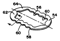
Fig. 13 is a further embodiment of a clip 54 in
accordance with the present invention. The clip 54 has the
same overall dimensions as the clip disclosed above, i.e.,
approximately 3 to 5 mm by 5 to 6 mm, so as to fit between
adjacent rectus muscles. However, the clip is oval or
round in shape and has a central opening 56 enclosed by a
continuous outer portion so that the clip 54 has a ring-
like appearance. This clip is applied to the sclera by
prolapsing the sclera through the central opening in the
clip by mechanical means, such as a twist hook or forceps,
or by the application of a vacuum. One or both of the
central opening 56 or outer edge 58 may be provided with
teeth 60, which are similar to teeth 46 described above,
for securing the clip to the sclera. Further, the teeth
may be bent out of the plane generally defined by the clip
so that they more firmly grip the sclera. With reference
t~ Fig. 14, the teeth on the outer edge or periphery 58 may
be bent downwardly an angle cc from between approximately 90
degrees to approximately 135 degrees, while the teeth on
the central opening or inner periphery 56 are bent
downwardly an angle ~ between approximately 20 degrees to
45 degrees.
The clip 54 is generally flat, with little or no angle
between the two arms or sides 62 , 64 , as def fined by the
center line through the clip, thus providing a very low
profile. Preferably, the clip 54 is sufficiently thin so
that it conforms to the natural shape or curvature of the
eye.
Thus, a method and a clip for performing the method
have been provided that fully meet the objects of the
present invention. While the invention has been described
in terms of a preferred method and clip, there is no intent
to limit the invention to the same. Instead, the invention
is defined by the scope of the following claims.