Note: Descriptions are shown in the official language in which they were submitted.
CA 02420350 2003-02-21
WO 02/18572 PCT/USO1/26421
MEMBRANE PENETRATING PEPTIDES AND USES THEREOF
This application claims the benefit of U.S. Provisional Application No.
60/227,647,
filed August 2S, 2000 and GB Application 0103110.3, filed February 7, 2001.
FIELD OF THE INVENTION
The invention relates to membrane penetrating peptides useful as in vitro, ex
vivo and
ih vivo delivery devices for intracellular delivery of a compound of interest
to cells ih vitro, ex
vivo and if2 vivo, compositions comprising the same and methods of using the
same. The
l0 invention also includes identification of additional membrane penetrating
peptides useful as
delivery devices for intracellular delivery of a compound of interest to cells
in vitro, ex vivo
and in vivo.
BACKGROUND OF THE INVENTION
The delivery of small molecules, oligonucleotides, and proteins through
biological
membranes is a major challenge facing therapy and validation paradigms. It has
recently been
established that transducing peptides derived from Antennapedia, TAT-HIV, and
VP22 can
penetrate biological membranes, act as cargo vehicles, and target to specific
subcellular
2o compartments. Here we show the identification of a nuclear localization
sequence (NLS)
within human Period 1 (hPERl) circadian protein that functions as a
transducing peptide.
More importantly, using database mining, we have uncovered additional
transducing peptides
embedded within the NLS's of other proteins and extend the number of gene-
encoded
transducing peptides from 3 to 14. Our data suggest that transducing peptides
are found within
NLS's and are prevalent, diverse, and distributed widely throughout the
genome. It is well
established that certain extracellular and intracellular proteins are targeted
to specific
organelles within a cell, transmembrane or secreted from the cell. The
biological mechanisms
by which intracellular protein targeting occurs continues to be characterized,
but is well
recognized that one mechanism for localization occurs by virtue of specific
leader sequence
3o contained within the protein of interest, or intraprotein sequence.
Localization of proteins
within selected cellular organelles is aided by specific targeting sequences.
A number of
nuclear localization sequences (NLSs) have been identified in proteins that
permit the protein
to be tranported or otherwise pass from the cytoplasm into the nuclear
membrane.
SUBSTITUTE SHEET (RULE 26)
CA 02420350 2003-02-21
WO 02/18572 PCT/USO1/26421
-2-
Fusion proteins containing the targeting sequence and another, otherwise non-
targeted
protein, are localized in the selected cellular organelle depending on the
targeting sequence
selected. For example, Ferullo, J. M. and Paget, E. FR 279695, disclose
selective
compartmentalization of an hydroxyphenylpyruvate dioxygenase (HPPD) fused to a
signal
sequence directing the enzyme to a cellular compartment other than the
cytosol, e.g., a
vacuole. Similarly, WO 0147950 (Wehrle-Halter, Bernhard M.; Imhof, Beat A)
identify a
new determinant responsible fox basolateral targeting and prolonged exposure
of cell-surface-
anchored growth factors at cell surfaces. The signal is a mono-leucine
dependent basolateral
sorting signal consisting, of the amino acid sequence X1h2X3h4Lp5p6, wherein:
Xl
to represents a polar amino acid residue or alanine, h2 represents any
hydrophobic amino acid
residue, X3 represents any amino acid residue, h4 represents any hydrophobic
amino acid
residue, except Ieucine and isoleucine, L represents a Ieucine residue, p5
represents any polar
amino acid residue, and p6 represents any polar amino acid. Richardson, A. E.,
et al., Plant J.
(2001), 25(6), 641-649 describe manipulation of the enzyme aspergillus phytase
to include
i5 the signal peptide sequence from the carrot extensin gene. The resulting
fusion protein was
only effective when secreted as an extracellular enzyme into the adjacent
soil, and resulted in
a 20-fold increase in total root phytase activity in transgenic lines and
subsequent improved
phosphorus nutrition, such that the growth and phosphorus content of the
plants was
equivalent to control plants supplied with inorganic phosphate. WO 0132894
(Lok, S.)
20 disclose use of the signal anchor domain sequences of type TI cell surface
proteins to anchor
recombinant proteins into surface of transfected cells. A characteristic
feature of type II cell
surface proteins is that they are held within the cellular membrane by a
single hydrophobic
transmembrane domain and are oriented with their C-terminus outside the cell.
More recently, a few proteins have been identified which are capable of
passing
25 through the cellular membrane without requiring active transport mechanisms
or'pores'. It is
recently established that membrane penetrating peptides (MPPs, also known as
protein
transduction domain, "PTD") derived from Antennapedia, TAT, and VP22 can
penetrate
biological membranes and target to specific subcellular compartments. None of
these
previously disclosed proteins are derived from mammalian proteins. The present
invention is
3o directed to the discovery that polypeptides derived from mammalian or yeast
proteins nuclear
localization sequences (NLSs) or overlapping with NLS's are capable of acting
as MPPs, and
identification of a specific polypeptide sequences capable of penetrating
cellular membranes,
SUBSTITUTE SHEET (RULE 26)
CA 02420350 2003-02-21
WO 02/18572 PCT/USO1/26421
-3-
even when conjugated to large proteins, such as biologically active proteins,
or other organic
compounds.
Nuclear transport is essential to a number of biological processes including
gene
expression and cell division, as well as to viral replication, tumorigenesis
and tumor cell
proliferation. The mechanism of nuclear transport has only recently been
characterized in
detail and has been shown to involve a number of discrete steps. Proteins that
are destined to
be transported into the nucleus contain within their amino acid sequence a
short stretch of
amino acids termed a nuclear localization sequence ("NLS"). These sequences
may occur
anywhere within the amino acid sequence and are typically four to about eight
amino acids.
to These sequences are generally basic (i.e., positively charged) in nature,
however, there has
been no consensus sequence identified. Thus, there is a wide variety of these
sequences that
appear to be specific for particular proteins.
Within the cell, these NLSs may be either masked or unmasked by accessory
proteins
or by conformational changes within the NLS-containing protein. An NLS may be
masked
because it is buried in the core of the protein and not exposed on the surface
of the protein.
Unmasking of NLSs, and nuclear translocation of cytoplasmic proteins may be
triggered by
phosphorylation, dephosphorylation, proteolytic digestion, subunit association
or dissociation
of an inhibitory subunit, or the like. Accordingly, the masking and unmasking
of NLSs
provides a mechanism by which the transport of these cytoplasmic proteins into
the nucleus
2o may be regulated. For example, the transcription factor NF-AT contains
nuclear Localization
sequences which allow NF-AT to translocate to the nucleus in the presence of
intracellular
calcium, but which are shielded by forming intramolecular associations with
other domains in
the NF-AT polypeptide in the absence of calcium.
Lee, H. C. and Bernstein, H. D. Proc. Natl. Acad. Sci. U. S. A. (2001), 9~(6),
3471-
3476 studied the mechanism involved for presecretory proteins such as maltose
binding
protein (MBP) and outer membrane protein A (OmpA) that are targeted to the E.
coli inner
membrane by the molecular chaperone SecB, in contrast to the targeting of
integral membrane
proteins by the signal recognition particle (SRl'). The authors found that
replacement of the
MBP or OmpA signal peptide with the first transmembrane segment of AcrB
abolished the
dependence on SecB for transport and rerouted both proteins into the SRP
targeting pathway.
Some proteins contain cytoplasmic localization sequences (CLS), or nuclear
export
sequences, which ensure the protein remains predominantly in the cytoplasm.
For example,
Hamilton, M. H. et aL, J. Biol. Chem. (2001), 276(20, 26324-26331 demonstrate
that the
SUBSTITUTE SHEET (RULE 26)
CA 02420350 2003-02-21
WO 02/18572 PCT/USO1/26421
-4-
ubiquitin-protein ligase (E3), hRPFl/Nedd4, a component of the ubiquitin-
proteasome
pathway responsible for substrate recognition and specificity, is capable of
entering the
nucleus, but the presence of a functional Rev-like nuclear export sequence in
hRPFl/Nedd4
ensures a predominant cytoplasmic localization. The cytoplasmic domains of
many
membrane proteins contain sorting signals that mediate their endocytosis from
the plasma
membrane.
Heineman, T. C. and Hall, S. L. Virology (2001), 28S(1), 42-49 studied three
consensus internalization motifs within the cytoplasmic domain of VZV gB and
determined
that internalization of VZV gB, and its subsequent localization to the Golgi,
is mediated by
to two tyrosine-based sequence motifs in its cytoplasmic domain. In mammalian
cells and
yeasts, amino acid motifs in the cytoplasmic tails of transmembrane proteins
play a prominent
role in protein targeting in the early secretory pathway by mediating
localization to or rapid
export from the endoplasmic reticulum (ER). Hoppe, H. C. and Joiner, K. A.
Cell. Microbiol.
(2000), 2(6), 569-578.
The mammalian endopeptidase, furin, is predominantly localized to the trans-
Golgi
network (TGN) at steady state. The localization of furin to this compartment
seems to be the
result of a dynamic process in which the protein undergoes cycling between the
TGN and the
plasma membrane. Both TGN localization and internalization from the plasma
membrane are
mediated by targeting information contained within the cytoplasmic domain of
furin.
2o Voorhees, P., et al., EMBO J. (1995), 14(20), 4961-75 report that there are
at least two
cytoplasmic determinants that contribute to the steady-state localization and
trafficking of
furin. The first determinant corresponds to a canonical tyrosine-based motif,
YI~GL (residues
758-761), that functions mainly as an internalization signal. The second
determinant consists
of a strongly hydrophilic sequence (residues 766-783) that contains a large
cluster of acidic
residues (E and D) and is devoid of any tyrosine-based or di-leucine-based
motifs. This
second determinant is capable of confernng Localization to the TGN as well as
mediating
internalization from the plasma membrane.
The trans-Golgi network (TGN) plays a central role in protein
sorting/targeting and the
sequence SXYQRL can by itself confer significant TGN localization. along,
S.H., and Hong,
3o W. J. Biol. Chem. (1993), 268(30), 22853-62 report detailed mutagenesis of
the 32-residue
sequence of TGN38, an integral membrane protein confined mainly to the TGN,
and
determined that the Ser, Tyr, and Leu residues at positions 23, 25, and 28,
respectively, are
essential for TGN localization. When the cytoplasmic 32-residue sequence of
TGN38 was
SUBSTITUTE SHEET (RULE 26)
CA 02420350 2003-02-21
WO 02/18572 PCT/USO1/26421
-5-
fused to the ecto- and transmembrane domains of glycophorin A (a surface
protein), the
resulting chimeric protein was localized to the TGN.
It is well recognized that certain proteins are either only active in a
specific organelle,
or are capable of different functions depending on their localization. Far
example, appropriate
subcellular localization is crucial for regulation of NF-~cB function. Huang,
T. T., et al., Proc.
Natl. Acad. Sci. U. S. A. (2000), 97(3), 1014-1019, show that latent NF-xB
complexes can
enter and exit the nucleus in preinduction states and identified a previously
uncharacterized
nuclear export sequence in residues 45-54 of IxBa that was required for
cytoplasmic
localization of inactive complexes. It appears that NF-~cB/hcBa complexes
shuttle between
l0 the cytoplasm and nucleus by a nuclear localization signal-dependent
nuclear import and a
CRMl-dependent nuclear export and that the dominant nuclear export over
nuclear import
contributes to the largely cytoplasmic localization of the inactive complexes
to achieve
efficient NF-~cB activation by extracellular signals.
Nuclear import of classical nuclear localization sequence-containing proteins
involves
the assembly of an import complex at the cytoplasmic face of the nuclear pore
complex (NPC)
followed by movement of this complex through the NPC and release of the import
substrate
into the nuclear interior. In combination with Ran, two other soluble factors
are thought to be
absolutely required to mediate the nuclear import of a protein containing a
classical or basic
NLS into the nucleus. The first is karyopherin/importin a (Kap a), which binds
a classical
2o NLS and then forms a complex with karyopherin/importin (31 (Kap(31). Adam,
S. A., and
Gerace, L. (1991) Cell 66, 837-847; Gorlich, D., et al. (1994) Cell79, 767-
778; Moroianu, J.,
et a1..(1995) Proc. Natl. Acad. Sci. U. S. A. 92, 2008-2011; Radu, A., et al.
(1995) P~oc. Natl.
Acad. Sci. U. S. A. 92, 1769-1773; Gorlich, D., ., et al. (I99S) Curs. Biol.
5, 383-392; Chi, N.
C., et al. (1995) J. Cell Biol. 130, 265-274. Kap (31 interacts with nuclear
pore complex
(NPC) proteins and appears to mediate movement of the import complex through
the NPC via
these interactions. Rexach, M., and Blobel, G. (1995) Cell 83, 683-692; Radu,
A., Blobel, G.,
and Moore, M. S. (1995) Proc. Natl. Acad. Sci. U. S. A. 92, 1769-1773; Iovine,
M. K.,
Watkins, J. L., and Wente, S. R. (1995) J. Cell Biol. 131, 1699-1713; Radu,
A., Moore, M. S.,
and Blobel, G. (1995) Cell 81, 215-222. Another protein, p10/NTF2, has also
been implicated
3o in nuclear import, but its function may only be to take Ran into the
nucleus, where it is
subsequently needed to disassemble an incoming import complex. Moore, M. S.,
and Blobel,
G. (1994) P~oc. Natl. Acad. Sci. U. S. A. 91, 10212-10216; Paschal, B. M., and
Gerace, L.
(1995) J. Cell Biol. 129, 925-937; Ribbeck, K., Lipowsky, G., Kent, H. M.,
Stewart, M., and
SUBSTITUTE SHEET (RULE 26)
CA 02420350 2003-02-21
WO 02/18572 PCT/USO1/26421
-6-
Gorlich, D. (1998) EMBO J. 17, 6587-6598; Smith, A., Brownawell, A., and
Macara, I. G.
(1998) Curr. Biol. 8, 1403-1406.
Although there is only one Kap a homologue in yeast (SRP1 or Kap60),
vertebrate
cells contain a number of proteins that can bind a classical NLS and share
sequence homology
S (see Ref. Nachury, M. V., Ryder, U. W., Lamond, A. L, and Weis, K. (1998)
Proc. Natl.
Acad. Sci. U. S A. 95, S82-587, and references therein). These proteins have
been given a
varietyof names but can be grouped into three major families. The Kap al
family contains the
human protein NPI-1/importin al/kaxyopherin al/Rch2/hSRPl and a second related
protein
importin a6, in addition to the mouse S2 protein. Moroianu, J., et al., (1995)
Proc. Natl.
l0 Acad. Sci. U. S A. 92, 2008-2011; Cortes, P., et al., (1994) Proc. Natl.
Acad. Sci. U. S. A. 91,
7633-7637; O'Neill, R. E., et al., (1995) J. Biol. Ghem. 270, 22701-22704;
Kohler, M., et al.,
(1997) FEBSLett. 417, 104-108; Tsuji, L., et al., (1997) FEBSLett. 416, 30-34.
The second
family, Kapa2, contains human Rchl/hSRPl/importin a2/karyopherin a2 and the
mouse
protein pendulin/PTAC S8. Gorlich, D., Prehn, S., Laskey, R. A., and Hartmann,
E. (1994)
15 Cell79, 767-778; Cuomo, C. A., Kirch, S. A., Gyuris, J., Brent, R., and
Oettinger, M. A.
(1994) P~oc. Natl. Acad. Sci. U. S. A. 91, 6156-6160; Kussel, P., and Frasch,
M. (1995) Mol.
Gera. Genet. 248, 3S1-363; Imamoto, N., Shimamoto, T., Takao, T., Tachibana,
T., Kose, S.,
Matsubae, M., Sekimoto, T., Shimonishi, Y., and Yoneda, Y. (1995) EMBO J. 14,
3617-
3626;, K., Mattaj, I. W., and Lamond, A. I. (1995) Science 268, 1049-S3. The
third family,
20 Kapa3, consists of the two human proteins, QIP-1/importin a3 and
KPNA3/hSPRl y/hSRP4,
and the mouse proteins Q1 and Q2. Nachury, M. V., et al., (I998) Pnoc. Natl.
Acad. Sci. U. S.
A. 95, 582-587; Kohler, M., et al., (1997) FEBSLett. 417, 104-108; Tsuji, L.,
et al., (1997)
FEBSLett. 416, 30-34; Takeda, S., et al., (1997) Cytogeraet. Cell Genet. 76,
87-93; Seki, T., et
al., (1997) Biochem. Biophys. Res. Commun. 234, 48-S3; Miyamoto, Y., et al.,
(1997) J. Biol.
2S Chem. 272, 26375-26381. Each of these classes share about SO% homology with
each other
and to the yeast SRPI, and each of these mammalianproteins has been shown to
be capable of
mediating the import of one or more classical NLS-containing proteins.
Nachury, M. V., et al.,
(1998) Pnoc. Natl. Acad. Sci. U. S. A. 95, S82-587; Sekimoto, T., et al.,
(1997) EMBO J. I6,
7067-7077; Nadler, S. G., et al., (1997) J. Biol. Chem. 272, 4310-4315;
Prieve, M. G., et al.,
30 (1998) Mol. Cell. Biol. 18, 4819-4832.
Stat-1 import is mediated by Kapal/NPI-1 but not Kapa2/Rchl, but activated
Stat-1
appears to bind to a COON-terminal region ofKapal distinct from the NLS
binding
SUBSTITUTE SHEET (RULE 26)
CA 02420350 2003-02-21
WO 02/18572 PCT/USO1/26421
_7_
Armadillo repeats. The binding differences of the different Kapas to RCC1
observed appear
to be due solely to the NLS on RCC1 and thereforeprobably due to the NLS
binding region of
Kapa3. Sekimoto, T., et al., (1997) EMBO J. 16, 7067-7077. Kamei, Y., et al.,
(1999) J.
Histoche~ra. Cytochern. 47, 363-372 showed that, in mice, the Kapa3 homologue
is expressed
in many tissues and theorized that Kapa3 may play a role in importing "a
limited number of
unique karyophilic proteins, such as helicase Q1." The results provided by
Talcott, B. and
Moore, M.S., 2000 JBiol Cherra, 275(14) 10099-10104 suggest that RCC1 should
be included
in the group of proteins that use Kapa3 to mediate their nuclear import.
USP 6,191,269 teaches the existence of a nuclear localization sequence
contained
to within the cDNA sequence of the N-terminal IL-1 alpha propiece, T76-
NGKVLKKRRL,
which had characteristics of a nuclear localization sequence (NLS) and could
mediate nuclear
localization of the propiece (Stevenson et al. (1997) Proc. Natl. Acad. Sci.
USA 94:508-13).
Introduction of the cDNA encoding the N-terminal IL-.alpha. propiece into
cultured mesangial
cells resulted in nuclear accumulation (Stevenson et al. id).
USP 5,877,282 teaches that the antennapedia homeodomain signal sequence
peptide is
the amino acid sequence RQIKIWFQNRRMKWKK; the fibroblast growth factor signal
sequence peptide is AAVALLPAVLLALLA; the HIV Tat signal sequence peptide is
the
amino acid sequence CFITKALGISYGRKI~R.RQRRRPPQGSQTH.
ti
Schwartze, S.R., et al., Science 285:1569-1572 (1999) report delivery of an ip
injected
2o reporter protein, 116 kD beta-galacatosidase, as a TAT fusion protein into
tissues and across
the blood-brain barrier. Schwartze used an 11 amino acid protein transduction
domain (PTD)
derived from H1V tat protein with an N-terminal fluorescein isothiocyanate
(FITC)-Gly-Gly-
Gly-Gly motif. The authors report that earlier attempts to transduce beta-Gal
chemically cross-
linked to the TAT PTD resulted in sporadic and weak beta-Gal activity in a
limited number of
tissues. They speculate that the improved transduction was due to the in-frame
fusion and
purification strategy used.
Nuclear localization of IFNy is mediated by a polybasic NLS in its C terminus,
which
is required for the full expression of biological activity of IFNy, both
extracellularly and
intracellularly. Subramaniam, Prem S., et al., J. Cell Sci. (2000), 113(15),
2771-2781. This
3o NLS is thought to play an integral intracellular role in the nuclear
translocation of the
transcription factor STATla activated by IFNy because treatment of IFNy with
antibodies to
the C-terminal region (95-133) containing the NLS blocked the induction of
STATla nucleax
SUBSTITUTE SHEET (RULE 26)
CA 02420350 2003-02-21
WO 02/18572 PCT/USO1/26421
_g_
translocation, but these antibodies had no effect on nuclear translocation of
STATla in IFNa
treated cells. A deletion mutant of human IFNy, IFNy(1-123), which is devoid
of the C-
terminal NLS region was biologically inactive, but was still able to bind to
the IFNy receptor
complex on cells with a I~ similar to that of the wild-type protein. Deletion
of the NLS
specifically abolished the ability of IFNy(1-123) to initiate the nuclear
translocation of
STATla, which is required for the biological activities of IFNy following
binding to the IFNy
receptor complex. A C-terminal peptide of murine IFNy, IFNy(9S-133), that
contains the NLS
motif, induced nuclear translocation of STATIa when taken up intracellularlyby
a murine
macrophage cell line. Deletion of the NLS motif specifically abrogated the
ability of this
to intracellular peptide to cause STATla nuclear translocation. In cells
activated with IfNy,
IFNy was found to as part of a complex that contained STATla and the importin-
a analog
Npi-1, which mediates STATla nuclear import. The tyrosine phosphorylation of
STATla,
the formation of the complex IFNy/Npi-1/STATla complex and the subsequent
nuclear
translocation of STAT1 a were all dependent on the presence of the IFNy NLS.
The peptide representing amino acids 95-132 of IFN-y (IFN-y(95-132)),
containing the
polybasic sequence lzsRKRKRSRi32, was capable of specifying nuclear uptake of
the
autofluorescent protein, APC, in an energy-dependent fashion that required
both ATP and
GTP. Nuclear import was abolished when the above polybasic sequence was
deleted.
Subramaniam, P., et al., 1999 JBiol Chem 274(1) 403-407. A peptide containing
the
prototypical polybasic NLS sequence of the SV40 large T-antigen was also able
to inhibit the
nuclear import mediated by IFN-y(95-132), suggesting that the NLS in IFN-y may
function
through the components of the Ranlimportin pathway utilized by the SV40 T-NLS.
Intact IFN-
y, when coupled to APC, was also able to mediate its nuclear import, and this
nuclear import
was blocked by the peptide IFN-y (95-132) and the SV40 T-NLS peptide,
suggesting that
intact IFN-y was also transported into the nucleus through the Ran/importin
pathway.
Nuclear proteins are imported into the nucleus through aqueous channels that
span the
nuclear envelope called nuclear pore complexes (NPCs). Although ions and
molecules less
than ~20-40 Da can diffuse passively through the nuclear pore complexes,
larger proteins are
transported by saturable pathways that are energy- and signal-dependent. The
signals that
specify nuclear protein import (NLSs)1 are commonly short stretches of amino
acids rich in
basic amino acid residues, although other classes of NLSs have been described
recently. The
initial step in the import of proteins containing basic amino acid-type NLSs
occurs in the
SUBSTITUTE SHEET (RULE 26)
CA 02420350 2003-02-21
WO 02/18572 PCT/USO1/26421
-g_
cytosol, Where the NLS-containing proteins are bound to a receptor (variously
called the NLS
receptor, importin a, and karyopherin (13). The substrate-receptor complex
then associates
with the cytoplasmic face of the nuclear pore complexes, and with the
participation of other
cytosolic factors, is transported through a gated channel in the nuclear pore
complexes to the
nuclear interior. The in vivo events of NLS-mediated nuclear import can be
duplicated in an in
vitro system using digitonin-permeabilized cells supplemented with cytosolic
extracts and
ATP (14). Transport in this in vitro assay is blocked by the same inhibitors
that block in vivo
import, is rapid, and is easily quantified.
The NLS the sequence NYKI~PKL in the N-terminus of fibroblast growth factor
(FGF)-1, the precursor fox acidic FGF, has been proposed to affect the long
term activities of
FGF-1 through its function as a nuclear translocation signal or its role in
stabilization of the
structure required to sustain binding and activation of the transmembrane
receptor kinase.
Luo, Y., et al., J. Biol. Chem. (1996), 271(43), 26876-26883. For example,
concurrent with
a marked increase in dependence on exogenous heparin for optimal activity,
sequential
deletion of residues in the NYKKPKL sequence in FGF-1 resulted in a
progressive loss of
thermal stability, resistance to protease, mitogenic activity, and affinity
for the transmembrane
receptor. The largest change resulted from deletion of the entire sequence
through the lysine-
leucine residues. In the presence of sufficiently high concentrations of
heparin, the deletion
mutants exhibited mitogenic activity equal to wild-type FGF-1.
2o Although FGF-1 contains an NTS, nuclear translocation requires an exogenous
and not
an endogenous pathway. The NTS of FGF-1, NYKI~PKL, is able to direct the
expression of
the bacterial (3-galactosidase ((3ga1) gene to the nucleus of transfected NIH
3T3 cells, but this
NTS is unable to target either FGF-1 itself of a FGF-1-(3gal fusion protein
into the nucleus,
suggesting that FGF-1 may contain an additional sequence which prevents
endogenously
expressed FGF-1 from being translocated into the nucleus. Zhan, X., et al.,
Biochem.
Biophys. Res. Common. (1992), 188(3), 982-91.
Interferon-y (IFN-y), a protein that uses the Jak-Stat pathway for signal
transduction,
translocates rapidly to the nucleus in cells treated extracellularly with the
cytokine. An NLS
has been identified and characterized in the C-terminus of human and marine
IFN-y. Laxkin,
3o J., et al., J. Interferon Cytokine Res. (2001), 21(6), 341-348 report that
human IFN-y
(HuIFN-y) contains a second NLS at an upstream site. The primary sequence,
analogous with
the NLS sequence identified in marine IFN-y, representing amino acids 122-132
of HuIFN-y
was capable of mediating the nuclear import of the autofluorescent protein
allophycocyanin
SUBSTITUTE SHEET (RULE 26)
CA 02420350 2003-02-21
WO 02/18572 PCT/USO1/26421
-10-
(APC) in an energy-dependent manner. The second sequence, representing amino
acids 78-92
of HuIFN-y, was also capable of mediating the nuclear import of APC in an
energy-dependent
manner but to a greatly reduced extent. The nuclear import of both sequences
conjugated to
APC was strongly blocked by competition with unconjugated HuIFN-y(122-132).
Competition by the sequence HuIFN-y(78-92) effectively blocked the import of
APC-
conjugated HuIFN-y(78-92) but, at the same concentration, was not capable of
inhibiting the
nuclear import of APC-conjugated HuIFN-y(122-132), suggesting that HuIFN-y(78-
92) was a
less efficient NLS than HuIFN-y(122-132). This is consistent with >90% loss of
antiviral
activity of HuIFN-y lacking the downstream NLS in 122-132. The nuclear import
of APC-
to conjugated HuIFN-y(122-132) was inhibited by a peptide containing the
prototypical
polybasic NLS of the SV40 T NLS, which suggests that the same Ran/importin
cellular
machinery is used in both cases.
There appears to be strong conservation of the NLS motif as a. mechanism for
nuclear
localization. Evolution seemed to have used part of the existing DNA-binding
mechanism
when compartmentalizing DNA-binding proteins into the nucleus. Cokol, M., et
al., EMBO
Rep,. (2000), 1(5), 411-415 estimate that greater than 17% of all eukaryotic
proteins may be
imported into the nucleus, and after analyzing a set of 91 experimentally
verified NLSs from
the literature and expanding this set to 214 potential NLSs through iterated
"in silico
mutagenesis". This final set matched in 43% of all known nuclear proteins and
in no known
2o non-nuclear protein. Cokel et al found an overlap between the NLS and DNA-
binding region
for 90% of the proteins for which both the NLS and DNA-binding regions were
known, but
only S6 of the 214 NLS motifs overlapped with DNA-binding regions. These 56
NLSs
enabled a de novo prediction of partial DNA-binding regions for approximately
800 proteins
in human, fly, worm and yeast.
More recently, it has been reported that NLS signal peptide can induce
structural
changes of DNA. The plant enzyme, glutaminyl-tRNA synthetase (GInRS) from
Lupinus
luteus, contains an NLS at the N-terminal, a lysine rich polypeptide,
KPKKKKEK.
Krzyzaniak, A., et al., Mol. Biol. Rep. (2000), 27(1), 51-54. Two synthetic
peptides (20 and
8 amino acids long), derived from the NLS sequence of lupin GInRS interact
with DNA. In
3o addition, the shorter 8 amino acid peptide caused the DNA to change its
conformation from
the B to the Z form. This observation clearly suggests that the presence of
the NLS
polypeptide in a leader sequence of GlnRS is required not only for protein
transport into
SUBSTITUTE SHEET (RULE 26)
CA 02420350 2003-02-21
WO 02/18572 PCT/USO1/26421
-11-
nucleus but also for regulation of a gene expression. This is the first report
suggesting. a role
of the NLS signal peptide in structural changes of DNA.
Typically there is strong conservation of the NLS sequence within species. For
example, the NLS in the N-terminal region of Smad 3 protein, the major Smad
protein
involved in TGF-(3 signal transduction, has a basic motif Lys4°- Lys-
Leu-Lys-Lys44, which is
conserved among all the pathway-specific Smad proteins, and is required for
Smad 3 nuclear
import in response to ligand. Smad proteins are intracellular mediators of
transforming growth
factor-(3 (TGF-[3) and related cytokines. Xiao, Z., et al., J. Biol. Chem.
(2000), 275(31),
23425-23428 identified the role the NLS plays in nuclear localization. The
authors
l0 demonstrated that the isolated Smad 3 MH1 domain displays significant
specific binding to
importin (3, which is diminished or eliminated by mutations in the NLS. Full-
size Smad 3
exhibits weak but specific binding to importin (3, which is enhanced after
phosphorylation by
the type I TGF-(3 receptor. In contrast, no interaction was observed between
importin a and
Smad 3 or its MH1 domain, indicating that nuclear translocation of Smad
proteins may occur
through direct binding to importin (3. The authors conclude that activation of
all of the
pathway-specific Smad proteins (Smads l, 2, 3, 5, 8, and 9) exposes the
conserved NLS motif,
- which then binds directly to importin (3 and triggers nuclear translocation.
In all cells, the lipid bilayer of cell membranes serves as a selective
barrier for the
passage of charged molecules, with the internalization of hydrophilic
macromolecules being
2o achieved through classical transport pathways (Hawiger, J., Curr Cpin Chena
Biol. 3, 89-94
(1999), Schwarze, S.R., et al., Trends in Cell Biology 10, 290-295 (2000)).
These classical
mechanisms of internalization involve receptor-mediated endocytosis or
transporter dependent
uptake (Cleves, A.E., Current Biology 7, 8318-8320 (1997)). In contrast, an
increasing
number of molecules have been discovered that lack classical import and/or
export signals
(Cleves, A.E., Current Biology 7, 8318-8320 (1997)). These molecules gain
direct access to
either cytoplasmic or nuclear compartments using unconventional processes of
which the
mechanisms remain largely unknown. These novel mechanisms are generally termed
"nonclassical" and refer to transport pathways being used that are atypical.
Relevant
examples of this latter type are found in the gene-encoded proteins of HIV-1
TAT (Frankel,
A.D. and Pabo, C.O. Cell 55,1189-1193 (1988)), herpes virus VP22 (Elliott, G.
and O'Hare, P.
Cell 88, 223-233 (1997)), and Antennapedia, Antp (Derossi, D., et al., J.
Biol. Che~ra.
269,10444-10450 (1994)). It is now well established that the full-length
proteins of HIV-1
TAT (Helland D.E., et al., J Virol 65, 4547-4549 (1991)), and VP22 (Pomeranz
L.E. and
SUBSTITUTE SHEET (RULE 26)
CA 02420350 2003-02-21
WO 02/18572 PCT/USO1/26421
-12-
Blaho J.A., J Tirol 73, 6769-6781 (I999)) rapidly translocate into and out of
cellular
membranes. In fact, distinct peptide regions have been identified within both
of these proteins
that are capable of translocating into cellular compartments either alone or
in combination
with chimeric cargo peptides, and proteins (Lindgren, M., et al., Trends
Pharmacol Sci. 3, 99-
103 (2000), Derossi, D., et al, Trends Cell Biol., 8, 84-87 (1998), Prochiantz
A., Current
Opinion in Cell Biology 12, 400-406 (2000), Steven R. Schwarze, S.R., et al.,
Trends in Cell
Biology 10, 290-295 (2000)). In contrast, full-length Antp protein has not
been shown to
traverse biological membranes; however, a 16 amino acid synthetic peptide
derived from
within its coding region does possess potent membrane penetrating abilities
(Derossi, D., et al,
Trends Cell Biol., 8, 84-87 (1998)). The accepted view of atypical transport
used by these
molecules has been termed "transduction" (Schwarze, S.R., et al., Trends in
Cell Biology 10,
290-295 (2000)), and is currently defined as an extremely rapid membrane
transport pathway
that is receptor and energy independent, and can occur at 4 C in all cell
types (Schwarze, S.R.
and Dowdy, S.F. Trends Pharmacol. Sci. 21, 45-48 (2000)). Interestingly, these
three proteins
are all nuclear proteins involved in transcriptional regulation, and their
respective transducing
peptides consist of strings of amino acids rich in arginine and lysine
(Lindgren, M., et al.,
Trends Pharmacol Sci. 3, 99-103 (2000), Schwarze, S.R. and Dowdy, S.F. Trends
Pharmacol.
Sci. 21, 45-48 (2000)). However, irrespective of these similarities, these
transducing peptides
possess many different characteristics such as amino acid sequence, length of
the sequence,
cellular localization, and potency of membrane penetration. Thus, though each
transducing
sequence can penetrate cells and tissues, it has not been established whether
they use the
identical atypical transport mechanisms.
Finally, USP 6,022,950 teaches the use of a hybrid molecule of a portion of
the
binding domain of a cell-binding polypeptide ligand effective to cause said
hybrid protein to
bind to a cell of an animal, a translocation domain of naturally occurring
protein which
translocates said third part across the cytoplasmic membrane into the cytosol
of the cell; and a
chemical entity to be introduced into the cell. However, the patent teaches
translocation
domains of toxins. Naturally-occurnng proteins which are known to have a
translocation
domain include diphtheria toxin and Pseudomonas exotoxin A, and may include
other toxins
3o and non-toxin molecules, as well. The translocation domains of diphtheria
toxin and
Pseudomonas exotoxin A are well characterized (see, e.g., Hoch et al., Proc.
Natl. Acad. Sci.
USA 82:1692-1696, 1985; Colombatti et al., J. Biol. Chem. 261:3030-3035, 1986;
and
Deleers et al., FEES 160:82-86, 1983), and the existence and location of such
a domain in
SUBSTITUTE SHEET (RULE 26)
CA 02420350 2003-02-21
WO 02/18572 PCT/USO1/26421
-13-
other molecules may be determined by methods such as those employed by Hwang
et al., Cell
48:129-136, 1987; and Gray et al., Proc. Natl. Acad. Sci. USA 81:2645-2649,
1984.
Given the considerable body of literature teaching control mechanisms of
cellular
localization, the proteins involved in regulation of intracellular transport,
the different
properties and control mechanisms for plasma membrane and the nuclear
envelope, it is
unexpected that polypeptides derived from mammalian proteins could transduce
through the
plasma membrane using nonclassical mechanisms and thus could be useful as
membrane
penetrating peptides useful as in vitro, ex vivo and in vivo delivery devices
of a compound of
interest. There is also considerable literature teaching non-protein derived
methods for
l0 delivering a compound of interest into cells, for example electroporation,
membrane fusion
with liposomes, high velocity bombardment with DNA-coated microprojectiles,
incubation
with calcium-phosphate-DNA precipitate, DEAE-dextran mediated transfection,
infection
with modified viral nucleic acids, and direct microinjection into single
cells, usually ova and
the like. Each of these methods is relatively inefficient, resulting in
relatively low percentage
of the cells containing the delivered compound of interest and most of the
methods are clearly
not capable of realistic in vivo delivery. Many of the methods are toxic to
the cells, resulting
in relatively high apoptosis. Therefore, there is a considerable need for
simple and more
efficient delivery of compounds of interest into cells.
SUMMARY OF THE INVENTION
The present invention is directed to polypeptides derived from mammalian and
yeast
proteins useful as a carrier for in vitro, ex vivo and in vivo delivery a
compound of interest.
The invention also provides compositions containing the same, and methods of
delivering a
compound of interest in vitro, ex vivo and ih vivo.
BRIEF DESCRIPTION OF THE DRAWINGS
Figure 1. (A). Schematic diagram of hPERl fusion constructs showing the
locations
of the PAS, cytoplasmic localization, and nuclear localization sequence (NLS,
but indicated as
nuclear localization domain (NLD) in Figure). The name and the position of the
fusion
3o constructs are listed on the left. The number indicates the first and last
amino acid residues in
the hPERl protein. The principal sites of accumulation of each fusion protein
are summarized
on the right, (n) nuclear, (no) nucleoli, (c) cytoplasmic, (diff) diffuse. All
constructs were N-
SUBSTITUTE SHEET (RULE 26)
CA 02420350 2003-02-21
WO 02/18572 PCT/USO1/26421
-14-
terminally tagged with EYFP. The alignment human and mouse PERT-NLS is shown
at the
bottom.
Figure 1. (B). Cellular localization of hPER1 fusion proteins as described in
Figure
1A, above, in living cells. CHO cells were transient transfected with the
fusion constructs
indicated on the top of each panel and the subcellular localization of EYFP
reporters (green)
was directly visualized using fluorescent microscopy 10 h post-transfection.
EYFP vector
alone is used as control (see 5. EYFP-VECTOR)
Figure 2. (A). Membrane penetration assay in CHO cells. N-terminal
biotinylated
synthetic peptides hPERI-PTD, Flag-hPERl-PTD, Flag-TAT-PTD (positive control),
and
to Flag-Flag (negative control) were assayed for their ability to penetrate
cellular membranes in
living CHO cells in culture. The subcellular localization of internalized
peptides was
determined using a two color staining method, either Streptavidin-Alexa 594
(red) or anti-flag
mAb (green). The third column is an overlay (yellow). Confocal microscopy was
employed to
further confirm intracellular and intranuclear localization. Single section of
confocal imaging
is shown.
Figure 2. (S). Nuclear targeting of biotinylated peptides hPERl-NLD (also
known as
hPERl-PTD) compared with TAT-PTD and Flag-Flag (negative control) using
Streptavidin
Alexa -594 fluorescence (green). Hoechst 33258 at 5ng/mI was used to stain the
nucleus
(blue, middle column). The third column is an overlay of confocal imaging.
Figure 3. Alanine scanning of hPERl-PDTs. Biotinylated hPERl-NPDs were
synthesized with a single amino acid residue substitution at the indicated
position with an
alanine and assayed for membrane penetration in CHO cells. Cells were
incubated for 10
minutes at 37 C at a peptide concentration of 10 ~M followed by washing,
fixation,
permeablization, and then detected with labeled Streptavidin Alexa-594 (red,
2~g/ml) for 15
minutes at the RT. Control peptide was from hPERl N-terminal amino acids
residues 486-
500.
Figure 4. Activation of serotonin SHT2A receptor with hPERl-MPP fusion
peptide.
(A). hPERl-MPP and TAT-PTD peptides were synthesized alone or in fusion with
either the
first intracellular loop I1 (SLEKKLQNATN), or the C-terminal Transmembrane 7
domain,
3o TM7 (KTYRSAFSRYIQYKENKKPLQLI) derived from the 5HT2A receptor, genebanl~
accession numbr, M86841). Receptor activities was assayed using standard
FLIl'R analysis
and measuring endogenous and exogenous Ca~2 levels. Peptide designations are
as follows: T
(TAT-PTD), P (hPERl-MPP), Il (intracellular loop 1), T-Il (TAT-PTD-Il), P-Il
(hPERl-
SUBSTITUTE SHEET (RULE 26)
CA 02420350 2003-02-21
WO 02/18572 PCT/USO1/26421
-15-
MPP-Il), TM7 (C-terminal domain), TTM7 (TAT-PTD-TM7), PTM7 (hPER1-MPP-TM7),
and S (Serotonin).
Figure 4 (B). Dose response of PTM7 (closed circles) and TTM7 (closed
diamonds)
peptides. Serotonin (control, open triangle) was used at the maximum receptor
stimulatory
concentration of 10 ~,M.
Figure 5. Identification of additional PTDs. Putative PTD sequences were
searched
using a combined bioinformatics method that included SwissPro, PRF, P1R-
Protein info
Resource, PDB with peptides sequences translated from the annotated protein
coding region in
GenBank with "transcription factor" as the key word. We initially searched for
all known or
1o putative NLS's. Secondly, we employed the PHI-BLAST (Pattern-Hit Initiate
BLAST) to
search for the degenerative pattern occurrence jR/I3/K]-[R/H/K]-[R/Ii/K]-
[R/H/I~], (X)n
where n is an integer of 4 or larger and X each time is independently selected
to be either
arginine, histidine, or lysine. 73?4 putative PTD sequences were identified.
From the two
searches we synthesized (A) biotinylated peptides to these sequences or (B)
created in frame
fusion proteins with GFP and transfected CHO cells. 9 of the 12 peptides were
found to
transduce, and all sequences localize to the nucleus in transfected cells.
hPERl-PTD, hPER3-
PTD, and TAT-PTD peptides were used as positive controls. Six positive
sequences and 2
negative sequences are shown. Numbers represent the amino acid residues within
the parental
protein sequence and Gene bank accession numbers for these proteins are
indicated as
follows: (M24899, human Thyroid hormone apha-I; L12699, human Homeobox protein
Engrailed 1 HME1; X16416, human Proto-oncogene tyrosine protein kinase ABLl;;
Q02575,
human HENl/NSLC1; Q02577, human HEN2/NSLC2; AAA74561, rat HNF-3; CAB65887,
Drosophila cAMP dependent transcription factor). Three negative peptides are
(V01512, c-
Fos; AAD53184, human cyclin L ania-6a; CAB66914, Arabidopsis /3-zip
transcription factor).
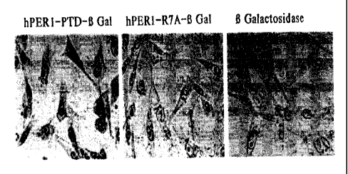
Figure 6. hPER-PTD cargo's [3-Galactosidase into cells: At least one feature
of HIV
TAT transducing peptide is its ability to cargo proteins into cells and
tissues. We therefore
sought to determine if hPERl. transducing peptide could cargo beta
galactosidase into cells.
To perform this experiment, we followed a protocol by Frankel et al. 1989
(19):7397-401,
whereby, we chemically linked hPERl-PTD or hPER-PTD R7A to full length (3-
3o galactosidase and assayed for the ability of these conjugates and beta-
galactosidase protein
alone to transduce into CHO cells. As shown in the figure 6, left, cells
incubated with hPER-
PTD (3-galactosidase fusion showed positive enzymatic activity for (3-
galactosidase as
indicated by the blue color in the cells after the addition of X-gal. However,
neither hPER-
SUBSTITUTE SHEET (RULE 26)
CA 02420350 2003-02-21
WO 02/18572 PCT/USO1/26421
-16-
MPP R7A (3-galactosidase (center) nor (3-galactosidase protein (right) alone
was able to enter
the cells as indicated by a no blue staining reactivity after the addition of
X-gal . These data
indicate that like TAT peptide, hPERI-PTD can cargo a large (120 kD) protein
into cells.
DETAILED DESCRIPTION OF THE INVENTION
The present invention is based on discovery that human Periodl (hPERl) protein
contains an NLS which has now also been identified as an MPP and is useful as
a delivery
device for intracellular delivery of a compound of interest. hPERl is involved
in regulation of
the circadian rhythm and the capacity of hPERl to translocate to adjacent
cells may be critical
to to its overall biological function ofregulating circadian rhythm. The NLS
identified within
hPERI does not fit within previously identified NLS sequences, and its
identification has
resulted in identification of an algorithm for searching for other NLS
sequences which may
also function as MPPs.
Period 1 (hPERl) is a nuclear protein involved with transcriptional
regulation. It is an
15 essential component in the "gears" of the biological clock (Brown, S.A.,
and Schibler, U.,
Current Opinion in Genetics ,& Development 9, 588-594 (1999), Dunlap, J.C.,
Cell 96, 271-
290 (1999)), and studies in mice have shown thatnuclear entry of PERI is
essential for the
down regulation of CLOCK/BMAL transcriptional complexes (Gekakis N, et al.,
Science 280,
1564~1569. (1998), Yagita, K., et al., Genes Dev 14,1353-1363 (2000), Lowrey,
P.L., et al.,
2o Science 288, 483-492 (2000)). However, to date, the functional NLS for
human PERT has not
been elucidated. The present inventors identified the NLS within hPERI, and
demonstrate
that the 16 amino acid and 13 amino acid sequence, see Figure 3.hPERl-NLS
peptide,
hPERI-MPP, has potent membrane penetrating ability. This work results in the
identification
of four additional MPPs also derived from nuclear proteins.
25 PERT is a central component in the circadian clock, and its nuclear entry
plays an
important role in the regulation of daily oscillations (Jin, X., et al., Cell
96, 57-68 (1999),
Sangoram, A.M., et al., Neuron 21, 1101-13 (1998 )). Using deletion and fusion
protein
analysis, we identified a NLS that is necessary and sufficient for hPERl
nuclear localization.
This functional analysis was necessary because the NL,S of hPERl does not
conform to
30 classical nuclear localizing consensus motifs; and therefore, was not
identified using standard
NLS search procedures. We show that a single copy of hPERI-NLS is sufficient
for inducing
nuclear localization of a reporter protein and of tagged hPER1 fragments (P1-
F2 to P1-F7) in
transfected cells. The PERT-NLS is located between amino acids (830-845) of
hPERl, is
SUBSTITUTE SHEET (RULE 26)
CA 02420350 2003-02-21
WO 02/18572 PCT/USO1/26421
-17-
embedded within a string. of 13 amino acids rich in arginine, histidine, and
lysine (see Table 1)
that is not found in other PERs or other nuclear proteins in available
databases. Therefore,
though PERs 2 and 3 are nuclear proteins (Tin, X., et al., Cell 96, 57-68
(1999)), they
apparently use alternative sequences and or mechanisms fox their nuclear
import.
Peptide fragments of a limited number of nuclear proteins that are rich in
basic
residues have been shown to penetrate into cellular membranes in a
receptorless, energy-
independent fashion. Sequences from three such proteins, TAT, Antp, and VP22
have been
demonstrated to possess the ability to penetrate and cargo fusion molecules
into cells and
tissues by an as yet undefined mechanism. See, for example, USP 5,804,604,
5,747,641,
l0 5,674,980, 5,670,617 and 5,652,122 issued to Frankel et al., which teach
the use of a nine-
amino acid HIV TAT-derived polypeptide (Arg-Lys-Lys-Arg-Arg-Gln-Arg-Arg-Arg)
for
intracellular delivery of cargo molecules.
The similarities between hPERl, the hPERl-NLS, and other MPPs prompted us to
investigate whether or not hPERl-MPP could have membrane penetrating
capability. The
15 immunohistochemical and cytological data presented herein indicates that
the hPER1-MPP
functions as a MPP in a variety of cell types. hPER1-MPP demonstrated intense
focal
staining in the nuclear plasma as well as in the nucleolus, suggesting that
the subnuclear
address of hPERI-MPP is different from the hPERl (P1-FL) protein that was
diffused in the
nucleus but not concentrated in the nucleolusThe cellular penetration of hPERl-
MPPs is not
20 blocked even under the conditions of reversing the sequence (reversed hPERl-
MPP), adding
negatively charged residues or pre-fixing cells with 4% PFA, unpublished
observation, the
latter supports the idea that penetration is receptor and membrane
independent. These results
are in contrast to other peptide classes that have been described that are
derived from signal
peptide sequences (Hawiger, J., Curr Opin Immunol. 9, 189-94 (1997)), DNA
antibodies
25 (Deng, S.X., et al., Iht Immunol. 12, 415-423 (2000)), and other protein
domains (Lindgren,
M., et al., Trends Pharmacol Sci. 3, 99-103 (2000)) that bind and cross the
cell membranes
using slow, temperature, energy, and receptor dependent mechanisms.
The identification of other MPPs, has been limited by our lack of
understanding the
mechanisms and structural requirements necessary for membrane peptide
penetration. The
3o likelihood that a specific peptide structure and/or charge is important for
membrane
penetration is demonstrated in the alanine scanning experiments whereby a
single amino acid
change at arginine 7 appears to be critical for MPP potential. By comparing
wild-type hPER-
MPP to modified P1- R7A, in live cells or pre-fixed and permeabilized cells
(data not show),
SUBSTITUTE SHEET (RULE 26)
CA 02420350 2003-02-21
WO 02/18572 PCT/USO1/26421
-18-
P1-R7A is only defective in penetration but not in nuclear targeting once the
cells have been
permeabilized. This finding suggests that arginine 7 has a major role in
structure based
penetration, and thus provides a useful model for the future structure-
function studies. No
structural determinants for TAT peptide have been described, but in the case
of Antp,
replacing the two tryptophan residues with two phenylalanines abolishes
penetration (Le
Roux, L, et al., Py-oc Natl Acad Sci USA. 90, 9120-9124 (1993)). Since hPERl-
MPP does
not contain any tryptophan residues, membrane penetration between these two
peptides may
occur by different mechanisms.
Full-length HIV TAT and VP22, both of which lack classical secretary signal
sequences and are therefore exported by non-classical mechanisms, can also be
imported "by
transduction", into cells in a non-classical manner (Prochiantz A., Current
Opiniorz irz Cell
Biology 12, 400-406 (2000)). Therefore, it is interesting to speculate that
perhaps hPERl
distributes circadian clock information to adjacent SCN neurons or to
circadian output
pathways by "transduction" mechanisms similar to full-length TAT and VP22
proteins.
However, simply having membrane penetrating sequences within the body of a
protein does
not necessarily confer membrane penetrating capability, as full-length Antp
protein is neither
exported from nor imported into cells. Thus, the non-classical penetration of
the Antp peptides
into the cells is unlikely to have physiological relevance, and Like Antp,
there is no evidence to
suggest that full-length hPERl is a cell membrane penetrating protein.
However, these
2o findings did encourage us to search for other MPP-containing proteins. By
searching protein
databases with an algorithm designed to identify strings of basic residues
within nuclear
proteins, we uncovered hundreds of proteins that contained potential membrane
penetrating
peptide regions and found 4 additional MPPs from several species (see Fig. 5).
These and
additional database mining searches suggest that MPP-like sequences are
common, and
present within a wide variety of proteins. However, like many putative NLSs
that do not
always confer nuclear localization when fused to reporter sequences (Moroianu,
J., J Cell
Biochem. 32-33, 76-83 (1999)), any potential MPPs must be functionally
determined
experimentally. Though it seems clear that either transducing or non-
transducing proteins can
encode MPP regions, the interesting question that remains is whether or not
proteins
containing MPP-like sequences use these domains to rapidly translocate
intracellularly into
cellulax domains to activate normal physiological processes. The efficiency
associated with
the transduction phenomena might be particularly useful where the rapid
delivery of
SUBSTITUTE SHEET (RULE 26)
CA 02420350 2003-02-21
WO 02/18572 PCT/USO1/26421
-19-
intercellular information is critical, as may be the case in cell
synchronization, development,
and differentiation paradigms.
The ability for MPPs to cargo molecules to intracellular compartments is
becoming
well-established (Lindgren, M., et al., Trends Plzarmacol Sci. 3, 99-103
(2000), Derossi, D., et
s al, Trends Cell Biol., 8, 84-87 (1998)). Similar to other MPPs, hPERl-MPP
and other MPPs
identified herein can deliver compounds of interest, such as large molecules,
i.e., peptides and
proteins, lipids, polysaccharides, other organic molecules, rapidly and
efficiently into cells.
The data presented herein demonstrates that hPERl-MPP in fusion with either
serotonergic
and/or adrenergic 7TM-receptor derived peptides mimic the effects of ligand
activated
to receptors (see Fig. 4, and data not shown), confirming that hPERl-MPP
translocates
compounds of interest to intracellular compartments, and supports the idea
that
physiologically relevant signaling can be initiated by MPPs linked to
compounds of interest.
Using the methods described herein, the present invention may be expanded to
provide target
validation using MPPs linked to targets, and/or therapeutic strategies using
MPPs linked to
15 specific enzymes or receptors as a method of altering, correcting or
compensating for
dysfunctional enzyme performance or within pathways. In addition, therapeutic
strategies
using MPPs linked to specific receptors may be used as a method of altering,
correcting or
compensating for dysfunctional receptor, low expression of normal or abnormal
receptors.
Taken together, the results provided herein demonstrate an MPP encoded by a
2o mammalian protein and more specifically, a human nuclear protein, whose
cellular penetration
is membrane independent and likely depends on the peptide structure. hPERl-MPP
targets to
specific subnuclear sites, but has the potential to efficiently deliver other
macromolecules to
intracellular targets.
More importantly, this invention also provides the first example for mapping a
novel
25 MPP based on a NLS domain, and suggests that many MPP-like regions are
contained within
a wide variety of proteins. The data provided herein demonstrate that an MPP
may be based
on part of an NLS, or overlap with part of the NLS, or alternatively, may be a
novel peptide.
Methods of identifying NLS sequences are well known in the art, and include
NLSs
previously identified as conferring the ability of the native protein to enter
the nucleus, or is a
3o putative NLS based on substantial sequence homology with a previously
identified NLS.
Alternatively, the NLS may be identified by sequence deletion experiments. See
for example,
Luo JC, Shibuya M A variant of nuclear localization signal of bipartite-type
is required for
the nuclear translocation of hypoxia inducible factors (lalpha, 2alpha and
3alpha). Oncogene.
SUBSTITUTE SHEET (RULE 26)
CA 02420350 2003-02-21
WO 02/18572 PCT/USO1/26421
-20-
2001 Mar 22;20(12):I435-44 or Hodel MR, Corbett AH, Hodel AE. Dissection of a
nuclear
localization signal. J Biol Chem. 2001 Jan 12;276(2):1317-25.
Preferred membrane penetrating peptides (MPPs, also known as peptide
transduction
domain or 'PTD') of the present invention are small polypeptides, and may be
derived from an
NLS, or overlapping with an NLS, of a mammalian or yeast protein. Preferred
mammalian
proteins are those of human, primate, marine or rat species. It is generally
preferred to use the
same species for the NLS-derived protein as the cell to be treated. Human
species are
especially preferred as the NLS-derived protein when being used to treat human
cells. NLSs
may be found within a broad class of enzymes, and is not limited to nuclear
proteins,
transcription factors, cytokines and kinases. Preferred MPPs are those derived
from nuclear
proteins or transcription factors. Alternatively, MPPs of the present
invention are small
polypeptides comprising a sequence -(X-X-X-X)"- where n is an integer 1 to 7,
and X each
time is independently selected from the group consisting of arginine,
histidine or lysine. It is
preferred that small MPPs are used, and therefore, it is preferred that n is
an integer 1 to 5, and
more preferred that n is an integer 1 to 3. Selected embodiments of suitable
MPPs are
provided in Table 1 and Example 5.
The MPP and/or compound of interest may be chemically synthesized separately,
for
example, by chemical synthetic routes and using commercially available
reagents.
Alternatively, if the MPP and/or compound of interest is a polypeptide, it may
be synthesized
by recombinant technology and purified according to known methods. Host cells,
cloning
vectors, promoters and oligonucleotide linkers are well known and commercially
available.
Methodolgies for using recombinant technology and purification methods are
also well
known, see Current Protocols in Molecular Biology, 4 Vols. Wiley. Generally,
recombinant
technology is preferred, as it is more amenable to larger scale production and
is more
economical for mass production. Alternatively, MMPs may be obtained by
specific protease
degradation of a precursor proteins.
The compound of interest rnay be attached or linked to the MPP via chemical
crosslinking at the N- or C-terminus of the MPP to create a conjugated (also
referred to a a
fusion) MPP and compound of interest, for example, via disulfide or ester
linkages. In an
alternative embodiment, if the compound of interest is a peptide, the peptide
may be
synthesized by recombinant technology with a host cell with an expression
vector encoding a
fusion of the MPP sequence and the compound of interest under conditions to
permit
expression of the vector and obtaining the fusion MPP and compound of
interest.
SUBSTITUTE SHEET (RULE 26)
CA 02420350 2003-02-21
WO 02/18572 PCT/USO1/26421
-21-
In another embodiment, the MPP and the compound of interest may be attached or
linked via a chemical linker. Chemical linkers are well known in the art, and
include but are
not limited to dicyclohexylcarbodiimide (DCC), N-hydroxysuccinimide (NHS),
maleiimidobenzoyl-N-hydroxysuccinimide ester (MBS), N-ethyloxycaxbonyl-2-
ethyloxy-1,2-
dihydroquinoline (EEDQ), N-isobutyloxy- carbonyl-2-isobutyloxy-1,2-
dihydroquinoline
(IIDQ). Preferred linkers may also be monomeric entities such as a single
amino acid,
especially preferred are those amino acids with small side chains, or a small
polypeptide
chain, or polymeric entities of several amino acids. Preferred polypeptide
linkers are fifteen
amino acids or less, more preferred are polypeptide linkers of ten or less
amino acids. Even
to more preferred are polypeptide linkers of five or less amino acids. In an
alternative
embodiment, the linker may be a nucleic acid encoding a small polypeptide
chain; preferred
linkers encode a polypeptide of fifteen or less amino acids. More preferred
linkers axe nucleic
acids encoding a small polypeptide chains of ten or less amino acids. Even
more preferred
linkers are nucleic acid encoding a small polypeptide of five or less amino
acids, such as Gly-
Phe-Leu-Gly, Gly-Gly, Gly-Leu or Gly, and the like.
Recombinant technology may be used to express a fusion MPP, linker and
compound
of interest, as described above and is well known in the art.
In another embodiment, the linker may be a cleavable linker, resulting in
cleavage of
the MPP and compound of interest once delivered to the tissue or cell of
choice. In such an
2o embodiment, the cell or tissue would have endogenous (either naturally
occuring enzyme or
be recombinantly engineered to express the enzyme) or have exogenous (e.g., by
injection,
absorption or the like) enzyme capable of cleaving the cleavable linker.
Suitable enzymes for
cleavage include, for example, use of a KEX2 protease recognition site (Lys,
Arg) inserted
between glucoamylase and the desired polypeptide to allow in vivo release of
the desired
polypeptide from the fusion protein as a result of the action of a native
Aspergillus KEX2-like
protease. (Contreras et aL, I991; Broekhuijsen et al., 1993; Ward et al.,
1995). Another
example of a cleavable linker peptide comprises the recognition sequence Asp-
Asp-Asp-Asp-
Lys, and wherein said fusion protein is cleavable by enterokinase.
Alternatively, the linker may be biodegradable such that the compound of
interest is
3o detached from the fusion MPP and compound of interest by hydrolysis and/or
enzymatic
cleavage inside cells. For example, tumors often express specific proteases,
and be used in the
delivery of prodrugs of cytotoxic agents. The linker may be selective for
lysosomal proteases,
such as cathepsin B, C, or D. Delivery of prodrugs and their subsequent
activation is well
SUBSTITUTE SHEET (RULE 26)
CA 02420350 2003-02-21
WO 02/18572 PCT/USO1/26421
-22-
recognized, and such an approach provides significantly less systemic toxicity
due to
premature linker hydrolysis in the blood, consequently a greater amount of
compound of
interest, i.e., drug or cytotoxic agent, is delivered to the tumor site. See
for example, T.
Higuchi and V. Stella provide a thorough discussion of the prodrug concept in
Pro-drugs as
Novel Delivery Systems, Vol. 14 of the A.C.S. Symposium Series, American
Chemical
Society (1975). Examples of readily-cleavable groups include acetyl,
trimethylacetyl,
butanoyl, methyl succinoyl, t-butyl succinoyl, ethoxycarbonyl,
methoxycarbonyl, benzoyl, 3-
aminocyclohexylidenyl, and the like.
The compound of interest is any organic molecule, and includes small organic
molecules, peptides, lipoproteins, and other modifed proteins,
polysaccharides,
oligonucleotides, antisense oligonucleotides, and any other compound thought
to have
pharmaceutical, prophylactic, diagnostic properties andlor research interest.
The compound of
interest may be a small organic molecule already known to have pharmaceutical
properties,
and thus the present invention may be used as a method of treating a patient
with the
compound of interest. Alternatively, the compound of interest may be a novel
protein of
unknown function, and thus the present invention may be used as a method of
identifying the
function of the compound of interest. In another embodiment, the compound of
interest may
be an antisense molecule, and thus the present invention may be used as a
method of altering
transcription. In yet another embodiment, the compound of interest may be a
prodrug, e.g. in
2o an inactive form but capable of being activated once within the cell. In
another embodiment,
the compound of interest may be a cytotoxic agent, and thus the invention may
be used as a
method of delivering a cytotoxic agent to a cell. The compound of interest
also includes
detectable proteins which are useful to generate conjugated MMP and the
detectable protein
for identification of new MMPs. Detectable proteins include GFP, beta
galactosidase,
radiolabeled proteins and biotinylated proteins, proteins capable of
conferring a detectable
phenotype in the cell.
The present invention may be used to deliver the compound of interest into a
cell in
vitro, ex vivo or in vivo. For example, delivery may be carried out in vitro
by adding the
conjugated MPP and compound of interest extracellularly to cultured cells.
Delivery may be
3o carried out ex vivo by adding the conjugated MPP and compound of interest
extracellularly or
exogenously to a cultured sample removed from a patient, for example, blood,
tissue or bone
marrow, and returning the treated sample to the patient. Delivery may be
carried out in vivo
SUBSTITUTE SHEET (RULE 26)
CA 02420350 2003-02-21
WO 02/18572 PCT/USO1/26421
-23-
by adminstering the conjugated MPP and compound of interest by transdermal
administration,
inhilation, or injection to a patient.
Any type of cell may used in the present invention. The cell may be of
mammalian,
bacterial, viral or yeast origin. The cell may be a cultured cell such as
commonly used for
oncology screening. Examples of cultured cells include CHO, HEK293T, HeLa, and
NIH3T3. The cell rnay be a cultured cell from a patient suitable for ex vivo
treatment with an
MPP conjugate and reintroduction into a patient. The cell may be from the same
or different
patient than the patient to be treated.
Compositions of the invention comprising the conjugated MPP and compound of
to interest may be used for therapeutic, prophylactic, diagnostic or research
purposes.
Compositions may further comprise adjuvants, stabilizers and the like to
improve the
handling, stability and storage properties of the compositions.
Methods to identify novel MPPs are also part of the present invention. One
method for
identification of a membrane penetrating peptide is to generate a conjugate
peptide comprising
the sequence -(X-X-X-X)"- where n is an integer 1 to 7, and X each time is
independently
selected from the group consisting of arginine, histidine or lysine, with a
detectable protein
such as GFP, beta galactosidase and the like, adding the conjugate peptide to
a cell and
determining if the conjugated peptide is located within the cytoplasm and/or
nucleus of the
cell. Another method for identification of a membrane penetrating peptide is
to generate a
2o conjugate peptide comprising a peptide derived from or overlapping with a
nuclear
localization sequence of a mammalian or yeast protein and a detectable protein
such as GFP,
beta galactosidase and the like, adding the conjugate peptide to a cell and
determining if the
conjugated peptide is located within the cytoplasm andlor nucleus of the cell.
The following abbreviations are used for amino acids:
A refers to Ala, or alanine;
C refers to Cys or cysteine;
D refers to Asp or aspartic acid;
E refers to Glu or glutarnic acid;
F refers to Phe or phenylalanine;
3o G refers to Gly or glycine;
H refers to His or histidine;
I refers to Ile or isoleucine;
K refers to Lys or lysine;
SUBSTITUTE SHEET (RULE 26)
CA 02420350 2003-02-21
WO 02/18572 PCT/USO1/26421
-24-
L refers to Leu or luecine;
M refers to Met or methionine;
N refers to Asn or asparagine;
P refers to Pro or proline;
Q refers to Gln or glutamine;
R refers to Arg or arginine;
S refers to Ser or serine;
T refers to Thr or threonine;
V refers to Val or valine;
W refers to Trp or tryptophan;
Y refers to Tyr or tyrosine.
Proteins are written with the N-terminus to the left.
The following abbreviations are used: 'v/v' refers to volume to volume; 'EYFP'
refers
to a peptide fragment of the sequence Glu-Tyr-Phe-Pro; 'ORF' refers to Open
Reading Frame;
'PCR' refers to polyrnerase chain reaction; 'CHO' refers to Chinese Hamster
Ovary cells;
'HEK.293T' refers to Human Embroyonic Kidney cells, 'HeLa' refers to
epithelial
adenocarcinoma cells; 'NIH3T3' refers to Swiss mouse embryo fibroblast cells;
' DMSO'
refers to dimethyl sulfoxide; 'FCS' refers to fetal calf serum; 'DMEM' refers
to Dulbecco's
Modified Eagle's Medium; 'PBS' refers to Phosphate buffered saline; ' BSA'
refers to bovine
2o serum albumin; 'C-terminus' refers to the carboxy-terminus; 'N-terminus'
refers to the amino-
terminus; 'PTD' refers to Peptide transduction domain; 'GPCR' refers to G-
protein coupled
receptor; 'TM' refers to a transmembrane domain of a GPCR; 'f refers to an
intracellular loop
of a GPCR; 'SHT2A' refers to serotonin receptor 2A; and'mAb' refers to
monoclonal
antibody.
as
EXAMPLES
Example 1 Identification of an NLS within hPERl
Plastnid Construction
All hPer1 fragments described here are cloned as in-frame C-terminal fusion to
EYFP.
3o EYFP-hPerl ORF, P1-N and P1-NX (fig.lA) is generated by insertion of EcoRI
and XhoI
digested fragments into EYFP-C1 vector (Clontech). The other fragments are PCR
amplified
from the full-length hPerl cDNA and subcloned into EYFP-CI vector. The first
and the last
SUBSTITUTE SHEET (RULE 26)
CA 02420350 2003-02-21
WO 02/18572 PCT/USO1/26421
-25-
residue present in each of fragment is indicated in Fig.lA. All constructs axe
verified by
automated DNA sequencing.
Cell culture and DNA Trausfectiou
CHO, HeLa and 293T cells are maintained in Dulbecco's Modified Eagle's Medium
(DMEM) supplemented with 10% fetal calf serum (FCS), 50 units/ml penicillin,
50 ~.g
streptomycin, and 4 mM L-glutamine (hereafter referred to as complete DMEM) at
37°C with
5% CO2. Transfection of the cells is carned in two-well Lab-Tek coverslips
(Nunc Inc.) with
LIPOFECT-AMINETM~ Reagent (Life Technologies) according to the manufacturer's
instructions.
Peptides and Peptide I>zterualizatiou
Peptides are synthesized by a conunercial vendor (Bio Synthesis). For peptides
internalization, cells are plated into two-well Lab-Tek coverslips (Nunc Inc.)
at a density of
2X10⁵ cells/well and cultured overnight. The peptides are dissolved in DMSO
diluted to
indicated concentration with PBS. The cell monolayers were incubated with the
appropriate
peptide/PBS solution at 1 ~,M standard concentration for 10 min at room
temperature (RT)
unless otherwise specified. For experiments at 4°C, the protocol was
the same except that all
incubations were performed at 4°C until the end of the fixation
procedure.
Irnmuho, fl'uo~esceuce afzd Microscopy
Fox direct detection of expression and subcellular localization of EYFP fusion
protein,
2o transfected cells were examined directly without fixation or after fixation
with 4%(v/v)
formaldehyde in PBS for 20 min at 4°C and washed with PBS. For indirect
immunodetection
of biotinylated peptides, fixed cell were washed twice with PBS and
permeabilized with 0.3%
Triton X-100 in PBS for 20 min at 4°C and blocked with 2% BSA in PBS
for 30 min at RT.
Cells were then washed with PBS and incubated with Streptavidin-FITC~ (Sigma)
or -
Alex499 (Molecular Probe), 1:400 diluted in 0.2% Tween 20, 2% BSA in PBS for 1
h at RT.
Following 2 x 5 min washes with PBS and once with 0.3% Triton X-100 in PBS for
20 min
RT. In some experiment, the nucleus was stained with 50 ng/ml Hoechst 3325
(Sigma) or 3
~.g/ml propidium iodide in PBS. The subcellular localization of the
fluorescence was analyzed
on an Olympus microscope. Confocal images were taken on a Zeiss confocal laser
scan
microscope (CLSM phoibos 1000).
Though it is known that nuclear entry of PERT is important for its function,
no
putative NLS was identified using a standard Profile Scanning program
(Shearman, L.P., et
al., Neuron 19, 1261-1269 (1997), Yagita, K., et al., Genes Dev. 14, 1353-1363
(2000)). To
SUBSTITUTE SHEET (RULE 26)
CA 02420350 2003-02-21
WO 02/18572 PCT/USO1/26421
-26-
determine the NLS of hPERl experimentally, three full-length hPER1 (P1-FL)
were
constructed and denoted as P1-N, P1-NM and P1-C (Fig. 1A). The ability of
these constructs
to localize to the nucleus in CHO cells then analyzed. An EYFP-tag was used to
facilitate the
detection of hPERl in living cells; however, the EYEP-tag had no apparent
contribution on
hPERI fusion protein localization since hPERl constructs made with an N-
terminal Flag-tag
presented an identical cytological distribution pattern (data not shown).
After transient
transfection, both P1-FL and P1-NM proteins were expressed in the nucleus of
transfected
cells as early as 10 hours post-transfection, while both P1-N and P1-C
accumulated only in the
cytoplasm (Fig.lB). The EYFP vector control was diffuse in both the nucleus
and cytoplasm.
to These results demonstrate that a functional NLS in hPERl is located between
Pl-N and P1-C
in what we designated as region M (see Fig.lA).
To further localize the NLS in region M (amino acids 481-890), a series of 8
deletion
constructs, P1-F1 to Pl-F8, were generated and the subcellular distribution of
each mutant
was assayed as indicated in Fig. 1A and B. Sequential deletion from amino acid
581 (Pl-F2)
to position 821(P1-F7) of region M resulted in nuclear localization. Further
deletion of amino
acids 821 to 841 (P1-F8) resulted in a diffused fluorescent pattern within
transfected cells with
a localization pattern similar to that of the EYFP vector: control. These data
indicate that a
NLS exists between amino acids 821 and 890, and is located at the C-terminus
of region M.
This observation was confirmed by the construction of an additional EYFP
fusion protein, P 1-
NLS, which contained hPERl amino acids 830-845. This region contains a string
of basic
residues that might function as a NLS (Weis, I~., TYehds Bioclaem. Sci. 23,
185-189 (1998),
Truant, R, and Cullen, B.R. Mol Cell Biol. 19, 1210-1217 (1999)). As expected,
P1-NLS
exhibited nuclear localization in 100% of transfected cells (Fig. 1 B). Other
regions of PERT
in additional fusion constructs failed to localize to the nucleus (data not
shown). Therefore,
we conclude that the NLS of hPERl (hPER1-NLS) is localized to within amino
acids 830-
845. Interestingly, construct P1-F1 has a strictly cytoplasmic localization
pattern irrespective
of the fact that it contains the NLS, supporting published observations that
this region also
contains and as yet unidentified cytoplasmic localization domain (Vielhaver,
E., et al., Mol
Cell Biol. 20, 4888-4899 (2000)). Sequence alignment shows that the hPERI-NLS
is
3o conserved between human and mouse PERI proteins (Fig. 1A), but not with
other putative
NLSs, or with other human, mouse or Drosophila PERs. After completion of our
studies,
Vielhaber et al. (2000), identified a longer mouse PERT-NLS that contains our
identified 16
SUBSTITUTE SHEET (RULE 26)
CA 02420350 2003-02-21
WO 02/18572 PCT/USO1/26421
-27-
amino acid sequence (Vielhaver, E., et al., Mol Cell Biol. 20, 4888-4899
(2000)); thus,
supporting our findings.
Example 2 hPER1-NLS encodes an MPP
Two common features of the three identified gene encoded MPPs (TAT, Antp, and
VP22) are that they axe derived from nuclear proteins and they consist of
basic amino acid
residues (Lindgren, M., et al., Trends Pharmacol Sci. 3, 99-103 (2000)), hPERl
is also a
nuclear protein whose NLS is rich in basic amino acids (SRRHHCRSKAKRSRHH, see
Fig.
1). These similarities led us to determine whether hPER-NLS might also
function as a MPP.
In order to test this hypothesis, we synthesized several N-terminally
biotinylated peptides:
IO hPERl-MPP, Flag-tagged hPERI-MPP, Flag-tagged TAT-PTD, Flag-Flag alone, See
Table I
below:
Table 1
Name Amino Acid Sequence TransducingNuclear
Peptides Localizati
on Fusion
Protein2
hPER1 GRRHHCRSKAKRSRHH + +
Flag-hPERl GMDYKDDDDKGSRRH~ICRSK + nd
AKRSHH
Flag-TAT GMDYKDDDDKG'YGRKKKRR + +
QRRR
Flag GMD~KDDDDKG - -
MDYKDDDDK
Antennapedia GRQIKIWFQNRRMKWKI~ + nd
9 Arginine G + nd
9 Lysine GKKKKKKKKK + nd
9 Histidine GHHI~IH~ - nd
NLSs:
SUBSTITUTE SHEET (RULE 26)
CA 02420350 2003-02-21
WO 02/18572 PCT/USO1/26421
_~g_
SV40 GDPKKKRKV - +
hPER2 GKKTGKNRKLKSKRVKPRD - +
hPER3 GRKGKHKRKKLP + +
Thyroid A-1 GKRVAKRKLIEQNRERRR + +
HME-1 GRKLKKKKNRKEDKRPRT + . +
ABL-1 GKKTNLFSALIKKKKTA + +
Nucleoplasmin.XGRRERNK1VIAAAI~CRNRRR + +
C-FOS GRRERNK1VIAA.AKCRNRRR - +
GCN-4 GB;RARNTEAARRSRARKL + +
[R/H/K] _
[RlH/K]-
[R/H/K]-
[R/H/K]
HEN1/NSLC1 GRRRRATAKYRTAH + +
HEN2/NSLCZ GK:RRRRATAKYRSAH + +
HNF3 GRRR.RKRLSHRT ~ + +
CAMP dependentGR:RRRRERNK + +
TF
Cyclin L aria-6aGKHRHERGHHRDRRER - +
beta Zip TF GKKKRKT SNRESAKRSR - +
GFP - nd -
Fn 1: Results shown fox selected MPPs, see Fig 5
Fn Z: Results shown for selected MPPs, see Fig 5
The peptides are assayed for their ability to penetrate cellular membranes.
Intracellular
localization is assayed by direct staining with labeled Streptavidin ALEXA
reagents or by
indirect staining with anti-Flag iizA.b followed by the addition of labeled
secondary antibodies.
When added to the cells in culture at a concentration of 10 ~.M, hPERl-MPP,
Flag-hPERl-
MPP and Flag TAT-PTD peptides are found to penetrate rapidly into 100% cells
(Fig. 2A and
Fig. 5). By both detection methods, hPERl-MPP, Flag-tagged hPERl-MPP, and Flag-
tagged
TAT-PTD are observed to be diffusely distributed throughout the cytoplasm, but
concentrated
1o within subnuclear domains that appear as distinct foci within the
nucleoplasm and the
SUBSTITUTE SHEET (RULE 26)
CA 02420350 2003-02-21
WO 02/18572 PCT/USO1/26421
-29-
nucleolus. In contrast, biotinylated negative control peptides, Flag-Flag and
several additional
peptides derived from other hPERl regions are only barely discernible
background staining,
with no staining in the nucleus or nucleoli, even at high concentrations (data
not shown).
Confocal microscopy is used to confirm the intracellular and intranuclear
staining of Flag-
tagged hPERl-MPP, and that the negative control peptides are not internalized
(Fig. 2A).
hPERl-MPP rapidly penetrated the cellular membranes and localized in nuclear
regions with efficiencies similar to the TAT-PTD peptide (Fig. 2B). Identical
results are
obtained using CHO, HEK293T, HeLa, NIH3T3 and cultured rat primary cortical
neurons
(data not shown), indicating cell type-independent penetration.
1o hPERl-MPP internalization occurrs rapidly (within 5 min), with similar
potencies at
4C and 37C and even after cell membrane fixation (data not shown). Thus, the
amino acid
sequence 830-845 of hPERl functions as both as a protein nuclear/nucleolar
localization
signal in the fusion protein and as a MPP, and that membrane penetration is
independent of
traditional receptor-mediated endocytic mechanisms.
Example 3 Arginine 7 is essential for hPERl-MPP activity
To date, the mechanisms as well.as the structural basis whereby MPPs
transverse
cellular membranes have not been elucidated. We therefore sought to determine
if there were
key residues within hPERl-MPP that were important for maintaining those
properties
2o essential for its membrane penetrating potential. We separately replaced
each amino acid in
hPERl-MPP to alanine (Table 2), and assayed for the ability of these mutated
peptides to
penetrate living cells relative to the wild-type hPERl-MPP.
Alanine scaring:
Name hPERl-PTD alanine Transducing
substitution Peptide
hPERl-PTD SRRHHCRSKAKR S RHH +
R2A SARHHCRSK A K R S R H H +
R3A SRAHHCRSK A K R S R H H +
H4A SRRAHCRSKAKR S RHH +
HSA SRRHACRSK A K R S R H H +
C6A SRRHHARSK A K R S R H H +
SUBSTITUTE SHEET (RULE 26)
CA 02420350 2003-02-21
WO 02/18572 PCT/USO1/26421
-30-
R7A SRRHHCASK A K R S R H H -
S8A SRRHHCRAK A K R S R H H +
K9A SRRHHCRSA A K R S R H H +
K1IA SRRHHCRSKAARSRHH +
R12A SRRHHCRSKAKASRHH +
S13A SRRHHCRSKAKRARHH +
R14A SRRHHCRSK A K R S A H H +
hPER1- RRHHCRSK A K R S R +
PTD 13
hPERl- QELSEQIHRLLLQPV -
Control
(484-
503)
As shown in Fig. 3, most of the single alanine substitutions had very little
effect on
membrane penetrating capabilities as compared with wild-type peptide. However,
changing
arginine 7 to an alanine (R?A) reduced the detectable cytological signal to
that observed for
the negative control peptides. Thus, the arginine 7 to alanine mutation
significantly reduced
the membrane penetrating properties of hPERl-MPP. Identical observations were
observed
after changing arginine ? to glutamic acid (R?E) (data not shown).
Furthermore, the
simultaneous deletion of the N-terminal serine and of the two C-terminal
histidines from
hPERl=MPP (hPERI-MPP13) had little overall effect on the positive membrane
penetrating
to potential of the peptide (Fig. 3).
The arginine 7 residue plays a critical role in the cell penetrating ability
of the hPERI-
MPP. We therefore sought to determine if the R?A mutation affected nuclear
translocation of
a fusion protein P1-NLS. CHO cells transfected with fusion protein P1-R7A
(arginine ?
mutated to alanine) have intense nuclear staining similar to the wild-type, F1-
NLS (data not
15 shown). Nuclear translocation appears to be normal in the P1-R?A mutant
fusion protein, but
subnuclear targeting to the nucleoli is disrupted (data not shown). These data
indicate that
membrane penetration and nucleoli targeting are affected by the single R?
amino acid residue
and indicate that nuclear translocation of hPERl-NLS has separate and distinct
determinants.
Example 4 hPER1-1VIPP delivery of functioning molecules
2o One of the features of MPPs is their ability to cargo proteins and peptides
into cells.
We were successful in coupling hPER1-MPP to B-galactosidase and in delivering
the fusion
SUBSTITUTE SHEET (RULE 26)
CA 02420350 2003-02-21
WO 02/18572 PCT/USO1/26421
-31-
protein into cells in culture (data not shown), as has been described by
Fawells et al., 1994
(Fawell, S., et al., Proc Natl Acad Sci U S A. 91, 664-668 (1994)). However,
to further extend
the functional utility of MPPs, we tested hPER1-MPP in fusion with a
physiologically
relevant and biologically active peptide. Wess and collegues (1993) have shown
a functional
role for the conserved transmembrane segment ? (TM7) of the G-protein coupled
receptor
(GPCR) superfamily. Along with TM7, the third intracellular loop (I3) plays a
significant role
in GPCR calcium signaling (bless, JM., et al., EMBO J. 12, 331-338 (1993))
while
intracellular loops 1 and 2 (I1 and I2) appear not to be important. Using the
serotonin
receptor, 5HT2A, we experimentally tested the ability of hPER1-MPP and TAT-PTD
in
fusion with peptides designed from Il and the TM7 domains to activate the
receptor.
Biotinylated peptides hPERI-MPP TM7, TAT-PTD TM7, hPERl-MPP I1, TAT-PTD Il,
hPER-MPP, TAT-PTD, TM7 or Il were incubated at a concentration of 10 ~,M with
a SHT2A
receptor CHO stable cell line. Peptide membrane penetration was assayed using
Streptavidin-
Alexa 594 as described above. As shown in Fig. 4A, receptor signaling is
activated by the
addition of exogenous serotonin, hPERl-MPP TM7, and TAT-PTD TM7 as measured by
the
level of the calcium response. However, TM7 alone nor any of the other
peptides were able to
generate a calcium response. . Furthermore, the activation of the receptor by
hPERl-MPP TM7
and TAT-PTD TM7 is peptide concentration dependent, Fig. 4B. The addition of
increasing
concentrations of the activating peptide, TM7, in fusion with hPERI-MPP or TAT-
PTD
2o results in a calcium response in a dose dependent manner. TAT-PTD TM7
appears to be a
more potent 5HT2A receptor activator than is hPERl-MPP TM7. A simple
explanation for
this result is that TAT-PTD TM7 is more cytoplasmically localized or has
greater cell
penetrating capablilites than hPERl-MPP TM7, although we have not observed
that to be the
case. Similar results were also obtained in this laboratory using hPER1-MPP in
fusion with ~i-
adrenergic activating peptides (unpublished data). These data support previous
results that
hPERl-MPP not only penetrates cell membranes, but also demonstrates that it is
capable of
cargoing peptides to intracellular compartments to initiate biologically
relevant signal
transduction events.
Example 5 Identification of other gene encoded MPPs
Since hPERl is a nuclear protein proposed to be involved in transcriptional
regulation,
and since, to date, all PTDs derived from naturally occurnng proteins are
transcription factors
(TAT, Antp, and VP22), we sought to determine if other PTD sequences existed
within the
genome. To this end, we used two approaches; first, we searched the NCBI non-
redundant
SUBSTITUTE SHEET (RULE 26)
CA 02420350 2003-02-21
WO 02/18572 PCT/USO1/26421
-32-
protein database for all known and putative NLS's (table 1, 10-17). We
synthesized peptides
corresponding to the NLS amino acid sequences and assayed for peptide
transduction. As
shown in table 1 and Fig. 5, 6 of the 7 peptides synthesized had membrane
penetrating
characteristics similar to hPER-PTD and TAT-PTD. These proteins included human
proteins .
of the thyroid hormone receptor alpha-1, homeobox protein HMEl, and proto-
oncogene
protein ABL-1. Furthermore, (table 1 and Fig. 5) when we create in frame
fusion proteins
between these peptide sequences and GFP then transfected into CHO or HEK 293T
cells, all
of the sequences conferred nuclear localization of the fusion protein.
Our second approach to identifying PTDs involved searching the NCBI non-
redundant
i0 protein database collection with a degenerative algorithm (see Fig. 5,
legend). Using these
search parameters, we found 533,291 sequences of which the conditions for the
algorithm
were satisfied 129,169 times (24%). By limiting our search to include either
"transcription
factors, cytokines or tyrosine kinases", we identified 8284 transcription
factor protein
sequences of which the algorithm pattern occurred 7374 times (89%); within
2333 cytokine
15 protein sequences the pattern occurred 450 times (19%); and within 2513
tyrosine kinase
protein sequences the pattern occurred 843 times (36%). Because the algorithm
occurred
most frequently in nuclear proteins, we synthesized peptides to putative PTDs
for 6 of the
"transcription factor" sequences and assayed for their ability to penetrate
into the cells. As
shown in table 1, results in lines 18-23 and fig 3A, 4 of the 6 peptides
tested had membrane
20 penetrating properties similar to hPER1-PTD and TAT-PTD. These proteins
included two
human proteins HENl/NSLC-l and HEN2/NSLC-2 which are reported to be involved
in
neuronal differentiation and development (Uittenbogaard, M., Peavy, D.R. and
Chiaramello,
A. 1999. Expression of the bHLH gene NSCL-1 suggests a role in regulation of
cerebellar
granule cell growth and differentiation. J. Neurosci. Res. 57:770-781,
Lipkowitz, S. et al.
25 1992. A comparative structural characterization of the human NSCL-1 and
NSCL-2 genes.
Two basic helix-loop-helix genes expressed in the developing nervous system.
J. Biol. Chem.
267:21065-21071), rat HNF-3 (17), and a D~osophila CAMP dependent
transcription factor
(18). Furthermore, (table l and Fig 5) when we create in frame fusion proteins
between these
peptides and GFP and transfected into CHO or HEK 293T cells, all of the
sequences conferred
3o nuclear localization of the fusion protein. These results indicate that PTD
sequences can be
found within or overlapping with NLSs. However not all NLSs are PTDs as is
apparent in
SV40, hPER2, C-FOS, Cyclin L ania-6 and beta Zip transcription factor NLSs
(table 1).
SUBSTITUTE SHEET (RULE 26)
CA 02420350 2003-02-21
WO 02/18572 PCT/USO1/26421
-33-
These results also suggest that PTDs sequences are prevalent throughout the
genome and in
particular within nuclear proteins.
Example 6. hPER-PTD with (3-Galactosidase
At least one feature of HIV TAT transducing peptide is its ability to cargo
proteins into
cells and tissues. We therefore sought to determine if hPERl transducing
peptide could cargo
beta galactosidase into cells. To perform this experiment, we followed a
protocol by Frankel
et al. PNAS 199 (19):7397-401, whereby, we chemically linked hPERl-PTD or hPER-
PTD
R7A(with Ala replacing Arg~) to full length (3-galactosidase and assayed for
the ability of
these conjugates and beta-galactosidase protein alone to transduce into CHO
cells. As shown
to in the figure 6, panel 1, cells incubated with hPER-PTD (3-galactosidase
fusion showed
positive enzymatic activity for (3-galactosidase as indicated by the blue
color in the cells after
the addition of X-gal. However, neither hPER-MPP R7A (3-galactosidase nor j3-
galactosidase
protein alone was able to enter the cells as indicated by a no blue staining
reactivity after the
addition of X-gal, panels 2 and 3. These data indicate that like TAT peptide,
hPERl-PTD can
cargo a large (120 kD) protein into cells.
SUBSTITUTE SHEET (RULE 26)