Note: Descriptions are shown in the official language in which they were submitted.
CA 02420567 2003-02-24
WO 02/18644 PCT/US01/25884
MASS SPECTROMETRIC
ANALYSIS OF BIOPOLYMERS
FIELD OF THE INVENTION
The present invention relates to the analysis of biopolymers in crude
solutions. In
particular, the invention relates to the determination, quantitation, and
identification of
biopolymers, such as polypeptides and oligonucleotides, using mass
spectroscopic data
obtained from fractioned mixtures.
REFERENCES
Allen G (1989) Sequencing of Proteins and Peptides. 2nd edn. Elsevier,
Amsterdam.
Bairoch A, Apweiler R (2000) The SWISS-PROT protein sequence database and its
supplement TrEMBL in 2000. Nucleic Acids Res 28:45-48.
Burks C, et al. (1990) GenBank: current status and future directions. Methods
Enzymol 183:3-22.
Chowdhury SK et al. (1995) Examination of Recombinant Truncated Mature Human
Fibroblast Collagenase by Mass Spectrometry: Identification of Differences
with the
Published Sequence and Determination of Stable Isotope Incorporation. Rapid
Communications in Mass Spectrometry 9:563-569.
Christianson T, Paech C (1994) Peptide mapping of subtilisins as a practical
tool for
locating protein sequence errors during extensive protein engineering
projects. Anal
Biochem 223:119-129.
Corthals G.L., et al. (1999) Identification of proteins by mass spectrometry,
in
Proteome research: 20 gel electrophoresis and detection methods, Ed.
Rabilloud, T.,
Springer, New York, pp. 197-231.
Deutscher MP, ed (1990) Guide to Protein Purification. Academic Press, New
York.
George DG, et al. (1996) PIR-International Protein Sequence Database. Methods
Enzymol 266:41-59.
Goddette DW, et al. (1992) The crystal structure of the Bacillus lentus
alkaline
protease, subtilisin BL, at 1.4 A resolution. J Mol Biol 228:580-595.
Guermant C, et al. (2000) Under proper control, oxidation of proteins with
known
chemical structure provides an accurate and absolute method for the
determination of their
molar concentration. Anal Biochem 277:46-57.
CA 02420567 2003-02-24
WO 02/18644 PCT/US01/25884
- 2 -
Gygi SP, et al. (1999) Quantitative analysis of complex protein mixtures using
isotope-coded affinity tags. Nat Biotechnol 17:994-999.
Hancock WS, ed (1996) New Methods in Peptide Mapping for the Characterization
of Proteins. CRC Press, Boca Raton.
Hsia C, et al. (1996) Active-site titration of serine proteases using a
fluoride ion
selective electrode and sulfonyl fluoride inhibitors. Anal Biochem 242:221-
227.
Janson JC, Ryden L, eds (1998) Protein Purification. 2nd edn. Wiley-Liss, New
York.
Kahn P, Cameron G (1990) EMBL Data Library. Methods Enzymol 183:23-31.
Kellner R, Lottspeich F, Meyer HE, eds (1999) Microcharacterization of
Proteins.
2nd edn. Wiley-VCH, Weinheim.
Kunst F, et al. (1997) The complete genome sequence of the gram-positive
bacterium Bacillus subtilis. Nature 390:249-256.
Lahm HW, Langen H (2000) Mass spectrometry: a tool for the identification of
proteins separated by gels. Electrophoresis 21:2105-2114.
Matsudaira P, ed (1993)A Practical Guide to Protein and Peptide Purification
for
Microsequencing. 2nd edn. Academic Press, San Diego.
Oda Y, et al. (1999) Accurate quantitation of protein expression and site-
specific
phosphorylation. Proc Natl Acad Sci USA 96:6591-6596.
Pace CN, et al. (1995) How to measure and predict the molar absorption
coefficient
of a protein. Protein Sci 4:2411-2423.
Scopes R (1994) Protein Purification. 3rd edn. Springer-Verlag, New York.
Stocklin et al., (1997) A Stable Isotope Dilution Assay for the In Vivo
Determination
of Insulin Levels in Humans by Mass Spectrometry. Diabetes 46:44-50.
BACKGROUND OF THE INVENTION
Protein concentration determination is at the heart of any study concerned
with the
catalytic efficiency of an enzyme. Even for highly purified enzymes the choice
of first-
principle methods for accurately measuring molar concentrations is restricted
to a few
techniques (amino acid, total nitrogen, and absorbance measurement (Pace et
al., 1995),
titration of oxidized sulfur (Guermant et al., 2000). For enzymes in crude
solution the
options are even smaller and techniques are much more elaborate (e.g., active-
site
titrations involving the stoichiometric release of a reporter group, enyme-
linked
immunosorbent assay (ELISA), densitometry after sodium dodecylsulfate
polyacrylamide
gel electrophoresis (SDS-PAGE)). Catalytic rate assays while highly specific
for an
CA 02420567 2003-02-24
WO 02/18644 PCT/US01/25884
- 3 -
enzyme and often quantitative in nature presuppose validation with purified
enzyme which
in turn requires first-principle methods for accurate mass quantitation.
The determination of the concentration of a specific protein among other
proteins in
crude solution, such as a fermenter broth, is a formidable challenge. Even
more
demanding is the task of verifying the presence of a specific protein and the
quantitation of
this protein in a cell or tissue extract without knowing the properties of the
protein and ever
having seen it before.
Most methods for estimating protein concentration are built on general
properties of
proteins, e.g., the chemistry and light absorbance of aromatic side chains and
the peptide
bond, and the binding affinity for chromophores. More specific techniques,
e.g.
immunoassay and active site titration, require some prior knowledge of the
targeted
protein. All such methods, however, suffer from interferences, as the
extensive literature
on protein assays documents, and none of the methods takes advantage of that
one
unique feature that differentiates non-identical proteins, the amino acid
sequence. On that
level there is no interference possible.
The use of isotopically labeled biopolymers to investigate cellular processes
is not
new. For example, Chowdhury et al. used mass spectrometry and isotopically
labeled
analogs to investigate the molecular weight of truncated mature collagenase,
and Stocklin
et al. have investigated human insulin concentration in serum samples that had
been
extracted and purified. Neither one discuss the use of crude solutions to
determine
biopolymer concentration without prior isolation of the biopolymer.
The present invention makes use of the subunit sequence as a unique tag of a
biopolymer (e.g., the amino acid sequence of a specific protein), that can be
exploited for
determining the concentration in crude solutions.
SUMMARY OF THE INVENTION
The present invention addresses the need for a straightforward and rapid
technique
for determining the specific concentration of one or more biopolymers (e.g.,
proteins,
oligonucleotides, etc.) in a mixture, e.g., a cell-free culture fluid, a cell
extract, or the entire
complement of proteins in a cell or tissue.
The present invention additionally provides a method for identifying a
biopolymer
fragment (e.g., peptide, oligonucleotide, etc.) derived from a larger
biopolymer added to a
solution that otherwise lacks such a biopolymer or fragment.
CA 02420567 2003-02-24
WO 02/18644 PCT/US01/25884
- 4 -
In one of its aspects, the present invention provides a method for determining
the
absolute quantity of a target polypeptide, such as a selected protein, in a
crude solution or
mixture, comprising the steps of:
(a) adding a known quantity of an analog of the target polypeptide to the
solution or
mixture;
(b) treating the target polypeptide and analog in the solution or mixture with
a
fragmenting activity (e.g., a protease) to generate a plurality of
corresponding peptide pairs;
(c) resolving the peptide content of the solution or mixture;
(d) determining by mass spectrometric analysis the ratio of a selected target
peptide
to its corresponding analog peptide; and
(e) calculating, from the ratio and the known quantity of the analog, the
quantity of
the target polypeptide in the solution or mixture.
The solution or mixture can be, for example, a crude fermenter solution, a
cell-free
culture fluid, a cell extract, or a mixture comprising the entire complement
of proteins in a
cell or tissue.
Another aspect of the present invention provides a method for determining the
absolute quantity of a target polynucleotide in a crude solution, comprising
the steps of:
(a) adding a known quantity of an analog of the target polynucleotide to the
solution;
(b) treating the target polynucleotide and analog with a fragmenting activity
(e.g., a
restriction enzyme) to generate a plurality of corresponding polynucleotide-
fragment pairs;
(c) resolving the polynucleotide-fragment content of the mixture;
(d) determining by mass spectrometric analysis the ratio of a selected target
polynucleotide fragment to its corresponding analog fragment; and
(e) calculating, from the ratio and the known quantity of the analog, the
quantity of
the target oligonucleotide in the mixture.
In one embodiment, the target polynucleotide is an oligonucleotide.
Yet a further aspect of the present invention provides a method for verifying
the
presence and, optionally, determining the absolute quantity of a selected
putative
polypeptide, such as a protein, in a mixture containing a plurality of isotope-
labeled cellular
proteins from a selected cell type. One embodiment of the method includes the
steps of:
selecting a putative polypeptide potentially present in said mixture;
generating a theoretical fragmentation of the putative polypeptide;
selecting a theoretical fragment from the theoretical fragmentation;
CA 02420567 2003-02-24
WO 02/18644 PCT/US01/25884
- 5 -
producing a peptide having an amino acid sequence corresponding to the
theoretical fragment;
adding a known amount of the produced peptide as an internal standard to the
mixture;
treating the mixture with a proteolytic activity;
resolving the cellular polypeptide fragments along with the internal standard
and
analyzing the same by mass spectrometry to provide a mass spectrograph;
locating a peak pair from the mass spectrograph comprised of a peak
representing
the internal standard and a peak representing a cellular polypeptide fragment
corresponding to the internal standard, thereby verifying the presence of the
putative
polypeptide;
optionally, upon verifying the presence of the putative polypeptide,
determining the
ratio of internal standard to its corresponding cellular polypeptide fragment;
and,
calculating, from the ratio and the known quantity of the internal standard,
the
absolute quantity of the putative polypeptide in the mixture.
The putative polypeptide can be derived, for example, from a database of
sequence
information.
Preferably, in connection with the fragmentation step, the fragmentation of
the
cellular polypeptide is determined to be substantially complete with respect
to the cellular
polypeptide fragment corresponding to the internal standard.
One embodiment provides the additional steps of:
after determining the absolute quantity of the putative polypeptide in the
mixture,
growing the selected cell type under a set of defined conditions,
querying an extract from the grown cell type for the presence, for an increase
or
decrease of the absolute concentration of the putative polypeptide by mixing
the extract
with a known amount of the isotope-labeled mixture as a new internal standard;
treating the extract with a proteolytic activity;
resolving the polypeptide fragment content of the extract and analyzing the
same by
mass spectrometry to provide a mass spectrograph;
locating a peak pair from said mass spectrograph comprised of a peak
representing
the new internal standard and a peak representing a cellular polypeptide
fragment
corresponding to the new internal standard, thereby verifying the presence of
the putative
polypeptide;
optionally, upon verifying the presence of the putative polypeptide,
determining the
ratio of the new internal standard to its corresponding cellular polypeptide
fragment; and,
CA 02420567 2003-02-24
WO 02/18644 PCT/US01/25884
- 6 -
calculating, from the ratio and the known quantity of the internal standard,
the
absolute quantity of the putative polypeptide in the extract.
In another of its aspects, the present invention provides a cell-culture
extract,
derived from a selected microorganism grown on media enriched in a specific
isotope, said
extract containing a known amount of a metabolically labeled polypeptide
determined by a
peptide-separation technique in combination with mass spectroscopy.
A further aspect of the present invention provides a method for determining
the
identity of a target polypeptide fragment in a solution, comprising the steps
of:
(a) adding an analog of the target polypeptide and the target polypeptide to
the
solution, in a selected fixed analog:target ratio;
(b) treating the target polypeptide and analog with a fragmenting activity to
generate
a plurality of corresponding peptide pairs;
(c) resolving the peptide content of the solution;
(d) identifying by mass spectrometric analysis those fragment pairs that
exhibit the
selected ratio; and, optionally,
(e) determining the amino acid sequence of the fragment pairs identified in
step (d).
In one embodiment, the target polypeptide is a protein.
In another embodiment, the crude solution contains a plurality of different
proteins.
For example, the solution can be a crude fermenter solution, a cell-free
culture fluid, a cell
extract, a mixture comprising the entire complement of proteins in a cell or
tissue, etc.
Other objects, features and advantages of the present invention will become
apparent from the following detailed description. It should be understood,
however, that
the detailed description and specific examples, while indicating preferred
embodiments of
the invention, are given by way of illustration only, since various changes
and modifications
within the scope and spirit of the invention will become apparent to one
skilled in the art
from this detailed description.
BRIEF DESCRIPTION OF THE DRAWINGS
Figure 1. UV traces of a tryptic co-digest of 15N-subtilisin-DAI, indexed
(15N), and
subtilisin, indexed (s). Peptide numbering refers to Table I.
Figure 2. Total ion current chromatogram of selected peptides in Figure 1. (A)
Peptide 3 of subtilisin (3 (s), upper panel) and peptide 3 of 15N-subtilisin-
DAI (3 (15N), lower
panel). (B) TIC of peptides 5, 6, and 9 of the co-digest of 15N-subtilisin-
DAI, indexed ('5N),
and subtilisin, indexed (s). Sequence differences between subtilisin-DAI and
subtilisin
CA 02420567 2012-01-20
WO 02/18644 PCT/US01/25884
- 7 -
reside on peptide 5 (N74D) and 6 (S101A, V1021). Amino acid sequence numbering
is
linear.
Figure 3. Rapid tryptic digest of subtilin-DAI and 15N-subtilisin-DAI and
separation
of peptides by RP-HPLC on a 2.0x50 mm C18 column (Jupiter, by Phenomenex). The
s quantitation by TIC peak area integration of corresponding peaks gave the
result expected
from enzyme activity assays and active site titrations (see Figures 1 and 2).
Figure 4. (A) SDS-PAGE of a fermentation broth concentrate of unknown origin.
(B) This material spiked with a known amount of 15N-labeled purified
subtilisin BPN'-Y217L
and was digested with trypsin. The peptide mixture was separated by RP-HPLC on
a C18
column (2.1 x 150 mm) and the eluate was recorded at 215 nm.
Figure 5. Totoal ion current chromatogram of peptides 1, 2, and 3 from Figure
3.
(1) Mass 980.6 (1+), left trace; mass 991.5 (1+), right trace, corresponding
to tryptic
peptide SSLENTTTK of BPN' and containing 11 nitrogen atoms. (2) Mass
765.6(2+), left
trace; mass 775.6 (2+), right trace corresponding to tryptic peptide
APALHSQGYTGSNVK
of BPN' and containing 20 nitrogen atoms. 'x' Is an unrelated peptide. (3)
Mass 627.0
(2+), left trace; mass 636.4(2+), right trace corresponding to tryptic peptide
HPNVVTNTQVR
of BPN' and containing 19 nitrogen atoms.
Figure 6. Table 1.: Sequence comparison, mlz values, and ratios of integrated
TIC
peak areas and UV absorbance peak areas for chromatogram in Figure 1. The
concentration measured by the co-digest technique for subtilisin and
subtilisin-DAI was
8.15 and 7.13 mg/ml, respectively, while the given concentration (established
by
independent methods) was 7.99 and 7.03mg/ml, respectively.
Figure 7. Table II. Determination of concentration, activity and conversion
factor for
subtilisin-DAI variants determined by peptide mapping (15N-isotope method) and
by active
site titration with a calibrated mung bean inhibitor solution using as
internal standard a
previously calibrated solution of subtilisin-DAI (Hsla et al., 1996). The
range of target
protein concentrations was 2 to 5 pg. m1-1.
DETAILED DESCRIPTION OF THE INVENTION
The invention will now be described in detail by way of reference only using
the
following definitions and examples.
The present invention provides methods for the quantitation of biopolymers in
36 crude, i.e., unpurified, solutions.
CA 02420567 2003-02-24
WO 02/18644 PCT/US01/25884
- 8 -
Definitions
Unless defined otherwise herein, all technical and scientific terms used
herein have
the same meaning as commonly understood by one of ordinary skill in the art to
which this
invention belongs. Singleton, et al., DICTIONARY OF MICROBIOLOGY AND MOLECULAR
BIOLOGY, 2D ED., John Wiley and Sons, New York (1994), and Hale & Marham, THE
HARPER COLLINS DICTIONARY OF BIOLOGY, Harper Perennial, NY (1991) provide one
of skill
with a general dictionary of many of the terms used in this invention.
Although any
methods and materials similar or equivalent to those described herein can be
used in the
to practice or testing of the present invention, the preferred methods and
materials are
described. Numeric ranges are inclusive of the numbers defining the range.
Unless
otherwise indicated, nucleic acids are written left to right in 5' to 3'
orientation; amino acid
sequences are written left to right in amino to carboxy orientation,
respectively. The
headings provided herein are not limitations of the various aspects or
embodiments of the
is invention which can be had by reference to the specification as a whole.
Accordingly, the
terms defined immediately below are more fully defined by reference to the
specification as
a whole.
Biopolymer
20 The term "biopolymer" as used herein means any large polymeric molecule
produced by a living organism. Thus, it refers to nucleic acids,
polynucleotides,
polypeptides, proteins, polysaccharides, carbohydrates, lipids and analogues
thereof. The
terms "biopolymer" and "biomolecule" are used interchangeably herein.
25 Isolated
As used herein an "isolated" biomolecule (such as a nucleic acid or protein)
has
been substantially separated or purified away from other biological components
in the cell
of the organism in which the component naturally occurs, i.e., other
chromosomal and
extrachromosomal DNA and RNA, and proteins. Nucleic acids and proteins which
have
30 been "isolated" thus include nucleic acids and proteins purified by
standard purification
methods. The term also embraces nucleic acids and proteins prepared by
recombinant
expression in a host cell as well as chemically synthesized nucleic acids.
Polypeptide or Protein
35 A macromolecule composed of one to several polypeptides. Each
polypeptide
CA 02420567 2003-02-24
WO 02/18644 PCT/US01/25884
- 9 -
consists of a chain of amino acids linked together by covalent (peptide)
bonds. They are
naturally-occurring complex organic substances composed essentially of carbon,
hydrogen,
oxygen and nitrogen, plus sulphur or phosphorus, which are so associated as to
form sub-
microscopic chains, spirals or plates and to which are attached other atoms
and groups of
atoms in a variety of ways. A protein may comprise one or multiple
polypeptides linked
together by disulfied bonds. Examples of the protein include, but are not
limited to,
antibodies, antigens, ligands, receptors, etc. The terms "polypeptide" and
"protein" are
used interchangeably herein to refer to a polymer of amino acid residues.
As the description of this invention proceeds, it will be seen that mixtures
are
produced which may contain individual components containing 100 or more amino
acid
residues or as few as one or two such residues. Conventionally, such low
molecular
weight products would be referred to as amino acids, dipeptides, tripeptides,
etc. However,
for convenience herein, all such products will be referred to as polypeptides
since the
mixtures which are prepared for mass spectrometric analysis contain such
components
together with products of sufficiently high molecular weight to be
conventionally identified
as polypeptides.
Polypeptides may contain amino acids other than the 20 gene encoded amino
acids. "Polypeptide(s)" include those modified either by natural processes,
such as
processing and other post-translational modifications, but also by chemical
modification
techniques. Such modifications are well described in basic texts and in more
detailed
monographs, as well as in a voluminous research literature, and they are well
known to
those of skill in the art. Polypeptides may be branched or cyclic, with or
without branching.
Cyclic, branched and branched circular polypeptides may result from post-
translational
natural processes and may be made by entirely synthetic methods, as well.
Peptide or oligopeptide
A linear molecule composed of two or more amino acids linked by covalent
(peptide) bonds. They are called dipeptides, tripeptides and so forth,
according to the
number of amino acids present. These terms may be used interchangeably with
polypeptide. See above.
Polynucleotide
A chain of nucleotides in which each nucleotide is linked by a single phospho-
diester bond to the next nucleotide in the chain. They can be double- or
single-stranded.
The term is used to describe DNA or RNA.
CA 02420567 2003-02-24
WO 02/18644 PCT/US01/25884
- 10 -
"Polynucleotide(s)" generally refers to any polyribonucleotide or
polydeoxribonucleotide, which may be unmodified RNA or DNA or modified RNA or
DNA.
"Polynucleotide(s)" include, without limitation, single- and double-stranded
DNA, DNA that
is a mixture of single- and double-stranded regions or single-, and double-
stranded regions,
single- and double-stranded RNA, and RNA that is mixture of single- and double-
stranded
regions, hybrid molecules comprising DNA and RNA that may be single-stranded
or, more
typically, double-stranded, or a mixture of single- and double-stranded
regions. The RNA
may be a mRNA.
As used herein, the term "polynucleotide(s)" also includes DNAs or RNAs as
described above that contain one or more modified bases. Thus, DNAs or RNAs
with
backbones modified for stability or for other reasons are "polynucleotide(s)"
as that term is
intended herein. Moreover, DNAs or RNAs comprising unusual bases, such as
inosine, or
modified bases, such as 4-acetylcytosine, to name just two examples, are
polynucleotides
as the term is used herein. It will be appreciated that a great variety of
modifications have
been made to DNA and RNA that serve many useful purposes known to those of
skill in the
art. The term "polynucleotide(s)" as it is employed herein embraces such
chemically,
enzymatically or metabolically modified forms of polynucleotides, as well as
the chemical
forms of DNA and RNA characteristic of viruses and cells, including, for
example, simple
and complex cells.
The length of the polynucleotides may be 10 kb. In accordance with one
embodiment of the present invention, the length of a polynucleotide is in the
range of about
50 bp to 10 Kb, preferably, 100 bp to 1.5 kb.
Oligonucleotide
A short molecule (usually 6 to 100 nucleotides) of single-stranded DNA.
"Oligonucleotide(s)" refer to short polynucleotides, i.e., less than about 50
nucleotides in
length. In a preferred embodiment, the oligonucleotides can be of any suitable
size, and
are preferably 24-48 nucleotides in length. In accordance with another
embodiment of the
present invention, the length of a synthesized oligonucleotide is in the range
of about 3 to
100 nucleotides. In accordance with a further embodiment of the present
invention, the
length of the oligonucleotide is in the range of about 15 to 20 nucleotides.
Size separation of the cleaved fragments is performed using 8 percent
polyacrylamide gel described by Goeddel et at., Nucleic Acids Res., 8:4057
(1980).
CA 02420567 2003-02-24
WO 02/18644 PCT/US01/25884
- 11 -
Restriction enzyme
Restriction enzyme and restriction endonuclease are used interchangeably
herein
and refer to a protein that recognizes specific, short nucleotide sequences
and cuts the
DNA at those sites. There are three types of restriction endonuclease enzymes:
Type I: Cuts non-specifically a distance greater than 1000 bp from its
recognition
sequence and contains both restriction and methylation activities.
Type II: Cuts at or near a short, and often palindromic recognition sequence.
A
separate enzyme methylates the same recognition sequence. They may make the
cuts in the two DNA strands exactly opposite one another and generate blunt
ends,
or they may make staggered cuts to generate sticky ends. The type II
restriction
enzymes are the ones commonly exploited in recombinant DNA technology.
Type III: Cuts 24-26 bp downstream from a short, asymmetrical recognition
sequence. Requires ATP and contains both restriction and methylation
activities.
The present invention contemplates the fragmentation of polynucleotides with
restriction enzymes. In a preferred embodiment the restriction enzyme is a
Type II. The
fragment polynucleotides are then resolved into individual components based on
size.
The Invention
In one of its aspects, the present invention makes use of the biomolecule
(e.g.,
amino acid or nucleotide) sequence as a unique tag of a specific biopolymer
(e.g.,
polypeptide or polynucleotide) that can be exploited for determining
biopolymer
concentration or identity in crude solutions, e.g., a crude fermenter
solution, a cell-free
culture fluid, a cell or tissue extract, etc. In one general embodiment, a
target biomolecule
is selected for analysis and an analog thereof is generated. The analog is
purified and
calibrated, and a known amount is added as an internal standard to the
solution to be
assayed. The biopolymers of the mixture are then fragmented, e.g., by
proteolytic
digestion for proteins, and the resulting biomolecule-fragments are resolved,
e.g., by way of
chromatography. One or more corresponding biomolecule-fragments pairs are then
identified and analyzed by selected ion monitoring of a mass spectrometer.
According to one general embodiment, a target polypeptide is selected for
analysis
and an analog of the target polypeptide is generated. The target protein can
be, for
example, a protein that is known to be in a mixture, a putative protein (e.g.,
derived from a
genome database search) that is potentially present in a mixture, or a known
or putative
CA 02420567 2003-02-24
WO 02/18644 PCT/US01/25884
- 12 -
protein segment or fragment (peptide). The analog of the target polypeptide
can be the
target polypeptide itself or a unique segment or fragment (peptide) of the
target
polypeptide. One or the other of the target polypeptide and analog is labeled
so that the
two can be distinguished from one another in subsequent mass analysis. The
analog is
purified and its absolute quantity is determined in a solid quantity or in a
solution by
standard techniques (the analog is now said to be 'calibrated'), and a known
amount is
employed as an internal standard in the solution to be assayed. The
polypeptides of the
mixture are treated with a fragmenting activity, and the peptide components of
the mixture
are then resolved. Corresponding peptide pairs are then analyzed by selected
ion
monitoring of a mass spectrometer. Peak area integration of such peptide pairs
provides a
direct measure for the amount of target polypeptide in the crude solution.
According to another embodiment, a target polynucleotide is selected for
analysis
and an analog of the target polynucleotide is generated. The target
polynucleotide can be,
for example, a gene sequence that is known to be in a mixture, a putative gene
(e.g.,
derived from a genome database search) that is potentially present in a
mixture, or a
known or putative polynucleotide or fragment (oligonucleotide). The analog of
the target
polynucleotide can be the target polynucleotide itself or a unique segment or
fragment
(oligonucleotide) of the target polynucleotide. One or the other of the target
polynucleotide
and analog is labeled so that the two can be distinguished from one another in
subsequent
mass analysis. The analog is purified and its absolute quantity is determined
in a solid
quantity or in a solution by standard techniques (the analog is now said to be
`calibrated'),
and a known amount is employed as an internal standard in the solution to be
assayed.
The polynucleotides of the mixture are treated with a fragmenting activity,
and the
oligonucleotide components of the mixture are then resolved. Corresponding
nucleotide-
fragment pairs are then analyzed by selected ion monitoring of a mass
spectrometer. Peak
area integration of such nucleotide-fragment pairs provides a direct measure
for the
amount of target polynucleotide in the crude solution.
In yet another embodiment, the biomolecule analog is labeled with a suitable
stable
isotope and calibrated. The sample containing (or suspected of containing) the
biomolecule of interest is aliquoted out such that the final concentration
(after addition of
the analog) in each aliquot is the same. Then decreasing amounts of the known
labeled
biomolecule analog is added to each aliquot. Each aliquot is subjected to mass
spectrometry and their spectra analyzed for peaks corresponding to the labeled
and
unlabeled biomolecule of interest. Corresponding biomolecule peaks of the same
magnitude, i.e., where the peak area ratio of labeled:unlabeled biomolecule
equals one,
CA 02420567 2003-02-24
WO 02/18644 PCT/US01/25884
- 13 -
indicates that the concentrations of each are the same. Thus, one is able to
determine the
concentration of the unlabeled biomolecule of interest from the sample with
the known
concentration of the labeled analog when the ratio equals one.
In a further embodiment, neither the biomolecule of interest nor the analog
are
labeled with a stable isotope. A known quantity of the analog is added in
decreasing
amounts to aliquots of the sample to be analyzed to yield a contaminated
sample. The
contaminated sample is treated with a fragmenting activity, and the
biomolecule
components of the mixture resolved. The resolved biomolecule-fragments, i.e.,
the
corresponding biomolecule-fragment pairs, are then analyzed by mass
spectrometry. The
contribution of the unlabeled contaminant will decrease as its concentration
in the sample
of interest decreases. At some concentration the contribution of the unlabeled
analog to
the spectral analysis becomes negligible and the concentration of the
biomolecule of
interest can be determined. The concentration of the biomolecule of interest
is determined
by the intensity of the signal when the contribution of the analog is
negligible and known
concentration of the analog.
Isotope Labeling of Proteins
Labeling of the target or analog can be effected by any means known in the
art. For
example, a labeled protein or peptide can be synthesized using isotope-labeled
amino
acids or peptides as precursor molecules. Preferred labeling techniques
utilize stable
isotopes, such as 180, 15N, 13C, or 2H, although others may be employed.
Metabolic
labeling can also be used to produce labeled proteins and peptides. For
example, cells
can be grown on a media containing isotope-labeled precursor molecules.
Particularly, an
organism can be grown on 15N-labeled organic or inorganic material, such as
urea or
ammonium chloride, as the sole nitrogen source. See Example 5.
In a preferred method, biopolymers are labeled with 15N. The following is a
preferred protocol.
This protocol may be used to produce 15N-labeled biomolecules. Due to the fact
that the only source of nitrogen is urea, this media lends itself to being a
very cost-effective
way to label proteins (the cell and all of its components as well) with 15N.
The one caveat is
that the host organism must be able to grow and produce the target protein in
a defined
media. A preferred host is Bacillus subtilis. Purification is made easier
because the
unwanted proteins are usually at level(s) lower than the target protein
reducing the amount
of contaminants to separate from this protein. The protocol is as follows:
CA 02420567 2003-02-24
WO 02/18644 PCT/US01/25884
- 14 -
1) Media Preparation, Innoculation and Growth
These are the media and shake flask conditions preferred in the preparation of
labeled biopolymers.
MOPS Medium-10X Base for 1.0 L volume
To a Milli-Q rinsed beaker add with stirring:
Milli-Q water 750mL
MOPS 83.72gm
Tricine 7.17gm
KOH Pellets 12.00gm
K2SO4 (Potassium Sulfate) 0.276M Stock 10.00mL
MgC12 (Magnesium Chloride) 0.528M Stock 10.00mL
NaCI (Sodium Chloride) 29.22gm
Micronutrients - 100X Stock (previously made; recipe 100.00mL
below)
Dissolve MOPS and Tricine, then add KOH. Add the remaining ingredients. Adjust
the pH of the solution to 7.4 by addition of more KOH pellets (don't use a KOH
solution as
that could effect the final volume >1L). Generally ¨2.13gm of additional KOH
pellets are
needed, be careful to ensure all KOH is solubilized before making additions of
KOH pellets.
With the pH at 7.4 adjust the liquid volume to 1.0L with additional Milli-Q
water and after
allowing the solution to mix well sterile-filter through a 0.22um filter unit.
Refrigeration of this media will help storage life, but it has been found that
after ¨1.5
to 2 months the MOPS media production level (for protease) decreases.
100X Micronutrients 1.00 liter
Add the following ingredients, sequentially, to 1L Milli-Q water mix to
solubilize then
sterile filter through a 0.22 um filter unit. (Note: the actual volume will be
1.02L)
FeSO4*7H20 (Ferrous Sulfate, Heptahydrat, 400mg
MnSO4*H20 (Manganese Sulfate, 100mg
Monohydrate)
ZnSO4*7H20 (Zinc Sulfate, Heptahydrate) 100mg
CuCl2*2H20 (Cupric Chloride, Dihydrate) 50mg
C0Cl2*6H20 (Cobalt Chloride, Hexahydrate) 100mg
NaMo04*2H20 (Sodium Molybdate, Dihydra 100mg
Na2B407*10H20 (Sodium Borate, 100mg
Decahydrate)
CaCl2 (Calcium Chloride) 1M Stock 10mL
C6H5Na307*2H20 (Sodium Citrate, Dihy-drat 10mL
0.5M Stock
CA 02420567 2003-02-24
WO 02/18644 PCT/US01/25884
- 15 -
Shake Flask Media: (For 1L volume)
10X Mops 100mL
21%Glucose/35% Maltrin M150 stock 100mL
solution
15N-labeled Urea(15N2Urea,99 Atom%) 3.6gm
k2HPO4(Potassium Phosphate, DiBasic) _ 523mg
dH20
Mix the above ingredients and add deionized H20 to 1L volume. Mix well and
adjust the pH to 7.3(or predetermined best production pH between 7.0 to 7.5)
with
50%Na0H. Add antibiotic(s) to desired concentration (e.g., 1mL of a 25mg/mL
chloramphenicol (Cmp) solution added to this volume will give a 25ppm Cmp
concentration) Sterile filter through a 0.22m filter unit.
Shake Flask conditions: Using sterilized (e.g., autoclaved) shake
flasks(bottom
baffled are best for aeration of culture) use a 10 to 20% liquid volume(eg
50mL in a 250mL
shake flask or 300mL in a 2800mL Fernbach)). For example, for protease
production a 10
to 15% volume works well, for amylase production a 20% volume works well.
Inoculation and Growth: Cultures should be inoculated from thawed
and mixed
glycerol stocks (which were made in the Mops/Urea media prior to the labeling
experiment)
at the level of 1504 per 250mL shake flask or 1 vial(1.5mL) per 2800mL shake
flask.
Once inoculated the cultures should be grown at 37 C and 325 to 350rpm for -
60hrs (spo-
host, cutinase production), -72hrs (spo- host) for protease production and -
90hrs (spo+
host or amylase production), to achieve a maximum yield.
2) Harvesting the culture(s)
Once the titers have reached their optimum level (or reasonably close as
predetermined in earlier experiments) the cultures should be harvested as the
titers will
only decrease and background biopolymers and by products will make the
purification/isolation more difficult. Remove the shake flasks from the
incubator and
measure the activities from each culture (along with O.D. and pH). If all the
activities are at
a desirable level the cultures are pooled, and the pH is adjusted to -6.0 with
acetic acid,
(add slowly so that the resulting pH doesn't drift lower than the target pH).
Centrifuge the
broth immediately using centrifuge bottles appropriate for the amount of
culture broth
obtained. The material may be centrifuged at a high rpm (e.g., 12,000 rpm for
250mL
CA 02420567 2003-02-24
WO 02/18644 PCT/US01/25884
- 16 -
bottles) for 30 minutes. Filter the supernatants through 0.8 micron filters
(Nalgene or
Corning 1L units are preferred). Measure the total titer of this supernatant.
The cell pellets
can be saved, stored at -70 C, and used in future experiments as all of this
material is
labeled with 15N.
3) Concentrating the Supernatant
This step should be done in a cold room (4 C) to minimize recovery loss. Use
400mL stirred cell(s) (Amicon 8400 series, 76mm diameter membranes) with a
10,000MWCO membrane (PM, polysulfone, is best, but may retain hydrophobic
1() molecules). Add 350mL of the supernatant to each of the stirred cells,
it is assumed that at
least 1000mL of supernatant is available. Cap the units with their appropriate
top and
connect to a nitrogen line (50psi input), open the pressurizing valve on the
unit and start
concentrating. These units should be put on a multicell stir plate with
¨130rpm stirring
action. Add more supernatant to the cell(s) as the level goes down in the cell
(usually 50-
100mL at a time), make sure to collect the permeate in an appropriate beaker
in case of a
leak through the membrane. When all of the supernatant has been concentrated
to at least
one-tenth the original volume (e.g., 3000mL concentrated to 300mL) stop
concentrating the
material. Remove all the liquid from each stirred cell to a graduated
cylinder, making sure
to rinse the sides, stir bar and membrane off with a minimal amount of
deionized water.
This volume should be measured and an (activity) assay done to check the
concentration
of the labeled protein so that the total labeled protein available can be
calculated (assays
can be done on the permeate(s) to check for loss, also this material can be
frozen away
because all the protein components are labeled).
4) Dialyzing the Concentrated 15N Biopolymer
If the first step in purifying the labeled protein will be ion-exchange the
concentrated
material should be dialyzed into an appropriate buffer system (if not the
sample is ready to
be run using the desired chromatographic method/system that will give the best
yield of
pure 15N biopolymer). This is set up with dialysis tubing of 10,000MWCO
(SpectraPor 7,
32mm), filling the tubing with the concentrate, never more than 75mL per tube,
clamping off
the set up and put into a graduated cylinder (in the 4 C cold room) filled
with buffer (20mM
MES, pH 5.5, 1mM CaCl2 works well for most applications) on a stir plate
(slowly stirring).
The quantity of buffer used is between 20 to 50 times the volume of
concentrate being
dialyzed, and fresh buffer should be used after 4hours to ensure a good
dialysis. It works
best to let the sample dialyze overnight in the second buffer exchange. When
done the
CA 02420567 2003-02-24
WO 02/18644 PCT/US01/25884
- 17 -
sample should be removed from the dialysis tubing very carefully so that all
the protein is
recovered. At this point the sample should be filtered with a 0.45micron
filter unit, activity
assays should be done along with a volume measurement.
5) Purification of the 15N Biopolvmer
As with any separation method one should know about the biopolymer that one is
working with, because with this information it is easier to exploit specific
characteristics of
the molecule such as PI, hydrophobicity, affinity or any property that will
distinguish it from
the others in the media. For example, ion-exchange chromatography is the
preferred
method used to separate the labeled proteins from their matrix and works best
if the PI of
the target protein is known. Essentially the two pH ranges we have worked with
so far is
either pH 6.0 or pH 8.0, this involves using a cation exchange resin for
binding the target
protein and a salt (NaCl) gradient for elution of this protein. For good
separation the load
onto the column should be 25 to 35 per cent of the total column capacity, a
25cv (column
volume) wash with the running buffer and a 50 to 100cv elution gradient where
the eluate is
collected in fractions. This ensures that the majority of the contaminants are
eliminated
from the protein sample fractions which will be pooled and assayed. At this
point the pool
is concentrated using a stirred cell in the cold room (4 C) and buffer
exchanged/diafiltered
to make another run using the either the same chromatographic procedure or a
complimentary procedure involving conservative fractionation of the eluate. It
is here that
the pooled target biopolymer should be buffer exchanged while concentrating
the sample in
the buffer system that will be used for sample storage, whether frozen at
minus20 C or
formulated for future use. The amount of concentration of the sample is
determined by the
desired final biopolymer concentration that is needed in future use.
6) Analysis of the 15N-Biopolymer Sample for Future Reference
Prior to the generation of the labeled biopolymer a pure sample of this
unlabelled
biopolymer should have been produced and well characterized by appropriate
means. For
example, for proteins SDS Page gel, activity assay, protein assay (e.g., BOA
titration),
amino acid analysis and a tryptic digest/peptide map along with MS analysis
should have
been done numerous times. With this information in hand the analysis of the
labeled
biopolymer is greatly facilitated as it is used for comparison to standardize
the labeled
biopolymer. All the analysis that was done for the unlabelled biopolymer
should be done for
the labeled biopolymer and compared the unlabelled biopolymer in different
concentration
ratios.
CA 02420567 2003-02-24
WO 02/18644 PCT/US01/25884
- 18 -
Purification and Calibration of Proteins and Peptides
The target biopolymer or analog, produced in isotope-labeled form either by
synthesis or in vivo, can be purified by any means known in the art. For
example, some
extracellular alkaline proteases of microbial origin can be obtained in pure
form by a single
cation exchange chromatography step at pH 7.8 to 8.0 (Christianson and Paech,
1994).
Other extracellular alkaline proteases can be obtained in pure form by cation
exchange
chromatography at pH 5.5 to 5.8 (Hsia et al., 1996), and yet other enzymes and
proteins
can be purified using one or more similar or different separation techniques,
such as anion
exchange, affinity, or hydrophobic interaction chromatography, size-exclusion
chromatography, chromatofocusing, preparative isoelectrofocusing,
precipitation,
ultrafiltration, and others (for overviews see Deutscher, 1990, Scopes, 1994,
and Janson
and Ryden, 1998).
Peptides of specific sequence can be synthesized by standard techniques,
purified
by reverse-phase chromatography (RP-HPLC).
Once the protein or peptide is purified, a proof of purity can be ascertained,
e.g. by
SDS-PAGE for proteins, by RP-HPLC for peptides, the protein or peptide
concentration can
be determined by quantitative amino acid analysis, by total nitrogen analysis,
by weight, or
by light absorbance of the denatured protein (provided the amino acid sequence
is known).
Herein, a solution of purified protein or peptide of known protein mass
content is called a
'calibrated solution'. The solution can be stabilized, as desired, by
refrigeration, freezing,
or by additives such as polyols and saccharides (1,2-propanediol, glycerol,
sucrose, etc.),
salt (sodium chloride, ammonium sulfate, etc.), and buffers adjusted to the pH
of optimal
stability.
Fragmentation of Proteins
The activity used in the practice of the present invention to fragment a
protein into
smaller fragments can be any enzyme or chemical activity which is capable of
repeatedly
and accurately cleaving at particular cleavage sites. Such activities are
widely known and
a suitable activity can be selected using conventional practices. Examples of
such enzyme
or chemical activities include the enzyme trypsin which hydrolyzes peptide
bonds on the
carboxyl side of lysine and arginine (with the exception of lysine or arginine
followed by
praline), the enzyme chymotrypsin which hydrolyzes peptide bonds preferably on
the
carboxyl side of aromatic residues (phenylalanine, tyrosine, and tryptophan),
and cyanogen
bromide (CNBr) which chemically cleaves proteins at methionine residues.
Trypsin is often
a preferred enzyme activity for cleaving proteins into smaller pieces, because
trypsin is
CA 02420567 2003-02-24
WO 02/18644 PCT/US01/25884
- 19 -
characterized by low cost and highly reproducible and accurate cleavage sites.
Techniques for carrying out enzymatic digestion are widely known in the art
and are
generally described by Allen, 1989, Matsudaira, 1993, Hancock, 1996, and
Kellner et al.,
1999.
Fragmentation of Polynucleotides
The various restriction enzymes used herein are commercially available and
their
reaction conditions, cofactors and other requirements would be known to the
ordinarily
skilled artisan. For analytical purposes, typically 1 pg of plasmid or DNA
fragment is used
io with about 2 units of enzyme in about 20 pl of buffer solution. For the
purpose of isolating
DNA fragments, typically 5 to 50 pg of DNA are digested with 20 to 250 units
of enzyme in
a larger volume. Appropriate buffers and substrate amounts for particular
restriction
enzymes are specified by the manufacturer. Incubation times of about 1 hour at
37 C are
ordinarily used, but may vary in accordance with the supplier's instructions.
After digestion
the reaction is electrophoresed directly on a polyacrylamide gel to isolate
the desired
fragment.
Peptide Resolution
Any suitable separation technique can be used to resolve the peptide
fragments. In
one embodiment, a chromatographic column is employed comprising a
chromatographic
medium capable of fractionating the peptide digests as they are passed through
the
column. Preferred chromatographic techniques include, for example, reverse
phase, anion
or cation exchange chromatography, open-column chromatography, and high-
pressure
liquid chromatography (HPLC). Other separation techniques include capillary
electrophoresis, and column chromatography that employs the combination of
successive
chromatographic techniques, such as ion exchange and reverse-phase
chromatography.
In a further embodiment, precipitation and ultrafiltration as initial clean-up
steps can be part
of the peptide separation protocol. Methods of selecting suitable separation
techniques
and means of carrying them out are known in the art. Herein, precipitation,
ultrafiltration,
and reverse-phase HPLC are preferred separation techniques.
Polynucleotide Resolution
Any suitable separation technique can be used to resolve the polynucleotide
fragments. In one embodiment, size-based analysis of polynucleotide samples
relies upon
separation by gel electrophoresis (GEP). Capillary gel electrophoresis (CGE)
may also be
CA 02420567 2003-02-24
WO 02/18644 PCT/US01/25884
- 20 -
used to separate and analyze mixtures of polynucleotide fragments having
different
lengths, e.g., the different lengths resulting from restriction enzyme
cleavage. In a
preferred embodiment, the polynucleotide fragments which differ in base
sequence, but
have the same base pair length, are resolved by techniques known in the art.
For
example, gel-based analytical methods, such as denaturing gradient gel
electrophoresis
(DGGE) and denaturing gradient gel capillary electrophoresis (DGGC), can
detect
mutations in polynucleotides under "partially denaturing" conditions.
Recently, a Matched
Ion Polynucleotide Chromatography (MIPC) separation method has been described
for the
separation of polynucleotides. See U.S. Patent No. 6,265,168.
Mass Spectrometric Identification of Peptides
Any suitable mass spectrometry instrumentation can be used in practicing the
present invention, for example, an electrospray ionization (ESI) single or
triple-quadrupole,
or Fourier-transform ion cyclotron resonance mass spectrometer, a MALDI time-
of-flight
mass spectrometer, a quadrupole ion trap mass spectrometer, or any mass
spectrometer
with any combination of source and detector. A single quadrupole and an ion-
trap ESI
mass spectrometer are especially preferred herein.
General Embodiments/Examples
As used herein, "percent homology" of two amino acid sequences or of two
nucleic
acid sequences is determined using the algorithm of Karlin and Altschul (Proc.
Natl. Acad.
Sci. USA 87:2264-2268, 1990), modified as in Karlin and Altschul (Proc. Natl.
Acad. Sci.
USA 90:5873-5877, 1993). Such an algorithm is incorporated into the NBLAST and
XBLAST programs of Altschul et al. (J. Mol. Biol. 215:403-410, 1990). BLAST
nucleotide
searches are performed with the NBLAST program, score = 100, wordlength = 12,
to obtain
nucleotide sequences homologous to a nucleic acid molecule of the invention.
BLAST
protein searches are performed with the XBLAST program, score = 50, wordlength
= 3, to
obtain amino acid sequences homologous to a reference polypeptide. To obtain
gapped
alignments for comparison purposes, Gapped BLAST is utilized as described in
Altschul et
al. (Nucleic Acids Res. 25:3389-3402, 1997). When utilizing BLAST and Gapped
BLAST
programs, the default parameters of the respective programs (e.g., XBLAST and
NBLAST)
are used. See http://wvvvv.ncbi.nlm.nih.gov.
A biopolymer or biopolymer fragment is said to "correspond" to an analog
thereof
when the biopolymer/fragment and analog have similar chemical and physical
properties,
but differ in at least one chemical or physical property. For example, an
analog of a target
CA 02420567 2003-02-24
WO 02/18644 PCT/US01/25884
- 21 -
polypeptide can comprise a polypeptide having an amino acid sequence identical
to that of
the target, the analog being formed, however, from amino acids that differ
isotopically from
those making up the target polypeptide. Or, the polypeptide analog can be
isotopically
identical to the target in terms of its amino acid content, but have an amino
acid sequence
that is homologous, but not identical, to the sequence of the target (e.g.,
the analog can
have one or more amino acid substitutions, insertions, or deletions (e.g., 1,
2, 3, 4, 5, 6, 7,
8, 9, or 10 substitutions)). In one embodiment, the analog shares at least 90,
95, and/or 98
percent homology with the tardet biopolymer. Alternatively, the analog can be
derivatized
(e.g., tagged) in a fashion so as to alter at least one chemical or physical
property as
compared to the target. The exact manner in which the analog differs from the
biopolymer
is not critical, provided only that the two are capable of producing a pair of
peaks that can
be distinguished one from the other, yet which occur relatively close to one
another, in
mass spectrographic analysis (i.e., a peak pair can be identified attributable
to the target
and analog).
Known Protein
In one embodiment of the present invention, which is especially useful for the
analysis of a known protein or a family of proteins that share a high degree
of sequence
homology with the known protein as in the case of genetically modified
variants of a parent
molecule, or closely related molecules with the same function, but from
different organisms,
(e.g., having at least 85%, 90%, 95%, and/or 98% sequence homology) a
purified, isotope-
labeled, calibrated form (analog) of a target protein is added to a solution
(e.g., a cell
extract) known or believed to contain the target protein. The resulting
mixture is subjected
in its entirety to rapid protein fragmentation, e.g., by trypsin digestion.
The resulting
peptides are briefly separated, e.g., by reverse-phase chromatography, and the
eluting
peptides are monitored by mass spectrometry. The ratio of integrated peak
areas of a
reconstructed ion current chromatogram of corresponding peptides (wildtype and
isotope-
labeled) provides a direct measure for the molar concentration of the unknown
concentration of the known protein.
As detailed in Example 1, the inventors have tested such a method with 15N-
Bacillus
lentus subtilisin-N76D-S103A-V1041 (15N-subtilisin-DAI), and accurately
determined the
unknown concentrations of subtilisin-DAI to 5%. In other experiments, correct
concentrations were obtained with a standard-to-target mass ratio of up to
10:1, with as low
as 2 pg mr1 and as little as 2 pg of target protein (see Table II). In yet
another
CA 02420567 2003-02-24
WO 02/18644 PCT/US01/25884
- 22 -
experiment, the fragmentation time was reduced to 1 min, and the total
chromatography
cycle was limited to 20 min (see Figure 3).
The technique has been validated by using the same internal standard for a
large
number of variants with as many as ten different mutations, some of which
affect the
catalytic properties so that rate measurements could not serve as a convenient
or reliable
way of quantifying the proteins in crude solutions. With an extended
chromatography
regime, one can pinpoint the approximate area of mutation, and in some cases
even the
exact mutation. It should be appreciated that there is no limit to the
sequence variation as
long as at least one peptide is shared between the internal standard and the
target protein.
io The application of the methods of the present invention to the
quantitation of variants that
have lost catalytic function is of particular interest. In one specific case,
this technique was
used to quantitate a putative alkaline serine protease in a commercially
available, solid
fermentation product, as detailed in Example 2.
Unknown Protein
The methods of the present invention can be applied to unknown (putative)
polypeptides, as well. Analysis of such polypeptides can be accomplished, for
example,
using synthetic isotope-labeled peptides, or by calibrating an isotope-labeled
cell extract
with peptides of natural abundance atomic composition. In an embodiment of the
latter, a
putative protein of interest is selected using one or more available databases
and software
tools. A number of sequence libraries can be used, including, for example, the
GenBank
database (now centered at the National Center for Biotechnology Information,
Bethesda,
summarized by Burks et al., 1990), EMBL data library (now relocated to the
European
Bioinformatics Institute, Cambridge, UK, summarized by Kahn and Cameron,
1990), the
Protein Sequence Database and FIR-International (summarized by George et al.,
1996),
and SWISS-PROT (described in Bairoch and Apweiler, 2000). The ExPASy (Expert
Protein Analysis System) proteomics server of the Swiss Institute of
Bioinformatics (SIB), at
http://www.expasy.ch/, provides information on, and URLs (links) for, numerous
available
databases and software tools for the analysis of protein sequences. Another
listing of
URLs to access tools for protein identification and databases on the Internet
is set out by
Lahm and Langen, 2000.
For example, in a case where it is desired to select a putative protein of a
Bacillus
species, one can search a database of Bacillus sequence information, e.g., as
described
by Kunst et al., 1997, and available over the Internet at
http://genolist.pasteurfr/SubtiList/.
It should be appreciated that the present invention is applicable to any
sequence
CA 02420567 2003-02-24
WO 02/18644 PCT/US01/25884
- 23 -
databases and analysis tools available to the skilled artisan, and is not
limited to the
examples described herein.
Once a putative protein has been selected, a theoretical fragmentation (e.g.
trypsin
digest) of the protein of interest is performed. Several programs to assist
with protease
digestion analysis are available over the Internet. MS-Digest, for example,
(available at
http://prospector.ucstedu/) allows for the "in silico" digestion of a protein
sequence with a
variety of proteolytic agents including trypsin, chymotrypsin, V8 protease,
Lys-C, Arg-C,
Asp-N, and ON Br. The program calculates the expected mass of fragments from
these
virtual digestions and allows the effects of protein modifications such as N-
terminal
acetylation, oxidation, and phosphorylation to be considered. From the
theoretical
fragmentation, a suitable peptide is selected, which can then be synthesized
and
calibrated. The suitability of the peptide can be checked by querying the
genome of
interest for redundancy. If the same peptide (string of amino acid residues)
occurs on more
than one protein then another peptide should be selected.
Next, the organism can be grown on isotope-enriched media. In a preferred
embodiment, the nitrogen content of the media is enriched in 15N. The
calibrated peptide is
added to a protein extract from the cells, and the entire mixture is digested
rapidly and
'cleaned up'; for example, and without limitation, by precipitation, ultra-
filtration, or ion
exchange chromatography. The choice of an optimal technique can be tailored by
the
skilled artisan to the properties of the peptide (size, charge, hydrophic
index, etc.) since
these features can be established prior to the use of the peptide as an
internal standard.
The resulting 'lean' solution is passed over a RP-HPLC column attached to a
mass
spectrometer. Since the characteristics of the internal standard peptide
(retention time,
mass) are known, the skilled artisan can focus the separation and the mass
measurement
on a very narrow window, both in time and mass, and thereby tremendously
increase the
sensitivity of the detection. If the expected peak pair is found (wild-type
from internal
standard, 15N from organism), peak area integration yields the absolute
concentration of
the targeted protein. Preferably, in this embodiment, a series of experiments
is carried out,
as appropriate, to assure that the fragmentation of the target protein is
substantially
complete with respect to the peptide of interest. The 15N-labeled extract can
be queried for
any number of proteins, even simultaneously, as long as mass and retention
times can be
properly spaced.
Advantageously, the just-described method provides a calibrated 15N-labeled
protein mixture (cell extract) that can be conserved (e.g., in small aliquots)
for later use.
For example, now possessing a calibrated 15N-labeled cell extract, the
organism can be
CA 02420567 2003-02-24
WO 02/18644 PCT/US01/25884
- 24 -
grown under defined conditions, and extracts queried for the presence, for an
increase or
decrease of the absolute concentration of the target protein by mixing it with
the calibrated
15N-labeled aliquot. It should be appreciated that, at this stage, the digest
does not have to
be quantitative as long as a little of the fragment of the molecule of
interest is formed.
Analysis can be carried out by LC/MS as above. The skilled artisan can
increase the
accuracy of absolute quantitation by searching for one or more other peptides
from the
target protein because they all must exist as pairs. A byproduct of this
approach is that any
protein other than the target proteins can be quantified relative to the level
in the isotope-
labeled sample similar to the approach taken by others using isotope labeling
(Oda et al.,
1999) and reporter groups (Gygi et al., 1999).
Additional General Embodiments/Examples
The teachings herein can be adapted to a number purposes. For example, the
selected target can be a polymer of nucleotides, e.g., one or more
polynucleotides and/or
oligonucleotides. According to one general embodiment, a target
oligonucleotide is
selected for analysis and an analog of the target oligonucleotide is
generated. The target
oligonucleotide can be, for example, an oligonucleotide that is known to be in
a mixture, a
putative oligonucleotide (e.g., derived from a genome database search) that is
potentially
present in a mixture, or a known or putative oligonucleotide segment or
fragment. The
analog of the target oligonucleotide can be the target oligonucleotide itself
or a unique
segment or fragment of the target oligonucleotide. One or the other of the
target
oligonucleotide and analog is labeled, using methods known in the art (e.g.,
32P labeling),
so that the two can be distinguished from one another in subsequent mass
analysis. The
analog is purified and its absolute quantity is determined in a solid quantity
or in a solution
by standard techniques (the analog is now said to be `calibrated'), and a
known amount is
employed as an internal standard in the solution to be assayed. The
oligonucleotides of
the mixture are treated with a fragmenting activity (e.g., an endonuclease),
and the
oligonucleotide fragments of the mixture are then resolved. Corresponding
oligonucleotide
fragment pairs are then analyzed by selected ion monitoring of a mass
spectrometer. Peak
area integration of such pairs provides a direct measure for the amount of
target
oligonucleotide in the crude solution.
The present teachings can be adapted for the identification of a target
biopolymer
fragment in a crude solution or mixture. In one embodiment, wherein a fragment
of a
target protein is identified in a solution otherwise not including such
fragment (i.e., the
fragment to be identified is not natively present in the solution), a selected
fixed ratio of an
CA 02420567 2003-02-24
WO 02/18644 PCT/US01/25884
- 25 -
analog of the target protein and the target protein are added to the solution.
The target
protein and analog are then subjected to fragmentation, e.g., by treatment
with a
fragmenting activity, thereby generating a plurality of corresponding peptide
pairs. The
peptide fragments are then resolved, e.g., by way of a suitable
chromatographic technique.
Mass spectrometric analysis is then employed to identify those fragment pairs
corresponding to the target protein that exhibit the selected ratio. In other
words, the
fragments that arose from the target protein are identified via their
characteristic (selected)
mass ratio. Next, the fragment pairs exhibiting the selected ratio can then be
sequenced
using any suitable technique, e.g., utilizing further mass spectrometric
analysis, database
query, etc. (see, e.g., Lahm and Langen, 2000; Corthals et al., 1999).
The following preparations and examples are given to enable those skilled in
the art
to more clearly understand and practice the present invention. They should not
be
considered as limiting the scope and/or spirit of the invention, but merely as
being
illustrative and representative thereof.
In the experimental disclosure which follows, the following abbreviations
apply: eq
(equivalents); M (Molar); pM (micromolar); N (Normal); mol (moles); mmol
(millimoles);
pmol (micromoles); nmol (nanomoles); g (grams); mg (milligrams); kg
(kilograms); pg
(micrograms); L (liters); ml (milliliters); pl (microliters); cm
(centimeters); mm (millimeters);
pm (micrometers); nm (nanometers); C. (degrees Centigrade); h (hours); min
(minutes);
sec (seconds); msec (milliseconds); Ci (Curies) mCi (milliCuries); pCi
(microCuries); TLC
(thin layer chromatography).
EXAMPLES
The following examples are illustrative and are not intended to limit the
invention.
Example 1
1A. Materials and Methods
Bacillus lentus subtilisin-N76D-S103A-V1041(subtilisin DAI) was expressed by
Bacillus subtilis grown on minimal media and 15N-urea as nitrogen source. The
protein was
purified (Goddette et al., 1992; Christianson and Paech, 1994) and calibrated
by amino
acid analysis and by active site titration (Hsia et al., 1996) as described
previously. Once
calibrated, succinyl-L-alanyl-L-alanyl-L-prolyl-L-phenylalanyl-p-nitroanilide
(sucAAPF-pNA)
supported catalytic activity in 0.1 M Tris/HCI, containing 0.005% (v/v) Tween
80, pH 8.6 at
25 C, recorded at 410 nm and measured in AU. min-1, was used to quantify the
enzyme
CA 02420567 2003-02-24
WO 02/18644 PCT/US01/25884
- 26 -
concentration (f = 0.020 mg. min. AU-1). Wildtype Bacillus lentus subtilisin
(subtilisin) was
purified, calibrated, and measured similarly (f = 0.053 mg. min. AU-1).
Standard peptide mapping with trypsin was carried out as outlined by
Christianson
and Paech, 1994, except that sample sizes ranged from 2 to 100 pg of protein.
Peptides
were separated by HPLC (Hewlett-Packard model 1090) on a 018 reverse-phase
column
(Vydac, 2.1x150 mm), heated to 50 C, using a gradient of 0.08% (v/v)
trifluoroacetic acid
(TFA) in acetonitrile and 0.1% (v/v) TFA in water. The column eluate was
monitored by UV
absorbance at 215 nm and by mass measurement on an ESI mass spectrometer
(Hewlett-
Packard, model 59896/59987B).
Rapid peptide mapping was performed with a trypsin-to-protein ratio of 1:1 for
15 s
to 1 min at 37 C. Peptides were separated on 2.0x50 mm C18 reverse-phase
column
(Jupiter, by Phenomenex).
1B. Results
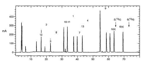
Figure 1: UV traces of a tryptic co-digest of 15N-subtilisin DAI and
subtilisin, .
Peptides are numerated in the order of occurrence beginning with the N-
terminus (see
Table l).
Figure 2. (A) Integrated total ion current (TIC) chromatogram of peptide 3 of
subtilisin (indexed (s)) and 15N-subtilisin DAI (indexed (15N). (B) TIC of
peptides 5, 6 and 9
of 15N-subtilisin DAI and subtilisin. The results of area integration for both
TIC and UV
peaks are summarized in Table I. Note that sequence differences of subtilisin
and
subtilisin-DAI reside on peptide 5 (N74D) and 6 (S1011, V102A). Amino acid
sequence
numbering is linear.
Table I.: Sequence comparison, m/z values, and ratios of integrated TIC peak
areas
and UV absorbance peak areas for chromatograms in Figure 1. The concentration
measured by the co-digest technique for subtilisin and subtilisin-DAI was 8.15
and 7.13
mg/ml, respectively, while the given concentration (established by independent
methods)
was 7.99 and 7.03mg/ml, respectively.
Example 2
A fermentation broth concentrate of unknown origin was suspected of containing
an
alkaline serine protease. A small sample was dissolved in buffer and spiked
with purified
15N-labeled subtilisin-Y217L. The mixture was digested with trypsin, peptides
were
separated by RP-HPLC, and the eluate monitored by UV absorbance and by mass
spectrometry. Figure 4 (A) shows an SDS-PAGE gel of the composition of the
sample.
Figure 4 (B) displays the peptide map, and Figure 5 gives a few examples of
TIC traces.
CA 02420567 2003-02-24
WO 02/18644 PCT/US01/25884
- 27 -
The data show that the sample contains an alkaline serine protease closely
related to
subtilisin BPN', and in this case, specifically at 0.54 mg. m1-1.
Example 3
Randomly generated variants of subtilisin-DAI were expressed by cultures grown
on
minimal media in microtiter plates. Aliquots of cell-free supernatants were
probed for the
presence of subtilisin-DAI variants by co-digests with 15N-labeled subtilisin-
DAI. In
separate experiments the catalytic activity was measured. In yet another
experiment, the
ratio of specific concentration to activity (referred to as 'conversion
factor' f) was measured
by active site titration with a mung bean inhibitor (MBI) solution calibrated
in the same
experiment with a previously standardized solution of subtilisin-DAI (Hsia et
al., 1996). The
data shown in Table 11 show convincingly the accuracy of the peptide mapping
method for
protein concentration measurements. A further advantage of the technique is
that the
protein variants can be queried for similarities and approximate location of
mutations.
Because all peptides of the internal standard are known, each can be checked
for the
presence of the unlabeled counterpart. If not present the target protein has a
mutation on
that sequence. Next one would search for a peptide of closely related mass and
verify that
it exists in the quantity, anticipated from the quantity of those peptides
identical in
sequence with the internal standard, using the UV trace.
Example 4
From the previous example one can extrapolate that the method should work with
equal efficiency and accuracy for proteins of unknown properties but known
sequence by
using instead of purified 15N-labeled protein a synthetic 15N-labeled peptide.
This will be
added to the sample ready for trypsin digestion. After digestion the sample
will be
analyzed as before.
Example 5
15N Protease
This example describes a method for the batch preparation of a 15N-labeled
protease. The Mops/Urea shake flask protocol (described above) was used with
all of the
chemicals, except for the urea, purchased from Sigma chemical in highest
purity available.
15N2 Urea(99 atom%) was purchased from Isotec,Inc. A 1.8L batch of media was
prepared
with chloramphenicol at 25ppm and sterile filtered. 300mL was added
aseptically to each
of the 6 sterilized 2.8L bottom baffled fernbachs. The inoculation was done by
adding the
thawed and mixed glycerol stocks, protease hyper producer prepared previously
in the
CA 02420567 2003-02-24
WO 02/18644 PCT/US01/25884
- 28 -
Mops/urea media and frozen, at 1vial(1.5mL) per shake flask. The shake flasks
were put
into a New Brunswick shaker/incubator, after inoculation, and run at 37 C and
350rpm for
78hours. At the harvest point, 78hours, AAPF activity assays were done on the
samples
and titers ranged from 0.7g/L to 1.4g/L. The contents from the shake flasks
were pooled
together, pH adjusted to 5.5 with acetic acid and centrifuged in 250mL bottles
at 12,000rpm
for 30minutes. The supernatants were filtered with a 0.8 micron Nalgene 1L
filter unit. The
pool was assayed at 1.1g/L for 1700mL with the total 15N protease being
1.9gms. The
supernatant was concentrated in the cold room (@4 C) to 135mL, using 3 Amicon
8400
stirred cells and PM10 (10,000MWCO) membranes. There was no loss of protein in
the
concentration step.
Dialysis was done using 20mM MES, pH 5.4, 1mM CaCl2 buffer in a 15L graduated
cylinder on a stir plate in the cold room, with the sample being added in two
67.5mL
aliquots respectively to 10,000MWCO Spectra Por 7 dialysis tubing, clamped off
and
placed into the cylinder with buffer. After the overnight dialysis the samples
were removed
from the graduated cylinder, the clamps removed from the dialysis tubing and
the contents
poured into and filtered using a 0.45micron Nalgene 500mL filter unit. Assays
run at this
time showed no loss of protein at 1.9gm total available in 250mL.
The protease protein was purified using a low pH buffer system with a cation
exchange column because the PI of the enzyme is around 8.6. An Applied
Biosystems
Vision was used to do the purification along with a 16x150mm (32mL) column of
POROS
HS 20 (Applied Biosystems cation exchange resin). The program used to do the
purification is as follows: Equilibrate the column at 50mL/minute with 20cv's
(colume
volumes) of 20mM MES, pH 5.4,1mM CaCl2 buffer, load the sample (150mL) onto
the
column at 15mL/minute, wash the column at 50mL/minute with a gradient from the
20mM
MES, pH 5.4 ,1mM Ca0I2 buffer to 20mM MES, pH 6.2, 1mM CaCl2 buffer in 25cv's.
Elute
the 15N protease protein with a gradient from 20mM MES, pH 6.2, 1mM CaCl2
buffer to
20mM MES, pH 6.2, 1mM CaCl2, 15mM NaCI buffer in 75cv's(start collecting the
fractions
at 5cv's into the gradient). Finally, clean the column off with a salt wash of
2M NaCI 10cv's,
rinse with lOcv's of H20. This run was made three times to purify all of the
labeled protein,
3o the 15N protease came off the column between 8 to 12mM NaCl, with 95
11mL fractions
collected each run. The labeled protease was concentrated from 1.8L to 150mL
using an
Amicon stirred cell with a 10,000MWCO PM membrane, with a buffer
exchange/diafiltration
to 20mM MES, pH 5.4, 1mM CaCl2 to prepare the sample for another run on the
same
system with the same method. Some of the labeled protease was lost because of
the cuts
made on the fractions collected, with the total available 15N protease down to
1.4gm. After
CA 02420567 2003-02-24
WO 02/18644 PCT/US01/25884
- 29 -
three more runs the purification was done. There was a pool of purified
material with a
1.3L total volume. This was concentrated down to 65mL using the Amicon
concentrator
and a buffer exchange to 20mM MES, pH 5.4, 1mM CaCl2 buffer. The 15N protease
purified sample was sterile filtered through a 0.22micron using the Nalgene
0.22micron
Analysis was done on these samples to confirm the concentration, the purity
and
the presence of the 15N labeling. An SOS-PAGE gel run against an unlabelled
protease
standard showed no molecular weight bands greater than 27,480, the intensity
of the
protease bands at 27,480 Daltons was about the same with the subsequent
breakdown
bands (3) to be of the same intensity also. An amino acid analysis showed that
the AAPF
activity concentration to be the same (20g/L) as well as the BCA total protein
concentration
run against the unlabelled protease standard. Tryptic digests/codigests with
protease
(unlabelled) and subsequent peptide mapping with MS analysis on the HP 59987A
engine
showed that the peptides were labeled with 15N. Thus, the material was shown
to be what
was intended, 15N labeled protease, suitable for analytical use.
Those skilled in the art will appreciate the numerous advantages offered by
the
present invention. For example, unlike the prior methods, the methods taught
herein can
yield absolute protein concentrations. In comparison, ICAT (Gygi et al., 1999)
measures
relative quantities, as does staining of 2D gels or the isotope technique by
Oda et al., 1999.
A further advantage of the present method is that it applies to all proteins,
while the ICAT
technology can capture only about 10% of all proteins since it relies on the
presence of free
SH groups. Yet a further advantage of the present invention is that this
methodology is
compatible with all automated equipment developed for protein identification
under the
rproteomics' umbrella.
The present invention is useful where only very dilute concentrations of
biopolymer
are available for analysis. With regard to quantity, for example, the present
invention can
be employed to determine the absolute quantity of a selected protein in a
solution
containing less than 25, less than 20, less than 15, less than 10, less than
5, and down to
about 2 micrograms, or less, of such protein. With regard to concentration,
the present
invention can be employed to determine the absolute quantity of a selected
protein in a
solution containing less than 25, less than 20, less than 15, less than 10,
less than 5, and
down to about 2 micrograms/ml, or less, of such protein.
CA 02420567 2012-01-20
WO 02/18644 PCMS01/25884
- 30 -
Various other examples and modifications of the foregoing description and
examples will be apparent to a person skilled in the art after reading the
disclosure without
departing from the scope of the invention.
io
CA 02420567 2003-07-24
=
31 --
SEQUENCE LISTING
<110> Genencor International, Inc,
<120> Mass Spectrometric Analysis of Biopolymers
<130 11816-56
<140> CA 2,420,56?
<141> 2001-0?-17
<150> US 60/228,198
<151> 2000-05-25
<a60> 15
<170> FastSEQ for Windows Version 4.0
<210> 1
<211> in
<212> PRT
<213> Artificial Sequence
<220>
<223> cryptic peptide of BPN'
<400> 1
Pro Sr Ser Leu Giu Asn Thr Thr Thr Lys
1 5 10
<210> 2
<211> 16
<212> PRT
<213> Artificial Sequence
<220>
<22-3> trypric pepride of EPN,
<400> 2
Pro Ala Pro Ala Leu His Ser Gin Gly Tyr Thr Gly Set Am Val Lys
1 5 10 15
<210> 3
<211> 11
<212> PRT
<213> Artificial Sequence
<220>
<223> tryptic peptide of BPN:
<400> 3
Pro His Pro As Trp Thr Asn Thr Gin Val Arg
1 5 10
<210> 4
<211> 10
<212> PRT
<213> Artificial Sequence
<220>
CA 02420567 2003-07-24
- 32 -
<223> tryptkc co-digest oi! 15N-subCilisin DAT and
subtilisin
<400> 4
Ala Gin Ser Val Pro Trp Gly Ile Ser Axg
1 5 10
<220> 5
<211> 9
<212> PRT
<213> Artificial Sequence
<220>
<223> cryptic Cc-digest of 15N-subti1ioin DAT and
eubtilisin
<400:, 5
Val Gin Ala Pro Ala Ala His An Arg
1 5
<210. 6
<211> 8
<212> PRT
213 Artificial Sequence
<220>
<223> cryptic co-digest ot 1SN-subtilisin DAT and
subcilisin
<400> 4
nly Leu Thr Gly Ser Giy Val Lys
<210> 7
<211> 17
<212> PRT
<2vA> Artificial Sequence
<220>
<223> cryptic co-digest of ISN-subCilisin DAT and
f;subtilisia
<400> 7
Val Ala Val Leu Asp 'Mr Gly Ile Ser Thr His Fro Asp Leu An Tle
1 5 10 2S
Arc
<210> S
<211-.. 40
<212> PRT
<213> Artifir-ial Sequence
<220>
<221- Cryptic co-digest of 15N-subtilisin DAT and
subtilisin
<400> 8
Gly Gly Ala Ser Phe Val Pro Gly mu Pro Ser Thr Gin Asp Gly An
1 5 15
Gly His Gly Thr Els Val Ala Gly Thr Ile Ala Ala Leu Asp An Ser
CA 02420567 2003-07724
=
33 -
20 25 30
Ile Gly Val Lem Gly Val Ala. Pro Ser Ala Giu Leu Tyr Ala Val Lya
35 45
<210> 9
<211> Si
<212> PRT
<213> Artificial Sequence
<220>
<223> tryptic co-digest pi' 15M-subtilloin DAI and
subtilisin
<400> 9
Val Leu Gly Ala Ser Gly Ser Gly Ala Ile Ser Ser Ile Ala Gln. Gly
1 5 10 15
Lem Glu Trp Ala Gly As ;Ion Gly Met His Val Ala. An Leu Ser Lau
20 25 10
Gly Ser Pro Sal- Pro Ser Ala. Thr Leu Giu Gin Ala Val An Sex Ala
35 4.045
Thr Ser Arg
<210> 10
<211> 21
<212> PRT
<213> Artificial Sequence
<220>
<223> tryptic co-digeot of 15N-subtillsin DAT and
subtilisin
<400> 10
Sly Val Lau Val Val Ala Ala Ser Sly Asn Ser. Gly Ala Gly Ser Ile
5 10 15
Ser Tyr Pro Ala Arg
<210> 11
<211> 16
<212> PRT
<213> Artificial Sequeace
<220>
<223> tryptic co-,diqest of 15N-subtiliein DA1 aad
subtilisin
<400> 11
Tyr Ala Asa Ala Met Ala Val Gly Ala Thr Aap Gin An .An Asa Arg
1 5 10 15
<210> 12
<211> 49
<212> PRT
<213> Artificial Sequence
<220>
<223> tryptic co-digest of 15N-sabtilisin MI and
<400> 12
CA 02420567 2003-07-24
Ala Ser Phe Ser Gin Tyr Gly Ala Gly Leu Asp Ile Val Ala Pro Gly
1 5 10 15
Val An Val Gin Sez Thr Tyr Pro Giy Ser Thr Tyr Ala. Sex Lea An
20 25 30
Gly Thr Ser Met Ala Thr Pro Hi..' Val Ala Gly Ala Ala Ala Leu Val
35 40 45
Lys
<210> 13
(.211> 12
.4212> PT
<213> Artificial Sequence
<220>
<223> tryDtic co-digest of 1511-eubtilioin DAI and
subtilisin
<400> 13
Gin Lys Asn Pro Ser Trp Ser An Val Gin Ile Arg
1 5 10
<210> 14
<211 4
<212> PRT
<213> Artificial Sequence
<220>
.4223> tryptic coest of 15N-subtilisin DAI and
subtilisin
<400> 14
An Hie La'. Lys
1
<210> :US
<211> 24
<212> PRT
<213> Artificial Sequence
<220>
<223> tryptic co-digest of 15N-subtilisin DAI and
subtilisin
4:400:. 15
Mn Thr Ala Thr Sex Leu nly Ser Thr An Leu Tyr Gly Ser Giy Leu
1 10 15
Val Asn Ala Giu Ala Ala Thr Arg