Note: Descriptions are shown in the official language in which they were submitted.
CA 02420676 2003-02-21
WO 02/15973 PCT/AU01/01051
1
CATHETER LOCATOR APPARATUS AND METHOD OF USE
This invention relates to a method of catheter and radiating coil location in
a human
body and in particular to the determination over time of the location of the
tip of a
catheter as it is inserted and during its use in the body and/or its route
through the
body.
BACKGROUND
For ease of explanation, the guidance and placement and ongoing maintenance of
a
catheter for enteral nutrition will be described in one example in this
specification. It
will however be clear to the person skilled in the art that the techniques and
equipment described are useful for similar placement requirements in other
parts of
the human body and animal bodies as well. Catheters are used for many
different
purposes and there exist many different catheter types. An example of the use
of the
invention in a human body will also be provided in the field of Central Venous
Catheter location.
Enteral nutrition includes both the ingestion of food orally and the non-
volitional
delivery of nutrients by tube into the gastrointestinal tract. Patients are
candidates for
enteral tube feeding that will not, should not, or cannot eat but who have a
functional
gastrointestinal tract. Benefits of enteral tube feeding are the maintenance
of
gastrointestinal structure and functional integrity, enhanced utilization of
nutrients,
ease and safety of administration.
Enteral tube feeding is contraindicated for patients with diffuse peritonitis,
intestinal
obstruction that prohibits use of the bowel, intractable vomiting, paralytic
ileus,
and/or severe diarrhoea that makes metabolic management typical. Other
potential
contraindications that depend on the clinical circumstances include severe
pancreatitis, enterocutaneous fistulae, and gastrointestinal ischemia. Enteral
tube
CA 02420676 2003-02-21
WO 02/15973 PCT/AU01/01051
2
feeding is not recommended during the early stages of short-bowel syndrome or
in
the presence of severe malabsorption.
The route selected for tube feeding depends upon the anticipated duration of
feeding, the condition of the gastrointestinal tract (e.g. esophageal
obstruction, prior
gastric or small-bowel resections), and the potential for aspiration. The
intestine can
be accessed at the bedside (naso intestinal tube, naso endoscopic gastrostomy)
or in
the operating room (gastrostomy and jejunostomy).
Nasal intubation for gastric feeding is the simplest and most often used
method for
tube feeding. This technique is preferred for patients who are expected to
eventually
resume oral feeding. Maximal patient comfort and acceptance is more likely
when a
soft feeding tube with a small external diameter is used. Access to the
duodenum and
jejunum is possible with longer tubes but placement of the tip into the
duodenum
and jejunum is more difficult and time consuming and has added risk factors.
Enteral tube feeding is considered safer than parenteral nourishment because
mechanical, infection, and metabolic complications are usually less severe
than those
encountered with parenteral nutrition. However, enteral feeding is not problem
free,
and significant complications can occur when the tube and feeding is managed
by
unskilled or untrained individuals or if monitoring is absent or
inappropriate.
Incorrect placement of the feeding tube is one of a number of major
complications.
Most serious is the unintended placement of the catheter during nasal
intubation into
the cardiovascular system or into the lungs. Both of these situations are
possible
when inexperienced medical staff perform intubation. A stylet (relatively
stiff small
gauge wire) is used to stiffen and support the otherwise flaccid catheter tube
during
its intubation.
CA 02420676 2003-02-21
WO 02/15973 PCT/AU01/01051
3
It is also prudent to check that the exit aperture of the feeding tube
(typically located
at the distal end /tip of the tube) remains in its desired location over the
period of
feeding. Protocols that address this requirement include frequent monitoring
for the
appropriate pH of fluids extracted from the feeding tube while not carrying
nutritional liquids and careful patient monitoring to ensure that nutritional
uptake is
as expected.
X-rays are often used to determine the location of the caudal/distal end of
the tube.
However, even X-rays are not necessarily conclusive as to its location. The
natural
and continuous movement of the internal organs can make it difficult for the
physician interpreting the X-ray to be sure of the actual location of the
caudal/distal
end of the tube because the movement described can change the position of the
catheter over time.
There exist a large variety of catheters, their names sometimes indicating
their use,
the part of the body they enter or treat and sometimes they are named after
the
physician or physicians who developed methodologies for their use.
This invention also relates to catheter location methods and means for central
venous
catheters.
Intravenous catheters are those that access the interior of a patient via an
opening in
the skin passed down one or more of the many branches of the venous system to
the
region requiring medical attention. These types of catheters are also referred
to as
Venous Access Catheters (VAC) and Central Venous Catheters (CVCs) and are used
generally in the cardiovascular system.
A sub-category of intravenous catheters is those that fall under the heading
of
Peripherally Inserted Central Catheter (PICC). These catheters have been used
by
clinicians for many years and many different techniques exist for their
insertion.
CA 02420676 2003-02-21
WO 02/15973 PCT/AU01/01051
4
One such insertion technique is the Seldinger technique and along with
advances in
devices and materials there now exists a modified Seldinger technique, useful
particularly for small or poor veins.
There exist a large selection of intravenous catheters such as for example
peripheral
catheters which are used for insertion into the body that are from one half to
three
inches in length; midline catheters which are used for insertion being greater
than
three inches in length; mid-clavicular and non-tunneled sub-clavian, tunneled
Groshong, Hickman and Broviac or subcutaneous implanted ports for longer
lengths.
Common to intravenous catheters is the use of a guide wire that is passed into
the
body and into a vein and then directed by the skill of the clinician to the
desired
location. Once the guide wire is in place a catheter is slid over its external
free end
and pushed till the distal end of the catheter reaches the end of the guide
wire.
The location of the tip of these types of catheters or the recordal of the
advancement
of the guide wire into the body is achieved in a number of ways.
Return checks are used to expire types of liquids expected to be at or near
the tip of
the catheter and the checking of length markings on the wires used within
catheters
are two methods used by clinicians who do not have ready access to
alternatives.
Ultrasound guidance, fluoroscopy and X-ray methodologies are preferred even
though they do not always provide an exact determination of the location of
the tip
or path of the guide wire or catheter.
More expensive and more time-consuming CT examinations provide the best means
of locating not only the tip but also the path of any of the types of catheter
described
above and others that are located in the body of a patient.
CA 02420676 2003-02-21
WO 02/15973 PCT/AU01/01051
The final location of the caudal/distal end of any catheter is critical to the
efficacy of
the purpose for the use of the catheter. In one example, the delivery of drugs
directly
into the heart can be best achieved by the location of the caudal/distal end
of the
catheter in the superior versa cave (CVC). Studies show that it is preferable
to locate
the caudal/distal end of the catheter in the upper portion of the superior
versa cave
(typically recognised as being more than 4.5cm above the cavoatrial junction).
Studies
indicate that these preferable locations appear to minimise catheter
malfunction.
More critical however, is to ensure that the catheter is in the superior versa
cave itself,
as there exist studies indicating that there is a significant association
between catheter
malfunction and catheter tip location in the venous system adjacent to the
superior
versa cave.
Fig. 7 depicts the various veins in the vicinity of and including the superior
versa
cave.
Where for example, the catheter tip is in either of the brachiocephilac veins
or near
the junction of a brachiocephilac vein there is a greater likelihood of the
unwanted
development of a fibrin sheath or the presence of thrombus around or at the
tip of the
catheter as compared to catheter tips located in the superior versa cave.
Greater
likelihood of unwanted developments can occur when a catheter tip is located
in the
sub-claviers vein or the cavoatrial junction or in the right atrium.
The effect of inappropriate or less than ideal catheter tip location is
shortened
survival which clearly is manageable but more serious effects may include
thrombosis and phlebitis infections more, particularly pheumothorax infections
and,
in more serious situations, occlusions.
When catheter tips suffer thrombosis, these studies demonstrate significantly
shorter
survival than those catheters that are not subject to this unwanted
development.
CA 02420676 2003-02-21
WO 02/15973 PCT/AU01/01051
6
Appropriate patient care requires consideration of a large number of factors
when
considering the need for intravenous catheter usage. Sometimes the benefit of
drug
delivery has to be carefully weighed against the likelihood of adverse effects
of
intravascular device related infections as mentioned previously.
Catheter selection is not a simple matter and factors to be considered include
the
following:
~ type of medication
~ osmolarity and pH of the solution to be infused
~ duration of therapy required
~ secondary risk factors and chronic diseases
~ patient age, activities, work and lifestyle
~ future intravenous needs and long term prognosis
~ current availability and status of access veins (typically peripheral veins
of the
limbs)
~ patient history of neurologic impairments, surgeries affecting veins or
lymphatic
system, bloods dyscrasias, thrombosis and previous intravenous use history
~ current patient diagnosis and preferences for treatment
The anticipated duration of therapy can readily suggest short peripheral
catheters for
periods of less than five days, and for periods of less than four weeks a
midline
catheter is generally suitable.
Midclavicular lines are an option and becoming more popular as the occurrence
of
thrombosis resulting from sub-optimal placement in sub-clavian regions other
than
the superior vena cava increase.
In fact mid-clavicular lines are often used in home care situations to avoid
the time
and cost of confirmatory X-rays. However, even mid-clavicular lines need to be
CA 02420676 2003-02-21
WO 02/15973 PCT/AU01/01051
7
optimally placed in the lower one third of the superior vena cava, close to
the
junction of the superior vena cave and the right atrium but should not advance
into
the right atrium itself.
The previously mentioned Peripherally Inserted Central Catheters (PICCs)
having a
tip location in the superior vena cava can be used for long term therapy (five
days to
one year). However, they should be critically checked by X-ray to determine
appropriate tip placement even though this is neither a totally satisfactory
nor certain
method of location checking.
It is thus a real need for physicians to be able to increase their confidence
that the
catheter has been placed at the desired location and remain there in the body
of their
patient. This is so whether that is for the purpose of enteral and parenteral
nutrition,
receiving vesicant chemotherapeutic agents, antibiotics and blood sampling or
for
other purposes.
BRIEF DESCRIPTION OF THE INVENTION
A broad form of the invention is a method of locating a coil used in relation
to a
catheter to be inserted into a subject body wherein said coil radiates signal
energy;
the method including the steps of: using a coil position measuring means
having at
least two signal energy detectors wherein said measuring means is located with
reference to a predetermined location on or part of said subject human body;
and
displaying a position measurement made by said coil position measuring means
wherein said position measurement is relative to said position measuring means
for
use by a clinician in determining the position of said coil and said catheter
in said
subject human body relative to said predetermined location on or part of said
subject
human body.
In a further aspect of the invention, the method includes the further step of
also
displaying with said position measurement a representation of a point or
region of a
CA 02420676 2003-02-21
WO 02/15973 PCT/AU01/01051
non-subject body referenced to said displayed position measurement for use by
the
clinician in determining the position of said coil and said catheter in said
subject
human body.
3. In another aspect of the invention, the position measuring means is located
with reference to a predetermined location being on or over the xiphoid
sternal
junction of said human body for use with a catheter inserted into the
alimentary
canal.
In another aspect of the invention, a region or delineation of a body part is
displayed
that is representative of the diaphragm of a non-subject human body thus
delineating
on said display the upper and lower chest cavities of a human body.
In another aspect of the invention, the position measuring means is located
with
reference to a predetermined loeation being on or over the caudal/mid sagital
aspect
of the jugular sternal notch of said human body use with a catheter inserted
into the
cardiovascular or respiratory system.
In yet a further aspect of the invention, a region or delineation of a body
part is
displayed that is representative of the sternum of the said human body thus
delineating on said display a portion of the upper skeleton of a human body.
In a yet further aspect of the invention, the method includes the further step
of
displaying a position measurement of said coil at predetermined intervals of
time.
In a further aspect of the invention, the coil is incorporated into a stylet
or guide wire
adapted for use with a catheter.
In yet a further aspect of the invention, the method includes the further step
of
displaying the position of said coil at predetermined intervals of time while
said
CA 02420676 2003-02-21
WO 02/15973 PCT/AU01/01051
9
stylet or guide wire is retracted from a catheter so that the path of said
coil and thus
said catheter can be tracked and displayed.
In an aspect of the invention, a coil is incorporated into a catheter and
usable for
locating the position of said catheter in a human body.
In another aspect of the invention a catheter locator apparatus for assisting
a user's
placement of a catheter into a subject body comprises: a pair of wires usable
with the
catheter, the wires having a first end and a second end; a processor in
electronic
communication with the first end of the wires; a radiating coil connected to
the
second end of the wires, the radiating coil having various locations in the
subject
body; a detector device in electronic communication with the processor, the
detector
device adapted to be positioned in relation to a predetermined portion of the
subject
body; reference data retrieved by the processor which specifies at least one
reference
image which represents at least one predetermined image; indicator data
generated
by the processor which specifies at least one indicator image which provides
information related to at least one location of the radiating coil in the
subject body;
and a monitor in electronic communication with the processor which displays
the
indicator image and the reference image.
In yet a further aspect of the invention a catheter adapted for use with a
catheter
locator apparatus includes: (a) a processor in electronic communication with a
coil;
(b) the coil radiating and incorporated into a coil positioning device,
stylet, guide
wire or into said catheter, the radiating coil having various locations in a
subject
body; (c) a detector device in electronic communication with the processor;
(d)
reference data generated by the processor which specifies at least one
reference
image which represents at least one predetermined portion of the subject body;
(e)
indicator data generated by the processor which specifies at least one
indicator image
which provides information related to at least one location of the radiating
coil in the
CA 02420676 2003-02-21
WO 02/15973 PCT/AU01/01051
subject body; and (f) a monitor in electronic communication with the processor
which
displays the indicator image and the reference image, said catheter
comprising:
a tube which is adapted to receive the stylet or guide wire or having a coil
incorporated thereto, the tube having a proximal end and a distal end; and
a tip included at the distal end.
A yet further aspect of the invention is a catheter locator apparatus
comprising: a
multi stranded wire insertable into a catheter, the wire having a proximal end
and a
distal end; a radiating coil connected to the distal end of two of the strands
in said
wire; at least two receiving coils; a monitor; a processor, in electronic
communication
with the wire, the receiving coils and the monitor, which: the processor
receives at
least one reference signal from at least two of the receiving coils after the
receiving
coil is positioned with reference to at least one predetermined bony landmark
on the
subject body; and retrieves reference data; and drives the monitor in order to
graphically represent the reference data; and receives at least one indicator
signal
from the at least two receiving coils that receive a signal from the radiating
coil after
the radiating coil is inserted into the subject body; as well as generates
indicator data;
and drives the monitor in order to graphically represent the indicator data.
Another method of facilitating proper placement of a catheter into a subject
body, the
method comprises the steps of:
(a) receiving at least one reference signal indicative of a location of at
least one
receiving coil positioned with reference to a predetermined landmark on the
subject
body;
(b) receiving at least one indicator signal from a radiating coil inserted
into the
subject body;
(c) retrieving reference data associated with the predetermined landmark;
(d) displaying a reference image derived from the reference data;
(e) generating data represented by an indicator signal indicative of the
relative
position of the at least two receiving coil positions on or over the subject
body;
CA 02420676 2003-02-21
WO 02/15973 PCT/AU01/01051
11
(f) displaying graphics represented by the indicator data; and
(g) repeating steps (e) and (f) after a change in the indicator signal is
received.
A further method of operating a catheter locator apparatus, comprises the
steps of:
(a) placing a predetermined area of a detector device over a predetermined
landmark on a subject body;
(b) inserting a catheter, stylet, guide wire or coil locating device, embedded
with a
radiating coil, into the subject body;
(c) viewing a graphical representation of a predetermined part or portion or a
representation of a reference image on a monitor;
(d) viewing a graphical representation of the radiating coil on a monitor; and
(e) manipulating the catheter in the subject body with aid of the display of
the
relative positions of the graphical representations.
A yet further aspect of the invention comprises a wire bundle for use with a
catheter
locator apparatus that includes: (a) a processor in electronic communication
with a
predetermined portion of a proximal end of the wire bundle; (b) a radiating
coil
connected to a predetermined portion of a distal end of the wire bundle, the
radiating
coil having various locations in a subject body; (c) a detector device in
electronic
communication with the processor, the detector device adapted to be positioned
in
relation to a predetermined portion of the subject body; (d) reference data
retrieved
by the processor which specifies at least one reference image which represents
at
least one predetermined image; (e) indicator data generated by the processor
which
specifies at least one indicator image which provides information related to
at least
one location of the radiating coil in the subject body; and (f) a monitor in
electronic
communication with the processor which displays the indicator image and the
reference image, said wire bundle comprising:
a first wire having a predetermined stiffness adapted to control positioning
of
a flexible tube;
CA 02420676 2003-02-21
WO 02/15973 PCT/AU01/01051
12
a second wire adapted to transmit signals between the radiating coil and the
processor; and
means for binding the second wire to the first wire.
Specific embodiments of the invention will now be described in some further
detail
with reference to and as illustrated in the accompanying figures. These
embodiments
are illustrative, and are not meant to be restrictive of the scope of the
invention.
Suggestions and descriptions of other embodiments may be included within the
description of the invention but may not be illustrated in the accompanying
figures
or alternatively features of the invention may be shown in the figures but not
described in the specification.
BRIEF DESCRIPTION OF THE FIGURES
Fig.1 depicts a catheter having a coil and exit aperture located near its
caudal/distal
end;
Fig. 2 depicts a stylet having a Boil located near its caudal/distal end;
Fig. 3 depicts a detector apparatus positioned appropriately on a patient and
an
outline of a display that delineates regions of the body;
Fig. 4 depicts a display of the location and depth of a coil on the tip of a
catheter or
stylet superimposed over a representation of a patient;
Fig. 5 depicts a typical display only of the coil position located in the
stomach of a
patient over time even while it is not being moved into a desired location;
Fig. 6 depicts the recorded path and the current location of a catheter in a
patient as
would be displayed;
Fig. 7 depicts the venous system of the upper torso;
Fig. S depicts an outline of a coil location device located in a preferred
manner over
the caudal/mid sagital aspect of the jugular sternal notch of a patient;
Fig. 9 depicts a pictorial representation of a correctly placed Central Venous
Catheter
(CVC) in the superior versa cava of a patient; and
CA 02420676 2003-02-21
WO 02/15973 PCT/AU01/01051
13
Fig. 10 depicts a pictorial representation of the upper torso of a patient
showing the
path of a CVC and its tip in the region of the superior vena cave.
DETAILED DESCRIPTION OF EMBODIMENTS OF THE INVENTION
Fig.1 depicts a single lumen catheter having located near its caudal/distal
end, a coil,
which is used to emit a signal that can be detected by an apparatus not unlike
that
described in US Patent 5,099,845. That patent is in the name of Micronix Pty
Ltd and
is hereby incorporated into this specification by reference. The incorporation
of the
above-mentioned patent does not and should not be construed as an admission of
the
content of the specification having entered the common general knowledge of
those
skilled in the art.
The apparatus of the invention described in the above-mentioned patent
provides a
means to determine both the depth and position of a coil located on the end of
a
catheter as well as its orientation. The type of catheter is of no great
importance to the
principle and method of the invention.
The depth and position determining apparatus, also referred to herein as the
detector
apparatus, is generally of the type disclosed in the above-mentioned
specification
and can be used in the method described herein but is not the only such device
that
will provide the required features.
The catheter 10 depicted in Fig.1 is suitable for use in parenteral nutrition
in
particular as it has a suitable tip shape 12 designed so that the exit
aperture 14 is less
likely to become blocked. The catheter shown has a single lumen (single
passageway
from proximal to caudal/distal end) but other catheters may have multiple
lumens.
A coil 16 is located near the tip 12 and the two ends of the pair of wires,
which will
run the length of the catheter. The pair of wires terminates at connector 18
near the
proximal end of the catheter. The entry aperture 20 to the catheter is located
at the
CA 02420676 2003-02-21
WO 02/15973 PCT/AU01/01051
14
extreme proximal end of the catheter. It is into this aperture that nutrients
are
pumped at the desired times and rates once the caudal/distal end of the
catheter has
been appropriately located within the patient.
It is intended that the connectors for catheters used for different uses will
eaeh be
different sizes, connection types and colors so as to make their use as safe
as possible.
By making the interconnection of catheters for different applications
difficult if not
impossible, it is intended to minimize accidental administration of the
incorrect or
inappropriate fluids particularly drugs to the patent via catheters not
intended for
such use.
When the catheter 10 is being intubated into the gastrointestinal tract of the
patient, a
metal stylet is used within the catheter.
The stylet can comprise the pair of wires described previously or can comprise
the
typical stylet wire or wires having a pair of wires incorporated therein. The
stylet
may be encapsulated in material know to be suitable for its intended use. The
stylet
itself is typically made of stainless steel but it could also be made of
plastic or other
suitable material as shown in Fig. 2.
In one embodiment, the processor is connected to a proximal end of a wire
bundle,
and the coil is connected to the distal end of the wire bundle. The wire
bundle
includes a guide wire suitably bound to a signal-carrying wires. The bundle is
preferably bound together at its ends and along its length and may also be
encapsulated in material know to be suitable for its intended use. The bundle
may for
example be wrapped together or wrapped with a suitable material, it may be
maintained by a suitable adhesive or fastener or suitable size and shape. See
Fig. 2
Intubation can be via the mouth or preferably via a nasal passageway of the
patient.
The stylet stiffens the otherwise formless catheter and is able to be
manipulated along
CA 02420676 2003-02-21
WO 02/15973 PCT/AU01/01051
its length as well as at its proximal end, so that its caudal/distal end
navigates an
appropriate route through the patient under the control of the health
professional or
trained staff performing the intubation.
In this specification, mention is made of clinicians performing intubations
but it is
possible for trained health professionals to insert catheters into patients
for a variety
of uses. It is also possible to use a suitably stiff catheter having a coil
incorporated
into the wall of the catheter typically at or near the caudal/distal end of
the catheter
that has a single lumen used to deliver the appropriate fluids.
The inventors have identified that there are economic and practical reasons
why the
arrangement of a coil integrated into a catheter is usable. However, it is
less desirable
than an alternative arrangement to be described herein which uses the stylet
to carry
the radiating coil.
The above is so, because the catheter/coil combination is expensive to
manufacture
to the high standards required of medical equipment. Catheter manufacturers
provide a specialist product and it is not always in their interest to
incorporate
changes that will markedly increase the cost of their product.
Furthermore, a catheter/coil combination is a single use item, thus the added
value
of the coil is not recoverable and therefore needs to compete with current
detection
techniques which involve X-rays, even though they are more time consuming.
Thus it has been identified that it is possible to incorporate a coil into the
caudal/distal end of a stylet.
The stylet is used not only to manipulate the caudal/distal end of the
catheter to a
desired location but it can be retracted and is also capable of being reused
after
CA 02420676 2003-02-21
WO 02/15973 PCT/AU01/01051
16
appropriate decontamination and cleaning according to a required protocol. The
inventors do not recommend such reuse unless a relevant protocol is in place.
The depth and position of the coil can be determined during the process of
incubation. Furthermore, when used to locate the tip of a catheter and
following
appropriate positioning of the caudal/distal end of the catheter the route of
the
catheter can be determined while the stylet is retracted from the catheter. An
example
of the trace displayed after a retraction of the stylet is provided by Fig. 6
the details of
which will be described later in the specification. A stylet with a coil
incorporated
thereon can also be used to determine the location in the patient of
particular
locations along the length of the catheter. Thus for multiple lumen catheters
that
have a plurality of ports along its length the loeation in the patient of each
of those
ports can be determined.
The manufacture of a coil on the end of a stylet is not trivial but it can be
automated
and will involve only a few different materials, not the many different
material and
processes involved in the manufacture of a catheter/coil combination as
described
above.
As described previously a particularly advantageous feature of a coil being
incorporated into a stylet is that while the stylet and coil is being
retracted from the
catheter, it is possible for the location apparatus to record the route
through the
patient taken by the catheter. The route can be recorded, including its X-Y
location
and its depth with respect to the detection apparatus. Most conveniently, the
route of
the catheter can be displayed in a manner, which directly relates it to the
anatomy of
a patient. Appropriate positioning of the detection apparatus on the patient
in the
manner described later in. this specification provides an ability to reference
the
position of the trace on the monitor with the position of internal parts of a
patient.
CA 02420676 2003-02-21
WO 02/15973 PCT/AU01/01051
17
Consequently, the procedure of re-checking the position of the caudal/distal
end of
the catheter and retracing the route of the catheter at future times can be
easily
conducted and the results compared with earlier records of the catheter tip
position.
Furthermore, the route displayed will have characteristics that are likely to
reassure a
clinician that a desired route of the catheter has in fact been taken.
Over time, displays including depth information will correlate with other
clinical
observations as to the correctness of the placement and therefore increase the
confidence of the clinician that the route and final placements are as they
should be.
The route displayed will, within expected anatomical variation, confirm that
the
caudal/distal end of the catheter or other portions of the catheter are
located in the
desired area of the patient. For example, when locating enteral feeding
catheters, the
region desired is that which is in the vicinity of the jejunum, as is
pictorially
represented in Fig. 6.
It is also possible for these techniques to be used during the intubation
process and
thus provide immediate feedback as to the route being taken by the catheter.
The techniques described herein, which assist the placement of a catheter, are
particularly applicable when tube enterostomies are necessary to provide long-
term
nutritional feeding or when obstruction makes nasal intubation impossible. A
conventional gastrostomy or jejunostomy requires a surgical procedure and that
is
obviously preferably avoided.
Percutaneous endoscopic placement of gastric feeding tubes can be performed at
the
bedside or in the endoscopy suite without general anesthesia. Jejunal
extensions may
also be noted through a percutaneous placed gastric port into patients who
require
post-pyloric intestinal feeding. Needle catheter or Witzel jejunostomy placed
at the
CA 02420676 2003-02-21
WO 02/15973 PCT/AU01/01051
18
time of the laparotomy allows early postoperative feeding because the small
bowel is
less affected than is the stomach and colon by postoperative ileus. Jejunal
feedings
minimize the risk of vomiting and aspiration compared with gastric feedings.
However, the techniques described herein are not a substitute for controlled
administration and careful monitoring to check for residual gastric fluids.
Clinical
observation of the patient must be continued. The technique however, does
lessen or
eliminate the costly and time consuming use of X-ray facilities and expertise
or other
even more expensive and time consuming catheter location procedures.
Fig. 2 depicts a stylet 22 which has a coil 24 at its caudal/distal end and
the proximal
ends of the wires which form the coil are located in a connector base 26 and a
manipulation block 28 is provided at the extreme proximal end of the stylet.
The
shape of the stylet is typically long and straight but shown in Fig. 2
conforms with
the shape of the catheter 10 shown in Fig.1, for illustrative purposes only.
The
clinician is capable of twisting the stylet, so that its caudal/distal end
when placed
inside and to the internal end of the catheter, turns the caudal/distal end of
the
catheter to navigate various passages and apertures within the patient body.
This
twisting of the stylet is not recommended but recognises a clinieal practice
noted in
the literature.
Fig. 3 depicts an anterior view of the skeleton 28 of a patient and marked in
overlay
are three circles representative of the preferred location of the three
detector coils of
the detector apparatus over the patient's body.
The external shape of the detector apparatus is of little consequence to the
way in
which the actual detector works but it is considered advantageous by the
inventors
that the external shape helps the clinician to appropriately locate the
detector device
on or over a predetermined location of the patient. For example a triangular
shape
places the upper apex of the apparatus towards the head of the patient and the
CA 02420676 2003-02-21
WO 02/15973 PCT/AU01/01051
19
longitudinal axis of the apparatus can be made to lie coincident with the mid-
sagittal
line of the patient 32.
Circle 30 can be the most important of the three detector coil portions as in
this
embodiment for the placement of the catheter in the alimentary canal (being
that
portion of the digestive system of the body, including without limitation, the
mucus
membrane-lined tube extending from the mouth to the anus including the
pharynx,
esophagus, stomach and the intestines) or in particular in the intestinal
tract, it is
positioned directly over the xiphoid sternal junction. This will also place
the
longitudinal axis of the three detector coils coincident with the median
antero-
posterior plane 32 of the body (mid-sagittal line). The longitudinal axis of
the three
detector coils is engraved on to the case of the detector housing for the
convenience
of the clinician to facilitate ease of positioning particularly since the
xiphoid sternal
junction is typically easily palpitated.
The xiphoid sternal junction is the point at which the diaphragm is connected
to the
human skeleton. The two arched lines (34 and 36) shown on the figure, are
representative of the upper limits of the quite complex domes of the musculo-
membranous partition (diaphragm) separating the abdominal and thoracic
cavities
and which serves as a major thoracic muscle.
It is of assistance to the clinician that these two ached lines, 34 and 36,
are displayed
on the monitor along with the mid-sagittal line 32 as is depicted in Figs. 5
and 6,
while the catheter is being located.
The display of the arched lines, 34 and 36 is a tangible consequence of the
correct
positioning of the detector coils in the manner described. The correlation of
the
position of the coil in circle 30 with the xiphoid sternal junction allows the
monitor to
depict the position and depth of the coil with reference to a position of the
body that
can be found on all patients. It is not imperative that the coil be coincident
with that
CA 02420676 2003-02-21
WO 02/15973 PCT/AU01/01051
particular bony landmark since the detector apparatus can accommodate any
predetermined offset created by having the outer housing of the apparatus
located in
a particular way that places the coil 30 other than on or over the xiphoid
sternal
junction.
The device is relatively effective even if the specific coil 30 is not placed
precisely in
the manner described. It is more important that the two or more signal
detector
elements are located on or over a part of the human body with reference to a
predetermined part or position of the human body. Thus when the position of
the
coil on the end of the guide wire or catheter is displayed it can be displayed
with
reference to the approximate position of a known body part or portion so as to
assist
the clinician during the placement process.
Clearly, the two arched lines 34 and 36 are only representations of the quite
complex
shape of the diaphragm. Since there is coincidence of at least a portion of
the
representations with the attachment point of the diaphragm at the xiphoid
sternal
junction, the lines will be sufficiently accurately depicted on the monitor to
assist the
clinician. With reference to the measurements taken by the sensor, the
representation
is an acceptable guide so as to provide confidence to the clinician that at
least the
caudal/distal end of the catheter is either below or above the diaphragm.
Similar
representations of parts or portions of the human body could be represented on
a
monitor referenced to the predetermined positioning of the detector coils.
As in most cases of intestinal intubation, once the caudal/distal end of the
catheter/stylet is displayed as passing below the two arches that represent
the shape
of the diaphragm, the clinician can be sure that the catheter is in the
gastrointestinal
tract rather than in the airways and/or lung of the patient. Indeed, if the
caudal/distal end of the catheter were to be mistakenly routed into the lung,
the path
of the catheter's caudal/distal end would be shown on the monitor to deviate
from
that expected.
CA 02420676 2003-02-21
WO 02/15973 PCT/AU01/01051
21
'The first noticeable deviation from the expected route would occur some 10-cm
above the two arches. That is, above the xiphoid sternal junction, at the
level of the
bifurcation of the trachea. This would also be measured as being deeper in the
body
from that which is expected of the correct route. Since the route shown in Fig
4 is as
expected for an intestinal incubation, it is on track to enter the stomach
shown, in
general, as region 38. The catheter will then move medially and cross the mid-
sagittal
line again at 40 entering the first part of the duodenum region 42 through the
pylorus
orifice. Once through the duodenum region 42, the catheter passes into the
jejunum
region 44 that is the portion of the small intestine, which extends from the
duodenum
to the ileum.
It is also possible for the detection method and apparatus to be used to
locate
particular portions of the catheter. For example, a dual cavity catheter that
is used
for enteral feeding at its distal end and decompression of gasses in the
stomach along
it length should ideally have the caudal/distal end located in the jejunum and
the
decompression cavity extend no further that the pylorus orifice. If the
decompression cavity of the catheter begins a known distance from the distal
end of
the catheter then a stylet having a coil on its end can be drawn back from the
distal
end of the catheter that known distance. The position of the radiating Boil
can then
be detected and compared with expected position measurements that will
indicate
whether the catheter is correctly positioned in the patient.
In another example, it will be advantageous to determine whether aspiration or
pressure measurement ports located along the length of a catheter are located
at the
desired position within the body of the patient. This is the ease not only at
the time of
intubation but also during the time the catheter is being used.
The advantage of having a reference point or points on the monitor, which
correlates
with an actual point, region or structure of the patient, is clearly apparent.
This
CA 02420676 2003-02-21
WO 02/15973 PCT/AU01/01051
22
feature is useful during intestinal intubation but it is just as useful when
locating a
Venous Access Catheter (VAC) which is a generic expression for the better
known
Central Venous Catheters (CVCs) some of which and their placement will be
described in detail later in this specification.
The monitoring of the passage of the catheter through the body and rechecking
of
correct location during treatment is enabled by means of the catheter locating
apparatus described herein. Advantageously, there is less erratic movement of
the
caudal/distal end of a VAC/CVC as it is placed into location in the upper
thoracic
region because it is not directly in contact with internal organs of the body
that move.
Movement caused by the breathing of the patient also affects the display of
the
position of the catheter tip, much less in this application.
A monitor display that provides lines or a symbol (representative of let us
say, the
diaphragm) assists the clinician and increases their confidence that the
correct path
and final position of the caudal/distal end of the catheter has been achieved.
However, the positioning of the detector coils in relation to a predetermined
reference point is important so that the lines or symbol displayed are
correctly
positioned relative to the trace and properly reflect the position of the
catheter in the
body of the patient.
In the case of inserting CVCs with any of the catheter types or methods
previously
described, it is preferable to position the detector apparatus over a well-
defined,
preferably bony landmark. In this example, a predetermined one of the three
detector
coils of the detector apparatus is located over such a landmark. In the
particular case
of locating the caudal/distal end of a Central Venous Catheter into the
superior vena
cava of a patient, it is preferable to locate coil 30, as depieted in Fig. 8,
on the
caudal/mid sagittal aspect of the jugular sternal notch that lies along the
mid sagittal
line.
CA 02420676 2003-02-21
WO 02/15973 PCT/AU01/01051
23
A possible detector apparatus shape 31 is also shown in Fig. ~. The shape of
the
apparatus is of triangular form in plan view and is preferable for its use on
the chest
of a patient as described previously. This shape is convenient for the
clinician to use
because its apex 33 can be located on the caudal/mid sagital aspect of the
jugular
sternal notch and its longitudinal axis made coincident with the midsagittal
line 32 of
the patient.
As an aid to the preferable positioning of the detector apparatus, the
external casing
of the apparatus is preferably marked near its lower corners "LEFT" and
"RIGHT"
respectively (not shown) ensuring a preferred orientation of this particular
shape of
apparatus in relation to the patient. Additionally, it is useful to have a
line marked or
engraved on the apparatus that runs along its longitudinal axis for assisting
the
visual alignment of that line with the mid sagital line of the patient.
However, it
would be possible to have a differently shaped detector housing to suit other
locations of use on the human body, whether that is for general use or adapted
for
patient specific reasons.
The caudal/mid sagital aspect of the jugular sternal notch appears to be an
ideal
point on the body for positioning of the detector apparatus by clinicians as
it is
common to all humans and readily located visually or palpitated regardless of
the
physical presentation of the patient. However, the use of the jugular sternal
notch as
a bony anatomy landmark is not the only bony point or region of the body that
could
be used for this purpose. There may be other points of the body to which the
measuring equipment can be reliably co-located or located with a predetermined
offset. Such an arrangement allows the monitor to be used to display a
reference
point or object, preferably shaped the same as a part of the body that will be
useful to
the clinician. Such as when intubating a catheter or guide wire or checking
the
location of a previously located catheter into which a stylet is located and
retracted
from.
CA 02420676 2003-02-21
WO 02/15973 PCT/AU01/01051
24
'The paths of the catheter/guide wire for enteral or CVC applications are
shown in
Figs. 4, 6 as well as 9 and 10 respectively. These paths are ideally
represented paths.
They are not what is necessarily seen by the clinician while using the locator
apparatus during the process of intubation of a catheter for enteral feeding
or CVC
positioning.
In practice the current position and depth of the signal emitting coil is
displayed in a
manner more like that shown in Fig. 5.
The coil at the caudal/distal end of the enteral feeding catheter when used in
certain
body regions of a living patient will be subjected to continual movement due
to the
involuntary movement of the internal organs of the patient. This can be due to
the
simple act of breathing (movement of the diaphragm). It can also be due to
peristalsis (movement by the tubular organs such as the stomach, duodenum and
jejunum in which both longitudinal and circular muscle fibres of those organs
propel
their contents). Furthermore there are other unavoidable movements that occur
during the intubation procedure, although the extent of movement is much less
for
intubation into organs of the upper thoracic cavity
Clearly, to provide the most useful form of display there needs to be a
balance
between the delay between drawing successive arrows (which represent the
current
position, depth and orientation of the Boil) and the need to display the
movement of
the coil to the clinician. Too long an interval may allow the coil to traverse
an
unacceptable distance along an incorrect path before it is recorded on the
screen and
displayed to the clinician. Too short an interval merely fills the screen with
arrows
that appear to jump about due to the movement factors described above. Either
of
these cases may confuse or mislead the clinician rather than being of
assistance.
The ideal delay is ultimately a matter of clinical preference and the
apparatus can be
adjusted by the clinician to deliver/provide a desired display
characteristics. Such a
CA 02420676 2003-02-21
WO 02/15973 PCT/AU01/01051
delay may be different when CVC catheters are being inserted or being inserted
into
other regions of the body.
The quantity of successively displayed location indicia is also a matter for
clinical
preference and in the example shown in Fig. 5 there are 14 successive
positions
shown at any one time. The tail of the display disappears as the route of the
coil
progresses through the internal organs of a patient so as to keep to a minimum
the
number of indicia on the screen at any one time. The monitor would otherwise
become cluttered with symbols, which would make it difficult to discern the
relevant
movement and location of the current position of the caudal/distal end of the
catheter/guide wire. Use of different colours to represent the newest versus
the last
and intermediate indicators is also contemplated to be advantageous to the
clinician.
With regard to an enteral feeding catheter, once the caudal/distal end of the
catheter
is located in the area of the jejunum, a location most suitable for enteral
tube feeding,
the guide wire can be retracted. At this point the location and depth
detection
equipment can be used in a recording mode.
If the guide wire is retracted over a period of say three seconds, the monitor
will be
used to trace the path (X-Y and depth) of the coil as it passes back through
the route
of the then properly located catheter. The detector apparatus is arranged to
record
the radial distance of the radiating coil from the two or more detector coils
at
predetermined intervals suitable for providing enough measurements to
calculate a
line of best fit. Clearly the more measurements the better the line of best
fit will be.
There are however, many techniques for transforming such measurements into a
visual indicator of the route of the coil as it is retracted from the
catheter. Those
skilled in the electronic and computing arts would readily be able to provide
such
functionality.
CA 02420676 2003-02-21
WO 02/15973 PCT/AU01/01051
26
The change in the signal detected can be used to determine the path being
taken by
the radiating coil and there exist many other ways in which the activity of
the coil can
be detected and processed to be displayed. A further way would be to display
the
coil position each time there is a predetermined change. The actual change
required
would be controllable by the user to suit the type of use the radiating coil
was being
put to at the time.
A processor having computer functions would be one of many ways of performing
the comparisons of various signals received by the detector device, that as is
disclosed in the referred to US patent by the applicants, comprises three
coils set in a
particular spatial relationship.
The processor would produce indicator data based on the signals received and
processed by the processor. The processor can then also produce indicator data
representative of the position of the radiating coil in the form most useful
to the user
of the apparatus. One such form is an indicator image. The figures display an
arrow
symbol that is an indication to the user of the position and direction of the
coil being
detected. This particular indicator image is useful but there will be many as
useful
alternatives.
The processor will also be capable of producing reference data that specifies
a
predetermined reference image. The reference image could be that of any line,
curve
or object. Preferably, the reference image is that of an appropriate part or
portion of a
body. That part or portion is not the actual part or portion of the patient
being
intubated but rather a created pictorial representation of such. In particular
it could
be of a non-subject body (that is not the patient). As stated previously, it
is intended
that different types of catheter locating apparatus be made to be incompatible
it may
be possible to use a common detector device. However, in the case of there
being
different devices it may not be necessary for them to provide any indication
to the
processor of their type. Since the two would be made for each other it may be
CA 02420676 2003-02-21
WO 02/15973 PCT/AU01/01051
27
possible for a data storage device containing one or more predetermined
reference
images to be available. Thus the data storage device can be used to provide an
appropriate image of say a sternum for the type of detector device and its
location
being used. The data storage device can also provide storage for symbols and
other
images useful in displaying information on a display relating to the position
of a
radiating coil (indicator data) used with a catheter and a reference image (an
appropriately located symbol of a point or part of a body).
The display shown in Fig. 6 is a typical result, which shows a trace in a
particular
patient. Other patients may display a slightly different path and depth. The
depth
measurements shown is a relative measure and not an absolute, but used in the
appropriate way it can greatly assist trained and experienced personnel
intubating a
catheter into a patient.
Thus the relative depths of the tip of the catheter are taken greatest note of
since the
ratio of change from person to person will very likely be small.
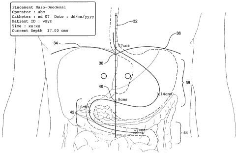
In an example of an enteral feeding catheter intubation the display shown in
Figure 6
shows that when the catheter passes below the xiphoid sternal junction 30 it
is very
deep (say l7cms below the location of the detector device). While the catheter
passes
through the stomach, its tip lies closer and less deep (say l4cms) and when
passing
under the mid sagital line 32 it is very shallow and closest to the surface of
the
patient (say 3cms). In the first part of the duodenum it is relatively shallow
(say
l0cms) and finally becomes very deep (say l7cms) when in the duodenum/jejunum.
After long term clinical use, an acceptable and reliable range of depths and
ratios at
the points or regions described above (or others) will be developed and most
useful
for assisting clinical assessment of the correctness of the route taken by the
catheter.
CA 02420676 2003-02-21
WO 02/15973 PCT/AU01/01051
28
Again, it can be seen that although not proof of the exact location of the
caudal/distal
end of the stylet mounted coil and hence the tip of the catheter, the
displayed
characteristics provide yet another aid to improving clirueal decision making
in
respect of the location of a catheter.
A stylet is capable of being reused in a catheter (after decontamination and
cleaning).
Thus, it can be cheaper to use in a clinical environment and encourages more
frequent checking of the catheter location than would otherwise be the case
because
the expense and time consuming nature of X-ray or other detection methods.
Stylet
reuse, although common in the clinical environment, is not recommended by the
authors of this document, until there is regulatory approval under a Bode of
practice
governing re-use of such devices.
It should not of course be forgotten that other clinical monitoring techniques
should
continue to be used thereby increasing the confidence of the clinician that
the
catheter is appropriately located, whether that be for enteral/parenteral
nutrition or
other purposes.
Patients are often intubated with VAC/CVCs using image guidance by means of X-
rays, fluoroscopy and ultrasonography. Percutaneous central venous access is
achieved when the tip of a catheter is located in the caval atrial region.
Tunneled
catheters travel through a subcutaneous tract prior to exiting the body via an
incision
in the skin. Such catheters are used for medicament delivery and dialysis.
Image-
guided techniques, although expensive, are less fraught with early
complications
than blind or external landmark intubation techniques.
VAC/CVCs are now used for long-term intravenous antibiotic support as well as
parenteral nutritional support and blood sampling.
CA 02420676 2003-02-21
WO 02/15973 PCT/AU01/01051
29
A selection from the many types of VAC/CVC in the marketplace can include
single-
or mufti-lumen short-term "central lines", tunneled catheters, such as
HickmanTM or
GroshongTM, and implanted catheters, such as Port-A-CathTM or InfusaPortTM. In
addition, Peripherally Inserted Central Catheters (PICCs) are also available.
The invention described herein can assist in the correct placement of most
catheters
and most advantageously can be used to check and confirm their correct
placement
at any time following initial placement.
Fig. 7 depicts an anterior view of the venous system of the upper torso
showing the
right subclavian vein 46 and the left subclavian vein 48 which meet medially
with
corresponding right and left brachiocephalic veins 50 and 52 respectively.
The junction of the brachiocephalic veins occurs at the upper region of the
superior
versa cava 54, which descends towards the cavoatrial junction 56 prior to
entering the
right atrium 60 of the heart 62.
Fig. 9 depicts, as would be seen on a screen visible to the clinician, a
constructed trace
64 of the path of a Peripherally Inserted Central Catheter (PICC) which has
its tip
ideally located in the central region of the superior versa cava 54. Fig. 9
also depicts
an outline of a sternum 66 thus providing a region of reference for the
clinician
between what is shown on screen and a known landmark of the patient's body.
Clearly, the outline is a pictorial representation of a generic sternum and
not the
sternum of the patient. This representation is, in any event, useful to the
clinician for
the task at hand.
During placement of the PICC, because of the relative stability of the organs
above
the diaphragm of the patient, the feed in trace provided on a monitor will be
similar
to the pull back trace described in respect of enteral feeding catheters. It
will be clear
that the tip of the catheter has entered the superior versa cava from not only
the two
CA 02420676 2003-02-21
WO 02/15973 PCT/AU01/01051
dimensional route displayed on the monitor, but also confirmed by its depth as
it
wends its way through the various branches of the venous system in the upper
torso
of the patient while being feed over the previously inserted guide wire.
Most obvious to the clinician from the monitor will be the sharp change in
direction
and relevant depth of the catheter as it transitions from the subclavian vein
into the
brachiocephalic vein (63 of Fig. 9).
As described previously, an independently identified measure of the most
preferred
location of the tip of the catheter occurs when, in an adult, there is about 9
cm of
linear distance between the catheter tip and the appropriately located
measuring
instrument. This is with reference to when a detector apparatus has been
placed on
the caudal/mid sagital aspect of the jugular sternal notch along the
midsagittal line
of the patient.
Such an explicit measurement is used herein as an example only, of the
practical
methodology associated with the use of the device but it may well not be
totally
accurate for all circumstances. Such measurements though, are likely as
described
previously, to become clinically acceptable as an indicator of the appropriate
location
of the tip of the catheter as the number of patients measured increases and
verification by other methods occur and further data is gathered and analyzed
in the
future.
The display is particularly useful to the clinician as the progress of the tip
of the
catheter is continuous and always displayed with reference to, in this
embodiment,
the position of the patient's sternum as is indicated by the shape 66 on the
monitor
display.
Fig. 10 depicts some of the venous system of the upper thoracic cavity and in
particular the placement of a PICC 70 terminating in the superior versa cava.
CA 02420676 2003-02-21
WO 02/15973 PCT/AU01/01051
31
It should be apparent from the foregoing that the part or portion of the body
displayed is not used to explicitly locate the signal radiating coil with
reference to it
but is used more as a guide.
The clinician will learn from their observations over time that the position
of the
inserted coil as determined by the signal detectors indicates a radial
distance of X
cms relative to a predetermined location on the signal detector apparatus and
not the
displayed symbol. The processor will be capable of providing the coordinates,
in say
X and Y in the horizontal plane (assuming a supine patient) of the location of
the coil
both as an image on the screen but also in figures.
When the signal detector apparatus itself is consistently located with
reference to a
predetermined body part or portion thereof, each measurement displayed will be
referenced to the displayed reference image. However, this is not meant to be
an
absolute and what the clinician experiences and assesses as the actual
position in say
a particular organ or channel in the body is a matter of acquired expertise.
Thus the display is used on two levels. One is to provide accurate radial
distance X or
(X,Y) from a predetermined point on the detector apparatus. The other is to
reference
that position measurement to a known body part or portion thereof (that might
be a
different one to that displayed).
The body part or portion displayed is only pictorial. It is only used as an
indicator of
a region.
The arched lines (depicted in Figs 3, 4 and 5) represent as discussed
previously the
complex shape on a vertical cross-section of the diaphragm of a typical human.
Since
the electronics used in the detector apparatus and the display expect that the
upper
coil of the detector Boils has been placed on a particular bony landmark, the
display
CA 02420676 2003-02-21
WO 02/15973 PCT/AU01/01051
32
can show the position the junction of the two curved lines and reference the
measurements of the position of the catheter coil accordingly.
It is thus possible, even recognizing that the display is only an indicator,
to provide
prompts additional to the obvious visual ones. These prompts may inform the
clinician that the signal-radiating coil is above or below the arched line
indicative of
the diaphragm of the patient. These prompts could be in the form of audible
signals.
Say for example, audible tones may increase in frequency as the depicted Boil
position gets closer from above the arched lines assuming that the head end of
the
patient is correctly determined by correct placement of the detector device.
Furthermore, as the coil position displayed moves below and a way from the
displayed arched lines an audible tone may pluses less quickly the further it
moves
away.
The audible signals could be of a type that is preferable to the user of the
apparatus.
It is also possible for the measured position to be used to indicate the
possibility of an
incorrect placement. This requires the equivalent of an expert system
information
database to be programmed into the display device. Ideally, for a
predetermined
location of the signal detector apparatus on the body, there are defined
regions in the
body consisting of certain radial distances from the signal detector, that if
measured
during an intubation would indicate that the signal radiating coil is in or
approaching an undesirable region or part of the body.
Thus not only does the clinician form over time a feel for the expected
position
measurements but the expert system can be used as a backup to warn the
clinician of
the progression of the radiating coil and thus the catheter into an
inappropriate
region of the patient. This further indication can be by way of visual
indicators on
the monitor screen or by additional audible signals. Recognizing that there
are
CA 02420676 2003-02-21
WO 02/15973 PCT/AU01/01051
33
variations in the anatomy of the human body from patient to patient, it is
important
to note that the method described provides guidance and is no substitute for
clinical
experience.
As clinical experience accumulates with the placement of catheters in other
parts of
the body, it will be possible to overlay on a monitor other reference images
such as
representations of static points, regions, or structures of the anatomy that
may assist
the clinician. Additional visual and audible information can also provide
guidance
to the skilled clinician or registered nurse authorised to locate catheters
into patients.
It will be appreciated by those skilled in the art, that the invention is not
restricted in
its use to the particular application described. Neither is the present
invention
restricted in its preferred embodiment with regard to the particular elements
and/or
features described or depicted herein. It will be appreciated that various
modifications can be made without departing from the principles of the
invention.
Therefore, the invention should be understood to include all such
modifications
within its scope.