Note: Descriptions are shown in the official language in which they were submitted.
CA 02421034 2003-02-28
WO 02/22200 PCT/US01/28561
METHOD AND SYSTEM FOR CLOSED CHEST
BLOOD FLOW SUPPORT
FIELD OF THE INVENTION
The present invention is related to a cardiac
assist system having a transseptal cannula that extends
through the atrial septum from the right atrium to the left
atrium of the patient and a perfusion cannula that extends
into the arterial system of the patient for circulating
oxygenated blood throughout the patient. More specifically,
the present invention is related to an extracorporeal blood
pump, connected to the transseptal cannula at the pump inlet
and the perfusion cannula at the pump outlet, that pumps
blood at specified flow rates over a range of physiological
pressure and is held in place on the patient's leg.
BACKGROUND OF THE INVENTION
For short term (hours to days) use in supporting
significant circulation (1-3.5 LPM) of oxygenated blood,
there is a need for simple equipment in a hospital that can
be quickly connected to the patient without surgical
intervention and that can provide bypass blood flow to the
patient. The present invention provides a,,quick and
relatively simple way of operation to assist the heart
without an open-chest surgery.
SUMMARY OF THE INVENTION
The present invention pertains to a system
supporting circulation of oxygenated blood. The system
comprises a transseptal cannula set adapted to be inserted
percutaneously in the femoral vein and extend through the
atrial septum from right,atrium to left atrium, a blood pump
CA 02421034 2003-02-28
WO 02/22200 PCT/US01/28561
-2-
mechanism, connected to the transseptal cannula through the
pump inlet and controlled by an external microprocessor based
blood pump controller, for pumping blood received from the
patient's heart, a perfusion cannula adapted to be inserted
percutaneously in the femoral artery and connected to the
pump outlet for returning oxygenated blood to the arterial
system of the patient. The cannula set consists of a
cannula, a catheter, and a dilator. The catheter and dilator
are used for insertion of the cannula.
The present invention pertains to a method and a
process for assisting a patient's heart. The method
comprises a step of inserting a transseptal cannula
percutaneously in the femoral vein of the patient and
extending through the atrial septum from the right atrium to
the left atrium. Next there is a step of inserting a
perfusion cannula percutaneously in the femoral artery for
returning oxygenated blood to the arterial system of the
patient. Next is preferably a step of connecting the two
cannulae to the pump. Then there is a step of pumping blood
with a blood pump connected to the transseptal cannula and
the perfusion cannula at specified flow rates over a range of
physiological pressures. This step preferably includes
control of the pump and monitoring of the control system and
pump by an external pump controller in such a manner as to
detect, manage, and alert the user to the applicable
potential system faults without dedicated human monitoring.
In addition, the system of the present invention
can be used to quickly access or redistribute apatient's
blood to a certain destination in a patient's body by
changing appropriate sizes of cannulae connected to the pump
inlet and outlet.
CA 02421034 2003-02-28
WO 02/22200 PCT/US01/28561
-3-
BRIEF DESCRIPTION OF THE DRAWINGS
In the accompanying drawings, the preferred
embodiment of the invention and preferred methods of
practicing the invention are illustrated in which:
Figure 1 is a schematic representation of the
transseptal cannula portion of the present invention.
Figure 2 is a schematic representation of a needle
and wire in a second catheter in a cannula.
Figure 3 is a schematic representation of a balloon
catheter at the distal end of the cannula.
Figure 4 is a schematic representation of a pigtail
cannula.
Figure 5 is a schematic representation of a pigtail
cannula with a straightening dilatory.
Figure 6 is a schematic representation of a
transseptal sheath over the port of a cannula.
Figure 7 is a schematic representation of the
transseptal sheath retracted from the port of the cannula.
Figure 8 is a schematic representation of an
alternative embodiment of a balloon at the distal end of the
cannula.
Figure 9 is a schematic representation of the
arterial connection system of the present'invention.
CA 02421034 2003-02-28
WO 02/22200 PCT/US01/28561
-4-
Figure 10 is a schematic representation of an
alternative arterial perfusion cannula of the present
invention.
Figure 11 is a schematic representation of the
fluid pathway components of the present invention.
Figure 12 is a schematic representation of an
exploded view of the pump with the clamp mechanism.
Figure 13 is a schematic representation of the pump
with the clamp mechanism.
Figure 14 is a schematic representation of the pump
assembly.
Figure 15is a schematic representation of a cross-
sectional view of the pump.
Figure 15a is a schematic representation of the
infusion system configuration.
Figures 16a-16e are schematic representations of
the blood pump controller.
Figure 17 is a schematic representation of a cross
sectional view of the purrip with a flow probe.
Figure 18 is a schematic representation of the
holding mechanism with the pump on the-leg of a patient.
Figure 19 is a schematic representation of the
holding mechanism.
CA 02421034 2008-06-13
-5-
Figure 20 is a schematic representation of the
holding mechanism with the pump on the leg of the patient.
Figure 21 is a schematic representation of the pump
in a normal position on the leg of a patient.
Figure 22 is a schematic representation of the pump
at an angle of 20 degrees from normal on the leg of a
patient.
Figure 23 is a. schematic representation of the
holding mechanism with.the pump on the leg of the patient.
Figure 24 is an illustration of how the ML4428
pulse train is processed.
Figure 25a and Figure 25b are illustrations of
pressure and pressure variation waveforms.
Figure 26a is a graph of pressure versus flow of
the blood pump.
Figure 26b is a graph of flow rate versus pressure
drop.
Figure 26c is a graph regarding pump inlet
pressure.
Figures 27a-27c are schematic representations of
the components of the cannula set.
Figure 28 is a schematic representation of slow
start circuitry.
CA 02421034 2003-02-28
WO 02/22200 PCT/US01/28561
-6-
Figure 29 is a schematic of timing circuitry.
DETAILED DESCRIPTION
Referring now to the drawings wherein like
reference numerals refer to similar or identical parts
throughout the several views, and more specifically to figure
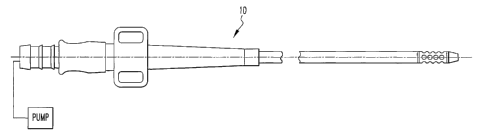
11 thereof, there is shown a system 300 for assisting flow of
blood by a patient's heart. The system 300 comprises a
transseptal cannula 12 adapted to be inserted percutaneously
in the femoral vein and extend through the atrial septum from
the right atrium to the left atrium. The system 300
comprises a perfusion cannula 100 adapted to be inserted
percutaneously in the femoral artery. The system 300
comprises a blood pump mechanism 30 having a blood pump 316
(driven by a brushless DC motor), connected to the
transseptal cannula 12 at the pump inlet, for pumping
oxygenated blood received from the left atrium through the
transseptal cannula 12 and returning the blood to the
arterial system of the patient through the perfusion cannula
100 connected to the blood pump 316 at the pump outlet. The
blood pump 316 is controlled by a controller 332 that
monitors key system operating parameters to detect and manage
faults and to alert operators.
Fault tolerance of the system is offered by
providing redundant mechanisms for pump operation and for
monitoring functions. Pump operation must propel blood at
sufficient flow rates (1-3.5 LPM) without destroying red
blood cells, without causing clotting of the blood, without
causing immune system or other biocompatibility compromise or
reaction, and must locate and center themselves without
offering a wear surface that can cause blood damage or
CA 02421034 2003-02-28
WO 02/22200 PCT/US01/28561
-7-
clotting over time or can cause variable power loss over
time, as power loss is used as a performance monitor.
Reliable operation must be maintained even through a wide
variety of system, component, or human faults.
The AB-180 XC System, as it is referred to, offers
atraumatic contact with red blood cells by virtue of the
smooth surface finishes of the blood contacting surfaces, the
gradual radiuses of the impeller, and does not allow
locations for stagnant blood to accumulate and form clots. In
addition to these smooth geometries, it is designed to be
placed within 3 feet of the blood egress from the body, as
the small amount of artificial material minimizes
complications. To provide further resistance to thrombus
formation, an anti-coagulant is infused directly into the
blood chamber of the pump, where concentration of anti-
coagulant is high enough to deter clotting. But since the
pump volume is small, the amount of infused anti-coagulant is
small enough to achieve minimal effect on systemic
concentration of anti-coagulant when the blood flowing out of
the pump is disbursed throughout the body. This is important,
as the anti-coagulant concentration throughout the body is
then small enough to prevent additional risk of internal
bleeding elsewhere in.the patient. Clots can also form when
blood flow drops to low rates (< 0.5 LPM), which can happen
if the transseptal cannula becomes clogged or kinked, if the
patient bleeds internally, or if a cannula.becomes dislodged.
Any of these events cause an alarm condition from the reduced
blood flow.
Pump wear is a major issue with blood pumps, as any
wear can cause blood damage and/or clotting. The infusion
system used to infuse anti-coagulant into the blood chamber
CA 02421034 2003-02-28
WO 02/22200 PCT/US01/28561
-8-
also performs a useful bearing function. While the main
bearing function., or centering and locating of the rotating
part of the pump, is performed by the interaction of the
stator and rotor electromagnetic interactions, the rotating
impeller is also centered by a) the impeller/seal sliding
contact and b) the fluid film and fluid' lift pads in the
motor chamber. Whenever the rotor becomes out of center, the
fluid film prevents rotor and stator contact, thus providing
fault tolerance of the electromagnetics. In the motor
chamber, the fluid film is propelled radially as the rotor
rotates. As lift pads are encountered in the lower chamber
surface, fluid is propelled axially into the surface of the
rotor, thus 'lifting' it away from the stator surface, where
contact would cause wear. This fluid is pumped by a constant
flow pump, which provides a stable flow of infusate into the
lower chamber. This constant flow builds pressure in the
lower chamber, and as this pressure exceeds the pressure
across the seal in the upper, or blood flow chamber, the
infusate is pressed through the seal and into the blood
chamber, where the anti-coagulation prevents clotting. The
infusate fluid thus provides a third bearing mechanism and a
cooling mechanism for the seal/rotor contact area. Loss of
infusate flow can then be used as an indicator of a fault in
the bearing system, which can then allow a user alarm to
correct the infusate system or the pump-bearing system prior
to any patient injury. The mechanism of the determination of
infusate flow is described elsewhere.
Another mechanism for system fault is the presence
of air in the system, which can happen in some circuits when
the pressure is reduced to dangerous levels. This can occur
in larger pumps or in longer and smaller cannula, which
involve more pressure drop. The pump design, by its small
CA 02421034 2003-02-28
WO 02/22200 PCT/US01/28561
-9-
si2e, prevents any such cavitation by means of low pressure.
The mechanism of connections between the components prevents
any injection of air into these joints, which can occuir with
the negative pressure associated with high pressure drop and
long, small diameter cannulae. Without this mechanism of
connection, and by connecting arbitrary components, dangerous
pressures can result and can cause cavitation andjor air
injection at the connection joints.
The flow characteristics of the Pump over a range
of conditions, performed with perfusate at the viscosity of
3.5 cP (35% Glycerol + 65% Saline @ 23 C) is shown in Figure
26a. The flow rates represent the relationship between RPM
and the pressure differential between the inflow port and the
outflow port. An estimated flow rate can be determined by
using this chart, the RPM of the Pump and the pressure
differential of the inlet to outlet ports. The pressure drop
characteristics of the transseptal cannula are shown in
Figure 26b, with the box indicating the limits as specified
with'the AB-1-80 pump. The key issue is that the low pressure
rise of the pump, in combination with the pressure drop of
the cannula, does not allow any point in the circuit to
obtain pressures lower than -350mmHg, which is the point at
which gases can be removed from the blood solution. Including
the flow characteristics of the pump, the transseptal
cannula, and the possible sizes of perfusion cannulae, the
vacuum pressure created at the pump inlet can be solved at
different pump speeds as shown in Figure 26c. Since the pump
inlet pressure is always higher than -350 mmHg, cavitation
will n.ot happen in the system over the recommended flow range
against,physiologic pressure.
CA 02421034 2003-02-28
WO 02/22200 PCT/US01/28561
-10-
All electrical control functions are redundant to
provide enhanced reliability. And alarms are used to notify
operators of potentially unsafe conditions rather than to
stop the pump until replacement of control is maintained. All
power systems are duplicated, and all control systems are
watch-dogged to warn of failures of standby monitor
functions. Without these failsafe operations, tolerance of
system, component, or human faults could not be offered.
Preferably, the blood pump mechanism 30 includes a
transseptal clamp mechanism 322 which clamps the blood pump
316 to the transseptal cannula 12 to avoid undesired
disconnection of the blood pump 316 and the transseptal
cannula 12, as shown in Figures 17 and 18. The blood pump
316, during operation, is preferably adapted to be within
three feet of where the transseptal cannula 12 and the
perfusion cannula are positioned to enter the patient.
Preferably, the blood pump mechanism 30 includes tubing 324
which connects the blood pump 316 to the transseptal cannula
12 and the perfusion cannula 100 and the clamping mechanism
clamps the tubing 324 between the blood pump 316 and the
transseptal cannula 12. The tubing 324 has a continuous
smooth inner surface 326.
Alternatives of the tubing 324 include:
= a piece of tubing that connects the
transseptal cannula to the pump inlet port
and a piece of tubing that connects the
arterial cannula to the pump outlet port,
= a quick connection device with a seamless
connection (smooth without a step) to
CA 02421034 2003-02-28
WO 02/22200 PCT/US01/28561
-11-
minimize the potential for thrombus formation
and a lock mechanism to avoid inadvertent
disconnection that can directly connect the
transseptal and arterial cannulae to the
pump,
= tubing integrals on the transseptal and
arterial cannulae which can be placed over
the barbs of the inflow and outflow ports of
the pump and can be secured with a clamping
mechanisms to the barbs at the pump inlet and
outlet ports.
Preferably, the blood pump 316 pumps a continuous
flow of blood. The blood pump 316 preferably has a rotor 328
and a stator 330, as shown in Figures 14 and 15. Preferably,
the blood pump mechanism 30 includes a controller 332
connected to the blood pump 316 through which the operation
of the blood pump 316 is adjusted, as shown in Figures 16a,
16b, 16c, 16d and 16e. The blood pump 316 includes an
impeller 334 which moves against the blood, and the user
adjusts the operation of the blood pump 316 by changing
impeller 334 speed. Preferably, the controller 332 measures
flow of blood from the pump only from impeller 334 speed and
stator 330 current. Alternatively, the blood pump mechanism
includes an electromagnetic or ultrasonic flow probe 336
25 in communication with the blood pump 316 and the controller
332 measures flow of blood through the pump with the
electromagnetic or ultrasonic flow probe 336, as shown in
Figure 17.
Preferably, the pump has a hydrodynamic bearing 338
30 between the rotor 328 and-the lower housing 330, shown in
CA 02421034 2008-06-13
-12-
Figure 15. The blood pump mechanism 30 preferably includes
a fluid reservoir 340 and a fluid pump 342 connected to the
fluid reservoir 340 and the blood pump 316 to pump fluid to
the hydrodynamic bearing 338 in the blood pump 316.
Preferably, the fluid reservoir 340 includes predetermined
concentrations of drugs. See U.S. Patent 5,711,753 for a
more complete discussion of the pump 316, except the
occluder described therein is not needed in the
extracorporeal application.
The pump controller 332 preferably provides current
to the blood=pump 316 and the controller 332 includes a
battery 344 that provides energy to run the controller 332
and the blood pump 316. The battery 344 is used for powering
the blood pump 316 and controller 332 when the patient is
being moved between remote locations. Preferably, the blood
pump 316 is made of biocompatible materials which have no
effect on blood or the patient. See the Appendix. The blood
pump 316 is preferably a centrifugal pump or an axial pump.
Alternatively, a pulsatile flow may be obtained by modulation
of pump speed through controller 332 and synchronizing
impeller speed variation with the patient's beating heart.
Alternatively, the blood pump 316 is a pulsatile,
electrical or pneumatic pump having an inflow valve and a
perfusion valve. Farrar, D.J., Compton, P.G., Lawson, J.H.,
Hershon, J.J., Hill, J.D., "Control Modes of a Clinical
Ventricular Assist Device" : IESL Engineering in Medicine and
Biology Magazine, pp. 19-25, vol. 5, 1986, incorporated by
.reference herein. . Preferably, the blood pump mechanism 30
includes a controller 332 connected to the blood pump 316
through which the operation of the blood 'pump 316 is
CA 02421034 2003-02-28
WO 02/22200 PCT/US01/28561
-13-
adjusted. The pump is preferably a pulsatile pump having a
stroke time, and the controller 332 adjusts the operation of
the blood pump 316 by adjusting stroke time. Preferably, the
pump controller 332 provides current to the blood pump 316
and the controller 332 includes a battery 344 that provides
energy to run the controller 332 and the blood pump 316, the
battery 344 is used for powering the blood pump 316 and the
controller 332 when the patient is being moved between remote
locations.
Preferably, the system 300 includes a holding
mechanism 346 which holds the blood pump 316 and attaches to
the patient, as shown in Figures 18-23. The holding
mechanism 346 preferably includes a pump holding portion 348
which holds the pump and a patient portion 350 which is
adapted to fit to the leg 352 of the patient and to secure to
the pump holding portion 348. Preferably, the pump holding
portion 348 is made of plastic having an imprint 352 of the
shape of the blood pump 316 in which the blood pump 316 fits
to be held by the pump holding portion 348, and the patient
holding portion 348 includes a band 354 with loops and with
straps 356 having hooks adapted to wrap about the leg 3.52 and
the pump holding portion 348 to hold the pump holding portion
348 to the leg 352. The holding mechanism 346 is preferably
adapted to attach to either leg 352 of the patient and allow
inflow or outflow to be connected to the contralateral side
of the patient. Preferably, the holding mechanism 346 is
adapted to hold the blood pump 316 in a normal position or at
an angle of 20 degrees from the normal position.
The present invention pertains to a method for
assisting flow of oxygenated blood. The method comprises the
steps of inserting percutaneously in the femoral vein of the
CA 02421034 2003-02-28
WO 02/22200 PCT/US01/28561
-14-
patient and extending through the atrial septum from the
right atrium to the left atrium a transseptal cannula 12.
Next there is the step of inserting percutaneously in the
femoral artery a perfusion cannula 100 for returning
oxygenated blood to the arterial system 300 of the patient.
Then there is the step of pumping blood with a blood pump 316
connected to the transseptal cannula 12 and the perfusion
cannula 100 at specified flow rates over a range of
physiological pressures with performance monitoring to offer
fault tolerance and management.
The transseptal cannula set contains:
1- 21 Fr. Percutaneous Venous Transseptal Cannula (PVTC)
1 - 13 Fr. Percutaneous Venous Transseptal Catheter
1 - 14/21 Fr. Percutaneous Venous Transseptal Two Stage
Dilator
The following instruments are needed to complete
the procedure and should be supplied by the user (all are
standard in the art):
Introducer Needle
Guidewire, super stiff, 0.035 in., at least 260 cm long.
Transseptal Puncture Kit
Transseptal Catheter/Dilator (as needed).
Components of the cannula set are shown in Figure
27.
The transseptal cannula is inserted in the
following manner:
CA 02421034 2003-02-28
WO 02/22200 PCT/US01/28561
-15-
Prior to performing the procedure, insert the PVTC
Catheter into the PVTC Cannula assuring that the Cannula
fitting on the Catheter fits snugly and is fully
inserted into the 3/8 in. barbed connector of the
Cannula. Assure that the tip of the Catheter is extended
fully from the Cannula.
Use standard transseptal puncture technique to gain
access into the left atrium from the femoral vein.
Dilate the transseptal puncture site (fossa ovalis) with
a transseptal puncture catheter in the usual manner.
Introduce the 0.035 in. guidewire into the left atrium.
Verify that the guidewire is in position in left atrium.
Verify patient ACT is in excess of 400 seconds.
Remove the transseptal puncture catheter.
Advance the Two Stage Dilator over the guidewire into
the left atrium to dilate the fossa ovalis. Monitor
progress using fluoroscopy to assure that the tip does
not penetrate the atrial chamber.
Remove the Two Stage Dilator.
Advance the PVTC Catheter/Cannula assembly over the
guidewire into the left atrium.
Position the PVTC Cannula tip in the left atrium using
fluoroscopy. Assure that all of the drai.nage holes are
CA 02421034 2003-02-28
WO 02/22200 PCT/US01/28561
-16-
in the left atrium and the marker band is near the
septum.
Remove the guidewire and Catheter together to allow the
cannula to be back filled with blood.
Clamp the adapter of the PVTC Cannula on the clamping
area of the clear adapter.
The arterial cannula is inserted in the following manner:
= Introduce an arterial guidewire into the artery location
chosen.
Advance the arterial cannula over the guidewire into the
artery.
Verify the blood is arterial blood.
= Remove the guidewire.
= Clamp the cannula to prevent blood loss prior to
connection to the blood pump.
The extracorporeal circuit is connected to the pump in
the following manner:
= Connect appropriate length standard 3/8 in.
extracorporeal blood circuit tubing to the inflow and
outflow ports of the Pump.
Connect the inflow tubing to the inflow cannula.
CA 02421034 2003-02-28
WO 02/22200 PCT/US01/28561
-17-
Release the inflow cannula clamp and prime the Pump and
outflow tubing with blood. Clamp the outflow tubing
ensuring no air between the inflow cannula and the clamp
on the outflow tubing.
Make a wet to wet connection of the outflow tubing. and
the outflow cannula.
If there is a vent on the outflow cannula, aspirate any
final air from the extracorporeal blood circuit.
If there is no vent on the outflow cannula, inspect the
extracorporeal circuit for air. If there is any air,
break and remake the wet to wet connection of the
outflow tubing and outflow cannula until all of the air
is purged from the extracorporeal blood circuit.
= Secure all tubing connections with sta-straps.
Release the hemostat on the outflow tubing followed by
the hemostat on the inflow cannula.
= Adjust pump speed to desired setting and place Pump in
Mounting Assembly and secure to patient's leg.
Preferably, before the pumping step, there is the
step of clamping a transseptal clamp mechanism 322 to the
transseptal cannula 12 and the blood pump 316 to avoid
undesired disconnection of the blood pump 316 and the
transseptal cannula 12. Before the pumping step, there is
preferably the step of positioning the blood pump 316 within
three feet of where the transseptal cannula 12 and the
perfusion cannula 100 are inserted into the patient.
CA 02421034 2003-02-28
WO 02/22200 PCT/US01/28561
-18-
Preferably, the pumping step includes the step of
pumping a continuous flow of blood with the blood pump 316.
Preferably, the pumping step includes the step of adjusting
the flow of blood pumped with a controller 332 connected to
the blood pump 316. The adjusting step preferably includes
the step of adjusting impeller 334 speed of an impeller 334
of the blood pump 316 to attain a desired flow of blood in
the patient due to the operation of the blood pump 316.
Preferably, after the pumping step, there is the step of
powering the controller 332 and the blood pump 316 with a
battery 344 as the patient is moved from a first location to
a second location remote from the first location.
Before the pumping step, there are preferably the
steps of attaching a holding mechanism 346 for the blood pump
316 to the patient and placing the blood pump 316 in the
holding mechanism 346 to hold the blood pump 316 in place
relative to the patient. Preferably, the attaching step
includes the step of attaching the holding mechanism 346 to
the leg 352 of the patient. The placing step preferably
includes the step of wrapping straps of a band 354 positioned
about the leg 352 of the patient, about the blood pump 316,
and fixing hooks 360 of the straps to loops 358 of the band
354 to secure the blood pump 316 to the leg 352 of the
patient.
Alternatively, the pumping step includes the step
of pumping pulses of blood through the patient with a
pulsatile pump. The pumping step then can include the step
of adjusting stroke timing of the pulsatile pump to obtain
the desired pulse of blood flow through the patient.
CA 02421034 2003-02-28
WO 02/22200 PCT/US01/28561
-19-
In the operation of the invention, and referring to
Figures 1, 2 and 3, the distal end 14 of the transseptal
cannula 12, is inserted into a patient and moved to the right
atrium of the patient's heart via the femoral vein, as is
well known in the art. Generally, this occurs in the
following way. The guide wire 30 is introduced into the
patient and threaded to the right atrium of the patient. The
cannula 12, the second catheter 60 (with the needle 58
disposed in the second catheter 60) are placed over the end
of the guide wire 30 extending from the patient via the
orifice 18 and the opening in the second catheter 60. The
cannula 12 and second catheter 60, with the needle 58 inside
the second catheter 60, are then inserted and moved' along the
guide wire 30 to the right atrium of the patient. When the
distal end 14 of the cannula 12 is in the right atrium, the
guide wire 30 is pulled back 46 into the cannula 12 freeing
the orifice 18 so there is nothing in the orifice 18. The
needle 58 is then advanced, as is the second catheter 60
through the orifice 18 so the second catheter 60 extends
through the orifice 18 of the cannula 12 and the needle 58
extends. through the opening of the second catheter 60. The
needle 58 and second catheter 60 are then forced into the
septum until they puncture the septum and move into the left
atrium. The needle 58 is then retracted from the opening of
the second catheter 60 and the guide wire 30 is moved forward
through the second catheter's opening into the left atrium.
The second catheter 60 is maintained in position while the
guide wire 30 is maintained in place in the left atrium. The
cannula 12 is then advanced forward into the left atrium
along the guide wire 30 and the second catheter 60 which
extend through the orifice 18. The presence of the second
catheter 60 acts as a stiffener for the cannula 12 to assist
in the placement of the cannula 12 in the left atrium. The
CA 02421034 2003-02-28
WO 02/22200 PCT/US01/28561
-20-
second catheter 60, needle 58 and guide wire 30 are then
removed from the cannula.
Preferably, the transseptal cannula 12 connection
step includes the step of connecting the transseptal cannula
12 to the tubing 324 connected to the blood pump 316.
Preferably, prior to clamping the clamping mechanism 322 to
connect the tubing 324 to the transseptal cannula 12, there
are preferably the steps of filling the transseptal cannula
12 with blood and confirming proper transseptal cannula 12
position by visualizing blood color, as is well known in the
art.
It should be noted that the aforementioned
procedure can be performed without the introducer catheter.
Instead, the second catheter 60 acts with a dual purpose, as
the introducer catheter and the second catheter 60. In this
case, the needle 58 and guide wire 30 are together inserted
in the second catheter 60, and the introducer catheter is not
present. When the second catheter 60 and needle 58 puncture
the septum and move into the left atrium, the second catheter
60 remains in place and the guide wire 30 and the needle 58
are removed to clear a blood flow passage through the second
catheter 60. This apparatus of second catheter 60, guide
wire 30 and needle 58, without any of the other features
described herein on the cannula 12, or with some or all of
them, in and of itself can be used to access the left atrium.
Again, the advantage of the combination of elements, is that
it can serve to access the left atrium without having to take
turns pulling the guide wire 30 out and then inserting the
needle 58 into the second catheter 60 since the guide wire 30
and the needle 58 are together present in the second catheter
60 simultaneously; and the second catheter 60 serves a dual
CA 02421034 2003-02-28
WO 02/22200 PCT/US01/28561
-21-
purpose of being the introducer catheter and second catheter
60, without needing the introducer catheter. Alternatively,
the needl-e can be inserted into the second catheter 60 after
the second catheter has reached the right atrium.
During the process of moving the cannula 12 to the
right atrium, removing the guide wire 30 from the orifice 18
and extending the needle through the orifice 18, an imaging
device, external to the patient is imaging the location of
the orifice 18 (and during the entire procedure) by noting
where an end marker 34, disposed about the orifice 18, is
located in the patient. Such an imaging system, for instance
with the end marker 34 being radio opaque, is well known in
the art. If it is desired, the guide wire 30 or a portion
thereof, such as the tip of the guide wire 30, and/or the
needle 58 or a portion thereof, such as the tip of the needle
58, can also be enhanced for imaging purposes, for example by
having a radio opaque material, so the guide wire 30 and
needle 58 can also be followed as they move through the
patient.-
Once the orifice 18 is positioned in the left
atrium and the port 20 of the cannula 12 is positioned in the
right atrium, a balloon 52 disposed adjacent the orifice 18
is inflated with saline, as shown in Figure 3, which travels
along an inflation tube 54 that runs the length of the
cannula 12 along the outside of the cannula 12 to a s'aline
supply 87 disposed outside of the patient. The inflated
balloon 52 serves to prevent the distal end 14 of the cannula
from_puncturing an atrium wall 50 of the left atrium where
the distal end 14 of the cannula is now disposed, for
instance when the,patient is being turned or moved. The
inflated balloon 52 also serves to prevent the cannula 12
CA 02421034 2008-06-13
-22-
from slipping back into the right atrium at undesired times,
such as when the patient is being turned or moved about. The
balloon 52 can be deflated by removing the saline that has
been introduced into it through the inflation tube 54, back
out of the inflation tube 54 with negative pressure applied
to the end of the inflation tube 54 extending externally from
the patient. In another embodiment of a balloon 52 with the
cannula 12, as shown in Figure 8, the balloon 52 is disposed
at.the distal end 14 of the cannula 12.
P,lternatively, a-pigtail cannula 78, as shown in
Figure 4, can-be used which has its distal end curling about.
As long as a straightening dilator 80 or needle 58 is present
in the pigtail cannula 78, the pigtail cannula 78 is
straight, as shown in Figure S. As soon as the dilator 80 is
removed, the- pigtail cannula's distal end curls about to
achieve the same results as.the inflated balloon 52. See
U.S. Patent No. 5,190,528 titled "Percutaneous Transseptal
Left Atrial Cannulation System" by James D. Fonger et al. and
WO 2000/071194 for further information about a transseptal
cannula and its use.
Alternatively, a transeeptal sheath 82 positioned
about the cannula 12 can be used, as shown in Figure 6. When
the transseptal sheath 82 is in a closed -position, it covers
over the port 20 so no blood can pass through the port 20.
When the transseptal sheath 82 is in an open position,
meaning it has been retracted by being pulled on from outside
the patient, the transseptal sheath $2 bas moved away from
the distal end 14 exposing the port 20, as shown in Figure 7.
The extent the transseptal sheath 82 has been retracted
determines how much of the port 20 is exposed. The
CA 02421034 2008-06-13
-23-
transseptal sheath 82 can also have a marker at its end, and
the cannula 12 can have gradations which are marked to
identify where the end of the transseptal sheath 82 is
relative to the cannula 12.
Holes 32 having an elongate shape and disposed
essentially in parallel with the axis of the cannula 12 and
between the orifice 18 and the port 20 further facilitates
movement of blood into and out of the cannula 12. The
elongate shape of the holes 32 minimizes damage to the
cellul.ar,structure of the blood cells as they pass through
the holes 32. Furthermore, all openings, such as the orifice
18 and the port 20, are made as smooth as possible and are
made of bio-inert materials such as plastic or =steel to
minimize or preclude the clotting of blood. In this way,
access to the left and right atriums of the patient is
achieved for whatever purpose, such as the attachment of a
pump to the cannula 12.
The perfusion cannula 100 connected to the pump
mechanism 30, as shown in Figure 9, is inserted=into the
femoral artery of a patient 'so the -distal end 116 of the tube
112 of the perfusion cannula 100 is disposed in the femoral
artery, as is well known in the art. See U.S. Patent No.
5,330,433 titled "A Bidirectional Femoral Arterial Cannula"
by James D. Fonger et al. and U.S. patent 6,676,650 for
25. further discussions and use of a perfusion cannula.
Preferably, the perfusion cannula 100 connection
step includes the step of connecting the perfusion cannula
100 to the tubing 324 connected to the blood pump 316.
Preferably, the connection step includes the step of priming
CA 02421034 2003-02-28
WO 02/22200 PCT/US01/28561
-24-
the perfusion cannula 100. Preferably, prior to clamping the
clamping mechanism 322 to connect the tubing 324 to the
perfusion cannula 100, there is the step of filling the
perfusion cannula 100 with oxygenated blood.
Selecting the size of perfusion cannula depends on
patient's body size. A bigger size of perfusion cannula can
allow higher blood flow rate and thus unloading the patient's
heart better: However, if the perfusion cannula size is too
big, the cannula may block the blood stream through patient's
leg. It is desirable to choose an appropriate perfusion
cannula size such that the total blood flow rate through the
cannula between 1 and 4 L/min, preferably 1 to 3.5 L/min. In
addition, the blood stream through patient's leg between
femoral artery and the perfusion cannula should have the flow
rate between 100 ml/min and 500 ml/min, preferably 200 m1/min
to 400 ml/min..
Prior to pump attachment, the two chambers (upper
and lower) of the pump are primed to prevent air from being
pumped into the patient after attachment to the other system
components. The lower chamber uses fluid infusate to provide
a bearing function that prevents motor wear, provides
cooling, and provides anti-coagulation directly to the upper
chamber, where blood flows during operation. First, the
infusate line is primed with sterile infusate from the
infusate supply system. The lower chamber, or motor chamber,
is then filled with sterile infusate from the infusate line.
A syringe is used to push fluid through the infusate system
and into the lower chamber. The pump is then started, with
the pumping action pulling all air through the seal
separating the upper and lower chambers. Alternately, a
syringe with two way stopcock can be used to suck air out of
CA 02421034 2003-02-28
WO 02/22200 PCT/US01/28561
-25-
the lower chamber prior to filling with infusate. The upper
chamber, or blood flow chamber, is filled with saline from
either the inflow or outflow port. Owing to the low pump
volume, this can be accomplished with saline.
Once the transseptal cannula and the perfusion
cannula are in position in the patient, the blood pump 316 is
connected to them, as shown in Figures 11-13. Connection of
the blood pump 316 to the transseptal cannula and the
perfusion cannula is accomplished with tubing 324 that
extends between the input cannula of the blood pump 316 and
the transseptal cannula, and the output cannula of the blood
pump 316 and the perfusion cannula. The tubing 324 is
secured in place by the clamping mechanism 322 that clamps
the tubing to the respective elements. The clamping
mechanism 322 is used to avoid inadvertent disconnection of
the elements and the tubing and to prevent leakage of air
into the system. The blood pump 316 is positioned in close
proximity, within 3 ft. to the ends of the perfusion cannula
and the transseptal cannula which extend from the patient to
minimize the system blood volume and to minimize heat loss of
the blood in the extracorporeal portion of the system. The
tubing 324 from the perfusion cannula and the transseptal
cannula is designed so there is no step transition from the
pump housing to the connecting tubing that would tend to
create areas of low, blood flow.
The blood pump 316 is placed into an imprint 352 of
the holding portion 348 which corresponds to the shape of the
blood pump 316, as shown in Figures 18-23. The holding
portion 348 is placed on a band 354 about the patient's leg
352. Straps 356 of the band 354 are then placed over the
blood pump 316. Loops 358 on the straps 356 are connected to
CA 02421034 2003-02-28
WO 02/22200 PCT/US01/28561
-26-
the hooks 360 on the band 354 to secure the straps 356 on the
band 354, thus holding blood pump 316 securely to the
patient. The holding mechanism 346 comprising the holding
portion 348 and the band 354 can be attached to either of
the patient's legs and allows for inflow or outflow
cannulation to the side opposite the pump fixation to the
leg. The holding mechanism can fix the blood pump 316 in a
position normal to the leg of the patient, or rotated 20
degrees from the normal to the patient's leg.
The blood pump 30 is a continuous flow blood pump,
electrical in nature and magnetically driven having a rotor
328 and a stator 330. The blood pump components are
constructed from biocompatible materials suitable for blood
contact for periods of up to 14 days.
The primary function of the pump mechanism 30 is to
pump blood. The pump 316 provides a range of volumetric flow
rates from 1 to 4 1/min. over a mean arterial pressure range
of 60 to 100 mmHg with .a minimum of 5 mmHg left atrium
filling pressure in persons with body surface areas (BSA)
between 1.2 and 2.7 sq. meters. This is accomplished by the
regulated rotation of the impeller at speeds of 3000 to 7500
rpm.
Secondary functions of the pump mechanism 30 provided by the
controller 332 include:
= Provide a fluid bearing and localized heparin in the pump.
= Provide a system to monitor the fluid bearing infusate
supply.
= Provide a system to monitor the blood flow rate through
the pump 316.
Provide a system to power the device.
CA 02421034 2003-02-28
WO 02/22200 PCT/US01/28561
-27-
Provide a system to interface to user.
Provide a performance monitor and alarm system.
Provide a software-based control system.
Alarm conditions are detected by the CPU and
communicated to the user through audible and visual alarms on
the controller display.
For single system faults, when a monitored system
parameter goes outside an acceptable operating range, an
alarm condition is set and a standard alarm sequence is
started. The alarrn is first issued by turning on an audible
alarm device, turning on, a flasYi.ing red alarm LED and
displaying one or more related alarm messages on the display.
In case of multiple alarms, each time a new
parameter goes out of range, a new audible alarm and flashing
indication are generated. The alarm list fills top to
bottom, that is, a new alarm message is added to the bottom
of the alarm list and earlier alarm messages maintain their
position on the list. The operator may be able to mute the
alarm and the flashing LED may become steady, depending on
the alarms present. As alarm conditions are cleared, the
related alarm message is removed from the display but the
alarm LED remains light. Only when all alarm conditions have
been cleared, the red LED turns off. If the number of
simultaneous alarms exceeds the available lines on the
display, normally 13, then no new alarms appear on the
display until previous alarms clear. The audible alarm as
well as the alarm LED are still activated in the event of a
new alarm condition during a alarm message overflow.
CA 02421034 2003-02-28
WO 02/22200 PCT/US01/28561
-28-
All muted Alarms reactivate within 2 minutes if the
alarm condition persists.
A type 1 alarm condition is an indication that some
critical pump electrical parameter has gone outside the
acceptable operating range. A standard alarm sequence is
started and pump power is removed. The operator is able to
mute the audible alarm but the light still flashes if a type
1 alarm is present.
Pump parameter checking only'occurs when the pump
is turned on but not start until after a brief delay to allow
the pump speed to settle to the desired setpoint. Battery
monitoring is always active.
Type 1 alarm messages will clears if a pump restart
is attempted.
A type 2 alarm condition is a warning to the
operator that some system parameter is approaching or has
produced an unacceptable operating condition. A standard
alarm sequence is s-tarted but the pump is not stopped. The
operator is able to mute the audible alarm and the red light
will change from flashing to steady if no type 1 alarm is
present. If the alarm condition is cleared either by the
operator or natural circumstances, the alarm clears.
The blood pump has a hydrodynamic bearing 338
between the rotor and the lower housing. See Figures 14 and
15. A lubrication system is used for_the bearing. The
purpose of the lubrication system is to provide a fluid
bearing to the internal components of the pump and to provide
CA 02421034 2003-02-28
WO 02/22200 PCT/US01/28561
-29-
a localized concentration of heparin to the blood in the
interior of the pump for prevention of thrombus formation.
Heparin is injected into a 1000 ml I.V. bag of
sterile water' (i.e., the infusate). The infusate flows
through the I.V. set tubing, to the infusion tubing within
the infusion pump in the controller. This infusion pump
forces the infusate, at a constant rate of 10 ml/hr., through
a bacteriologic filter, the 12 ft. of lube tubing in the
external communicating line and into the lower housing as
shown in Figures 15-15a.
The infusate in the lower housing flows between the
rotor assembly and journal to provide a fluid bearing,
thereby lubricating these components. The infusate flows
from the lower housing through the center hole in the baffle
seal and into the blood chamber. As a result of positive
infusate fluid flow, blood will not pass below the seal into
the lower housing. This infusate provides a localized source
of heparin to the blood in the pumping chamber. This is
shown in Figure 15.
Blood pumps must not destroy red blood cells, must
not cause clotting of the blood, and must locate and center
themselves without offering a wear surface tha-t can cause red
cell damage or clotting over time. The location function must
also pr,event undesired contact between rotor and stator
parts, which can cause pump heating, particulate accumulation
in the blood, or pump seizure. The fluid bearing provides a
force that aligns the rotating surface to be in the center of
the stator, thus preventing contact. The closer the rotor and
stator parts come to each other (as a result of
electromagnetic forces due to motor operation or due to
CA 02421034 2003-02-28
WO 02/22200 PCT/US01/28561
-30-
faults or inconsistencies in the components) , the greater the
force provided by the fluid, as generated by the flow and the
geometries of the bearing surfaces. This centering force
provides fail safe centering and location. The primary
single point fault in this system is the loss of infusate
flow, which is detected well in advance by the infusion
management system, described elsewhere. If diluted with an
anti-coagulant, the flow of this infusate also provides an
anti-clotting mechanism directly to the pump blood chamber,
where it is most needed. Without this anti-coagulation, the
patient must be provided with a systemic anti-coagulation,
which affects the clotting of all the blood in the patient.
This carries the risk of internal bleeding. With the anti-
coagulation involved with the infusion system of the AB-180
XC System, the concentration of anti-coagulation is large
while it is in the blood chamber of the pump but is small by
the time it is diluted with the other blood in the body.
Therefore, the risk of internal bleeding associated with
systemic anti-coagulation is negated.
The infusate supply is monitored by four distinct
systems: 1) bag weigh system, 2) I.V. set drip chamber
observation, 3) lube line pressure measurements, and 4) air
detector. The purpose of the bag weigh system is to provide
a monitoring and alarm system to alert the user of a low
infusate volume condition because infusate flow is required
for lubrication of the fluid bearing between the journal seal
and the impeller shaft and to provide a constant infusion of
heparin into the upper housing of the pump for localized
anticoagulation.
A 1000 ml of sterile water containing 90,000 units
of heparin provides 90 hours of operation before a warning
CA 02421034 2003-02-28
WO 02/22200 PCT/US01/28561
-31-
alert is initiated. A large safety margin has been provided
by designating. a warning alarm to be sounded and displayed on '
the controller when the infusate volume remaining in the I. V.
bag is 100 10 ml. This provides a maximum 10 hour interval
before a run-dry condition could occur. A drip chamber has
been provided in the design of the lubrication system so that
visual checks can be made by bedside personnel that fluid is
constantly leaving the infusate bag.
A third layer of protection is the warning alarm
system based on the pressure measured in the lubrication
system during pump operation. High and low pressure alarm
warning limits provide safety by warning the operator that
the lube line may have become disconnected (low pressure
warning) or that the lube line may be kinked or the
particulate bacteriologic filter has become clogged and needs
to be changed (high pressure warning).
In order to mitigate the hazards associated with
the infusion system and to.manage the infusate delivery, a
multi-alarm system has been implemented. This system is
based on monitoring the infusion pump operation, monitoring
pressure in the infusion line, and monitoring for any air, at
the most downstream location that is practical, in the line.
Although the infusate flow rate is too low for
conventional flow measurement techniques, infusion system
operation can be confirmed considering the cyclical nature of
the pressure in the infusion line downstream from the
infus.ion pump. As the infusion pump rotates one full cycle,
the pressure in the downstream line also cycles accordingly.
CA 02421034 2003-02-28
WO 02/22200 PCT/US01/28561
-32-
By measuring pressure in the downstream line, high
pressure conditions can be monitored that would signal a kink
or an occlusion in the line, and the frequency of pressure
variation and the slope of that variation can be measured.
The waveforms of pressure and pressure variation are shown in
Figure 25.
The frequency of the pressure variation within the
infusion line is a measure of the speed of the infusion pump
rotation, and it can be measured either by counting the
number of zero crossings of the derivative of pressure or by
measuring the time between consecutive zero crossings of
pressure. The derivative of pressure is calculated as the
difference in two discrete pressure measurements divided by
the time between those measurements.
A high or low frequency of the pressure waveform
indicates an infusion pump which is rotating faster or slower
than normal, indicating an infusion flow rate which is out of
specification.
The slope of the pressure variation is related to
the infusate flow rate (i.e., below a specified limit) and
can be used to detect a low flow condition. If the magnitude
of dP/dt is 'low' for an extended period, then the infusate
flow rate is considered to be low. Generally, this situation
occurs when the infusion pump is pushing into an open line
(constant, ambient pressure in the infusion line) or a kink
in the line upstream to the infusion pump has stopped
supplying fluid.
Specifically, alarms are generated by the infusion
monitoring system under the following conditions:
CA 02421034 2003-02-28
WO 02/22200 PCT/US01/28561
-33-
HIGH INFUSION RATE
LOW INFUSION RATE
LOW LUBE FLOW (infusion pump stopped)
LUBE LINE OPEN (if air >= 2 ul detected in the line).
LUBE OFFSET ERROR (see following discussion of pressure
transducer).
LUBE PHASE SHIFT (if the time between successive zero
crossings of derivative of the pressure is out of spec.)
LUBE PRESSURE HIGH
LUBE VOLUME LOW (if lube volume < 90 ml, or 9 hours of
infusate supply)
LUBE XDUCER REMOVED (if the perfusion system pressure
transducer is removed)
Lubrication pressure is monitored at 1 second
intervals. If a pressure above the high lube pressure limit
is observed for 40 10 sec a standard alarm sequence is
started and the LUBE PRESSURE HIGH message is displayed. If
the alarm condition is observed not to occur for a continuous
period of 2 10 sec or the LUBE PRESSURE LOW alarm condition
occurs, the alarm cleaxs. This alarm cannot occur when the
lube transducer is disconnected. This alarm can occur prior
to starting the pump.
Lubrication pressure shall be monitored at 1 second
intervals. If a pressure lower than low lube pressure limit
is observed for a continuous period of 40 10 sec while the
pump is running, a standard alarm sequence is started and the
LUBE PRESSURE LOW message is displayed. The alarm is not
issued if the pump is stopped. If the alarm condition is
observed not to occur for a continuous period of 2 10 sec
or the LUBE PRESSURE HIGH condition occurs, the alarm clears.
This alarm cannot occur if the lube transducer is
CA 02421034 2003-02-28
WO 02/22200 PCT/US01/28561
-34-
disconnected. This alarm cannot occur unless the pump has
been started at least once. This alarm does not occur in the
"XD" version of the controller.
If the LOW LUBE FLOW condition is satisfied for 40
10 sec, while the pump is running, a standard alarm
sequence is started and the LOW LUBE FLOW message is
displayed. The alarm is not issued if the pump is stopped.
If the alarm condition is observed not to occur for a
continuous period of 40 10 sec or the HIGH INFUSION RATE
condition occurs, the alarm clears. This alarm cannot occur
when the lube transducer is disconnected or when the pump is
off.
If the LOW INFUSION RATE condition is satisfied,
while the pump is running, a standard alarm sequenc.e is
started and the LOW INFUSION RATE message is displayed. The
alarm is not issued if the pump is stopped. If the alarm
condition is observed not to occur for a continuous period of
10 + 1 min or the HIGH INFUSION RATE condition occurs, the
alarm clears. This alarm cannot occur when the lube
transducer is disconnected or when the pump is off.
If the HIGH INFUSION RATE condition is satisfied,
while the pump is running, a standard alarm sequence is
started and the HIGH INFUSION RATE message is displayed. The
alarm is not issued if the pump is stopped. If the alarm
condition is observed not to occur for a continuous period of
10 1 min or the LOW INFUSION RATE condition occurs, the
alarm clears. This alarm cannot occur when the lube
transducer is disconnected or when the pump is off.
CA 02421034 2003-02-28
WO 02/22200 PCT/US01/28561
-35-
Bag weight is monitored at 1 second intervals. If
bag volume is observed to drop below the alarm threshold for
3 consecutive 1 second intervals, a standard alarm sequence
is started and the LUBE VOLUME LOW message is displayed. The
message is removed and the alarm cleared when the bag volume
is greater than the alarm threshold for 3 consecutive 1
second intervals.
When a lube transducer is disconnected and then
placed the controller begins monitoring the new transducer
offset voltage. If the offset voltage is not between the
limits after an 8 sec delay and then an 8 sec test period, a
standard alarm sequence is started and the LUBE OFFSET ERROR
message displayed. When the faulty transducer is
disconnected and replaced with new one, the controller
monitors offset voltage for another 8 consecutive one second
intervals. This cycle is repeated until a lube transducer
offset is determined to be within range. The alarm clears if
the offset voltage is within range for the 8 second period.
When an acceptable offset has been identified, the controller
then uses the offset value until the transducer is removed.
This alarm cannot occur if the lube transducer'is removed.
When a lube transducer is disconnected or not
properly inserted a standard alarm sequence is started and
the LUBE XDUCER REMOVED message displayed. The alarm clears
when a lube transducer is properly connected. No lube related
alarms, except LUBE VOLUME LOW and LUBE XDUCER REMOVED, can
occur when a transducer disconnect is verified. The alarm can
occur before, during or after the pump has been started but
not before a transducer has been inserted at least once.
CA 02421034 2003-02-28
WO 02/22200 PCT/US01/28561
-36-
The time period (OT) between consecutive zero
crossings of dP/dt is monitored. If OT is outside the range
for 6 consecutive zero crossings then a standard alarm
sequence is started and the LUBE PHASE SHIFT message is
displayed. If the alarm condition is observed not to occur
for a continuous time period of 1 second then the alarm
clears.
This alarm cannot occur when the lube transducer is
disconnected. The alarm is not be issued if the pump is
stopped.
The lube line is monitored for the presence of air
at one second intervals. If a bubble of sufficient volume
passes the bubble detector transducer then a standard alarm
sequence is started and the LUBE LINE OPEN message is
displayed.
The alarm is mutable. The alarm is latched and
only cleared if the mute button has been pressed while the
message appears on the alarm list and no air is detected in
the lube line.
As a secondary indication, the yellow LED is
illuminated during a LUBE LINE OPEN alarm.
The blood pump 316 is controlled by the controller
332, as shown in Figure 11. Therefore, the flow rate through
the blood pump 316 can be adjusted by the technician. This
is accomplished in the blood pump 316 by changing speed.of
the impeller 334. A technician adjusts the controller 332
for attaining a desired impeller speed based on the arterial
pressure of the patient and the flow rate of blood through
CA 02421034 2008-06-13
-37-
the blood pump 316. Generally, it is desired to maintain a
flow rate of blood of between 1.0 and 3.5 liters per minute
through the blood pump, and an arterial pressure of 60-70 mm
of mercury in the patient. The arterial pressure of the
patient'can be obtained from standard techniques of obtaining
blood,pressure.
The flow rate of the blood through the blood punmp
316 can be identified by measuring the impeller speed and
stator current, as described in WO 2000/007643. The flow rate
is displayed on the controller, where the technician uses the
information to change the impeller speed. The impeller speed
is maintained between 3000 and 7500 rpm. If the estimated flow
is below a preset low flow alarm limit, a low flow alarm is
activated to warn the technician of potentially unsafe
conditions, such as impeller speed too low or too high and
cannula kinking or distortion. impeller under speed can cause
regurgitant flow from patient's artery, through the pump, and
into the heart and thus impair patient's heart function.
Excessive impeller speed can cause patient's heart collapse
and intr-oduce heart damage. Cannula kinking can cause blood
clotting in the cannula and lead to pump failure. Cannula
distortion may cause patient's organ under perfusion.
The low flow alarm also provide's prevention of
removing gases from the circulated blood stream into
patient's body due to an excessive vacuum pressure created at
the pump inlet.
Since the controller is able to detect low flow
conditions, which might be due to unsafe operation of the
CA 02421034 2003-02-28
WO 02/22200 PCT/US01/28561
-38-
system, this would provide an easier way for patient care and
a better safety feature to patients.
When the patient is stationary, and thus the
controller is stationary, the controller and the blood pump
are powered from the AC mains available through the walls of
the room. The controller also has a battery operation, which
is used for patient transfers, and more particularly from the
catheter lab to the ICU or operating room.
The power supply system provides power to the pump.
The power supply system consists of a switching power supply,
a battery charging supply, and the necessary control
circuitry, as shown in Figures 16c-16e..
The switching power supply provides power to the
controller when the controller is connected to AC power (with
power switch on or off). The battery provides a minimum of
30 minutes of operation under maximum load conditions.
A warning is generated in the controller when
- switching to battery operation to tell the user the system is
operating onbattery power. A low battery alarm is
generated, which cannot be muted, when the battery output is
less than specification giving the user warning of 10 minutes
remaining on battery operation. When the battery voltage has
decayed to the battery depleted threshold, new alarm messages
are displayed (PUMP AUTO OFF; RECONNECT AC PWR), the pump is
shut off. The ALARM/MUTE switch will not silence this alarm.
This alarm can only be silenced by connection to AC power.
CA 02421034 2003-02-28
WO 02/22200 PCT/US01/28561
-39-
The system is designed for a single mode of
operation. All setup and service operations are done from
the normal operating mode.
The control system is divided into three distinct
parts: an operator control panel, a support stand and a power
assembly. The system is shown in Figures 16a-16e:
The operator control panel is an electronic control
system that includes the system computer, status display and
operator controls. It is attached at about shoulder height
to the support stand to provide optimum display readability
and easy hand access to the operator controls. The height of
the control panel can be adjusted. The operator panel has
dual control units, a primary unit and a backup unit. The
dual unit design provides dual integral displays and dual
computers for redundant operation. A hinged door is used to
cover the display panel not being used (normally the backup
display). A knob is provided to manually switch control to
the backup unit as described below. The door can be moved to
cover the primary display and is mechanically interlocked
with the "primary - backup" control knob to eliminate
inadvertently covering the active controller.
The support stand is a vertical post with a square
pedestal base with castors. It provides an attachment point
for the control panel, power assembly and lubricating fluid
components. It also contains the wires which interconnect
the system components and the height adjustment subsystem.
The power assembly is located at the base of the
support stand and contains the power system, the battery and
CA 02421034 2003-02-28
WO 02/22200 PCT/US01/28561
-40-
implant pump drive electronics. The system power switch is
located here.
The control system architecture provides for fault
tolerance while minimizing the failure rate of the system
controlling pump operation. As shown in Figure 16e, the user
can only turn the pump on or off and change the speed between
minimum and maximum speeds. Speed control is performed by
hardware, with the user input determining voltage from a
potentiometer that is then fed directly to a motor
control/drive circuit, which then feeds current directly to
the motor. All other control functions provide only
monitoring of the system parameters, which are performed in
parallel with the motor and pump operation. Should a monitor
function fail, the system will maintain pump operation but
will alert the user to the failure of the monitor system. The
user can then switch to a backup control system to again
begin effective pump monitoring. In other words, the
monitoring is not directly involved with motor operation, in
which case any monitor failure might affect patient
treatment.
Pump speed is controlled by the selected (primary
or backup) potentiometer (speed pot) on the control panel
which is wired directly to the motor control chip. The motor
control chip independently starts the motor and controls the
speed in accordance with the speed potentiometer setting.
The CPU monitors the speed pot voltage and compares it to the
actual pump speed determined by the voltage-controlled
oscillator (VCO) frequency to confirm that they are within
the specified tolerance. The CPU monitors pump direction via
a direction detection circuit located on the power board. If
CA 02421034 2003-02-28
WO 02/22200 PCT/US01/28561
-41-
the CPU detects pump direction reversal, it provides an
appropriate type 2 alarm.
The control system is designed with redundancy for
all subsystems (except for the lubricant infusion subsystem) .
The redundancy is structured as two parallel control units
with the exception of the battery, which is structured with
independent redundancy. Under normal operation, both
computers are operati.onal but only one is in control,
recording data and generating alarms based on the setting of
the primary/backup knob. A circuit is included to alarm a
dual control unit failure. The primary and backup computers
are separate without exchanging information.
The control panel is divided into two halves: 1
primary system and 1 backup system. Under normal operation,
the blood pump is controlled using the primary control unit.
If a runtime failure occurs in the primary control unit,
control can be manually switched to the backup control unit.
Circuitry is also provided to monitor the backup computer.
Because of the requirement to always have two
operational control units available before implanting the
cardiac pump, when power is turned on, both primary and
backup control units perform a startup self test sequence.
If either computer is not functional, an alarm condition is
evident. If the primary computer does not function the
primary light on the primary/backup select knob indicates the
failure, the backup light flashes green and a non-mutable
audible alarm is sounded. A PRIMARY CPU FAIL message appears
on the backup controller. If the backup computer does not
run, a BACKUP CPU FAIL alarm is posted on the primary control
panel and the backup light on the primary-backup select knob
CA 02421034 2003-02-28
WO 02/22200 PCT/US01/28561
-42-
indicates the failure. A mutable audible alarm occurs. If
the proper functioning of components in the system other than
the computers must be verified, they should be checked by the
operator.
Both primary and backup systems must be fully
operational for the control system to be used. If a startup
failure is detected in either the primary or backup control
unit, the entire control system should be rejected. If the
BACKUP CPU FAIL appears on the display, at any time, then the
controller should be replaced since backup patient support is
unavailable.
The system power and pneumatic components are
located in the power assembly. If a failure of any power or
pneumatic component occurs, the backup component set can be
selected by manually switching the control unit on the
control panel. The two redundant batteries are always
connected and use semiconductor and mechanical fusing to
automatically disconnect a failed battery.
If.a primary to backup switchover is performed
while the pump is running, the pump shuts down. If a primary
to backup switchover is performed while the pump is stopped,
the pump remains stopped. =
All switches are labeled and recessed or guarded to
prevent inadvertent operation. All controls occur in
duplicate except the primary/backup select knob and the power
switch.
A recessed rotary knob is supplied to control pump
speed. The knob does not have any quantitative markings but
CA 02421034 2003-02-28
WO 02/22200 PCT/US01/28561
-43-
is labeled Pump RPM and "---~" to indicate that turning
the knob clockwise increases speed (hence flow rate). When
the pump is started, it comes to a speed determined by the
angular position of the knob. The speed can then be
increased or decreased by rotating the knob. The actual pump
speed is determined by observing the RPM readout on the
display which is measured directly from the pump drive
electronics.
When an alarm condition occurs, a flashing RED
light occurs, the audible alarm is activated and an alarm
message is displayed.
A rotary knob is built into the control panel which
selects the operable system components. It is labeled
"Primary" and "Backup" and has a circular indent to point to
the selected subsystem.
The display screen is touch sensitive and has three
main buttons: SERVICE DATA, CONFIG MENU and WEAN MENU. The
CPU activates an audible chirp when it senses a touchscreen
button press.
The SERVICE DATA is used to display system
parameters not needed for normal pump operation. Pressing the
SERVICE DATA button again exits the system parameter display.
The CONFIG MENU button is displayed when the
SERVICE DATA button is pressed after power on but before the
pump is started (this prevents CPU controller reset while the
pump is running). The configuration menu is a user interface
that consists of clearly labeled and intuitive touchscreen
CA 02421034 2003-02-28
WO 02/22200 PCT/US01/28561
-44-
buttons for adjusting the controller time, RS-232 port
setting, date; and language. The settings are stored in non-
volatile memory and are recalled when the controller is
powered up. The CPU maintains the time/date and the CPU
EEPROM holds the latest language and RS-232 configuration
setting. The selectable languages are English, Spanish,
German, Italian and French. The selectable RS-232 port
settings are direct (to PC) or modem.
The WEAN MENU button only appears while the pump is
'10 running or after the pump has been stopped. The WEAN MENU
button replaces the CONFIG MENU button if the pump is started
when the service data is being displayed. The WEAN MENU
allows the operator to temporarily disable the PUMP FLOW LOW
alarm during patient weaning.
System power is directly controlled by a guarded,
manual switch. If the system is off, pressing the power
button supplies power to the controller. Selection of
primary or backup power is controlled by the primary/backup
knob on the control panel as described above, however,
select'ion of primary or backup battery is automatic. Status
of system power is displayed by visible LED indicators.
If the system is on and the power button is
pressed, system power is 'removed and the controller stops.
Any audible alarms are silenced. Battery charging continues
as long as AC power is supplied.
All parameters and messages are displayed on a
backlit monochromatic graphics display with alpha numeric
capability and a touch-sensitive overlay. The messages are
CA 02421034 2003-02-28
WO 02/22200 PCT/US01/28561
-45-
grouped into three sections: Normal operating parameters,
alarms and system parameters.
During normal operation, with the pump on and no
alarms, several messages are presented on the display. These
include: SYSTEM READY, SERVICE DATA, WEAN MENU, XXXX RPM and
Y.YY LPM where XXXX is the pump speed and Y.YY is the pump
flow rate. If the pump is stopped, XXXX RPM and Y.YY LPM are
replaced by the message PUMP OFF HH:MM:SS.
If an out of range reading is observed for any of
the monitored system parameters, the audible alarm is sounded
and the red alarm light is illuminated. Appropriate alarm
messages are displayed.
An audible alarm is provided to indicate that a new
alarm condition has occurred. The alarm can be silenced by
pushing a mute button except under certain conditions.
A number of indicator lights are provided to tell
the operator the status of the pump, the occluder, alarms,
system power, and bubble detector status.
The controller has external connections for the
implant pump, system power and external data communications.
The controller is designed so that initial set-up
and testing can be performed by a single non-sterile
operator.
The pump is started by pushing the PUMP START/STOP
button while the pump is in the OFF state.
CA 02421034 2003-02-28
WO 02/22200 PCT/US01/28561
-46-
The pump is stopped by pushing and holding the Pump
Start/Stop button for 5 seconds while the pump is in the ON
state. The typical pump stop sequence would be as follows:
= Press PUMP START/STOP button.
The controller initiates the pump stop sequence by
displaying the HOLD BUTTON X message (a short beep
is issued).
= The HOLD BUTTON X message where X= the seconds
remaining until the pump shuts down is the
controller's confirmation to the operator that the
button is being pushed.
= After 5 seconds, the pump is turned off.
= The PUMP START/STOP light will switches green to
off.
= The PUMP OFF HH:MM:SS message is displayed and
starts counting. The PUMP STOPPED alarm is posted.
If any of the alarm conditions that require
shutting off the pump occurs, the pump is shut down and the
PUMP STOPPED alarm message is displayed. When the pump stops,
either from an operator command or an alarm condition, a
"PUMP OFF" timer is started and the elapsed time since pump
stop is shown on the display with the message PUMP OFF
HH:MM:SS. If an primary to backup switchover is performed
while the pump is running the pump shuts down and the
messages PUMP STOPPED and PUMP OFF HH:MM:SS are displayed on
the backup display.
If a primary to backup switchover is performed when
the pump is off then the pump will remain off and the
messages PUMP STOPPED and PUMP OFF HH:MM:SS will be displayed
on the backup display.
CA 02421034 2003-02-28
WO 02/22200 PCT/US01/28561
-47-
If the pump is restarted and runs for at least 1
second, the PUMP OFF timer is reset to zero.
Stator current is monitored at 1 second intervals.
An alarm condition is established and latched if the running
average of any 5 consecutive measurements is out of
specification.
If an alarm condition is detected, a standard alarm
sequence is started and the PUMP CURRENT HIGH message
displayed.
10. Pump speed is monitored at 1 second intervals. An
alarm condition is established if 3 successive speed readings
are out of specification. If the alarm condition occurs,
the pump will be stopped.
Pump current is checked first. If the current for
any of the previous 3 readings was below the low current
limit,~ the alarm shall be handled as a pump current low
failure. If all 3 previous readings were above the low
current limit, the alarm is handled as a high speed failure.
If the alarm is to be handled as a pump speed high
alarm, the pump will be stopped. A standard alarm sequence
shall be started and the PUMP SPEED HIGH message displayed.
It is normalto transport the patient from the
operating room to a recovery room with the system attached to
the patient and operating. This requires removal of AC
power. The control system is designed for a minimum of 30
minutes of battery operation.
CA 02421034 2003-02-28
WO 02/22200 PCT/USO1/28561
-48-
If the voltages on batteries A and B drop below the
threshold and the AC power is disconnected, a system power
failure alarm condition is established within 1 second. The
pump is stopped, a standard alarm sequence is started and the
BATTERY DEPLETED message displayed.
The audible alarm cannot be muted. The alarm
clears when the AC power is restored.
System power (DC at the power supply) is monitored
at 1 second intervals. If 3 consecutive AC Power Lost
conditions are observed, a standard alarm sequen.,ce is
started. An AC POWER LOST message is displayed. If the AC
power restored condition is established for 3 consecutive 1
second intervals, the message will clear. The AC power
status is also echoed by the status panel indicators.
If battery A or battery B voltage drops below the
battery fail voltage threshold for 1 second, but both
batteries are not below the battery fail voltage threshold,
a standard alarm sequence is started. The BATTERY BACKUP
FAIL message is displayed. If both batteries exceed the
BATTERY BACKUP FAIL voltage threshold for 1 second,.the
BATTERY BACKUP FAIL alarm will clear.
If both the A and B battery voltages drop below
battery depleted voltage threshold for 1 second, but the
system DC voltage is normal, i.e., AC power connected, a
standard alarm sequence is started. The BATTERY DEPLETED
message is displayed. If the DC system voltage is normal and
at least one battery reaches the battery depleted voltage
threshold, for 1 second, the BATTERY DEPLETED alarm will
clear.
CA 02421034 2003-02-28
WO 02/22200 PCT/US01/28561
-49-
If either the C or D watchdog (WD) battery voltages
drop below the WD battery low voltage limit or rises above
the WD battery high voltage limit for 3 seconds, a standard
alarm sequence is started. The WD BATTERY FAIL message is
displayed. If both C and D battery voltages rise above a
threshold but less than WD battery high voltage limit for 1
second, the alarm will clear. A watchdog battery is
considered depleted below the WD battery low voltage limit
and is considered removed from the circuit if the voltage is
greater than the WD battery high voltage limit.
If, for 3 consecutive 1 second. readings, the
voltage on both batteries drops below the battery low voltage
threshold and the system voltage is below the specified
limit, a low battery condition is set. A standard alarm
sequence is started and the BATTERY LOW message is displayed.
The audible alarm cannot be muted while the low battery
condition exists. The BATTERY LOW message will clear and the
audible alarm will automatically silence if the system DC
voltage and the voltage on both batteries exceeds the alarm
limit, specified in the alarm table, for 3 consecutive 1
second readings.
If the backup unit fails, a standard alarm sequence
is started. The BACKUP CPU FAIL message is displayed. If a
primary unit failure occurs while the backup unit is selected
then a standard alarm sequence is started and a PRIMARY CPU
FAIL message will be displayed. The PRIMARY/BACKUP CPU FAIL
alarm can occur at any time when the controller is on. This
alarm is non-mutable.
The temperature of the power board case in the
region of the primary and backup motor control chips are
CA 02421034 2003-02-28
WO 02/22200 PCT/US01/28561
-50-
measured at 1 second intervals. If the temperature exceeds
the temperature threshold, a standard alarm sequence is
started and a POWER ASSY TEMP HIGH message displayed. The
alarm clears if the temperature drops below the alarm limit.
The AB-180 pump is controlled by a dedicated motor
control chip which has three nested control loops. For each
loop, the parameters must be selected by the designer to give
the desired dynamic operation over the full range of pump
speeds.
The loop parameter settings for optimum dynamic
operation are not consistent with reliable startup
performance. Startup can be erratic with chatter and in the
worst case the pump refuses to start at all. These symptoms
vary with the setting of the speed control knob. Also at
higher speed settings there is significant speed overshoot
which could be damaging to blood cells. The control circuit
parameters for reliable startup at the minimum pump speed
were determined, considering the special algorithms in the
control chip that operate during startup to limit current and
sense rotor position. The parameters for optimum dynamic
operation were then determined over the full speed range.
Referring to Figure 28, the key speed control
concept employed to offer stable speed and soft speed change
without overshoots involves switching between two different
sets of loop filter components, A and B. The point at which
switching occurs is determined by the circuitry at D. The
switching also changes the speed between upper and lower
settings provided by the user. The circuitry at C determines
the rate of speed change between the upper and lower speed
settings.
CA 02421034 2003-02-28
WO 02/22200 PCT/US01/28561
-51-
The pump speed setting is electronically overridden
and held at minimum during the startup sequence, regardless
of the setting of the speed control knob. Then the loop
parameters are configured for reliable startup and the pump
is started. The pump starts and its speed begins to ramp up.
When it reaches a predetermined threshold, the loop
parameters are automatically reconfigured to their optimum
values for dynamic operation and the override of the pump
speed setting is released. The pump speed is then allowed to
slowly increase to the knob setting.
Pump startup is reliable and smooth since the loop
parameters are optimized for startup at one particular speed
which is set by the override and startup always occurs at
that speed.
Refer to Figure 28 for a description of this speed
modulation control circuitry. Ul is the motor control IC.
Only the pertinent connections are shown. The pump speed is
proportional to the voltage at the VSPEED input (pin 8).
RUN/STOP signal is low when the pump is off and the voltage
at CISC (Ul, pin 21) is high. This forces comparator U2's
output to the low state which holds transistor Q4 off and
relay Ki de-energized. In the de-energized position K1 has
two effects. The lower contacts of Kl cause the loop filter
components connected to Ul's CSC line (Ul, pin 5) to be set
for proper startup. Capacitor Cl is connected from pin 5
directly to ground and components Rl, C2, and C3 are
connected to ground through resistor R2 which provides
isolation and a DC return path. The upper contacts of Kl
pull the non-inverting input of op amp U3 to ground. This
causes the voltage determined by the minimum speed set
potentiometer R3 to appear at Ul's VSPEED input.
CA 02421034 2003-02-28
WO 02/22200 PCT/US01/28561
-52-
When the RUN/STOP signal is taken high, Ul's BRAKE
line (pin 25) is released and the pump is started. Its speed
begins to ramp up to the minimum speed setting. As U1's
control loops settle to their locked condition, the voltage
at CISC (pin 21) begins to drop. This CISC voltage is
filtered and delayed by the R4/C4 network and its drop is
sensed by comparator U2. U2 then turns on 'Q4 which energizes
relay KI. U2 incorporates hysteresis which is not shown in
the figure and is a high input impedance device to avoid
loading the CISC signal. When K1 is energized, its lower
contacts connect C2 and C3 directly to ground which sets the
loop filter components to the desired configuration for
normal pump operation. K1's upper contacts release U3's non-
inverting input and capacitor C5 begins to slowly charge to
the voltage set by the run speed potentiometer R5. Op amps
U3 and U4 then cause the voltage at U1's VSPEED input to be
determined by the run speed setting.
It can be appreciated from the above description
that the speed modulation circuitry provides a convenient
means of ramping the pump speed between two user selected
settings in a controlled manner. This technique would allow
the pump output to be varied in a pulsatile manner using only
a simple digital signal for control. The control signal
could be provided by a simple oscillator which could have an
unequal duty cycle. The time constants of the ramp rates can
be adjusted as desired by proper component selection or minor
changes to the circuitry of Figure 28. Note that other more
complex means could also be used to implement a pulsatile
flow control. Pulsatile control with this circuit_ concept
could be implemented in software.
CA 02421034 2003-02-28
WO 02/22200 PCT/US01/28561
-53-
It is possible to simulate the varying blood
pressure of the normal heartbeat by modulating the speed of
the pump to obtain pulsatile flow.
The actual pump speed is compared to the speed
potentiometer setting at one second intervals. If a deviation
greater than that specified is detected, for 6 consecutive
intervals, then a standard alarm sequence is initiated and
the PUMP SPEED ERROR message is displayed. The alarm clears
if the deviation is less than the limit specified in the
alarm table for one second.
The actual pump speed is monitored by the CPU after
the CPU sends the pump shutdown signal. If the actual pump
speed is greater than the alarm threshold specified then a
standard alarm sequence is initiated and the PUMP SHUTDOWN
ERROR alarm is displayed. The alarm clears if the pump is
restarted. The alarm clears if the pump speed decreases below
the alarm threshold when the CPU has established a pump off
condition.
The pump flow estimate is monitored at one second
intervals. If a blood flow rate less than the limit specified
is detected then a standard alarm sequence is initiated and
the PUMP FLOW LOW message is displayed. The alarm clears if
the flow rate rises above the limit specified in the alarm
table. This alarm can only be disabled via the WEAN MENU.
The alarm defaults to enabled at controller power up. The
alarm automatically re-enables within a specified period of
being disabled.
Another safety feature of the system is its ability
to detect if the motor controller is improperly causing the
CA 02421034 2003-02-28
WO 02/22200 PCT/US01/28561
-54-
pump to spin in the reverse direction. The control IC has the
ability to spin the pump in either direction. Unintended
reverse operation is a concern to regulatory agencies
regardless of its low likelihood of occurrence.
Pump direction is monitored via a direction
detection circuit located on the power board. The circuit
outputs logic high for forward direction and logic low for
reverse. The direction detection circuit is monitored at one
second intervals. If pump reversal is detected then a
standard alarm sequence is initiated and the PUMP DIRECTION
ERROR message will be displayed. The alarm can occur with the
pump on or off. When the pump is on the PUMP DIRECTION ERROR
alarm clears if the pump reverses direction. A PUMP DIRECTION
ERROR alarm when the pump is off suggests a direction
detection circuit failure and the alarm can only be cleared
if the fault is repaired by service personnel.
Referring again to Figure 28, Ul signals Pl, P2,
and P3 (pins 2, 3, and 4) are the drive lines to the P
channel FETS in a standard three phase bridge motor driver.
These signals are logic outputs which are driven in a
specific sequence as the three phases of the motor are
energized. When the pump is spinning in the forward
direction the sequence is P1, P2, P3 and in reverse it is Pl,
P3, P2. The pulses are low for two cycles of the VCO clock
output (pin 13) and transitions occur on the positive going
VCO clock edge. Note that the N channel outputs (pins 9, 10,
and 11) are not suitable because in an analog control scheme
they vary in amplitude and in a pulse width modulated scheme-
they are high frequency pulses that bear no relationship to
the phase sequence.
CA 02421034 2003-02-28
WO 02/22200 PCT/US01/28561
-55-
The direction of pump drive is determined by
examining the P line sequence using digital logic. Figure 28
is a timing diagram which shows both the foreword and reverse
sequences. To determine direction, the-P2 phase is sampled
shortly after the high going edge of the P1 phase. If the
sample is low, the direction is foreword. If the sample is
high, the direction is reverse. It will be appreciated that
any phase could be used for the reference and either of the
remaining phases could be sampled. Also it is possible to
sample both of the remaining phases.
D type flip flop U5 is used to delay the P1 signal
by one VCO clock period. The rising edge of the delayed P1
signal then clocks D type flip flop U6 to sample the P2
signal. The sampled P2 signal appears at the Q output of U6.
It is filtered by the R10/C6 network and applied to
comparator U7 which produces the DIRECTION output.
Comparator U7 also incorporates hysteresis which is not
shown: Resistors R6, R7, R8, and R9 reduce the voltage of
the P1 and P2 signals to the logic level required by U5 and
U6. The R11/C7 network holds the direction output at foreword
until the control loops have stabilized.
A runtime random access memory (RAM) test is
implemented by periodically checking critical values against
duplicate inverted values. If any of the critical variables
cannot be verified then a standard alarm sequence is started
and the RAM TEST FAILURE message is displayed. The alarm
clears if all critical variables can be verified in a
subsequent cycle of_the runtime RAM test.
A runtime read only memory (ROM) test checks the
application code space in Flash memory. This test is
CA 02421034 2003-02-28
WO 02/22200 PCT/US01/28561
-56-
completed approximately every 5 minutes or less. If the test
is not completed successfully then a standard alarm sequence
is started and the message ROM TEST FAILURE is displayed. The
alarm clears if a subsequent ROM test passes.
The CPU supply voltage is monitored in 1 second
intervals. If the CPU supply voltage exceeds the alarm limit
then a standard alarm sequence occurs and the message CPU
VOLTAGE HIGH is displayed. The alarm clears if the CPU
voltage decreases below the specified alarm limit for one
interval.
The CPU supply voltage is monitored in 1 second
intervals. If the CPU supply voltage drops below the alarm
limit then a standard alarm sequence occurs and the message
CPU VOLTAGE LOW is displayed. - The alarm clears if the CPU
15, voltage increases above the specified alarm limit for one
interval.
A runtime CPU self test is completed approximately
every 10 seconds or less. If the test is not completed
successfully then a standard alarm sequence is started and
the message CPU TEST FAILURE is displayed. The alarm clears
if a subsequent CPU self test passes.
The WD (PIC) toggles a signal to the CPU when the
WD sends its strobe. When the CPU fails to detect the holdoff
signal from the PIC within 1 second then a standard alarm
sequence is started and the message PIC FAILURE is displayed.
The alarm clears if the WD resumes sending CPU holdoff
signals in less than 1 second intervals. In addition, the WD
also monitors the CPU communications to confirm CPU status.
CA 02421034 2003-02-28
WO 02/22200 PCT/US01/28561
-57-
The CPU initiates an air bubble detector self test
approximately once every 1 minute. If, during the self test,
the air in line response is not confirmed by the CPU within
1 second then a standard alarm sequence is started and the
message AIR DETECTOR FAILURE is di.splayed. The alarm clears
if a subsequent air detector self test passes.
When the controller passes its internal startup
self tests successfully, the CPU displays the SYSTEM READY
message. The SYSTEM READY message remains posted while the
computers are functioning normally as established by the
runtime self tests. The only exceptions are that the SYSTEM
READY message is not displayed when the configuration menu is
displayed and SYSTEM READY is not displayed when BATTERY ON
XXX MIN is displayed.
The pump can only be started when SYSTEM READY
appears in the display.
Hardware watchdogs are provided for both the
primary and backup control units. If the control unit
selected by the primary/backup selector knob fails to start
or fails during operation, the watchdog circuit alarms. The
alarm is a type 2 mutable. The primary/backup switch LEDs
indicate system status.
Pump power is controlled by removing motor power
from the motor drive and pulling the brake pin of the motor
control chip low. Removing power from the drive circuit is
needed to assure that current to the motor can be removed
even if drive transistors short. When the CPU sends the
signal to shutdown the pump it continues to monitor the VCO
frequency to verify that the pump stopped.
CA 02421034 2008-06-13
-58-
To turn the pump on, the CPU will send a voltage
level command to the power board. This controls a relay that
applies power to the pump drive chip and releases the brake
line.
Pump speed is measured by counting VCO pulses from
the motor drive chip. The drive chip produces 12 pulses per
revolution of the motor, so RPM = pulse rate per sec X 60 sec
per.min / 12 pulses per revolution = Pulse rate (Hz) X S.
Figure 24 serves as an illustration of how the
motor control chip pulse train is processed.
pump flow is derived using Non-Invasive Flow
Estimation Algorithm (see WO 2000/007643). The algorithm
samples two analog voltages (16 bit, 200Hz) that are
proportional to pump current and pump speed to compute flow
to within +10% of full scale. The algorithm is derived
based on the force balance between the electric torque
generated by the motor and the load torque, including the
mechanical losses. The flow estimator equation is given by
Q= (1.5KI - )"do) - Bw)/(fW)
dt
where Q is the estimated pump flow rate, o is the sampled
pump angular velocity, f(w) is an empirically determined
function of speed, K* is the product of the number of rotor
poles and the motor torque constant, J is the rotor's
inertia, B is the viscous friction coefficient, and I is the
sampled pump current.
CA 02421034 2003-02-28
WO 02/22200 PCT/US01/28561
-59-
The controller continuously logs system parameters
and events on an event driven basis. The log includes a
System Parameter Record and an Event List. The entire log is
sent to the serial port every 15 seconds. The serial port is
configurable for modem or direct PC connection. The maximum
data rate is 19.2kbps.
The System Parameter Record contains the following
entries: current time & date; last system power-up time &
date; total elapsed run time for the CPU since power-up; pump
speed when pump on; pump flow when pump on and the current
Service Data Items.
The Event List contains each occurrence of an
operator button press, each occurrence or clearing of. an
alarm and each occurrence of a primary/backup switchover.
If the log takes longer than 15 seconds to send
then thenext transmission is delayed until the log has
completed. This is to ensure that when connecting to the
modem or serial port the authorized service technician
receives a complete list of the most recent parameters and
events.
The current date and time are determined from the
real time clock supplied with the CPU board. The clock is
settable by the user via the CONFIG MENU button on the
touchscreen.
Paged RAM, other than common RAM is available on
the QED4 board, address 0-7FFF in Pages 1, 2, and 3. This
RAM is used for the log message queue. The runtime RAM test
performs a non-destructive read/write memory test on this
CA 02421034 2003-02-28
WO 02/22200 PCT/US01/28561
-60-
RAM. A test is implemented by periodically checking critical
values against duplicate inverted values. This test is
completed every 10 seconds. The critical values to be
verified, based on the risk analysis, are pump state,
occluder state, pump_speed, pump_flow, avg_pump_current,
pump_current and battery voltage.
A runtime ROM test implements a Fletcher's checksum
to test the application code space in flash memory.
All available flash memory, including the system
code is included in the startup CRC (cyclic redundancy check)
test. A 16 bit CRC is computed. The message ROM TEST is
displayed during the test.
An EEPROM startup test performs a 16 bit checksum
on the EEPROM memory and compares it with the expected
checksum stored in a non-volatile EEPROM memory location. The
message EEPROM TEST is displayed during the test.
A CPU startup and runtime self test performs a te'st
of the CPU to verify that the registers, including the
accumulators, the index registers, and the flags operate
correctly. Integer and floating point arithmetic and shifting
instructions are also tested. The message CPU TEST is
displayed during the startup test only.
A startup display test activates all pixels on the
display screen and subsequently clears all of the pixels. The
operator must visually verify that all pixels are activated
and then cleared to confirm proper display functionality. No
messages are associated with this test.
CA 02421034 2003-02-28
WO 02/22200 PCT/US01/28561
-61-
A startup and runtime air detector test are
implemented by toggling the air detector's self test line and
confirming that the proper response is received from the air
detector!s signal line. The message AIR DET. TEST is
displayed during the startup self test only.
Common RAM is the page zero memory used by all
modules for the storage of local and global variables. This
startup test performs a destructive read/write memory test
and restarts the QED4 after setting a unique pattern in
memory to indicate that the test has been run and that it has
passed. The message COMMON RAM is displayed during the test.
A number of human factors considerations have been
applied to the system. These include:
Simultaneous display of system data and
alarms
= Functional grouping of operating parameters,
operator messages and alarms on the display
Consistent alarm and data nomenclature
Positioning of the display for easy viewing
with adjustable height
Audible alarming
Kick space at the base of the unit
= Closed geometry handles to eliminate
entanglement of clothing and equipment
= The handles sized to fit the full range of
nursing personnel
An equipment box for storage of manuals,
cords and other materials
= Display lighting adequate for both low and
high brightness areas
CA 02421034 2003-02-28
WO 02/22200 PCT/US01/28561
-62-
All connections between the implant pump and the
controller are made at waist height at the control panel.
All connections to remote devices are made from the power
assembly located at the base of the stand. Connections
critical for safety are provided with strain reliefs and/or
locking mechanisms.
The system shall have 2 connections for data
output. One RS-232 connector is used to send system data to
remote computers; standard DB-9 connector with sockets, base
panel mounted and menu configurable for use as modem or
direct PC connection. One dry contact relay output is
provided for connection to a nursing alarm panel; normally
closed, access via base panel mounted 1/" female phone jack.
The relay opens, to signal an alarm condition, only when the
unit is powered on and a type 1 or type 2 alarm occurs.
The pump is a three phase brushless DC (BLDC)
motor. The motor is driven by a Micro Linear Corporation
Sensorless Smart-StartTM BLDC Motor Controller Model ML4428.
The circuit is configured for linear mode operation to
minimize noise and to facilitate flow estimation.
A data dump is made to the external data ports
every 15 seconds for authorized service personnel use only.
The data stream is write only. No external control of the
control system is provided. The signal is sent to 1 serial
port - corinected at the rear base panel: RS-232, half
duplex, optically isolated, using standard DB9 connector. A
cover is provided to prevent the operator from accessing the
serial port.
CA 02421034 2003-02-28
WO 02/22200 PCT/US01/28561
-63.-
A battery load test detects certain failure modes
of the system batteries that are currently not identified by
either the charging system or the software monitoring.
Failures that are detected are:
= single shorted cells
high impedance in a cell
When the load test is activated, a high current is
drawn independently from each battery by connecting load
resistors across them. This high current simulates the
system load during battery operation. The system software
monitors the battery voltage during the test. The voltage
must remain above a threshold to pass the test. The load
test gives an indication of capacity since during testing the
battery's internal impedance increases as the capacity
decreases.
The system can also be used for treatment of
oxygenated blood without surgical procedures, such as
radiation or drug or gene insertion. Drug insertion can be
accomplished by injecting a specified amount of a specific
drug into the system infusion IV bag. Delivery of the drug
is then specified by the amount of drug inserted and the
constant flow rate of the infusion system. Alternatively,
drug insertion can be accomplished by connecting a bag,
containing fluid or drug to be professed, to the pump inlet,
either with a tube, or a channel if the fluid supply and the
pump are in a single housing, and an appropriate size of
perfusion cannul.a.to the pump outlet to access a patient's
blood stream. The desired perfusion rate can be achieved by
adjusting impeller speed of the pump. Radiation treatment
CA 02421034 2003-02-28
WO 02/22200 PCT/US01/28561
-64-
can be accomplished by accessing blood flowing through the
extracorporeal circuit.
The system can be used for the treatment of
hemorrhagic shock, providing circulatory support to treat
patients suffering from extreme blood loss. Alternatively,
the system can be used for regional blood redistribution when
re-circulating blood of a patient is needed. The application
would be similar to those already discussed, with the
mechanics of setup and operation being identical. Locations
for blood access are viable depending on the purposes of
treatments. For example, blood access at the pump inflow side
could be from subclavian vein, sephalicvein, jugular vein,
femoral artery, or axillary artery. On the other hand, blood
can be returned to patients through the outflow cannulation
at axillary artery, femoral artery, or descending aorta via
axillary or femoral artery. Selection of cannulae for
different applications depends on the location of blood
access and patient's size.
The pump fixation (holster) mechanism as described
is to be attached to patient's leg. If other blood access
sites are used to provide circulatory support, the pump
fixation mechanism could be located at different places of
patient's body, such as the arm, torso, or shoulder, near the
blood access sites by modifying the bottom shape of the
holster.
Although the invention has been described in detail
in the foregoing embodiments for the purpose of illustration,
it is to be understood that such detail is solely for that
purpose and that variations can be made therein by those
skilled in the art without departing from the spirit and
CA 02421034 2003-02-28
WO 02/22200 PCT/US01/28561
-65-
scope of the invention except as it may be described by the
following claims.
CA 02421034 2003-02-28
WO 02/22200 PCT/US01/28561
-66-
APPENDIX
CA 02421034 2003-02-28
WO 02/22200 PCT/US01/28561
-67-
Description Material Direct Duration Biocompatability
Blood Level
Contact
Upper housing Polysulfone Yes < 30 days Class VI
Inflow/Outflow Silicone Yes < 30 days Class VI
Connector Tube
Silicone Adhesive Silicone Yes < 30 days Class VI
Impeller Polysulfone Yes < 30 days Class VI
Journal Polycarbonate Yes < 30 days Class VI
Seal Coating Polycarbonate Yes < 30 days Class VI
based
polyurethane