Note: Descriptions are shown in the official language in which they were submitted.
CA 02421060 2003-02-21
WO 02/15823 PCT/USO1/26250
1
METHOD OF MANUFACTURING CUSTOM INTRAVASCULAR DEVICES
FIELD OF THE INVENTION
The present invention relates to intravascular devices that are custom-sized
to fit a
particular diseased vessel or vessels within a patient.
BACKGROUND OF THE INVENTION
The present invention relates to a method and apparatus for the manufacture of
an
intravascular device, such as an endoluminal stmt or stmt graft, suited for
delivery into the
vascular system of a patient. As is known by those skilled in the art, the
term "lumen" refers to a
charnel or cavity within an organ, such as an artery or other blood vessel.
Each of the terms
"intravascular device" "endoluminal device," "intraluminal device" and
"endoprosthesis" means a
device designed for placement inside of a blood vessel, i.e., in the lumen of
the blood vessel, and
such device can be a stmt, scent graft, occluder, pump, catheter or other
device. The term
"transluminal," as used herein, refers to a device that connects the lumen of
one vessel to the lumen
of another. The term "occlude" means closed off or blocked, and an "occluder"
is a device
designed to close off or block a lumen. As used herein, the term "vessel" or
"blood vessel" refers
to a structure, such as an artery or vein, that transports blood. A "catheter"
is a generally tubular,
surgical instrument inserted into a body cavity, such as a lumen.
A stmt is a device that provides support to a weakened or diseased vessel.
Stems are
usually circular wires that apply outward pressure against a vessel to keep
the vessel open. Stems
may be used to repair compronused coronary arteries which have become narrowed
(called a
stenosis) or altogether blocked by the build up of plaque. Stems may also be
used to support
structures that are being anastomosed.
A stmt covered or lined with biocompatible material is known as a stmt graft,
prosthetic
vascular graft or endoluminal graft. Known stmt grafts are tubular structures
(either right-
cylindrical tubes or other shapes, such as conical) that allow for the passage
of fluid, such as blood,
through the stmt graft. For example, prosthetic vascular grafts formed of
biocompatible materials
(e.g:,, Dacron or expanded, porous polytetrafluoroethylene (PTFE) tubing) have
been employed to
replace or bypass occluded or damaged blood vessels. They may also be used to
repair or replace
weakened or diseased blood vessels, such as the aorta, which have developed
dilated (or enlarged),
weakened areas known as aneurysms. In the aorta, aneurysms may often occur in
the areas where
the aorta divides into two secondary arteries, such as the two common iliac
arteries, which supply
blood to the lower limbs. Stent grafts are disposed in aneurysms to remove the
pressure on the
weakened blood vessel to reduce the risk of rupture, which is when the natural
wall bursts. The use
of stems and stmt grafts for treatment or isolation of vascular aneurysms and
vessel walls which
have been thinned or thickened by disease (this use is called endoluminal
repair or exclusion) is
known. Examples of stmt grafts are described in U.S. Patent Nos. 4,955,899 and
5,152,782.
CA 02421060 2003-02-21
WO 02/15823 PCT/USO1/26250
2
There are many types of stems and stmt grafts. For example, stems and stmt
grafts that
are "self expanding," i.e., that are inserted into the vascular system in a
compressed or contracted
state, and permitted to expand upon removal of a restraint, are known. Some
self expanding stems
and stmt grafts employ a wire of suitable material, such as stainless steel or
nitinol (nickel-
s titanium), configured to provide an outward radial force. Such a stmt
typically has a tubular
configuration and a slightly greater diameter than the diameter of the lumen
in which it is intended
to be used.
Some stents are generally flexible so they can be easily maneuvered through
the various
body vessels for deployment. Once in position, the stmt may be deployed by
allowing the stmt to
expand to its uncompressed state or by expanding the stmt by use of a catheter
balloon. Various
forms of stems and/or stmt grafts are described in U.S. Patent Nos. 5,873,906,
5,302,317,
5,662,713, 5,575,816, 5,0507,767, 5,415,664, 4,800,882, 4,907,336, and
5,718,724.
The use of trigger or release wires to control expansion of a self expanding
endoprosthesis
are known. Additionally, U.S. Patent No. 5,415,664 describes a stmt delivery
and deployment
apparatus including three concentric tubes, an interior hollow tube and an
outer sheath, and an inner
tubular actuation member with a cup-like gripping member rigidly attached to
its distal end. Other
devices for deploying self expanding endoprosthesis are described in U.S.
Patent Nos. 5,484,444,
5,833, 5,776,142, 5,873,906, and 5,700,269.
A vascular stmt which comprises a length of sinuous or "zigzag" wire formed
into a helix
is disclosed in U.S. Patent No. 4,886,062. The helix defines a generally
cylindrical wall which, in
use, constitutes a prosthetic intraluminal wall. The sinuous configuration of
the wire permits radial
expansion and compression of the stmt. The patent discloses that the stmt can
be delivered
percutaneously and expanded in situ using a balloon catheter.
An expandable intraluminal stmt graft which is constituted by a tubular member
formed
from a plurality of intersecting elongate members which permit radial
expansion and compression
of the stmt is disclosed in U.S. Patent No. 4,733,665.
An intraluminal stmt which is constituted by a sinuous wire formed into a
helix is
disclosed in EP-A-0556850. Juxtaposed apices of the wire are secured to one
another so that each
hoop of the helix is supported by its neighboring hoops to increase the
overall strength of the stmt
and to minimize the risk of plaque herniation. In some embodiments the stmt of
EP-A-0556850
further comprises a tubular graft member to form an endoluminal prosthesis.
Intravascular devices, such as stems, stmt grafts and occluders, are deployed
using
different methods. They may be deployed using a "cut-down" procedure, i.e.,
cutting directly into
the lumen from an entry point proximate to the site where the prosthesis is to
be deployed and
placing the device into the lumen. Alternatively, they may be deployed using a
less invasive
percutaneous method, such as cutting through the skin to access a lumen at a
convenient, and
relatively low-trauma, entry point, and routing the device through the lumen
until the site where the
CA 02421060 2003-02-21
WO 02/15823 PCT/USO1/26250
3
device is to be deployed is reached. Deployment of an intravascular device is
sometimes effected
using a delivery catheter with coaxial inner (plunger) and outer (sheath)
tubes arranged for relative
axial movement. The device is compressed and disposed within the distal end of
the outer catheter
tube in front of the inner tube. The distal end of the catheter is then
maneuvered, typically routed
though a lumen, until it (and thus the intravascular device) is positioned in
the vicinity of the
intended treatment site. The inner tube is then held stationary when the outer
tube of the delivery
catheter is withdrawn. The inner tube prevents the intravascular device from
being withdrawn with
the outer tube, so that, as the outer tube is withdrawn, the intravascular
device radially expands into
a substantially conforming surface contact with the interior of the lumen. An
example of such a
delivery system is described in U.S. Patent No. 4,655,771, the disclosure of
which is incorporated
herein by reference.
It is also known to insert and advance an intravascular device in the form of
a unitary
bifurcated graft through a single branch of the femoral arterial system, to a
point beyond the
treatment site, then pull or draw one of the limbs into the contralateral
(opposite) branch by
manipulation of a contralateral-femoral wire catheter or snare. Such a system
is described in U. S.
Patent 5,639,278, the disclosure of which is incorporated herein by reference.
An example of
another deployment system is one that requires cross-femoral wire catheter and
guide wires, and is
described in U.S. Patent No. 5,489,295 and PCT Application No. WO 98/36708. It
is also
suggested in U.S. Patent No. 4,617,932 that blood flow entering the graft can
be utilized to cause
the graft to float free in the blood stream so that it may be directed to the
proper position.
The application of bifurcated stmt grafts to branched lumen (such as the
infrarenal portion
of the aortic artery where it bifurcates to the common iliac arteries) is also
known. However, the
deployment of a bifurcated stmt graft is typically relatively invasive because
the respective
portions of bifurcated stmt grafts often must be joined in situ and require a
plurality of
catheterizations. Stent grafts for bifurcated lumen are described in U.S.
Patent Nos. 5,906,640,
5,755,734, and 5,827,320. These devices require large access ports in a
portion of the vessel that is
usually much deeper under the skin tlian the preferred femoral artery entry
site.
The respective disclosures of the following U.S. Patents and patent
applications are
incorporated herein by reference: U.S. Patent Application No. 09/401,599
entitled "Delivery
System for Self expanding Stents and Grafts," filed on September 22, 1999;
U.S. Patent
Application No. 09/361,192 entitled "A Balloon-assisted Intraluminal Stent
Graft," filed on July
26, 1999; U.S. Patent Application No. 09/574,870 entitled "Expandable Vascular
Prosthesis" filed
on May 19, 2000; and U.S. Patent Application No. 09/244,343 entitled "A Method
of Making
Large Diameter Vascular Prosthesis Made by Said Method," filed on February 4,
1999.
The prior art intravascular devices are generally satisfactory for the
treatment of
aneurysms, stenoses and other angeological diseases at sites in blood vessels,
as long as an
intravascular device is available to properly fit the dimensions of the blood
vessel requiring repair.
CA 02421060 2003-02-21
WO 02/15823 PCT/USO1/26250
4
The prior art does not disclose, however, intravascular devices that are
custom-sized to fit the
lumen of a particular vessel.
An intravascular device must accurately fit the vessel lumen requiring repair
or the repair
may not be adequate. Further, the more closely the dimensions of the
intravascular device fit the
lumen of the diseased vessel, the less surgical intervention required to
achieve a proper fit.
Because of the desirability to have an intravascular device that closely fits
the dimensions of a
vessel requiring repair, intravascular device manufacturers often manufacture
numerous sizes i.e.,
different widths and lengths) of intravascular devices to fit different sized
diseased vessels..
Further, medical facilities often stock numerous sizes. Despite the many sizes
manufactured and
stocked some diseased or weakened vessels develop a shape for which there is
no available
intravascular device. Currently, such vessels can only be repaired using
highly invasive surgical
techniques that require at least several weeks of convalescence.
SUMMARY OF THE INVENTION
The present invention solves the above-referenced problems. An intravascular
device
according to the method of the invention is custom-sized to fit the diseased
vessel. Consequently,
diseased vessels that heretofore could not be adequately treated using
standard-sized intravascular
devices, and that could only be treated utilizing invasive surgical
procedures, can be treated
utilizing the invention. Further, the invention simplifies or eliminates
inventory management and
the need for specialized knowledge of intravascular device sizes by hospital
personnel or doctors.
Because each device is custom sized for each vessel no inventory must be
maintained and no
selection is required by medical personnel.
The benefits are (1) less time spent by medical personnel on inventory
management and
device selection, and (2) an intravascular device properly sized to fit the
vessel requiring repair.
Further, unusual sized lumens, for which there are currently no available
iiitravascular devices, can
be repaired utilizing the present invention without the need for highly
invasive surgery.
Disclosed herein are (1) a method for manufacturing a custom-sized
intravascular device,
(2) custom-sized intravascular devices made by the method, and (3) a method of
performing a
surgical procedure using a custom-sized intravascular device of the invention.
BRIEF DESCRIPTION OF THE DRAWINGS
The preferred exemplary embodiment of the present invention will hereinafter
be described
in conjunction with the appended drawing, where like designations denote like
elements, and:
Figure 1 depicts a diseased blood vessel.
Figure 1A shows side views of custom-sized intravascular devices manufactured
according to the
invention for the repair of the vessel shown in Fig. 1.
Figure 2 depicts another diseased blood vessel.
Figure 2A shows a side view of a custom-sized intravascular device
manufactured according to the
invention for the repair of the vessel shown in Fig. 2.
CA 02421060 2003-02-21
WO 02/15823 PCT/USO1/26250
Figure 3 depicts another diseased blood vessel including a custom-sized
intravascular device
according to the invention.
Figure 4 depicts another diseased blood vessel including a custom-sized
intravascular device
according to the invention.
5 Figure 5 depicts another diseased blood.
Figure SA shows a side view of custom-sized intravascular devices according to
the invention for
the repair of the vessel shown in Fig. 5.
Figure 6 depicts measurements taken from another diseased blood vessel.
Figure 6A is a side view of a custom-sized intravascular device manufactured
to repair the vessel
shown in Fig. 6.
Figure 6B is a side view of a mandrel used to manufacture the custom-sized
intravascular device
shown in Figure 6A.
DETAILED DESCRIPTION OF A PREFERRED EMBODIMENT
Turning now to the drawings where the purpose is to describe a preferred
embodiment of
the invention and not to limit same, Fig. 1 shows a diseased or weakened blood
vessel that can
receive a custom-sized intravascular device made according to the method of
the invention. As
used herein, the term "vessel," when used to define the invention, shall mean
any vessel for which a
custom-sized intravascular device according to the invention is to be
implanted, for any reason,
such as disease or because the vessel has become weak.
Generally, the method according to the invention includes the following steps:
(1)
measuring one or more dimensions of a vessel lumen, or one or more dimensions
of each of a
plurality of vessel lumen, and (2) producing an intraluminal device, or
plurality of intraluminal
devices, according to the dimensions of the vessels) lumen(s). Measuring the
lumens) can be
accomplished by any suitable method. For example, the dimensions) may be
measured by an
angiographical technique (x-ray examinations of blood vessels or lymphatics
following the
injection of a radiopaque substance). This is usually done by injecting
radiopaque dye into a vessel
and photographing the dye with an X-ray machine as it moves through the
vessel. It is also known
to use Computerized Tomography (CT) scans and the like to measure arterial
diameters from which
the length of intravascular devices can be extrapolated. Other more novel
methods for visualizing a
vessel, and thus measuring its diameter and length, include spiral CT scan and
intravascular
ultrasound (IVUS). In many cases, only three dimensions need be known to
produce a custom-
sized device according to the invention: (1) the diameter of the lumen at
which one end (the first
end) of the intravascular device will be placed (for example, this may be the
diameter of the lumen
of the distal neck of an aorta); (2) the diameter of the lumen at which the
second end of the
intravascular device will be placed (for example, this may be the diameter of
the lumen of one of
the iliac arteries or the diameter of the lumen of the proximal neck of the
aorta), and (3) the length
between the locations at which the diameters referenced in (1) and (2) above
are measured. Other
CA 02421060 2003-02-21
WO 02/15823 PCT/USO1/26250
6
dimensions, however, may be measured, and in some cases other dimensions may
have to be
measured.
The measurements are normally made by a physician, typically the physician
that will
deploy the intravascular devices made according to the inventor. The decision
about which
measurements to take, at what locations) in the vessels) to take them, and any
instructions
provided the manufacturer of the custom-sized intravascular device with
respect to the final
dimensions of the intravascular device, is usually left to the physician.
The measurements are then preferably transmitted from a medical (first)
facility to a
manufacturing (second) facility using any suitable method. Facsimile
transmission, e-mail, mail,
overnight delivery, courier, telephone or auy other method can be used. It is
possible, however,
that the manufacture could take place at the same facility in which the
dimensions were measured
and/or that the device could be deployed at the facility that manufactures the
custom-sized
intravascular device.
Once the dimensions of the lumens) are known, one or more custom-sized
intravascular
devices according to the invention can be produced using any suitable
technique, such techniques
being known to those skilled in the art. If the device is a stmt or stmt
graft, mandrels are
preferably produced using techniques known to those skilled in the art. The
stmt graft is then cast
on the mandrel. In other instances, the stmt graft may be hand crafted using
tubing, stents and any
means to affix the two. Furthermore, an intravascular device according to the
invention may be
bifurcated. For example, it may include a single passage that extends through
the aorta and branch
into two passages to transfer blood into each of the iliac arteries.
As is known to those skilled in the art, the dimensions of an intraluminal
device are usually
slightly larger than the dimensions of the vessel in order to create a
pressure fit (or, in the case of a
scent, the intraluminal device is larger than the lumen because it is designed
to push outward
against the lumen wall and enlarge the lumen). Therefore, an intraluminal
device according to the
invention is generally not manufactured to the exact diameter of the lumen,
but is preferably
slightly oversized to create a pressure fit when deployed.
As used herein, the term "deployed" refers to any method for placing a custom-
sized
intravascular device according to the invention into the lumen of a vessel
requiring repair. A
custom-sized intraluminal device according to the invention may be deployed
using any suitable
technique. For example, it could be deployed using the previously mentioned
cut-down procedure,
any type of delivery catheter, or any other method.
The preferred embodiments of the invention shall be explained by reference to
the
following examples.
EXAMPLE 1
Fig. 1 shows a blood vessel network 10 that has a weakened or diseased aorta
20. Aorta 20
has a distal neck 20A, a proximal neck 20B and a dilated section 22 wherein
the wall of aorta 20
CA 02421060 2003-02-21
WO 02/15823 PCT/USO1/26250
7
has expanded to form an aneurysm. The common iliac arteries 30A and 30B branch
off beneath
aorta 20 and the renal arteries 40A and 40B branch to either side above aorta
20. To manufacture a
custom-sized intravascular device for aorta 20 each of the following was
measured: (1) the
diameter A of distal neck 20A of aorta 20 beneath renal arteries 40A, 40B, (2)
a length B which
includes the length of aneurysm 22 plus an added distance beneath and above
aneurysm 22 because
the intravascular device is preferably pressure fit against nondiseased lumen
of a vessel (either the
proximal neck 20B of aorta 20 or iliac artery 30A), (3) the length C of distal
neck 20A from
aneurysm 22 to renal arteries 40A, 40B, (4) the diameter D of iliac artery
30A, (5) length E, which
includes the length of the proximal neck 20B of aorta 20, and (6) part of the
length of iliac artery
30A. As used herein, the term "diameter" refers to the width of a blood vessel
lumen. The blood
vessel lumen may not be cylindrical, so the width may not be uniform.
Therefore, the term
diameter, as used herein refers to both a single width of a lumen, which is
currently what is
measured, and the cross-sectional dimensions of a noncylindrical lumen. After
the measurements
were taken they were transmitted to a second facility by facsimile where
custom-sized intravascular
devices were manufactured according to the dimensions to fit and repair vessel
20.
Fig. 1A shows three custom-sized intravascular devices manufactured to fit the
vessel
shown in Fig. 1. Device 50 is a stmt graft having an outer diameter of 34 mm
and a length of 11
cm. Device 60 is a stmt graft open at the top. Device 60 includes a graft (or
covered) portion 64, a
stmt (or uncovered) portion 62 and markers 66. Device 60 has a total length of
6 cm and a width
of 34 mm. Device 70 is an optional stmt graft having a length of S cm and a
width of 34 mm.
Each stmt (or open end of a stmt graft) referred to herein is preferably
comprised of nitinol
wire having a diameter of .018 inches. Each stmt graft referred to herein is
preferably comprised
of nitinol wire having a diameter of .018 inches surrounded by PTFE. The
preferred stmt and stmt
graft are described in more detail in U.S. Patent Application No. 09/244,343
to William M. Colone
et al., entitled "A Method of Making Large Diameter Vascular Prostheses and a
Vascular Prosthesis
made by Said Method" and filed on February 4,1999, the disclosure of which is
incorporated
herein by reference.
Devices 50, 60 and (optionally) 70 are placed inside of aorta 20 and iliac
artery 30A such
that they overlap to create a single conduit for the transfer of blood.
Generally, the intraluminal
devices are deployed from top to bottom, with the uppermost device being
deployed first and the
lower most device being deployed last. In this case, device 60 is first
positioned in distal neck 20A
to create an anchor for device 50. The open end 62 of device 60 can be in the
path of renal arteries
40A, 40B without blocking the blood flow to arteries 40A, 40B. Preferably
device 60 includes a
marker 66 that is visible by the physician when utilizing an imaging process
to position the device.
The purpose of marker 66 is to avoid positioning closed portion 64 in the path
of renal arteries 40A,
40B, which could seriously obstruct the blood flow to arteries 40A, 40B.
Device 50 is positioned next. Preferably, distal end 52 of device 50 overlaps
(it fits inside
CA 02421060 2003-02-21
WO 02/15823 PCT/USO1/26250
8
of) portion 64 of device 60 to create a continuous passage for the transfer of
blood. Device 50
extends through dilated portion 22, into proximal section 20B, and preferably
into iliac artery 30A.
If, during the course of the deployment, it is discovered that extra length is
needed to property
anchor proximal end 54 of device 50, device 70 may be deployed. Device 70 is
optional and is
sometimes referred to as a cuff or an extension. If used, end 72 of device 70
overlaps with (it fits
inside of) proximal end 54 of device 50 to create a continuous passage for the
transfer of blood, and
end 74 forms a pressure fit against the lumen of artery 30A to anchor it and
device 50 in place. Not
shown here is a transluminal graft that would be used to transfer part of the
blood from artery 30A
to artery 30B and an occulder that would be positioned in artery 30B between
the transluminal graft
and proximal end 20B of aorta 20 in order to eliminate any back pressure. (The
width of artery
30B and preferably the length from (1) the position of the transluminal graft
in artery 30B, to (2)
proximal and 20B of aorta 20, should be measured to manufacture a custom-sized
occluder
according to the invention.)
EXAMPLE 2
Fig. 2 shows another diseased or weakened blood vessel network 100. Vessel
network
100 includes an aorta 120, having a distal neck 120A, a proximal neck 120B,
and a dilated portion
122 that has formed an aneurysm. Vessel network 100 also includes iliac
arteries 130A, 130B and
renal arteries 140A, 140B. As can be seen in Fig. 2, numerous widths and
lengths were measured
for aorta 120 and iliac arteries 130A, 130B. These measurements were
transmitted by facsimile to
a second facility for the manufacture of custom-sized intravascular devices to
fit and repair vessel
120.
Fig. 2A is a custom-sized, cone-shaped stmt graft 150 made in accordance with
the
invention to fit vessel 120. Stent graft 150 has a first end 152 that is open
i.e., end 152 is an
uncovered, wire stmt), so as to not block the flow of blood into renal
arteries 140A, 140B. Main
body 154 is closed and has a second end 156.
In use, open end 152 is positioned in distal neck 120A and may be positioned
in the path of
renal arteries 140A, 140B. Body portion 154 extends through dilated portion
122 and end 156 is
positioned in, and is pressure fzt against the lumen of, iliac artery 130A.
Not shown here is a
transluminal graft that would be used to transfer part of the blood from
artery 130A to artery 130B
and an occluder that would be positioned in artery 130B between the
transluminal graft and
proximal end 120B of aorta 120 in order to eliminate any back pressure. (The
width of artery 130B
and preferably the length from (I) the position of the transluminal graft in
artery 30B, to (2)
proximal end 20B of aorta 20, should be measured to manufacture a custom-sized
occluder
according to the invention.)
EXAMPLE 3
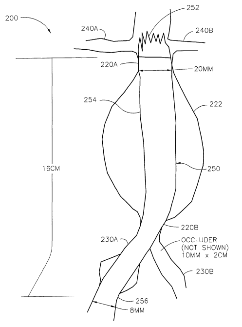
Fig. 3 shows a custom-sized stent graft 250 according to the invention
deployed in vessel
network 200. Network 200 includes an aorta 220 having a distal neck 220A, a
proximal neck 220B
CA 02421060 2003-02-21
WO 02/15823 PCT/USO1/26250
9
and a dilated portion 222 that forms an aneurysm. Network 200 also includes
iliac arteries 230A,
230B and renal arteries 240A, 240B. Measurements were taken of the: (1) width
of the distal neck
220A, (2) the length from distal neck 220A to a position in iliac artery 230A,
(3) the width of iliac
artery 230A, and the width of iliac artery 230B. The attending physician
decides, based on his
experience and the facilities available at his disposal, the preferred form
and location of each
measurement. Based on his/her judgment, a custom-sized stent or stmt graft
will be specified. In
this case, once measured, the dimensions were transmitted to a second facility
by facsimile where
custom-sized intravascular devices according to the invention were produced.
A 10 mm diameter by 2 cm in length custom-sized occluder (not shown) was
manufactured
and deployed in iliac artery 230B to eliminate back pressure from iliac artery
230B into aorta 220.
Tntravascular device 250 was also manufactured and deployed as shown in aorta
220 and
iliac 230A. Device 250 has an open end 252, a closed main body 254 and a
closed end 256.
Device 250 extends from aorta 220 immediately below renal arteries 240A, 240B
and into iliac
artery 230A. Open end 252 as shown, extends into the path of renal arteries
240A, 240B. Closed
end 256 is positioned in the lumen of iliac 230A. Not shown here is a
transluminal graft that would
be used to transfer part of the blood from artery 230A to artery 230B.
EXAMPLE 4
Fig. 4 depicts a custom-sized intravascular device according to the invention
deployed in a
blood vessel network 300. Network 300 includes an aorta 320 having a distal
neck 320A, a
proximal neck 320B and a dilated portion 322 that forms an aneurysm. Network
300 also includes
iliac arteries 330A, 3308 and renal arteries 340A, 340B. Measurements were
taken of the: (1)
width of the distal neck 320A, (2) the length from distal neck 320A to a
position in iliac artery
230A, (3) the width of iliac artery 330A, and the width of iliac artery 330B.
These dimensions
were transmitted to a second facility via facsimile where custom-sized
intravascular devices
according to the invention were produced.
A 16 mm diameter by 2 cm in length custom-sized occluder (not shown) was
manufactured
and deployed in iliac artery 330B to eliminate back pressure from iliac artery
330B into aorta 320.
Intravascular device 350 was also manufactured and deployed as depicted in
aorta 320 and
iliac 330A. Device 350 has an open end 352, a closed main body 354 and a
closed end 356.
Device 350 extends from aorta 320 immediately below renal arteries 340A, 340B
and into iliac
artery 330A. Open end 352 as shown, extends into the path of renal arteries
340A, 340B. Closed
end 356 is positioned in the lumen of iliac 330A. Not shown here is a
transluminal graft that would
be used to transfer part of the blood from artery 330A to artery 330B.
EXAMPLE 5
Fig. 5 shows a diseased blood vessel network S00 that includes aorta 520.
Aorta 520
includes distal neck SZOA, proximal neck SZOB and a dilated portion 522 that
has formed an
aneurysm. Vessel network 500 fiuther includes renal arteries 540A, 540B and
iliac arteries 530A,
CA 02421060 2003-02-21
WO 02/15823 PCT/USO1/26250
530B. As shown in Fig. 5, several measurements were taken at various locations
on vessel network
500. Fig. 5A shows custom-sized intravascular device 550 and 560 made to
repair aorta 520.
Device 550 is a scent graft made to extend from distal neck 520A to at least
proximal neck 520B,
and preferably into iliac artery 530A. Open stent graft 560 has a first
portion 562 and a second
5 portion 564. First portion 562 is open i.e., it is a wire stmt) and can be
positioned in the path of
renal arteries 530A, 530B without blocking the flow thereto. Portion 564 is a
stmt graft and is
positioned beneath renal arteries 530A, 530B to create a pressure fit against
the lumen of aorta 520
at distal end 520A. Device 550 has a first end 552, a body 554 and a second
end 556. End 552 is
overlapped i.e., placed inside of) portion 564 in order to create a continuous
passage for the
10 transfer of blood. Graft 550 extends through dilated section 522 and into
at least proximal end
520B of aorta 520. Preferably , second end 556 of graft 550 extends into iliac
artery 530A where it
forms a pressure fit against the lumen of artery 530A. If it does not extend
into artery 530A, the
physician has the option of adding a properly sized extension or cuff of the
type previously
described.
EXAMPLE 6
Fig. 6 illustrates measurements taken of a vessel network (not shown). The
measurements
included the (1) diameter of the distal neck of the aorta, (2) length from the
renal arteries to the iliac
artery, (3) length into the first iliac artery, where the graft would
terminate and form a pressure fit
against the lumen of the iliac artery, (4) the width of the second iliac
artery, and (5) a length along
the second iliac artery. Fig. 6A shows a custom-sized stmt graft 650
manufactured in accordance
with these dimensions. Stent graft 650 includes a closed first end 652, a
closed body 654 and a
closed second end 656. Graft 650 is designed to extend from the distal neck of
the aorta and into
the first iliac artery of the vessel network.
Fig. 6B shows a mandrel 670 that was custom manufactured in order to produce
the stmt
graft shown in Fig. 6A. The preferred method for manufacturing the mandrel is
disclosed in U.S.
Patent Application No. 09/244,343 to William M. Colone, entitled "A Method of
Making Large
Diameter Vascular Prostheses and a Vascular Prostheses Made by Said Method"
and filed on
February 4, 1999, the disclosure of which is incorporated herein by reference.
Any suitable method
could be used, however, to manufacture mandrel 670 or to produce device 650.
Having now described preferred embodiments of the invention, alterations and
modifications that do not depart from the spirit of the invention will occur
to others. The invention
is thus not limited to the preferred embodiments, but is set forth in the
following claims and legal
equivalents thereof. Unless specifically stated otherwise in the claims or the
specification, (1) any
method or device according to the claimed invention may include additional
steps or structures, and
(2) method steps may be performed in any order suitable for producing a device
according to the
invention.